experimental autoimmune encephalitis activation of the sting
TRANSCRIPT
of April 7, 2018.This information is current as
Experimental Autoimmune EncephalitisActivation of the STING Adaptor Attenuates
Munn and Andrew L. MellorPacholczyk, Glen N. Barber, Yoshihiro Hayakawa, David H.Mohamed, Guilherme R. Souza, Lingqian Li, Gabriela Henrique Lemos, Lei Huang, Phillip R. Chandler, Eslam
ol.1303258http://www.jimmunol.org/content/early/2014/05/03/jimmun
published online 5 May 2014J Immunol
MaterialSupplementary
8.DCSupplementalhttp://www.jimmunol.org/content/suppl/2014/05/03/jimmunol.130325
average*
4 weeks from acceptance to publicationFast Publication! •
Every submission reviewed by practicing scientistsNo Triage! •
from submission to initial decisionRapid Reviews! 30 days* •
Submit online. ?The JIWhy
Subscriptionhttp://jimmunol.org/subscription
is online at: The Journal of ImmunologyInformation about subscribing to
Permissionshttp://www.aai.org/About/Publications/JI/copyright.htmlSubmit copyright permission requests at:
Email Alertshttp://jimmunol.org/alertsReceive free email-alerts when new articles cite this article. Sign up at:
Print ISSN: 0022-1767 Online ISSN: 1550-6606. Immunologists, Inc. All rights reserved.Copyright © 2014 by The American Association of1451 Rockville Pike, Suite 650, Rockville, MD 20852The American Association of Immunologists, Inc.,
is published twice each month byThe Journal of Immunology
by guest on April 7, 2018
http://ww
w.jim
munol.org/
Dow
nloaded from
by guest on April 7, 2018
http://ww
w.jim
munol.org/
Dow
nloaded from
The Journal of Immunology
Activation of the STING Adaptor Attenuates ExperimentalAutoimmune Encephalitis
Henrique Lemos,* Lei Huang,* Phillip R. Chandler,* Eslam Mohamed,*
Guilherme R. Souza,*,† Lingqian Li,* Gabriela Pacholczyk,* Glen N. Barber,‡
Yoshihiro Hayakawa,x David H. Munn,* and Andrew L. Mellor*
Cytosolic DNA sensing activates the stimulator of IFN genes (STING) adaptor to induce IFN type I (IFN-ab) production.
Constitutive DNA sensing to induce sustained STING activation incites tolerance breakdown, leading to autoimmunity. In this
study, we show that systemic treatments with DNA nanoparticles (DNPs) induced potent immune regulatory responses via STING
signaling that suppressed experimental autoimmune encephalitis (EAE) when administered to mice after immunization with
myelin oligodendrocyte glycoprotein (MOG), at EAE onset, or at peak disease severity. DNP treatments attenuated infiltration
of effector T cells into the CNS and suppressed innate and adaptive immune responses to myelin oligodendrocyte glycoprotein
immunization in spleen. Therapeutic responses were not observed in mice treated with cargo DNA or cationic polymers alone,
indicating that DNP uptake and cargo DNA sensing by cells with regulatory functions was essential for therapeutic responses to
manifest. Intact STING and IFN-ab receptor genes, but not IFN-g receptor genes, were essential for therapeutic responses to
DNPs to manifest. Treatments with cyclic diguanylate monophosphate to activate STING also delayed EAE onset and reduced
disease severity. Therapeutic responses to DNPs were critically dependent on IDO enzyme activity in hematopoietic cells. Thus,
DNPs and cyclic diguanylate monophosphate attenuate EAE by inducing dominant T cell regulatory responses via the STING/
IFN-ab/IDO pathway that suppress CNS-specific autoimmunity. These findings reveal dichotomous roles for the STING/IFN-ab
pathway in either stimulating or suppressing autoimmunity and identify STING-activating reagents as a novel class of immune
modulatory drugs. The Journal of Immunology, 2014, 192: 000–000.
Self-tolerance is an active and constitutive process that pre-vents autoimmunity. Recent studies on mice with defectiveDNA repair enzyme expression emphasize the potential for
DNA to incite lethal autoimmunity by stimulating cytosolic DNAsensors that activate STING to induce IFN-ab production (1, 2).Multiple sclerosis (MS) is a chronic demyelinating autoimmunedisease in which neuronal tissues are progressively targeted (3) dueto loss of tolerance to CNS Ags such as myelin basic protein andmyelin oligodendrocyte glycoprotein (MOG).IDO is a natural immunomodulatory enzyme that attenuates
autoimmunity in murine disease models, including experimental
autoimmune encephalitis (EAE; a model of MS), autoimmunerheumatoid arthritis, type I diabetes, systemic lupus erythematosus,and inflammatory bowel disease (4). In these syndromes IDOablation accelerated disease onset and enhanced disease severity.Moreover, the immunomodulatory properties of soluble forms ofCTLA4 (5, 6) and CD83 (7) depend, in part, on their ability toinduce IDO-dependent regulatory phenotypes in dendritic cells(DCs), which promote de novo regulatory T cell (Treg) genera-tion, activate resting Tregs, and suppress effector T cell responses(8, 9). Moreover, some immune stimulatory reagents (adjuvants)coinduce IDO, and this property of TLR9 ligands (CpGs) inhibitedtype I diabetes progression in NOD female mice (10–12). Diametricresponses to immune adjuvants underscore the need to evaluateinnate immune responses to inflammatory stimuli to discern un-derlying pathways that induce dominant stimulatory or regulatoryresponses by T cells (13). Sustained IFN-ab production is a keyfeature of chronic immune activation at sites of persistent infectionssuch as HIV-1 (14). Sustained IFN-ab release, especially by plas-macytoid DCs, correlates strongly with risk of autoimmune syn-dromes such as systemic lupus erythematosus (15). IFN-ab andIFN type II (IFN-g) have well-documented immune stimulatoryproperties, but IFNs are also potent IDO inducers, providing a ra-tionale for increased IDO-mediated T cell regulation, particularly atsites of chronic inflammation.Previously, we reported that small populations of DCs coex-
pressing the B cell marker CD19 upregulated IDO selectively(among DCs) in response to systemic treatments with solubleCTLA4 (CTLA4Ig), TLR9 ligands (CpGs), and DNA nanoparticles(DNPs) containing the cationic polymer polyethylenimine (PEI)and cargo DNA (12, 16, 17). IDO induction in CD19+ DCs wasmediated by IFN-ab, not IFN-g, and by CD19+ DCs expressingIDO-stimulated Foxp3-lineage CD4 T cells (Tregs) to acquire
*Cancer Immunology, Inflammation and Tolerance Program, Cancer Center, GeorgiaRegents University, Augusta, GA 30912; †Department of Pharmacology, School ofMedicine of Ribeirao Preto, University of Sao Paulo, Ribeirao Preto, SP 14049-900Sao Paulo, Brazil; ‡Sylvester Comprehensive Cancer Center, University of MiamiSchool of Medicine, Miami, FL 33136; and xDepartment of Applied Chemistry,Faculty of Engineering, Aichi Institute of Technology, Toyota 470-0392, Japan
Received for publication December 6, 2013. Accepted for publication April 10, 2014.
This work was supported by National Institutes of Health Grants AI83005 andAI103347 and the Trustees of the Carlos and Marguerite Mason Trust (to A.L.M.).H.L. was supported by a postdoctoral fellowship from the Juvenile Diabetes ResearchFoundation, and G.R.S. was supported by a postdoctoral fellowship from the SaoPaulo Research Foundation.
Address correspondence and reprint requests to Dr. Andrew L. Mellor, Cancer im-munology, Inflammation and Tolerance Program, Cancer Center, Georgia Regent’sUniversity, 1120 15th Street, Augusta, GA 30912. E-mail address: [email protected]
The online version of this article contains supplemental material.
Abbreviations used in this article: B6, C57BL/6; c-diGMP, cyclic diguanylate mono-phosphate; DC, dendritic cell; DNP, DNA nanoparticle; EAE, experimental autoim-mune encephalitis; GFAP, glial fibrillary acidic protein; KO, knockout; MOG, myelinoligodendrocyte glycoprotein; MS, multiple sclerosis; 1MT, 1-methyl-D-tryptophan;PEI, polyethylenimine; STING, stimulator of IFN genes; Treg, regulatory T cell; WT,wild-type.
Copyright� 2014 by The American Association of Immunologists, Inc. 0022-1767/14/$16.00
www.jimmunol.org/cgi/doi/10.4049/jimmunol.1303258
Published May 5, 2014, doi:10.4049/jimmunol.1303258 by guest on A
pril 7, 2018http://w
ww
.jimm
unol.org/D
ownloaded from
regulatory phenotypes that suppressed Th1 responses (8, 17).Tolerogenic responses to DNPs were dependent on cargo DNAsensing by small populations of myeloid DCs to activate STINGand induced selective IFN-ab release that stimulated CD19+ DCsto express IDO (17, 18). In the present study, we tested the hy-pothesis that STING activation to induce IDO via IFN-ab sig-naling following systemic DNP treatment inhibits EAE progressionand reduces disease severity.
Materials and MethodsMice and induction of EAE
Micewere bred under specific pathogen-free conditions at Georgia RegentsUniversity or purchased from Taconic Farms, and procedures were ap-proved by the Institutional Animal Care and Use Committee. IDO1-knockout (KO), IFNAR-KO, IFN-gR-KO and STING-KO mice weredescribed previously (19–21). To induce EAE, mice aged 8–12 wk wereimmunized s.c. at two sites on the rear flank with 100 mg myelin oligo-dendrocyte glycoprotein peptide (MOG35–55, MEVGWYRSPFSRVVH-LYRNGK; Bio Basic Canada) emulsified in CFA (Difco Laboratories,Detroit, MI) containing 4 mg/ml Mycobacterium tuberculosis H37Ra(Difco Laboratories). Pertussis toxin (200 ng; Sigma-Aldrich) was giveni.p. on days 0 and 2 after immunization. Clinical symptoms of EAE werescored on a scale from 0 to 5 as described by Das Sarma et al. (22), withslight modifications as follows: 0, no clinical signs; 0.5, partial limp tail;1, full limp tail or waddling gait; 1.5, limp tail and waddling gait/lumbarweakness; 2, partial paralysis of one hindlimb; 2.5, paralysis of onehindlimb or partial paralysis of both hindlimbs; 3, paralysis of onehindlimb and partial paralysis of the other hindlimb; 3.5, paralysis of bothhindlimbs or partial paralysis of both hindlimbs and weakness of theupper limb; 4, ascending paralysis, that is, complete paralysis of bothhindlimbs and weakness of the upper limb; 4.5, three paralyzed limbs; 5,four paralyzed limbs/moribund or dead. Some mice were given drinkingwater containing the IDO inhibitor 1-methyl-D-tryptophan (1MT; 2 mg/ml) and Nutrasweet (to increase palatability) as described (23).
DNP and cyclic diguanylate monophosphate treatments
DNPs were prepared by mixing 7 ml PEI (150 mM) with 21 mg CpGfree
LacZ pDNA (InvivoGen, San Diego, CA) in 200 ml 5% glucose solution(N/P = 16.7) as described (17). Chemically synthesized cyclic diguanylatemonophosphate (c-diGMP) was dissolved in PBS at 500 mg/ml. DNPs andc-diGMP (100 mg/dose) were injected i.v.
Immunohistochemistry
After sacrifice, mice were perfused (PBS then paraformaldehyde at 4%)and lumbar spinal cord tissues were harvested, left in paraformaldehyde(3 d) and embedded in paraffin. Five-micrometer sections were depar-affinized, rehydrated, submerged in Target retrieval solution (20 min; Dako,catalog no. S-1699) and incubated in 3% H2O2 (5 min). Nonspecificbiotin/avidin (Vector Laboratories, Burlingame, CA) and Fc/Fab stainingwas blocked using kits from Jackson ImmunoResearch Laboratories(catalog no. 015.000.008). Mouse on Mouse kits (Vector Laboratories)were used to dilute primary mouse anti-mouse/human IDO mAb (E7,1:50; Santa Cruz Biotechnology, catalog no. 365086), neurons (NeuNrabbit polyclonal, 1:500; Millipore, catalog no. ABN78), microglia (IBArabbit, 1:500; Wako Chemicals USA, catalog no. 019-19741), andastrocytes (glial fibrillary acidic protein [GFAP] rabbit monoclonal,1:500; Cell Signaling Technology, catalog no. 123895). After 60 min,sections were incubated with biotinylated anti-mouse IgG Ab from theMouse on Mouse kit and Alexa Fluor 488–conjugated AffiniPure F(ab9)2fragment donkey anti-rabbit IgG (H+L) (Jackson ImmunoResearchLaboratories, catalog no. 711-546-152) for 45 min. After washing, strepta-vidin conjugated with Alexa Fluor 555 (1:400; Invitrogen/Molecular Probes,catalog no. S32355) was applied for 15 min. Slides were mounted in Flu-orSave reagent (Calbiochem, catalog no. 345789) and analyzed by confocalmicroscopy (Zeiss LSM 510).
Cytokines and Proliferative Responses to MOG
Splenocytes (106) were cultured in RPMI 1640 in triplicate in 96-well U-bottom plates in the presence or absence of 20 mg/ml MOG peptide for72 h. Cytokine levels in culture supernatants were analyzed using multi-plex kits from Bio-Rad or eBioscience. For proliferation assays, cells werepulsed with 0.5 mCi [3H]thymidine for the last 16 h of incubation andmean thymidine incorporation in triplicate wells was measured.
IDO enzyme activity
Cell-free spleen homogenates were added to IDO enzyme cocktails, andkynurenine generated after 2 h was measured by HPLC as described (24).
Flow cytometry
Single-cell suspensions of spleen and CNS (pooled brain and spinal cord)prepared as described (25) were stimulated in medium containing 20 mg/ml MOG peptide (37˚C, 5% CO2, 18 h); 1 ml/ml brefeldin (BD Bio-sciences) was added for the last 4 h of culture. After staining the surfacemarker CD4 (BD Pharmingen, clone RM4-5), cells were fixed and per-meabilized (Foxp3/transcription factor fixation/permeabilization buffer;eBioscience), followed by staining with mAbs to mouse Foxp3 (eBio-science, clone FJK-16S), IL-17A (eBioscience, clone eBio17B7), and IFN-g(BD Pharmingen, clone XMG1.2). Flow cytometric analyses were per-formed using a LSRII cytometer (BD Biosciences), and data generatedwere analyzed using FACSDiva (BD Biosciences) or FlowJo (Tree Star,Ashland, OR) software.
Radiation chimeric mice
Mice were irradiated (900 rad) and after 24 h mice received ∼107 nucleatedbone marrow cells i.v. harvested from the long bones of donor mice and wereallowed to recover for 10 wk before use in EAE induction experiments.
Statistical analysis
EAE scores were evaluated with two-way ANOVA. The unpaired Student ttest was used for statistical evaluations of cytokine and cell frequencymeasurements. Two-tailed p values ,0.05 were considered significant.GraphPad Prism was used to perform all data analyses.
ResultsDNP treatment slows EAE onset and reduces disease severity
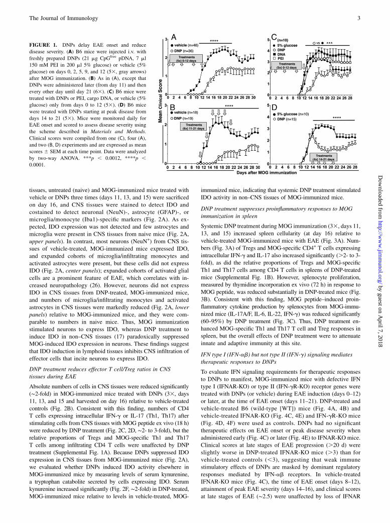
C57BL/6 (B6) mice were immunized with MOG and treated withpertussis toxin to induce EAE (see Materials and Methods). Theeffects of systemic DNP treatment on EAE progression were evalu-ated after administering DNPs (PEI/CpGfree pDNA) i.v. to groups ofmice starting at the time of MOG immunization (day 0) and on days2, 5, 9, and 12 (Fig. 1A, gray arrows). Control mice were given ve-hicle (5% glucose) and all mice were monitored to detect EAE onsetand score clinical disease severity (see Materials and Methods). Asexpected, EAE symptoms began to manifest in vehicle-treated mice8–12 d after MOG immunization (partial/full limp tail, clinical score0.5–1), and peak disease severity occurred 14–24 d after immuniza-tion (mean clinical score.2), with minor relapse thereafter (Fig. 1A,filled symbols). DNP treatment delayed disease onset by ∼4 d andreduced peak disease severity (mean clinical score ∼1.5) significantlyrelative to vehicle-treated mice (Fig. 1A, open symbols).Next, we tested whether administering DNPs later, when EAE first
started to manifest, attenuated EAE. DNPs were administered i.v.from day 11 and every other day until day 21 (Fig. 1B, gray arrows).DNP treatment prevented disease onset by ∼7 d, and subsequentdisease severity was reduced significantly (mean clinical score ,1)relative to outcomes in vehicle-treated mice (Fig. 1B, open andfilled symbols, respectively). Delayed vehicle treatment had noimpact on disease progression, peak disease severity, or minor re-lapse at late times (Fig. 1B, filled symbols) relative to mice givenvehicle earlier (Fig. 1A). In contrast, treatments with PEI or cargoDNA alone induced no therapeutic responses in MOG-immunizedmice relative to mice treated with vehicle (5% glucose, Fig. 1C).DNP treatments administered to MOG-immunized mice at peakEAE disease (starting on day 14 and given every other day until day21) also induced significant therapeutic responses (Fig. 1D), indi-cating that regulatory responses to DNPs overcame establishedCNS-specific autoimmunity.
DNP treatment inhibits IDO expression in neurons during EAE
DNPs induce rapid IDO upregulation in mucosal and lymphoidtissues (18). To evaluate whether DNPs induced IDO in CNS
2 STING ACTIVATION ATTENUATES EAE
by guest on April 7, 2018
http://ww
w.jim
munol.org/
Dow
nloaded from
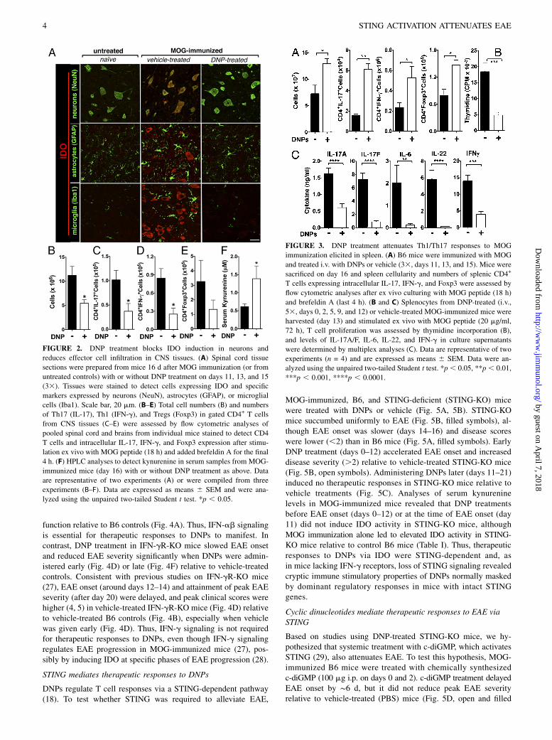
tissues, untreated (naive) and MOG-immunized mice treated withvehicle or DNPs three times (days 11, 13, and 15) were sacrificedon day 16, and CNS tissues were stained to detect IDO andcostained to detect neuronal (NeuN)-, astrocyte (GFAP)-, ormicroglia/monocyte (Iba1)-specific markers (Fig. 2A). As ex-pected, IDO expression was not detected and few astrocytes andmicroglia were present in CNS tissues from naive mice (Fig. 2A,upper panels). In contrast, most neurons (NeuN+) from CNS tis-sues of vehicle-treated, MOG-immunized mice expressed IDO,and expanded cohorts of microglia/infiltrating monocytes andactivated astrocytes were present, but these cells did not expressIDO (Fig. 2A, center panels); expanded cohorts of activated glialcells are a prominent feature of EAE, which correlates with in-creased neuropathology (26). However, neurons did not expressIDO in CNS tissues from DNP-treated, MOG-immunized mice,and numbers of microglia/infiltrating monocytes and activatedastrocytes in CNS tissues were markedly reduced (Fig. 2A, lowerpanels) relative to MOG-immunized mice, and they were com-parable to numbers in naive mice. Thus, MOG immunizationstimulated neurons to express IDO, whereas DNP treatment toinduce IDO in non–CNS tissues (17) paradoxically suppressedMOG-induced IDO expression in neurons. These findings suggestthat IDO induction in lymphoid tissues inhibits CNS infiltration ofeffector cells that incite neurons to express IDO.
DNP treatment reduces effector T cell/Treg ratios in CNStissues during EAE
Absolute numbers of cells in CNS tissues were reduced significantly(∼2-fold) in MOG-immunized mice treated with DNPs (33, days11, 13, and 15 and harvested on day 16) relative to vehicle-treatedcontrols (Fig. 2B). Consistent with this finding, numbers of CD4T cells expressing intracellular IFN-g or IL-17 (Th1, Th17) afterstimulating cells from CNS tissues with MOG peptide ex vivo (18 h)were reduced by DNP treatment (Fig. 2C, 2D, ∼2- to 3-fold), but therelative proportions of Tregs and MOG-specific Th1 and Th17T cells among infiltrating CD4 T cells were unaffected by DNPtreatment (Supplemental Fig. 1A). Because DNPs suppressed IDOexpression in CNS tissues from MOG-immunized mice (Fig. 2A),we evaluated whether DNPs induced IDO activity elsewhere inMOG-immunized mice by measuring levels of serum kynurenine,a tryptophan catabolite secreted by cells expressing IDO. Serumkynurenine increased significantly (Fig. 2F, ∼2-fold) in DNP-treated,MOG-immunized mice relative to levels in vehicle-treated, MOG-
immunized mice, indicating that systemic DNP treatment stimulatedIDO activity in non–CNS tissues of MOG-immunized mice.
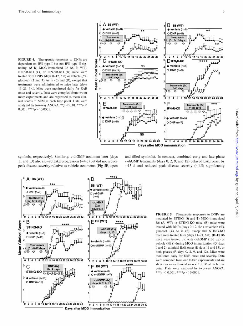
DNP treatment suppresses proinflammatory responses to MOGimmunization in spleen
Systemic DNP treatment during MOG immunization (33, days 11,13, and 15) increased spleen cellularity (at day 16) relative tovehicle-treated MOG-immunized mice with EAE (Fig. 3A). Num-bers (Fig. 3A) of Tregs and MOG-specific CD4+ T cells expressingintracellular IFN-g and IL-17 also increased significantly (.2- to 3-fold), as did the relative proportions of Tregs and MOG-specificTh1 and Th17 cells among CD4 T cells in spleens of DNP-treatedmice (Supplemental Fig. 1B). However, splenocyte proliferation,measured by thymidine incorporation ex vivo (72 h) in response toMOG peptide, was reduced substantially in DNP-treated mice (Fig.3B). Consistent with this finding, MOG peptide–induced proin-flammatory cytokine production by splenocytes from MOG-immu-nized mice (IL-17A/F, IL-6, IL-22, IFN-g) was reduced significantly(60–95%) by DNP treatment (Fig. 3C). Thus, DNP treatment en-hanced MOG-specific Th1 and Th17 T cell and Treg responses inspleen, but the overall effects of DNP treatment were to attenuateinnate and adaptive immunity at this site.
IFN type I (IFN-ab) but not type II (IFN-g) signaling mediatestherapeutic responses to DNPs
To evaluate IFN signaling requirements for therapeutic responsesto DNPs to manifest, MOG-immunized mice with defective IFNtype I (IFNAR-KO) or type II (IFN-gR-KO) receptor genes weretreated with DNPs (or vehicle) during EAE induction (days 0–12)or later, at the time of EAE onset (days 11–21). DNP-treated andvehicle-treated B6 (wild-type [WT]) mice (Fig. 4A, 4B) andvehicle-treated IFNAR-KO (Fig. 4C, 4E) and IFN-gR-KO mice(Fig. 4D, 4F) were used as controls. DNPs had no significanttherapeutic effects on EAE onset or peak disease severity whenadministered early (Fig. 4C) or later (Fig. 4E) to IFNAR-KO mice.Clinical scores at late stages of EAE progression (.20 d) wereslightly worse in DNP-treated IFNAR-KO mice (.3) than forvehicle-treated controls (,3), suggesting that weak immunestimulatory effects of DNPs are masked by dominant regulatoryresponses mediated by IFN-ab receptors. In vehicle-treatedIFNAR-KO mice (Fig. 4C), the time of EAE onset (days 8–12),attainment of peak EAE severity (days 14–16), and clinical scoresat late stages of EAE (∼2.5) were unaffected by loss of IFNAR
FIGURE 1. DNPs delay EAE onset and reduce
disease severity. (A) B6 mice were injected i.v. with
freshly prepared DNPs (21 mg CpGfree pDNA, 7 ml
150 mM PEI in 200 ml 5% glucose) or vehicle (5%
glucose) on days 0, 2, 5, 9, and 12 (53, gray arrows)
after MOG immunization. (B) As in (A), except that
DNPs were administered later (from day 11) and then
every other day until day 21 (63). (C) B6 mice were
treated with DNPs or PEI, cargo DNA, or vehicle (5%
glucose) only from days 0 to 12 (53). (D) B6 mice
were treated with DNPs starting at peak disease from
days 14 to 21 (53). Mice were monitored daily for
EAE onset and scored to assess disease severity using
the scheme described in Materials and Methods.
Clinical scores were compiled from one (C), four (A),
and two (B, D) experiments and are expressed as mean
scores 6 SEM at each time point. Data were analyzed
by two-way ANOVA. ***p , 0.0012, ****p ,0.0001.
The Journal of Immunology 3
by guest on April 7, 2018
http://ww
w.jim
munol.org/
Dow
nloaded from
function relative to B6 controls (Fig. 4A). Thus, IFN-ab signalingis essential for therapeutic responses to DNPs to manifest. Incontrast, DNP treatment in IFN-gR-KO mice slowed EAE onsetand reduced EAE severity significantly when DNPs were admin-istered early (Fig. 4D) or late (Fig. 4F) relative to vehicle-treatedcontrols. Consistent with previous studies on IFN-gR-KO mice(27), EAE onset (around days 12–14) and attainment of peak EAEseverity (after day 20) were delayed, and peak clinical scores werehigher (4, 5) in vehicle-treated IFN-gR-KO mice (Fig. 4D) relativeto vehicle-treated B6 controls (Fig. 4B), especially when vehiclewas given early (Fig. 4D). Thus, IFN-g signaling is not requiredfor therapeutic responses to DNPs, even though IFN-g signalingregulates EAE progression in MOG-immunized mice (27), pos-sibly by inducing IDO at specific phases of EAE progression (28).
STING mediates therapeutic responses to DNPs
DNPs regulate T cell responses via a STING-dependent pathway(18). To test whether STING was required to alleviate EAE,
MOG-immunized, B6, and STING-deficient (STING-KO) micewere treated with DNPs or vehicle (Fig. 5A, 5B). STING-KOmice succumbed uniformly to EAE (Fig. 5B, filled symbols), al-though EAE onset was slower (days 14–16) and disease scoreswere lower (,2) than in B6 mice (Fig. 5A, filled symbols). EarlyDNP treatment (days 0–12) accelerated EAE onset and increaseddisease severity (.2) relative to vehicle-treated STING-KO mice(Fig. 5B, open symbols). Administering DNPs later (days 11–21)induced no therapeutic responses in STING-KO mice relative tovehicle treatments (Fig. 5C). Analyses of serum kynureninelevels in MOG-immunized mice revealed that DNP treatmentsbefore EAE onset (days 0–12) or at the time of EAE onset (day11) did not induce IDO activity in STING-KO mice, althoughMOG immunization alone led to elevated IDO activity in STING-KO mice relative to control B6 mice (Table I). Thus, therapeuticresponses to DNPs via IDO were STING-dependent and, asin mice lacking IFN-g receptors, loss of STING signaling revealedcryptic immune stimulatory properties of DNPs normally maskedby dominant regulatory responses in mice with intact STINGgenes.
Cyclic dinucleotides mediate therapeutic responses to EAE viaSTING
Based on studies using DNP-treated STING-KO mice, we hy-pothesized that systemic treatment with c-diGMP, which activatesSTING (29), also attenuates EAE. To test this hypothesis, MOG-immunized B6 mice were treated with chemically synthesizedc-diGMP (100 mg i.p. on days 0 and 2). c-diGMP treatment delayedEAE onset by ∼6 d, but it did not reduce peak EAE severityrelative to vehicle-treated (PBS) mice (Fig. 5D, open and filled
FIGURE 2. DNP treatment blocks IDO induction in neurons and
reduces effector cell infiltration in CNS tissues. (A) Spinal cord tissue
sections were prepared from mice 16 d after MOG immunization (or from
untreated controls) with or without DNP treatment on days 11, 13, and 15
(33). Tissues were stained to detect cells expressing IDO and specific
markers expressed by neurons (NeuN), astrocytes (GFAP), or microglial
cells (Iba1). Scale bar, 20 mm. (B–E) Total cell numbers (B) and numbers
of Th17 (IL-17), Th1 (IFN-g), and Tregs (Foxp3) in gated CD4+ T cells
from CNS tissues (C–E) were assessed by flow cytometric analyses of
pooled spinal cord and brains from individual mice stained to detect CD4
T cells and intracellular IL-17, IFN-g, and Foxp3 expression after stimu-
lation ex vivo with MOG peptide (18 h) and added brefeldin A for the final
4 h. (F) HPLC analyses to detect kynurenine in serum samples from MOG-
immunized mice (day 16) with or without DNP treatment as above. Data
are representative of two experiments (A) or were compiled from three
experiments (B–F). Data are expressed as means 6 SEM and were ana-
lyzed using the unpaired two-tailed Student t test. *p , 0.05.
FIGURE 3. DNP treatment attenuates Th1/Th17 responses to MOG
immunization elicited in spleen. (A) B6 mice were immunized with MOG
and treated i.v. with DNPs or vehicle (33, days 11, 13, and 15). Mice were
sacrificed on day 16 and spleen cellularity and numbers of splenic CD4+
T cells expressing intracellular IL-17, IFN-g, and Foxp3 were assessed by
flow cytometric analyses after ex vivo culturing with MOG peptide (18 h)
and brefeldin A (last 4 h). (B and C) Splenocytes from DNP-treated (i.v.,
53, days 0, 2, 5, 9, and 12) or vehicle-treated MOG-immunized mice were
harvested (day 13) and stimulated ex vivo with MOG peptide (20 mg/ml,
72 h), T cell proliferation was assessed by thymidine incorporation (B),
and levels of IL-17A/F, IL-6, IL-22, and IFN-g in culture supernatants
were determined by multiplex analyses (C). Data are representative of two
experiments (n = 4) and are expressed as means 6 SEM. Data were an-
alyzed using the unpaired two-tailed Student t test. *p , 0.05, **p , 0.01,
***p , 0.001, ****p , 0.0001.
4 STING ACTIVATION ATTENUATES EAE
by guest on April 7, 2018
http://ww
w.jim
munol.org/
Dow
nloaded from
symbols, respectively). Similarly, c-diGMP treatment later (days11 and 13) also slowed EAE progression (∼4 d) but did not reducepeak disease severity relative to vehicle treatments (Fig 5E, open
and filled symbols). In contrast, combined early and late phasec-diGMP treatments (days 0, 2, 9, and 12) delayed EAE onset by∼15 d and reduced peak disease severity (∼1.5) significantly
FIGURE 4. Therapeutic responses to DNPs are
dependent on IFN type I but not IFN type II sig-
naling. (A–D) MOG-immunized B6 (A, B; WT),
IFNAR-KO (C), or IFN-gR-KO (D) mice were
treated with DNPs (days 0–12, 53) or vehicle (5%
glucose). (E and F) As in (C) and (D), except that
treatments were administered to mice later (days
11–21, 63). Mice were monitored daily for EAE
onset and severity. Data were compiled from two or
more experiments and are expressed as mean clin-
ical scores 6 SEM at each time point. Data were
analyzed by two-way ANOVA. **p, 0.01, ***p,0.001, ****p , 0.0001.
FIGURE 5. Therapeutic responses to DNPs are
mediated by STING. (A and B) MOG-immunized
B6 (A, WT) or STING-KO mice (B) mice were
treated with DNPs (days 0–12, 53) or vehicle (5%
glucose). (C) As in (B), except that STING-KO
mice were treated later (days 11–21, 63). (D–F) B6
mice were treated i.v. with c-diGMP (100 mg) or
vehicle (PBS) during MOG immunization (D, days
0 and 2), at initial EAE onset (E, days 11 and 13), or
both phases (F, days 0, 2, 9, and 12). Mice were
monitored daily for EAE onset and severity. Data
were compiled from one to two experiments and are
shown as mean clinical scores 6 SEM at each time
point. Data were analyzed by two-way ANOVA.
***p , 0.001, ****p , 0.0001.
The Journal of Immunology 5
by guest on April 7, 2018
http://ww
w.jim
munol.org/
Dow
nloaded from
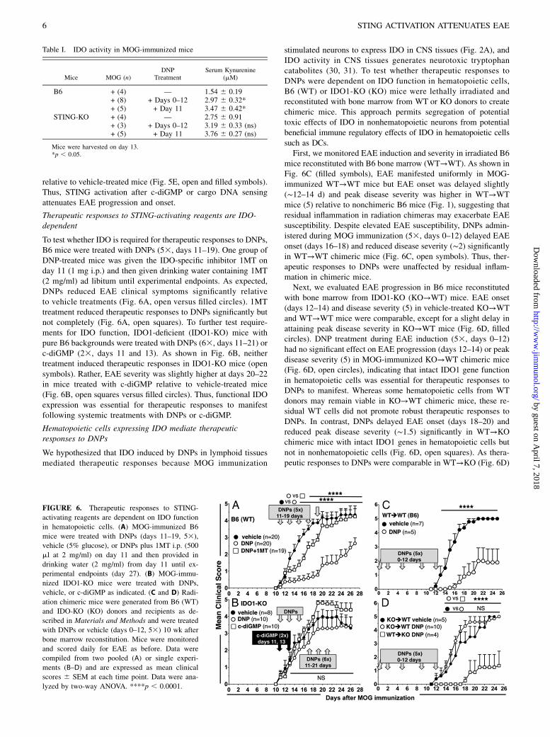
relative to vehicle-treated mice (Fig. 5E, open and filled symbols).Thus, STING activation after c-diGMP or cargo DNA sensingattenuates EAE progression and onset.
Therapeutic responses to STING-activating reagents are IDO-dependent
To test whether IDO is required for therapeutic responses to DNPs,B6 mice were treated with DNPs (53, days 11–19). One group ofDNP-treated mice was given the IDO-specific inhibitor 1MT onday 11 (1 mg i.p.) and then given drinking water containing 1MT(2 mg/ml) ad libitum until experimental endpoints. As expected,DNPs reduced EAE clinical symptoms significantly relativeto vehicle treatments (Fig. 6A, open versus filled circles). 1MTtreatment reduced therapeutic responses to DNPs significantly butnot completely (Fig. 6A, open squares). To further test require-ments for IDO function, IDO1-deficient (IDO1-KO) mice withpure B6 backgrounds were treated with DNPs (63, days 11–21) orc-diGMP (23, days 11 and 13). As shown in Fig. 6B, neithertreatment induced therapeutic responses in IDO1-KO mice (opensymbols). Rather, EAE severity was slightly higher at days 20–22in mice treated with c-diGMP relative to vehicle-treated mice(Fig. 6B, open squares versus filled circles). Thus, functional IDOexpression was essential for therapeutic responses to manifestfollowing systemic treatments with DNPs or c-diGMP.
Hematopoietic cells expressing IDO mediate therapeuticresponses to DNPs
We hypothesized that IDO induced by DNPs in lymphoid tissuesmediated therapeutic responses because MOG immunization
stimulated neurons to express IDO in CNS tissues (Fig. 2A), andIDO activity in CNS tissues generates neurotoxic tryptophancatabolites (30, 31). To test whether therapeutic responses toDNPs were dependent on IDO function in hematopoietic cells,B6 (WT) or IDO1-KO (KO) mice were lethally irradiated andreconstituted with bone marrow from WT or KO donors to createchimeric mice. This approach permits segregation of potentialtoxic effects of IDO in nonhematopoietic neurons from potentialbeneficial immune regulatory effects of IDO in hematopoietic cellssuch as DCs.First, we monitored EAE induction and severity in irradiated B6
mice reconstituted with B6 bone marrow (WT→WT). As shown inFig. 6C (filled symbols), EAE manifested uniformly in MOG-immunized WT→WT mice but EAE onset was delayed slightly(∼12–14 d) and peak disease severity was higher in WT→WTmice (5) relative to nonchimeric B6 mice (Fig. 1), suggesting thatresidual inflammation in radiation chimeras may exacerbate EAEsusceptibility. Despite elevated EAE susceptibility, DNPs admin-istered during MOG immunization (53, days 0–12) delayed EAEonset (days 16–18) and reduced disease severity (∼2) significantlyin WT→WT chimeric mice (Fig. 6C, open symbols). Thus, ther-apeutic responses to DNPs were unaffected by residual inflam-mation in chimeric mice.Next, we evaluated EAE progression in B6 mice reconstituted
with bone marrow from IDO1-KO (KO→WT) mice. EAE onset(days 12–14) and disease severity (5) in vehicle-treated KO→WTand WT→WT mice were comparable, except for a slight delay inattaining peak disease severity in KO→WT mice (Fig. 6D, filledcircles). DNP treatment during EAE induction (53, days 0–12)had no significant effect on EAE progression (days 12–14) or peakdisease severity (5) in MOG-immunized KO→WT chimeric mice(Fig. 6D, open circles), indicating that intact IDO1 gene functionin hematopoietic cells was essential for therapeutic responses toDNPs to manifest. Whereas some hematopoietic cells from WTdonors may remain viable in KO→WT chimeric mice, these re-sidual WT cells did not promote robust therapeutic responses toDNPs. In contrast, DNPs delayed EAE onset (days 18–20) andreduced peak disease severity (∼1.5) significantly in WT→KOchimeric mice with intact IDO1 genes in hematopoietic cells butnot in nonhematopoietic cells (Fig. 6D, open squares). As thera-peutic responses to DNPs were comparable in WT→KO (Fig. 6D)
Table I. IDO activity in MOG-immunized mice
Mice MOG (n)DNP
TreatmentSerum Kynurenine
(mM)
B6 + (4) — 1.54 6 0.19+ (8) + Days 0–12 2.97 6 0.32*+ (5) + Day 11 3.47 6 0.42*
STING-KO + (4) — 2.75 6 0.91+ (3) + Days 0–12 3.19 6 0.33 (ns)+ (5) + Day 11 3.76 6 0.27 (ns)
Mice were harvested on day 13.*p , 0.05.
FIGURE 6. Therapeutic responses to STING-
activating reagents are dependent on IDO function
in hematopoietic cells. (A) MOG-immunized B6
mice were treated with DNPs (days 11–19, 53),
vehicle (5% glucose), or DNPs plus 1MT i.p. (500
ml at 2 mg/ml) on day 11 and then provided in
drinking water (2 mg/ml) from day 11 until ex-
perimental endpoints (day 27). (B) MOG-immu-
nized IDO1-KO mice were treated with DNPs,
vehicle, or c-diGMP as indicated. (C and D) Radi-
ation chimeric mice were generated from B6 (WT)
and IDO-KO (KO) donors and recipients as de-
scribed in Materials and Methods and were treated
with DNPs or vehicle (days 0–12, 53) 10 wk after
bone marrow reconstitution. Mice were monitored
and scored daily for EAE as before. Data were
compiled from two pooled (A) or single experi-
ments (B–D) and are expressed as mean clinical
scores 6 SEM at each time point. Data were ana-
lyzed by two-way ANOVA. ****p , 0.0001.
6 STING ACTIVATION ATTENUATES EAE
by guest on April 7, 2018
http://ww
w.jim
munol.org/
Dow
nloaded from
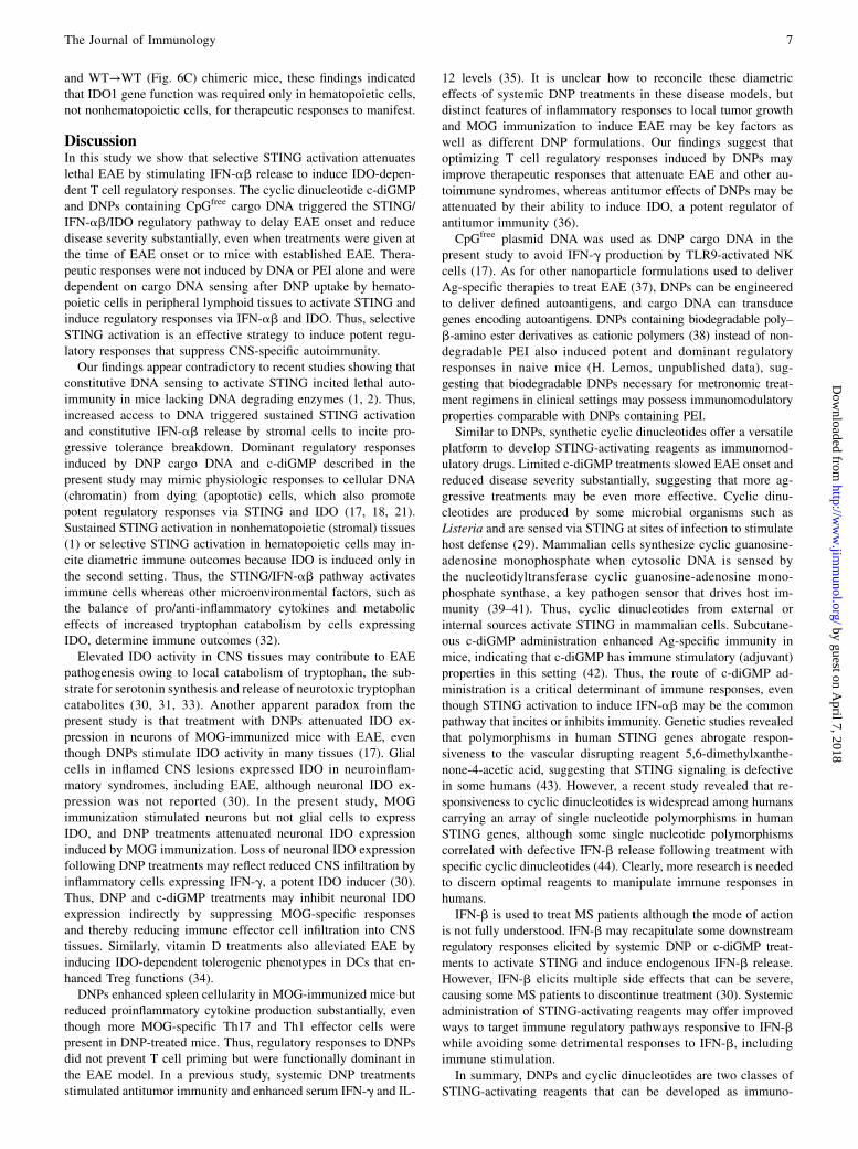
and WT→WT (Fig. 6C) chimeric mice, these findings indicatedthat IDO1 gene function was required only in hematopoietic cells,not nonhematopoietic cells, for therapeutic responses to manifest.
DiscussionIn this study we show that selective STING activation attenuateslethal EAE by stimulating IFN-ab release to induce IDO-depen-dent T cell regulatory responses. The cyclic dinucleotide c-diGMPand DNPs containing CpGfree cargo DNA triggered the STING/IFN-ab/IDO regulatory pathway to delay EAE onset and reducedisease severity substantially, even when treatments were given atthe time of EAE onset or to mice with established EAE. Thera-peutic responses were not induced by DNA or PEI alone and weredependent on cargo DNA sensing after DNP uptake by hemato-poietic cells in peripheral lymphoid tissues to activate STING andinduce regulatory responses via IFN-ab and IDO. Thus, selectiveSTING activation is an effective strategy to induce potent regu-latory responses that suppress CNS-specific autoimmunity.Our findings appear contradictory to recent studies showing that
constitutive DNA sensing to activate STING incited lethal auto-immunity in mice lacking DNA degrading enzymes (1, 2). Thus,increased access to DNA triggered sustained STING activationand constitutive IFN-ab release by stromal cells to incite pro-gressive tolerance breakdown. Dominant regulatory responsesinduced by DNP cargo DNA and c-diGMP described in thepresent study may mimic physiologic responses to cellular DNA(chromatin) from dying (apoptotic) cells, which also promotepotent regulatory responses via STING and IDO (17, 18, 21).Sustained STING activation in nonhematopoietic (stromal) tissues(1) or selective STING activation in hematopoietic cells may in-cite diametric immune outcomes because IDO is induced only inthe second setting. Thus, the STING/IFN-ab pathway activatesimmune cells whereas other microenvironmental factors, such asthe balance of pro/anti-inflammatory cytokines and metaboliceffects of increased tryptophan catabolism by cells expressingIDO, determine immune outcomes (32).Elevated IDO activity in CNS tissues may contribute to EAE
pathogenesis owing to local catabolism of tryptophan, the sub-strate for serotonin synthesis and release of neurotoxic tryptophancatabolites (30, 31, 33). Another apparent paradox from thepresent study is that treatment with DNPs attenuated IDO ex-pression in neurons of MOG-immunized mice with EAE, eventhough DNPs stimulate IDO activity in many tissues (17). Glialcells in inflamed CNS lesions expressed IDO in neuroinflam-matory syndromes, including EAE, although neuronal IDO ex-pression was not reported (30). In the present study, MOGimmunization stimulated neurons but not glial cells to expressIDO, and DNP treatments attenuated neuronal IDO expressioninduced by MOG immunization. Loss of neuronal IDO expressionfollowing DNP treatments may reflect reduced CNS infiltration byinflammatory cells expressing IFN-g, a potent IDO inducer (30).Thus, DNP and c-diGMP treatments may inhibit neuronal IDOexpression indirectly by suppressing MOG-specific responsesand thereby reducing immune effector cell infiltration into CNStissues. Similarly, vitamin D treatments also alleviated EAE byinducing IDO-dependent tolerogenic phenotypes in DCs that en-hanced Treg functions (34).DNPs enhanced spleen cellularity in MOG-immunized mice but
reduced proinflammatory cytokine production substantially, eventhough more MOG-specific Th17 and Th1 effector cells werepresent in DNP-treated mice. Thus, regulatory responses to DNPsdid not prevent T cell priming but were functionally dominant inthe EAE model. In a previous study, systemic DNP treatmentsstimulated antitumor immunity and enhanced serum IFN-g and IL-
12 levels (35). It is unclear how to reconcile these diametriceffects of systemic DNP treatments in these disease models, butdistinct features of inflammatory responses to local tumor growthand MOG immunization to induce EAE may be key factors aswell as different DNP formulations. Our findings suggest thatoptimizing T cell regulatory responses induced by DNPs mayimprove therapeutic responses that attenuate EAE and other au-toimmune syndromes, whereas antitumor effects of DNPs may beattenuated by their ability to induce IDO, a potent regulator ofantitumor immunity (36).CpGfree plasmid DNA was used as DNP cargo DNA in the
present study to avoid IFN-g production by TLR9-activated NKcells (17). As for other nanoparticle formulations used to deliverAg-specific therapies to treat EAE (37), DNPs can be engineeredto deliver defined autoantigens, and cargo DNA can transducegenes encoding autoantigens. DNPs containing biodegradable poly–b-amino ester derivatives as cationic polymers (38) instead of non-degradable PEI also induced potent and dominant regulatoryresponses in naive mice (H. Lemos, unpublished data), sug-gesting that biodegradable DNPs necessary for metronomic treat-ment regimens in clinical settings may possess immunomodulatoryproperties comparable with DNPs containing PEI.Similar to DNPs, synthetic cyclic dinucleotides offer a versatile
platform to develop STING-activating reagents as immunomod-ulatory drugs. Limited c-diGMP treatments slowed EAE onset andreduced disease severity substantially, suggesting that more ag-gressive treatments may be even more effective. Cyclic dinu-cleotides are produced by some microbial organisms such asListeria and are sensed via STING at sites of infection to stimulatehost defense (29). Mammalian cells synthesize cyclic guanosine-adenosine monophosphate when cytosolic DNA is sensed bythe nucleotidyltransferase cyclic guanosine-adenosine mono-phosphate synthase, a key pathogen sensor that drives host im-munity (39–41). Thus, cyclic dinucleotides from external orinternal sources activate STING in mammalian cells. Subcutane-ous c-diGMP administration enhanced Ag-specific immunity inmice, indicating that c-diGMP has immune stimulatory (adjuvant)properties in this setting (42). Thus, the route of c-diGMP ad-ministration is a critical determinant of immune responses, eventhough STING activation to induce IFN-ab may be the commonpathway that incites or inhibits immunity. Genetic studies revealedthat polymorphisms in human STING genes abrogate respon-siveness to the vascular disrupting reagent 5,6-dimethylxanthe-none-4-acetic acid, suggesting that STING signaling is defectivein some humans (43). However, a recent study revealed that re-sponsiveness to cyclic dinucleotides is widespread among humanscarrying an array of single nucleotide polymorphisms in humanSTING genes, although some single nucleotide polymorphismscorrelated with defective IFN-b release following treatment withspecific cyclic dinucleotides (44). Clearly, more research is neededto discern optimal reagents to manipulate immune responses inhumans.IFN-b is used to treat MS patients although the mode of action
is not fully understood. IFN-b may recapitulate some downstreamregulatory responses elicited by systemic DNP or c-diGMP treat-ments to activate STING and induce endogenous IFN-b release.However, IFN-b elicits multiple side effects that can be severe,causing some MS patients to discontinue treatment (30). Systemicadministration of STING-activating reagents may offer improvedways to target immune regulatory pathways responsive to IFN-bwhile avoiding some detrimental responses to IFN-b, includingimmune stimulation.In summary, DNPs and cyclic dinucleotides are two classes of
STING-activating reagents that can be developed as immuno-
The Journal of Immunology 7
by guest on April 7, 2018
http://ww
w.jim
munol.org/
Dow
nloaded from
modulatory drugs to treat patients with hyperimmune syndromessuch as autoimmune diseases. Diametric responses to STINGactivators highlight pivotal roles for the STING/IFN-b pathway inactivating immune cells but they emphasize the importance ofrigorous evaluation of innate and adaptive immune responses tothese treatments to discern dominant immune outcomes in par-ticular settings of inflammatory diseases and treatments.
AcknowledgmentsWe thank Janice Randall for expert technical assistance with mice used in
this study, as well as our colleagues in the Georgia Regents University Can-
cer Immunology, Inflammation and Tolerance program for constructive
comments on various aspects of this study.
DisclosuresA.L.M. and D.H.M. are consultants and shareholders for NewLink
Genetics, Inc., which licensed intellectual technology on manipulating
the IDO pathway to modify immune responses. The other authors have no
financial conflicts of interest.
References1. Gall, A., P. Treuting, K. B. Elkon, Y. M. Loo, M. Gale, Jr., G. N. Barber, and
D. B. Stetson. 2012. Autoimmunity initiates in nonhematopoietic cells andprogresses via lymphocytes in an interferon-dependent autoimmune disease.Immunity 36: 120–131.
2. Ahn, J., D. Gutman, S. Saijo, and G. N. Barber. 2012. STING manifests self DNA-dependent inflammatory disease. Proc. Natl. Acad. Sci. USA 109: 19386–19391.
3. Simmons, S. B., E. R. Pierson, S. Y. Lee, and J. M. Goverman. 2013. Modelingthe heterogeneity of multiple sclerosis in animals. Trends Immunol. 34: 410–422.
4. McGaha, T. L., L. Huang, H. Lemos, R. Metz, M. Mautino, G. C. Prendergast,and A. L. Mellor. 2012. Amino acid catabolism: a pivotal regulator of innate andadaptive immunity. Immunol. Rev. 249: 135–157.
5. Grohmann, U., C. Orabona, F. Fallarino, C. Vacca, F. Calcinaro, A. Falorni,P. Candeloro, M. L. Belladonna, R. Bianchi, M. C. Fioretti, and P. Puccetti. 2002.CTLA-4-Ig regulates tryptophan catabolism in vivo. Nat. Immunol. 3: 1097–1101.
6. Mellor, A. L., B. Baban, P. Chandler, B. Marshall, K. Jhaver, A. Hansen,P. A. Koni, M. Iwashima, and D. H. Munn. 2003. Cutting edge: induced indo-leamine 2,3 dioxygenase expression in dendritic cell subsets suppresses T cellclonal expansion. J. Immunol. 171: 1652–1655.
7. Lan, Z., W. Ge, J. Arp, J. Jiang, W. Liu, D. Gordon, D. Healey, M. DeBenedette,C. Nicolette, B. Garcia, and H. Wang. 2010. Induction of kidney allograft tol-erance by soluble CD83 associated with prevalence of tolerogenic dendritic cellsand indoleamine 2,3-dioxygenase. Transplantation 90: 1286–1293.
8. Baban, B., P. R. Chandler, M. D. Sharma, J. Pihkala, P. A. Koni, D. H. Munn, andA. L. Mellor. 2009. IDO activates regulatory T cells and blocks their conversioninto Th17-like T cells. J. Immunol. 183: 2475–2483.
9. Fallarino, F., U. Grohmann, K. W. Hwang, C. Orabona, C. Vacca, R. Bianchi,M. L. Belladonna, M. C. Fioretti, M. L. Alegre, and P. Puccetti. 2003. Modu-lation of tryptophan catabolism by regulatory T cells. Nat. Immunol. 4: 1206–1212.
10. Fallarino, F., C. Volpi, T. Zelante, C. Vacca, M. Calvitti, M. C. Fioretti, P. Puccetti,L. Romani, and U. Grohmann. 2009. IDO mediates TLR9-driven protection fromexperimental autoimmune diabetes. J. Immunol. 183: 6303–6312.
11. Guillonneau, C., J. D. Mintern, F. X. Hubert, A. C. Hurt, G. S. Besra, S. Porcelli,I. G. Barr, P. C. Doherty, D. I. Godfrey, and S. J. Turner. 2009. CombinedNKT cell activation and influenza virus vaccination boosts memory CTL gen-eration and protective immunity. Proc. Natl. Acad. Sci. USA 106: 3330–3335.
12. Mellor, A. L., B. Baban, P. R. Chandler, A. Manlapat, D. J. Kahler, andD. H. Munn. 2005. Cutting edge: CpG oligonucleotides induce splenic CD19+
dendritic cells to acquire potent indoleamine 2,3-dioxygenase-dependent T cellregulatory functions via IFN type 1 signaling. J. Immunol. 175: 5601–5605.
13. Fallarino, F., and P. Puccetti. 2006. Toll-like receptor 9-mediated induction of theimmunosuppressive pathway of tryptophan catabolism. Eur. J. Immunol. 36: 8–11.
14. Boasso, A. 2011. Wounding the immune system with its own blade: HIV-inducedtryptophan catabolism and pathogenesis. Curr. Med. Chem. 18: 2247–2256.
15. Banchereau, J., and V. Pascual. 2006. Type I interferon in systemic lupuserythematosus and other autoimmune diseases. Immunity 25: 383–392.
16. Baban, B., A. M. Hansen, P. R. Chandler, A. Manlapat, A. Bingaman,D. J. Kahler, D. H. Munn, and A. L. Mellor. 2005. A minor population of splenicdendritic cells expressing CD19 mediates IDO-dependent T cell suppression viatype I IFN signaling following B7 ligation. Int. Immunol. 17: 909–919.
17. Huang, L., H. P. Lemos, L. Li, M. Li, P. R. Chandler, B. Baban, T. L. McGaha,B. Ravishankar, J. R. Lee, D. H. Munn, and A. L. Mellor. 2012. EngineeringDNA nanoparticles as immunomodulatory reagents that activate regulatoryT cells. J. Immunol. 188: 4913–4920.
18. Huang, L., L. Li, H. Lemos, P. R. Chandler, G. Pacholczyk, B. Baban,G. N. Barber, Y. Hayakawa, T. L. McGaha, B. Ravishankar, et al. 2013. Cuttingedge: DNA sensing via the STING adaptor in myeloid dendritic cells inducespotent tolerogenic responses. J. Immunol. 191: 3509–3513.
19. Ishikawa, H., Z. Ma, and G. N. Barber. 2009. STING regulates intracellular DNA-mediated, type I interferon-dependent innate immunity. Nature 461: 788–792.
20. Jung, S., D. Unutmaz, P. Wong, G. Sano, K. De los Santos, T. Sparwasser, S. Wu,S. Vuthoori, K. Ko, F. Zavala, et al. 2002. In vivo depletion of CD11c+ dendriticcells abrogates priming of CD8+ T cells by exogenous cell-associated antigens.Immunity 17: 211–220.
21. Ravishankar, B., H. Liu, R. Shinde, P. Chandler, B. Baban, M. Tanaka,D. H. Munn, A. L. Mellor, M. C. Karlsson, and T. L. McGaha. 2012. Toleranceto apoptotic cells is regulated by indoleamine 2,3-dioxygenase. Proc. Natl. Acad.Sci. USA 109: 3909–3914.
22. Das Sarma, J., B. Ciric, R. Marek, S. Sadhukhan, M. L. Caruso, J. Shafagh,D. C. Fitzgerald, K. S. Shindler, and A. Rostami. 2009. Functional interleukin-17receptor A is expressed in central nervous system glia and upregulated in ex-perimental autoimmune encephalomyelitis. J. Neuroinflammation 6: 14.
23. Hou, D. Y., A. J. Muller, M. D. Sharma, J. DuHadaway, T. Banerjee, M. Johnson,A. L. Mellor, G. C. Prendergast, and D. H. Munn. 2007. Inhibition of indole-amine 2,3-dioxygenase in dendritic cells by stereoisomers of 1-methyl-tryptophan correlates with antitumor responses. Cancer Res. 67: 792–801.
24. Hoshi, M., K. Saito, A. Hara, A. Taguchi, H. Ohtaki, R. Tanaka, H. Fujigaki,Y. Osawa, M. Takemura, H. Matsunami, et al. 2010. The absence of IDOupregulates type I IFN production, resulting in suppression of viral replication inthe retrovirus-infected mouse. J. Immunol. 185: 3305–3312.
25. Rothhammer, V., S. Heink, F. Petermann, R. Srivastava, M. C. Claussen,B. Hemmer, and T. Korn. 2011. Th17 lymphocytes traffic to the central nervoussystem independently of a4 integrin expression during EAE. J. Exp. Med. 208:2465–2476.
26. Sriram, S. 2011. Role of glial cells in innate immunity and their role in CNSdemyelination. J. Neuroimmunol. 239: 13–20.
27. Hamilton, N. H., J. L. Banyer, A. J. Hapel, S. Mahalingam, A. J. Ramsay,I. A. Ramshaw, and S. A. Thomson. 2002. IFN-g regulates murine interferon-inducible T cell a chemokine (I-TAC) expression in dendritic cell lines andduring experimental autoimmune encephalomyelitis (EAE). Scand. J. Immunol.55: 171–177.
28. Sakurai, K., J. P. Zou, J. R. Tschetter, J. M. Ward, and G. M. Shearer. 2002.Effect of indoleamine 2,3-dioxygenase on induction of experimental autoim-mune encephalomyelitis. J. Neuroimmunol. 129: 186–196.
29. Burdette, D. L., K. M. Monroe, K. Sotelo-Troha, J. S. Iwig, B. Eckert,M. Hyodo, Y. Hayakawa, and R. E. Vance. 2011. STING is a direct innateimmune sensor of cyclic di-GMP. Nature 478: 515–518.
30. Kwidzinski, E., and I. Bechmann. 2007. IDO expression in the brain: a double-edged sword. J. Mol. Med. 85: 1351–1359.
31. Kim, H., L. Chen, G. Lim, B. Sung, S. Wang, M. F. McCabe, G. Rusanescu,L. Yang, Y. Tian, and J. Mao. 2012. Brain indoleamine 2,3-dioxygenase con-tributes to the comorbidity of pain and depression. J. Clin. Invest. 122: 2940–2954.
32. Munn, D. H., and A. L. Mellor. 2013. Indoleamine 2,3 dioxygenase and meta-bolic control of immune responses. Trends Immunol. 34: 137–143.
33. Anderson, G., M. Maes, and M. Berk. 2012. Inflammation-related disorders inthe tryptophan catabolite pathway in depression and somatization. Adv. ProteinChem. Struct. Biol. 88: 27–48.
34. Farias, A. S., G. S. Spagnol, P. Bordeaux-Rego, C. O. Oliveira, A. G. Fontana,R. F. de Paula, M. P. Santos, F. Pradella, A. S. Moraes, E. C. Oliveira, et al. 2013.Vitamin D3 induces IDO+ tolerogenic DCs and enhances Treg, reducing theseverity of EAE. CNS Neurosci. Ther. 19: 269–277.
35. Rodrigo-Garzon, M., P. Berraondo, L. Ochoa, J. J. Zulueta, and G. Gonzalez-Aseguinolaza. 2010. Antitumoral efficacy of DNA nanoparticles in murinemodels of lung cancer and pulmonary metastasis. Cancer Gene Ther. 17: 20–27.
36. Munn, D. H. 2012. Blocking IDO activity to enhance anti-tumor immunity.Front. Biosci. (Elite Ed.) 4: 734–745.
37. Yuan, B., L. Zhao, F. Fu, Y. Liu, C. Lin, X. Wu, H. Shen, and Z. Yang. 2014. Anovel nanoparticle containing MOG peptide with BTLA induces T cell toleranceand prevents multiple sclerosis. Mol. Immunol. 57: 93–99.
38. Zugates, G. T., W. Peng, A. Zumbuehl, S. Jhunjhunwala, Y. H. Huang,R. Langer, J. A. Sawicki, and D. G. Anderson. 2007. Rapid optimization of genedelivery by parallel end-modification of poly(b-amino ester)s. Mol. Ther. 15:1306–1312.
39. Sun, L., J. Wu, F. Du, X. Chen, and Z. J. Chen. 2013. Cyclic GMP-AMP syn-thase is a cytosolic DNA sensor that activates the type I interferon pathway.Science 339: 786–791.
40. Wu, J., L. Sun, X. Chen, F. Du, H. Shi, C. Chen, and Z. J. Chen. 2013. CyclicGMP-AMP is an endogenous second messenger in innate immune signaling bycytosolic DNA. Science 339: 826–830.
41. Schoggins, J. W., D. A. Macduff, N. Imanaka, M. D. Gainey, B. Shrestha,J. L. Eitson, K. B. Mar, R. B. Richardson, A. V. Ratushny, V. Litvak, et al. 2014.Pan-viral specificity of IFN-induced genes reveals new roles for cGAS in innateimmunity. Nature 505: 691–695.
42. Gray, P. M., G. Forrest, T. Wisniewski, G. Porter, D. C. Freed, J. A. DeMartino,D. M. Zaller, Z. Guo, J. Leone, T. M. Fu, and K. A. Vora. 2012. Evidence forcyclic diguanylate as a vaccine adjuvant with novel immunostimulatory activi-ties. Cell. Immunol. 278: 113–119.
43. Conlon, J., D. L. Burdette, S. Sharma, N. Bhat, M. Thompson, Z. Jiang,V. A. Rathinam, B. Monks, T. Jin, T. S. Xiao, et al. 2013. Mouse, but not humanSTING, binds and signals in response to the vascular disrupting agent 5,6-dimethylxanthenone-4-acetic acid. J. Immunol. 190: 5216–5225.
44. Yi, G., V. P. Brendel, C. Shu, P. Li, S. Palanathan, and C. Cheng Kao. 2013.Single nucleotide polymorphisms of human STING can affect innate immuneresponse to cyclic dinucleotides. PLoS ONE 8: e77846.
8 STING ACTIVATION ATTENUATES EAE
by guest on April 7, 2018
http://ww
w.jim
munol.org/
Dow
nloaded from