exercise during pregnancy increases hippocampal brain-derived neurotrophic factor mrna expression...
TRANSCRIPT

Exercise during pregnancy increases hippocampal brain-derived
neurotrophic factor mRNA expression and spatial learning in
neonatal rat pup
Panaree Parnpiansila, Nuanchan Jutapakdeegula, Thyon Chentanezb, Naiphinich Kotchabhakdia,*
aNeuro-Behavioral Biology Center, Institute of Science and Technology for Research and Development, Mahidol University, Salaya Campus, Nakornpathom,
73170, ThailandbDepartment of Physiology, Faculty of Science, Mahidol University, Bangkok, 10400, Thailand
Received 27 June 2003; received in revised form 8 August 2003; accepted 14 August 2003
Abstract
Physical activities for a few days can increase brain-derived neurotrophic factor (BDNF) mRNA in rat hippocampus. To investigate the
influence of maternal exercise during pregnancy on rat pup hippocampal BDNF mRNA, we studied its expression by a semi-quantitative RT-
PCR method after young pregnant rats were exercised on a motor driven treadmill. Pups of exercised mothers had significantly increased
hippocampal BDNF mRNA expression compared to the control rat pups at birth (on postnatal day 0) (P , 0:001). In contrast, hippocampal
BDNF mRNA expression in pups of exercised mothers decreased significantly on postnatal day 28 (P , 0:002). Spatial learning of rat pups
was examined by multiple T maze training for 7 consecutive days between postnatal days 40 and 47. Pups of exercised mothers showed a
significant increase in spatial learning ability as demonstrated by significant decreases in total time from starting to target and total number of
errors as compared to age-matched control pups during the first 4 days of 7 consecutive days on multiple T maze training (P , 0:05). Thus,
physical exercise during gestation in pregnant mothers can increase hippocampal BDNF mRNA expression of postnatal pups and result in an
improvement in spatial learning in pups from exercised dams.
q 2003 Published by Elsevier Ireland Ltd.
Keywords: Brain-derived neurotrophic factor mRNA; Prenatal exercise; Spatial learning; Semiquantitative RT-PCR; T maze; Hippocampus
Moderate aerobic exercise during pregnancy is thought to
benefit the mother by maintaining her aerobic fitness during
pregnancy [19], and reducing pregnancy-associated dis-
comforts [8]. Exercise is presumed to have a positive
influence on fetal health as well, though such effects have
not been documented.
Physical exercise can also improve the performance of
experimental animals in tests of spatial learning [4,5] and
passive avoidance memory [14]. An increase in brain-
derived neurotrophic factor (BDNF) was postulated to play a
role in these processes [7]. The expression of BDNF mRNA
in rat brain is increased by both physical activity and by tasks
associated with learning and memory. In rat hippocampus,
BDNF mRNA increased in an age-dependent manner while
the pattern for its protein levels was mixed [2]. Further
increases in BDNF mRNA can be stimulated by running [13],
or by the radial arm maze test [10]. However, it is unknown
whether exercise in a pregnant mother can have effects on the
learning and memory of her offspring. This study was
performed to determine the effects of maternal exercise
during the gestation period on the postnatal expression of
BDNF in the hippocampus of rat pups. Furthermore, we
explored whether maternal exercise has effects on multiple T
maze, a test of spatial learning in these pups.
Eight-week-old pregnant Sprague–Dawley rats were
purchased from The National Laboratory Animal Center,
Mahidol University, Salaya. Gestational day was timed
from the appearance of a vaginal plug after mating. Rats
were housed individually in stainless steel cages. The rat
room was maintained on a reversed light/dark cycle (lights
off 08:00 h; lights on 20:00 h). The pregnant rats were
equally divided into control (n ¼ 20) and exercised groups
(n ¼ 20). During gestation exercised pregnant rats were
exercised by running on a horizontal motorized treadmill at
20 m/min for 30 min/day (approximately equal to 70% VO2
0304-3940/03/$ - see front matter q 2003 Published by Elsevier Ireland Ltd.
doi:10.1016/S0304-3940(03)01008-5
Neuroscience Letters 352 (2003) 45–48
www.elsevier.com/locate/neulet
* Corresponding author. Tel.: þ66-2-4419321; fax: þ66-2-4419743.
E-mail address: [email protected] (N. Kotchabhakdi).
max) [16], 5 consecutive days/week. One-third of the total
number of pups (n ¼ 9) from each group were sacrificed on
postnatal days 0, 14 and 28, respectively, for measurement
of hippocampal BDNF. The remaining pups (n ¼ 15) of
each group were trained in a multiple T maze for 3
consecutive days and then tested on the maze daily on
postnatal days 40–47. These animals were sacrificed on
postnatal day 47, and their two sides of hippocampi were
collected for BDNF mRNA measurement.
Total RNA was isolated from rat pup hippocampus using
guanidine thiocyanate [1]. The b-actin sense primer is 50-
CCCAGAGCAAGAGAGGCATC-30 and the antisense
primer is 50-CTCAGGAGGAGCAATGATCT-30 (GenBank
accession number: NM031144). In the RT-PCR protocol 25
pmol of each b-actin primer, 200 mM dNTPs, 1.5 mM
MgCl2 and 1 unit of Taq DNA polymerase were used. For
BDNF, 2 mg total RNA was preheated with 40 pmol oligo
(dT) primer at 70 8C for 10 min in the RT reaction. The
mRNA was reversed transcribed into single stranded cDNA
using AMV reverse transcriptase (Pacific Science Com-
pany, Finland). Ten units of AMV reverse transcriptase was
activated at 42 8C for 60 min. The Oligo (dT) primer
sequence was obtained from Singapore (GENSET Singa-
pore Biotech. Pte Ltd, Singapore). BDNF primers obtained
from GenBank accession number M61175 were used [17].
To amplify BDNF 25 pmol of each sense and antisense
BDNF primer, 200 mM dNTPs, 2 mM MgCl2 and 1.5 units
of Taq DNA polymerase were used. PCR amplification of
BDNF and b-actin was carried out using the Gene Amp
PCR system (Perkin Elmer 9700). Initial denaturation of
BDNF mRNA was performed for 5 min. Each PCR cycle of
BDNF consisted of denaturation at 94 8C for 30 s, annealing
at 58 8C for 30 s, and extension at 72 8C for 45 s with final
extension at 72 8C for 7 min. For b-actin, AMV reverse
transcriptase was activated at 42 8C for 30 min. Then
denaturation was performed at 94 8C for 30 s, annealing at
58 8C for 30 s, and extension at 72 8C for 30 s. PCR products
of BDNF and b-actin were separated by electrophoresis on a
1.5% agarose gel and recorded by a gel documentation
system (Gel Doc, Bio-Rad). The density of the BDNF band
was analyzed with the Scion image software program
(http://www.scioncorp.com). The amount of PCR products
of hippocampal BDNF is normalized to that of PCR
products of b-actin in each sample.
Finally, RT-PCR of BDNF and b-actin conditions were
used for optimized semiquantitative RT-PCR conditions
using 2 and 0.25 mg of total RNA, respectively. The cycle
number for BDNF and b-actin amplification was varied
from 15 to 44 cycles to optimize condition. Thirty-four and
24 cycles were chosen for optimal conditions of BDNF and
b-actin amplification, respectively.
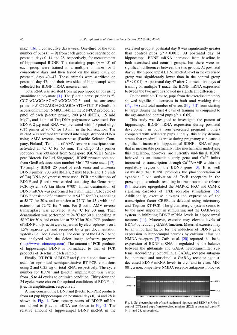
A time course of the BDNF and b-actin RT-PCR products
from rat pup hippocampus on postnatal days 0, 14 and 28 is
shown in Fig. 1. Densitometry scans of BDNF mRNA
normalized to b-actin mRNA are shown in Fig. 2. The
relative amount of hippocampal BDNF mRNA in the
exercised group at postnatal day 0 was significantly greater
than control pups (P , 0:001). At postnatal day 14
hippocampal BDNF mRNA increased from baseline in
both exercised and control groups, but there were no
significant differences between the two groups. At postnatal
day 28, the hippocampal BDNF mRNA level in the exercised
group was significantly lower than in the control group
(P , 0:01). At postnatal day 47 after 7 consecutive days of
training on multiple T maze, the BDNF mRNA expression
between the two groups showed no significant difference.
On the multiple T maze, pups from the exercised mothers
showed significant decreases in both total working time
(Fig. 3A) and total number of errors (Fig. 3B) from starting
to target during the first 4 days of training as compared to
the age-matched control pups (P , 0:05).
This study was designed to investigate the pattern of
hippocampal BDNF mRNA expression during postnatal
development in pups from exercised pregnant mothers
compared with sedentary pups. Finally, this study demon-
strates that treadmill exercise during pregnancy can cause a
significant increase in hippocampal BDNF mRNA of pups
that is measurable postnatally. The mechanisms underlying
this regulation, however, are currently undefined. BDNF
behaved as an immediate early gene and Ca2þ influx
increased its transcription through Ca2þ/cAMP within the
regulatory region of the BDNF gene [3]. It is well
established that BDNF promotes the phosphorylation of
synapsin I via activation of TrkB receptors in the
presynaptic terminals, resulting in neurotransmitter release
[9]. Exercise upregulated the MAP-K, PKC and CaM-K
signaling cascades of TrkB receptor stimulation [15].
Additionally, exercise elevates the expression of the
transcription factor CREB, as detected using microarray
and Taqman RT-PCR. The glutamatergic system seems to
be the most important in stimulating and the GABAergic
system in inhibiting BDNF mRNA levels in hippocampal
neurons [11]. Moreover, exercise may elevate levels of
BDNF by reducing GABA function. Maternal exercise may
be an important factor for the induction of BDNF gene
expression in hippocampal neurons by calcium influx via
NMDA receptors [7]. Zafra et al. [20] reported that basic
expression of BDNF mRNA is regulated by the balance
between the glutamate and GABA neurotransmitter sys-
tems. Accordingly, bicucullin, a GABAA receptor antagon-
ist, increased and muscimol, a GABAA receptor agonist,
decreased BDNF mRNA levels in vivo and in vitro. MK-
801, a noncompetitive NMDA receptor antagonist, blocked
Fig. 1. Gel electrophoresis of rat b-actin and hippocampal BDNF mRNA in
control (CTL) and pups from exercised mothers (EXS) at postnatal days (P)
0, 14 and 28, respectively.
P. Parnpiansil et al. / Neuroscience Letters 352 (2003) 45–4846

the increase in BDNF mRNA produced by bicucullin [20],
suggesting that the effect of glutamate on the basal
expression of BDNF mRNA is mediated through NMDA
rather than non-NMDA receptors [21]. Further studies have
demonstrated that MK-801 reduced the basal levels of
BDNF mRNA in the rat hippocampus in vitro and in vivo
[20]. On the other hand, MK-801, which specifically blocks
NMDA receptors, was ineffective in blocking the increase
in BDNF mRNA in hippocampal neurons. Zafra et al. [20]
found that the NMDA receptor antagonists, MK-801 and
ketamine could block the increase in BDNF and NGF
mRNA in vivo. However, neither MK-801 nor ketamine
inhibited the kainic acid mediated increase in hippocampal
BDNF mRNA, although both drugs effectively suppressed
seizures resulting from NMDA receptor activation. It has
been proposed that the placenta and amniotic fluid may also
be other sources of neurotrophic factors for the developing
fetus [6,18]. In addition, BDNF and NGF are expressed in
human amniotic epithelial tissue [18]. The sources of BDNF
and NGF in the amniotic fluid are unknown, though it is
likely that placenta amniotic epithelial tissue is one source.
There is evidence that some maternally derived growth
factors cross the placenta and are active in the fetus [6].
Exercise upregulates maternal hippocampal neuron activity
via glutamatergic neurons resulting in a significant increase
in hippocampal BDNF mRNA. Maternal BDNF gene
expression may mediate the signal transduction pathways
via release of glutamate, depolarization and Ca2þ dependent
BDNF transcription to cross the placenta via an autocrine or
paracrine mechanism and indirectly activate fetal BDNF
gene expression in hippocampal neurons. Therefore,
hippocampal BDNF mRNA of pups from exercised dams
increases immediately after birth via still unknown
mechanisms.
During neural development, hippocampal BDNF mRNA
expression reached the peak level at postnatal day 14 while
BDNF protein reached its highest level between postnatal
days 14 and 21 [2]. The hippocampus plays a central role in
spatial learning and memory [12]. Spatial learning is
associated with an increase in BDNF mRNA levels in the
hippocampus within 15 and 30 min after radial arm maze
test [10]. The induction of BDNF mRNA is relatively rapid,
detectable within 30 min and peaking at about 3 h [21]. In
this study we collected pup brain 24 h after T maze training
for quantitative measurement of hippocampal BDNF
mRNA. Thus, we could not detect an increased hippocam-
pal BDNF mRNA level at P47; it may return to the level in
control pups at 24 h after T maze training. With respect to
total working time and total number of errors from starting
to target, the maze shows a significant difference between
two groups of pups at the first 4 days of 7 consecutive days
of training. The behavioral differences at the first 4 days in
our study result from an elevation of hippocampal BDNF
mRNA expression after T maze training.
In conclusion, these results indicate that maternal
exercise by treadmill running at submaximal intensity
during pregnancy can upregulate rat pup hippocampal
BDNF mRNA expression at birth associated with an
increase in spatial learning. Increased BDNF mRNA
expression in the pups by physical exercise in pregnant
Fig. 2. Alterations of relative hippocampal BDNF mRNA expression in the
control and pups from exercised mothers at different postnatal ages: P0, P14
and P28, respectively. Each bargraph represents three sessions of RNA
determinations prepared totally from both sides of nine hippocampi for
each postnatal group. BDNF mRNA data are expressed as a BDNF/actin
ratio. Each value represents the mean ^ SEM of three experiments:
***P , 0:001, **P , 0:01, NS means no significant difference.
Fig. 3. (A,B) Bargraphs show the total working time from starting to target
and total number of errors in T maze during 7 consecutive days of T maze
training respectively. * means a significant difference between control and
pups from exercised mothers at P , 0:05.
P. Parnpiansil et al. / Neuroscience Letters 352 (2003) 45–48 47
mothers may have beneficial effects on spatial learning and
other intellectual development. Future studies will explore
whether maternal exercise during pregnancy and lactation
can maintain the neonatal elevation of hippocampal BDNF
mRNA expression, and spatial learning from exercised
mothers.
References
[1] P. Chomczynski, N. Sacchi, Single-step method of RNA isolation by
acid guanidinium thiocyanate-phenol-chloroform extraction, Anal.
Biochem. 162 (1987) 156–159.
[2] K.P. Das, S.L. Chao, L.D. White, W.T. Haines, G.J. Harry, H.A.
Tilson, S. Barone Jr., Differential patterns of nerve growth factor,
brain-derived neurotrophic factor and neurotrophin-3 mRNA and
protein levels in developing regions of rat brain, Neuroscience 103
(2001) 739–761.
[3] S. Finkbeiner, Calcium regulation of the brain-derived neurotrophic
factor gene, Cell. Mol. Life Sci. 57 (2000) 394–401.
[4] D.E. Fordyce, R.P. Farrar, Effect of physical activity on hippocampal
high affinity choline uptake and muscarinic binding: a comparison
between young and old F344 rats, Brain Res. 541 (1991) 57–62.
[5] D.E. Fordyce, J.M. Wehner, Physical activity enhances spatial
learning performance with an associated alteration in hippocampal
protein kinase C activity in C57BL/6 and DBA/2 mice, Brain Res. 619
(1993) 111–119.
[6] J.H. Gilmore, L.F. Jarskog, S. Vadlamudi, Maternal infection
regulates BDNF and NGF expression in fetal and neonatal brain
and maternal-fetal unit of the rat, J. Neuroimmunol. 138 (2003)
49–55.
[7] F. Gomez-Pinilla, V. So, J.P. Kesslak, Spatial learning induces
neurotrophin receptor and synapsin I in the hippocampus, Brain Res.
904 (2001) 13–19.
[8] A.E. Heffernan, Exercise and pregnancy in primary care. Nurse Pract.,
25 (2000) 42, 49, 53–56, passim.
[9] J.N. Jovanovic, A.J. Czernik, A.A. Fienberg, P. Greengard, T.S. Sihra,
Synapsins as mediators of BDNF-enhanced neurotransmitter release,
Nat. Neurosci. 3 (2000) 323–329.
[10] M. Mizuno, K. Yamada, A. Olariu, H. Nawa, T. Nabeshima,
Involvement of brain-derived neurotrophic factor in spatial memory
formation and maintenance in a radial arm maze test in rats,
J. Neurosci. 20 (2000) 7116–7121.
[11] R. Molteni, Z. Ying, F. Gomez-Pinilla, Differential effects of acute
and chronic exercise on plasticity-related genes in the rat hippo-
campus revealed by microarray, Eur. J. Neurosci. 16 (2002)
1107–1116.
[12] R.G. Morris, P. Garrud, J.N. Rawlins, J. O’Keefe, Place navigation
impaired in rats with hippocampal lesions, Nature 297 (1982)
681–683.
[13] S.A. Neeper, F. Gomez-Pinilla, J. Choi, C.W. Cotman, Physical
activity increases mRNA for brain-derived neurotrophic factor and
nerve growth factor in rat brain, Brain Res. 726 (1996) 49–56.
[14] T. Samorajski, C. Delaney, L. Durham, J.M. Ordy, J.A. Johnson, W.P.
Dunlap, Effect of exercise on longevity, body weight, locomotor
performance, and passive-avoidance memory of C57BL/6J mice,
Neurobiol. Aging 6 (1985) 17–24.
[15] R.A. Segal, M.E. Greenberg, Intracellular signaling pathways
activated by neurotrophic factors, Annu. Rev. Neurosci. 19 (1996)
463–489.
[16] R.E. Shepherd, P.D. Gollnick, Oxygen uptake of rats at different work
intensities, Pflug. Arch. 362 (1976) 219–222.
[17] W. Tokuyama, T. Hashimoto, Y.X. Li, H. Okuno, Y. Miyashita,
Highest trkB mRNA expression in the entorhinal cortex among
hippocampal subregions in the adult rat: contrasting pattern with
BDNF mRNA expression, Brain Res. Mol. Brain Res. 62 (1998)
206–215.
[18] S. Uchida, Y. Inanaga, M. Kobayashi, S. Hurukawa, M. Araie, N.
Sakuragawa, Neurotrophic function of conditioned medium from
human amniotic epithelial cells, J. Neurosci. Res. 62 (2000) 585–590.
[19] A.M. Uzendoski, R.W. Latin, K.E. Berg, S. Moshier, Physiological
responses to aerobic exercise during pregnancy and post-partum,
J. Sports Med. Phys. Fitness 30 (1990) 77–82.
[20] F. Zafra, E. Castren, H. Thoenen, D. Lindholm, Interplay between
glutamate and gamma-aminobutyric acid transmitter systems in the
physiological regulation of brain-derived neurotrophic factor and
nerve growth factor synthesis in hippocampal neurons, Proc. Natl.
Acad. Sci. USA 88 (1991) 10037–10041.
[21] F. Zafra, B. Hengerer, J. Leibrock, H. Thoenen, D. Lindholm, Activity
dependent regulation of BDNF and NGF mRNAs in the rat
hippocampus is mediated by non-NMDA glutamate receptors, Eur.
Mol. Biol. Org. J. 9 (1990) 3545–3550.
P. Parnpiansil et al. / Neuroscience Letters 352 (2003) 45–4848