excretory system biology 20. four excretory organs excretion rids the body of metabolic wastes...
TRANSCRIPT
Excretory System
Biology 20


Four Excretory Organs• Excretion rids the body of metabolic wastes
• Kidneys are the primary excretory organ but other organs also function in excretion: skin, liver and lungs
• Sweat glands in skin secrete perspiration– Sweat is water, salt and some urea– Helps rid the body of waste products but
mainly cools the body down by evaporation

Four Excretory Organs
• Liver excretes bile pigments–Bile pigments results from the
breakdown of hemoglobin–Urochrome is yellow pigment derived
from heme breakdown and is excreted by the kidneys
–Liver also excretes cholesterol and excess fat-soluble vitamins

Four Excretory Organs
• Kidneys produce urine–Urine contains 95% water plus
nitrogen wastes and inorganic salts–Nitrogenous wastes are products of
metabolism of amino acids and nucleotides
–Amino acid metabolism produces urea as main nitrogenous end products in humans

Urinary System Anatomy

Urinary System Anatomy
• Kidneys contribute to homeostasis, producing urine to rid body of nitrogenous wastes and keeping pH and salt/water balance of blood within normal range
• The Path of Urine– Kidneys
• Located on either side of the vertebral column just below the diaphragm
• Function is to produce urine
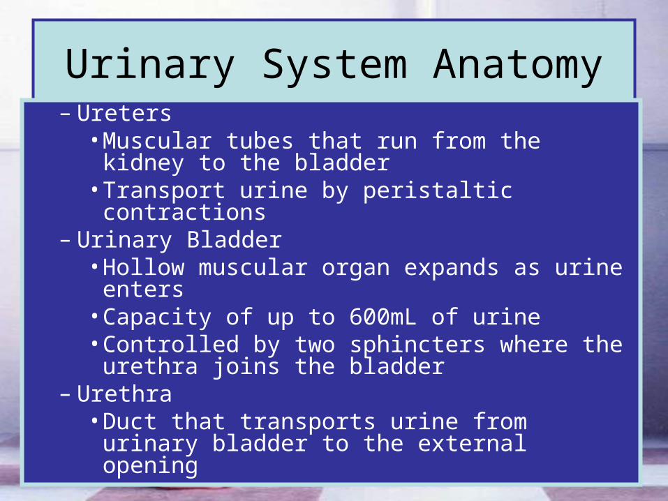
Urinary System Anatomy– Ureters
• Muscular tubes that run from the kidney to the bladder
• Transport urine by peristaltic contractions– Urinary Bladder
• Hollow muscular organ expands as urine enters
• Capacity of up to 600mL of urine• Controlled by two sphincters where the
urethra joins the bladder– Urethra
• Duct that transports urine from urinary bladder to the external opening

Urination and the Nervous System
• Urinary bladder fills with urine
• Stretch receptors send nerve impulses to the brain
• Nerve impulses from spinal cord causes the urinary bladder muscles to contact and the sphincters to relax making urination possible (micturating reflex)

Kidney Anatomy
• On concave side there is a depression where renal blood vessels and ureters enter
• Renal cortex is outer layer (filtration)

Renal Anatomy
• Renal medulla is the middle layer (reabsorption) with cone-shaped masses call renal pyramids
• Renal pelvis is central space or cavity that is continuous with the ureters

Kidney Anatomy

Nephrons
• Nephrons are microscopic
• Each kidney contains over one million nephrons
• Functional unit of the kidney
• Several nephrons enter one collecting duct

Afferent Arteriole
Efferent Arteriole

Blood Flow Through the Nephron
• Renal Artery brings waste filled blood into the kidney
• Afferent arteriole is a dilated blood vessel leading into the glomerulus

Blood Flow Through the Nephron
• Glomerulus is a ball of capillaries responsible for blood filtration
• Efferent arteriole is a constricted blood vessel leaving the nephron
• Blood collects in the venules that joins the renal vein
Bowman’s Capsule
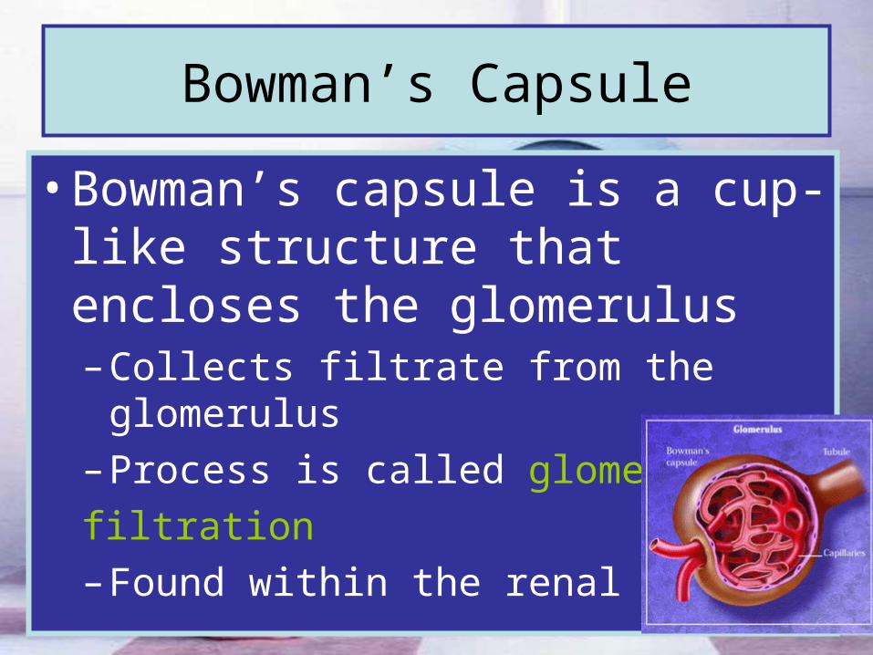
• Bowman’s capsule is a cup-like structure that encloses the glomerulus– Collects filtrate from the glomerulus
– Process is called glomerular
filtration
– Found within the renal cortex

Proximal Convoluted Tubule
• Has large surface area for tubular reabsorption
• Tubular reabsorption is the transfer of water and solutes from the nephron to the surrounding capillaries
• Cells have many mitochondria to supply energy for active transport
• Found within the renal medulla

Loop of Henle
• Descending loop of Henle allows the reabsorption of water
• Ascending loop of Henle in impermeable to water

Distal Convoluted Tubule and Collecting Duct
• Distal Convoluted Tubule– Permeable to water in the
presence of Antidiuretic Hormone
• Collecting Duct– Permeable to water in the
presence of Antidiuretic Hormone

Filtration
• The first event to occur in urine formation is filtration of the blood
• Filtrate will include mostly water and dissolved solutes including wastes and nutrients– Afferent arteriole is dilated, which
increases blood volume and pressure into the glomerulus
– Efferent arteriole is constricted, which increases pressure as blood backs up in the glomerulus
– Filtrate is collected by the Bowman’s capsule

Filtration
• You need a minimum of 70mmHg of systolic pressure for proper filtration to occur
• Extremely high blood pressure can damage the glomerulus and allow RBC and proteins to pass into the nephron

Urine Solute Concentration• The nutrients and salts in the filtrate need to be
reabsorbed into the bloodstream. This process includes:– 60% of the solutes are reabsorbed by active
transport at the proximal convoluted tubule– 20% is reabsorbed by active transport at the loop of
Henle– The remaining 20% is either reabsorbed at the
distal convoluted tubule or excreted as part of the urine

Solute Concentration
Bowman’s capsule
60% of solutes reabsorbed by active transport
20% of solutes reabsorbed by active transport
20% of solutes reabsorbed or excreted in the urine

Water Regulation
• The kidney filters about 180 to 200L of water a day
• 99% of the water is reabsorbed
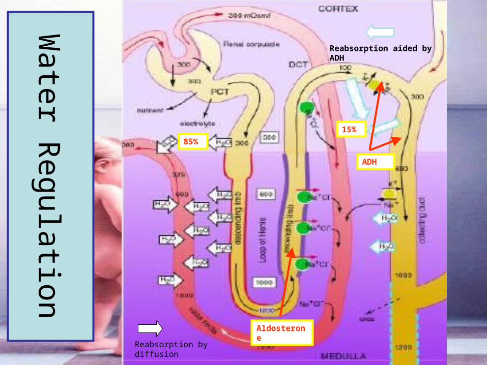
• About 85% of the water diffuses out of the water permeable proximal convoluted tubule and descending loop of Henle and is reabsorbed by the capillaries

Water Regulation
• Osmoreceptors in the hypothalamus are stimulated by the high osmotic pressure in the blood. As a result the hypothalamus stimulates:– The adrenal gland to release the
hormone aldosterone– The pituitary gland to release antidiuretic
hormone (ADH)– And other areas in the brain to produce
the sensation of thirst

Water Regulation
• At the ascending loop of Henle NaCl is actively transported out in the presence of the hormone Aldosterone
• The distal convoluted tubule and collecting duct become permeable to water in the presence of ADH
• The remaining 15% of water is reabsorbed based on osmotic pressure of the blood (regulated by ADH)
Water R
egulation
85%
Reabsorption by diffusion
Reabsorption aided by ADH
15%
Aldosterone
ADH

Dialysis

Kidney Disease
• Diabetes Mellitus– Lack of insulin produced from the
pancreas– Results in increased blood sugar levels,
which in turn will result in increased sugar concentration in the nephron
– Draws water into the nephron– Results in increased sugar and water in
the urine
Kidney Disease
• Diabetes Insipidus
– Destruction of ADH producing cells of the hypothalamus
– No reabsorption of water – urine output increases dramatically (20L/day)
– Must drink large quantities of water, regulated with injections of ADH

Kidney Disease
• Brights Disease (Nephritis)– Inflammation of the nephron– Proteins found in the urine
• Kidney Stones– The clumping and hardening of minerals
from the blood– Must pass through pelvis ureters
urinary bladder urethra (painful in males)