excitatory/inhibitory synaptic imbalance leads to ... · neuron article excitatory/inhibitory...
TRANSCRIPT

Neuron
Article
Excitatory/Inhibitory Synaptic Imbalance Leadsto Hippocampal Hyperexcitabilityin Mouse Models of Tuberous SclerosisHelen S. Bateup,1 Caroline A. Johnson,1 Cassandra L. Denefrio,1 Jessica L. Saulnier,1 Karl Kornacker,2
and Bernardo L. Sabatini1,*1Department of Neurobiology, Howard Hughes Medical Institute, Harvard Medical School, Boston, MA 02115, USA2Division of Sensory Biophysics, The Ohio State University, Columbus, OH 43210, USA*Correspondence: [email protected]
http://dx.doi.org/10.1016/j.neuron.2013.03.017
SUMMARY
Neural circuits are regulated by activity-dependentfeedback systems that tightly control network excit-ability and which are thought to be crucial for properbrain development. Defects in the ability to establishand maintain network homeostasis may be central tothe pathogenesis of neurodevelopmental disorders.Here, we examine the function of the tuberous scle-rosis complex (TSC)-mTOR signaling pathway, acommon target of mutations associated with epi-lepsy and autism spectrumdisorder, in regulating ac-tivity-dependent processes in the mouse hippocam-pus. We find that the TSC-mTOR pathway is a centralcomponentof apositive feedback loop thatpromotesnetwork activity by repressing inhibitory synapsesonto excitatory neurons. In Tsc1 KO neurons, weak-ened inhibition caused by deregulated mTOR altersthe balance of excitatory and inhibitory synaptictransmission, leading tohippocampal hyperexcitabil-ity. These findings identify the TSC-mTOR pathwayas a regulator of neural network activity and have im-plications for the neurological dysfunction in disor-ders exhibiting deregulated mTOR signaling.
INTRODUCTION
To preserve stability during changing environmental conditions
and developmental stages, neural networks have intrinsic regu-
latory mechanisms that maintain activity levels within a bounded
range (Davis, 2006). Individual neurons homeostatically regulate
their excitability via finely tuned mechanisms that detect and
respond to changes in action potential firing and network
activity. These include modulation of excitatory and inhibitory
postsynaptic strength, alterations in neurotransmitter release
probability, and adjustment of intrinsic membrane excitability
(Marder and Goaillard, 2006; Turrigiano, 2011). These forms of
plasticity are thought to be especially important during early
postnatal brain development when circuits adapt to the onset
and maturation of sensory input. Notably, several neurodevelop-
510 Neuron 78, 510–522, May 8, 2013 ª2013 Elsevier Inc.
mental disorders become manifest during this period of experi-
ence-dependent learning and circuit refinement (Zoghbi, 2003),
suggesting that disrupted homeostasis may be a contributing
factor (Ramocki and Zoghbi, 2008). In fact, a favored hypothesis
for autism spectrum disorders (ASDs) is that they arise from
imbalanced synaptic excitation and inhibition in specific neural
circuits (Bourgeron, 2009; Rubenstein and Merzenich, 2003).
One such neurodevelopmental disorder, tuberous sclerosis
complex (TSC), is caused by loss-of-function mutations in the
mTOR-negative regulators TSC1 or TSC2, resulting in a constel-
lation of neurological phenotypes that can include epilepsy,
autism, and intellectual disability (Prather and de Vries, 2004).
The mTOR kinase complex is the central component of a cell
growth pathway that responds to changes in nutrients, energy
balance, and extracellular signals to control cellular processes,
including protein synthesis, energy metabolism, and autophagy
(Laplante and Sabatini, 2012). Loss of function of the TSC1/2
protein complex results in deregulated and constitutively active
mTOR complex 1, which promotes cell growth and contributes
to tumor formation in dividing cells, including the hamartomas
that are characteristic of TSC (Kwiatkowski and Manning,
2005). However, the ways in which perturbations of TSC-mTOR
signaling alter the function of neurons or neural circuits to give
rise to the neurological pathologies associated with TSC are
not well understood.
Mouse models of TSC exhibit behavioral changes paralleling
human disease phenotypes, including seizures, decreased
social interaction, altered vocalizations, and deficits in learning
and memory (Ehninger et al., 2008; Meikle et al., 2007; Tsai
et al., 2012; Young et al., 2010). Like many other molecules
genetically linked to ASDs (Bourgeron, 2009), the TSC-mTOR
pathway regulates synapses such that loss-of-function muta-
tions in Tsc1 or 2 alter excitatory synapse structure, function,
and plasticity (Auerbach et al., 2011; Bateup et al., 2011; Che-
vere-Torres et al., 2012; Ehninger et al., 2008; Tavazoie et al.,
2005). Nevertheless, although perturbations of Tsc1/2 and
mTOR clearly alter aspects of neuronal function, given the
many homeostatic feedback pathways that influence neural cir-
cuit and brain development, it is unclear which perturbations are
directly causal and which are induced secondarily as a conse-
quence of altered brain function.
Here, we use in vitro and in vivo approaches to determine
the cell-autonomous and network phenotypes resulting from

Neuron
Loss of Tsc1 Disrupts Hippocampal E/I Balance
genetic deletion of Tsc1 in the mouse hippocampus, a brain re-
gion important for learning and memory that is involved in the
generation of temporal lobe seizures (Meador, 2007). We find
that loss of Tsc1 results in hippocampal network hyperexcitabil-
ity manifested by elevated spontaneous activity in dissociated
cultures and increased seizure susceptibility in vivo. Prolonged
high levels of network activity chronically engage activity-depen-
dent homeostatic pathways that secondarily alter the biochem-
ical, functional, and transcriptional state of neurons in vitro.
Network hyperexcitability cannot be attributed to alterations in
homeostatic excitatory synaptic plasticity, intrinsic neuronal
excitability, or glutamatergic synaptic drive. Rather, hippocam-
pal hyperexcitability results from a primary imbalance in excita-
tion and inhibition due to reduced inhibition onto Tsc1 knockout
(KO) pyramidal neurons. The loss of inhibition and upregulation
of network activity, as well as many of the pursuant secondary
responses in Tsc1 KO neurons, can be reversed by treatment
with the mTOR inhibitor rapamycin. These findings support the
hypothesis that disrupted excitatory/inhibitory (E/I) balance is
an initiating factor leading to perturbed circuit function in neuro-
developmental disorders.
RESULTS
NetworkHyperactivity in Tsc1KOHippocampal CulturesIn order to determine how loss of function of the Tsc1/2 complex
alters circuit function, we investigated whether genetic deletion
of Tsc1 affected the development of hippocampal network activ-
ity and the ability of neurons to respond to changes in activity.
We examined this in a culture system in which bidirectional
manipulation of activity can be accomplished pharmacologically
and biochemical, gene transcriptional, and synaptic analyses
can be performed in parallel (Figure 1). Dissociated hippocampal
cultures were prepared from mice carrying conditional alleles of
Tsc1 (Tsc1fl/fl) (Kwiatkowski et al., 2002). At 2 days in vitro (DIV)
cultures were infected with high titer lentivirus encoding either
synapsin-driven GFP (control) or GFP-IRES-Cre to delete Tsc1
from all neurons (Tsc1 KO). To address whether loss of Tsc1
affected neural network activity, we monitored the development
of spontaneous activity in neurons plated ontomultielectrode ar-
rays. Multiunit activity in control and Tsc1 KO neural networks
was measured simultaneously in dual-chamber arrays daily
over 2 weeks in culture (Figures 1A and 1B). We found that action
potential rates were significantly increased in Tsc1 KO networks
by 10 DIV and further increased to more than double control
levels by 14 DIV (Figures 1B and 1C). Activity in the DIV 14
Tsc1 KO cultures displayed a bursting pattern reminiscent of
an epileptic-like state (Figure 1B). The time point when activity
in Tsc1 KO neurons began to diverge from control levels corre-
sponded to the time when there was significant loss of Tsc1 pro-
tein, assessed by western blotting and upregulation of mTOR
signaling, determined by phosphorylation of the mTOR pathway
target ribosomal protein S6 (Figure 1D). Therefore, loss of Tsc1
has profound effects on the development of hippocampal
networks in vitro, resulting in severe hyperactivity.
We tested whether overactive mTOR signaling was respon-
sible for the deregulated activity in Tsc1 KO neurons by applying
the mTOR inhibitor rapamycin, beginning at 12 DIV. Network
activity decreased gradually in rapamycin such that after
4 days of treatment, activity levels were statistically indistin-
guishable between the two genotypes (Figure 1E). Compared
to controls, the rapamycin-dependent drop in activity was
greater in Tsc1 KO neurons, indicating that a larger proportion
of spiking activity was mTOR dependent in the latter condition
(Figure 1E, inset). The decrease in activity following rapamycin
was not likely to be due to cell death as acute treatment with
picrotoxin, a drug that blocks inhibitory receptors and increases
network activity, was able to robustly increase spiking activity in
cultures of both genotypes (control, 306.1% ± 58% of baseline,
p < 0.001; Tsc1 KO, 413.1%± 101.7%of baseline, p < 0.01). This
indicates that the networks were still responsive and capable of
generating high levels of activity after chronic treatment with
rapamycin.
Chronic Engagement of Activity-RegulatedTranscriptional Networks in Tsc1 KO NeuronsThe hyperactivity of Tsc1 KO cultures suggests a defect in the
activity-dependent processes that respond to and set network
activity levels. To test if loss of Tsc1 affects the ability of neural
networks to activate transcriptional programs in response to
changes in activity, we performed unbiased microarray analysis
of control and Tsc1 KO neurons in different activity states
(Figure 2; Tables S1 and S2 available online). Network activity
was elevated for 1, 6, or 24 hr by blocking inhibitory neurotrans-
mission with the glycine and GABAA/C receptor antagonist
picrotoxin. Activity was inhibited for the same time periods by
blocking action potential firing with the voltage-gated sodium
channel antagonist tetrodotoxin (TTX). Three biologically inde-
pendent samples per condition were collected and analyzed in
two batches: (1) basal and 6 hr treatments and (2) basal, 1 and
24 hr treatments. Therefore, the heatmap contains two basal
state control and Tsc1 KO conditions (Figure 2A). As expected,
the two sets of basal state samples show similar gene expres-
sion patterns within the genotype.
Hierarchical clustering of the experimental conditions revealed
that basal state Tsc1 KO neurons clustered with picrotoxin-
treated control neurons, denoted by the right cluster in the
dendrogram (Figure 2A), indicating basal alterations in many ac-
tivity-regulated genes due to loss of Tsc1. Conversely, pro-
longed (R6 hr) activity blockade in Tsc1 KO neurons reversed
many of the transcriptional changes such that TTX-treated KO
neurons clustered with control neurons in the basal state, shown
by the left cluster in the dendrogram (Figure 2A and Table S2).
Therefore, the majority of transcriptional changes in Tsc1 KO
neurons are a consequence of prolonged elevated network ac-
tivity and not a direct effect of loss of Tsc1.
Cluster analysis of the genes confirmed the presence of
constitutively engaged activity-dependent transcriptional pro-
grams in Tsc1 KO neurons (Figures 2A and 2B). Sets of genes
that were up- (Group 1) or down- (Group 3) regulated by long-
term elevations in network activity in control neurons showed
tonic changes in Tsc1 KO neurons that could be partially
reversed by prolonged activity blockade. Additionally, immedi-
ate early genes (Group 4) showed rapid and transient induction
following acute upregulation of activity in both Tsc1 KO and con-
trol networks. A subset of genes (Group 2) were elevated in Tsc1
Neuron 78, 510–522, May 8, 2013 ª2013 Elsevier Inc. 511

1 3 4 5 6 7 8 9 10 11 12 13 140
4
8
12
16
DIV
ControlTsc1 KO
virusAve
rage
spi
ke ra
te (H
z)
0 5 10 15 20 250
5
10
15
20
25
Control
Tsc1
KO
14 DIVC
D
*
2
*
* * *
2-3 4-5 6-7 8-9 10-11 12-13 14-15DIV
% o
f Con
trol
300
200
100
0
Tsc1 proteinp-S6
virus **
*
* *
** * *
E
2 3 4 5 6 7 8 9 10 11 12 13 14DIV
15 16 17 18 190
4
8
12
16
Ave
rage
spi
ke ra
te (H
z)
Rapamycin25
20
15
10
5
0
Spik
e Fr
eque
ncy
(Hz)
**
DIV12
DIV19
Control + Rapa
Tsc1 KO + Rapa*
* **
................................
150 μM150 μM
A B
0 5 10 15 20Time (s)
0 5 10 15 20Time (s)
10
20
30
40
50
60
Ele
ctro
de #
10
20
30
40
50
60E
lect
rode
#
DIV5 DIV14
ControlTsc1 KO
5s5s
Figure 1. Tsc1 KO Hippocampal Cultures Exhibit an mTOR-
Dependent Increase in Spontaneous Network Activity
(A) Image of a MED64 dual probe with 32 electrodes per chamber. The dotted
circles indicate the approximate plating area (�19 mm2) over the planar
electrode arrays, 150 3 150 mm interelectrode distance. Figure adapted from
http://www.MED64.com.
(B) Example raster plots of multiunit activity recorded from Tsc1fl/fl hippo-
campal neurons plated on a dual-chamber probe recorded on days 5 and 14
in vitro (DIV). Each line represents a single spike detected in a given channel
during 20 s of a recording. Neurons plated on the top array (electrodes #1–32)
were treated at 2 DIV with a GFP lentivirus (control, black). Neurons on the
bottom array (electrodes #33–64) were treated with GFP-IRES-Cre lentivirus
(Tsc1 KO, red). (Bottom) Example spiking data from one electrode on days 5
and 14, demonstrating a bursting pattern in Tsc1 KO cultures at DIV 14; scale
bar = 5 s.
(C) Average spike rate per electrode in hertz from control (black) and Tsc1 KO
(red) cultures across days in vitro (DIV). Lentivirus was added at 2 DIV (arrow).
Data are represented as mean ± SEM. * indicates significant difference
(p < 0.05) from control on that day. Inset scatterplot shows the average spike
rate per electrode on DIV 14 from pairs of control (x axis) and Tsc1 KO (y axis)
cultures (n = 15).
(D) Bar graphs display western blot data from Tsc1 KO neurons harvested on
the indicated DIV. Black bars represent Tsc1 protein levels (normalized to
b-actin loading control), and gray bars represent phosphorylated S6 levels
(p-S6, Ser240/244, normalized to total S6), expressed as a percentage of
control levels harvested on the same day (n = 4–8). Data are represented as
mean ± SEM. * indicates significant difference (p < 0.05) from control on that
day. Dashed line at 100% indicates control levels.
(E) Average spike rate per electrode in hertz from control (black) and Tsc1 KO
(red) neurons across days in vitro (DIV). At 12 DIV, 50 nM rapamycin was added
to both sets of cultures (n = 5–7). Data are represented as mean ± SEM. * in-
dicates significant difference (p < 0.05) from control on that day. Inset shows
the average spike rate on day 12 and day 19 from untreated cultures (dashed
lines) and rapamycin-treated cultures (solid lines). Data from control cultures
are in black, and data from Tsc1 KO cultures are in red. There was a significant
(p < 0.05) reduction of spiking activity from day 12 to 19 in the rapamycin-
treated cultures of both genotypes, denoted by the asterisks.
Neuron
Loss of Tsc1 Disrupts Hippocampal E/I Balance
512 Neuron 78, 510–522, May 8, 2013 ª2013 Elsevier Inc.
KO neurons across all activity conditions and showed little or no
modulation by activity in control neurons, indicating that these
genes are regulated by TSC-mTOR independently of activity.
Taken together, this transcriptional analysis demonstrates that
the increased activity of Tsc1 KO networks drives many consti-
tutive secondary changes in gene expression. However, it also
reveals that the core transcriptional responses to alterations in
network activity are generally preserved in Tsc1 KO neurons.
Tonic Upregulation of Arc and Engagementof Homeostatic Synaptic Plasticity in Tsc1 KO CulturesGene cluster 4 in the microarray data contains immediate early
genes that are rapidly induced by activity, including fos, zif268,
and Arc. Among these, Arc is known to be amediator of synaptic
plasticity, including a type of homeostatic plasticity described in
cultured neurons in which chronic decreases or increases in
network activity induce neuron-wide up- or downregulation,
respectively, of synaptic glutamate receptors as a means to
normalize excitatory drive (Shepherd et al., 2006; Turrigiano
et al., 1998). We and others have reported a deficit in another
form of Arc-dependent synaptic plasticity, metabotropic gluta-
mate receptor-induced long-term depression (mGluR-LTD),
following loss of function of Tsc1 or 2 (Auerbach et al., 2011; Ba-
teup et al., 2011; Chevere-Torres et al., 2012), which may be due
to an inability to activate mTOR-dependent translation of Arc

TTX (hr)Tsc1
PTX (hr)24 246 61 1- - - - - - - - - -
246124 61----------
TTX
24 h
rTT
X 6
hrTT
X 1
hrba
sal A
basa
l BPT
X 1
hrPT
X 6
hrPT
X 24
hr
Control Tsc1 KO
2
3
4
A B
-1
0
1
log
fold
log
fold
-1
0
1 2
-1
0
1
-1
0
1 4
log
fold
log
fold
Gene clusters
1
1
C C C CC C C CKO KO KO KO KO KO KO KO
3
Activity
Figure 2. Transcriptional Profiles of Control
and Tsc1 KO Neurons in Different Activity
States
(A) Hierarchical clustering of data from microarray
analysis of gene expression in control (C) and Tsc1
KO (KO) hippocampal neurons treated with 1 mM
TTX or 50 mM picrotoxin (PTX) for the indicated
times in hours. Treatment groups with similar pat-
terns of gene expression cluster together as indi-
cated by the dendrogram at the top of the figure.
The left cluster includes control neurons in the basal
condition and Tsc1 KO neurons treated for R6 hr
with TTX. The right cluster includes basal state Tsc1
KOneuronsandpicrotoxin-treatedcontrol neurons.
The heatmap displays the top 250 differential
expression profiles across all treatment groups; red
indicates higher expression; and blue indicates
lower expression relative to the median for all
groups. Data were obtained from two separate mi-
croarraybatches; therefore, there are twountreated
basal samples for each genotype indicated by the
red and black boxes. The numbers on the right
denote clusters of genes displaying similar patterns
of regulation determined by cluster analysis.
(B) Average ± SEM log-fold changes in expression
for each gene cluster are shown for low, basal,
and high network activity conditions across the x axis for control (black) and Tsc1 KO (red) cultures. For each gene, fold changes were calculated relative to the
average level across conditions such that no change from the mean results in log = 0 (dashed lines). Clusters 1 and 3 contain genes whose levels are up- or
downregulated, respectively, by activity showing constitutive changes in Tsc1 KO networks that are partially reversed by prolonged activity blockade. Cluster 4
contains immediate early genes that are robustly and transiently increased by activity in both control and Tsc1 KO networks. Cluster 2 contains genes
upregulated by loss of Tsc1 in an activity-independent manner.
See also Tables S1 and S2.
Neuron
Loss of Tsc1 Disrupts Hippocampal E/I Balance
mRNA at stimulated synapses (Waung and Huber, 2009). There-
fore, we hypothesized that deregulation of mTOR due to loss of
Tsc1 could perturb Arc-mediated homeostatic plasticity of excit-
atory synapses, leading to neuronal hyperactivity. To investigate
this possibility, we examined whether loss of Tsc1 disrupts the
activity-dependent production of Arc protein or the ability to
downregulate synaptic glutamate receptors.
Quantification of Arc mRNA levels by quantitative real-time
PCR confirmed basally high levels in Tsc1 KO neurons and
revealed significant bidirectional modulation by activity in a
manner similar to control neurons (Figure 3A). This confirms
that the activity-dependent transcriptional pathways that control
ArcmRNA production are not perturbed by loss of Tsc1.We next
investigated whether signaling through TSC-mTOR is required
for the activity-dependent translation of Arc protein. Consistent
with a possible role in this process, the mTOR pathway itself
was bidirectionally regulated by activity in control cultures
reflected by modulated phosphorylation of S6 following treat-
ment with picrotoxin or TTX (Figure 3B). In Tsc1 KO cultures,
p-S6 was constitutively elevated and no longer responsive to
manipulations of network activity (Figure 3B), indicating that
the Tsc1/2 complex is required to relay changes in network
activity to targets downstream of mTOR. Nevertheless, the
activity-dependent regulation of Arc protein was generally pre-
served in Tsc1 KO cultures (Figure 3C). In addition, short-term
rapamycin treatment (6 hr) did not affect basal or picrotoxin-
induced Arc protein levels in control or Tsc1 KO neurons,
whereas it reversed activation of the mTOR pathway target S6
(Figure S1). Thus, in contrast to our hypothesis, the Tsc1/
2-mTOR pathway does not directly control the basal or
activity-dependent production of Arc.
Arc mediates homeostatic plasticity by stimulating the
removal of glutamate receptors from the synapse; therefore,
we investigated whether the constitutive upregulation of Arc pro-
tein in Tsc1 KO cultures (see Figure 3C) had an effect on cell-
surface levels of AMPA-type glutamate receptors. As expected
from tonically active Arc-mediated endocytosis, surface levels
of the AMPA receptor subunits GluA1 and GluA2 were signifi-
cantly reduced in Tsc1 KO neurons compared to controls (Fig-
ure 3D). There was also a significant reduction in total levels of
GluA1 and GluA2 protein (GluA1, 51.7% ± 3.8% of control,
p < 0.001; GluA2, 80.6% ± 5.6% of control, p < 0.05;
n = 10–12) indicative of a global downregulation of glutamate
receptors in Tsc1 KO cultures. This was associated with a func-
tional reduction in glutamatergic synaptic strength and number
in Tsc1 KO neurons demonstrated by decreased amplitude
and number of spontaneous miniature excitatory postsynaptic
currents (mEPSCs) (Figures 3E–3G). These alterations probably
reflected an active homeostatic response to chronically high
network activity since both elevated Arc protein levels and the
biochemical downregulation of glutamate receptors could be
reversed in Tsc1 KO neurons by restoring activity levels with
chronic rapamycin treatment (Figures 3H–3J).
These findings indicate that homeostatic mechanisms for
regulating excitatory synaptic function are tonically engaged in
Tsc1 KO cultures. Therefore, a failure to activate these pro-
cesses cannot account for the network hyperactivity in Tsc1
KO cultures. Taken together with the microarray data, these
Neuron 78, 510–522, May 8, 2013 ª2013 Elsevier Inc. 513

ControlTsc1 KO
B
*
*
250
200
150
100
50
0
% o
f Con
trol
- +6h PTX -TTX - - +
- + -- - +
p-S6
* * * 1000
800
600
400
200
0
% o
f Con
trol
Arc protein
*
*- +6h PTX -
TTX - - +- + -- - +
#
#*
*
C
C KO C KO
GAPDH
GluA1GluA2
IEI (s)Amp (pA)
1.0
0.5
0
1.0
0.5
0
ControlTsc1 KO
ControlTsc1 KO
mEPSC Amplitude mEPSC IEI
cum
ulat
ive
prob
abili
ty
cum
ulat
ive
prob
abili
ty
0.5 1 1.5 2.05040302010
Pull-downInput
0
E
300ms25pA
Control Tsc1 KO
0
25
50
75
100
125
% o
f Con
trol
GluA1
GluA2
* *
Surface proteins
D
F
IH Tsc1 KOControl- + - +
GluA1β-Actin
7d Rapa:
100
50
0
25
75
125
-7d Rapa + - +
GluA1
*
#
% o
f Con
trol
Tsc1 KOControl- + - +
GluA2β-Actin
7d Rapa:
GluA2
-7d Rapa + - +
100
50
0
25
75
125
*
*#
% o
f Con
trol
- +6h PTX -TTX - - +
- + -- - +
2500
2000
1500
1000
500
0
% o
f Con
trol
Arc mRNAA
**
#
*
*#
G
250
200
150
100
50
0
% o
f Con
trol
Arc
*
*
*
-7d Rapa + - +
Arc
Tsc1 KOControl-7d Rapa: + - +
#
β-Actin
ControlTsc1 KO
J
ControlTsc1 KO
Figure 3. Activity-Dependent Homeostatic Pathways Are Tonically Engaged in Tsc1 KO Cultures
(A) Quantitative real-time PCR analysis of Arc mRNA levels in control (black) and Tsc1 KO (red) hippocampal cultures following treatment with 50 mMpicrotoxin or
1 mM TTX for 6 hr (n = 2–6).
(B and C)Western blot data of phosphorylated S6 (B) (p-S6 Ser240/244, normalized to total S6) and Arc protein levels (C) (normalized to b-actin loading control) in
control (black) and Tsc1 KO (red) cultures following treatment with 50 mM picrotoxin or 1 mM TTX for 6 hr (n = 7–20).
(D) (Top)Representativewesternblots fromabiotin-surface-protein-labelingexperiment. Left lanesare total cell lysates (input), and right lanesarecell-surfaceproteins
(pull-down). C, control; KO, Tsc1 knockout. (Bottom) Quantification of surface GluA1 and GluA2 levels from control (black) and Tsc1 KO (red) cultures (n = 5–7).
(E) Representative traces of miniature excitatory postsynaptic currents (mEPSCs) recorded from control (black) and Tsc1 KO (red) neurons in culture.
(F and G) Cumulative distributions of mEPSC amplitudes (F) and interevent intervals (IEI) (G) from control (black) and Tsc1 KO (red) neurons in culture (n = 9–10).
(H–J) (Top) Representative western blots of Arc (H), GluA1 (I), and GluA2 (J) protein in control and Tsc1 KO cultures following 7 days of treatment with 50 nM
rapamycin. (Bottom) Bar graphs displaying summary western blot data for Arc (H), GluA1 (I), and GluA2 (J) expressed as a percentage of untreated control
(n = 9–10). Protein levels were normalized to b-actin loading control.
Data in bar graphs are represented as mean ± SEM, normalized to the control baseline condition. * indicates significant difference (p < 0.05) from untreated
control; # indicates significant difference (p < 0.05) from untreated Tsc1 KO. See also Figure S1.
Neuron
Loss of Tsc1 Disrupts Hippocampal E/I Balance
514 Neuron 78, 510–522, May 8, 2013 ª2013 Elsevier Inc.

p-S6A B
C MaximumSeizure Severity
Seizure Behavior
0 30 60 90 120 150 180Time (min.)
0
1
2
3
4
5
6
7
Seiz
ure
seve
rity
scor
e
cKOControl
0
20
40
60
80
100
Perc
enta
ge o
f mic
e
Cont. cKO
Stage 3Stage 4Stage 5Stage 6Mortality
D
100
80
60
40
20
0
Perc
ent s
urvi
val
cKOControl
0 10 20 30 40 50 60Days
Survival ProportionE
*
Cont. cKO
10
30
50
Min
utes
70Latency to stage 4
Control
cKO
Figure 4. Loss of Tsc1 in Forebrain
Excitatory Neurons Increases Seizure
Severity, Resulting in Premature Death
(A) Immunohistochemistry staining for phosphor-
ylated S6 (p-S6, Ser240/244) in hippocampal brain
sections from a control (CamkIIaCre+;Tsc1wt/wt)
and Tsc1 conditional knockout mouse (cKO,
CamkIIaCre+;Tsc1fl/fl) at 4 weeks of age.
(B) Severity of seizure behavior over time following
i.p. injection of 15 mg/kg kainic acid in 4- to
5-week-old Tsc1 cKO (n = 5) and littermate control
mice (n = 20). Higher scores correspond to
more severe seizure status; a score of seven
indicates mortality. Data are represented as
mean ± SEM.
(C) Tsc1 cKO mice displayed increased seizure
severity demonstrated by a higher percentage of
mice with a maximum seizure score of six (tonic-
clonic seizures) or seven (mortality) within the 3 hr
test period.
(D) Scatterplot summary of the time in minutes to
reach seizure stage 4 (forelimb clonus with
intermittent rearing) in control and Tsc1 cKO
mice. * indicates significant difference (p < 0.001)
from control.
(E) Kaplan-Meier survival curve for untreated
control (CamkIIaCre+;Tsc1wt/wt, n = 40) and
Tsc1 cKO (CamkIIaCre+;Tsc1fl/fl, n = 41) littermate
mice.
Neuron
Loss of Tsc1 Disrupts Hippocampal E/I Balance
results also suggest that many alterations observed in Tsc1 KO
neurons are actually secondary, compensatory changes result-
ing from unrestrained activity and not acutely due to elevated
mTOR signaling.
Loss of Tsc1 in Forebrain Excitatory Neurons CausesHyperexcitability and SeizuresThe above results indicate that loss of Tsc1 leads to hyperactivity
of hippocampal networks and secondary engagement of
homeostatic synaptic plasticity in vitro. However, despite down-
regulation of synaptic AMPA receptors, network activity
remained elevated, suggesting that the primary trigger of hyper-
excitability in Tsc1 KO networks cannot be fully compensated by
reduced glutamatergic drive. To determine the functional mech-
anism behind this hyperactivity in a more physiological context,
we generated an in vivo model in which Tsc1 was conditionally
deleted from excitatory forebrain neurons. To do this, Tsc1fl/fl
mice were crossed with mice expressing Cre recombinase
from theCamkIIa promoter (Tsien et al., 1996). Since Cre expres-
sion does not turn on until approximately 21 days of age in these
mice, this approach also allows investigation of the effects of
perturbed mTOR signaling on network activity in a more mature
circuit.
mTOR signaling was elevated in the hippocampus of
CamkIIaCre+;Tsc1fl/fl (cKO) mice judged by immunostaining for
p-S6 at 4 weeks of age (Figure 4A). To determine whether these
mice displayed a hyperexcitability phenotype, we assessed
seizure induction at postnatal day (PND) 29–32, following admin-
istration of the convulsant kainic acid. Seizure severity was
determined using a previously established rating system (Morri-
son et al., 1996) with higher values corresponding tomore severe
seizures and a score of seven indicating death. Over the 3 hr test
period, cKO mice displayed dramatically increased severity of
seizures (Figure 4B) such that 80% of the cKO mice died during
the 3 hr observation period in contrast to zero mortalities among
the littermate controls (Figure 4C). Moreover, Tsc1 cKO mice
exhibited significantly decreased latency to reach seizure stage
four (forepaw clonus with rearing) (Figure 4D). The hyperexcit-
ability phenotype in the Tsc1 cKO mice was severe enough
that even without experimental manipulation we observed spon-
taneous seizures in some mice and premature death (Figure 4E),
as reported previously (Ehninger et al., 2008). These findings
indicate that selective loss of Tsc1 in excitatory pyramidal
neurons causes severe behavioral hyperexcitability, even in the
absence of developmental abnormalities.
Loss of Tsc1 Does Not Increase Intrinsic Excitabilityor Glutamatergic Synaptic DriveThe network hyperexcitability phenotype we observed both
in vitro and in vivo could arise from several possible mecha-
nisms, including alterations in intrinsic membrane excitability,
synaptically driven excitability, or inhibitory synapse function.
Due to the early lethality of the Tsc1 cKO mice and the possible
secondary changes in neuronal function due to spontaneous
seizures, we investigated these possibilities in acute brain slices
from Tsc1fl/fl mice injected with an adeno-associated virus (AAV)
expressing a Cre-EGFP fusion protein in the CA1 subregion of
the hippocampus. We diluted the virus to achieve sparse Cre
Neuron 78, 510–522, May 8, 2013 ª2013 Elsevier Inc. 515
# of
AP
s
010
20
30
40
50
60
70
Control Tsc1 KO
40m
V
500ms200 pA
150 pA
100 pA
Current (pA)0 100 200 300 400
ms
100
200
300
400
500
0
LatencyControlTsc1 KO
Action Potentials
Current (pA)0 100 200 300 400
ControlTsc1 KO
B C D
Stimulus Duration (ms)0.2 0.4 0.6 0.8 1.0
0
10
20
30
40
# of
APs
Control
Tsc1 KO
500ms
40m
V
20Hz Stimulation
ControlTsc1 KO
G
A
*
*
0 5 10 15Control (mV)
0
5
10
15
Tsc1
KO
(mV
)
150ms
2mV
Single EPSP
1 2 3 4 5Stimulus #
ControlTsc1 KO
4
6
8
10
Ampl
itude
(mV)
5Hz StimulationControl
Tsc1 KO
500ms
5mV
E F
Cre-EGFP
p-S6
Overlay
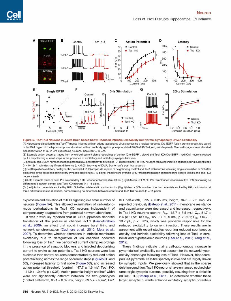
Figure 5. Tsc1 KO Neurons in Acute Brain Slices Show Reduced Intrinsic Excitability but Normal Synaptically Driven Excitability
(A) Hippocampal section from a Tsc1fl/flmouse injected with an adeno-associated virus expressing a nuclear-targeted Cre-EGFP fusion protein (green, top panel)
in the CA1 region of the hippocampus and stained with an antibody against phosphorylated S6 (Ser240/244, red, middle panel). Overlaid image shows elevated
phosphorylation of S6 in Cre-expressing neurons. Scale bar = 10 mm.
(B) Example action potential traces from whole-cell current clamp recordings of control (Cre-EGFP�, black) and Tsc1 KO (Cre-EGFP+, red) CA1 neurons evoked
by 1 s depolarizing current steps in the presence of excitatory and inhibitory synaptic blockers.
(C and D)Mean ± SEMnumber of action potentials (C) and latency to first spike (D) in control and Tsc1 KO neurons following injection of depolarizing current steps
(n = 9–12). * indicates significant difference (p < 0.05, two-way ANOVA, Bonferroni’s post hoc analysis).
(E) Scatterplot of excitatory postsynaptic potential (EPSP) amplitude in pairs of neighboring control and Tsc1 KO neurons following single stimulation of Schaffer
collaterals in the presence of inhibitory synaptic blockers (n = 19 pairs). Inset shows overlaid EPSP traces from a pair of neighboring control (black) and Tsc1 KO
neurons (red).
(F) (Left) Example trains of five EPSPs evoked by 5 Hz Schaffer collateral stimulation. (Right) Mean ± SEM of EPSP amplitudes for a train of five EPSPs showing no
differences between control and Tsc1 KO neurons (n = 16 pairs).
(G) (Left) Action potentials evoked by 20 Hz Schaffer collateral stimulation for 1 s. (Right) Mean ± SEM number of action potentials evoked by 20 Hz stimulation at
three different stimulus durations, demonstrating no difference between control and Tsc1 KO neurons (n = 11 pairs).
Neuron
Loss of Tsc1 Disrupts Hippocampal E/I Balance
expression and elevation of mTOR signaling in a small number of
neurons (Figure 5A). This allowed examination of cell-autono-
mous perturbations in Tsc1 KO neurons independent of
compensatory adaptations from potential network alterations.
It was previously reported that mTOR suppresses dendritic
translation of the potassium channel Kv1.1 (Raab-Graham
et al., 2006), an effect that could increase burst firing and
network synchronization (Cudmore et al., 2010; Metz et al.,
2007). To determine whether alterations in intrinsic membrane
excitability due to deregulation of ion channels occurred
following loss of Tsc1, we performed current clamp recordings
in the presence of synaptic blockers and injected depolarizing
current to evoke action potentials. Tsc1 KO neurons were less
excitable than control neurons demonstrated by reduced action
potential firing across the range of current steps (Figures 5B and
5C), increased latency to first spike (Figure 5D), and increased
action potential threshold (control, �47.7 ± 1.6 mV; Tsc1 KO,
�41.9 ± 1.9 mV; p < 0.05). Action potential height and half-width
were not significantly different between the two genotypes
(control half-width, 0.91 ± 0.02 ms, height, 89.5 ± 2.5 mV; Tsc1
516 Neuron 78, 510–522, May 8, 2013 ª2013 Elsevier Inc.
KO half-width, 0.95 ± 0.05 ms, height, 84.6 ± 2.5 mV). As
reported previously (Bateup et al., 2011), membrane resistance
and capacitance were decreased and increased, respectively,
in Tsc1 KO neurons (control Rm, 167.7 ± 5.5 mU; Cm, 81.7 ±
2.6 pF; Tsc1 KO Rm, 127.0 ± 10.9 mU, p < 0.01; Cm, 115.7 ±
10.2 pF, p < 0.01), which was probably responsible for the
reduced excitability to current injection. These results are in
agreement with recent studies reporting reduced spontaneous
activity and intrinsic excitability following loss of Tsc1 in cere-
bellar and hypothalamic neurons (Tsai et al., 2012; Yang et al.,
2012).
These findings indicate that a cell-autonomous increase in
pyramidal cell excitability cannot account for the network hyper-
activity phenotype following loss of Tsc1. However, hippocam-
pal CA1 pyramidal cells fire sparsely in vivo and are largely driven
by synaptic inputs. We previously showed that in the sparse
deletion condition, Tsc1 KO neurons have enhanced evoked glu-
tamatergic synaptic currents, possibly resulting from a deficit in
mGluR-LTD (Bateup et al., 2011). To determine whether these
larger synaptic currents enhance excitatory synaptic potentials

Control
1s
20pA
Tsc1 KO
A
ControlTsc1 KO
pA30252015105
1.0
0.5
0
cum
ulat
ive
prob
abili
ty
mIPSC Amplitude
ControlTsc1 KO
seconds1 2 30
1.0
0.5
0
cum
ulat
ive
prob
abili
ty
mIPSC IEI
0 100 200 300 400Control (pA) Con
trol
Tsc1 K
O
*
0
100
200
300
400
Tsc1
KO
(pA
)
0
50
100
150
200
250
Am
plitu
de (p
A)
IPSC Amplitude
100ms50pA
Control
Tsc1 KO100ms
100p
A
D E
ControlTsc1 KO
0 100 200 300 400ISI (ms)
5000.2
0.4
0.6
0.8
1.0
Rat
io
Paired Pulse Ratio
B C
*
Cont. KO
12
10
8
6
*
Cont. KO
.75
.50
.25
0
pA sec
Figure 6. Decreased Amplitude of Inhibitory Synaptic Currents in Tsc1 KO Neurons
(A) Example recordings of miniature inhibitory postsynaptic currents (mIPSCs) from control (black) and Tsc1 KO (red) CA1 neurons from an acute brain slice.
(B andC) Cumulative distributions ofmIPSCamplitudes (B) and interevent intervals (IEI) (C) from control (black) and Tsc1KO (red) neurons (n = 11–12). Insets show
scatterplot summaries of cell averages; horizontal lines indicate the mean, and error bars denote SEM. * indicates significant difference (p < 0.05) from control.
(D) (Left) Scatterplot of evoked IPSC amplitude in pairs of neighboring control and Tsc1 KO neurons following stimulation of the CA1 pyramidal cell layer with
excitatory synaptic transmission blocked (n = 16 pairs). Inset shows overlaid IPSC traces from a pair of neighboring control (black) and Tsc1 KO neurons (red).
Scale bar = 100 pA3 25ms. (Right) Mean ± SEM evoked IPSC amplitude in control and Tsc1 KO neurons. * indicates significant difference (p < 0.05) from control.
(E) (Left) Representative overlaid traces for sets of two IPSCs evoked by stimuli delivered at different interstimulus intervals (ISI). (Right) Mean ± SEM paired pulse
ratios of IPSCs from neighboring control and Tsc1 KO neurons at different ISIs (n = 14 pairs), demonstrating no difference between genotypes.
Neuron
Loss of Tsc1 Disrupts Hippocampal E/I Balance
or synaptically driven firing, we performed simultaneous current
clamp recordings of neighboring pairs of control and Tsc1 KO
neurons, while stimulating Schaffer collateral axons at different
frequencies. There was no significant difference in the amplitude
of excitatory postsynaptic potentials (EPSPs) following either a
single stimulation or a 5 Hz train (Figures 5E and 5F). Further-
more, no differences in firing frequency were observed following
20 Hz stimulation at three different stimulus intensities (Fig-
ure 5G). We did, however, observe a significant decrease in
the resting membrane potential of Tsc1 KO neurons compared
to controls (control, �66.6 ± 1.0 mV; Tsc1 KO, �69.1 ±
1.2 mV, p < 0.05). These results demonstrate that the increased
glutamatergic synaptic currents we observed previously (Bateup
et al., 2011) are largely canceled out by the reduced intrinsic
excitability such that there is no net change in glutamatergic syn-
apse-driven excitability in isolated Tsc1 KO neurons. Taken
together with the reduction in glutamatergic synapses we
observed in the highly active cultures (see Figure 3), this indi-
cates that an enhancement of excitatory synaptic drive does
not account for the network hyperexcitability caused by loss of
Tsc1.
Loss of Tsc1 Reduces Inhibitory Synaptic TransmissionIn addition to intrinsic neuronal firing rate and excitatory synaptic
drive, neural network activity is dependent upon inhibition, which
controls overall activity level, shapes the temporal pattern of
activity, and limits bursting (Isaacson and Scanziani, 2011; Kull-
mann, 2011). To assess the strength and number of inhibitory
synapses onto CA1 pyramidal neurons, we recorded sponta-
neous miniature inhibitory synaptic currents (mIPSCs) following
sparse loss of Tsc1. Both mIPSC amplitude and interevent inter-
val were significantly reduced in Tsc1 KO neurons, which is
indicative of reduced ionotropic GABA receptor content per syn-
apse but a greater number of inhibitory synapses (Figures
6A–6C). We determined how these alterations affected evoked
inhibition by recording IPSCs in pairs of neighboring control
and Tsc1 KO neurons following stimulation of interneurons in
the CA1 pyramidal cell layer. Ionotropic glutamate receptors
were blocked to allow direct activation of inhibitory interneurons
and to evoke monosynaptic IPSCs recorded as inward currents
with a high chloride internal solution. We found a significant
reduction in the amplitude of evoked inhibitory currents in Tsc1
KO neurons relative to controls (Figure 6D). Consistent with the
decreased mIPSC amplitude, this effect was probably due to a
change in postsynaptic function as there were no differences
between control and Tsc1 KO neurons in paired pulse ratios,
which are a measure of presynaptic release probability at inhib-
itory synapses (Figure 6E).
These results indicate that loss of Tsc1 in CA1 pyramidal
neurons causes a cell-autonomous weakening of inhibitory
Neuron 78, 510–522, May 8, 2013 ª2013 Elsevier Inc. 517

A B CmIPSC Amplitude mIPSC IEI
pA30252015105
1.0
0.5
0
cum
ulat
ive
prob
abili
ty ControlTsc1 KO
ControlTsc1 KO
seconds2 4 60 8
1.0
0.5
0
cum
ulat
ive
prob
abili
ty
D E FmEPSC Amplitude mEPSC IEI
ControlTsc1 KO
pA35
1.0
0.5
0
cum
ulat
ive
prob
abili
ty
seconds1 2 30 4 5 6
1.0
0.5
0
cum
ulat
ive
prob
abili
ty ControlTsc1 KO
Control
Tsc1 KO
1s
20pA
1s
20pA
Control
Tsc1 KO
25155
Widespread Cre-EGFP
Cont. KO
4
8
12
pA
*
Cont. KO
0
2
4
sec
*
Cont. KO
6
10
14
pA
Cont. KO
0
1
2
3
sec
0 1 2 3 4Control
5 6 70
1
2
3
4
5
6
7
Tsc1
KO
E/I ratio
0
1
2
3
Cont.
KO
*
G
4mV
50ms
Tsc1 KO
Control
Sparse knock-outEPSP
0
2
4
6
8
mV
H
C KO
Tsc1 KO
3mV
50ms
Widespread knock-outI
Control
6mV
50ms
0
2
4
6
8
mV
C KO
EPSP543
0
mV
21
C KO
*
IPSP
IPSP6
4
2
0
mV
*
C KO
Rapamycin Treated
J
EPSP IPSP
Sparse knock-out
E/I
Rat
io
Widespread knock-out
Sparse knock-out
50ms
3mV
Tsc1 KO
Control
0
2
4
6
8
mV
C KO C KO
6
4
2
0
mV
Figure 7. Excitatory-Inhibitory Synaptic
Imbalance in Tsc1 KO Neurons
(A) (Top) A high titer adeno-associated virus
expressing a nuclear localized Cre-EGFP fusion
proteinwas stereotaxically injected unilaterally into
the CA1 region to delete Tsc1 from >90% of neu-
rons (‘‘widespreadknockout’’). Scale bar =100mm.
(Bottom) Example recordings of miniature inhibi-
tory postsynaptic currents (mIPSCs) from a control
neuron (black) in the uninjected hemisphere and a
Tsc1 KO neuron (red) in the injected hemisphere.
(B and C) Cumulative distributions of mIPSC
amplitudes (B) and interevent intervals (IEI) (C)
from control neurons in the uninjected hemisphere
and Tsc1 KO neurons in the injected hemisphere
(n = 12). Insets show scatterplot summaries of cell
averages; horizontal lines indicate the mean, and
error bars denote SEM. * indicates significant
difference (p < 0.05) from control.
(D) Example recordings of miniature excitatory
postsynaptic currents (mEPSCs) from a control
neuron (black) in the uninjected hemisphere and a
Tsc1 KO neuron (red) in the injected hemisphere
following widespread injection of Cre.
(E and F) Cumulative distributions of mEPSC
amplitudes (E) and interevent intervals (IEI) (F) from
control neurons in the uninjected hemisphere and
Tsc1 KO neurons in the injected hemisphere
(n = 11–14). Insets show scatterplot summaries
of cell averages, demonstrating no difference
between genotypes; horizontal lines indicate the
mean, and error bars denote SEM.
(G) Scatterplot of excitatory/inhibitory (E/I) ratio in
pairs of neighboring control and Tsc1 KO neurons
following sparse deletion of Tsc1 (‘‘sparse knock-
out,’’ as inFigure5A). Insetshowsmean±SEMofE/I
ratio in cells of both genotypes (n = 13 pairs). * in-
dicates significant difference (p < 0.05) fromcontrol.
(H) Sparse knockout. (Left) Overlaid traces of compound excitatory (EPSP) and inhibitory (IPSP) postsynaptic potentials evoked by Schaffer collateral stimulation
in a neighboring pair of control (black) and Tsc1 KO (red) neurons. Dashed line indicates the baseline. (Right) Mean ± SEM of EPSP and IPSP amplitude in control
and Tsc1 KO neurons (n = 13 pairs). * indicates significant difference (p < 0.05) from control.
(I) Widespread knockout. (Left) Example traces of compound postsynaptic potentials evoked by Schaffer collateral stimulation in a control neuron in the
uninjected hemisphere (black) and a Tsc1 KO neuron in the injected hemisphere (red) following widespread unilateral injection of Cre. Dashed lines indicate the
baseline. (Right) Mean ± SEM. EPSP and IPSP amplitude in control and Tsc1 KO neurons (n = 8–9). * indicates significant difference (p < 0.05) from control.
(J) Sparse knockout. Mice were injected daily with 5 mg/kg rapamycin for 6 days prior to and on the day of electrophysiological analysis. (Top) Overlaid
recordings of compound postsynaptic potentials evoked by Schaffer collateral stimulation in a neighboring pair of control (black) and Tsc1 KO (red) neurons.
Dashed line indicates the baseline. (Bottom) Mean ± SEM of EPSP and IPSP amplitude after 7 days of treatment with rapamycin, demonstrating no difference
between control and Tsc1 KO neurons (n = 8 pairs).
See also Figures S2–S4.
Neuron
Loss of Tsc1 Disrupts Hippocampal E/I Balance
input, which could result in hyperexcitability at the network level.
To determine whether reduced inhibition persists when there is
more widespread loss of Tsc1, we injected a high concentration
of the Cre-EGFP-expressing virus to delete Tsc1 from >90% of
CA1 neurons on one side of the brain (Figure 7A; Figure S2).
This resulted in robust activation of mTOR signaling in area
CA1 (Figure S2), but probably due to the unilateral and spatially
confined nature of themanipulation, did not induce spontaneous
seizures. Comparison of Tsc1 KO neurons from the injected
hemisphere to control neurons from the uninjected hemisphere
revealed a significant reduction in mIPSC amplitude due to
loss of Tsc1 (Figures 7A and 7B). Moreover, and in contrast to
our findings following sparse loss of Tsc1 (see Figure 6C), the in-
terevent interval (IEI) of mIPSCs was significantly increased after
518 Neuron 78, 510–522, May 8, 2013 ª2013 Elsevier Inc.
widespread deletion of Tsc1 (Figure 7C), which is indicative of a
reduction in inhibitory synapse number. These changes in inhib-
itory synaptic transmission could be due to alterations in presyn-
aptic inhibitory interneurons, postsynaptic pyramidal neurons, or
both.We found that the AAV serotype 1 used to deliver Cre either
did not infect or did not express in interneurons of the hippocam-
pus (Figure S3), suggesting that the changes in inhibitory syn-
apse strength and number were probably due to alterations in
postsynaptic pyramidal cells. To determine if synapse loss was
specific to inhibitory synapses, we measured miniature excit-
atory synaptic currents following the same widespread loss of
Tsc1 in area CA1. We found no significant changes in mEPSC
amplitude or IEI between neurons of the two genotypes (Figures
7D–7F). This is in contrast to the downregulation of mEPSCs we

Neuron
Loss of Tsc1 Disrupts Hippocampal E/I Balance
observed in the highly active Tsc1 KO cultures (see Figure 3).
Since our in vivo manipulation affected only postsynaptic CA1
neurons on one side of the brain, it is possible that network
activity was not elevated enough to engage homeostatic synap-
tic scaling under these conditions.
The above data, based on measurements of spontaneous
excitatory and inhibitory transmission in different populations
of neurons, suggest an imbalance in excitation and inhibition
following loss of Tsc1. To determine whether loss of Tsc1 causes
E/I imbalance in individual Tsc1 KO neurons following stimula-
tion of the local circuit, current clamp recordings were used to
measure compound synaptic potentials elicited by Schaffer
collateral stimulation. This analysis was performed in the
absence of synaptic blockers to allow activation of both excit-
atory and inhibitory synapses. We performed these experiments
first in the sparse deletion condition to allow direct comparison of
synaptic potentials in neighboring pairs of control and Tsc1 KO
neurons evoked by the same stimulus. In support of our hypoth-
esis, loss of Tsc1 resulted in a significant increase in the E/I ratio
in Tsc1 KO neurons relative to controls (Figure 7G) that was pri-
marily due to a decrease in the amplitude of inhibitory hyperpo-
larizing potentials (Figure 7H). This imbalance persisted and was
exacerbated following widespread loss of Tsc1 in CA1 neurons
(Figure 7I), consistent with our mIPSC findings.
In Tsc1 KO cultures, network hyperactivity could be restored
with chronic rapamycin treatment. To test whether mTOR
blockade in vivo could reverse the E/I imbalance in Tsc1 KO
neurons, we treatedmice with rapamycin for 7 days prior to elec-
trophysiological analysis. We confirmed that this treatment
resulted in reduced mTOR signaling in the hippocampus (Fig-
ure S4). We found that rapamycin was sufficient to normalize
E/I ratios (control, 1.83 ± 0.31; Tsc1 KO, 1.43 ± 0.52; p = 0.31)
by restoring inhibitory synaptic function in Tsc1 KO neurons
(Figure 7J). Taken together, these data indicate that postnatal
loss of function of the Tsc1/2 complex in CA1 pyramidal neurons
results in decreased inhibitory synapse function and enhanced
E/I ratio, alterations that can be reversed by chronically inhibiting
mTOR signaling with rapamycin.
DISCUSSION
A major question concerning neurodevelopmental disorders,
such as autism, is at what level the mutations in the diverse
molecules genetically associated with these disorders converge
to produce a common set of behavioral abnormalities. Disrupted
network homeostasis has been proposed as a pathophysiology
contributing to autism spectrum disorders (Ramocki and Zoghbi,
2008). This could be caused by perturbations in synaptic E/I
balance, as the protein products of many genes associated
with neurodevelopmental disorders regulate aspects of synaptic
function (Bourgeron, 2009; Kelleher and Bear, 2008). In this
study, we addressed this possibility using molecular, biochem-
ical, behavioral, and electrophysiological approaches in in vitro
and in vivo mouse models of the epilepsy- and autism-associ-
ated disorder TSC. Our goal was to link molecular and bio-
chemical alterations associated with loss of Tsc1 to changes
in synaptic and neuronal function to determine how deregulated
mTOR signaling affects the ability to maintain balanced hippo-
campal network activity. Our results demonstrate a primary
defect in inhibition onto pyramidal neurons, resulting in
enhanced E/I ratio and dramatically elevated hippocampal
network excitability both in vitro and in vivo. This in-
creased activity caused secondary alterations, including
tonic activation of homeostatic excitatory synaptic plasticity in
cultures, which surprisingly, was unable to normalize network
activity. Importantly, the loss of inhibition occurred following
cell-autonomous deregulation of mTOR signaling and therefore
could be an initiating mechanism that drives the network to an
unstable state.
The mTOR Pathway as a Regulator of E/I Balanceand Hippocampal Network ExcitabilityWe demonstrate that loss of Tsc1 in CA1 pyramidal neurons
results in a deficit in inhibitory synaptic function manifested by
decreased amplitude of spontaneous miniature inhibitory cur-
rents, reduced evoked inhibitory currents, and reduced synaptic
inhibitory potentials. Reduced inhibition is due to loss of Tsc1 in
the postsynaptic pyramidal neuron as inhibitory interneurons did
not express Cre under our conditions, and we did not observe
changes in presynaptic release probability. Whether the TSC-
mTOR pathway globally regulates inhibition by modulating
numbers or trafficking of GABA receptors or whether it specif-
ically regulates subclasses of inhibitory synapses in the hippo-
campus are questions for future investigation. Notably, the
reduction in inhibition is reversed by blocking mTOR, suggesting
that amelioration of the signaling perturbation can restore synap-
tic balance.
Our data indicate that a primary deficit in inhibition onto pyra-
midal neurons is sufficient to alter E/I balance. However, TSC is
caused by germline mutations affecting all cells, and therefore it
is possible that perturbations in inhibitory neuron function or glia
could also contribute to the pathogenesis of seizures in humans
with the disease. In line with this, loss of Tsc1/2 in glia has been
shown to perturb glutamate transport (Wong et al., 2003), which
would further exacerbate hyperexcitability in an unbalanced
network.
Analysis of Tsc1 KO hippocampal cultures indicates that
mTOR signaling is both upregulated by activity and promotes
activity at the network level. Thus, mTOR may act as a positive
feedback regulator of network excitability. In support of this, in
a rodent model of temporal lobe epilepsy independent of TSC,
mTOR signaling was both stimulated by seizure activity and
contributed to subsequent epileptogenesis (Zeng et al., 2009).
Such a positive feedback pathway might be beneficial during
the development of neural circuits. For example, a gradual
positive feedback system that allows neurons to incrementally
increase their excitability in proportion to their network drive
would allow the contribution of an individual neuron to the
network to increase as it becomes functionally incorporated.
The ability to downregulate inhibition could also be a way to pro-
mote synaptic potentiation and enhance learning and memory,
as recently demonstrated for the translational regulatory kinase
PKR (Zhu et al., 2011). It is vital that such a mechanism be tightly
regulated, as even a small imbalance will have severe conse-
quences for network function. We find that the Tsc1/2 complex
is required for the activity-dependent regulation of mTOR
Neuron 78, 510–522, May 8, 2013 ª2013 Elsevier Inc. 519

Neuron
Loss of Tsc1 Disrupts Hippocampal E/I Balance
signaling. Therefore, it probably provides the brake that normally
prevents runaway activation of mTOR.
Primary versus Secondary AlterationsA complexity in the analysis of mouse models of human neuro-
logical disease is the multiple levels at which changes in the
activity of individual cells and networks induce secondary,
compensatory alterations. For this reason, any primary defect
that alters cellular excitability will lead to amyriad of downstream
changes, and it is often difficult to identify the primary alteration
directly caused by the mutation. To attempt to disambiguate
these processes, we performed our analyses in low, basal, and
high network activity states and compared the changes induced
by loss of Tsc1 in a sparse number versus in the majority of hip-
pocampal CA1 neurons.
Our findings in networks of cultured neurons indicate that
many biochemical, transcriptional, and functional changes in
Tsc1 KO neurons arise secondarily due to increased network
activity. For example, chronically high firing rates caused consti-
tutive transcriptional activation of immediate early genes, such
as Arc, a central mediator of homeostatic excitatory synaptic
plasticity (Shepherd et al., 2006). For this reason, hyperexcitable
Tsc1 KO networks may appear to have a dysfunctional homeo-
stat; however, we find that the activity-dependent induction of
the Arc gene, production of the Arc protein, and downregulation
of surface AMPA receptors occur independently of mTOR and
are intact in Tsc1 KO cultures. Instead, these processes appear
to be constitutively engaged in vitro because reduced glutama-
tergic synaptic function is unable to compensate for the primary
change in activity.
Using viral delivery of Cre in vivo to delete Tsc1 from the ma-
jority of CA1 neurons, we did not observe significant changes
in glutamatergic synapse strength or number. This is most likely
due to differences in activity levels between the dissociated cul-
tures and the hippocampus in vivo. In the dissociated cultures,
all neurons have deletion of Tsc1 and activity is very high,
whereas in the viral model, we delete Tsc1 from postsynaptic
CA1 neurons on one side of the brain only. Therefore, global hip-
pocampal network activity may not be increased enough to drive
homeostatic changes in glutamatergic synapses. Alternatively,
homeostatic synaptic plasticity in the hippocampus has largely
been studied in vitro, and it is possible that this type of global
scaling is not as readily expressed at later ages in the hippocam-
pus in vivo. Regardless of the differences between the two sys-
tems, in neither case is glutamatergic transmission enhanced;
therefore, we can conclude that changes in excitatory synaptic
strength do not account for hippocampal network hyperexcit-
ability following loss of Tsc1.
The change we observed that most plausibly accounts for
network hyperactivity was a weakening of inhibition. This occ-
urred cell autonomously and was exacerbated following wide-
spread loss of Tsc1 such that half of pyramidal neurons
exhibited a near complete loss of functional inhibitory synapses.
Therefore, disrupted inhibitory synaptic transmission is probably
a primary consequence of altered TSC-mTOR signaling that
cannot be effectively counterbalanced. Because appropriate
inhibition is integral to circuit function, this could indeed account
for the network hyperexcitability following loss of Tsc1.
520 Neuron 78, 510–522, May 8, 2013 ª2013 Elsevier Inc.
Relevance to Neurodevelopmental DisordersThe increased network activity observed after loss of Tsc1 in
mouse hippocampal neurons has clear relevance for the high
prevalence of epilepsy observed in TSC patients (Thiele, 2010).
Our data frommouse models suggest that an mTOR-dependent
loss of inhibition could be a contributing factor. In line with this,
recent clinical studies analyzing tissue samples from TSC
patients have reported alterations in inhibitory receptors, specif-
ically decreased benzodiazepine binding and reduced expres-
sion of the a1 GABAA receptor subunit in the cortex (Mori
et al., 2012; Talos et al., 2012). The fact that we were able to
reverse both the increased network activity and E/I imbalance
with rapamycin after dysfunction had already occurred strongly
suggests that mTOR may be a useful therapeutic target even
after the onset of seizures. Furthermore, it supports the idea
that TSC is an mTOR-overactivation syndrome whereby
neuronal and network dysfunction can contribute to disease
phenotypes independent of the structural brain abnormalities
observed in some patients (de Vries, 2010). Lastly, the fact that
mTOR regulates inhibition supports the idea that rapamycin
may be effective in other forms of epilepsy not associated with
mutations in TSC1 or 2 (McDaniel and Wong, 2011; Wong and
Crino, 2012).
There is a clear clinical link between ASDs and epilepsy, and
reduced GABAergic inhibition may be a common pathophysio-
logical mechanism (Hussman, 2001). One-third of ASD patients
develop seizures (Gillberg and Billstedt, 2000), and more than
60% of autistic children have epileptiform activity in EEG
recordings suggestive of unstable cortical networks (Spence
and Schneider, 2009). TSC1 and 2 were recently shown to be
susceptibility genes in nonsyndromic autism, independent of
TSC (Kelleher et al., 2012). Therefore, E/I imbalance resulting
from deregulated TSC-mTOR signaling may contribute to
autistic phenotypes as well. Notably, another autism spectrum
disorder with a high prevalence of epilepsy, fragile X syndrome
(FXS), has also been associated with reduced functional inhibi-
tion (Paluszkiewicz et al., 2011). This suggests that despite
differences in the molecular mechanisms, the pathophysiology
of these disorders could converge at the level of altered E/I
balance.
EXPERIMENTAL PROCEDURES
Dissociated Hippocampal Cultures
Primary dissociated hippocampal cultures were prepared from P0-1 Tsc1fl/fl
mice (Kwiatkowski et al., 2002) using standard protocols. On DIV 2, lentivirus
expressing either GFP or GFP-IRES-Cre from the synapsin promoter was
added. For biochemical experiments, 1.8–2 3 105 cells were plated onto
24-well plates precoated with Poly-D-lysine (PDL). For multielectrode array
recordings, neurons were plated onto MED64 dual-chamber probes
(MED-P5D15A) precoated with PDL and laminin at a density of �4.2 3 103
cells/mm2.
Multielectrode Array Recordings
Daily recordings were performed for 2–5 minutes with a MED64Multielectrode
Array System using a Panasonic 64-channel amplifier and Mobius software
(AutoMate Scientific, Berkeley, CA, USA). Spikes were detected using
Mobius software with the threshold set at ±0.009 mV (R2-fold the baseline
noise).

Neuron
Loss of Tsc1 Disrupts Hippocampal E/I Balance
Microarray Preparation and Data Analysis
Dissociated hippocampal cultures were prepared from Tsc1fl/fl mice and
treated at 14 DIV with 50 mM picrotoxin or 1 mM TTX for 0, 1, 6, or 24 hr.
RNA was prepared using an RNeasy Kit (QIAGEN, Hilden, Germany) and sub-
mitted to the Microarray Core at the Dana-Farber Cancer Institute in biologic
triplicate for each condition. Samples were submitted in two separate batches
on two dates. The first set contained baseline and 6 hr time points, and the sec-
ond set contained baseline, 1 hr, and 24 hr time points for each genotype. See
Supplemental Experimental Procedures for details of microarray analysis.
Seizure Behavior
Male and female littermates were housed on a reversed light-dark cycle and
tested for seizure behavior in the dark phase on PND 29–32. Seizures were
induced by intraperitoneal (i.p.) administration of 15 mg/kg kainic acid.
Seizures were video recorded for 3 hr, and behaviors were scored by two
independent observers blinded to genotype on a 0–7 rating scale as previously
described (Morrison et al., 1996).
Stereotaxic Injections
Unilateral injections into the CA1 region of the hippocampus were made at A/P
�3.0 mm, M/L �3.4 mm, and D/V �2.3 mm relative to Bregma with 1 ml of an
AAV serotype 1 Cre-EGFP-expressing virus (Lu et al., 2009) (1.2 3 1013
genome copy/ml) in P14–P16 mice. To achieve sparse infection, the virus
was diluted 10–20 times in 13 PBS. Mice were used for experiments
11–14 days following the virus injection.
Electrophysiology
Recordings from Dissociated Cultures
Hippocampal neurons from Tsc1fl/fl mice were plated onto PDL-coated glass
coverslips and treated at 2 DIV with either GFP or GFP-IRES-Cre lentivirus.
To record mEPSCs, coverslips were perfused with ACSF, including (in mM)
10 CPP, 1 TTX, and 10 gabazine. For all voltage clamp recordings, �3 MU
recording pipettes were filled with cesium-based internal solution, and cells
were held at �70 mV.
Recordings from Acute Brain Slices
Hippocampal slices from P25–P32 virus-injected Tsc1fl/fl mice were cut in
ice-cold choline-based external solution and transferred to ACSF. To measure
mIPSCs, the external solution contained (in mM) 1 TTX, 10 CPP, and 10 NBQX.
For evoked IPSC recordings, paired voltage-clamp recordings were obtained
from neighboring CreEGFP-positive and CreEGFP-negative CA1 neurons in
external solution containing (in mM) 10 NBQX, 10 CPP, and 500 AIDA. The
pyramidal cell body layer was stimulated to evoke IPSCs.
Current clamp recordings were performed at 32�C using potassium-based
internal solution. To measure intrinsic excitability, the membrane potential
was held at �70 mV, and depolarizing current steps were given in the pres-
ence of (in mM) 10 NBQX, 10 CPP, and 50 picrotoxin to block synaptic trans-
mission. For the synaptic excitability experiments, paired recordings were
obtained from neighboring CreEGFP-positive and CreEGFP-negative neu-
rons without adjustment of the membrane potential, and inhibition was
blocked with 50 mM picrotoxin and 0.4 mM CGP55845. Excitatory postsyn-
aptic potentials (EPSPs) and action potentials were evoked by Schaffer
collateral stimulation at 0.33, 5, or 20 Hz for 1 s. To measure E/I ratio, current
clamp recordings from paired (sparse knockout) or single neurons
(widespread knockout) were made in the absence of synaptic blockers.
Schaffer collaterals were stimulated to evoke both monosynaptic
EPSPs and compound disynaptic/monosynaptic inhibitory postsynaptic
potentials (IPSPs). See Supplemental Experimental Procedures for detailed
methods.
Statistical Analysis
For comparisons between two groups, unpaired or paired two-tailed Student’s
t tests were used. If the variance between groups was significantly different, a
Welch’s correction was used. For comparisons between multiple groups, a
one- or two-way ANOVA with Bonferroni post hoc analysis was used.
SUPPLEMENTAL INFORMATION
Supplemental Information includes four figures, two tables, and Supplemental
Experimental Procedures and can be found with this article online at http://dx.
doi.org/10.1016/j.neuron.2013.03.017.
ACKNOWLEDGMENTS
We thank members of the Sabatini lab for helpful comments and critically
reading our manuscript. This work was supported by an NINDS grant
(NS052707) (to B.L.S.) and a Nancy Lurie Marks postdoctoral fellowship
(to H.S.B.).
Accepted: March 20, 2013
Published: May 8, 2013
REFERENCES
Auerbach, B.D., Osterweil, E.K., and Bear, M.F. (2011). Mutations causing syn-
dromic autism define an axis of synaptic pathophysiology. Nature 480, 63–68.
Bateup, H.S., Takasaki, K.T., Saulnier, J.L., Denefrio, C.L., and Sabatini, B.L.
(2011). Loss of Tsc1 in vivo impairs hippocampal mGluR-LTD and increases
excitatory synaptic function. J. Neurosci. 31, 8862–8869.
Bourgeron, T. (2009). A synaptic trek to autism. Curr. Opin. Neurobiol. 19,
231–234.
Chevere-Torres, I., Kaphzan, H., Bhattacharya, A., Kang, A., Maki, J.M.,
Gambello, M.J., Arbiser, J.L., Santini, E., and Klann, E. (2012). Metabotropic
glutamate receptor-dependent long-term depression is impaired due to
elevated ERK signaling in the DRG mouse model of tuberous sclerosis com-
plex. Neurobiol. Dis. 45, 1101–1110.
Cudmore, R.H., Fronzaroli-Molinieres, L., Giraud, P., and Debanne, D. (2010).
Spike-time precision and network synchrony are controlled by the homeostat-
ic regulation of the D-type potassium current. J. Neurosci. 30, 12885–12895.
Davis, G.W. (2006). Homeostatic control of neural activity: from phenomenol-
ogy to molecular design. Annu. Rev. Neurosci. 29, 307–323.
de Vries, P.J. (2010). Targeted treatments for cognitive and neurodevelopmen-
tal disorders in tuberous sclerosis complex. Neurotherapeutics 7, 275–282.
Ehninger, D., Han, S., Shilyansky, C., Zhou, Y., Li, W., Kwiatkowski, D.J.,
Ramesh, V., and Silva, A.J. (2008). Reversal of learning deficits in a Tsc2+/-
mouse model of tuberous sclerosis. Nat. Med. 14, 843–848.
Gillberg, C., and Billstedt, E. (2000). Autism and Asperger syndrome: coexis-
tence with other clinical disorders. Acta Psychiatr. Scand. 102, 321–330.
Hussman, J.P. (2001). Suppressed GABAergic inhibition as a common factor
in suspected etiologies of autism. J. Autism Dev. Disord. 31, 247–248.
Isaacson, J.S., and Scanziani, M. (2011). How inhibition shapes cortical activ-
ity. Neuron 72, 231–243.
Kelleher, R.J., 3rd, and Bear, M.F. (2008). The autistic neuron: troubled trans-
lation? Cell 135, 401–406.
Kelleher, R.J., 3rd, Geigenmuller, U., Hovhannisyan, H., Trautman, E., Pinard,
R., Rathmell, B., Carpenter, R., and Margulies, D. (2012). High-throughput
sequencing of mGluR signaling pathway genes reveals enrichment of rare var-
iants in autism. PLoS ONE 7, e35003.
Kullmann, D.M. (2011). Interneuron networks in the hippocampus. Curr. Opin.
Neurobiol. 21, 709–716.
Kwiatkowski, D.J., andManning, B.D. (2005). Tuberous sclerosis: a GAP at the
crossroads of multiple signaling pathways. Hum. Mol. Genet. 14 Spec No. 2,
R251–R258.
Kwiatkowski, D.J., Zhang, H., Bandura, J.L., Heiberger, K.M., Glogauer, M.,
el-Hashemite, N., and Onda, H. (2002). A mouse model of TSC1 reveals
sex-dependent lethality from liver hemangiomas, and up-regulation of
p70S6 kinase activity in Tsc1 null cells. Hum. Mol. Genet. 11, 525–534.
Laplante, M., and Sabatini, D.M. (2012). mTOR signaling in growth control and
disease. Cell 149, 274–293.
Neuron 78, 510–522, May 8, 2013 ª2013 Elsevier Inc. 521

Neuron
Loss of Tsc1 Disrupts Hippocampal E/I Balance
Lu,W., Shi, Y., Jackson, A.C., Bjorgan, K., During,M.J., Sprengel, R., Seeburg,
P.H., and Nicoll, R.A. (2009). Subunit composition of synaptic AMPA receptors
revealed by a single-cell genetic approach. Neuron 62, 254–268.
Marder, E., and Goaillard, J.M. (2006). Variability, compensation and homeo-
stasis in neuron and network function. Nat. Rev. Neurosci. 7, 563–574.
McDaniel, S.S., and Wong, M. (2011). Therapeutic role of mammalian target of
rapamycin (mTOR) inhibition in preventing epileptogenesis. Neurosci. Lett.
497, 231–239.
Meador, K.J. (2007). The basic science of memory as it applies to epilepsy.
Epilepsia 48(Suppl 9 ), 23–25.
Meikle, L., Talos, D.M., Onda, H., Pollizzi, K., Rotenberg, A., Sahin, M., Jensen,
F.E., and Kwiatkowski, D.J. (2007). A mouse model of tuberous sclerosis:
neuronal loss of Tsc1 causes dysplastic and ectopic neurons, reduced myeli-
nation, seizure activity, and limited survival. J. Neurosci. 27, 5546–5558.
Metz, A.E., Spruston, N., and Martina, M. (2007). Dendritic D-type potassium
currents inhibit the spike afterdepolarization in rat hippocampal CA1 pyramidal
neurons. J. Physiol. 581, 175–187.
Mori, K., Mori, T., Toda, Y., Fujii, E., Miyazaki, M., Harada, M., and Kagami, S.
(2012). Decreased benzodiazepine receptor and increased GABA level in
cortical tubers in tuberous sclerosis complex. Brain Dev. 34, 478–486.
Morrison, R.S., Wenzel, H.J., Kinoshita, Y., Robbins, C.A., Donehower, L.A.,
and Schwartzkroin, P.A. (1996). Loss of the p53 tumor suppressor gene pro-
tects neurons from kainate-induced cell death. J. Neurosci. 16, 1337–1345.
Paluszkiewicz, S.M., Martin, B.S., and Huntsman, M.M. (2011). Fragile X
syndrome: the GABAergic system and circuit dysfunction. Dev. Neurosci.
33, 349–364.
Prather, P., and de Vries, P.J. (2004). Behavioral and cognitive aspects of
tuberous sclerosis complex. J. Child Neurol. 19, 666–674.
Raab-Graham, K.F., Haddick, P.C., Jan, Y.N., and Jan, L.Y. (2006). Activity-
and mTOR-dependent suppression of Kv1.1 channel mRNA translation in
dendrites. Science 314, 144–148.
Ramocki, M.B., and Zoghbi, H.Y. (2008). Failure of neuronal homeostasis
results in common neuropsychiatric phenotypes. Nature 455, 912–918.
Rubenstein, J.L., and Merzenich, M.M. (2003). Model of autism: increased
ratio of excitation/inhibition in key neural systems. Genes Brain Behav. 2,
255–267.
Shepherd, J.D., Rumbaugh, G., Wu, J., Chowdhury, S., Plath, N., Kuhl, D.,
Huganir, R.L., and Worley, P.F. (2006). Arc/Arg3.1 mediates homeostatic
synaptic scaling of AMPA receptors. Neuron 52, 475–484.
Spence, S.J., and Schneider, M.T. (2009). The role of epilepsy and epileptiform
EEGs in autism spectrum disorders. Pediatr. Res. 65, 599–606.
Talos, D.M., Sun, H., Kosaras, B., Joseph, A., Folkerth, R.D., Poduri, A.,
Madsen, J.R., Black, P.M., and Jensen, F.E. (2012). Altered inhibition in tuber-
ous sclerosis and type IIb cortical dysplasia. Ann. Neurol. 71, 539–551.
522 Neuron 78, 510–522, May 8, 2013 ª2013 Elsevier Inc.
Tavazoie, S.F., Alvarez, V.A., Ridenour, D.A., Kwiatkowski, D.J., and Sabatini,
B.L. (2005). Regulation of neuronal morphology and function by the tumor sup-
pressors Tsc1 and Tsc2. Nat. Neurosci. 8, 1727–1734.
Thiele, E.A. (2010). Managing and understanding epilepsy in tuberous
sclerosis complex. Epilepsia 51(Suppl 1 ), 90–91.
Tsai, P.T., Hull, C., Chu, Y., Greene-Colozzi, E., Sadowski, A.R., Leech, J.M.,
Steinberg, J., Crawley, J.N., Regehr, W.G., and Sahin, M. (2012). Autistic-like
behaviour and cerebellar dysfunction in Purkinje cell Tsc1mutant mice. Nature
488, 647–651.
Tsien, J.Z., Chen, D.F., Gerber, D., Tom, C., Mercer, E.H., Anderson, D.J.,
Mayford, M., Kandel, E.R., and Tonegawa, S. (1996). Subregion- and cell
type-restricted gene knockout in mouse brain. Cell 87, 1317–1326.
Turrigiano, G. (2011). Too many cooks? Intrinsic and synaptic homeostatic
mechanisms in cortical circuit refinement. Annu. Rev. Neurosci. 34, 89–103.
Turrigiano, G.G., Leslie, K.R., Desai, N.S., Rutherford, L.C., and Nelson, S.B.
(1998). Activity-dependent scaling of quantal amplitude in neocortical
neurons. Nature 391, 892–896.
Waung, M.W., and Huber, K.M. (2009). Protein translation in synaptic plas-
ticity: mGluR-LTD, Fragile X. Curr. Opin. Neurobiol. 19, 319–326.
Wong, M., and Crino, P.B. (2012). mTOR and Epileptogenesis in develop-
mental brain malformations. In Jasper’s Basic Mechanisms of the
Epilepsies, J.L. Noebels, M. Avoli, M.A. Rogawski, R.W. Olsen, and A.V.
Delgado-Escueta, eds. (Bethesda, MD: National Center for Biotechnology
Information).
Wong, M., Ess, K.C., Uhlmann, E.J., Jansen, L.A., Li, W., Crino, P.B.,
Mennerick, S., Yamada, K.A., and Gutmann, D.H. (2003). Impaired glial gluta-
mate transport in a mouse tuberous sclerosis epilepsy model. Ann. Neurol. 54,
251–256.
Yang, S.B., Tien, A.C., Boddupalli, G., Xu, A.W., Jan, Y.N., and Jan, L.Y. (2012).
Rapamycin ameliorates age-dependent obesity associated with increased
mTOR signaling in hypothalamic POMC neurons. Neuron 75, 425–436.
Young, D.M., Schenk, A.K., Yang, S.B., Jan, Y.N., and Jan, L.Y. (2010). Altered
ultrasonic vocalizations in a tuberous sclerosis mouse model of autism. Proc.
Natl. Acad. Sci. USA 107, 11074–11079.
Zeng, L.H., Rensing, N.R., and Wong, M. (2009). The mammalian target of
rapamycin signaling pathwaymediates epileptogenesis in amodel of temporal
lobe epilepsy. J. Neurosci. 29, 6964–6972.
Zhu, P.J., Huang, W., Kalikulov, D., Yoo, J.W., Placzek, A.N., Stoica, L., Zhou,
H., Bell, J.C., Friedlander, M.J., Krnjevi�c, K., et al. (2011). Suppression of PKR
promotes network excitability and enhanced cognition by interferon-g-medi-
ated disinhibition. Cell 147, 1384–1396.
Zoghbi, H.Y. (2003). Postnatal neurodevelopmental disorders: meeting at the
synapse? Science 302, 826–830.