examining calit2’s technology toolbox
TRANSCRIPT

California Institute for Telecommunications and Information Technology University of California, Irvine division
4100 Calit2 Building Innovate. Integrate. Ignite. Irvine, CA 92697-2800
Examining Calit2’s Technology Toolbox Calit2 – UCI School of Medicine Event
Wednesday, June 3, 2009 5:30 pm Calit2 Research Demonstrations
7 pm Networking Dinner at University Club First Floor Atrium
Microbiomechanics Lab Samples Micro/Nano Fluidics Fundamentals Focus Center (MF3) Telios MedCart
Microscopy Lab 1302
Carl Zeiss Center of Excellence Scanning Electron Microscopy Materials Characterization Center (MC2)
Training Room 1322
Artifacts in Agile Software Development & Code Retrieval on the Web iPubMed Prototype & the PSearch System Telios for Training
Second Floor Landing
XAR – Information Extraction System eMedia Studio 2100
Active Space and eDance Network Orthopedic Anatomy & Mechanics of the Lower Extremity in Dancers
Visualization Lab 2301
3-D Medical Visualization HIPerWall Seamless High-Resolution Multi-Projector Displays
Networking Lab 2402
Experimental Mobile Ad-hoc NETwork (MANET) Wireless Mesh Networks (WMNs) for a Medical Setting
Wireless Sensor Technology Lab 2421
Bionic and Assistive Technologies (BATs) EcoNodes: Endless Possibilities Telios in the Home CMOS Code-Multiplexing Path Sharing Multi-channel Receiver Front-End for Future Biomedical Implants Fully Integrated Dual-Band Transceiver for Automotive Radars and Medical Imaging

Calit2 Atrium Microbiomechanics Lab Samples Electronic Vestibular Prosthesis
2
ature type.
Custom-designed one-axis MEMS gyroscope is incorporated as the sensing unit of a unilateral vestibular prosthesis. Similar to the natural semi-circular canal, the MEMS gyroscope senses angular motion of the head and generates voltages proportional to the corresponding angular acceleration. The voltage is then converted into electric current pulses according to the physiological data relating angular acceleration to the spike count in the vestibular nerve. The current pulses can be delivered to stimulate the corresponding vestibular nerve branch. Scalability is the unique feof this implant proto
Dielectrophoretic Blood Panel Current methods for performing a complete blood count require the use of expensive equipment and trained technicians. We are exploring the possibility of dielectrophoresis as a means of miniaturizing the process into a portable device that a patient can operate independently. Such a device would allow untrained individuals to obtain the results of a complete blood count and remotely report the information to a healthcare professional. Human blood cells can be divided into three broad groups: red blood cells, white blood cells, and platelets. White blood cells can be further divided into several types. To facilitate analysis, our first step is to separate the mixed cell population. Dielectrophoretic cell separation is based on different cell types experiencing unique translational forces in nonuniform E-fields at certain AC frequencies. We are working towards a red blood cell and two-way white blood cell differential (those with and those without granules in their cytoplasms), with plans to increase to a five-way differential in the future. Rigid Microfluidics Rigid and photopatternable microfluidic materials are sought to enable precise alignment, prevent fluid leakage and provide interconnect to PCB. A photosensitive resin, 1002F was used instead by standard lithography, and produced the micro patterns with high resolutions. A thin epoxy layer was then cured and sealed against the 1002F resin layer to form closed channels. An acid-base titration was performed to test the capability of the epoxy prototype device. Epoxy can also be utilized as the interconnect layer between microfluidic channels and PCB. Flow experiments are tested successfully in such devices. Micropallet Arrays Micropallet arrays is a novel microtechnology that allows the isolation and recovery of a single adherent cell from a large population. The micropallets, each one designed to hold a single cell, are so small that 500,000 can be arrayed on a single glass slide. The micropallets are composed of high-aspect negative photoresist, and are fabricated using photolithography techniques. Cells are grown on the tops of the micropallets where they can be observed for many days or analyzed using microscopy and/or immunofluorescent techniques. Single cells can be selected and recovered from the arrays for single-cell analysis or expansion into clonal populations. Microneedle Arrays Microneedle arrays can play a crucial role in medical devices, especially when interfacing with the skin. Our skin protects us from the outside world by preventing chemicals, electricity, sunlight, and anything else you can think of from entering our bodies. This is usually a good thing, but when a medical device needs to penetrate skin it can be troublesome. Unlike typical needles, microneedles provide a way to painlessly penetrate the skin. These microneedles are long enough to penetrate the outer skin layer (the stratum corneum) but they are short enough to not contact the nerves in the skin, making them painless. Applications of microneedle arrays include administer drugs through the skin and improving electrical connections with underlying tissue and nerves. The microneedles displayed here are 100 micrometers (0.004 inches) long and are designed to improve electrical stimulation and recording or nerves. They will enable new medical devices to communicate with the brain and open up new types of medical treatments.
(see reverse side)

3
Microbiomechanics Laboratory Microfluidic platform with integrated thin-film piezoelectric transducers is developed for the goal of performing mechanical characterization of cellular events. A ZnO piezoelectric thin-film bridge transducer operated in bending mode vibration serves as a less invasive and real time sensor for monitoring mechanical behaviors of adherent cells. This is a device that will potentially lead to a micro-platform with massive arrays of micro chambers, each instrumented with a resonant transducer capable of interrogating the mechanical properties of a cell in a fully automated system. 3D Lithium-Ion Microbattery We have fabricated and tested three-dimensional carbon anodes for lithium-ion batteries, which are fabricated through the pyrolysis of lithographically patterned epoxy resins. This technique, known as Carbon-MEMS, provides great flexibility and an unprecedented dimensional control in shaping carbon microstructures. Variations in the pattern density and in the pyrolysis conditions result in anodes with different specific and gravimetric capacities, with a three to six times increase in specific capacity with respect to the current thin-film battery technology. Newly designed cross-shaped Carbon- MEMS arrays have a much higher mechanical robustness (as given by their moment of inertia) than the traditionally used cylindrical posts, but the gravimetric analysis suggests that new designs with thinner features are required for better carbon utilization. Pyrolysis at higher temperatures and slower ramping up schedules reduces the irreversible capacity of the carbon electrodes. We also analyze the addition of Meso-Carbon Micro-Beads (MCMB) particles on the reversible and irreversible capacities of new three-dimensional, hybrid electrodes. This combination results in a slight increase in reversible capacity and a big increase in the irreversible capacity of the carbon electrodes, mostly due to the non-complete attachment of the MCMB particles. Contacts: Bill Tang, [email protected], (949) 824-9892 Andrei Shkel, [email protected], (949) 824-3843 **************************************************************************************************************************** Micro/Nano Fluidics Fundamentals Focus (MF3) Center Our center is composed of academic, government and commercial institutions across the country dedicated to the development of the basic science and technology of micro/nanoscale fluidics and their advancement toward applications in a number of commercial and military arenas. Comprised of 17 leading micro/nanofluidics professors at 10 different universities nationwide, MF3 includes leading researchers from biomedical engineering, mechanical engineering, electrical engineering, and chemistry. This research team is working together to address the critical challenges facing the microfluidics and industry sectors. These advances will be critical for drug discovery/delivery, in-situ health monitoring, drinking and environmental water quality analysis, etc. Manufacture of Dual-Layer Microbubble Lipospheres as Drug Delivery Vehicles in Microfluidic Devices The precision engineering of micrometer-sized lipid-stabilized microbubble drug delivery vehicles using digital “droplet-based” microfluidics technology is reported. These vehicles have a drug carrying capacity greater than a microbubble, acoustic activity greater than that of a liposome, controlled drug loading, and a monodisperse size distribution with consistent stability. The encapsulation of an extra oil layer between the outer lipid shell and inner bubble gaseous core allows the transport of highly hydrophobic drugs such as Paclitaxel at high concentrations. Photolithography techniques are applied to fabricate novel PDMS-based microfluidic devices that feature a combined triple hydrodynamic flow-focusing region and expanding nozzle geometry with a narrow orifice. Spherical vehicles are formed through flow-focusing by the self-assembly of phospholipids to a lipid membrane around the gas core and oil layer, followed by a forced shape transformation at the orifice. Following a tightly controlled recipe of shell materials, geometrical conditions, pressure differences from flow parameters, and surrounding medium composition allows for the formation of monodisperse stabilized vehicles with consistent drug loading. Additionally, we demonstrate that we can functionalize these vehicles for cell-specific targeting, create hybrid vehicles for multimodal imaging, and locally deliver chemotherapeutics. Contact: Abe Lee, [email protected], (949) 824-9691
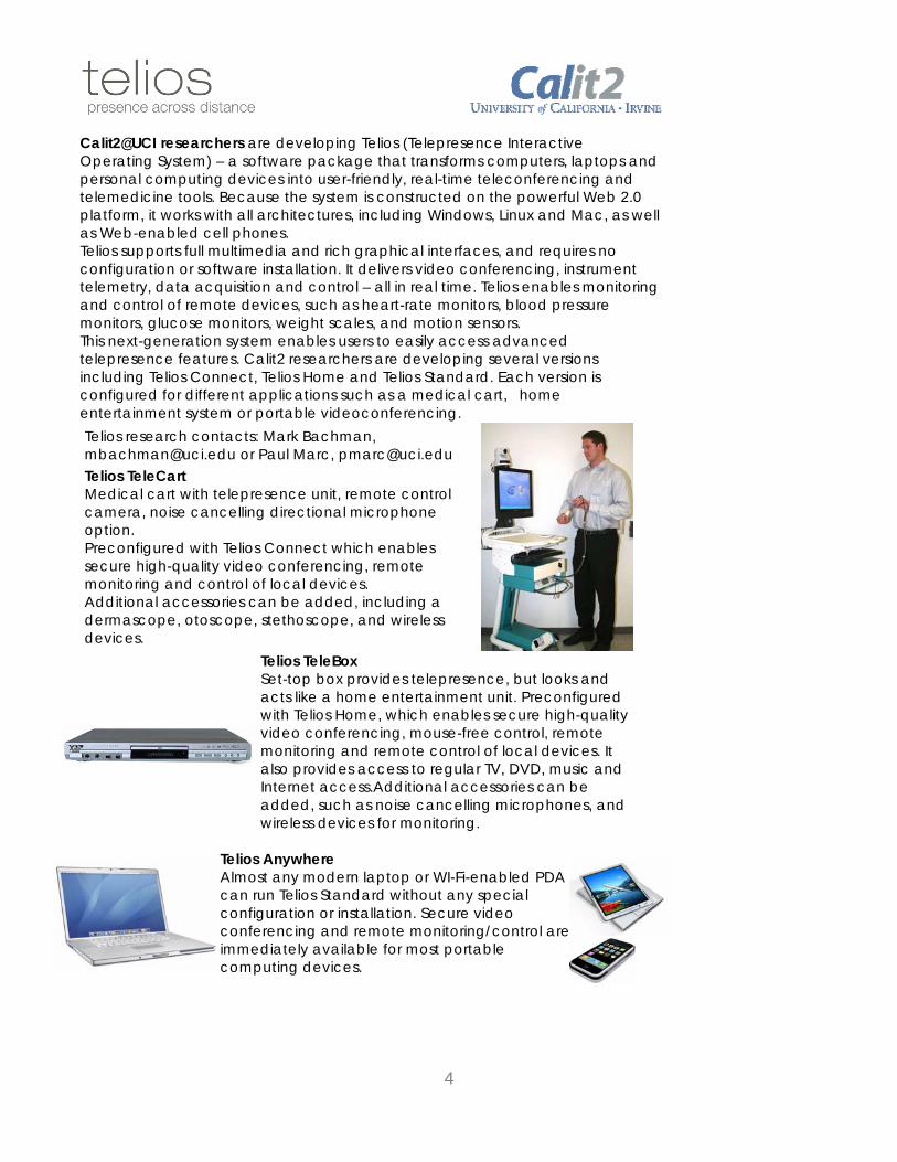
Telios TeleCart Medical cart with telepresence unit, remote control camera, noise cancelling directional microphone option. Preconfigured with Telios Connect which enables secure high-quality video conferencing, remote monitoring and control of local devices. Additional accessories can be added, including a dermascope, otoscope, stethoscope, and wireless devices.
Telios TeleBox Set-top box provides telepresence, but looks and acts like a home entertainment unit. Preconfigured with Telios Home, which enables secure high-quality video conferencing, mouse-free control, remote monitoring and remote control of local devices. It also provides access to regular TV, DVD, music and Internet access.Additional accessories can be added, such as noise cancelling microphones, and wireless devices for monitoring.
Telios Anywhere Almost any modern laptop or WI-Fi-enabled PDA can run Telios Standard without any special configuration or installation. Secure video conferencing and remote monitoring/control are immediately available for most portable computing devices.
Calit2@UCI researchers are developing Telios (Telepresence Interactive Operating System) – a software package that transforms computers, laptops and personal computing devices into user-friendly, real-time teleconferencing and telemedicine tools. Because the system is constructed on the powerful Web 2.0 platform, it works with all architectures, including Windows, Linux and Mac, as well as Web-enabled cell phones. Telios supports full multimedia and rich graphical interfaces, and requires no configuration or software installation. It delivers video conferencing, instrument telemetry, data acquisition and control – all in real time. Telios enables monitoring and control of remote devices, such as heart-rate monitors, blood pressure monitors, glucose monitors, weight scales, and motion sensors. This next-generation system enables users to easily access advanced telepresence features. Calit2 researchers are developing several versions including Telios Connect, Telios Home and Telios Standard. Each version is configured for different applications such as a medical cart, home entertainment system or portable videoconferencing. Telios research contacts: Mark Bachman, [email protected] or Paul Marc, [email protected]
4

Microscopy Lab 1302
Carl Zeiss Center of Excellence
Nanotechnology is expected to change the face of healthcare, electronics, packaging, pharmaceuticals, environmental protection, homeland security and many other industries in the near future.
The Zeiss Center features two state-of-the-art scanning electron microscopes that aid scientists working with tiny nanosystems. The center offers advanced microscopy to the community with the EVO® multi-purpose scanning electron
microscope with variable chamber pressure capability; and the Ultra 55 CDS ultra-high-resolution field-emission SEM.
5
The EVO, with its environmental chamber pressure capability, can be used for examining wet specimens, while the Ultra 55 offers higher resolution and a high-vacuum capability.
The Zeiss Center is a partnership between Calit2@UCI and Carl Zeiss, SMT, a global semiconductor and nanotechnology instrument manufacturer. The strategic alliance provides Southern California with a regional center for nanotechnology and biotechnology research, as well as advanced materials development and innovation. The equipment is
shared by Calit2’s researchers, its industry partners and Carl Zeiss SMT’s application development team.
The center also offers six specimen-preparation tools donated by South Bay Technology, Inc. The instruments are used to prepare SEM cross-sections, deposit high-resolution films onto samples, and clean samples and/or microscope parts.
Contacts: John Porter, [email protected], (949) 824-0270; Dan Mumm, [email protected], (949) 824-3858
Material Characterization Center (MC2) This Center is a university-wide shared-user facility, focusing on the characterization of crystal structure, surface topography, materials microstructure and chemical microanalysis, with capabilities to reveal materials features from tens of microns down to 0.2 nm. Once users are trained on the instruments, they can reserve time on a recharge basis to conduct their own research.
The center offers a wide range of instrumentation and techniques in microscopy and diffraction, including light optical microscopy (LOM), scanning electron microscopy (SEM), transmission electron microscopy (TEM), atomic force microscopy (AFM) and X-ray diffraction (XRD), together with a comprehensive specimen-preparation facility (SPF). Various materials can be prepared and examined in the center, from bulk to nanostructured materials and from inorganic to organic materials, including but not limited to metals, ceramics, semi-conductors, superconductors, magnetic materials, ferroelectric materials, composites, polymers, biological
materials, biochemical materials and medical materials. The center is open 24 hours a day/7 days a week to all UCI faculty, staff and students, as well as outside researchers, including industrial companies and partners. It serves the scientific and engineering community through education, collaborative research and technical service. Academic professionals who have rich research experience with various materials in the center provide hand-on training, short courses and technical assistance. The center is maintained by Calit2, but is located in Room 140, Engineering Tower on the UCI main campus.
Contact: Jian-Guo Zheng, [email protected], (949) 824-0441

Training Room 1322 Artifacts in Agile Software Development Documentation of software requirements is a major concern among software developers and software researchers. Agile Software Development denotes a different relationship to documentation, one that warrants investigation. While a common perception is that Agile lacks rigorous documentation, in fact documentary artifacts abound in Agile development practice, though they are used in unexpected ways. We conducted qualitative fieldwork at two Agile software development companies to investigate the role of artifacts in the software development work as well as the relationship between these artifacts and Software Process. Our study examines the role of work artifacts and conversations in negotiating between models of Software Process and the contingencies of work in practice. Findings are presented which suggest a new understanding of the relationship between artifacts and Software Process. We argue that Software Process is a generative system that emerges out of interplay between Software Process models and enactments, shaped by artifacts and conversation. Through this interplay the boundary of Software work is collectively and individually negotiated by members of the software development team as they forge new modes of participation in software work. Code Retrieval on the Web Internet-Scale Code Search is the problem of finding source on the Internet. Developers are typically searching for code to reuse as-is on a project or as a reference example. This phenomenon has emerged due to the increasing availability and quality of open source and resources on the web. Solutions to this problem will involve more than the simple application of information retrieval techniques or a scaling-up of tools for code search. Instead, new, purpose-built solutions are needed that draw on results from these areas, as well as program comprehension and software reuse. Contact: Susan Elliott Sim, [email protected], (949) 824 2373 *****************************************************************************************************
The PSearch System
6
This system (http://psearch.ics.uci.edu/) supports powerful type-ahead fuzzy search on the UCI directory. The techniques can badopted in the medical domain to support powerful search on data such as patient records and clinical trials.
e
The iPubMed Prototype This prototype’s (http://ipubmed.ics.uci.edu/) goal is to support type-ahead search on millions of MEDLINE publication records.
This system is actively being developed, and we wish to make it fully functioning in a few months. Contact: Chen Li, [email protected], (949) 824-9470

7
Calit2 Second Floor Landing XAR – Information Extraction System XAR is a state of the art information extraction system for information distillation from free text that we have developed at UCI in the last 3 years. XAR provides a unique framework that allows for the integration of “traditional” extraction techniques based on declarative rules and machine learning with semantic information. Amongst many domains, we have applied XAR for the task of information extraction from unstructured clinical notes in free text. This demo will showcase the XAR technology as well the application to the clinical notes entities and relationships extraction task. Contact: Naveen Ashish 2074 Bren Hall ICS UC-Irvine, Irvine CA 92697 Tel: 949 824 2991 http://www.ics.uci.edu/~ashish [email protected]

eMedia Studio 2100 The eMedia Studio is a "Distributed Arts Collaboratory" where artistic inquiry meets technology innovation, used for telepresence performance events and a wide range of digital media arts projects. Research in the eMedia Studio centers on creating real-world environments, augmented by computer and communications technology, to explore embodied interaction in dance, theatre, music and visual arts. Active Space Active Space is an interactive media programming system that generates visuals and sound in response to movement. It has been used to create interdisciplinary performances, public art projects, gallery installations and teaching environments. The system continually senses, measures and responds to the movement of participants, providing an array of tools with which to engage and "play the space" as an instrument. In continuous development since 1994 by media artist and software designer John Crawford in association with choreographer Lisa Naugle and composer Martin Gotfrit, the Active Space system is designed to be a platform for building responsive environments where participants can explore integration of body-centered performance practices with video-based motion tracking software, motion capture animation, real-time video and audio synthesis, high bandwidth networking, and multi-channel visuals and sound. eDance Network eDance Network is a public art project consisting of a series of live video “photo booth” exhibits where visitors dance with professionals to create dance videos that become a permanent part of the exhibit. Currently in
development, the eDance Network is envisioned as a collective participatory media environment, with dance/media kiosks installed in different community locations and connected by computer networks to a central server. Two prototype exhibits already have been installed in San Francisco and Orange County.
8
Participants entering eDance kiosks can view and interact with a series of pre-recorded “demo dance” sequences, then record themselves dancing along. The newly recorded clips are stored by the system, and subsequently played back in a “dance montage” which automatically selects from all the clips recorded by eDance kiosks over time, resulting in a continually growing and ever-
changing visual representation of the movement contributions of eDance participants from different locations. Contact: John Crawford Associate Professor, Dance & Media Arts 949.528.6222 [email protected] www.embodied.net

eMedia Studio 2100 Orthopaedic Anatomy and Mechanics of the Lower Extremity in Dancers Focus of research to date:
9
nd
The ability of a ballerina to amaze an audience with beauty and grace belies the intense demands she places on her ankles and feet. Perhaps surprisingly, dancers suffer more injuries in their physical activities than athletes do. This is no surprise to those who work inside dance, however. Research is needed to better
understand the anatomy and mechanical function of dancers’ lower extremities because more injuries occur here than in any other body region. The purpose of the present research is to explore the bones, muscles, tendons, and ligaments of dancers’ ankles and feet by going where the naked eye can’t see: inside the body. This comprises mapping of bone locations and position changes during the extreme motions of dance and investigating the role of soft tissues in stabilizing the bony framework of the ankle and foot. Using x-rays amagnetic resonance imaging, data are collected and analyzed to show with good precision where the individual structures lie and how they respond during dance motions. A view from the inside provides a basis for developing more effective injury prevention techniques and equipping healthcare providers for better injury care delivery.
Further areas of research being developed:
Magnetic resonance imaging of dancers’ ankles and feet en pointe Surface electromyography of leg musculature during dance movements Three-dimensional motion analysis of the lower extremity in dance Proprioceptive role of the leg musculature in dancers’ ability to balance Fiber optic measurement of ankle ligament tension Potential for isolated vastus medialis obliquus strengthening to treat patellofemoral pain
Contact: Jeff Russell Assistant Professor of Dance Science University of California, Irvine 300 Mesa Arts Building [email protected] (949)824-1054

Visualization Lab 2301
3-D Medical Visualization
We will demonstrate high-resolution imaging from Computed Tomography (CT), Magnetic Resonance Imaging (MRI) and OpticaCoherence Tomography (OCT) using volumetric rendering on a distributed rendering computer cluster disp
l
layed on HIPerWall.
y.
Contact: Joerg Meyer, [email protected], (949) 824-9321
HIPerWall HIPerWall (Highly Interactive Parallelized Display Wall) is a mega-high-resolution, grid-based display that allows researchers to visualize and manipulate massive data sets. The display measures nearly 23 x 9 feet and consists of 50 flat-panel monitors providing a total resolution of 200 million pixels. Each panel, with a resolution of 2560 x 1600 pixels (4 megapixels), is powered by a dual-processor 2.7GHz G5 node, with nVIDIA 6800 Ultra DDL graphics, that has access to an initial storage capacity of 10 terabytes.
The display brings to life data sets generated by research in biomedical engineering, Earth system science, medical imaging, satellite photography, structural engineering, physics and genetics. It facilitates multidisciplinary research by offering a framework that permits researchers from different departments to display data simultaneousl
10
HIPerWall allows researchers to see concurrently both the broad view and the fine details of the data by offering several display options. Images can be viewed as a single full-screen visual; in
tiled mode, as a series of smaller pictures – from data streams or 3D models – that are displayed concurrently for comparison purposes; or as an animation. Contacts: Steve Jenks, [email protected], (949) 824-9072 Sung-Jin Kim, [email protected], (949) 824-9822

Visualization Lab 2301
11
act
e workspace.
Seamless High-Resolution Multi-Projector Displays Tiling multiple projectors allows us to create a large scale high-resolution seamless display in a cost-effective manner. These displays are critical for applications like medical visualization, scientific data visualization, training and simulation, education and entertainment. Currently such displays are built of high-end high-quality projectors aided by custom proprietary software and a set of trained technicians for maintenance, making them expensive and cumbersome to build and maintain. We develop algorithms to build such displays from commodity products making them affordable. More importantly, we provide a tremendous ease of deployment and maintenance by designing automated camera-based registration techniques. This allows any one,
including a historian in a museum, a doctor in a hospital, or a teacher in a school, to deploy and maintain their own displays at a cost and labor which is a couple of orders of magnitude lower than current displays. More recently, we have used our work on automated registration of multi-projector displays as a launching pad to start our new project on Ubiquitous displays, supported by NSF CAREER 2009. The key element of ubiquitous displays is an active display unit made of a projector with embedded sensor(s) (like camera, accelerometer), computation and communication unit. The goal is to instrument a workspace with distributed network of such active displays which are active members of the workspace – an interacting medium
between the user, data, environment, and other displays. In other words, these are active agents that are autonomous and smart entities with the capability to sense changes in the environment, data, or user and reto those changes. We are developing methodologies to instrument a workspace with a pool of such active displays which can collectively and automatically (a) scale to any size and resolution; (b) reconfigure to any form factor; (c) migrate from one location to another; (d) be flexible in using almost any kind of surface; and finally (e) pool resources together to interact with other components of th In our demo today, you will see our novel multi-projector automated registration techniques on planar and cylindrical displays -- probably the best in the country right now. You will also see some preliminary works on ubiquitous displays including an early distributed registration and gesture based interaction technique. This project utilizes camera-based calibration techniques and custom algorithms to produce a realistic, high-resolution, seamless display using multiple projectors. Contact: Aditi Majumder, [email protected], (949) 824-8877

12
Networking Lab 2402 Experimental Mobile Ad-hoc NETwork (MANET) We demonstrate the functionality of an experimental Mobile Ad-hoc NETwork (MANET) testbed formed by a collection of Software Defined Radios (SDRs). Each SDR consists of a Linux-based PC attached to a Universal Software Radio Peripheral (USRP) unit. Relying on a hybrid design and implementation of the protocol stack, we show the delivery of text and multimedia (both stored and live) content over our MANET testbed. Utilizing a set of identified small form factor off-the-shelf and/or custom-tailored radio components to host our MANET protocol technology, we propose forming a sensor network testbed appropriate for use by medical monitoring applications. Contacts: Homayoun Yousefi'zadeh, [email protected], (949) 824-4839 Hamid Jafarkhani, [email protected], (949) 824-1755 Wireless Mesh Networks in Medical Settings This demo shows the potential applications of wireless mesh networks (WMNs) in medical and hospital application scenarios. A WMN provides anywhere, anytime, mobile Internet access, but requires very few Internet outlets in order to cover a large geographic area for Internet access purposes. This means WMNs eliminates more wires than commodity WLAN systems. For instance, the demostrated WMN provides the Internet access over the whole Calit2 second floor using 6 wireless mesh nodes, but only one Internet outlet. We have 7 slides demonstrating the layout of the Calit2 building and the WMN coverage on this floor. In the slides, we will also show the performance with respect to the data traffic throughput capacity and delay. You may access the demo WMN network by connecting to "uci-med" on this floor. The WMN demonstrated here is cheap to deploy ($30/unit or $180/system), and can securely protect your privacy-sensitive medical information. Contact: Lichun Bao, [email protected], (949) 824-8870

Wireless Sensor Technology Lab 2421
Bionic and Assistive Technologies (BATs)
The Bionic and Assistive Technologies (BATs) group helps doctors solve real-life medical problems with a combination of cutting edge technology and custom fabricated devices. Our group is skilled in fast prototyping methods and micro-fabrication processes, with devices ranging from the nano-scale to the macro-scale. Projects are generally initiated by the doctor and currently include balance monitoring and restoration, next generation electro-larynx devices, vision improvement systems, advanced acupuncture patches, chronic penetrating electrodes, and thermo-tactile displays. Contacts: Mark Bachman, [email protected], (949) 824-6421;
lo, [email protected] Mark Mer
re babies.
EcoNodes: Endless Possibilities Ultra-compact wireless MEMS-based sensors about the size of a dime are opening doors to a wide range of new applications. Comprised of minuscule eco-nodes that measure three-axial acceleration, temperature and
light, the sensors are only 13x11x7 millimeters, including the battery. Their size and wireless capability makes them useful for monitoring the movements of prematu
13
The smaller these sensors get, the more functional they can become. Future applications could include sewing them into clothing to detect falls, embedding them into waterproof balls to measure the movement of sand under water or packaging them into consumer goods to track buying habits. The sensors utilize chip antennas and rechargeable batteries, and contain an expansion interface that allows them to be connected to external sensors. The challenge for researchers is to continue to
improve their performance without compromising their size.
Contact: Pai Chou, [email protected], (949) 824-3229

Wireless Sensor Technology Lab 2421
A Novel CMOS Code-Multiplexing Path Sharing Multi-channel Receiver Front-End for Future Biomedical Implants The demo shows an integrated chip embedded on a board that implements a novel multi-channel receiver front-end. Designed for the 5-GHz frequency and fabricated in 0.18μm CMOS, the 76mW 2.3mm2 two-antenna receiver front-end prototype achieves a 10-2 symbol error rate (SER) at -64, -77, and -78 dBm of input power for SM, SD, and BF, respectively, while providing 21~85 dB gain, 6.2dB NF, and -10.6dBm IIP3.
The First Fully Integrated Dual-Band 22-29GHz/77-81GHz BiCMOS Transceiver for Automotive Radars and Medical Imaging A fully integrated millimeter-wave transceiver chip embedded on a board has been developed. The system demonstrates the first effort of integrating such complexity on a single die and is utilized for next generation medical imaging and automotive radars.
Contact: Payam Heydari, [email protected], (949) 824-9324
14
An Ultra Wideband (UWB) Two-Stage Distributed CMOS Mixer A Novel Three-Stage Differential Non-Uniform Downsized Distributed Amplifier