examination - american veterinary dental college
TRANSCRIPT
1
EXAMINATION INFORMATION FOR CANDIDATES
Reviewed and revised 2013. Version current for 2014 Examination
Candidates: Complete the Examination Security Form at the end of this document and return it
to the AVDC Executive Secretary by December 31st.
Available in This Document
Page 1: Dates, Times, Location
Page 1: Disabilities and Other Health Issues
Page 2: Examination Format
Page 2: Written Examination
Page 10: Bench Examination
Page 10: Practical Examination
Page 12: Possible Practical Examination Procedure List
Page 14: Reasons for Failure of Practical Examination Procedures
Page 16: Suggested Reading List
Page 18: Passing Score, Examination Results, Repeat Examinations
Page 19: Examination Security and Candidate Misconduct
Page 19: Appeal of Adverse Decision
Page 20: Examination Security Form
Dates, Times, Location
Candidates will be informed of the dates, times and location of the examination, including information
about a convenient hotel and the date and time of a mandatory meeting with the Examination
Committee on the evening before the examination.
Disabilities and Other Health Issues
Within the constraints of an examination environment requiring maintenance of anonymity of the
candidates and use by the candidates of equipment during the practical examination, AVDC will
endeavor to accommodate disabilities or other health concerns that are made known to the AVDC
prior to the examination. Any health-related information you elect to submit will be held in
confidence. A separate Disability Accommodation Request document and form is available in the
Examination section of the Information For Registered Trainees web page.
2
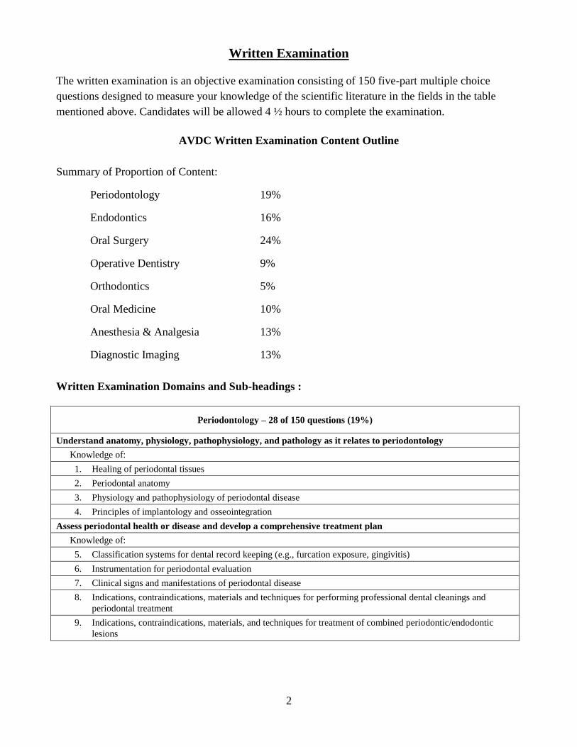
Written Examination
The written examination is an objective examination consisting of 150 five-part multiple choice
questions designed to measure your knowledge of the scientific literature in the fields in the table
mentioned above. Candidates will be allowed 4 ½ hours to complete the examination.
AVDC Written Examination Content Outline
Summary of Proportion of Content:
Periodontology 19%
Endodontics 16%
Oral Surgery 24%
Operative Dentistry 9%
Orthodontics 5%
Oral Medicine 10%
Anesthesia & Analgesia 13%
Diagnostic Imaging 13%
Written Examination Domains and Sub-headings :
Periodontology – 28 of 150 questions (19%)
Understand anatomy, physiology, pathophysiology, and pathology as it relates to periodontology
Knowledge of:
1. Healing of periodontal tissues
2. Periodontal anatomy
3. Physiology and pathophysiology of periodontal disease
4. Principles of implantology and osseointegration
Assess periodontal health or disease and develop a comprehensive treatment plan
Knowledge of:
5. Classification systems for dental record keeping (e.g., furcation exposure, gingivitis)
6. Instrumentation for periodontal evaluation
7. Clinical signs and manifestations of periodontal disease
8. Indications, contraindications, materials and techniques for performing professional dental cleanings and
periodontal treatment
9. Indications, contraindications, materials, and techniques for treatment of combined periodontic/endodontic
lesions
3
10. Indications, contraindications, materials, and techniques for periodontal splinting, guided tissue regeneration,
bone augmentation, and periodontal membranes
11. Techniques, principles and materials to implement home care programs
12. Presence of severe cases of periodontal disease requiring staged treatment, including recognition of systemic or
immunopathic effects
13. Assessment of pretreatment systemic, general and local immunologic health of the animal as it relates to
treatment options
Utilize appropriate periodontal instruments, materials, and techniques and assess outcome/complications for the
treatment plan, and develop follow-up plan
Knowledge of:
14. Materials and techniques to treat periodontal pockets and exposed root surfaces
15. Care and use of hand instrumentation including curettes and scalers
16. Materials and patterns used to suture a periodontal flap
17. Materials and techniques to perform gingivectomy/gingivoplasty
18. Polishing equipment and materials
19. Care, use, and mechanism of action of power equipment
20. Visualization equipment such as light source, magnification, and mirrors
21. Combined periodontic/endodontic monitoring post-treatment
22. Dietary products, treats, and toys that may promote oral health by retarding plaque and calculus
23. Home care products – indications, use, and contraindications
24. Mechanisms of action of home care products
25. Postoperative care, long-term prognosis, and future assessment
26. Strategies for periodontal disease prevention, maintenance and improvement
27. Evaluation of home care product efficacy and safety
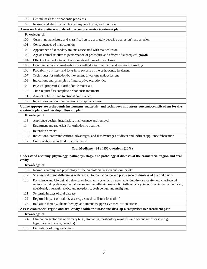
Endodontics - 24 of 150 questions (16%)
Understand anatomy, physiology, pathophysiology, and pathology as it relates to endodontics
Knowledge of:
28. Gross and microscopic endodontic and periapical anatomy
29. Physiology and pathophysiology of the pulp-dentin complex and periapical tissues
Assess endodontic health or disease and develop a comprehensive treatment plan
Knowledge of:
30. Clinical signs of, and methods to assess, endodontic disease, including tooth fractures, tooth resorption,
pulpitis, and developmental defects
31. Tooth-fracture classifications and nomenclature
32. Indications, contraindications, materials, and techniques for vital pulp therapy, plus or minus coronal reduction
33. Indications, contraindications, materials, and techniques for standard (orthograde) endodontic therapy
34. Indications, contraindications, materials, and techniques for surgical (retrograde) endodontic therapy
35. Indications, contraindications, materials, and techniques for hemisection and root resection
36. Indications, contraindications, materials, and techniques of apexification procedures
37. Physical properties of endodontic materials
4
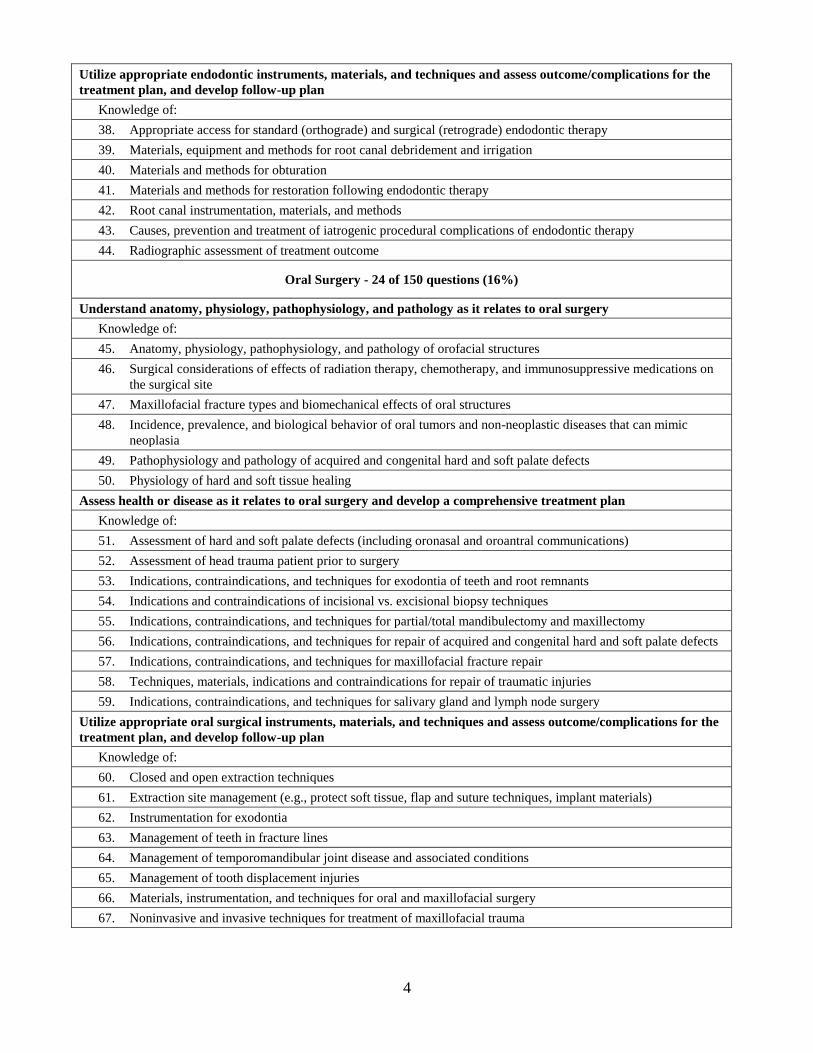
Utilize appropriate endodontic instruments, materials, and techniques and assess outcome/complications for the
treatment plan, and develop follow-up plan
Knowledge of:
38. Appropriate access for standard (orthograde) and surgical (retrograde) endodontic therapy
39. Materials, equipment and methods for root canal debridement and irrigation
40. Materials and methods for obturation
41. Materials and methods for restoration following endodontic therapy
42. Root canal instrumentation, materials, and methods
43. Causes, prevention and treatment of iatrogenic procedural complications of endodontic therapy
44. Radiographic assessment of treatment outcome
Oral Surgery - 24 of 150 questions (16%)
Understand anatomy, physiology, pathophysiology, and pathology as it relates to oral surgery
Knowledge of:
45. Anatomy, physiology, pathophysiology, and pathology of orofacial structures
46. Surgical considerations of effects of radiation therapy, chemotherapy, and immunosuppressive medications on
the surgical site
47. Maxillofacial fracture types and biomechanical effects of oral structures
48. Incidence, prevalence, and biological behavior of oral tumors and non-neoplastic diseases that can mimic
neoplasia
49. Pathophysiology and pathology of acquired and congenital hard and soft palate defects
50. Physiology of hard and soft tissue healing
Assess health or disease as it relates to oral surgery and develop a comprehensive treatment plan
Knowledge of:
51. Assessment of hard and soft palate defects (including oronasal and oroantral communications)
52. Assessment of head trauma patient prior to surgery
53. Indications, contraindications, and techniques for exodontia of teeth and root remnants
54. Indications and contraindications of incisional vs. excisional biopsy techniques
55. Indications, contraindications, and techniques for partial/total mandibulectomy and maxillectomy
56. Indications, contraindications, and techniques for repair of acquired and congenital hard and soft palate defects
57. Indications, contraindications, and techniques for maxillofacial fracture repair
58. Techniques, materials, indications and contraindications for repair of traumatic injuries
59. Indications, contraindications, and techniques for salivary gland and lymph node surgery
Utilize appropriate oral surgical instruments, materials, and techniques and assess outcome/complications for the
treatment plan, and develop follow-up plan
Knowledge of:
60. Closed and open extraction techniques
61. Extraction site management (e.g., protect soft tissue, flap and suture techniques, implant materials)
62. Instrumentation for exodontia
63. Management of teeth in fracture lines
64. Management of temporomandibular joint disease and associated conditions
65. Management of tooth displacement injuries
66. Materials, instrumentation, and techniques for oral and maxillofacial surgery
67. Noninvasive and invasive techniques for treatment of maxillofacial trauma

5
68. Nonsurgical and surgical methods for treatment of hard and soft palate defects
69. Nonsurgical and surgical treatment of osteomyelitis
70. Nutritional management of the oral surgery patient
71. Complications of extraction procedures and their management
72. Complications of hard and soft palate repair procedures and their management
73. Complications of maxillofacial trauma repair and their management
74. Complications of oral biopsies and their management
75. Complications of partial/total mandibulectomy and maxillectomy and their management
76. Postoperative and follow-up management of the oral surgery patient
Operative Dentistry - 13 of 150 questions (9%)
Understand anatomy, physiology, pathophysiology, and pathology of tooth structure
Knowledge of:
77. Normal anatomy and histology of tooth structure, including occlusal contacts
78. Tooth structure, pathophysiology, and pathology and classification of defects
Assess structural integrity of teeth and develop a comprehensive treatment plan
Knowledge of:
79. Presence of direct or indirect pulp exposure
80. Effects of alteration of normal anatomy or structural integrity
81. Periodontal considerations for restorations
82. Indications and contraindications for placement of dental prostheses
83. Indications, contraindications, types, uses, and wear characteristics for restorative and prosthodontic materials
84. Periodontal considerations for restorations
85. Physical properties of restorative materials
86. Principles of micro- and macro-mechanical retention of dental restorative materials
Utilize appropriate operative dentistry instruments, materials, and techniques and assess outcome/complications
for the treatment plan, and develop follow-up plan
Knowledge of:
87. Cavity preparation
88. Instrumentation for operative dentistry
89. Materials and techniques for crown buildup procedures
90. Placement and finish of restoration material
91. Techniques and materials for obtaining impressions and model fabrication
92. Techniques for appropriate crown reduction methods
93. Techniques, materials, indications, and contraindications for marginal finish and prosthesis cementation
94. Complications of operative dentistry and their management
95. Postoperative and follow-up management of restorative dentistry patient
Orthodontics - 7 of 150 questions (5%)
Understand anatomy, physiology, pathophysiology, and pathology of occlusal patterns
Knowledge of:
96. Occlusal characteristics and skull types
97. Developmental anatomy and embryology
6
98. Genetic basis for orthodontic problems
99. Normal and abnormal adult anatomy, occlusion, and function
Assess occlusion pattern and develop a comprehensive treatment plan
Knowledge of:
100. Current nomenclature and classification to accurately describe occlusion/malocclusion
101. Consequences of malocclusion
102. Appearance of secondary trauma associated with malocclusion
103. Age of animal relative to performance of procedure and effects of subsequent growth
104. Effects of orthodontic appliance on development of occlusion
105. Legal and ethical considerations for orthodontic treatment and genetic counseling
106. Probability of short- and long-term success of the orthodontic treatment
107. Techniques for orthodontic movement of various malocclusions
108. Indications and principles of interceptive orthodontics
109. Physical properties of orthodontic materials
110. Time required to complete orthodontic treatment
111. Animal behavior and treatment compliance
112. Indications and contraindications for appliance use
Utilize appropriate orthodontic instruments, materials, and techniques and assess outcome/complications for the
treatment plan, and develop follow-up plan
Knowledge of:
113. Appliance design, installation, maintenance and removal
114. Equipment and materials for orthodontic treatment
115. Retention devices
116. Indications, contraindications, advantages, and disadvantages of direct and indirect appliance fabrication
117. Complications of orthodontic treatment
Oral Medicine - 14 of 150 questions (10%)
Understand anatomy, physiology, pathophysiology, and pathology of diseases of the craniofacial region and oral
cavity
Knowledge of:
118. Normal anatomy and physiology of the craniofacial region and oral cavity
119. Species and breed differences with respect to the incidence and prevalence of diseases of the oral cavity
120. Prevalence and biological behavior of local and systemic diseases affecting the oral cavity and craniofacial
region including developmental, degenerative, allergic, metabolic, inflammatory, infectious, immune mediated,
nutritional, traumatic, toxic, and neoplastic, both benign and malignant
121. Systemic impact of oral disease
122. Regional impact of oral disease (e.g., sinusitis, fistula formation)
123. Radiation therapy, chemotherapy, and immunosuppressive medication effects
Assess craniofacial region and oral cavity health or disease and develop a comprehensive treatment plan
Knowledge of:
124. Clinical presentations of primary (e.g., stomatitis, masticatory myositis) and secondary diseases (e.g.,
hyperparathyroidism, petechia)
125. Limitations of diagnostic tests
7
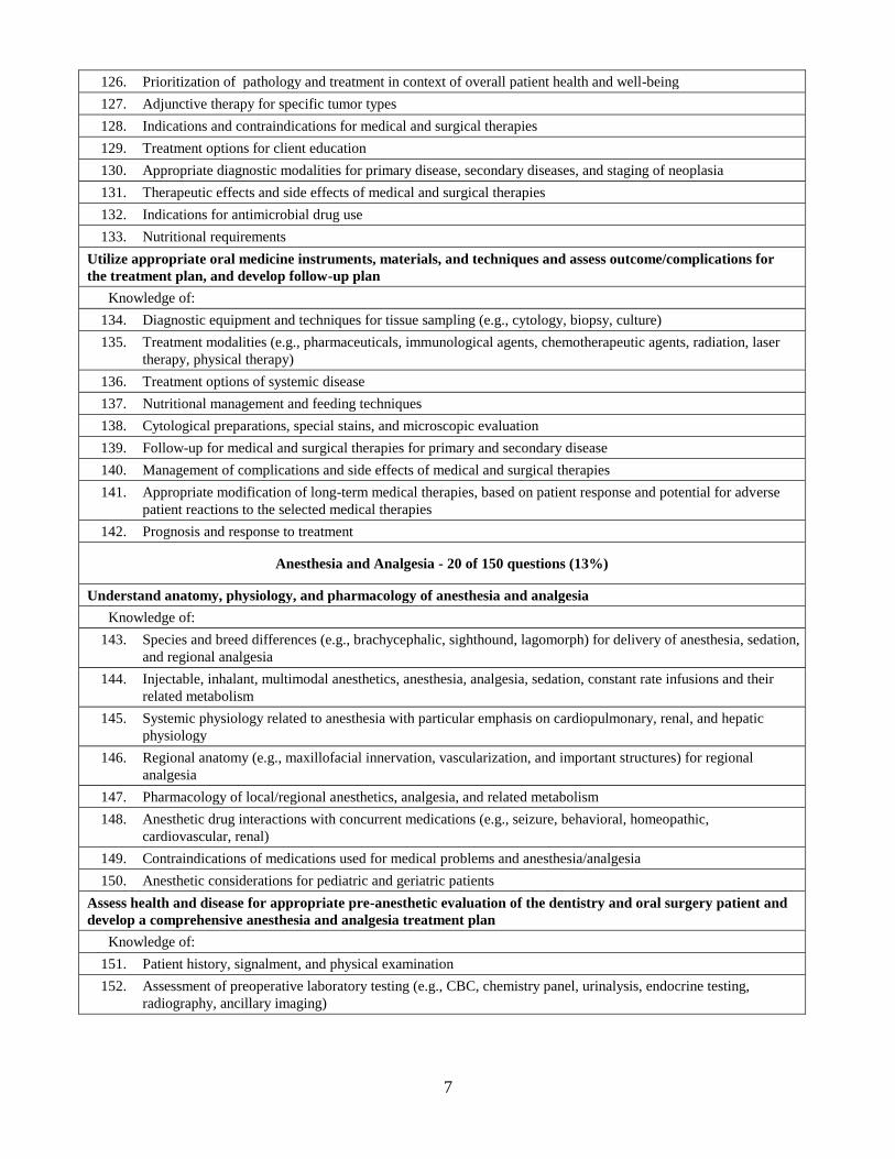
126. Prioritization of pathology and treatment in context of overall patient health and well-being
127. Adjunctive therapy for specific tumor types
128. Indications and contraindications for medical and surgical therapies
129. Treatment options for client education
130. Appropriate diagnostic modalities for primary disease, secondary diseases, and staging of neoplasia
131. Therapeutic effects and side effects of medical and surgical therapies
132. Indications for antimicrobial drug use
133. Nutritional requirements
Utilize appropriate oral medicine instruments, materials, and techniques and assess outcome/complications for
the treatment plan, and develop follow-up plan
Knowledge of:
134. Diagnostic equipment and techniques for tissue sampling (e.g., cytology, biopsy, culture)
135. Treatment modalities (e.g., pharmaceuticals, immunological agents, chemotherapeutic agents, radiation, laser
therapy, physical therapy)
136. Treatment options of systemic disease
137. Nutritional management and feeding techniques
138. Cytological preparations, special stains, and microscopic evaluation
139. Follow-up for medical and surgical therapies for primary and secondary disease
140. Management of complications and side effects of medical and surgical therapies
141. Appropriate modification of long-term medical therapies, based on patient response and potential for adverse
patient reactions to the selected medical therapies
142. Prognosis and response to treatment
Anesthesia and Analgesia - 20 of 150 questions (13%)
Understand anatomy, physiology, and pharmacology of anesthesia and analgesia
Knowledge of:
143. Species and breed differences (e.g., brachycephalic, sighthound, lagomorph) for delivery of anesthesia, sedation,
and regional analgesia
144. Injectable, inhalant, multimodal anesthetics, anesthesia, analgesia, sedation, constant rate infusions and their
related metabolism
145. Systemic physiology related to anesthesia with particular emphasis on cardiopulmonary, renal, and hepatic
physiology
146. Regional anatomy (e.g., maxillofacial innervation, vascularization, and important structures) for regional
analgesia
147. Pharmacology of local/regional anesthetics, analgesia, and related metabolism
148. Anesthetic drug interactions with concurrent medications (e.g., seizure, behavioral, homeopathic,
cardiovascular, renal)
149. Contraindications of medications used for medical problems and anesthesia/analgesia
150. Anesthetic considerations for pediatric and geriatric patients
Assess health and disease for appropriate pre-anesthetic evaluation of the dentistry and oral surgery patient and
develop a comprehensive anesthesia and analgesia treatment plan
Knowledge of:
151. Patient history, signalment, and physical examination
152. Assessment of preoperative laboratory testing (e.g., CBC, chemistry panel, urinalysis, endocrine testing,
radiography, ancillary imaging)
8
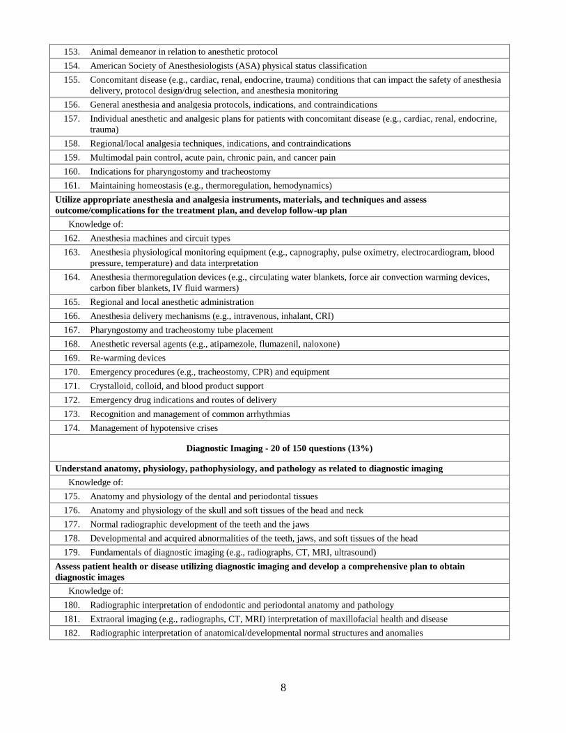
153. Animal demeanor in relation to anesthetic protocol
154. American Society of Anesthesiologists (ASA) physical status classification
155. Concomitant disease (e.g., cardiac, renal, endocrine, trauma) conditions that can impact the safety of anesthesia
delivery, protocol design/drug selection, and anesthesia monitoring
156. General anesthesia and analgesia protocols, indications, and contraindications
157. Individual anesthetic and analgesic plans for patients with concomitant disease (e.g., cardiac, renal, endocrine,
trauma)
158. Regional/local analgesia techniques, indications, and contraindications
159. Multimodal pain control, acute pain, chronic pain, and cancer pain
160. Indications for pharyngostomy and tracheostomy
161. Maintaining homeostasis (e.g., thermoregulation, hemodynamics)
Utilize appropriate anesthesia and analgesia instruments, materials, and techniques and assess
outcome/complications for the treatment plan, and develop follow-up plan
Knowledge of:
162. Anesthesia machines and circuit types
163. Anesthesia physiological monitoring equipment (e.g., capnography, pulse oximetry, electrocardiogram, blood
pressure, temperature) and data interpretation
164. Anesthesia thermoregulation devices (e.g., circulating water blankets, force air convection warming devices,
carbon fiber blankets, IV fluid warmers)
165. Regional and local anesthetic administration
166. Anesthesia delivery mechanisms (e.g., intravenous, inhalant, CRI)
167. Pharyngostomy and tracheostomy tube placement
168. Anesthetic reversal agents (e.g., atipamezole, flumazenil, naloxone)
169. Re-warming devices
170. Emergency procedures (e.g., tracheostomy, CPR) and equipment
171. Crystalloid, colloid, and blood product support
172. Emergency drug indications and routes of delivery
173. Recognition and management of common arrhythmias
174. Management of hypotensive crises
Diagnostic Imaging - 20 of 150 questions (13%)
Understand anatomy, physiology, pathophysiology, and pathology as related to diagnostic imaging
Knowledge of:
175. Anatomy and physiology of the dental and periodontal tissues
176. Anatomy and physiology of the skull and soft tissues of the head and neck
177. Normal radiographic development of the teeth and the jaws
178. Developmental and acquired abnormalities of the teeth, jaws, and soft tissues of the head
179. Fundamentals of diagnostic imaging (e.g., radiographs, CT, MRI, ultrasound)
Assess patient health or disease utilizing diagnostic imaging and develop a comprehensive plan to obtain
diagnostic images
Knowledge of:
180. Radiographic interpretation of endodontic and periodontal anatomy and pathology
181. Extraoral imaging (e.g., radiographs, CT, MRI) interpretation of maxillofacial health and disease
182. Radiographic interpretation of anatomical/developmental normal structures and anomalies
9
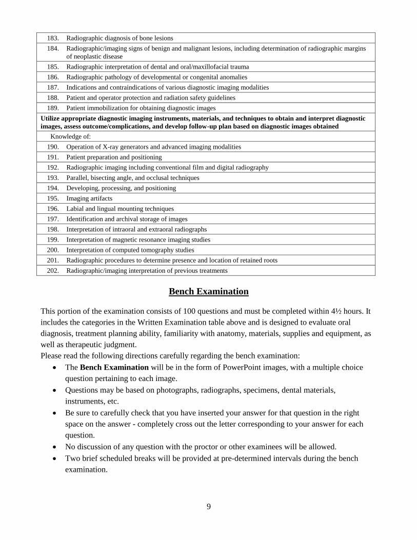
183. Radiographic diagnosis of bone lesions
184. Radiographic/imaging signs of benign and malignant lesions, including determination of radiographic margins
of neoplastic disease
185. Radiographic interpretation of dental and oral/maxillofacial trauma
186. Radiographic pathology of developmental or congenital anomalies
187. Indications and contraindications of various diagnostic imaging modalities
188. Patient and operator protection and radiation safety guidelines
189. Patient immobilization for obtaining diagnostic images
Utilize appropriate diagnostic imaging instruments, materials, and techniques to obtain and interpret diagnostic
images, assess outcome/complications, and develop follow-up plan based on diagnostic images obtained
Knowledge of:
190. Operation of X-ray generators and advanced imaging modalities
191. Patient preparation and positioning
192. Radiographic imaging including conventional film and digital radiography
193. Parallel, bisecting angle, and occlusal techniques
194. Developing, processing, and positioning
195. Imaging artifacts
196. Labial and lingual mounting techniques
197. Identification and archival storage of images
198. Interpretation of intraoral and extraoral radiographs
199. Interpretation of magnetic resonance imaging studies
200. Interpretation of computed tomography studies
201. Radiographic procedures to determine presence and location of retained roots
202. Radiographic/imaging interpretation of previous treatments
Bench Examination
This portion of the examination consists of 100 questions and must be completed within 4½ hours. It
includes the categories in the Written Examination table above and is designed to evaluate oral
diagnosis, treatment planning ability, familiarity with anatomy, materials, supplies and equipment, as
well as therapeutic judgment.
Please read the following directions carefully regarding the bench examination:
The Bench Examination will be in the form of PowerPoint images, with a multiple choice
question pertaining to each image.
Questions may be based on photographs, radiographs, specimens, dental materials,
instruments, etc.
Be sure to carefully check that you have inserted your answer for that question in the right
space on the answer - completely cross out the letter corresponding to your answer for each
question.
No discussion of any question with the proctor or other examinees will be allowed.
Two brief scheduled breaks will be provided at pre-determined intervals during the bench
examination.
10
Practical Examination
The practical examination is given to test the clinical technical skills of the candidate. The
examination will be given in three half-day sessions (Friday morning, Saturday morning, Sunday
morning), with on average four procedures to be performed per half day session. The candidates will
perform procedures in periodontics, endodontics, oral surgery, restorative dentistry/prosthodontics and
orthodontics. The core disciplines are periodontics, endodontics, oral surgery and operative dentistry
(restorative dentistry and prosthodontics), and there will be a minimum of two procedures per core
discipline. An additional, non-core, discipline is orthodontics.
The format of the examination will be explained further at the beginning of the examination.
After you have received the assignment sheet for that session (see Possible Practical Examination
Procedure List on page 5), plan your work sequence at the start of the session, and continue to be
aware of the time during the session.
Examinees are responsible for staging their procedures accordingly to satisfy all examination
requirements. As in all clinical cases, not all specimen(s) are exactly the same. Each examinee should
work with their specimen(s) to the best of their ability.
Candidates are to work independently and no candidate is allowed to receive help from any other
candidate or other person on any phase of the practical examination. Planned sharing of equipment
or materials among candidates is no longer permitted, as this has been found to be disruptive to the
examination process. Candidates are not to discuss the examination with each other during the
examination. Minimal conversation directed toward the use of AVDC-provided equipment is
permissible. Otherwise, candidates are not to engage in conversation during the examination. If a
candidate has a question, he or she should come to the proctor. Pets, family members, friends, staff,
and personal belongings not related to the examination will not be allowed in the examination area.
Electronic music players and ear phones are not allowed (see Examination Security and Candidate
Misconduct). You may use ear plugs if you wish to reduce ambient noise.
Work-station: Each candidate will be provided with an individual work area equipped with air-and-
water sources for conventional 4-hole hand-pieces. Candidates with preferences for specific
equipment are allowed to bring their own provided it does not interfere with the work of other
candidates or the Examination Committee. Planned sharing of equipment or materials among
candidates is not permitted, as this has been found to be disruptive to the examination process. The
Examination Committee has discretion as to how to manage equipment emergencies that occur on site.
11
Instruments and materials such as radiographic film, hand-pieces, restorative material, curing lights,
impression materials, dental stone, etc. are not supplied by the AVDC. The Examination Committee
discourages the use of thermoplasticized gutta percha for endodontic procedures due to the
temperature of the materials provided. The use of surgical adhesives for closure is not allowed because
this prevents evaluation of surgical technique. Due to safety regulations, use of two-part, liquid-
powder methyl methacrylate products and chloroform are not allowed. Candidates must find an
alternate material for procedures that might call for the use of such products. Candidates will not be
required to use amalgam for restorative procedures. To summarize, all materials necessary to
complete the practical examination sessions, and which were not mentioned in this document as being
supplied by the AVDC, are the responsibility of the candidate. No reading materials associated with
dental equipment or supplies may be brought into the examination room, except product information
sheets that were originally packaged with the equipment or material.
Set-up: The candidates will be allowed into the examination room 30 minutes prior to the scheduled
start time of each session, to set up their equipment.
Radiographs: A digital radiographic system will be used for the 2013 examination by all candidates.
Further details will be provided to all candidates shortly.
Submission of items for grading:
Examination materials must be handed in on time. Materials will not be accepted after
candidates have been told that there is no time left, and a score of zero will be assigned for the
procedures performed on those materials. Time remaining in the session will be announced
periodically by the proctor.
Any items being submitted for evaluation (such as impression trays) must be completely devoid of any
identifying mark other than the specimen numbers that will be assigned at the time of the examination.
Candidates must submit only what is specifically requested on the examination instructions. Any other
material submitted will not be evaluated and may compromise the anonymity of the candidate.
Safety issues: Taking dangerous chemicals (e.g. chloroform) on airplanes is illegal. Candidates
currently residing outside the USA should be aware that the voltage in the United States is 110v.
Given the changes in travel security arrangements resulting from the September 11th 2001 disaster,
review carefully what you need to bring with you. Contact your airline and/or the US Transport
Security Administration if you have any questions or concerns.
POSSIBLE PRACTICAL EXAMINATION PROCEDURE LIST
While this list is representative of the types of procedures that will be included in the
examination, the AVDC and the Examination Committee reserve the right to include other
12
procedures. Any procedures not on the list will not require equipment or supplies beyond those
necessary for performing the procedures on the list.
The goal of the practical examination is to evaluate clinical skills, judgment, and treatment
planning.
The choice of technique and materials to be used for each procedure is part of treatment
planning, and it is up to the examinee to select an appropriate technique and to execute the
procedure. Radiographs will be required for some procedures.
Use this list to determine what equipment, instruments and supplies may be needed, so that
you are fully prepared.
Periodontics
1. Perform routine periodontal treatment (“prophylaxis”) on an assigned area.
2. Open curettage of single or multiple teeth.
3. Flap procedures of assigned type and location.
4. Use of a guided tissue regeneration technique for management of a periodontal defect. You will
NOT be required to provide and place an actual guided tissue regeneration membrane.
5. Type II Crown Lengthening procedure.
Endodontics
1. Pulpectomy (standard root canal treatment) or partial coronal pulpectomy (vital pulp therapy),
specific tooth as directed.
2. Apicoectomy (surgical root canal treatment), specific tooth as directed.
3. Pulp capping, specific tooth as directed.
4. Treatment of endodontic complications (e.g. perforated root).
Restorative
1. Crown preparation for a metal or porcelain crown in response to a fractured crown with most of
the crown intact; appropriate impressions, bite registrations, and models.
2. Functional direct crown build-up on a tooth fractured off 2 mm coronal to the gingival margin; this
may involve placement of post and/or pins.
3. Restoration of a specific class and type of defect with an appropriate restorative for such, or with a
restorative such as glass ionomer or composite as specified in the examination.
4. Crown wall restoration including subgingival finish.
5. Crown lengthening procedure.
Oral Surgery
1. Surgical and/or non-surgical extraction of specified tooth or teeth.
2. Repair of an oronasal fistula on a specified area.
3. Intra-osseous or interdental wiring of specified teeth or area.
4. Palatal surgery.
5. Dental arch resection for the treatment of a neoplasm.
6. Noninvasive fracture repair techniques (intraoral splints)
Orthodontics
1. Take an impression of a specified area with an appropriate material.
2. Pour and prepare a stone model from the impression taken.
13
3. Cementation of brackets and buttons.
4. Application of a bracket, button, appliance, wire, elastics, or power chain as required or requested,
appropriate for movement of a specified tooth or teeth.
5. Diagnosis of a malocclusion, recommendation of a treatment plan and preparation of laboratory
instructions.
Reasons for Failure of Practical Examination Procedures
This list summarizes common reasons for low scores recorded by graders in recent AVDC Practical
Examinations. Often, it is a series of faults rather than a single major fault that adds up to a Fail
decision on a particular procedure, and some faults are weighted more heavily than others in
determining the Pass-Fail decision.
This list is not intended to be a comprehensive list of possible reasons for failure.
No two practical examinations are the same - each examination includes a limited number of
procedures selected from the full list of possible AVDC Practical Examination Procedures shown in
the Possible Practical Examination Procedure List, above.
General:
Some requested items were not submitted.
Requested radiographs do not show the required structure(s).
Stated specifications have not been met (e.g. mm of crown length to be created).
Soft Tissue:
Inappropriate location or length of incision.
Irregular edges of incised tissues.
Un-necessary exposure of bone on completion of procedure.
Inappropriate size of suture material.
Gaps between sutures, sutures are too loose or too tight or are crowded, or suture knots are not secure.
Tension at suture line.
Debris present.
Adjacent soft tissue has been damaged.
Dental structures:
Inadequate or excessive removal of enamel or dentin, or unsupported enamel is present.
Exposed dental surfaces have not been smoothed.
Root is gouged or rough.
Tooth gouged during preparation of adjacent tissues.
Gingiva and Periodontal Bone:
Calculus remaining on teeth.
Biologic width is inadequate, gingiva is damaged or poorly adapted.
Bone is rough or inappropriately shaped.
Root is exposed.
Perforation near or into nasal cavity.
14
Flap is poorly designed, and is insufficient to cover the defect without tension, or width : length ratio
is inadequate.
Flap is loose.
Flap perforated.
Tooth damage created during preparation of bone.
Impressions and Bite Registrations:
Impression tray is not included, is flexible or is too large.
Impression material is not fully mixed; light body/wash not appropriately distributed.
Not all relevant teeth are included in the impression.
Cuspal show-through as a result of insufficient height of impression material.
Bubbles or other defects such as drag lines are included in the impression.
Bite registration is inappropriately designed and fabricated to correctly register the bite.
Oral Surgery:
Inadequate or excessive extent of tissue resection (for mass removal).
Inappropriate design and length of flap incisions.
Excessive bone removal and inadequate alveoloplasty (extractions).
Excessive undermining of flaps and damage to adjacent tissue.
Inadequate preparation or over-preparation of flap recipient site.
Bone surfaces rough and irregular, debris in alveolus beneath suture line or on exposed bone.
Poor or absent blood vessel management.
Retained root tip.
Root tip in mandibular canal.
Exposure of mandibular canal (extractions).
Canine alveolus fractured and mobile.
Poor wiring technique in fracture repair.
Splint is excessive or design is poor - prevents occlusal closure or causes excessive soft-tissue
coverage.
Weak bonding of splint to teeth.
Endodontics
Access is too shallow or over-prepared or there is damage of adjacent enamel.
Canal is over-instrumented or is inappropriately instrumented.
Failure to clean the coronal portion if a separate access is made.
Obturation is incomplete or of variable density or has obvious voids.
Tooth split by excess obturation pressure.
Debris in access site, or sealer is present on walls of access site.
Excessive apical extrusion.
Apicoectomy: Inappropriate location or length of incision, excessive or rough edges to cavity in bone,
root surface at apicoectomy not smooth; perforation into nasal cavity; inadequate preparation; overfill
and flash; site closure incomplete.
Operative Dentistry:
Preparation is insufficient, or restoration extends to the bone edge or restoration margins and surface
are not smooth, or bone management is poor.
15
Root trauma.
Buccal bulge is excessive or preparation is over-filled.
Crown preparation margin is irregular in width, in height to gingiva or is undercut, and surface of
preparation is gouged or rough.
Restorative material is not cured.
Soft tissue damage.
Orthodontics
Inappropriate choice of or location of attachment device for anchor and target teeth.
Active force device not appropriately loaded.
Appliance will cause occlusal interference or soft tissue damage.
Appliance not securely attached.
Appliance design will not cause required tooth movement.
Appliance not finished.
Gingival or tooth damage created.
SUGGESTED READING LIST for CANDIDATES AND TRAINEES
The following list is provided as suggested reading material. It is not all inclusive of every potential
reference and publication as the body of scientific literature is fluid and always changing. No attempt
is made to restrict examination questions to the material in these references. The examination
reflects the current state of knowledge in veterinary dentistry rather than material from a particular
group of references. Much of veterinary dental knowledge has been derived from human dentistry.
This is reflected in the suggested reading list and will also be reflected in the examination itself.
BOOKS
Anatomy:
1. Evans HE. Miller’s Anatomy of the Dog. 3rd ed. Philadelphia: WB Saunders, 1993.
2. Nanci A. Ten Cate’s Oral Histology: Development, Structure, and Function. 7th ed. St. Louis:
Mosby, 2007.
3. Schroeder HE. Oral Structural Biology. New York: Thieme Medical Publishers, 1991.
Anesthesia:
1. Tranquilli WJ and et al eds. Lumb & Jones Veterinary Anesthesia and Analgesia. 4th ed.
Baltimore: Williams & Wilkins, 2007.
Dental Materials:
1. Anusavice KJ. Philips’ Science of Dental Materials. 11th ed. Philadephia: WB Saunders, 2003.
2. Powers JM. Craig’s Restorative Dental Materials. 12th ed. St. Louis: Mosby Elsevier, 2006.
Endodontics:
1. Hargreaves KM and Cohen MA. Cohen’s Pathways of the Pulp. 10th ed. St. Louis: Mosby,
2010.
Equine:
1. Easley J, Dixon PM, and Schumacher J. Equine Dentistry. 3rd ed. Philadelphia: Saunders, 2010.
Exotics:
1. Capello V, Gracis M, and Lennox, A. Rabbit and Rodent Dentistry. Philadelphia: WB
Saunders, 2005.

16
Orthodontics:
1. Proffit WR and Fields HW. Contemporary Orthodontics. 4th ed. St. Louis: Mosby-Year Book,
2007
Pathology:
1. Regezi JA, Sciubba JJ, Jordan RCK. Oral Pathology: Clinical Pathologic Correlations. 5th ed.
Philadelphia: Saunders, 2007.
Periodontology:
1. Newman MG, Takei H, Klokkevold PR, Carranza FA. Carranza’s Clinical Periodontology.
11th ed. Philadelphia: Saunders, 2011.
2. Wolf HF, Rateitschak KH, Rateitschak EM, Hassell TM. Color Atlas of Dental Medicine –
Periodontology. 3rd ed. New York: Thieme Medical Publishers, 2005.
Radiology:
1. DuPont GA and DeBowes LJ. Atlas of Dental Radiography in Dogs and Cats. St. Louis:
Saunders Elsevier, 2009.
2. Mulligan TW, Aller MS, Williams CA. Atlas of Canine & Feline Dental Radiography.
Trenton: Veterinary Learning Systems, 1998.
3. White SC and Pharoah MJ. Oral Radiology: Principles and Interpretation. 6th ed. St. Louis:
Mosby, 2008.
Restorative Dentistry / Prosthodontics:
1. Roberson TM, Heyman HO, and Swift EJ. Sturdevant’s Art and Science of Operative
Dentistry. 5th ed. St. Louis: Mosby, 2006.
Surgery:
1. Fossum TW. Small Animal Surgery. 3rd ed. St. Louis: Mosby, 2007.
2. Hupp JR, Ellis III E, Tucker MR. Contemporary Oral and Maxillofacial Surgery. 5th ed. St.
Louis: Mosby, 2008.
3. Slatter D. Textbook of Small Animal Surgery. 3rd ed. Philadelphia: WB Saunders, 2003.
4. Verstraete FJM and Lommer MJ. Oral and Maxillofacial Surgery in Dogs and Cats.
Philadelphia: Saunders, 2012.
Small Animal Dentistry:
1. Harvey CE and Emily PP. Small Animal Dentistry. St. Louis: Mosby, 1993.
2. Holmstrom SE. Canine Dentistry. Veterinary Clinics of North America: Small Animal
Practice. 28(5). Philadelphia: WB Saunders, 1998.
3. Holmstrom SE, Frost P, Eisner ER. Veterinary Dental Techniques. 3rd ed. Philadelphia: WB
Saunders, 2004.
4. Holmstrom SE. Veterinary Dentistry. Veterinary Clinics of North America: Small Animal
Practice. 35(4), p. 763-1072. Philadelphia: Saunders-Elsevier, 2005.
5. Tutt C, Deeprose J, Crossley DA, eds. BSAVA Manual of Small Animal Dentistry. 3rd ed.
Quedgeley: British Small Animal Veterinary Association, 2007.
6. Verstraete FJM. Self-Assessment Color Review of Veterinary Dentistry. London: Manson
Publishing and Ames: Iowa State University Press, 1999.
7. Wiggs RB and Lobprise HB. Veterinary Dentistry Principles & Practice. Philadelphia:
Lippincott-Raven, 1997.
JOURNALS AND PERIODICALS:
1. Journal of Veterinary Dentistry: Volume 16(1) 1999 to present.
2. Dental Abstracts. Elsevier Science International. (Last five years)
17
3. Dental Clinics of North America. Philadelphia, WB Saunders Co. (Last five years)
4. Year Book of Dentistry®, St. Louis: Mosby-Year Book. (Last five years)
5. Dixon PM (2008). Equid Dentistry. The Veterinary Journal, 178(3), 307 – 424.
Other suggested journals with valuable dental articles in them include: Compendium on Continuing
Education for the Practicing Veterinarian, Compendium of Continuing Education in Dentistry,
Veterinary Surgery, Journal of the American Animal Hospital Association, Journal of the American
Veterinary Medical Association, and American Journal of Veterinary Research.
PASSING SCORE, EXAMINATION RESULTS, REPEAT EXAMINATIONS
The minimum passing score for each of the three parts of the examination is 70 %, and
candidates must pass all three parts to become a Diplomate of the College.
‘Criterion-referencing’ is used to ensure that the 70% Pass cut score reflects the level of expertise
expected of a minimally qualified entry-level veterinary dental specialist.
The scoring standards for the practical portion of the examination are based on generally accepted
dental techniques as found in current textbooks and practiced by Diplomates of the AVDC. For each
procedure, a grading system of 0-100 based on predetermined criteria is used. The passing grade for a
single procedure is 70% and the passing grade for each core discipline and the examination as a whole
is 70%. All four core disciplines must be passed to pass the practical portion of the examination. The
core disciplines are periodontics, endodontics, oral surgery and operative dentistry. The scores of the
graders will be averaged for each candidate. Fractional scores shall stand as is, and are not rounded.
The final grade of the practical examination shall be the average score of the procedures assigned.
The Examination Committee reserves the right to recommend to the AVDC Board of Directors to fail
a candidate in the practical examination, irrespective of the score obtained, if an error was performed
by the candidate that would, in a clinical situation, result in serious harm to the patient.
Disclosure of Examination Results
Candidates will receive written notification of whether or not they passed the examination within
forty-five days of the date of the examination. Examination results will be sent to all candidates on
the same day. Candidates who are not successful in the examination, including the practical
examination, will be provided with an explanation of the deficiencies that prevented their passing the
examination. For the written and the bench examinations, information will be made available on
whether the candidate passed or failed each category of the examinations. For the practical
examination the candidate will be informed of the general areas where the candidate was found to be
deficient. Candidates will not be allowed to review their examination papers or other examination
materials after the examination.
18
Repeat Examinations
The AVDC certifying examination has three parts: written, bench and practical. Candidates failing one
or two parts of the examination do not have to repeat the part(s) of the examination that they have
previously passed. Candidates may take the examination three times within the five-year period
following acceptance of credentials to successfully complete all three parts of the examination.
Candidates wishing to retake all or part of the examination are to complete, sign and submit the Re-
Examination Form, which is available for down-loading from the AVDC web site, in the Examination
documents section of the Information for Registered Trainees page.
Any candidate who has not successfully completed all parts of the examination within five years will
be permitted to re-apply by submitting a new credentials application, pay all application and
examination fees, satisfy all current credentials requirements and repeat the credential review process.
An applicant whose new credentials have been accepted will be required to take the entire
examination and complete all three parts successfully.
EXAMINATION SECURITY AND CANDIDATE MISCONDUCT
Any questions before the examination regarding the examination are to be directed via e-mail to the
Executive Secretary of the AVDC ([email protected]) or, if the Executive Secretary is
unavailable, to the Chair of the Examination Committee. Questions will be answered in writing and
copies will be sent to all candidates. It is strictly forbidden to have direct or indirect contact with other
members of the Examination Committee (including the ‘on-deck’ members) regarding the process,
format or content of the examination, from the date that an applicant is notified that s/he is a candidate
for the examination and until the examination has been completed. Any breach of these rules can be
considered reason for action by the Board of Directors to deny a candidate admission to the
examination.
The Committee for the 2013 Examination consists of: Judith Yee (chair), Boaz Arzi, Stanley
Blazejewski, Sarah Bonner, Susan Crowder, Jerome D’Astous, Viacheslav Eroshin, Wade Gingerich,
Paul Mitchell, Jennifer Rawlinson, Judy Rochette, Christopher Snyder, Kevin Stepaniuk, Bob Ulbricht
and Tony Woodward.
Examination security is a primary concern for AVDC. Do not bring personal materials (e.g. notes,
books, tape recorders, photographic devices, calculators, computers, cellular phones) to the
examination room. References are not to be consulted during the examination process. The
examination material is not to be divulged to others.
Candidates: Complete and sign the Examination Security Form at the end of this document and
return it to the AVDC Executive Secretary by December 31st of the year before the examination.

19
AVDC POLICY ON APPEAL OF ADVERSE DECISIONS
The AVDC policy on appeal of adverse decisions is available on the AVDC web site by link from the
Information for Registered Trainees page
20
Examination
Security Form
This form is to be completed, signed and returned to the AVDC Executive Secretary by
December 31st in the year prior to taking the examination
Ethical and Professional Standards Statement
AVDC endorses the AVMA Principles of Veterinary Medical Ethics and the American Board of Veterinary
Specialties (ABVS) statement that members of ABVS-recognized colleges are to “Demonstrate unquestionable moral
character and ethical professional behavior”. I understand and accept that candidates for the AVDC examination are
required to adhere to this standard.
I understand and accept that I am required to abide by the AVDC Guidelines for Use of Specialty Titles (available
on the Information for Registered Applicants page on the AVDC web site), and specifically that I may not use the terms
“board eligible” or “board qualified” and that no connection with the AVDC may be made or implied until I have
successfully completed the examination and am certified as a Diplomate of AVDC.
Specific Limitations on Examination Conduct and Communications:
I understand and accept that the giving or receiving of aid in the examination as evidenced by observation at the
time of the examination, or the use of notes, or the taking of notes other than on the examination booklet, or removal of
materials from the examination room, or discussion of the examination with other individuals, or any other forms of
misconduct or cheating may be sufficient cause for the American Veterinary Dental College to terminate any participation
in the examination, and/or to invalidate the results of my examination, and/or to deny my entrance to all future
examinations.
I understand and accept that the format and content of the examination are the property of the AVDC, and that I
may not divulge information about the examination to others. I accept that the only exception to this rule is that, should I
fail any part of the examination, I am permitted to discuss my performance with my mentor/program director in order to
better prepare myself for my next examination attempt.
Printed Name of Candidate: _________________________________
Signature of Candidate: _________________________________ Date: ______________
The results of the examination usually are available by late April. Unless indicated otherwise below, the results
of the examination will be communicated to candidates by e-mail to the e-mail address on record with AVDC.
Please check that your AVDC DMS contact information is correct.
I wish to be notified of the result of the examination by the following means of communication:
US Mail at this postal address: ______________________________________
______________________________________
Fax: ________________ ______________________________________
Please give the name, address, telephone number, fax number or e-mail address for a back-up contact
person should AVDC not be able to contact you when the examination results are available:
Return the completed form to: Colin Harvey, Executive Secretary, AVDC, 622 Maple Court,
Haddonfield, NJ 08033, USA