evolutionary ecology of mammalian placental...
TRANSCRIPT

EVOLUTIONARY ECOLOGY OF MAMMALIAN PLACENTAL INVASIVENESS
Michael G. Elliot M.A., University of Oxford, 1998
THESIS SUBMITTED IN PARTIAL FULFILLMENT OF THE REQUIREMENTS FOR THE DEGREE OF
MASTER OF SCIENCE
In the Department of Biological Sciences
O Michael G. Elliot 2007
SIMON FRASER UNIVERSITY
Fa11 2007
All rights reserved. This work may not be reproduced in whole or in part, by photocopy
OF other means, without permission of the author.

APPROVAL
Name: Michael G. Elliot
Degree: Master of Science
Title of Thesis:
Evolutionary ecology of mammalian placental invasiveness
Examining Committee:
Chair: Dr. R. Ydenberg, Professor
Dr. B. Crespi, Professor, Senior Supervisor Department of Biological Sciences, S.F.U.
Dr. A. Mooers, Associate Professor Department of Biological Sciences, S.F.U.
Dr. T. Williams, Professor Department of Biological Sciences, S.F.U.
Dr. J. Reynolds, Professor Department of Biological Sciences, S.F.U. Public Examiner
4 December Date Approved

Declaration of Partial Copyright Licence The author, whose copyright is declared on the title page of this work, has granted to Simon Fraser University the right to lend this thesis, project or extended essay to users of the Simon Fraser University ~ibrary, and to make partial or single copies only for such users or in response to a request from the library of any other university, or other educational institution, on its own behalf or for one of its users.
The author has further granted permission to Simon Fraser University to keep or make a digital copy for use in its circulating collection (currently available to the public at the 'Institutional Repositoryn link of the SFU Library website <www.lib.sfu.ca> at: <http://ir.lib.sfu.ca/handle/l892/112>) and, without changing the content, to translate the thesislproject or extended essays, if technically possible, to any medium or format for the purpose of preservation of the digital work.
The author has further agreed that permission for multiple copying of this work for scholarly purposes may be granted by either the author or the Dean of Graduate Studies.
It is understood that copying or publication of this work for financial gain shall not be allowed without the author's written permission.
Permission for public performance, or limited permission for private scholarly use, of any multimedia materials forming part of this work, may have been granted by the author. This information may be found on the separately catalogued multimedia material and in the signed Partial Copyright Licence.
While licensing SFU to permit the above uses, the author retains copyright in the thesis, project or extended essays, including the right to change the work for subsequent purposes, including editing and publishing the work in whole or in part, and licensing other parties, as the author may desire.
The original Partial Copyright Licence attesting to these terms, and signed by this author, may be found in the original bound copy of this work, retained in the Simon Fraser University Archive.
Simon Fraser University Library Burnaby, BC, Canada
Revised: Fall 2007
ABSTRACT
Eutherian mammals differ markedly in placental form and function. In
species with invasive placentation the fetal epithelium of the placenta is bathed in
flowing maternal blood, giving the fetus an opportunity to extract resources
directly from the mother and to secrete substances which modify maternal blood
chemistry. In species with non-invasive placentation the fetal tissues are
separated from maternal blood by a barrier of maternal cell layers which limits
the ability of the fetus to control resource transfer during gestation. My thesis
explores the role played by placental invasiveness in the evolution of eutherian
brain size, life history and reproductive isolation. I find that the relationships
between brain size, body size and lifespan, and the rate at which hybrid inviability
evolves, differ strikingly between species with invasive versus non-invasive
placentation. I propose a number of physiological mechanisms that may account
for such differences among eutherian mammals.
Keywords: Eutheria; Placenta; Reproductive physiology; Evolution; Brain size; Life history
Subject headings: Mammals - reproduction; Mammals - evolution; Reproduction - physiology; Placenta

DEDICATION
I dedicate this thesis to my partner, lover, intellectual sparring-partner and
best friend, Sonia Memetea. Without her unfailing support and encouragement,
far beyond the call of duty or reason, it would never have been possible.

I would like to thank all the members of the FAB* Lab for their
encouragement, and for keeping me interested with their own weird and
wonderful work. I especially wish to thank Jeff Joy, Christine Parent, Rutger Vos,
Patrik Nosil, Erica Jeffreys and Sampson Wu for making the Crespi lab such a
pleasant and invigorating place to work. I w o ~ ~ l d like to thank Arne Mooers for his
kindness and support, and for always encouraging my computational interests.
Without Marlene Nguyen's support I would long ago have drowned in a sea of
unsigned forms and missed deadlines. I will always be grateful for the generosity
and kindness of Livia Memetea since my arrival in Canada, and hope she is back
with us soon. Finally I would like to thank Bernie Crespi. He has been a better
supervisor than I could possibly have hoped for, always putting the interests of
his students first and always ready with fresh insights on what seem like tired
problems. His broad interests, deep knowledge and consistently crazy
hypotheses have interested, entertained and educated me.

TABLE OF CONTENTS
.. Approval .............................................................................................................. 11
... Abstract .............................................................................................................. 111
Dedication ........................................................................................................... iv
Acknowledgements ............................................................................................ v
Table of Contents ............................................................................................... vi ...
List of Figures .................................................................................................. VIII
...................................................................................................... List of Tables ix
Chapter I : lntroduction ....................................................................................... The role of physiology in life history evolution .................................................... 2 Evolution of brain-body allometry in mammals .................................................. 4
.............................................................. Placentation. brain size and life history 6 .................................................................................... Parent-offspring conflict 7
.......................................................................................................... Speciation 9
Chapter 2: Divergent brain-body allometry and heterochrony between eutherian mammals with invasive versus non-invasive placentation ....................................................................................................... 11
Introduction ...................................................................................................... 11 ......................................................................................................... Methods 16
.......................................................................................................... Results 18 ..................................................................................... Brain-body allometry 18
.............................................................................. Encephalization quotient 25 Proportion of brain and body growth occurring prenatally versus
..................................................................................... postnatally 26 ............................................................................ Prenatal brain growth rate 27
...................................................................... Precocious and altricial young 28 ................................................................................. Alternative hypotheses 30
....................................................................................................... Discussion 34 Physiological mechanisms ........................................................................... 34
......................................................................................... Concluding remarks 40
Chapter 3: Prenatal effects on brain size evolution: life history. ecology and parent-offspring conflict ............................................................. 44
Introduction ...................................................................................................... 44 Materials and methods ..................................................................................... 49
............................................................................................................ Results 52

............................... Str~~ctural equation modelling of mammalian life history 52 ............................................................... The slow-fast life hisory continuum 55
....................................................................... Offspring quantity and quality 57 ............................................................... Growth rates before and after birth 59
Discussion .................... ... ............................................................................ 62 Conclusions ..................................................................................................... 68
Chapter 4: Placental invasiveness mediates the evolution of hybrid ...................................................................................... inviability in mammals 71
Introduction ...................................................................................................... 71 ..................................................................................... Materials and methods 74
.............................................................. Database of hybridisable mammals 74 Measurement of genetic distances ............................................................... 74 Measurement of range sympatry .................................................................. 75 Analysis of hybridisable species pairs .......................................................... 76 Analysis of sympatry data ............................................................................. 76
.......................................................................................................... Results 77 Discussion ....................................................................................................... 86
Chapter 5: Conclusions .................................................................................... 87
Appendices ........................................................................................................ 91 Appendix 1 ....................................................................................................... 91 Occurrence of invasive (hemochorial) and non-invasive
(endotheliochorial or epitheliochorial) placentation in eutherian mammal species ............................................................................... 91
Reference List ................................................................................................... 97
vii

LIST OF FIGURES
Figure 1 : lnvasive and non-invasive forms of placentation in eutherian ........................................................................... mammals.. .14
Figure 2: Brain-body allometry in adult and newborn mammals ......................... 20
Figure 3: Brain-body allometry in four orders exhibiting placental variability ....... 22
Figure 4: Structural equation models of mammalian life history and brain size ......................... .. ...................................................
Figure 5: Fecundity and brain size in mammal exhibiting invasive versus non-invasive placentation ..............................................
Figure 6: Hypothesized relationships between placental invasiveness and the slow-fast life history continuum, in connection with life history trade-offs over reproductionlmaintenance and offspring qualitylquantity ........................................
Figure 7: Maximum genetic distance at which hybridization occurs between species with invasive or non-invasive placentation ......................................................................... .79
Figure 8: Distribution of placental and hybridization data on a phylogenetic tree of eutherian mammals ................................................... 83
viii

LIST OF TABLES
Table 1 : Distribution of invasive (hemochorial) and non-invasive (epitheliochorial or endotheliochorial) placentation in eutherian mammals .............................................................. 15
Table 2: Principal hypotheses, tests and results considered in Chapter 2 .......... 33
Table 3: Pearson's R of the bivariate correlations between residual growth rates of prenatal brain, prenatal body, postnatal brain and postnatal body, split by placental type ........................... 61
Table 4: Pearson's R of the bivariate correlations between independent contrasts in residual growth rates of prenatal brain, prenatal body, postnatal brain and postnatal body, split by placental type.. ................................................................ .62

CHAPTER 1 : INTRODUCTION
The development of a permanent chorioallantoic placenta during gestation
is one of the characteristics that distinguishes eutherian mammals from other
vertebrate groups (Nowak 1999). The placenta is an organ of fetal origin that
bears the fetal genotype (Mossman 1987). It varies markedly in morphology and
physiology within and between most eutherian orders (Enders and Carter 2004;
Mossman 1987; Ramsey 1982). A vast amount of research effort has been
devoted to documenting this placental diversity (Mossman 1987; Appendix 1)
and, more recently, to tracing its evolutionary ancestry (Carter et al. 2004;
Wildman et al. 2006). Nevertheless, the functional role of the placenta in the
evolution of mammalian life history, behaviour and ecology remains obscure.
While 'the possession of a chorioallantoic placenta is the most prominent
physiological distinction between eutherian mammals and other vertebrate
groups, hypotheses regarding the evolution of characters unique or exaggerated
in the eutherians - including large brain and body size, long gestation and long
lifespan (Eisenberg 1981) - have been understood in terms largely divorced from
details of reproductive anatomy (for example, Bielby et al. 2007; Boyce 1988;
Charnov 1991; Charnov and Morgan Ernest 2006; Eisenberg 1981). This thesis
is an approach toward unravelling the relationships between placentation and
eutherian evolution. I focus on five outstanding problems in the evolutionary
biology of mammals: the role of physiology in life history evolution, the evolution

of brain size, the role of parent-offspring conflict in the evolution of maternal-fetal
interactions, and the nature of reproductive incompatibilities resulting in the
divergence and speciation of mammalian taxa.
The role of physiology in life history evolution
Life history theory is based upon the notion that individual animals must
divide their finite resources between a variety of physiological functions at a
variety of different times. The quantities of resources devoted to growth,
reproduction and maintenance are often negatively correlated with each other,
and are thus seen as reflecting strategic economic trade-offs between functions
whose common currency is energetic expenditure. Life history theory analyzes
,these strategic trade offs not only as individual choices but also as the products
of natural selection optimizing fitness over each individual's entire course of life,
and this body of theory has been successful in predicting a variety of
relationships and associations between economic trade-offs, behaviour and
demography (Charnov 1993; Roff 1992; Stearns 1992).
While mammalian life history may be understood as a long-term game of
evolutionary economics, a major question in life history theory is to explain how
evolutionary trade-offs between investment in various functions at various times
is ultimately the product of physiological structures and mechanisms (Ricklefs
and Wikelski 2002). The role of physiological mechanisms in life history evolution
has often been discussed under the rubric of "constraint", in the trivial sense that
the activity or growth of an individual may be limited by the availability of an
energetic surplus over and above maintenance requirements (i.e., Gittleman and
2

Thompson 1988). The last one or two decades, however, have seen the rising
popularity of a coevolutionary view of life history evolution in which physiological
structures set bounds on the range of possible life histories, but are also subject
to change due to natural selection on life history itself (Ricklefs 1991 ; Ricklefs
and Wikelski 2002). As such, research in evolutionary physiology has
increasingly involved the description of functional physiological interactions
among various components of life history traits, and the tracing of the
transformations of these physiological interactions over evolutionary time (Wikelsi
and Ricklefs 2001 ; Zera and Harshman 2001).
The eutherian placenta is a physiological structure of significance to life
history evolution since one of its primary roles is to mediate the transfer of
maternal resources - including nutrients, structural materials and oxygen - to
provision the growth and development of offspring (Mossman 1987; Pere 2003).
In this thesis, I consider the importance to life history evolution of placental
invasiveness, an important character related to the number of maternal cell
layers separating fetal from maternal blood. In general I refer to placentation in
which fetal tissues are bathed in maternal blood as "invasive" and placentation in
which fetal tissues are separated from maternal blood by cell layers of maternal
origin as "non-invasive" (Figure I ) , which is a coarse version of Grosser's
tripartite classification of placental types (Grosser 1909; King 1992). The nature
of this interhemal barrier is expected to be important with respect to life history
evolution since it is the locus of resource exchange between mother and
offspring. Chapters 2 and 3 of this thesis assess the role of placental variability

on mammalian life history evolution by identifying life I- ist tory trade-offs which
differ between species grouped as placentally-invasive versus non-invasive. I
attempt to provide specific physiological hypotheses for the observed results,
with a special emphasis on differences in the evolution of brain size in the two
groups of mammals.
Evolution of brain-body allometry in mammals
Chapter 2 of this thesis uses an allometric approach (Harvey and Pagel
1988) to examine the relationships between brain size and placental
invasiveness. Body parts or life history parameters are said to scale isometrically
when their magnitudes are a fixed proportion of total body size; for example, the
total volume of blood in a mammal's body scales isometrically with body mass,
being around 7% of body mass in species of any size. Body parts or life history
parameters are said to scale allometrically when their magnitudes scale
disproportionately with body size; for example, as a function of increasing body
size, bone mass increases more rapidly than total body mass, such that the
skeleton of a shrew accounts for just 5 per cent of its total body mass while that
of an elephant accounts for nearly 20 per cent (Lindstedt 1987). The scaling of
brain size (BR) to body size (BO) is modelled by the allometric equation, BR =
~ . B o ~ , where a is the intercept and b is the gradient of the linear relationship
between log(BR) and log(B0) (Harvey and Pagel 1988). The exponent b of the
brain-body allometry of mammals is considered to be 0.75 (Martin and Harvey
1985). However, marked departures from this value within large taxonomic
groups such as mammalian orders (Martin and Harvey 1985; Pagel and Harvey

1988), lead us to view the notion of a single brain-body allometric slope for all
mammals with suspicion, while the cause and sigr~ificance of such variation
remains uncertain.
In Chapter 2 1 use traditional and comparative statistical methods to show
that the slope of mammalian brain-body allometry is s~~ibstantially and
significantly steeper in species with invasive placentation than in species with
non-invasive placentation. This pattern holds across mammals as a whole,
across age classes, and within mammalian clades that exhibit diversity in
placental form. The steeper allometric slope is associated with relatively more
rapid prenatal brain growth and precocious neurological development at birth, in
species with invasive placentation. Studies of comparative placentation indicate
a simple mechanism for these differences: in species with invasive placentation,
fatty acids essential for mammalian brain development can be extracted by the
fetus from the maternal bloodstream, but in species with non-invasive
placentation they must be synthesized by the fetus itself. These results suggest
that mammalian brain-body scaling cannot be described adequately by a single
allometric equation, casting doubt over the utility of general models of allometry
applicable uniformly to all taxa (i.e., Blum 1977; West et al. 1997; White and
Seymour 2005). Instead, these results urge us to evaluate the physiological basis
of mammalian scaling, especially the underlying mechanisms of resource
acquisition exhibited by structures during their development.

Placentation, brain size and life history
Evolutionary explanations for the diversity of brain size among mammalian
species have long been a major area of research and controversy (Gayon 2000).
While social and ecological hypotheses predominate (Allman et al. 1993a;
Allman et al. 1993b; Barton 1996; Dunbar 2OO3a; Dunbar 2OO3b; Fish and
Lockwood 2003; Gittleman 1986; Jones and MacLarnon 2004; Lindenfors 2005;
Mann et al. 1988; Sawaguchi and Kudo 1990; Sh~lltz and Dunbar 2006), a
complementary body of work rooted in evolutionary physiology has based its
hypotheses on the fact that brain tissue is metabolically expensive and in a
sense competes for energy with other structures and functions during growth and
development (Aiello 1997; Aiello and Wheeler 1995; Hladik et al. 1999). Due to
the fact that brain tissue requires a great investment in its life-long maintenance
and especially its initial development during pregnancy and childhood (i.e.
Laughlin, van Steveninch et al. 1998), brain size seems a likely candidate for
involvement in life history trade-offs over resource usage.
As discussed above, understanding the relationship between physiology
and life history outcomes is a major aim of contemporary evolutionary biology. I
approach the issue by drawing upon evolutionary economic models of brain size
evolution. The growth of neural tissue is an unusual form of investment in that the
"value of neural capital" increases throughout the life span of an individual as
new knowledge, skills and information are acquired (Kaplan et al. 2003a).
Evolutionary economics models predict the existence of synergistic evolutionary
processes relating brain size, longevity and parental investment. High levels of

encephalization provide a context in which longevity is favoured by natural
selection, since the overall return on neural investment accumulates over the full
lifespan of the animal and since the size of these returns increases with age.
Conversely, since knowledge, skills and the ability to learn may promote the
survival of an animal over a long period of time, natural selection should favour
large brains in long-lived animals. In both cases, selection will favour a shift in the
timing of initial investment in brain growth to ever earlier ages, freeing up time for
an extended period of learning and experimentation in subadult mammals
(Kaplan et al. 2003a; Kaplan et al. 2003b; Kaplan and Robson 2002). These
considerations lead to the prediction that mammals are better able to reap the
cognitive rewards of slow life histories when they are equipped with a mechanism
for the rapid prenatal transfer of large quantities of nutritive and structural
resources. Similarly, invasive placentation should be selectively favoured in
species with extensive brain growth and longevity, and correlations between life
history "slowness1' and encephalization should be stronger in species with
invasive placentation than in species with non-invasive placentation. These
hypotheses are described and tested in Chapter 3, and the results are discussed
within the general context of mammalian life history evolution.
Parent-offspring conflict
Conflict over the level of resource transfer dwing early life is expected to
arise from asymmetries between the i~iclusive fitnesses of parents and offspring.
The role of the placenta in such conflict has been suggested by a number of
7

authors (Crespi and Semeniuk 2004; Haig 1993; Wells 2003; Zeh and Zeh 2000).
All else being equal, a mother (or more specifically, a maternal autosonial gene)
is equally related (by one half) to all of her offspring, and her optimal strategy
should be to divide resources equitably between all of her young. Each offspring,
on the other hand, is more related to itself (with a coefficient of relatedness of
one) than to its full siblings (with which it shares one half of its genes) or half
siblings (with which it shares one quarter of its genes). Genes expressed in the
fetus or placenta acting to increase the proportion of maternal resources received
by some focal offspring at the expense of its siblings or future siblings will thus be
favoured by natural selection. To the contrary, maternally-expressed genes
which equalize the distribution of maternal resources between offspring should
be favoured by natural selection acting on the mother (Godfray 1995; Lessells
and Parker 1999; MacNair and Parker 1979; Trivers 1974). These ideas have
been used to interpret a number of unusual characteristics of human placentation
and pregnancy including the extremely high titers of placentally-secreted
hormones and the invasion of maternal spiral arteries by tissues of fetal origin
(Haig 1993).
Such evolutionary conflicts of interest may play out in a variety of ways,
ranging from one participant "winning" the conflict, to both participants engaging
in a never-ending "tug of war" in which each adaptation that arises in one party
selects for a counteradaptation in the other party. I use data on prenatal and
postnatal growth rates to assess the evidence for parent-offspring conflict over
resource allocation during pregnancy. I consider the control of nutrient transfer

during pregnancy to differ between species with invasive versus non-invasive
placentation. In the former group, fetuses can directly modify maternal blood
chemistry by extracting resources and secreting hormones and other substances
(Haig 1993), while in the latter group the ability of fetuses to directly modify
maternal blood chemistry is likely limited. After birth this disparity should
disappear, as mothers find themselves in a much stronger position to exert
control over resource transfer to offspring by modifying the frequency and
duration of suckling. Conflict over the allocation of resources under invasive
placentation will thus be evidenced by high prenatal and perhaps low postnatal
growth rates compared to the rates found in species with non-invasive
placentation. My findings are broadly consistent with this hypothesis; I find that
the correlation between prenatal brain growth rate and postnatal body growth
rate differs in sign between species grouped as placentally-invasive versus non-
invasive. A discussion and interpretation of these findings is presented in
Chapter 3.
Speciation
Accounting for the strikingly different rates and patterns of ,the evolution of
reproductive isolation between animal lineages has long been a central issue in
evolutionary biology. Recent theoretical and empirical work has emphasized the
role of barriers to gene flow as a primary cause of population divergence and
speciation in nature (Coyne and Allen Orr 2004). In Chapter 4 1 consider the
possibility that differences in placental structure translate into differences in the
strength of reproductive incompatibility between divergent mammalian

populations. Using rr~itochondrial DNA sequences I show that the maximum
genetic distance at which interspecific mammalian pregnancies yield viable
neonates is significantly greater in clades with invasive placentation than in
species with non-invasive placentation. Moreover, sister species with invasive
placentation exhibit higher allopatry in their geographic ranges, suggesting that
formerly separated populations in mammals with this placental type fuse more
readily on recontact. These differences are apparently driven by the stronger
downregulation of maternal immune responses under invasive placentation,
under which fetal antigens directly contact the maternal bloodstream. These
results suggest that, in addition to its interactions with brain size and life history,
placental invasiveness mediates a major component of reproductive isolation in
mammals.
The following three chapters of my thesis explore the role of placentation
in these outstanding problems in the evolution of eutherian mammals. I end with
a discussion of the principal results and suggestions for future directions of
research.

CHAPTER 2: DIVERGENT BRAIN-BODY ALLOMETRY AND HETEROCHRONY BETWEEN EUTHERIAN MAMMALS WITH INVASIVE VERSUS NON-INVASIVE PLACENTATION
Introduction
Body size allometry, in which the dimensions of body parts and the values
of life history variables scale consistently with body size across species, has
been the subject of biological study, speculation and controversy for over a
century (Gould 1966; Stearns 1980). A central notion in the study of allometry is
that such patterns of scaling reflect fundamental and perhaps universal
constraints on the transfer of energetic resources within living organisms, and
ultimately on their development, function and evolution. Biological research has
documented interspecific differences in the intercept and gradient of allometric
slopes, and in deviation of each species from an observed allometric slope, with
mammalian brain-body allometry being one of the best studied patterns (Gayon
2000). Such studies help to identify ecological or life history correlates of
variation in the scaling of body size and it components, which have been
explained in terms of trade-offs in the allocation of limited energetic resources to
different body parts, functions, activities and time periods throughout an ar~imal's
life span.
Analysis of the development of energetically expensive tissues (Aiello
1997; Aiello and Wheeler 1995) is expected to yield important insights into the

origin and evol~rtionary diversification of animal allometry for two reasons. First,
such tissues place strong energetic demands on life-history trade-offs over
allocation of resources to growth versus fecundity. Second, expensive tissues
also mediate strong selection on the proximate mechanisms of resource
acquisition duriug prenatal and infant growth. In terms of its maintenance energy
requirements, the brain is the most costly tissue of the mammalian body,
consuming over twenty times the energy of skeletal muscle per unit mass at rest
(Aiello 1997; Aiello and Wheeler 1995; Laughlin et al. 1998). The brain is also
extremely costly in terms of the structural components that are required for its
growth, to such an extent that brain growth may be the main rate-limiting process
operating during fetal development (Martin 1996).
Previous studies have described various social and ecological correlates
of adult mammalian encephalization (Allman et al. 1993a; Allman et al. 1993b;
Barton 1996; Dunbar 2003a; Dunbar 2003b; Fish and Lockwood 2003; Gittleman
1986; Jones and MacLarnon 2004; Lindenfors 2005; Mann et al. 1988;
Sawaguchi and Kudo 1990; Shultz and Dunbar 2006). Here we adopt a
physiological perspective in order to explore how the growth and allometry of
developing brains may also be influenced by functional constraints and tradeoffs
in resource transfer and allocation during the prenatal period. The anatomical-
physiological structure of most significance to the developing mammalian fetus is
the placenta, which develops from fetal extra-embryonic ectoderm and varies
markedly in form and function among eutherians (Mossman 1987). The placenta
has also been identified as an arena in which genetic conflicts over the rate and

magnitude of resource allocation are made manifest (Crespi and Semeniuk 2004;
Haig 1993). Specifically, invasive forms of placentation may be associated with
enhanced fetal mobilization of maternal resources, and the fetal manipulation of
maternal energy budgets by secretion of hormones and other substances into
her bloodstream, resulting in improved resource acquisition by 'the fetus during
pregnancy (Haig 1993).
Our primary hypothesis is that invasive forms of placentation are
associated with accelerated prenatal brain growth, which may translate into
differences in patterns of brain-body allometry. In order to test this hypothesis we
group mammalian species according to their form of placentation. We refer to
species with hemochorial placentas (in which fetal tissue is in direct contact with
flowing maternal blood) as exhibitirlg invasive placentation, while species with
epitheliochorial or endotheliochorial placentas (in which fetal tissue is separated
from flowing maternal blood by maternal epithelia) exhibit non-invasive
placentation (see Figure 1). Phylogenetic analysis indicates that the ancestral
eutherian placental condition was apparently of an invasive form, and the
occurrence of non-hemochorial placentation in extant taxa is the result of 9 to1 I
independent evolutionary transitions occurring in the Insectivora, primates, bats,
rodents, Afrotheria, Xenarthra and at the root of Laurasiatheria (Elliot and Crespi
2006; Wildman et al. 2006). The taxonomic distribution of these placental types
is described in Table 1 .

Figure 1. lnvasive and non-invasive forms of placentation in eutherian mammals. Left: schematic representations of invasive and non-invasive forms of placentation (top: hemochorial placentation in human beings; middle: endotheliochorial placentation in canines; bottom: epitheliochorial placentation in horses; the placenta is of fetal origin, and maternal tissues -which surround the fetal-placental unit - are not drawn for the sake of clarity). Right: arrangement of maternal (M) and fetal (F) tissue layers corresponding to the invasive and non- invasive forms illustrated; open nucleated cells represent epithelial layers, dark grey nucleated cells represent the endothelial wall of blood vessels, light gray areas represent connective tissue and open dumbell-shaped cells represent hemocytes. Only in invasive placentation is the placenta in direct contact with maternal blood. See text and Table 1 for further details.

Table 1. Distribution of invasive (hemochorial) and non-invasive (epitheliochorial or endotheliochorial) placentation in eutherian mammals analysed in this study
1 Afrosoricidae ( Hemochorial I I family, I I species I Glade
Placentation
~%odactyla Carnivora
i H y racoidea 1 Hemochorial 1 1 family, 1 species ~
Number of families and species included in this studv
Cetacea Chiroptera Chiro~tera
Lipotyphla Hemochorial 3 families, 18 species I
Epitheliochorial Endotheliochorial
8 families, 87 species I I families. 38 species
Epitheliochorial Hemochorial Endotheliochorial
7 families, 25 spkcies 6 families, 95 species 8 families. 41 species
Lagomorpha Hemochorial 1 family, 4 species Macroscelidea Perissodactyla Primates (Haplorrhini) Primates (Stre~sirrhini)
Rodentia Rodentia (Heteromyidae) Scandentia Xenarthra (Dasypodidae) Xenarthra (Bradypodidae)
Hemochorial Epitheliochorial Hemochorial E~itheliochorial
1 family, 2 species 3 families, 5 species 9 families, 53 species 7 families. 18 species
Hemochorial Endotheliochorial Endotheliochorial Hemochorial Endotheliochorial
13 families, 43 species J
1 family, 21 species 1 family, 1 species 1 family, 1 species 1 family, 1 species

We use classical and phylogenetic statistical methods to test for
differences in the brain-body allometric slope exhibited by species with invasive
vs. non-invasive placentation, across mammals as a whole and within focal
clades. We further test for differences in prenatal and postnatal brain and body
growth rates between these two groups, and analyze data on relative precocity at
birth, our prediction being that invasive hemochorial placentation is associated
with relatively advanced neurosensory development at birth. Finally, we describe
a physiological mechanism that may account for our findings, compare it with
alternative hypotheses, and discuss our results in the context of existing
evolutionary theories of encephalization.
Methods
Data on brain mass and body mass at birth and adulthood, gestation
length, age and mass at weaning, and age at first reproduction for 471 mammals
of known placental type were gathered from the literature (Burton 2006; Carey
and Judge 2000; Fish and Lockwood 2003; Hayssen et al. 1993; Jones and
MacLarnon 2004; Lindenfors 2002; Marino 2006; Morgan Ernest 2003; Perez-
Barberia and Gordon 2005; Symonds 2001 ; White and Seymour 2003). In order
to boost the size of the Chiropteran dataset, placentation was assumed to be
uniforni within each genus of bat (a pattern found in anatomical studies of all bat
and non-bat mammalian genera to date); however, inclusion or exclusion of this
Chiropteran data did not have substantive effects on the overall results presented
below. Unless stated otherwise, adult body mass was based on female data. In
order to account for covariance between body size and the value of life history
variables such as gestation length or weaning age, we carried out linear
regression of each Loglo-transformed life history variable against Loglo body
mass, and used ,the residual for each species in our analysis.
Coniparison of allometric slopes was accomplished by fitting univariate
general linear models to the data in SPSS (SPSS 2006) using placental type as a
fixed factor. Slopes were considered to be significantly different when the
interaction term (placenta x independent variable) was significant at p<0.05.
Where possible, these classical approaches were replicated under a
phylogenetic model using independent contrasts as implemented in the computer
program PDAP:PDTREE/Mesquite (Midford et al. 2007). Tests conducted across
the entire mammalian dataset were based on a recent species-level supertree of
the mammals (Bininda-Edmonds and Cardillo 2007). Ordinal phylogenies used in
these tests were obtained from the literature (Grenyer and Purvis 2003; Jones et
al. 2002; Purvis 1995), except in a single case, a phylogeny of Geomyidae and
Heteromyidae, which was reconstructed by maximum likelihood analysis of
cytochrome b sequences using the computer program PAUP (Swofford 2003).
Data on the degree of infant precocity for 206 mammalian species was
gathered from the literature (see Appendix). Measures of precocity were based
on the Loglo-transformed age at which five developmental milestones are
reached in each species: eyes open, internal auditory meatus opens,
independent quadrupedal locomotion first occurs; solid food is first ingested; and
weaning is complete. Univariate general linear models using a stepwise selection

protocol were used to assess the effect and significance of placentation as a
predictor of precocity independent of the confounding effects of body size,
gestation length, and other relevant reproductive parameters.
Results
Results are summarized in Table 2 and discussed in detail below.
Brain-body allometry
When considering all eutherian mammals in our dataset, species with
invasive (hemochorial) placentation were found to exhibit a strikingly steeper
neonate brain-body allometric slope than species with non-invasive placentation
(P=1 .020 versus 0.724; N=117, F=49.797, pc0.001; Figure 2). Indeed, while
brain mass increases as an allometric fmction of body mass in species with non-
invasive placentation, it increases isonietrically in species with invasive
placentation. The slope of the adult brain-body mass allometry was also found to
be significantly steeper for the group of species with invasive placentation,
though the difference was less pronounced (P=0.861 versus 0.721 ; N=471,
F=21.788, pc0.00 1 ; Figure 2), suggesting that some of the difference associated
with placental invasiveness in brain mass at birth is diminished by patterns of
brain growth durirlg the juvenile period, consistent with the prenatal effects of
placentation. Moreover, the relatively uniform slope within each placental type at
both age points is remarkable, given the broad and diverse range of taxa that
make up each group in the analysis (see Table 1). These results may be
influenced by the clustering of data points within taxonorrric groups of mammals,

especially the existence of a clade with invasive placentation (Rodentia)
characterized by small brains and small bodies, and a clade with invasive
placentation (Primates) characterized by large brains and medium-sized bodies.
In order to acco~mt for such potential confounding effects, the analyses were
repeated using phylogenetically independent contrasts. From a phylogenetic tree
of 404 mammals, we considered 267 internal contrasts for which placental type
could be inferred unambiguously by maximum likelihood. A single outlying
contrast was removed from the dataset prior to analysis. Regression lines forced
through the origin for nodes classified as invasive or non-invasive were
consistent with the results presented in Figure 2, the slope for species with
invasive placentation being significantly steeper than the slope for species with
non-invasive placentation (P=0.664 versus 0.558, p<0.001, F=7.045, N=120
invasive and 147 non-invasive contrasts).
Of interest is the fact that species with invasive placentation tend to have
smaller adult bodies than noninvasive species in this dataset (mean log adult
body mass = 2.08 versus 3.54; F=89.473, p<0.001). Possible explanations for
this result are provided later in this chapter. In the meantime, it is necessary to
ensure that the apparent divergence in allometry is not the result of a statistical
difference in body mass between the two groups of mammals in our dataset. In
order to do so we used a resampling approach, selecting species randomly but
with weighted probabilities such that the same uniform distribution, mean and
range of body mass was reflected in each placental category. One hundred and

Placenta: Imrask Nokinvasive
2 4 6
Log,, Body Mass (g)
Figure 2. Brain-body allometry in adult and newborn mammals. Species with invasive (hernochorial) placentation (red) exhibit significantly steeper allometric slopes than species with non-invasive (endotheliochorial or epitheliochorial) placentation (blue).

sixty species were sampled from the original dataset (eighty invasive and eighty
non-invasive) across one hundred replicates; we found no replicate in which the
difference in allometric slope between the two groups was not significant
(F=18.298, p<0.001). For this reason the observed pattern appears to be robust
with respect to differences in mean and range of body mass between the two
groups. An alternative possibility is that the data in Figure 2 is best described by
a single curvilinear relationship in which the apparent distinction between the
allometry of species with invasive versus non-invasive placentation is the result
of the former species having a lower average body mass. While both models fit
the data very well, a quadratic curve explained less variance in log brain mass
than the two-line model depicted in Figure 2 (~*=0.974 versus 0.985).
An alternative way of testing the robustness of this result is to conduct the
same test independently for each order of mammals exhibiting diversity in
placental type. Four such orders were available within our dataset. In all of them,
the allometric exponent of species with invasive placentation is steeper than that
of species with non-invasive placentation, and ,this difference in slope is
statistically significant in three of the four cases (Figure 3).

1 5 . Bats
1 0 -
Placenta: lnvasive Mon-invastve
Rodents /
Placenta: 4 lnvasive
Non-invasive
Placenta:
Figure 3. Brain-body allometry in four orders exhibiting placental variability. Species with invasive, hemochorial placentation (red) exhibit steeper allometric slopes than species with non-invasive endotheliochorial or epitheliochorial placentation. The difference in slope is statistically significant (a=0.05) for primates, bats and Lipotyphlans ("Insectivores").

Amongst the bats, Megachiroptera exhibit uniformly invasive placentation,
while Microchiroptera exhibit both invasive and non-invasive forms. Average
body mass did not differ significantly between groups (F=2.341, p=O.I27). Brain-
body allometry differs significantly between invasive (N=163) and non-invasive
(N=115) categories, with the former exhibiting a significantly steeper allometric
slope (P=0.839 versus 0.699; F=6.074, p=0.014). The same pattern was found
when testing independent (P=0.737 for hemochorial versus 0.680 for non-
hemochorial species; F=6.474, p=0.002).
All rodents exhibit invasive placentation expect for the Heteromyidae
(kangaroo rats and pocket mice) which develop a non-invasive form. We tested
for a difference in allometric slope between Heteromyidae and their closest sister
clade, the Geomyidae (pocket gophers). In this case, data on brain size was
available only in the form of endocranial volume rather than brain mass. In
contrast with the situation across mammals as a whole, members of the invasive
clade (N=9) were found to have significantly larger body masses than members
of the clade with non-invasive placentation (N=21, F=10.932, p=0.001). The
Geomyidae exhibited a steeper allometric slope than their non-hemochorial
sister clade (P=0.76 versus 0.66) though this difference in slope was not found to
be statistically significant (p=0.221). Under analysis of independent contrasts, the
slope for the invasive group was also found to be steeper (P=0.72 versus 0.56)
and this difference approached significance (F=3.577, p=0.071).

Within Lipotyphla, invasive forms of placentation are found in hedgehogs,
gymnures, solenodons and shrews, while moles and desmans develop a non-
invasive placenta. The combined non-invasive species (N=6) were found to be
significantly larger than the insectivores with invasive placentation (N=18;
F=5.176, p=0.034). As expected, the brain-body allometry of the invasive group
was found to be significantly steeper than that of the species with non-invasive
placentation (P=0.623 versus 0.395; F=6.456, p=0.020). A similar pattern was
identified from analysis of independent contrasts (P=0.625 versus 0.383;
F=4.670, p=0.042).
Amongst primates, strespirhines (lemurs, lorises, galagos and allied
species) exhibit non-invasive placentation, while haplorhines (tarsiers,
marmosets, monkeys, gibbons and apes) exhibit invasive placentation. The
haplorhines have a significantly higher mean body mass (F=23.424, p<0.001).
The brain-body allometry of adult haplorhines is significantly steeper than that of
adult strepsirhines (P=0.802 versus 0.639; N=19 strepsirhines and 51
haplorhines; F=5.188, p=0.026); for newborn primates the difference in slope is
not statistically significant (p=0.552), though when newborn brain mass is
predicted by adult body mass, the slope of the invasive clade is again
significantly steeper than that of the non-invasive clade (P=0.704 versus 0.520;
F=6.078, p=0.020). The analysis of independent contrasts, however, rendered
these results statistically non-significant (though the group of species with
invasive placentation always exhibited a steeper slope than the group of species
with non-invasive placentation). This lack of significance under independent

contrasts may be a function of low statistical power, given that in Primates there
is only one transition between placental types.
These results indicate that invasive hemochorial placentation tends to be
associated not with accelerated prenatal brain growth (which would be reflected
in a difference in allometric intercept but no difference in allometric slope) but
with a steeper brain-body mass allometry. Although there was an overall
tendency for the hemochorial species in our dataset to be larger than the non-
hemochorial species, bootstrapping tests show that this tendency does not
account for the differences in allometry between mammals grouped by placental
type. Tests carried out within four orders of mammals demonstrated no
systematic correlation between body size and placental type, while often finding
a steeper allometric slope in groups of species with hemochorial placentation,
though in Primates the result depended on whether the test was carried out using
classical statistics or phylogenetically independent contrasts.
Encephalization quotient
Relative brain size in mammals is frequently described in terms of an
encephalization quotient, calculated for each species as the ratio of observed
brain mass to expected brain mass, the latter based on a global regression of log
brain mass against log body mass (Jerison 1973). The steeper allometric slopes
found in taxa with invasive placentation do not necessarily translate into higher
encephalization quotients, because the intercept of the allometric slope generally
appears to be lower in these groups (see Figures 2 and 3).

Across all mammalian species in our dataset, mean encephalization
quotient did not differ between placental categories (F=1.862, p=0.173); nor did it
differ within the orders of mammals exhibiting placental diversity (Chiroptera:
p=0.934; Insectivora: p=0.667; Primates: p=0.122; Geomyoid rodents excluded
since only brain volume, not brain mass, is available). When encephalization
quotient was based on a regression of brain mass against body mass within
orders rather than globally across all mammals, hemochorial encephalization
quotients were significantly higher than non-hemochorial encephalization
quotients in the Primates (F=5.245, p=0.025), but significantly lower in the
Insectivores (F=9.440, p=.006) and not significantly different in the Chiroptera
(F=0.004, p=0.951). These results can be understood by inspection of Figure 2,
which shows that species with invasive placentation tend to have relatively large
brains at large body sizes, but relatively small brains at small body sizes.
The lack of any consistent association between relative brain size and
placental type leads us to interpret differences in allometric slope as the result of
differences in the pattern of brain growth (for example, the rate of prenatal brain
growth or, alternatively, the proportion of brain growth occurring prenatally
compared to postnatally) rather than differences in the total relative quantity of
brain growth. These avenues are explored below.
Proportion of brain and body growth occurring prenatally versus postnatally
If invasive forms of placentation permit more rapid or extended prenatal
growth of the brain, then we predict that species with invasive hemochorial

placentation will accomplish a larger proportion of their total brain growth during
gestation than is accomplished by non-hemochorial species. The proportion of
brain growth occurring prenatally was found to correlate with adult brain size,
such that larger-brained species tend to grow a lower proportion of their total
brain mass prior to birth (N=113, F=6.435, p=0.013). We adjusted for this pattern
by considering the residual proportion of total brain growth occurring prenatally,
after regression against log-transformed adult brain mass. Across all mammals,
species with non-invasive placentation were found to accomplish on average
12.5% less brain growth prior to birth than species with invasive placentation
(mean = 0.35 versus 0.40, N=113, F=6.165, p=0.015). In order to test the
significance of this pattern at lower taxonomic resolutions, we repeated the
analysis using primate data (primates being the only order of mammals exhibiting
placental diversity and with sufficient available neonatal brain mass data for
statistical analysis). Consistent with the previous result, the proportion of total
brain growth occurring prenatally in strepsirhines was 20% less than that
occurring in the haplorhines (mean = 0.40 versus 0.50, N=29 haplorhine and 11
strepsirhine species, F=8.868, p=0.005).
Prenatal brain growth rate
We calculated average prenatal brain growth rate by dividing neonate
brain mass by gestation length. Since the log-transformed growth rate was found
to correlate in a linear fashion with log adult body size, residuals of the
regression against log adult body mass was used as a metric of relative prenatal
growth rate. Across all mammals, the average prenatal brain growth rate of

species with invasive placentation appeared to be higher than that of species
with non-invasive placentation, independent of body size (N=85, F=3.954,
p=0.050). The same trend was found within the primates, the only order of
mammals to contain sufficient data for independent analysis. Haplorhine
primates, with invasive placentation, exhibited significantly higher prenatal brain
growth rates than strepsirhine primates, with non-invasive placentation (N=20,
F=50.230, p<O.OOI).
Precocious and altricial young
Since the different classes of placentation are apparently associated with
different prenatal brain growth rates, and since the brain is thought to act as a
pacemaker in the rate of prenatal development (Hofman 1983; Sacher and
Staffeldt 1974), we expect that the patterns described above will translate into an
increase in the degree of precocity exhibited by species with invasive
placentation. For 206 mammalian species we gathered data on the timing of five
developmental milestones: age at first opening of the eyes; age at opening of the
internal auditory meatus; age at first locomotion; age at first ingestion of solid
food; and age at the completion of weaning. The first two milestones have
occasionally been used as proxies for the level of neurosensory development at
birth, in opposition to measures of musculoskeletal development (Grand 1992). It
is important to note that precocity can vary across a number of dimensions,
human beings, for example, being neurologically precocious (Finlay et al. 2001)
but otherwise physically altricial. Similarly, ungulates are often considered to be
examples of precocious mammals because of their high level of musculoskeletal

development at birth, yet long gestation lengths in ungulates may mask a slow
developmental process. For this reason we consider the timing of developmental
milestones since conception rather than since birth.
From a consideration of standard life history variables, gestation length
and body size were found to be strong predictors of the timing of all milestones
except age at first ingestion of solid food, which varied randomly with respect to
candidate explanatory variables and was excluded from further analysis. In
combination, body mass and gestation length accounted for 56% of the variance
in age at eye-opening, 28% of the variance in age at first locomotion, and 73% of
the variance in age at weaning. Body mass was not a significant predictor of age
at opening of the auditory meatus, gestation length alone accounting for 65% of
the variance. Nevertheless, under a stepwise selection protocol, placental type -
independent of body mass and gestation length - was found to be a significant
predictor of the age at which eyes open (p<0.001) and the age at which the
internal auditory meatus opens (p=0.040). For both variables, invasive
placentation is associated with a more precocial life history, reducing the age
since birth at which each milestone is reached. Surprisingly, placental type
accounts for more variance than both body mass and gestation length in the age
at which eye opening occurred (partial r12 = 0.224 versus 0.058 for body mass
and 0.209 for gestation length; r2 for total model = 0.576), and has a stronger
effect than body mass on age at ear opening (partial q2 = 0.058 versus 0.002;
partial q2 of gestation length = 0.242; r2 of total model = 0.666). Placentation had
a similar effect on the age at which locomotion first occurs, but the fit of the
model to the data was relatively low (partial t12 = 0.1 11 versus 0.1 16 for body
mass; gestation partial t12 = 0.004; r2 of three-parameter model = 0.326). In
predicting the age at which weaning ends, placental type was found to play no
role.
These results indicate that while gestation length is the primary
determinant of precocity at birth (long gestations tending to result in more-
developed young), invasive placentation is also significantly associated with the
production of more precocious offspring, especially with respect to age at eye
opening, the variable most commonly used as a measure of neurosensory
development.
Alternative hypotheses
The results presented above imply that mammals fall into two natural
groups which differ in brain-body relations as a result of differences in the
physiological structures and mechanisms underlying prenatal brain development.
Species with invasive placentation exhibit steeper brain-body allometric slopes
than species with non-invasive placentation, accomplish a larger proportion of
their brain growth prior to birth, and appear to exhibit more rapid prenatal brain
growth and development. Here we consider a pair of alternative hypotheses that
might explain these results without proposirlg a causal role for placental
structure.
First, placental type may covary with overall growth during gestation, so
that the relationship between placentation and growth rate is not exclusive to the

brain but also maintains with other structures of the developing body. In order to
evaluate this possibility we conducted an ad hoc test of the hypothesis that
species with invasive placentation not only accomplish a greater proportion of
brain growth, but also accomplish a greater proportion of total body growth
(exclusive of the brain) than species with non-invasive placentation. However,
the proportion of total body growth occurring prenatally did not differ significantly
between invasive and non-invasive groups (N=106, F=0.912, p=0.342), and
within the primates the same lack of association was found (N=33, F=l .I 24,
p=0.297).
We also found that prenatal brain growth rate was significantly
higher for species with invasive placentation that non-invasive placentation.
Again, is it possible that invasive placentation increases overall growth rate and
its effects are do not operate exclusively or predominantly on the brain? We
tested this hypothesis by calculating prenatal body growth rates (exclusive of the
brain) for the species in our dataset, and found that while the groups differ in
prenatal brain growth rate, they do not differ in prenatal body growth rate (N=85,
F=0.314, p=0.577). The difference in the proportion of brain mass grown
prenatally does not appear to be the result of species with invasive placentation
having longer gestation lengths, since size-corrected gestation length does not
differ between the two groups of mammals (N=251, F=1.199, p=0.275).
These additional tests provide evidence that species with invasive
placentation, as a result of exhibiting high prenatal brain growth rates,
accomplish a larger proportion of their total brain growth during gestation than is

accomplished by species with non-invasive placentation, and that this interaction
between placentation and prenatal growth appears to operate exclusively or
predominantly on brain development, rather than on development of the body as
a whole. As such it is consistent with the notion that variation in placental form
may be specifically associated with the development of the brain.
A second alternative hypothesis is that, while species with invasive
placentation exhibit more rapid prenatal brain growth, this difference reflects high
levels of parental investment not restricted to pregnancy, hence not ultimately the
result of placentation. To test this possibility we further tested for a difference in
postnatal growth rates. The postnatal body growth rate of species with invasive
placentation, calculated from the change in mass during lactation and the
duration of lactation, was not significantly higher than that of species with
invasive placentation, indeed it was significantly lower (N=47, F=5.151, p=0.028).
This pattern may provide a partial explanation for the overall larger size of non-
invasive species: the majority of body mass growth - irrespective of placental
type - occurs postnatally; hence for a mammal of a given newborn mass,
species with non-invasive placentation (hence higher postnatal growth rates)
may tend to achieve a larger adult body mass.
A similar comparison of postnatal brain growth rates is difficult due to the
lack of mammalian data on brain mass at weaning. In an attempt to circunivent
this difficulty we assumed that, for female mammals, the age of sexual maturity is
proportional to the age at which adult size is reached. We then calculated brain
growth rate between birth and adulthood and compared this metric (corrected for

its covariance with adult body mass) across mammals grouped by placental type.
The results were similar, the postnatal brain growth rate of species with non-
invasive placentation being significantly higher than that of invasive species
independent of body mass (N=88, F=7.746, p=0.007).
The alternative hypotheses presented in this section -that the effects of
placentation are not unique to the brain, and that invasive placentation is a
correlate of overall growth rate including postnatal as well as prenatal growth -
are not supported by our dataset. These results indicate that variation in
placental invasiveness is associated with variation in the quantity of maternal
resources invested specifically in the brain, and specifically during pregnancy.
Table 2. Principal hypotheses, tests and results considered in Chapter 2.
The significance of results is noted as follows: ** = pe0.05; * = pe0.1.
Hypothesis The slope of the brain- body allometry in species with invasive placentation differs from that in species with non-invasive placentation
Test Traditional statistics, all mammals, with bootstrapping Traditional statistics, Chiroptera
Phylogenetically independent contrasts, Chiroptera Traditional statistics, Geomyidae and Heteromyidae (Rodentia) Phylogenetically independent contrasts, Geomyidae and Heteromyidae (Rodentia) Traditional statistics, Li potyphla
Result lnvasive placentation slope > noninvasive placentation slope (**) lnvasive placentation slope > noninvasive placentation slope (**) lnvasive placentation slope > noninvasive placentation slope (**) lnvasive placentation slope > nor~invasive placentation slope lnvasive placentation slope > noninvasive placentation slope (*)
lnvasive placentation slope > noninvasive placentation

Species with invasive placentation accorr~plish more brain growth prenatally than species with noninvasive placentation
Species with invasive placentation exhibit higher prenatal brain growth rates than species with non- invasive placentation Species with invasive placentation are more neurologically precocious at birth
Phylogenetically independent contrasts, ~ i ~ o t ~ p hla Traditional statistics, Primates
Phylogenetically independent contrasts, Primates Traditional statistics, all mammals
Traditional statistics, Primates
Traditional statistics, all mammals
Traditional statistics, all mammals
lnvasive placentation slope > noninvasive placentation slope (**) lnvasive placentation slope > noninvasive placentation slope (**) lnvasive placentation slope > noninvasive placentation slope Species with noninvasive placentation accomplish an average of 12.5% more brain growth prenatally (**) Species with noninvasive placentation accomplish an average of 20% more brain growth prenatally (**) Species with invasive placentation exhibit higher prenatal brain growth rates (**) while prenatal body growth rates and gestation lenaths do not differ The eyes and ears of young with invasive open significantly earlier than the eyes and ears of young with noninvasive placentation (**)
Discussion
Physiological mechanisms
What placental physiological mechanism might be responsible for
divergence in brain-body allometry and associated traits between species with
invasive versus non-invasive placentation? One possible explanation is that the
invasive placenta is able to transfer nutritional resources at a faster rate than the

non-invasive placenta, permitting an increase in parental investment during
pregnancy. Comparative studies of prenatal nutrition do not indicate any
consistent differences in the placental consumption of carbohydrates or proteins
between species with invasive placentation versus non-invasive placentation.
Placentally non-invasive species such as sheep exhibit a placental glucose
utilization rate similar to that found in placentally invasive species such as human
beings; and the amino acid composition of newborn carcasses is similar in the rat
(with invasive placentation), the pig (with non-invasive placentation) and even the
chick (with no viviparity) (Pere 2003). As a result, we conclude that absolute
levels of fetal energy consumption during gestation - which depend largely upon
carbohydrate transfer from mother to fetus - may not be a major constraint on
brain growth rate in normal pregnancies and may not be involved in generating
the observed allometric patterns described above.
Studies of fatty acid nutrition during pregnancy, to the contrary,
demonstrate a more pronounced distinction between species with invasive
versus non-invasive placentation. In studies of pregnancy in species with
invasive placentation - such as primates (Dancis et al. 1976; Haggarty et al.
1997; Hendrickse et al. 1985; Hull and Elphick 1978; Portman et al. l969),
rodents (Hershfield and Nemeth 1968; Honda et al. 1990; Hummel et al. 1975;
Koren and Shafrir 1964; Thomas and Lowy 1982; Thomas and Lowy 1983;
Thomas and Lowy 1984) and lagomorphs (Edson et al. 1975; Elphick et al. 1975;
Elphick and Hull 1977a; Elphick and Hull 1977b; Gilbert et al. 1984; Stephenson
et al. 1990) - maternal fatty acids are found to be readily and rapidly transferred

to the fetus. Under normal nutritional conditions, the rate of placental transport is
responsive to changes in maternal serum lipid concentration, such that the levels
of fatty acid in fetal and maternal tissues are correlated. Under fasting conditions,
the rate of placental transport may increase and it shows signs of selectivity for
essential and long-chain fatty acids over non-essential fatty acids. In all cases,
fetal levels of serum fatty acid are considerably higher than maternal levels, and
in those species whose neonatal body composition has been studied, lipids
derived directly from the mother (as opposed to being synthesized by the fetus)
constitute a majority of the total lipid content of the carcass.
In contrast, studies of pregnancy in species with non-invasive,
(epitheliochorial) placentation - such as bovids (Elphick et al. 1979; Leat and
Harrison 1980; Shand and Noble 1979) and swine (Elphick et al. 1980; Pere
2001 ; Thulin et al. 1989) - or (also non-invasive) endotheliochorial placentation -
such as felines (Elphick and Hull 1984) - find that the transfer of fatty acids from
mother to fetus is minimal or non-existent. Placental uptake rate is not correlated
with maternal serum fatty acid concentration, and there is no evidence of
placental selectivity for essential or long-chain fatty acids. Fetal serum fatty acid
level is as low as one percent of that of the mother, and it appears that only trace
amounts of lipid derived directly from the mother (as opposed to being
synthesized by the fetus) are present in the neonatal body.
In all species, maternally-derived carbohydrates form the main energy
supply for the developing fetus, and their transfer rate does not appear to covary
with placental type. Lipids, however, are of particular importance to the brain not

because they provide an energy supply but because they perform important
structural roles. Lipids constitute around one half of the dry matter of the
mammalian brain, the most structurally- and metabolically-important being long
chain polyunsaturated derivatives of essential fatty acids, such as
docosahexaenoic acid and arachidonic acid (Crawford et al. 1976).
Fatty acids required for brain development may be synthesized by the
fetal liver or extracted from the mother via the fetally-derived placenta. However,
the essential fatty acids, being of dietary origin, must follow the latter route (Pere
2003). The mechanisms by which they do so fall into three broad classes:
During pregnancy the majority of fatty acids circulate in the
maternal serum as phospholipids, triglycerides or cholesterol ester,
in each of which the fatty acid is attached to another molecule by
an ester bond (Berghaus et al. 2000; Hoving et al. 1994; Otto et al.
1997). A minority of fatty acids circulate as free non-esterified
molecules associated with a carrier protein, albumin, and a tiny
fraction circulate in an unbound form (Benassayag et al. 1999;
Patel et al. 1997). Esterified fatty acids are unable to cross directly
from maternal serum to fetal tissue, but fetal lipoprotein receptors
bind them to the placental surface where they are hydrolysed to
yield non-esterified fatty acids (Haggarty 2004). These are
transferred from mother to fetus by a process of diffusion driven by
a concentration gradient in unbound albumin (Stephenson et al.
1993), which is up to 20% more concentrated on the fetal side of

the placental barrier than on the maternal side (Benassayag et al.
1999).
The transfer of non-esterified fatty acids from mother to fetus is
further promoted by the existence of fetal fatty acid
bindingltransport proteins borne by the maternal-facing membranes
of the placenta (Dutta-Roy 2000).Such proteins are common in
organs with high metabolic requirements, and are thought to
facilitate ,the transmernbrane and cytoplasmic transport of long-
chain molecules (Van Nieuwenhoven et al. 1996). In humans, the
placenta is unusual in bearing its own tissue-specific surface fatty
acid binding protein that is not expressed elsewhere in the body.
Unlike other forms, which will bind with any fatty acid, placental
fatty acid binding protein binds selectively with essential fatty acids
and their long-chain polyunsaturated derivatives, particularly
docosahexaenoic acid, arachidonic acid and linoleic acid (Campbell
and Dutta-Roy 1995; Campbell et al. 1996; Campbell et al. 1998;
Haggarty et al. 1997). Consequently, its presence exclusively on
the maternal face of the placenta serves to drive a selective
enrichment of long-chain polyunsaturated fatty acids known to play
a central role in brain development, and protect the supply of
polyunsaturated fatty acids to the fetus during critical periods of
development.

Finally, two putative functions of placentally-derived leptin may
contribute to the uptake and metabolism of fatty acids by the
placenta. First, the hormone may play a role in mobilizing maternal
lipid reserves, making them available for fetal use in late pregnancy
(Hoggard et al. 2001). Second, it may increase the rate at which
maternal esterified fatty acids are hydrolysed at the placental
surface through the hormone's enhancement of placental nitric
oxide production (White et al. 2006).
Despite the fact that these mechar~isnis have been elucidated from the
study of only a handful of primates, rodents and lagomorphs (all of which have
invasive placentation), it is of interest that all of them involve the binding of
maternal serum fatty acids on the placental surface, and hence require direct
contact between the maternal bloodstream and fetal epithelium. Such an
arrangement is found only in species exhibiting the hemochorial (invasive) form
of placentation. This notion is consistent with the placental perfusion and
radioactive labelling experiments described above. These results suggest that
divergence in mammalian brain-body allometry may in part be the result of
differences in the ability of species of different placental type to effect prenatal
fatty acid transport from maternal blood to developing fetal brain. Such
differences may also account, in part, for the tendency of species with invasive
placentation to be smaller than those with non-invasive placentation. Recent
comparative research indicates that small mammals tend to exhibit higher long-
chain polyunsaturation of cell membrane fats than large mammals (Hulbert and

Else 2005). If invasive placentation provides a better supply of such fatty acids
during development then it may be favoured by natural selection not only in
species of large brain size but also in species of low body size (see Chapter 3)
Concluding remarks
Previous studies have generally viewed mammalian brain-body
allometry as a unitary relationship, with a single allometric slope, albeit
differentiated by "grades", describing the correlation between brain and body size
from mouse to elephant (Jerison 1973). Studies which seek correlations between
life history or ecology and encephalization quotient (i.e., Armstrong 1983;
Eisenberg and Wilson 1978; lwaniuk et al. 2001 ; O'Shea and Reep 1990; Worthy
and Hickie 1986) implicitly assume that a single unitary allometric slope is a valid
description of mammalian brain-body relationships -for it is this single unitary
allometric slope by which species are judged to be highly or poorly encephalized.
OL I~ analysis of prenatal effects on brain-body allometry in 471 mammalian
species indicates that mammals, with respect to encephalization, are actually
structured into two fundamentally distinct groups differentiated by the
physiological source of fatty acids destined for early brain development. The
notable difference in allometric exponent between species with invasive versus
non-invasive placentation is consistent across both neonate and adult data, it is
qualitatively similar across major taxonomic groups, and it is independent of
differences between clades in absolute body size. Our findings draw into
question the validity of generalized measures of relative brain size that are

supposedly applicable to all mammals, and they suggest that previous studies of
encephalization that do not account for placental type may usefully be revisited.
Our analysis of brain growth rates suggests that the steeper allometric
slope found in species with invasive placentation is the result of earlier and more
rapid prenatal investment in encephalization. This interpretation is in agreement
with mathematical models of allometry as a time-independent representation of
two growth curves, which predict that retardation in the growth of an organ or
structure should result in a reduction of the allometric exponent (Vincinius and
Mirazon Lahr 2003). For this reason, the results of our analysis of growth rates
and allometry are consistent with the hypothesis that evolutionary transitions in
placentation are associated with heterochronic shifts in the growth schedule of
the brain, and that invasive placentatior~ is associated with earlier brain
development.
How might such heterochronic shifts fit in with existing hypotheses of brain
size evolution? There have historically been a number of coexisting hypotheses.
First, a number of ecological and life history parameters have been found to be
involved in the development of large brains, including longevity (Allman et al.
1993a; Allman et al. 1993b; Hakeem et al. 1996; Sacher 1975), diet (Fish and
Lockwood 2003) and metabolic expenditure (Aiello 1997; Aiello and Wheeler
1995; Leonard and Robertson 1994). These views have often been seen as
alternatives to - or competitors with - the social brain hypothesis, which argues
for a causal connection between the level of social complexity and the brain size
characteristic of a species (Brothers 1990; Byrne and Whiten 1988; Dur~bar

2003a; Dunbar 2003b; Humphrey 1976). However, recent life history theoretical
treatments rooted in primatology incorporate these diverse views into a single
model of simultaneous selection acting on brain size and longevity, in which the
cost of initial brain growth and development during infancy, and the maintenance
of neural tissue over the lifespan of the animal, are balanced against the future
social and ecological benefits accruing from the possession of a large brain later
in life (Kaplan et al. 2003a; Kaplan et al. 2003b; Kaplan and Robson 2002). In
this view, improvements in diet quality and reduced niortality rates result in
selection for increased longevity, which in turn favours greater investment in the
storage and processing of information - especially at early life stages and during
an extended childhood learning phase, where expenditure is balanced against
future adult reproductive success.
Fundamental to such theories of primate brain size evolution is the notion
that energetic expenditure on brain development can be shifted to an earlier time
in the life history of the individual, and that the duration of the brain's growth
phase can be increased by extension of gestation and infancy (Kaplan and
Robson 2002; Walker et al. 2006). We argue that the physiological mechanisms
of resource acquisition by the brain during its early growth are hence important
but previously unexplored components of mammalian brain size evolution. The
results presented here support our more general view that evolutionary
transitions in placental structure may be one of the mechanisms by which
modifications to the timing and rate of eutherian brain growth evolve, just as the
evolution of extended placentation was initially involved in the diversification of

mammalian reproduction into its eutherian and Metatherian modes, the former
being associated with large brains and the latter with small ones. To the extent
that maternal-fetal conflict was involved in the origin of invasive placentation, and
the subsequent evolution of placental form and function as eutherian mammals
diversified (Crespi and Semeniuk 2004; Haig 1993), it may play an important role
in the macroevolution of mammalian allometry and life history.

CHAPTER 3: PRENATAL EFFECTS ON BRAIN SIZE EVOLUTION: LIFE HISTORY, ECOLOGY AND PARENT-OFFSPRING CONFLICT
Introduction
The evolution of mammalian brain size has generally been explained in
terms of a trade-off between the cognitive benefits of large complex brains and
the high energetic costs of growing and sustaining them. The cognitive benefits
of large brains are implied by the existence of positive correlations between brain
size and social complexity in various mammalian taxa (Barton 1996; Dunbar
1992; Gittleman 1986; MacLaren et al. 1992; Sawaguchi and Kudo 1990; Shultz
and Dunbar 2006); between brain size and intensity of mammalian mating
competition (Jones and MacLarnon 2004); between brain size and problem-
solving ability in primates (Deaner et al. 2007); and by the identification of an
apparent survival advantage in novel environments enjoyed by large-brained
birds (Sol et al. 2005). However, the energetic costs of brain growth and
maintenance are high (Laughlin et al. 1998), such that the ability of a mammal to
sustain those costs from dietary sources may limit attainable brain size (Fish and
Lockwood 2003; Mann et al. 1988) unless alternative expensive tissues can be
sacrificed in the brain's favour (Aiello 1997; Aiello and Wheeler 1995). Of central
evolutionary concern, then, is whether the growth and development of a large
brain will yield sufficient "returns" over the course of an animal's life history to
offset the high initial energetic investment.

As an investment, neural tissue is unusual in that its value appreciates
over time. Possession of a complex brain with the capacity for learning and
negotiating complex social interactions or physical environments is of limited
value at birth, when innate behaviours such as suckling and seeking protection
are likely of greater significance to survival. However the value of neural capital
increases throughout the life span of an individual as new knowledge, skills and
information are acquired (Kaplan et al. 2003a). This phenomenon is expected to
yield synergistic evolutionary pressures relating brain size, longevity and parental
investment. First, high levels of encephalization provide a context in which
longevity is favoured by natural selection, since the overall return on neural
investment accrues over time; conversely, since knowledge, skills and the ability
to learn may promote the survival of an animal over long durations, natural
selection is expected to favour large brains in long-lived animals (Kaplan et al.
2003a; Kaplan et al. 2003b; Kaplan and Robson 2002). Second, the relatively
high reproductive value of adult animals relative to young ones under conditions
of high encephalization will, under economic models, encourage the production
of smaller litter sizes and an increase in per capita investment in offspring
(Chesnais 1992); and at the same time, these higher levels of investment in
offspring during the prenatal period and infancy provide a context in which
longevity and high levels of encephalization are both facilitated and selected for.
Such synergistic processes are thought to have played a role in the evolution of
the large brains, intergenerational transfers of skills and knowledge during

extended childhood, and longevity in hominids and other primates (Kaplan et al.
2003a; Kaplan et al. 2003b; Kaplan and Robson 2002; Walker et al. 2006).
To the extent that evolutionary transitions toward larger brain sizes are
dependent upon the existence of mechanisms for early maternal investment in
the brain during pregnancy, the mammalian placenta is expected to be an
important physiological mechanism upon which patterns of brain size evolution
with the mammals depend. The placenta, an organ of fetal genotype which
controls and regulates resource 'transfer from mother to offspring during
pregnancy, and which varies markedly in structure and function between
eutherian taxa, has been identified as an arena in which genetic conflicts over
the rate and magnitude of resource allocation manifest themselves (Crespi and
Semeniuk 2004; Haig 1993). Specifically, invasive forms of placentation may be
associated with enhanced fetal mobilization of maternal resources, and the fetal
manipulation of maternal energy budgets by secretion of hormones and other
substances into her bloodstream, resulting in improved resource acquisition by
the fetus during pregnancy (Haig 1993; Petry et al. 2007). Maximum likelihood
reconstructions of placental characters indicate that the ancestral eutherian
placental condition was of an invasive form, and the occurrence of non-invasive
placentation in extant taxa is the result of 9 to 11 independent evolutionary
transitions occurring within eight mammalian orders (Elliot and Crespi 2006;
Wildman et al. 2006).
Adult cognitive abilities depend in part upon processes of brain
development during gestation. Mammals in utero are highly sensitive to a wide

variety of stressors, which can result in alterations to brain morphology and
behaviour (Weinstock 2001) including deficits in social interaction, brain
symmetry, anxiety, circadian rhythm and sleeping. Of special relevance to the
study of placentation are the effects on brain development of prenatal nutritional
status (in part determined by the extractive properties of the placenta; (Pere
2003)) and gestation length (in part determined by the timing of placental
hormone secretions; (Jenkin and Young 2004)). In human and animal models,
pregnancies compromised in length and nutritional status are associated with
reductions in regional brain volumes (Peterson 2003), cognitive impairments
such as reduced learning or memory (Briscoe et al. 2001 ; Erhard and Boissy
2004; Gomez-Pinilla and Vaynman 2005; lsaacs et al. 2001; Landon et al. 2007;
Lefebvre et al. 1988; Morgane et al. 1993; Peters 1979), and mental illness
(Dauncey and Bicknell 1999).
Evolution of life history is one possible mechanism by which mammals
may accommodate the nutritional properties of different types of placenta;
similarly, the demands of life history and ecology (such as the production of a
large number of offspring or the production of offspring with large brains) may
selectively promote placental evolution. The importance of brain development to
mammalian fitness, and the predictions of the evolutionary economics model of
brain size and life span, lead us to predict coevolution between aspects of life
history, placentation and brain size.
In this chapter I continue my analysis of differences in comparative brain
growth resulting from variation in the prenatal interactions of mother and fetus, by

focusing upon the relationship between placental invasiveness, brain size and
the slow-fast life history continuum (Promislow and Harvey 1990). The slow-fast
continuum - describing variation in life history ranging from a slow syndrome
characterized by slow metabolism, lorlg lifespan and large size, to a fast
syndrome characterized by the opposite - incorporates the major axes of life
history variation in mammals and other taxa (Bennett and Owens 2002; Bielby et
al. 2007; Promislow and Harvey 1990; Sibly and Brown 2007). In particular, I
focus upon the theoretical expectation of a positive correlation between slow life
histories and large brain sizes. We expect that mammals are better able to reap
the cognitive rewards of slow life histories when they are equipped with a
mechanism for the rapid prenatal transfer of large quantities of nutritive
resources. Hence we predict both that invasive placentation will be selectively
favoured in species with extensive brain growth and longevity, and that
correlations between life history slowness and encephalization will be stronger in
species with invasive placentation than in species with non-invasive placentation.
In addition, we consider the role of parent-offspring conflict in the evolution
of patterns of mammalian brain growth. Under invasive placentation, control of
the rate and quantity of fatty acid and other nutrient transfer is likely exercised to
a great extent by the fetus, which can not only extract materials directly from the
blood stream of the mother, but also secrete hormones directly back into the her
circulatory system that act to mobilize maternal resources in favour of the fetus
(Haig 1993; Petry et al. 2007). Under non-invasive placentation, on the other
hand, control is likely more in the hands of the mother, since the fetus lacks the

capacity to directly modify the physiology of maternal-fetal nutritional systems.
Postnatal maternal investment in the form of milk composition and provision will
be largely under the control of the mother regardless of placental type. The
optimal level of prenatal investment likely differs between mothers and their
offspring (Trivers 1974). We expect that species with non-invasive placentation
will exhibit an amelioration of parent-offspring conflict due to the greater control
of a single actor (the mother) throughout both prenatal and postnatal phases of
growth. This amelioration is expected to be evidenced by consistency between
prenatal and postnatal growth rates; species with invasive placentation, to the
contrary, should be characterized by relatively high prenatal growth rates and
relatively low postnatal growth rates, since parturition should result in a partial
transfer of control (from fetus to mother) over the rate of resource exchange.
Materials and methods
Data on the life history and placentation of placental mammals were
obtained from the study of brain-body allometry presented in Chapter 2 (Table 1,
Appendix 1). A practical difficulty in analysing these data is the existence of
strong covariance among all of ,the variables. Typically, life history variables are
analysed after being corrected for their correlation with body mass. In order to
avoid making an a priori judgement of how best to "correct" brain mass in these
analyses we first used structural equation modelling to establish the relationships
between the nine Ln-transformed life history variables of our dataset, then used a
stepwise selection protocol to fit brain mass into the model (SPSS 2006).
Structural equation modelling can be considered a form of multiple regression

which explicitly models associations between traits by considering each variable
to be measured by more than one empirical indicator, and which offers more
flexible assumptions regarding collinearity (Bentler and Stein 1992). The nine
variables (body mass at birth, adulthood and weaning; duration of gestation and
weaning, age at first reproduction, and lifespan; litter size and annual number of
offspring per year) were taken to be the manifest, or measurable variables in the
model. In addition, we consider three latent, or unobservable, variables of wide
currency in life history theory, namely size, the position of a species' life history
along the slow-fast continuum (slowness), and reproductive output, each of
which is indicated by a number of the manifest variables. Previous empirical and
theoretical work (Roff 1992; Stearns 1992) leads us to expect co-occurrence of
large size, slow life history and low fecundity, a view which was modelled using
S.E.M. and contrasted with the more naive model in which each life history
variable is simply correlated with log body mass independently.
Our hypothesis predicts that bivariate correlations between life history
variables in mammals differ depending upon the form of placentation exhibited by
each species. Specifically, we predict that the correlation between brain size and
variables related to the slow-fast life history continuum (gestation length, weaning
period, age at first reproduction, and lifespan) will be more pronounced in
mammals with invasive placentation than in mammals with non-invasive
placentation. Similarly, we expect that the apparent effects of placental
invasiveness on the prenatal nutritional status of young will be manifest as

differences in slope and/or intercept of correlations between brain size and
variables representing offspring quantity and growth rate.
In order to explore these bivariate correlations, the life history variables
were analysed using traditional regression analysis (SPSS 2006) and
phylogenetically independent contrasts (Felsenstein 1985; Maddison and
Maddison 2005). In the traditional analysis, we corrected for the covariance of life
history variables with body mass by considering the residuals of the regression of
each variable against log maternal body mass. In order to compare the
relationship between life history variables under independent contrasts, the
ancestral placental state under maximum likelihood was inferred for each node of
a recent species-level mammalian phylogeny (Bininda-Edmonds and Cardillo
2007), and independent contrasts generated for each log-transformed life history
variable were grouped by the placental type inferred for that node of the tree.
These sets of independent contrasts were then analysed using regression forced
through the origin. Where necessary, contrasts in body mass were partialed out
of these analyses or residuals were analysed in order to correct for the
covariance of contrasts in life history variables with contrasts in body mass.
Finally, the relationships between prenatal and postnatal growth rates of
brain and body were examined. Again, the residuals of the regression of log
growth rates against log maternal body mass were used, in order to remove the
confounding effects of body size variation. Average prenatal growth rates are
simply the ratio of weight of brainlbody at birth to gestation length, but average
postnatal growth rates are more difficult to estimate since data on brain and body

size at weaning are available for few species. As an alternative, we use the ratio
of adult brain or body mass rr~inus neonatal brain or body mass, to female age at
first reproduction; the latter is considered a proxy for the approximate age at
which adult size is attained.
Results
Structural equation modelling of mammalian life history
The structural equation model of mammalian life history based on
theoretical considerations of the trade-off between growth, fecundity and the
lifespan, illustrated in Figure 4 (top), was found to fit the data significantly better
than a theoretically-nai've null model in which each life history variable is simply
correlated with body mass in the absence of any latent variables (Akaike
Information Criterion = 945.8 versus 2989.0, Comparative Fit Index = 0.926
versus 0.749; N=1438). Regression weights on each path of the diagram are
consistent with theoretical expectations that larger marr~malian species have
slower life histories and lower fecundity.
Log-transformed adult brain mass was fit into the life history model under
a stepwise selection protocol in which weak correlations between brain size and
each of the three latent variables were excluded from the analysis until a
significant reduction in fit was observed. The best fitting models for species
grouped as placentally invasive or non-invasive are illustrated in Figure 4 (middle
and bottom). The principal difference between the two models is that slow life
histories are positively correlated with adult brain mass, independent of the
confounding effects of body mass, only in the group of species with invasive
placentation. These results are consistent with the hypothesis that evolutionary
extension of the lifespan and the adoption of a slow life history may evolve so as
to increase the evolutionary payoff accruing to mothers following costly invasive
placentation and relatively high levels of early parental investment, and accruing
to offspring following the devotion of resources to brain growth rather than
reproduction.


Figure 4. Structural equation models of mammalian life history and brain size. Top: all mammals; middle: mammals with non-invasive placentation; bottom: mammals with invasive placentation. Latent variables are black, manifest variables are white. Single headed arrows represent hypothesized causal relationships while double-headed arrows represent correlation. The "slowness" of a mammal's life history appears to interact with brain size only amongst species with invasive placentation.
The slow-fast life hisory continuum
Our analysis of bivariate correlations between relative brain size (as
measured by the residual of the regression of log adult brain size against log
adult body size) and a number of variables reflecting the position of each species
along ,the slow-fast life history continuum, found differences in regression slope
related to placentation consistent with those illustrated by the SEM model
presented above. Slow life histories, as measured by residual gestation length,
residual weaning period and residual age at first female reproduction, are
statistically more strongly (positively) correlated with residual brain mass in
species with invasive placentation than in species with non-invasive placentation
(gestation length: B = 0.489 in species with invasive placentation versus 0.318 in
species with non-invasive placentation, F=16.973, N=239, p<0.001; weaning
period: B = 0.562 versus 0.378, F=56.840, N=188, p<0.001; age at first
reproduction: B = 0.501 versus 0.354, F=30.234, N=211, p<0.001; ANCOVA).
Relative lifespan was found to be positively correlated with relative brain size
amongst species with invasive placentation, but this association was marginally
non-significant in species with non-invasive placentation (for species with

invasive placentation B = 0.603, R2=0.498, N=75, p<0.001, F=34.683; for species
with non-invasive placentation B=0.215, R2=0.215, N=121, p=0.079, F=3.132).
These tests were repeated using phylogenetically independent contrasts
based on a recent species-level phylogeny of the mammals (see Materials and
Methods). No sigr~ificant correlation was found to exist between residual
contrasts in brain size and residual contrasts in gestation length, weaning age or
age at first reproduction, and when categorized by placental invasiveness the two
groups of marnmals did not differ significantly in this respect (p=0.563, 0.435, and
0.664 respectively). Residual contrasts in lifespan, on the other hand, were
significantly positively correlated with their corresponding residual contrasts in
brain mass in the group of species with invasive placentation (B=0.237, p=0.017,
N=67) but not in the group of species with non-invasive placentation (B=0.013,
p=0.107, N=96), and the interaction between placenta and lifespan was a
significant component of the regression model (p=0.002, F=6.545). Similar
results were obtained from analysis of body and brain weight at birth. The
previous section identified an apparent restriction of the association between
slow life histories and large brains to those species with invasive placentation.
This pattern is also recovered using traditional linear regression, as well as
independent contrasts, demonstrating that it is not an incidental by-product of the
phylogenetic structure of the mammalian dataset or statistical methods used.
Together, these results are consistent with the hypothesis that lifespan and brain
size may be linked through an economic mechar~ism that involves evolution of
the placenta as a mediator of early parental investment.

Offspring quantity and quality
Since neural tissue is metabolically expensive to grow and maintain, it is
expected to be involved in mediating the mammalian life history trade-off over
"offspring quality1' versus "offspring quality1' (Bell and Koufopanou 1986; Roff
1992; Stearns 1992). Figure 5b indicates that, among the group of species with
invasive placentation, those of large size tend to have larger brains than their
non-invasive counterparts, but those of small size tend to have smaller brains
than their non-invasive counterparts. However, Figure 5a also indicates that
small placentally-invasive species give birth to significantly more offspring per
year than placentally-non-invasive species of similar size, while large placentally-
invasive species give birth to significantly fewer offspring. Hence the trade-off
between offspring quality and quantity appears to be more pronounced in taxa
with invasive placentation, such that these species are found at both extremes of
the mammalian spectrum, exhibiting both the largest litters and largest brains
found in mammals.

LL - 1- rrJ 3 C
C J
-1 - \ -.
-2- - 9
I I 1 I I
0 5 10 15 20
Ln Adult Body Mass ( g )
8-
- 13, Y
: 5- P c 0 = . - 2 2- n m 3
Q, CI crJ
5 0- Q, z 5 ,/
-2- - lnvasive placentation
Y -- o Non-invasive placentation I
1 1 I I b I
2 5 8 10 12 15 Ln Adult Body Mass (g)
Figure 5. Fecundity and brain size in mammals exhibiting invasive versus non-invasive placentation. Differences in slopes of regression lines between invasive and non-invasive groups are statistically significant (log annual fecundity: p=0.001; log neonate brain mass: p<0.001). Among mammals of small size, species with invasive placentation exhibit larger litters but smaller neonatal brains than species with non-invasive placentation; among mammals of large size, species with invasive placentation exhibit smaller litters but larger neonatal brains than species with non-invasive placentation.

Growth rates before and after birth
The timing of life history events such as gestation and lifespan cannot be
considered in isolation from the rate of growth. The evolution of short gestation
lengths or fast life histories, for example, may be "compensated" by increased
growth rates during gestation, mitigating any consequences of adult brain size
evolution. The relationship between growth rates before and after birth is of
special interest, since this differential will likely determine the effects of
evolutionary changes to the nutritive qualities of placentation, which naturally
have no direct effect on growth after birth.
In order to explore these issues we first compared the average prenatal
and postnatal brain and body growth rates in the two groups of mammals. In
order to correct for the covariance of growth rates with maternal body mass, we
used the residual of each growth rate against maternal body mass. Prenatal
brain growth rates may be higher in the mammals with invasive placentation
(mean residual brain growth rate = 0.16) than in mammals with non-invasive
placentation (mean residual brain growth rate = -0.14), a difference that is
marginally non-significant (p=0.074, F=3.285, N=75). On the other hand, mean
prenatal body growth rate (exclusive of the brain) show no indication of a
difference between the two groups (mean residual body growth rate = -1.35
(invasive) versus 1.5 (non-invasive), p=0.579, F=0.311; N=75).
In contrast to prenatal brain growth rates, postnatal brain growth rates
were found to be significantly lower in species with invasive placentation (mean

residual brain growth rate = -0.22) than in species with non-invasive placentation
(mean residual brain growth rate = 0.20), a statistically significant difference
(p=0.003, F=9.058, N = 86). The same pattern was found for postnatal body
growth rate (mean residual body growth rate = -0.20 (invasive) versus 0.1 8 (non-
invasive), p=0.031, F=4.789; N=86).
Bivariate correlations between prenatal and postnatal brain and body
growth rates (with the effect of maternal body mass partialed out of the data) are
described in Table 3. Within both placental groups, species with rapid body
growth also experience rapid brain growth (residual brain and body growth rates
are positively correlated before and after birth). For mammals with non-invasive
placentation only, species with high prenatal body growth rate also tend to have
high postnatal body growth rate. Interestingly, there was no such correlation in
species with invasive placentation; furthermore, the postnatal brain and postnatal
body growth rates of species with invasive placentation are significantly
negatively correlated with prenatal brain growth rate. Broadly similar results arise
from analysis of independent contrasts (N=76 contrasts; Table 4). Positive
correlations between residual prenatallpostnatal brain and body growth rates are
found only in species with non-invasive placentation, suggesting a relative
disconnection between control of growth rates before versus after birth in species
with invasive placentation. Consistent with the parent-offspring conflict
hypothesis, the correlation between prenatal brain growth rate and postnatal
body growth rate is positive in species with non-invasive placentation but
negative in species with invasive placentation.

Table 3. Pearson's R of the bivariate correlations between residual growth rates of prenatal brain, prenatal body, postnatal brain and postnatal body, split by placental type. Correlations significant at alpha = 0.1 are labelled with a single asterisk, while correlations significant at alpha = 0.05 are labelled with a double asterisk. Residuals arise from the regression of each log transformed growth rate (in average grams per day) against log adult body mass. For both placental groups, residual brain and body growth rates are positively correlated before and after birth, and for species with non-invasive placentation, prenatal and postnatal residual body growth rates are also positively correlated. Interestingly - and suggestive of parent-offspring conflict - the postnatal brain and body growth rates of species with invasive placentation are significantly negatively correlated with prenatal brain growth rates.
Non-invasive Placentation
0.816**
O.644**
0.040
0.333*
0.178
Residual Growth Rate Correlation Prenatal Brain x Prenatal Body Postnatal Brain x Postnatal Body Prenatal Brain x Postnatal Brain Prenatal Body x Postnatal Body Prenatal Body x Postnatal Brain Prenatal Brain x Postnatal Body
lnvasive Placentation
0.338*
0,797**
-0.422**
-0.013
0.007
-0.689** -0.031

Table 4. Pearson's R o f the bivariate correlations between independent contrasts i n residual growth rates of prenatal brain, prenatal body, postnatal brain and postnatal body, split by placental type. Correlations significant at alpha = 0.1 are labelled with a single asterisk, while correlations significant at alpha = 0.05 are labelled with a double asterisk. Positive correlations between residual prenatallpostnatal brain and body growth rates are found only in species with non-invasive placentation, suggesting a relative disjunction between control of growth rates at different times in species with invasive placentation. Consistent with parent- offspring conflict hypothesis, the correlation between prenatal brain growth rate and postnatal body growth rate is positive in species with non-invasive placentation but negative in species with invasive placentation.
Residual Growth Rate Correlation Prenatal Brain x Prenatal Body
Prenatal Brain x Postnatal I Bodv 1 -0.336**
lnvasive Placentation
Postnatal Brain x Postnatal Body Prenatal Brain x Postnatal Brain
Discussion
Our analysis of the relationship between brain size and the slowness of
mammalian life history support our view that evolutionary increases in brain size
may be accomplished by an extension of the lifespan and the adoption of a slow
life history, but only in the context of physiological mechanisms such as invasive
placentation which perrnit high levels of early parental investment in the brain
growth of offspring. In the absence of such a mechanism, as under non-invasive
placentation, high levels of investment in early brain development may be
Non-invasive Placentation
-0.339* 0.290
0.600**
-0.190
0.681 **
0.51 I * *

hampered, reducing the fitness benefits that might otherwise result from the
adoption of slower life histories. If longevity is a means by which mammals
increase the fitness benefits accruing from cognitive development and offset the
costs of early investment in the development of a brain, then selection for
longevity will likely be reduced in strength among species with non-invasive
placentation, which make a relatively small initial investment in early brain
development. Hence we find that a correlation between brain size and longevity
is evident only in those species with invasive placentation making large early
investments in neural capital.
As the ancestral state of the common ancestor of rodents, lagomorphs
and primates, invasive placentation is also found in numerous species with fast
life histories. In our view, animals exhibiting invasive placentation but with a fast
life history are unlikely to devote high levels of prenatal maternal investment to
brain development, because a rapid life history does not provide sufficient time
for a sufficient return on the resources invested in early brain growth.
Nevertheless, invasive placentas are still expected to make a relative excess of
resources - especially the energetically-expensive fatty acids - available to
young during pregnancy, in comparison with non-invasive placentas (Pere 2003).
An interesting characteristic of the data presented in Figure 2 is that while the
brain masslbody mass and fecunditylbody mass regression slopes differ
between species grouped as placentally invasive or non-invasive, the intercepts
of those slopes also differ, such that small species with invasive placentation
exhibit relatively small brains and large litter sizes, while large species with

invasive placentation exhibit relatively large brains and small litter sizes. We
suggest that these life-history extremes reflect alternative strategies for the
prenatal investment of resources provided by invasive placentation: investment in
"quality", or large brains, in the context of large body size and slow life history, or
"quantity", or large litters, in the context of small body size and fast life histories.
The relationship between fecundity and the slow-fast life history continuum
has previously been identified as an inverse correlation between litter size and
gestation length (Read and Harvey 1989). One possible explanation for these
patterns is that competition within the litter results in a race between littermates
to acquire maternal resources (Haldane 1932). Competition among littermates to
extract the greatest quantity of maternal resources is likely to conflict with the
mother's interest in maximizing litter size while preventing the energetic costs of
doing so from inflating to unsustainable levels (Trivers 1974). Our analysis of
prenatal and postnatal growth rates provides a possible means of testing for
evidence of such parent-offspring conflict. Under invasive placentation, control of
the rate and quantity of fatty acid and other nutrient transfer is likely exercised to
a great extent by the fetus, which can not only extract materials directly from the
blood stream of the mother, but also secrete hormones directly back into the her
circulatory system that act to mobilize maternal resources in favour of the fetus
(Haig 1993; Petry et al. 2007). Under non-invasive placentation, on the other
hand, control is likely more in the hands of the mother, since the fetus lacks the
capacity to directly modify maternal blood chemistry. Postnatal maternal
investment in the form of milk composition and provision, on the other hand, will

be largely under the control of the mother irrespective of placental type. Conflict
over the allocation of resources under invasive placentation will thus be
evidenced by high prenatal and perhaps low postnatal growth rates compared to
the rates found in species with non-invasive placentation.
The results presented in Table 3 are broadly consistent with the
hypothesis of parent-offspring conflict over allocation of resources. Thus, among
species with non-invasive placentation, those with high prenatal body growth
rates also tend to have high postnatal body growth rates, which is expected
based on our view that niother exerts control over both prenatal and postnatal
nutrient transfer in these animals. However, this correlation is absent in species
with invasive placentation, indicative of the independence of prenatal and
postnatal growth patterns consistent with a transfer of control from fetus to
mother at birth. Furthermore, the postnatal brain and body growth rates of
species with invasive placentation are significantly negatively correlated with
prenatal brain growth rate. In other words, postnatal growth rates controlled
largely by the mother are balanced against prenatal resource extraction
controlled largely by the fetus. In comparison with fetuses with non-invasive
placentation, or with invasive placentation but moderate rates of resource
extraction, fetuses using invasive placentation to extract resources at rates
higher than the maternal optimum may tend therefore to suffer reduced maternal
provision after birth.
The apparent existence of parent-offspring conflict over the allocation of
resources from mother to offspring before birth may help to explain one lingering

problem: if invasive placentation provides benefits for its bearers under
conditions of both slow and fast life histories, then why do many taxa exhibit non-
invasive placentation, and why has non-invasive placentation arisen
independently within a number of mammalian clades? One possibility raised by
the results presented above is that non-invasive placentation is a means
whereby fetuses and mothers have evolved to avoid conflict over resource
transfer that is costly to one or both of the parties. Avoidance of parent-offspring
conflict may be beneficial in species that produce single offspring rather .than
litters, in species with high levels of intra-litter relatedness, or ,in species in which
mothers cannot sustain high rates of nutrient transfer during pregnancy, or
cannot sustain pregnancies over a long period of time. An ad hoc analysis of
existing databases of mammalian mating systems and litter sizes do not support
the hypothesis that singleton births or monogamous mating systems (which
increase intra- and inter-litter relatedness and are hypothesized to reduce parent-
offspring conflict) are concentrated in clades with non-invasive placentation. An
alternative possibility which remains to be considered in detail is the role of diet.
Data on diet composition in primates, rodents and Lipotyphlans suggests that the
origin of non-invasive placentation may be associated with the adoption of a
nutritionally impoverished or seasonally-restricted diet. Haplorhine primates (with
invasive placentation) tend to feed on a variety of vegetable and animal matter
including flowers, fruit, leaves, insects and vertebrates, wh~le strepsirhine
primates (with non-invasive placentation) tend to be more specialized predators,
especially upon tree gums, nectar, arthropod prey, and/or a seasonally-restricted

range of fruit (Andriant and Rahandra 1973; Atsalis 1999; Corbin and Schmid
1995; Donati and Borgognin-Tarli 2006; Fietz and Ganzhorn 1999; Fietz and
Tataruch 2003; Genin 2003; Ossi and Kamilar 2006; Radespiel and Reimann
2006; Schulke 2003; Tarnaud 2006a; Tarnaud 2006b; Tecot and Overdorff 2006;
Ullrey et al. 2003; Vasey 2004). In rodents, the only taxon exthibiting non-invasive
placentation is Heteromyidae (kangaroo rats, kangaroo mice and pocket mice).
These species inhabit unpredictable environments characterized by extremes in
temperature and food availability (Murray et al. 2006). In most genera, such as
Dipodomys or Perognathus, the grains of a small number of grass species
comprise up to 90% of nutritional intake by volume (Alcoze and Zimmerman
1971 ; Bradley and Mauer 1971; French 1974; Lemen and Freeman 1986;
Messerve 1976; Reichman 1975; Soholt 1973). In Microdipodops, insect prey
constitute a similar proportion of the diet (Harris 1986; Lemen and Freeman
1986). Such dietary specialization is extreme in comparison to the more
generalist habits of the sister taxa of Heteromyidae, the Geomyidae (pocket
gophers) whose extremely varied diet incorporates the leaves, shoots, roots and
tubers of a wide range of forbs, grasses and shrubs (Miller 1964; Williams and
Cameron 1986). Similarly, within Lipotyphla, the only clade to have evolved non-
invasive placentation is Talpidae (the moles and desmans). Over 75% of animals
predated upon by moles of the genus Talpa are earthworms (Beolchini and Loy
2003), in contrast with a highly varied, generalist diet in shrews (Dickman 1995;
Punzo 2003; Ritzi et al. 2005; Whitaker 2006) and hedgehogs (Jones et al. 2005;
Nowak 1999).

These findings are consistent with our view that non-invasive placentation
may have been selected for in part because of its dampening effects on parent-
offspring conflict under nutritionally-straitened conditions. In order to test these
hypotheses fully it will be necessary to obtain information on the diet of pregnant
and non-pregnant females in a variety of taxonomic groups. Other nutritional
matters, such as the effect of capital breeding versus income breeding the quality
of fetal brain fatty acid composition, are also attractive lines of future research.
Conclusions
The life histories of marr~mals are characterized by two fundamental trade-
offs: first, the investment of resources in growth and maintenance versus
reproduction, and second, the investment of resources in offspring quality versus
offspring quantity (Bell and Koufopanou 1986; Roff 1992; Stearns 1992). The
analyses and discussion presented here suggest that placental structure plays
an important role in determining the outcome of such trade-offs (Figure 6), and
show that analysis of mammalian brain evolution cannot proceed in the absence
of hypotheses regarding the physiological underpinnings of resource distribution
between different components of life history. One important implication of this

Fasr Lifc / H i s t o r y /
Slow Lifc History \
Figure 6. Hypothesized relationships between placental invasiveness and the slow-fast life history continuum, in connection with life history tradeoffs over reproductionlrnaintenance and offspring qualitylquantity.
work is that studies searching for universal life-history and allometric patterns or
trends within Eutheria need to consider the existence of two fundamentally
different groups of eutherian mammals which differ in their modes of maternal-
offspring interaction. Data on the ontogeny of brain size .throughout the lifespan
of species with different fornis of placentation, perhaps made available through
technologies such as magnetic resonance imaging of living individuals and
museum specimens of known age (Marino and Murphy 2001), would be of great
value in exploring this idea further. In addition, our work yields predictions that
the processes of maternal fat deposition prior to pregnancy, diet during

pregnancy, and milk composition after birth, will differ significantly between
species with invasive versus non-invasive placentation, consistent with the
parent-offspring conflict hypothesis. Species with invasive placentation should,
for example, exhibit higher levels of pre-maternal fat deposition and diets of
higher energetic quality, but less nutritive lactation behaviour, than species with
non-invasive placentation. Our results also suggest that the acquisition and
analysis of cross-species physiological data on prenatal and postnatal fat usage
would be an invaluable means of better understanding the physiological basis of
life history evolution in mammals. More generally, our findings indicate that
parent-offspring conflict, and its physiological underpinnings in placental
development, exert profound effects on the evolution of mammalian life histories,
which should be accounted for in future studies of how mammalian life history,
physiology, development and ecology coevolve together.

CHAPTER 4: PLACENTAL INVASIVENESS MEDIATES THE EVOLUTION OF HYBRID INVIABILITY IN MAMMALS
Introduction
Speciation is a consequence of the evolution of barriers to gene flow
between diverging populations. Studies in vertebrates indicate that barriers
resulting from behavioural, ecological, life-history, and geographical divergence
are common in disparate groups, including mammals, rept~les, birds and
amphibians. Despite the commonality of mechanisms generating speciation
within vertebrates, components of reproductive isolation such as hybrid inviability
evolve at markedly different rates in each vertebrate group. For example,
amphibians have been shown to evolve hybrid inviability ten times more slowly
than eutherian mammals (Wilson et al. 1974), and birds have been shown to do
so around three times more slowly (Fitzpatrick 2004). Furthermore, different
taxonomic groups of eutherian mammals exhibit considerable diversity in the rate
at which hybrid inviability has evolved (Fitzpatrick 2004). Here we provide
evidence that these marked differences in the rate of evolution of hybrid
inviability in mammals can in part be accounted for by differences among clades
in the degree of placental invasiveness, that is, how deeply the placenta invades
the maternal uterine lining. In particular, clades with invasive placentation
(contacting the maternal bloodstream, Figure 7) evolve hybrid inviability much

more slowly, apparently as a result of increased downregulation of the maternal
immune system during pregnancy.
Mammals are unique among the vertebrates in being almost universally
viviparous. Unlike in the vast majority of other oviparous vertebrates, prenatal
nutrition of eutherian mammals is accomplished by a chorioallantoic placenta,
which is in close contact with both the fetal and maternal blood circulatory
systems and which consequently provides an interface at which fetal antigens,
including those of paternal origin, are presented to the maternal immune system
(Bainbridge 2000). Pregnancy therefore represents an immunological paradox for
the mammalian mother in that the conceptus can be considered a natural
allograft that must be recognized as foreign but not rejected in order for
pregnancy to proceed (Medawar 1953). While the initiation of pregnancy requires
maternal recognition of the blastocyst as foreign tissue, the progression of
pregnancy into later stages is accompanied by a transformation of the maternal
immune environment that protects the developing fetal allograft from rejection.
Different taxonomic groups of eutherian mammals vary in the extent to
which they have evolved means to suppress the immunological consequences of
proximity between mother and fetus. Physiological research suggests that the
maternal immune response is more strongly suppressed in taxa exhibiting
invasive hemochorial placentation (in which maternal circulatory blood spaces
are enclosed directly by fetal tissues) than in taxa exhibiting noninvasive
endotheliochorial or epitheliochorial placentation (in which maternal blood is
separated from fetal surface antigens by cell layers of maternal origin) (Baker et

al. 1999; Meeusen et al. 2001). Placental invasiveness is also associated with an
increasingly strong downregulation of the adaptive inflammatory immune
response (Hunt et al. 2000; Meeusen et al. 2001) and with reduced MHC antigen
expression on the placental surface (Ait-Azzouzene et al. 1998; Baker et al.
I 999).
Failure of pregnancy, both within and between species, is commonly due
to inappropriate immunological interactions between mother and fetus (Clark
2003; Croy et al. 1982; MacLaren et al. 1992; Ruffing et al. 1993). On the basis
of the apparent higher degree of downregulation of the maternal immune
response in species with invasive placentation, we hypothesize that species with
invasive placentation are less likely to abort interspecific conceptuses (or
conceptuses from divergent populations) because of immunological
incompatibility between mother and offspring. Consequently, our first prediction is
that species with hemochorial (invasive) placentation should evolve hybrid
inviability more slowly than species with epitheliochorial or endotheliochorial
(noninvasive) placentation, because they experience reduced zygote loss during
gestation.
Differences among mammal clades and placental forms in the rate of
evolution of hybrid inviability should translate into effects on reproductive
isolation and speciation. Allopatric speciation is widely regarded as the norm in
vertebrates because their ability to travel relatively large distances promotes
gene flow between divergent populations or incipient species. Sympatry
combined with the ability to hybridize is known to result in the loss of species via

fusion, demonstrated, for example, by concern over the future of endangered
animals such as the dingo Canis lupus dingo and the wildcat Felis sylvesfris in
the face of widespread hybridization with domesticated sister taxa. We expect a
sustained period of allopatry to be found more frequently in sister species that
have the ability to maintain interspecific pregnancy by virtue of their placental
morphology, because sympatry of divergent populations within such species
should more readily result in the fusion of populations by hybridization and the
loss of incipient species. Consequently, our second prediction is that hemochorial
sister species will, on average, exhibit a lower degree of sympatry than
epitheliochorial or endotheliochorial sister taxa. We test our two predictions using
data on placental type, ability of pairs of species to hybridize, genetic distances
between pairs of species that can hybridize, and species geographic ranges.
Materials and methods
Database of hybridisable mammals
We used the compendium of mammalian species known to hybridize
successfully by Gray (1 972). An interspecific pregnancy was judged to be
successful if it resulted in the birth of life offspring; we make no assumption about
the likelihood of gene flow resulting from such hybridizations, though many such
hybridizations are known to result in fertile or semifertile adults.
Measurement of genetic distances
We obtained genetic distance data via GenBank fro mammals known to
hybridize successfully. We report genetic distances as the percentage of sites

that have undergone substitutions, using two putatively neutral loci (cytochrome
b and 12s ribosomal RNA). In order to obtain a phylogenetically independent
sample of hybridisable species pairs within clades where all species can
hybridize, only the most divergent non-overlapping hybridisable species within
each clade were included in the analysis; this resulted in a dataset unaffected by
the bias caused by some clades being more heavily sampled than others.
Genetic distances were calculated according to Kimura's two-parameter method
(Kimura 1980), as implemented in the distmat program of EMBOSS (Rice et al.
2000). Distances calculated using this method are based on the number of
transitions/transversions apparent from comparison of a pair of nucleotide
sequences, corrected for the probability of multiple substitutions at a single site.
Measurement of range sympatry
We obtained species range maps from a number of sources including
Natureserve (Patterson et al. 2003) and the African Mammal Databank (IEA
2006). Maps not available in digital format were digitized before analysis.
Species range area was calculated by counting pixels in Adobe Photoshop.
Range maps were overlaid to calculate the area of overlap between species
ranges. Syrnpatry was measured by dividing the area of overlap between the
range of two species by the area of the smaller species range. This measure
results in a value between 0 (no overlap) and 1 (the smaller range is entirely
enclosed by the larger range). Before analysis, measures of sympatry were
normalized by arcsine transformation.

Analysis of hybridisable species pairs
We used ANOVA to test for the difference in the mean genetic distance
found between pairs of hybridisable species grouped by placental type. In order
to account for the phylogenetic nonindependence of data, we also repeated the
analysis using only the most divergent non-overlapping pairs of hybridisable
species within each clade that contained multiple pairs of hybridizab le species,
as described in a previous section. We used data from studies of the mammalian
molecular clock to test the alternative hypothesis that the observed patterns of
genetic divergence between hybridizing sister species can be explained by
differences in the age of each clade rather than differences in placentation.
Finally, we also analyzed the genetic distance data using overall mean values for
higher taxa (families or subfamilies) that were homogenous for placental type;
this highly conservative analysis assumes that the effects of placental type on
hybridizability are independent only at the highest taxonomic levels at which
placental variation is found.
Analysis of sympatry data
We based our analysis of sympatry on the comparison of sister species.
Where possible, sister species were determined from published phylogenetic
sources. For 14 of our sister pairs, no published phylogeny could be found, so
phylogenies for undisputed monophyletic groups were reconstructed using
genetic sequences available in GenBank. For a number of our species pairs, no
published phylogeny was available, nor were genetic sequences available within
GenBank. Twenty-five such pairs were included in our analysis on the basis of

taxonomy, given that they occur in genera containing only two species. We used
ANOVA to test for a difference between the mean sympatry of sister species
grouped by placental type. To assess the extent to which our results are robust
across different clades, we repeated the analysis separately for each order of
mammals exhibiting both invasive and noninvasive placental forms. We used
genetic distance data (as a surrogate for time since divergence) to test the
alternative hypothesis that the observed patterns in range sympatry can be
explained by differences in the age of each sister pair.
Results
To test the hypothesis that species with invasive placentation evolve hybrid
inviability more slowly than species with noninvasive placentation, we examined
208 pairs of mammalian species (representing seven orders and 23 families)
known to be hybridizable (capable of bringing interspecific pregnancies to term
and birthing live offspring; (Gray 1972)). We predicted that mean genetic
distances would be lower between pairs of species exhibiting noninvasive
placentation than they are between hybridizable pairs of species exhibiting
invasive placentation, because the former group must, in general, be more
closely related if they are to be immunologically compatible in utero.
As predicted, hybridizable pairs of species with noninvasive
placentation are much less genetically distant than hybridizable pairs of species
with invasive placentation (mean = 10.28 + 1.84 and 6.69 + 0.92 substi.tutions per
hundred base pairs of cytochrome b for species with invasive and noninvasive
placentation, respectively; P < ,001, N = 159, F = 46.5; mean = 6.00 k 1.27 and
77

2.84 k 0.51 substitutions per hundred base pairs of 12s for species with invasive
and noninvasive placentation, respectively; P ,001, N = 143, F = 50.0). This
analysis may overrepresent certain laboratory, farm, and zoo animals that have
been subject to many attempted hybridizations and are thus present in nlultiple
pairs of species in our data set. In order to circumvent this possible bias, we
repeated the analysis using only the most divergent pairs of taxa within each
monophyletic group (Figure 7). The results with this subset of most divergent
species pairs were consistent with the previous analysis (mean = 12.64 + 1.79
and 7.37 + 1.2 substitutions per hundred base pairs of cytochrome b for species
with invasive and noninvasive placentation, respectively; P c .001, N = 61, F =
26.7; mean = 7.97 k 2.7 and 3.71 k 0.83 substitutions per hundred base pairs of
12s for species with invasive and noninvasive placentation, respectively; P C
.001, N = 35, F = 14.74). Finally, these results are not biased by the relative ages
of clades with invasive versus noninvasive placentation; analysis of molecular
clock data for orders and suborders shows that these clades do not differ in
estimated divergence dates (P > .30 by ANOVA, using topologies and branch
length data from recent mammalian trees (Hasegawa et al. 2003; Murphy et al.
2001 ; Springer et al. 2003), comparing the placentally invasive Haplorhine
primates, Lipotyphla, Rodentia, and Lagomorpha with the non-invasive
Strepsirhine primates, Carnivora, Perissodactyla, and Artiodactyla).
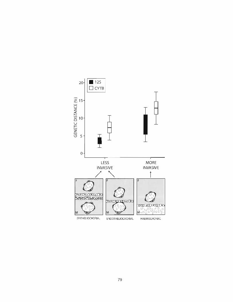
LESS INVASIVE
MORE INVASIVE

Figure 7. Maximum genetic distance at which hybridization occurs between species with invasive or non-invasive placentation. Placental invasiveness is quantified in terms of the number of maternal cell layers separating fetal tissues from the maternal circulatory system (see Mossman 1987; Wildman et al. 2006). With high invasiveness, there is a higher genetic distance between pairs of hybridizable mammal species. In species with epitheliochorial placentation- including all strepsirhine Primates, Cetartiodactyla, Perissodactyla, Pholidota, and some Lipotyphla-the fetal and maternal component of the interhemal membrane both typically consist of endothelium, connective tissue, and epithelium, making a total of six cell layers between the two circulatory systems. In species with endotheliochorial placentation-including all Carnivora, Serenia, and Proboscidea, some Rodentia, most lnsectivora and Xenarthra, and the sole member of Tubulidentata-the maternal epithelium and connective tissue are stripped away during development of the placenta, resulting in an interhemal interface consisting of just four cell layers. Finally, in species with hemochorial placentation-including all haplorhine Primates, Lagomorpha, Dermoptera, and Hyracoidea, most members of Rodentia and Chroptera, and many members of Afrotheria-all of the maternal cell layers are removed during development of the placenta, and maternal blood spaces are directly enclosed by the fetal chorion. The hemochorial placenta is thus unique in that it involves the presentation of fetal surface antigens directly to the blood circulatory system of the mother, unimpeded by cell layers of maternal origin. Bottom, schematic representation of blood flow relations between maternal (M) and fetal (F) sides of the three types of placenta. Open nucleated cells represent epithelial tissue; dark gray nucleated cells represent endothelial tissue; light gray areas represent connective tissue; open dumbbell-shaped cells represent hemocytes. Top, box plot showing cytochrome b and 12s genetic distance between hybridizable pairs of mammals w ~ t h less invasive versus more invasive placentation. Horizontal bars show mean values; boxes show 95% confidence intervals; error bars show 1 SD either side of the mean. The mean value for mammals with less invasive placentation is approximately half that for mammals with more invasive placentation, indicating that the former species evolve reproductive isolation via hybrid inviability more rapidly than the latter

Data within each clade on the evolution of hybrid inviability and on
placental type are not statistically independent under usual models of character
evolution, such as Brownian motion (Felsenstein 1985; Martins 2000). Given that
only four major clades in our analysis exhibit invasive placentation (Haplorhine
primates, Lipotyphla, Rodentia, and Lagomorpha) and four major clades show
non-invasive placentation (Strepsirhine primates, Carnivora, Perissodactyla, and
Artiodactyla), the tests conducted above may be subject to unsuitably high risk of
Type I error. However, analyses using mean values for each of these eight major
groups yielded essentially the same results, with hemochorial (invasive) taxa
exhibiting roughly twofold higher genetic distances for hybridizable species pairs
(cytochrome b, mean genetic distance 6.41 + 1.46 vs. 3.01 f 0.95, Mann-Whitney
U-test, P = ,021, N = 8; 12S, mean genetic distance 11.52 + I .92 vs. 6.29 ? 1.12,
Mann-Whitney U-test, P= ,034, N = 7; both results also remain significant if the
related groups Perissodactyla and Artiodactyla are combined). These results
demonstrate the strength and among-taxon consistency, across major eutherian
orders, ofthe differences in hybridization effects between placental type.
An explicitly phylogeny-based test of our hypothesis requires inferring a
species-level phylogeny for the bulk of eutherian mammals, inferring the
transitions between invasive and non-invasive placentation, and relating these
transitions to changes in the genetic distances at which species can hybridize.
Nine to 11 evolutionary transitions in placental type can be inferred on such a
phylogeny, but data on hybridization distances are currently available for taxa
spanning only two of these transitions, and in both cases, the clades with invasive
81

versus non-invasive placentation are much more distant than sister lineages
(Figure 8; see also Wildman et al. (Wildman et al. 2006) who inferred I I such
transitions). Both inferred changes in hybridization distance are in the predicted
direction, but robust phylogeny-based tests of our hypothesis require data on
hybridization from additional taxonomic groups that closely bracket the transitions
in placental type. Until then, our analysis of this prediction can be considered as a
species-level test that describes current patterns of trait association (Ricklefs and
Stark 1996) subject to the presumptions that the immunological effects of
invasive placentation on hybrid inviability exhibit macroevolutionary lability and
that unobserved, phylogenetically distributed third variables do not drive the
observed patterns (Ridley 1989). Given the >I00 million years of mammalian
radiation represented here, the wide range of taxa already included in our
analyses, and the striking magnitude (around twofold) of the differences between
placental types in hybridization distance, we believe that the strength of these
species-level results should compel more comprehensive tests.


Figure 8. Distribution of our placental and hybridization data on a phylogenetic tree of eutherian mammals. This tree of mammalian families was inferred from five genes-RAG-1 (recombination activating gene l ) , RAG-2 (recombination activating gene 2), cytochrome b, VWF (von Willebrand's factor), and IRBP (interphotoreceptor binding protein)-and 405 species using maximum likelihood, and it is fully compatible with other recent mammalian trees (i.e., see Wildman et al. 2006). Placental invasiveness datawas taken from a number of published sources (Bernirschke and Miller 1982; Carter et al. 2004; Gopalakrishna and Karim 1979; King 1993a; Luckett 1974a; Mess 2003; Mossman 1987; Rasweiler 1993b). Placental invasiveness was mapped onto the branches of the tree by maximum likelihood reconstruction using Mesquite (Maddison and Maddison 2005), and the reconstructions correspond very closely with those of Wildman et al. (2006). Solid branches represent more invasive placentation (hemochorial), and open branches represent less invasive placentation (endotheliochorial or epitheliochorial). Families of mammals whose names are written in bold are those for which hybridizability data and genetic distance data are available. Families whose names are written in regular type are those for which data were not available and are not included in this study. Each family has a small square, either solid or open, to the left of its name. Solid squares denote the availability of species range data (resulting in a measure of sympatry) for sister species within each family; open squares denote the current unavailability of such data.
An additional means of testing our hypothesis that placental invasiveness
mediates aspects of speciation is to use independent lines of evidence. The
hypothesis also predicts that a sustained period of allopatry should be found
more frequently in sister species that have the ability to maintain interspecific
pregnancy by virtue of their placental morphology, because sympatry of
divergent populations within such species should more readily result in the fusion
of populations by hybridization and the loss of incipient species. We tested this
prediction by gathering data on species ranges for 166 pairs of sister taxa of
known placental type (representing 11 orders and 46 families of nonaquatic,
nonvolant mammals) and testing for a mean difference in allopatry between
groups with invasive versus noninvasive placentation. We followed Barraclough
and Vogler (2000) in measuring the degree of allopatry between two species as
the area of range overlap divided by the area of the smaller species range,
normalized by arcsine transformation. By considering only recent biological sister

species, as determined from published phylogenies or analyses of published
DNA sequences, we avoided the phylogenetic inference of ancestral species
ranges from present-day species ranges, which has come under criticism (Losos
and Glor 2003), and we thus minimized the potentially confounding effect of
range shifts since speciation.
Consistent with our prediction, hemochorial sister species were on
average less sympatric (mean 0.22 + 0.06) than grouped epitheliochorial and
endotheliochorial (non-invasive) species (mean 0.55 + 0.1 ; P<0.01, N = 166, F =
33.46; ANOVA). This difference does not result from the possibility that sister
species with invasive placentation tend to be younger than those with
noninvasive placentation and have had less time for their species ranges to
become sympatric: cytochrome b and 12s genetic distance, surrogates for time
since divergence (Gissi et al. 2000), did not differ between invasive and non-
invasive groups (cytochrome b: P = .51, N = 58, F = 0.4; 12s: P = .26, N = 50, F
= 1.3; ANOVA), irrespective of their ability to hybridize. Hemochorial species
were also less sympatric, on average, in the two orders (Primates and
Insectivora) where both invasive and noninvasive placental forms are
represented in our data set (primates: P = .001, F = 12.41; insectivores: P = .007,
F = 14.42; ANOVA), which suggests that the overall mean difference was robust
to taxon-specific effects other than placentation. Such reduced range overlap
between sister species pairs and invasive placentation is an expected
biogeographic consequence of their slower evolution of hybrid inviability.

Discussion
Our analyses suggest that invasive placentation engenders an
approximate halving of the rate at which eutherian mammal species evolve
hybrid incompatibility, compared with species with noninvasive placentation. The
results provide evidence for the importance of physiological adaptations to
placental viviparity in determining the rate at which different clades of mammals
evolve reproductive isolation. In particular, the balance between placental
antigenicity and maternal immune response may differ between taxa with
invasive versus noninvasive placentation, such that immune system
downregulation in taxa with invasive placentation allows for viable hybrid
production at much greater genetic distances. The lower degree of sympatry
between sister species in hemochorial clades provides independent support for
this hypothesis and suggests that placental invasiveness has important
implications for modes of speciation. The evolution of placental form and
function, under conditions of ongoing conflict over resource allocation between
mother and offspring (Crespi and Semeniuk 2004; Haig 1993; Wildman et al.
2006; Zeh and Zeh 2000), may thus generate strong effects on rates and
patterns in the evolution of reproductive isolation. Further studies of eutherian
phylogeny, comparative reproductive immunology, the causes of hybrid inviability
in mammals, and placental evolution should illuminate the mechanisms driving
these results.
CHAPTER 5: CONCLUSIONS
In the ir~troductory chapter I describe a number of important evolutionary
questions which this thesis is intended to explore. First, there is the question of
what role physiology (specifically, the placenta) plays in the evolution of life
history trade-offs. Life history trade-offs have been of interest to evolutionary
biologists for many years. However, the notion of trade-off has often been used
to describe negative associations between traits without evidence of how those
traits interact functionally and causally (Zera and Harshman 2001). In my thesis I
attempt to ground trade-offs over resource allocation during pregnancy in
physiological outcomes resulting from the degree of placental invasiveness of the
fetus. I find that variation in the rate and magnitude of fatty acid transport across
the placenta is a possible causal mecharrisn~ underlying hitherto unrecognized
systematic differences in the brain-body allometry of mammals with invasive
versus non-invasive placentation. Similarly this variation appears to influence the
evolutionary economics of life history evolution, especially the outconies of
evolution along the slow-fast life history continuum. Chapter 3 discusses some
potential future research that might be conducted to further test and elaborate the
hypotheses presented in my thesis. In particular we should test for associations
between placental type and maternal gestational strategies such as income
breeding (in which resources used for reproduction are acquired during the
reproductive period) versus capital breeding (in which resources used for

reproduction are acquired from existing maternal energy stores), for example
through measurement of maternal body fat composition before and after
pregnancy. Shifts in maternal diet that occur with the onset of the reproductive
period should also illustrate the importance of maternal access to fatty acids to
fetal brain growth. Such future studies may also be able to focus on within-
species variation, for example using human medical data on the outcome of
individual pregnancies. Studies of the molecular evolution of genes involved in
fatty acid transport are an additional potential future line of research, my
prediction being that these genes undergo periods of accelerated evolution along
branches of the phylogenetic tree upon which placentation undergoes structural
transformations.
A second, related question is the nature of allometry in mammals. In
Chapter 2, 1 present evidence that some of the between-taxon variation in brain-
body allometry results from differences in the timing and magnitude of brain
growth resulting from variation in the invasiveness of the placenta. These results
lead me to question the validity of considering the realized brain size of each
mammalian species to be a departure from a single mammalian allometric slope,
or a member of a taxon-level "grade". Instead, variation in brain-body allometry is
likely the result, in part, of differences in the physiological basis of resource
acquisition by the fetus during pregnancy. This view might be tested profitably in
other taxa exhibiting placental variation, such as the fish or reptiles.
The fact that allometry and life history trade-offs are constrained by the
physiological basis of resource allocation to different functions and life stages is,

in itself, not partic~~larly surprising. To the extent that this physiological basis
varies between taxa, however, a functional understanding of life history trade-offs
is necessary if we are to understand the evolution of alternative strategies within
a diverse group of species such as mammals. Of particular interest in the case of
placentation is that placental invasiveness is not merely an external constraint
limiting the availability of resources in such a way as to generate conflict between
physiological functions over a finite pool of resources. In addition, the placenta
may be involved in conflict between parents and offspring. In Chapter 3, 1
suggest that placental invasiveness is not merely a cause of life history trade-
offs, but that the evolution of non-invasive placentation may also be a result of
selection against the escalation of conflict between mother and offspring or
between littermates. This hypothesis yields direct predictions regarding the
evolution of non-invasive placentation that could be tested using in-depth studies
of mammalian clades exhibiting placental variation, such as the primates or
insectivores. The existence of parent-offspring conflict is also expected to leave a
molecular signature, namely the accelerated rate evolution of genes involved in
parent-offspring interaction along branches of the phylogenetic tree exhibiting
invasive placentation, in comparison with that of branches exhibiting non-invasive
placentation. The role of genomic imprinting of placentally-expressed genes has
also been described in terms of parent-offspring conflict, yet these views remain
to be tested in a broad comparative context.
A final question in my introductory chapter relates to the role of
placentation in generating reproductive incompatibility between divergent

populations or species of mammal. I provide an explanation rooted in
reproductive immunology to account for divergence in hybridization rate and
allopatry in sister-species with invasive versus non-invasive placentation. Again,
testing such hypotheses in a focal group of species within which we can study
hybridization in a comprehensive way may be a useful way of proceeding. Direct
experimental measurement of hybridization potential within sister-clades of
viviparous versus oviparous fish, for example, would permit the hypothesis of
Chapter 4 to be tested independently. The mammalian data, on the whole,
supports our view that placental structure may influence the ability of species to
hybridize, and may therefore influence the rate at which speciation occurs in
sympatry or between neighbouring allopatric species separated by a hybrid zone.
Together, the results presented in my thesis underscore the importance of
functional studies in generating hypotheses and explaining the origin and
outcome of life history trade-offs, allometry, and other aspects of mammalian
evolution. Fostering a deeper understanding of the physiology of parent-offspring
interactions, incorporating methods derived from the study of molecular
evolution, and conducting within-species studies of pregnancy outcome, are the
best ways of testing and refining these ideas further.
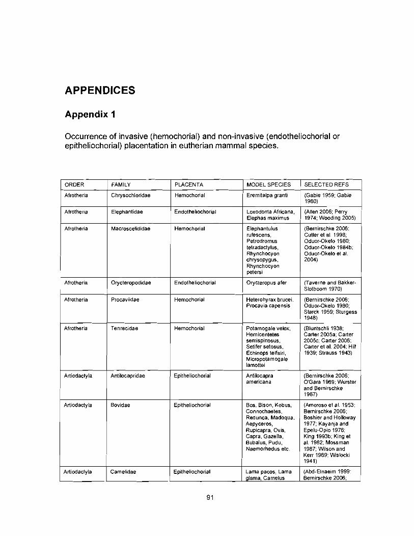
APPENDICES
Appendix 1
Occurrence of invasive (hemochorial) and non-invasive (endotheliochorial or epitheliochorial) placentation in eutherian mammal species.
ORDER
Afrotheria
PLACENTA
Hemochorial
MODEL SPECIES
Eremitalpa granti
FAMILY SELECTED REFS
Chrysochloridae (Gabie 1959; Gabie 1960)
Afrotheria Elephantidae Endotheliochorial Loxodonta Africana, Elephas maximus
(Allen 2006; Perry 1974; Wooding 2005)
Afrotheria Macroscelididae Hemochorial Elephantulus rufescens, Petrodromus tetradactylus, Rhynchocyon chrysopygus, Rhynchocyon petersi
(Bernirschke 2006; Cutler et al. 1998; Oduor-Okelo 1980; Oduor-Okelo 1984b; Oduor-Okelo et at. 2004)
Afrotheria Orycteropodidae -
Orycteropus afer Endotheliochorial (Taverne and Bakker- Slotboom 1970)
Afrotheria Procaviidae Hemochorial (Bernirschke 2006; Oduor-Okelo 1980; Starck 1959; Sturgess 1948)
Heterohyrax brucei, Procavia capensis
Afrotheria Tenrecidae Hemochorial -
Potamogale velox, Hemicentetes semispinosus, Setifer setosus, Echinops telfairi, Micropotamogale lamottei
Antilocapra americana
-
(Bluntschli 1938; Carter 2005a; Carter 200%; Carter 2006; Carter et al. 2004; Hill 1939; Strauss 1943)
Antilocapridae (Bernirschke 2006; O'Gara 1969; Wurster and Bernirschke 1967)
Artiodacty la Epitheliochorial
Artiodacty la Bovidae Epitheliochorial Bos, Bison, Kobus, Connochaetes, Redunca, Madoqua, Aepyceros, Rupicapra, Ovis, Capra, Gazella, Bubalus, Pudu, Naemorhedus etc.
(Amoroso et al. 1953; Bernirschke 2006; Boshier and Holloway 1977; Kay anja and Epelu-Opio 1976; King 1993b; King et al. 1982; Mossman 1987; Wilson and Kerr 1969; Wislocki 1941)
Artiodactyla Camelidae Epitheliochorial Lama pacos, Lama glama, Camelus
(AM-Elnaeim 1999; Bernirschke 2006;

drornedarius. Vicugna vicugna
bactrianus, Camelus 1990; Ghazi et al. 1994; Morton 1961; Olivera 2003a: Olivera 2003b; Skidrnore et al. 1996; Steven 1980)
Fowler and Olander
Artiodactyla Odocoileus, Elaphurus, Cervus, etc.
(Bernirschke 2006; Harrison and Hamilton 1952; Mossrnan 1987; Sinha 1969; Wurster and Bernirschke 1967)
Artiodacty la Giraffidae Epitheliochorial Giraffa camelopardalis
(Bernirschke 2006; Deka et al. 1980)
Artiodactyla Choeropsis liberiensis, Hippopotamus amphibious
Moschus rnoschiferus
Hippopotamidae (Bernirschke 2006; MacDonald and Bosrna 1975)
Epitheliochorial
Artiodactyla (Bernirschke 2006) Moschidae
Artiodactyla
Epitheliochorial
Suidae Epitheliochorial Sus scrofa, Phacochoerus aethiopicus, Hy lochoerus rneinertzhageni
(MacDonald and Bosrna 1975)
Artiodactyla Tayassuidae Epitheliochorial Tayassu pecari, Catagonus wagneri
(Bernirschke 2006; Dieffenbach 1907; MacDonald and Bosma 1975)
Artiodacty la Tragulidae Epitheliochorial Tragulus javanicus (Bernirschke 2006; Kirnura 2004)
Carnivora Ailuridae I Endotheliochorial Ailurus fulgens (Bernirschke 2006)
Carnivora Canidae Endotheliochorial Canis farniliaris, Vulpes vulpes, Canis adustus, Canis mesomelas, Otocyon megalotis
(Kehrer 1973; Mossman 1987; Stoffel et al. 1998)
Carnivora Felidae Endotheliochorial Felis catus, Panthera leo, Panthera tigris, etc
(Kehrer 1973; Leiser and Koob 1993; Tiedemann 1979)
Carnivora Hyaenidae 1 Hemochorial Crocuta crocuta (Wynn et al. 1990)
Carnivora Mephitidae 1 Endotheliochoiral Spilogale putorius (Sinha 1976; Sinha and Mean 1978)
(Bernirschke 2006; Gularnhusein and Beck 1975; Pfarrer 1999; Sinha and Mossman 1966)
(Mossrnan 1987)
(Bernirschke 2006)
Carnivora Mustela putorius furo, Enhydra lutris, Mustela vison, Aonyx cinerea
Mustelidae
Carnivora
Carnivora
Endotheliochorial
Nandinia
Odobenus rosrnarus
Nandiniidae
Odobenidae
Carnivora
Endotheliochorial
Endotheliochorial
Arctocephalus pusillus, Callorhinus ursinus
3tariidae (Rand 1955) Endotheliochorial
Carnivora Leptonychotes weddelli, Phoca vitulina, Halichoerus
Fhocidae (Arnoroso and Matthews 1952;
Endotheliochorial
92

SrYPUS I Davies 1950)
Endotheliochorial (Bernirschke 2006; Creed and Harrison 1965)
Carnivora Procy onidae Procyon lotor, Nasua narica
Carnivora Ursidae Ursus americanus, Thalarctos maritimus. Melursus ursinus
Arctictis, Paradoxus, Genetta, Helogale, Ichenumia, Cynictis, etc.
Tursiops truncatus, Globicephala melaena, Cephalorhynchus commersoni, Lissodelphis borealis
(Bernirschke 2006; Wmsatt 1974; Young 1969)
Endotheliochorial
Viverridae Endotheliochorial (Moghe 22B; Young 1978)
Carnivora
Epitheliochorial (Bernirschke 2006; Morton and Mulholland 1961; Wslocki and Enders 1941)
Cetacea Delphinidae
Cetacea Monodontidae Epitheliochorial Delphinapterus leucas
(Bernirschke 2006)
Cetacea Phocoenidae Epitheliochorial Phocaenaphocaena (Wslocki 1933)
Cetacea Platanistidae Epitheliochorial Platanista gangetica, Platanista indi, Pontoporia blainvillei
Taphozous melanopogon
(Harrison 1972; Pilleri and Gihr 1976a; Pilleri and Gihr 1976b)
(Bhigwade 1990) Emballonuridae Endotheliochorial Chiroptera
Chiroptera Hipposideridae Hemochorial Hipposideros fulvus (Bhigwade 1990)
Chiroptera Megadermatidae Endotheliochorial Megaderma sp. (Bhigwade 1990)
Chiroptera Molossidae Hemochorial and endotheliohorial
Molossus ater, Tadarida brasiliensis, Tadarida tragata
(Bernirschke 2006; Gopalakrishna and Badwaik 1990; Rasweiler 1991 : Rasweiler 1993a)
Chiroptera Mormoopidae Hemochorial Pteronotus parnellii (Badwaik and Rasweiler 1998)
Chiroptera Natalidae Endotheliochorial Natalus sp. (Carter and Enders 2004)
Chiroptera Noctilionidae Endotheliochorial Noctilio albiventris (Rasweiler 1993a)
Chiroptera Phyllostomidae Endotheliochorial Glossophaga soricina
(Rasweiler 1993a)
Chiroptera Pteropodidae Hemochorial Pteropus giganteus, rousettus leschenaulti, Cynopterus sphinx
(Bhigwade 1990; Bhigwade et al. 2000; Karim and Bhatnagar 1996)
Chiroptera Rhinolophidae Endolheliochorial Rhinolophus rouxi (Bhigwade 1990)
Chiroptera Rhinopomatidae Endotheliochorial Rhinopoma hardwickei
(Bhigwade 1990)
Chiroptera Thy ropteridae Hemochorial Thyroptera tricolor spix
(Wmsatt and Enders 1980)
(Bhigwade 1992; Gopalakrishna and Karim 1972; Kimura and Uchida 1984; Rasweiler 1993a;
Chiroptera Hemochorial Corynorhinus mexicanus, Miniopterus schreibersii, Myotis lucifugus. Myotis

I moluccarum. I Sanchez Hernandez ~,
Pipistrellus mimus,
Dermoptera Cynocephalidae Hemochorial Cynocephalus volans
(Starck 1959)
lnsectivora Erinaceidae Hemochorial Erinaceus europaeus
(Morris 1957)
lnsectivora Solenodontidae Hemochorial Solenodon paradoxus
(Wislocki 1940)
lnsectivora Soricidae - - -
Suncus murinus, Blarina brevicauda, Sorex fumeus
- -
(Owers 1960; Wimsatt et al. 1973; Wimsatt and Wislocki 1947)
(Carter 2005b; Malassine and Leiser 1984; Prasad et at. 1979)
Hemochorial
Talpidae lnsectivora Epitheliochorial or Endotheliochorial
Scalopus aquaticus, Talpa europaea
Lagomorpha Leporidae Hemochorial Oryctolagus cuniculus
(Samuel et al. 1975)
Lagomorpha Ochotonidae Hemochorial Ochotona princeps (Harvey l959b)
Perissodacty la Equidae Epitheliochorial Equus burchelli. Equus asinus. Equus caballus, Equus grevyi, Equus kiang
(Allen and Short 1997)
Perissodactyla Rhinocerotidae Epitheliochorial Diceros bicornis, Rhinoceros unicornis, Ceratotherium simum
(Benirschke and Lowenstine 1995; Bernirschke 2006)
Perissodactyla Tapiridae Epitheliochorial Tapirus sp (Bernirschke 2006; Schauder 1945)
Pholidota Manidae Epitheliochorial
Hemochorial
Hemochorial
Manis (Mossman 1987)
Primates
Primates
Atelidae
Callithrichidae
Ateles geoffroy i
Callithrix jacchus, Callimico goeldii
Cebus, Alouatta, Mandrillus, Lagothrix, etc.
(Bernirschke 2006)
(Jollie 2005; Smith and Moore 1990)
(Bernirschke 2006) (Mossman 1987)
Primates Cebidae Hernochorial
Primates Cercopithecidae Hemochorial Cercopithecus, Papio, Presbytis, Colobus, Pygathrix, Trachypithecus, Macaca etc.
(Bernirschke 2006; Burton 1980a; Burton 1980b; McLean 2001; Owiti 1986)
Primates Daubentoniidae Epitheliochorial Daubentonia rnadagascariensis
(Bernirschke 2006; Hill and Burne 1922)
Primates Galagidae Epitheliochorial Galago :rassicaudata, Salago senegalensis
(King 1984; Njogu 2005)
Hemochorial Primates - -
Pan, Pongo, Homo, Sorilla
Hominidae (Bernirschke 2006) (Mossman 1987)
Primates Epitheliochorial ndri indri, Jropithecus rlerreauxi
(Hill 1932) lndriidae
Primates Lemuridae Epitheliochorial Wicrocebus nurinus, Eulemur
[Bernirschke 2006;

1 fulvus, Lemur catta, I Strauss 1978)
Primates
Primates
Primates
1 Rodentia I Aplodontiidae I Hemochorial I Aplodontia rufa 1 (Hawey 1959a)
Lorisidae
Pitheciidae
Tarsiidae
Rodentia
Rodentia
Rodentia
Epitheliochorial
Hemochorial
Hemochorial
Agoutidae
Anomaluridae
Rodentia
Rodentia
Caviidae
Varecia variegatus
Loris tardigradus
Pithecia pithecia
Tarsius spectrum
Hemochorial
Hemochorial
Bathyergidae
Castoridae
Hemochorial
(Hill 1932)
(Bernirschke 2006)
(Luckett 1974b)
Rodentia
Cavia porcellus, Kerodon rupestris
Agouti paca
Anomalurus
Hemochorial
Hemochorial
Chinchillidae
(Bonatelli 2002; Bonatelli 2005; Miglino 2002)
(Luckett 1971)
(Bernirschke 2006; Kaufmann and Davidoff 1977; Oliveira 2006)
(King and Tibbets 1976)
Bathyergus janetta
Castor canadensis
Hemochorial
1 Rodentia Ctenodactylidae Hemochorial Ctenodactylus I (Luckett 1980) gundii
(Luckett and Mossman 1981)
(Fischer 1971)
Chinchilla laniger, Lagostomus maximus
Rodentia
Rodentia
Rodentia
1 Rodentia
Rodentia Heteromy idae Endotheliochorial Dipodomys. (King and Tibbets Microdipodops 1 2005)
Rodentia
Dasyproctidae
Dinomy idae
Dipodidae
Erethizontidae
Rodentia Myocastoridae Hemochorial Myocastor coypus (Hillemann and I Gaynor 1961)
Geomyidae
1 Rodentia
Rodentia
Rodentia
Hemochorial
Hemochorial
Hemochorial
Hemochorial
Hemochorial
Hydrochaeridae
Hystricidae
Muridae
Rodentia
Rodentia
Rodentia
Dasyprocta azarae
Dinomys brannicki
Jaculus jaculus
Erethizon dorsatum
(Becher 1921 a; Becher 1921 b; Miglino 2002)
(Bernirschke 2006)
(King and Mossman 1974)
(Perrotta 1959)
Geomys bursarius
Hemochorial
Hemochorial
Hemochorial
Octodontidae
Pedetidae
Sciuridae
(Mossman 1987; Mossman and Strauss 1963)
Hydrochaerus hydrochaeris
Hystrix cristata
Clethrionomys, Dicrostonyx, Lemmus, Microtus, Peromyscus, Phodopus, Rattus, Mus, Mesocricetus, etc.
Hemochorial
Hemochorial
Hemochorial
(Miglino 2002)
(Luckett and Mossman 1981)
(Carpenter 1982; Kertschanska 2000; King and Hastings 1977; Ogura 1991)
Octodon degus
Pedetes capensis
Spermophilus
(Bosco 2006; Kertschanska et al. 1997)
(Fischer and Mossman 1969)
(Carter and Enders

I lateralis
Rodentia Thryonomyidae Hemochorial Thryonomys swinderianus
(Oduor-Okelo 1984a; Oduor-Okelo and Gombe 1991)
(King and Mossman 1974)
Hemochorial Rodentia Zapus sp.
Scandentia Endotheliochorial Tupaia (Bernirschke 2006; Luckhardt et al. 1985)
Tupaiidae
Sirenia Trichechidae Endotheliochorial Trichechus manatus (Enders and Carter 2004)
Xenarthra Bradypodidae Endotheliochorial Bradypus variegatus (Amorim 2004; Bernirschke 2006)
(Wislocki 1928b) Hemochorial Cyclopes didactylus Xenarthra Cyclopedidae
Xenarthra Dasypodidae Hemochorial Cabassous chacoensis, Chaetophractus villosus, Dasypus hybridus, D. novemcinctus. Toly peutes matacus
(Adamoli 2001)
-
Xenarthra -
Megalonychidae Choloepus hoffmanni, Choloepus didactylus
Myrmecophaga jubata, Myrmecophaga tridactyla, Tamandua tetradactyla
(Bernirschke 2006; Wislocki 1928a)
Endotheliochorial
Xenarthra Myrmecophagidae Hemochorial (Becher 1931; Bernirschke 2006; Walls 1939)
REFERENCE LIST
Abd-Elnaeim, M. M. 1999. Fetomaternal attachment and anchorage in the early
diffuse epitheliochorial placenta of the camel (Camelus dromedarius).
Light, transmission and scanning electron microscopic study. Cells
Tissues Organs 164:141-154.
Adamoli, V. C. 2001. Corr~parative morphological placental types in Dasypodidae
(Chaetophractus villosus, Cabassous chacoensis, Tolypeutes matacus
and Dasypus hybridus). Biocell 25: 17-22.
Aiello, L. C. 1997. Brains and guts in human evolution. Brazilian Journal of
Genetics 20: 141 -148.
Aiello, L. C., and P. Wheeler. 1995. The expensive tissue hypothesis: the brain
and digestive system in human and primate evolution. Current
Anthropology 36: 1 99-221.
Ait-Azzouzene, D., M. C. Gendron, M. Houdayer, A. Langkopf, K. Burki, D.
Nemazee, and C. Kanellopoulos-Langevin. 1998. Maternal B lymphocytes
specific for paternal histocompatibility antigens are partially deleted during
pregnancy. Journal of Immunology 161 :2677-2683.

Alcoze, T., and E. Zimmerman. 1971. Food habits and dietary overlap of two
heteromyid rodents from the mesquite plains of Texas. Journal of
Mammalogy 54:900-908.
Allen, W. R. 2006. Ovulation, pregnancy, placentation and husbandry in the
African elephant (Loxodonta africana). Philosophical Transactions of the
Royal Society of London 361 B:821-834.
Allen, W. R., and R. V. Short. 1997. Interspecific and extraspecific pregnancies in
equids: anything goes. Journal of Heredity 88:384-392.
Allman, J. M., T. McLaughlin, and A. Hakeem. 1993a. Brain structl-~res and life
span in primate species. Proceedings of the National Academy of the
United States of America 90:3559-3563.
-. 1993b. Brain weight and life span in primate species. Proceedings of the
National Academy of the United States of America 90:118-122.
Amorim, A. L. M. J. A. 2004. Anatomical aspects of the placenta of the sloth.
International Journal of Morphology 22:9-18.
Amoroso, E. C., L. M. Kellas, and L. Harrison-Matthews. 1953. The foetal
membranes of an African waterbuck, Kobus defassa. Proceedings of the
Zoological Society of London 123:477.
Amoroso, E. C., and L. H. Matthews. 1952. The foetal placenta and membranes
of Halichoerus grypus. Jo~~rnal of Anatomy 86:487-488.
Andriant, R., and T. Rahandra. 1973. Influence of intrinsic factors (species, age,
gestation, lactation) on food consumption of Microcebus murinus murinus

and Microcebus murinus rufus. Comptes Rendus hebdomadaires des
Seances de I'Academie des Sciences D 277: 1893-1 896.
Armstrong, E. 1983. Relative brain size and metabolism in mammals. Science
220:1302-1304.
Atsalis, S. 1999. Diet of the brown mouse lemur (Microcebus rufus) in
Ranomafana National Park, Madagascar. International Journal of
Primatology 20: 193-229.
Badwaik, N., and J. J. Rasweiler. 1998. The interhaemal barrier in the
chorioallantoic placenta of the greater mustache bat, Pteronotus parnellii,
with observations on amplification of its intrasyncytial lamina. Placenta
I9:391-4Ol.
Bainbridge, D. R. J. 2000. Evolution of mammalian pregnancy in the presence of
the maternal immune system. Reviews of Reproduction 5:67-74.
Baker, J. M., A. I. Barnford, and D. F. Antczak. 1999. Modulation of allospecific
CTL responses during pregnancy in equids: an immunological barrier to
interspecies matings? Journal of lmmunology 162:4496-4501.
Barton, R. A. 1996. Neocortex size and behavioural ecology in primates.
Proceedings of the Royal Society of London 263: 173-1 77.
Becher, H. 1921 a. Der Feinere Bau der Reifen Placenta von Aguti (Dasyprocta
azarae, Schl.). Zeitschrift fiir Anatomie und Entwicklungsgeschichte
67:381-458.

-. 1921 b. Die Enwicklung des Mesoplacentariums und die Placenta bei Agout
(Dasyprocta azarae Schl.). Zeitschrift fijr Anatomie und
Entwicklungsgeschichte 61 :337-364.
-. 1931. Placenta und Uterusschleimhaut von Tamandua tetradactyla
(Myrmeophaga). Morphologie Jahrbuch 67:381-458.
Bell, G., and V. Koufopanou. 1986. The cost of reproduction. Oxford Surveys in
Evolutionary Biology 3:83-131.
Benassayag, C., V. Rigourd, T. M. Mignot, J. Hassid, M. J. Leroy, B. Robert, and
C. Civel. 1999. Does high polyunsaturated free fatty acid level at the feto-
maternal interface alter steroid hormone message during pregnancy? .
Prostaglandins, leukotrines, and essential fatty acids 60:393-399.
Benirschke, K., and L. J. Lowenstine. 1995. The placenta of the Rhinocerotidae.
Verhandlungsbericht der Erkrankungen der Zootiere 37:15-23.
Bennett, P. M., and I. P. F. Owens. 2002, Evolutionary Ecology of Birds: Life
Histories, Mating Systems and Extinction: Oxford Series in Ecology and
Evolution. Oxford, Oxford University Press.
Bentler, P. M., and J. A. Stein. 1992. Structural equation models in medical
research. Statistical Methods in Medical Research 1 :159-181.
Beolchini, F., and A. Loy. 2003. Diet of syntopic moles Talpa romana and Talpa
europaea in central Italy. Mammalian Biology 69: 140-1 44.
Berghaus, T. M., H. Demmelmair, and B. Koletzko. 2000. Essential fatty acids
and their long-chain polyunsaturated metabolites in maternal and cord

plasma triglycerides during late gestation. Biology of the Neonate 77:96-
100.
Bernirschke, B., and C. Miller. 1982. Anatomical and functional differences in the
placenta of primates. Biology of Reproduction 26:29-53.
Bernirschke, K. 2006, Comparative Placentation Website.
http://medicine.ucsd.edu/cpa. Accessed 2006-7.
Bhigwade, D. A. 1990. Comparative electron microscopy of chorioallantoic
placental barrier in some Indian Chiroptera. Acta Anatomica 128:302-317.
-. 1992. Ultrastructural development of chorioallantoic placenta in the Indian
Miniopterus bat, Miniopterus schreibersii fuliginosus (Hodgson). Acta
Anatomica 145:248-264.
Bhigwade, D. A., S. Taskar, and K. P. Bhatnagar. 2000. The interhemal
membrane of the bats, Rousettus leschenaulti and Cynopterus sphinx
(Megachiroptera): an ultrastructural investigation. Acta Chiropterologica
2~107-122.
Bielby, J., G. M. Mace, 0 . R. P. Bininda-Edmonds, M. Cardillo, J. L. Gittleman, K.
E. Jones, C. D. L. Orme et al. 2007. The fast-slow continuum in
mammalian life history: an empirical reevaluation. American Naturalist
1691748-757.
Bininda-Edmonds, 0. R. P., and M. Cardillo. 2007. The delayed rise of present-
day mammals. Nature 446:507-512.
Blum, J. J. 1977. The geonietry of four dimensions and relationship between
metabolism and body mass. Journal of Theoretical Biology 64:599-601.
Bluntschli, H. 1938. Le developpement primaire et I'implantation chez un
centetine (Hemicentetes). Comptes Rendus de I'Association des
Anatomistes.
Bonatelli, M. 2002. Morphological analysis of the placenta of paca (Agoutipaca
Linnaeus 1766). Study with light microscopy and transmission electron
microscopy. Biota Neotropica 2.
-. 2005. Placentation in the paca (Agouti paca L). Reproductive Biology and
Endocrinology 3.
Bosco, C. 2006. Placentation in the degu (Octodon degus): analogies with
extrasubplacental trophoblast and human extravillous trophoblast.
Comparative Biochemistry and Physiology A Early Access.
Boshier, D. P., and H. Holloway. 1977. The sheep trophoblast and placental
function: an ultrastructural study. Journal of Anatomy 124.
Boyce, M. S. 1988. Evolution of life histories of mammals, Yale University Press.
Bradley, W. G., and R. A. Mauer. 1971. Reproduction and food habits of
Merriam's kangaroo rat, Dipodomys merriami. Journal of Mammalogy
52:497-505.
Briscoe, J., S. E. Gathercole, and N. Marlow. 2001. Everyday memory and
cognitive ability in children born very prematurely. Journal of Child
Psychology 42:749-754.

Brothers, L. 1990. The social brain: a project for integrating primate behaviour
and neurophysiology in a new domain. Concepts in Neuroscience 1.
Burton, G. J. 1980a. Early placentation in the dusky leaf monkey (Presbytis
obscura). Placenta 1 : 187-1 95.
-. 1980b. Light microscopy of the dusky leaf monkey (Presbytis obscurus).
Journal of Reproduction and Fertility 57: lO9- l l l .
B~~r ton, R. F. 2006. A new look at the scaling of size in mammalian eyes. Journal
of Zoology 2691225-232.
Byrne, R. W., and A. Whiten. 1988, Machiavellian Intelligence. Oxford, Oxford
University Press.
Campbell, F. M., and A. K. Dutta-Roy. 1995. Plasma membrane fatty-acid-
binding protein (FABPpm) is exclusively located in the maternal-facing
membranes of the human placenta. FEBS Letters 375:227-230.
Campbell, F. M., M. J. Gordon, and A. K. Dutta-Roy. 1996. Preferential uptake of
long chain polyunsaturated fatty acids by isolated human placental
membranes. Molecular and Cellular Biochemistry 155:77-83.
-. 1998. Placental membrane fatty acid-binding protein preferentially binds
arachidonic and docosahexaenoic acids. Life Sciences 63:235-240.
Carey, J. S., and D. S. Judge. 2000, Longevity Records: Life Spans of Mammals,
Birds, Amphibians, Reptiles and Fish: Monographs in Population Aging, v.
8.

Carpenter, S. J. 1982. Trophoblast invasion and alteration of niesometrial
arteries in the pregnant hamster: light and electron microscopic
observations. Placenta 3:219-242.
Carter, A. M. 2005a. Development of the haemophagous region and labyrinth of
the placenta of the tenrec, Echinops telfairi. Placenta 26:251-261.
-. 2005b. Placentation in an American mole, Scalopus aquaticus. Placenta
261597-600.
-. 2005c. Structure of the definitive placenta of the tenrec, Echinops telfairi.
Placenta 25:2l 8-232.
-. 2006. The fetal membranes of the otter shrews and a synapomorphy for
Afrotheria. Placenta 27:258-268.
Carter, A. M., and A. C. Enders. 2004. Comparative aspects of trophoblast
development and placentation. Reproductive Biology and Endocrinology
2:46-.
Carter, A. M., A. C. Enders, H. Kunzle, D. Oduor-Okelo, and P. Vogel. 2004.
Placentation in species of phylogenetic importance: the Afrotheria. Animal
Reproduction Science 82:35-48.
Charnov, E. L. 1991. Evolution of life history variation among female mammals.
Proceedings of the National Academy of the United States of America
88: 1 134-1 137.
-. 1993, Life History Invariants: Some Explorations of Symnietry in Oxford
Series in Ecology and Evolution, Oxford University Press.

Charnov, E. L., and S. K. Morgan Ernest. 2006. The offspring/clutch-size trade-
off in mammals. American Naturalist 167:578-582.
Chesnais, J.-C. 1992, The Demographic Transition: Stages, Patterns and
Economic Implications. Oxford, Clarendon Press.
Clark, D. A. 2003. Is there any evidence for immunologically mediated or
immunologically modifiable early pregnancy failure. Journal of Assisted
Reproduction and Genetics 20:63-72.
Corbin, G. D., and J. Sclimid. 1995. Insect secretions determine habitat use
patterns by a female lesser mouse lemur (Microcebus murinus). American
Journal of Primatology 36:223-237.
Coyne, J. A., and H. Allen Orr. 2004, Speciation, Sinauer Associates.
Crawford, M. A., A. G. Hassam, and G. Williams. 1976. Essential fatty acids and
fetal brain growth. The Lancet 1 :452-453.
Creed, R. F. S., and J. Harrison. 1965. Preliminary observations on the
ultrastructure of the raccoon (Procyon lotor) placenta. Journal of Anatomy
99:933.
Crespi, B. J., and C. Semeniuk. 2004. Parent-offspring conflict in the evolution of
vertebrate reproductive mode. American Naturalist 163:635-653.
Croy, B. A,, J. Rossant, and D. A. Clark. 1982. Histological and immunological
studies of post implantation death of Mus caroli embryos in the Mus
musculus uterus. Journal of Reproductive lrr~munology 4:277-293.

Cutler, J. B., A. Dumas, and R. M. Wynn. 1998. lmmunohistochemical analysis of
the interhemal merr~brane of the elphant shrew. Gynecologic and Obstetric
Investigations 46:46-48.
Dancis, J., V. Jansen, and M. Levitz. 1976. Transfer across perfused human
placenta. IV. Effect of protein binding on free fatty acids. Pediatric
Research 10:5-10.
Dauncey, M. J., and R. J. Bicknell. 1999. Nutrition and neurodevelopment:
mechanisms of developmental dysfunction and disease in later life.
Nutrition Research Reviews l2:231-253.
Davies, D. V. 1950. The fetal membranes of the Weddell seal (Leptonychotes
weddelli). Journal of Anatomy 84:408.
Deaner, R. O., K. Isler, and C. van Schaik. 2007. Overall brain size, and not
encephalization quotient, best predicts cognitive ability across non-human
primates. Brain, Behavior and Evolution 70:115-124.
Deka, B. C., K. C. Nath, and B. N. Borgohain. 1980. Clinical note on the
morphology of the placenta in a giraffe (Giraffa camelopardalis). Journal of
Zoo Animal Medicine 11 : 11 7-1 18.
Dickman, C. R. 1995. Diets and habitat preferences of 3 species of crocidurine
shrews in arid southern Africa. Journal of Zoology 237:499-514.
Dieffenbach, L. 1907. Ueber die Semiplacenta diffusa incompleta von Dicotyles
labiatus (Cuvier). Anatonlische Hefie 34527-553.

Donati, G., and S. M. Borgognin-Tarli. 2006. Influence of abiotic factors on
cathemeral activity: the case of Eulemur fulvus coronatus in the littoral
forest of Madagascar. Folia Primatologica 77:104-122.
Dunbar, R. I. M. 1992. Neocortex size as a constraint on group size in primates.
Journal of Human Evolution 22.
-. 2003a. Neocortex size as a constraint on group size in primates. Journal of
Human Evolution 22.
-. 2003b. The social brain: mind, language and society in evolutionar
perspective. Annual Review of Anthropology 32.
Dutta-Roy, A. K. 2000. Cellular uptake of long-chain fatty acids: role of
membrane-associated fatty acid bindingltransport proteins. Cellular and
Molecular Life Sciences 57: 1360-1 372.
Edson, J. L., D. G. Hudson, and D. Hull. 1975. Evidence for increased fatty acid
transfer across placenta during a maternal fast in rabbits. Biology of the
Neonate 2750-55.
Eisenberg, J. F. 1981, The Mammalian Radiations: An Analysis of Trends in
Evolution, Adaptation and Behavior, University of Chicago Press.
Eisenberg, J. F., and D. E. Wilson. 1978. Relative brain size and feeding
strategies in the Chiroptera. Evolution 32:740-751.
Elliot, M. G., and B. J. Crespi. 2006. Placental invasiveness mediates the
evolution of hybrid inviability in mammals. American Naturalist 168: 1 14-
120.

Elphick, M. C., P. Flecknell, D. Hull, and I. R. McFadyen. 1980. Plasma ,free fatty
acid umbilical venous-arterial concetration differences and placental
transfer of 14C palmitic acid in pigs. Journal of Developmental Physiology
21347-356.
Elphick, M. C., D. G. Hudson, and D. Hull. 1975. Transfer of fatty acids across
the rabbit placenta. Journal of Physiology 252:29-42.
Elphick, M. C., and D. Hull. 1977a. Rabbit placental clearing-factor lipase and
transfer to the foetus of fatty acids derived from triglycerides injected into
the mother. Journal of Physiology 252:29-42.
-. 1977b. The transfer of free fatty acids across the rabbit placenta. Journal of
Physiology 264.
-. 1984. Transfer of fatty acid across the cat placenta. Journal of Developmental
Physiology 6:517-525.
Elpl-lick, M. C., D. Hull, and F. Broughton Pipkin. 1979. The transfer of fatty acids
across the sheep placenta. Journal of Developmental Physiology 1 :31-45.
Enders, A. C., and A. M. Carter. 2004. What can comparative studies of placental
structure tell us? A review. Placenta 25:3-9.
Erhard, H. W., and A. Boissy. 2004. Effects of prenatal undernutrition on
emotional reactivity and cognitive flexibility in adult sheep. Behavioural
Brain Research 151 :25-35.
Felsenstein, J. 1985. Phylogenies and the comparative method. American
Naturalist 125:l-15.

Fietz, J., and J. U. Ganzhorn. 1999. Feeding ecology of the hibernating primate
Cheirogaleus medius: how does it get so fat? Oecologia 121 : I 57-164.
Fietz, J., and F. Tataruch. 2003. White adipose tissue composition in the free-
ranging fat-tailed dwarf lemur (Cheirogaleus medius; Primates), a tropical
hibernator. Journal of Comparative Physiology B 173: 1-1 0.
Finlay, B. L., R. B. Darlington, and N. Nicastro. 2001. Developmental structure in
brain evolution. Behavioral and Brain Sciences 24:263-308.
Fischer, T. V. 1971. Placentation in ,the American beaver (Castor canadensis).
American Journal of Anatomy 131 : 159-1 84.
Fischer, T. V., and H. W. Mossman. 1969. The fetal membranes of Pedetes
capensis, and their taxonomic significance. American Journal of Anatomy
124189-1 16.
Fish, J. L., and C. A. Lockwood. 2003. Dietary constraints on encephalization in
primates. American Journal of Physical Anthopology 120:171-181.
Fitzpatrick, B. M. 2004. Rates of evolution of hybrid inviability in birds and
mammals. Evolution 58:1865-1870.
Fowler, M. E., and H. J. Olander. 1990. Fetal membranes and ancillary structures
of llamas (Lama glama). American Journal of Veterinary Research
51 :l495-l5OO.
French, N. R. 1974. A population study of irradiated desert rodents. Ecological
Monographs 44.

Gabie, V. 1959. The early embryology of Eremitalpa granti (Broom). Journal of
Morphology 104: 181 -204.
-. 1960. The placentation of Eremitalpa granti (Broom). Journal of Morphology
107:61-78.
Gayon, J. 2000. History of the concept of allometry. American Zoologist 40:748-
758.
Genin, F. 2003. Female dominance in competition for gum trees in the grey
mouse lemur Microcebus murinus. Revue dlEcologie 58:397-410.
Ghazi, S. R., A. Oryan, and H. Pourmirzaei. 1994. Some aspects of macroscopic
studies of the placentation in camel (Camelus dromedarius). Anatomy,
Histology and Embryology 23:337-342.
Gilbert, M., S. Hauquel, and M. Bouisset. 1984. Uterine blood flow and substrate
uptake in conscious rabbit during late gestation. American Journal of
Physiology 247:E574-580.
Gissi, C., A. Reyes, G. Pesole, and C. Saccone. 2000. Lineage-specific
evolutionary rate in mamnialian mtDNA. Molecular Biology and Evolution
17:1022-1031.
Gittleman, J. L. 1986. Carnivore brain size, behavioral ecology, and phylogeny.
Journal of Mammalogy 67:23-36.
Gittleman, J. L., and S. D. Thompson. 1988. Energy allocation in mammalian
reproduction. American Zoologist 28:863-875.

Godfray, H. C. J. 1995. Evolutionary theory of parent-offspring conflict. Nature
376:133-138.
Gomez-Pinilla, F., and S. Vaynman. 2005. A "deficient environment" in prenatal
life may compromise systems important for cognitive function by affecting
BDNF in the hippocampus. Experimental Neurology 192:235-243.
Gopalakrishna, A., and N. Badwaik. 1990. Fetal membranes and placentation in
2 species of molossid bats. Periodicum Biologorum 92:397-403.
Gopalakrishna, A., and K. 6. Karim. 1972. Arrangement of fetal membranes and
occurrence of a hemodichorial placenta in a Vespertilionid bat, Pipistrellus
mimus mimus. Current Science 41 : 144-.
-. 1979. Fetal membranes and placentation in Chiroptera. Journal of
Reproduction and Fertility 56:417-429.
Gould, S. J. 1966. Allometry and size in ontogeny and phylogeny. Biological
Reviews 41 :587-640.
Grand, T. 1. 1992. Altricial and precocial mammals: a model of neural and
muscular development. Zoo Biology 1 1 :3-15.
Gray, A. P. 1972, Mammalian hybrids: a check-list with bibliography. Slough, UK,
Commonwealth Agricultural Bureaux.
Grenyer, R., and A. Purvis. 2003. A composite species-level phylogeny of the
'Insectivoral (Mammalia: Order Lipotyphla Haeckel, 1866). Journal of
Z O O ~ O ~ Y 260:245-257.

Grosser, 0 . 1909, Vergleichende Anatomie und Entwicklungsgeschichte der
Eihaute und der Placenta. Vienna, Wilhelm Braumuller.
Gulamhusein, A. P., and F. Beck. 1975. Development and structure of the
extraembryonic membranes of the ferret. A light microscopic and
ultrastructureal study. Journal of Anatomy 120:349-365.
Haggarty, P. 2004. Effect of placental function on fatty acid requirements during
pregnancy. European Journal of Clinical Nutrition 58: 1559-1 570.
Haggarty, P., K. Page, D. R. Abramovich, J. Ashton, and D. Brown. 1997. Long-
chain polyunsaturated fatty acid transport across the perfused human
placenta. Placenta 18:635-642.
Haig, D. 1993. Genetic conflicts in human pregnancy. Quarterly Review of
Biology 68:495-532.
Hakeem, A., R. Sandoval, M. Jones, and J. M. Allman. 1996. Brain and lifespan
in primates, Pages 78-1 04 in J. Birren, ed. Handbook of the Physiology of
Aging, Academic Press.
Haldane, J. 6. S. 1932. The time of action of genes, and its bearing on some
evolutionary problems. American Naturalist 702:5-24.
Harris, J. 1986. Microhabitat segregation in two desert rodent species: the
relation of prey availability to diet. Oecologia 68: 1943-1 951.
Harrison, R. J. 1972. Reproduction and reproductive organs in Platanista indi and
Platanista gangetica, Pages 69-82 in G. Pilleri, ed. Investigations on
Cetacea.

Harrison, R. J., and W. J. Hamilton. 1952. The reproductive tract and the
placenta and membranes of Pere David's deer (Elaphurus davidianus
Milne Edwards). Journal of Anatomy 86:203-225.
Harvey, E. B. 1959a. Placentation in Aplodontidae. American Journal of Anatomy
105163-89.
-. 1959b. Placentation in Ochotonidae. American Journal of Anatomy 104:61-
85.
Harvey, P. H., and M. D. Pagel. 1988. The allometric approach to species
differences in brain size. Human Evolution 3:461-472.
Hasegawa, M., J. L. Thorne, and H. Kishino. 2003. Time scale of Eutherian
evolution estimated without assuming a constant rate of molecular
evolution. Genes and Genetic Systems 78:267-283.
Hayssen, V., A. Van Tienhoven, and V. D. Hayssen. 1993, Asdell's Patterns of
Mammalian Reproduction: A Compendium of Species-Specific Data,
Comstock Publishing.
Hendrickse, W., J. P. Stammers, and D. Hull. 1985. The transfer of free fatty
acids across the human placenta. British Journal of Obstetrics and
Gynaecology 92:945-952.
Hershfield, M. S., and A. M. Nemeth. 1968. Placental transport of free palmitic
and linoleic acids in the guinea pig. Journal of Lipid Research 9:460-468.
Hill, J. P. 1932. The developmental history of the Primates. Philosophical
Transactions of the Royal Society of London 221 B:45-178.
-. 1939. The microscopic features of the placentation of the water-shrew
(Potamogale velox). Biomorphosis 1 :331-332.
Hill, J. P., and R. H. Burne. 1922. Fetal membranes of Chiromys
madagascariensis (with appendix on external characters of fetus by R.J.
Pocock). Proceedings of the Zoological Society of London: 1 145-1 170.
Hillemann, H. H., and A. I. Gaynor. 1961. The definitive architecture of the
placentae of nutria Myocastor coypus (Molina). American Journal of
Anatomy 1 O9:299-318.
Hladik, C. M., D. J. Chivers, and P. Pasquet. 1999. On diet and gut size in non-
human primates and humans: is there a relationship to brain size? Current
Anthropology 40:695-697.
Hofman, M. A. 1983. Evolution of brain size in neonatal and adult placental
mammals: a theoretical approach. Journal of Theoretical Biology 105:317-
322.
Hoggard, N. P., P. Haggarty, L. Thomas, and R. G. Lea. 2001. Leptin expression
in placental and fetal tissues: does leptin have a functional role?
Biochemical Society Transactions 29:57-63.
Honda, M., C. Lowy, and C. R. Thomas. 1990. The effects of maternal diabetes
on placental transfer of essential and non-essential fatty acids in the rat.
Diabetes Research l5:47-51.

Hoving, E. B., C. M. van Beusekom, H. J. Nijeboer, and F. A. Muskiet. 1994.
Gestational age dependency of essential fatty acids in cord plasma
cholesterol esters and triglycerides. Pediatric Research 35:461-469.
Hulbert, A. J., and P. L. Else. 2005. Membranes and the setting of energy
demand. Journal of Experimental Biology 208:1593-1599.
Hull, D., and M. C. Elphick. 1978. Evidence for fatty acid transfer across the
human placenta. Ciba Foundation Symposium 63:75-91.
Hurnmel, L., W. Schirrmeister, and H. Wagner. 1975. Quantitative evaluation of
the maternal-fetal transfer of free fatty acids in the rat. Biology of the
Neonate 26:263-267.
Humphrey, N. K. 1976. The social function of ,the intellect, Pages 303-31 7 in P.
P. G. Bateson, and R. A. Hinde, eds. Growing points in ethology.
Cambridge, Cambridge University Press.
Hunt, J. S., M. G. Petroff, and T. G. Burnett. 2000. Uterine leukocytes: key
players in pregnancy. Seminars in Cell and Developmental Biology
22: 127-1 37.
IEA. 2006, African Mammals Databank, http://www.gisbau.uniromal .it/amd.php,
Universita degli studi di Roma "La Sapienza".
Isaacs, E. B., C. J. Edmonds, A. Lucas, and D. G. Gadian. 2001. Calculation
difficulties in children of very low birthweight: a neural correlate. Brain
124:1701-1707.

Iwaniuk, A. N., J. E. Nelson, and S. M. Pellis. 2001. Do big-brained animals play
more? Comparative analyses of play and relative brain size in mammals.
Journal of Comparative Psychology 1 15:29-41.
Jenkin, G., and I. R. Young. 2004. Mechanisms responsible for parturition; the
use of experimental models. Animal Reproduction Science 82-83:567-581
Jerison, H. J. 1973, Evolution of the brain and intelligence. New York, Academic
Press.
Jollie, W. P. 2005. Fine structural changes in the palcental membrane of the
marmoset with increasing gestational age. Anatomical Record 176:307-
31 9.
Jones, C., K. Moss, and M. Sanders. 2005. Diet of hedgehogs (Erinaceus
europaeus) in the upper Waitaki Basin, New Zealand: implications for
conservation. New Zealand Journal of Ecology 29:29-35.
Jones, K. E., and A. M. MacLarnon. 2004. Affording larger brains: testing
hypotheses of mammalian brain evolution on bats. American Naturalist
I64:2O-31.
Jones, K. E., A. Purvis, A. M. MacLarnon, 0. R. P. Bininda-Edmonds, and N. B.
Simmons. 2002. A phylogenetic supertree of the bats (Mammalia:
Chiroptera). Biological Reviews of the Cambridge Philosophical Society
77:223-259.

Kaplan, H. S., J. 6. Lancaster, and A. J. Robson. 2003a. Embodied capital and
the evolutionary economics of the human lifespan. Population and
Development Review 29: 152-1 82.
Kaplan, H. S., T. Mueller, S. Gangestad, and J. 6. Lancaster. 2003b. Neural
capital and lifespan evolution in primates and humans, Pages 69-98 in C.
E. Finch, J.-M. Robine, and Y. Christen, eds. The Brain and Longevity,
Springer.
Kaplan, H. S., and A. J. Robson. 2002. The emergence of humans: the
coevolution of intelligence and longevity with intergenerational transfers.
Proceedings of the National Academy of the United States of America
99:lO22l-lO226.
Karim, K. B., and K. P. Bhatnagar. 1996. Observations on the chorioallantoic
placenta of the Indian flying fox, Pteronotus giganteus giganteus. Annals
of Anatomy 178:523-530.
Kaufmann, P., and M. Davidoff. 1977. The guinea pig placenta. Advances in
Anatomy, Embryology and Cell Biology 53: 1-91.
Kayanja, F. I. B., and J. Epelu-Opio. 1976. The fine structure of the placenta of
the impala, Aepyceros melampus (Lichtenstein, 1812). Anatorr~ischer
Anzeiger 139:396-410.
Kehrer, A. 1973. Chorionic development and structure of the placenta zonaria in
the cat, dog and fox. Zeitschrift fur Anatomie und Entwicklungsgeschichte
143125-42.

Kertschanska, S. 2000. Distensible transtrophoblastic channels in the rat
placenta. Placenta 21 :670-677.
Kertschanska, S., H. Schroder, and P. Kaufmann. 1997. The ultrastructure of the
trophoblastic layer of the degu (Octodon degus) placenta: a re-evaluation
of the 'channel problem'. Placenta l8:219-225.
Kimura, J. 2004. Anatomical and histological characterization of the female
reproductive organs of mouse deer (Tragulidae). Placenta 25:705-711.
Kimura, K., and T. A. Uchida. 1984. Development of the main and accessory
placentae in the Japanese long-fingered bat, Miniopterus schreibersii
fuliginosus. Journal of Reproduction and Fertility 71 : 1 19-1 26.
Kimura, M. 1980. A simple method for estimating evolutionary rate of base
substitutions through comparative studies of nucleotide sequences.
Journal of Molecular Evolution 16: l l l -120.
King, 6. F. 1984. The fine structure of the placenta and chorionic vesicles of the
bush baby, Galago crassicaudata. American Journal of Anatomy 169: 101 -
116.
-. 1992. Comparative studies of structure and function in mammalian placentas
with special reference to maternal-fetal transfer of iron. American
Zoologist 32:331-342.
-. 1993a. Development and structure of the placenta and fetal membranes of
nonhuman primates. Journal of Experimental Zoology 266:528-540.

King, 6. F., and R. A. Hastings. 1977. The comparative fine structure of the
interhemal membrane of chorioallantoic placentas from six genera of
myomorph rodents. American Journal of Anatomy l49:165-179.
King, 6. F., and H. W. Mossman. 1974. Fetal membranes and unusual giant cell
placenta of the jerboa (Jaculus) and jumping mouse (Zapus). American
Journal of Anatomy l40:405-432.
King, 6. F., and F. D. Tibbets. 1976. The fine structure of the chinchilla placenta.
American Journal of Anatomy 145:33-56.
-. 2005. The ultrastructure of the placental labyrinth in the kangaroo rat,
Dipodomys. Anatomical Record 163:543-554.
King, G. J. 1993b. Comparative placentation in ungulates. Journal of
Experimental Zoology 266:588-602.
King, G. J., 6. A. Atkinson, and H. A. Robertson. 1982. Implantation and early
placentation in domestic ungulates. Journal of Reproduction and Fertility
31S117-30.
Koren, L., and E. Shafrir. 1964. Placental transfer of free fatty acids in the
pregnant rat. Proceedings of the Society for Experimental Biology and
Medicine 116:411-414.
Landon, J., M. Davison, C. U. Krageloh, N. M. Thompson, J. L. Miles, N. H.
Vickers, M. Fraser et al. 2007. Global undernutrition during gestation
influences learning during adult life. Learning & Behavior 35:79-86.

Laughlin, S. B., R. R. D. van Steveninch, and J. C. Anderson. 1998. The
metabolic cost of neural information. Nature Neuroscience 1 :36-41.
Leat, W. M., and F. A. Harrison. 1980. Transfer of long-chain fatty acids to the
fetal and neonatal lamb. Journal of Developmental Physiology 2:257-274.
Lefebvre, L., H. Bard, A. Veilleux, and C. Martel. 1988. Outcome at school age of
children with birthweights of 1000 grams or less. Developmental Medicine
and Child Neurology 30: 170-1 80.
Leiser, R., and B. Koob. 1993. Development and characteristics of placentation
in a carnivore, the domestic cat. Journal of Experimental Zoology 266:642-
656.
Lemen, C. A., and P. W. Freeman. 1986. Interference corr~petition in a
Heteromyid cornmur~ity in the Great Basin of Nevada, USA. Oikos 46:390-
396.
Leonard, W. R., and M. L. Robertson. 1994. Evolutionary perspectives on human
nutrition: the influence of brain and body size on diet and metabolism.
American Journal of Human Biology 6:77-88.
Lessells, C. M., and G. A. Parker. 1999. Parent-offspring conflict: the full-sib-half-
sib fallacy. Proceedings of the Royal Society B: Biological Sciences
266:1637-1643.
Lindenfors, P. 2002. Sexually antagonistic selection on primate size. Journal of
Evolutionary Biology 159:595-607.

-. 2005. Neocortex evolution in primates: the 'social brain' is for females.
Biology Letters 1.
Lindstedt, S. L. 1987. Allonietry: body size constraints in animal design, Pages
65-79 Pharmacokinetcs in Risk Assessment, National Academies Press.
Losos, J. B., and R. E. Glor. 2003. Phylogenetic comparative methods and the
geography of speciation. Trends in Ecology & Evolution 18:220-227.
Luckett, W. P. 1971. The development of the chorioallantoic placenta of the
African scaly-tailed squirrels (family Anomaluridae). American Journal of
Anatomy 130: 159-1 78.
-. 1974a. Comparative development and evolution of the placenta in primates.
Contributions to Primatology 3:142-234.
-. 1974b. Comparative development and evolution of the placenta in primates.
Contributions to Primatology:142-234.
-. 1980. Fetal mernbranes and placental development in the African
hystricomorphous rodent, Ctenodactylus. Anatomical Record 196:116A.
Luckett, W. P., and H. W. Mossman. 1981. Development and phylogenetic
significance of the fetal membranes and placenta of the African
hystricognathous rodents, Bathyergus and Hystrix. American Journal of
Anatomy 162:266-285.
Luckhardt, M., P. Kaufmann, and W. Elger. 1985. The structure of the tupaia
placenta. 1. Histology and vascularization. Anatomy and Embryology
171 :201-210.

MacDonald, A. A., and A. A. Bosma. 1975. Notes on placentation in the Suina.
Placenta 6:83-92.
MacLaren, L. A., G. B. Anderson, R. H. Bondurant, A. J. Edmondson, and D.
Bernoco. 1992. Maternal serum reactivity to species-specific antigens in
sheep-goat interspecific pregnancy. Biology of Reproduction 46:l-9.
MacNair, M. R., and G. A. Parker. 1979. Models of parent-offspring conflict. 3.
Intra-brood conflict. Animal Behaviour 27: 1202-1209.
Maddison, W. P., and D. R. Maddison. 2005, MESQUITE: a modular system for
evolutionary analysis. Version 1.06. http://mesquiteproject.org.
Malassine, A., and R. Leiser. 1984. Morphogenesis and fine structure of the
near-term placenta of Talpa europaea. 1. Endotheliochorial labyrinth.
Placenta 5:145-158.
Mann, M. D., S. E. Glickman, and A. L. Towe. 1988. Brainlbody relations among
myomorph rodents. Brain, Behavior and Evolution 31 : 1 1 1-1 24.
Marino, L. 2006. Does diving limit brain size in Cetaceans? Marine Mammal
Science 22:413-425.
Marino, L., and T. L. Murphy. 2001. Magnetic resonance imaging and three-
dimensional reconstructiosn of the brain of a fetal common dolphin,
Delphinus delphis. Brain Structure and Function 203:393-402.
Martin, R. D. 1996. Scaling of the mammalian brain: the maternal energy
hypothesis. News in Physiological Sciences 11 : 149-1 56.

Martin, R. D., and P. H. Harvey. 1985. Brain size allometry: ontogeny and
phylogeny, Pages 147-173 in W. L. Jungers, ed. Size and scaling in
primate biology. New York, Plenum Press.
Martins, E. 2000. Adaptation and the comparative method. Trends in Ecology &
Evolution 15296-299.
McLean, S. 2001. Uterine development in the baboon (Papio cynocephalus):
aspects of endonletrial organization in the fetus and neonate Society for
the Study of Reproduction 34th Annual Meeting, University of Ottawa.
Medawar, P. B. 1953. Some immunological and endocrinological problems
raised by the evolution of viviparity in vertebrates. Symposia of the Society
for Experimental Biology 7:320-338.
Meeusen, E. N., R. J. Bischof, and C. S. Lee. 2001. Comparative T-cell
responses during pregnancy in large animals and humans. American
Journal of Reproductive Immunology 46: 169-1 79.
Mess, A. 2003. Evolutionary transformations of chorioallantoic placental
characters in Roder~tia with special reference to hystricognath species.
Journal of Experimental Zoology 299.
Messerve, P. L. 1976. Food relationships of a rodent fauna in a California coastal
sage scrub community. Journal of Mammalogy 57:300-319.
Midford, P. E., T. Garland, and W. P. Maddison. 2007, PDAP:PDTREE, version
1 .O8. http://mesquiteproject.org/pdap~niesquite.

Miglino, M. A. 2002. Placentation in the capybara (Hydrochaeris hydrochaeris),
agouti (Dasyprocta aguti), and paca (Agouti paca). Placenta 23:416-428.
Miller, R. S. 1964. Ecology and distribution of pocket gophers (Geomyidae) in
Colorado. Ecology 45:256-272.
Moghe, S. 226. Some observations on the foetal membranes of the Indian palm
civet Paradoxurus hermaphroditus (Schrater). Proceedings of the National
Institute of Science, India 22B:41-47.
Morgan Ernest, S. K. 2003. Life history characteristics of placental nonvolant
mammals. Ecology 84:3402.
Morgane, P. J., R. Austinlafrance, J. Bronzino, J. Tonkiss, S. Diazcintra, L.
Cintra, T. Kemper et al. 1993. Prenatal malnutrition and development of
the brain. Neuroscience and Biobehavioral Reviews 17:91-128.
Morris, B. 1957. Some histochemical observations on the endometrium and yolk
sac placenta of Erinaceus europea. Journal of Embryology and
Experimental Morphology 5:184-200.
Morton, W. R. M. 1961. Observations on the full-term foetal membranes of three
members of the Camelidae (Camelus dromedarius, Camelus bactrianus
and Lama glama). Journal of Anatomy 95:200-209.
Morton, W. R. M., and H. C. Mulholland. 1961. The placenta of the ca'ing whale,
Globicephala melaena (Traill). Journal of Anatomy 95:605.
Mossman, H. W. 1987, Vertebrate Fetal Membranes. New Brunswick, Rutgers
University Press.

Mossman, H. W., and F. Strauss. 1963. The fetal membranes of the pocket
gopher illustrating an intermediate type of rodent membrane formation.
American Journal of Anatomy 1 13:447-477.
Murphy, W. J., E. Eizirik, W. E. Johnson, Y. P. Zhang, 0. A. Ryder, and S. J
O'Brien. 2001. Molecular phylogenetics and the origins of placental
mammals. Nature 409:614-618.
Murray, A., A. Barber, and S. Jenkins. 2006. Competitive environment affects
food hoarding behavior of Merriam's kangaroo rats (Dipodomys merriam~].
Journal of Mammalogy 87:571-578.
Njogu, A. 2005. Ultrastructure of the chorioallantoic placenta and chorionic
vesicles of the lesser bush baby (Galago senegalensis). Placenta 27:771-
779.
Nowak, R. A. 1999, Walker's Mammals of the World, Johns Hopkins University
Press.
O'Gara, B. W. 1969. Unique aspects of reproduction in the female pronghorn
(Antilocapra americana Ord). American Journal of Anatomy 125:217-232.
O'Shea, T. J., and R. L. Reep. 1990. Encephalization quotients and life-
historyaits in the Sirenia. Journal of Mammalogy 71:534-543.
Oduor-Okelo, D. 1980. The placenta and fetal membranes of the short-nosed
elephant shrew, Elephantulus rufescens. Saugertierkundliche Mitteilungen
281293-301.

-. 1984a. An electron rnicroscopic study of the chorioallantoic placenta and the
subplacenta of the cane rat (Thryonomys swinderianus Temminck).
Placenta 5:433-442.
-. 1984b. Histology of the chorioallantoic placenta of the golden-rumped
elephant shrew (Rhynchocyon chrysopygus Gunther, 1881).
Anatomischer Anzeiger 157:395-407.
Oduor-Okelo, D., and S. Gombe. 1991. Development of the fetal membranes in
the cane rat (Thryonomys swinderianus) - a reinterpretation. African
Journal of Ecology 29:157-167.
Oduor-Okelo, D., R. M. Katema, and A. M. Carter. 2004. Placenta and fetal
membranes of the four-toed elephant shrew, Petrodromus tetradactylus.
Placenta 25:803-809.
Ogura, A. 1991. The development of the uteroplacental vascular system in the
golden hamster Mesocricetus auratus. Journal of Anatomy 175:65-77.
Oliveira, M. F. 2006. Placentation in the rock cavy, Kerodon rupestris (Wied).
Placenta 27:87-97.
Olivera, L. V. 2003a. Developmental changes at the materno-embryonic interface
in early pregnancy of the alpaca, Lama pacos. Anatomy and Embryology
207131 7-331.
-. 2003b. Placentation in the alpaca Lama pacos. Anatomy and Embryology
207145-62.

Ossi, K., and J. M. Kamilar. 2006. Environmental and phylogenetic correlates of
Eulemur behavior and ecology (Primates: Lemuridae). Behavioral Ecology
and Sociobiology 61 :53-64.
Otto, S. J., A. C. Houwelingen, M. Antal, A. Manninen, K. Godfrey, P. Lopez-
Jaramillo, and G. Hornstra. 1997. Maternal and neonatal essential fatty
acid status in phospholipids: an international comparative study. European
Journal of Clinical Nutrition 51 :232-242.
Owers, N. 0 . 1960. The endothelio-endothelial placenta of the Indian musk
shrew, Suncus murinus - a new interpretation. American Journal of
Anatomy 106: 1-25.
Owiti, G. E. 1986. Early placentation in the African green monkey (Cercopithecus
aethiops). Acta Anatomica 127: 184-1 94.
Pagel, M. D., and P. H. Harvey. 1988. The taxon-level problem in the evolution of
mammalian brain size: facts and artefacts. The American Naturalist
1 32:344-359.
Patel, M. N., A. M. Kleinfeld, G. V. Richeiri, S. Ruben, M. Hiatt, and T. Hegyi.
1997. Serum levels of unbound free fatty acids. I. Normative data in term
newborn infants. Journal of the American College of Nutrition 16:81-84.
Patterson, B. D. G., G. Ceballos, W. Sechrest, M. F. Tognelli, T. Brooks, L. Luna,
P. Ortega et al. 2003. Digital distribution maps of the mammals of the
Western hemisphere, version 1 .O. Arlington, Virginia, USA, Natureserve.

Pere, M.-C. 2001. Effects of meal intake on materno-foetal exchanges of
energetic substrates in the pig. Reproduction Nutrition Development
41 :285-296.
-. 2003. Materno-foetal exchanges and utilisation of nutrients by the foetus:
comparison between species. Reproduction Nutrition Development 43: 1 -
15.
Perez-Barberia, F. J., and I. J. Gordon. 2005. Gregariousness increases brain
size in ungulates. Oecologia 145:41-52.
Perrotta, C. A. 1959. Fetal membranes of the Canadian porcupine, Erethizon
dorsatum. American Journal of Anatomy 104:35-39.
Perry, J. S. 1974. Implantation, fetal membranes and early placentation of the
African elephant, Loxodonta africana. Philosophical Transactions of the
Royal Society of London 269: 109-1 35.
Peters, D. P. 1979. Effects of prenatal nutrition on learning and motivation in rats.
Physiology & Behavior 22:1067-1071.
Peterson, B. S. 2003. Brain imaging studies of ,the anatortical and functional
consequences of preterm birth for human development. Annals of the New
York Academy of Sciences 1008:219-237.
Petry, C. J., K. K. Ong, and D. B. Dunger. 2007. Does the fetal genotype affect
maternal physiology during pregnancy? Trends in Molecular Medicine
l3:415-421.

Pfarrer, C. 1999. The development of the endotheliochorial mink placenta: light
microscopy and scanning electron microscopical morphometry of the
maternal vascular casts. Anatomy and Embryology 199:63-74.
Pilleri, G., and M. Gihr. 1976a. On the embryology of Platanista gangetica, Pages
65-89 in G. Pilleri, ed. Inves,tigations on Cetacea.
-. 1976b. On the err~bryology of the La Plata dolphin, Pontoporia blainvillei,
Pages 65-89 in G. Pilleri, ed. Investigations on Cetacea.
Portman, 0. W., R. E. Behrman, and P. Soltys. 1969. Transfer of free fatty acids
across the primate placenta. American Journal of Physiology 216:143-
147.
rasad, M. R. N., H. W. Mossman, and G. L. Scott. 1979. Morphogenesis of th
fetal membranes of an American mole, Scalopus aquaticus. American
Journal of Anatomy 155:31-68.
Promislow, D. E. L., and P. H. Harvey. 1990. Living fast and dying young: a
comparative analysis of life history variation among mammals. Journal of
Zoology 220.
Punzo, F. 2003. Observations on the diet composition of the gray shrew
Notiosorex crawfordi (Insectivora) including interactions with large
arthropods. Texas Journal of Science 55:75-86.
Purvis, A. 1995. A composite estimate of primate phylogeny. Philosophical
Transactions: Biological Sciences 348:405-421.

Radespiel, U., and W. Reimann. 2006. Feeding ecology of sympatric mouse
lemur species in northwestern Madagascar. International Journal of
Primatology 27:311-321.
Ramsey, E. M. 1982, The Placenta: Human and Animal. New York, Praeger.
Rand, R. W. 1955. Reproduction in the female Cape fur seal, Arctocephalus
pusillus (Schreber). Proceedings of the Zoological Society of London
l27:717-740.
Rasweiler, J. J. 1991. Development of the discoidal hemochorial placenta in the
black mastiff bat, Molossus ater - evidence for a role of maternal
endothelial cells in the control of trophoblast growth. American Journal of
Anatomy 191 : 185-207.
-. 1993a. Pregnancy in Chiroptera. Journal of Experimental Zoology 266:495-
51 3.
-. 1993b. Pregnancy in Chiroptera. Journal of Experimental Zoology 266:495-
51 3.
Read, A. F., and P. H. Harvey. 1989. Life history differences among the
eutherian radiations. Journal of Zoology 21 9:329-353.
Reichman, 0 . 1975. Relationships of desert rodent diets to available resources.
Journal of Mammalogy 56:731-751.
Rice, P., I. Longden, and A. Bleasby. 2000. EMBOSS: The European Molecular
Biology Open Software Suite. Trends in Genetics 16:276-277.

Ricklefs, R. E. 1991. Structures and transformations of life histories. Functional
Ecology 5: 174-1 83.
Ricklefs, R. E., and J. M. Stark. 1996. Applications of phylogenetically
independent contrasts: a mixed progress report. Oikos 77: 167-1 72.
Ricklefs, R. E., and M. Wikelski. 2002. The physiology/life-history nexus. Trends
in Ecology & Evolution 10:462-468.
Ridley, M. 1989. Why not to use species in comparative tests. Journal of
Theoretical Biology 136:361-364.
Ritzi, C. M., B. C. Bartels, and D. W. Sparks. 2005. Ectoparasites and food habits
of Elliot's short-tailed shrew, Blarina hylophaga. Southwestern Naturalist
50188-93.
Roff, D. A. 1992, The Evolution of Life Histories. London, Chapman and Hall.
Ruffing, N. A., G. B. Anderson, R. H. Bondurant, R. L. Pashen, and D. Bernoco.
1993. Antibody response of ewes and does to chimeric sheep-goat
pregnancy. Biology of Reproduction 49: 1260-1 269.
Sacher, G. A. 1975. Maturation and longevity in relation to cranial capacity in
hominid evolution, Pages 417-441 in R. Tuttle, ed. Primate Functional
Morphology and Evolution. The Hague, Mouton.
Sacher, G. A., and E. F. Staffeldt. 1974. Relation of gestation time to brain weight
for placental mammals: implications for the theory of vertebrate growth.
American Naturalist 108:593-615.
Samuel, C. A., P. M. B. Jack, and P. W. Nathanielsz. 1975. Ultrastructural
studies of the rabbit placenta in the last third of gestation. Journal of
Reproduction and Fertility 45:9-14.
Sanchez Hernandez, M. G. 2003. Estructura histologica de la placenta en el
murcielago de orejas grandes Corynorhinus mexicanus (Chiroptera:
Vespertilionidae). Naturaleza y Desarrollo 1 :3.
Sawaguchi, T., and H. Kudo. 1990. Neocortical development and social structure
in primates. Primates 31 :283-290.
Schauder, W. 1945. Der gravide Uterus und die Placenta des Tapirs im Verleich
von Uterus und Placenta des Schweines und Pferdes. Morphologie
Jahrbuch 89:407-456.
Schiilke, 0. 2003. To breed or not to breed - food competition and other factors
involved in female breeding decisions in the pair-living fork-marked lemur
(Phaner furcifer). Behavioral Ecology and Sociobiology 55: 1 1-2 1.
Shand, J. H., and R. C. Noble. 1979. The role of maternal triglycerides in the
supply of lipids to the ovine fetus. Research in Veterinary Science 2 6 : l l l -
119.
Shultz, S., and R. I. M. Dunbar. 2006. Both social and ecological factors predict
ungulate brain size. Proceedings of the Royal Society B: Biological
Sciences 273:207-215.

Sibly, R. M., and J. H. Brown. 2007. Effects of body size and lifestyle on
evolution of mammalian life histories. Proceedings of the National
Academy of the United States of America PNAS Early Edition.
Sinha, A. 1969. Morphogenesis of the fetal membranes of the white-tailed deer.
American Journal of Anatomy 127:369-395.
-. 1976. Morhpological changes in the trophoblast, uterus, and corpus luteum
during delayed implantation and implantation in the western spotted
skunk. American Journal of Anatomy 145:331-356.
Sinha, A., and R. A. Mean. 1978. Development of fetal membranes during late
implantation through mid-term pregnancy in the western spotted skunk,
Spilogale putorius latifrons. Anatomical Record 190543-544.
Sinha, A., and H. W. Mossman. 1966. Placentation of the sea otter. American
Journal of Anatomy 11 9:52l-554.
Skidmore, J. A., F. B. Wooding, and W. R. Allen. 1996. Implantation and early
placentation in the one-humped camel (Camelus dromedarius). Placenta
17:253-262.
Smith, C. A., and H. D. Moore. 1990. An ultrastructural study of early chorionic
villus formation in the marmoset monkey (Callithrix jacchus). Anatomy and
Embryology 1 81 59-66.
Soholt, L. F. 1973. Consumption of primary production by a population of
kangaroo rats (Dipodomys merriami) in the Mojave Desert. Ecological
Monographs 43:357-376.

Sol, D., R. P. Duncan, T. M. Blackburn, P. Cassey, and L. Lefebvre. 2005. Big
brains, enhanced cognition, and response of birds to novel environments.
Proceedings of the National Academy of the United States of America
1 0215460-5465.
Springer, M. S., W. J. Murphy, E. Eizirik, and S. J. O'Brien. 2003. Placental
mammalian diversification and the Cretaceous-Tertiary. Proceedings of
the National Academy of the United States of America 100:1056-1061.
SPSS. 2006, Statistics Package for the Social Sciences, version 15.0.
Starck, D. 1959. Ontogenie und Entwickl~~ngsphysiologie der Saugetiere.
Handbuch der Zoologie 9:l-276.
Stearns, S. C. 1980. A new view of life history evolution. Oikos 35:266-281
-. 1992, The Evolution of Life Histories. Oxford, Oxford University Press.
Stephenson, T. J., J. P. Stammers, and D. Hull. 1990. Maternal to fetal transfer
of free fatty acids in the in situ perfused rabbit placenta. Journal of
Developmental Physiology 13:117-123.
-. 1993. Placental transfer of free fatty acids - importance of fetal albumin
concentration and acid-base status. Biology of the Neonate 63:273-280.
Steven, D. H. 1980. Ultrastructural observations on the placenta of the alpaca
(Lama pacos). Placenta 1 :21-32.
Stoffel, M. H., U. Gille, and A. E. Friess. 1998. Scanning electron microscopy of
the canine placenta. Italian Journal of Anatomy and Embryology 103:291-
300.
134

Strauss, F. 1943. Die Placentation von Ericulus setosus. Revue Suisse
Zoologique 50: 17-87.
-. 1978. The ovoimplantation of Microcebus murinus Miller (Primates,
Lemuroidea, Strepsirhini). American Journal of Anatomy 152:99-110.
Sturgess, 1. 1948. The early embryology and placentation of Procavia capensis.
Acta Zoologica 29:393-479.
Swofford, D. L. 2003, Phylogenetic Analysis Using Parsimony (*and Other
Methods). Version 4. . Sunderland, Massachusetts, Sinauer Associates.
Symonds, M. R. E. 2001. Life histories of the Insectivora: the role of phylogeny,
metabolism and sex differences. Journal of Zoology 249:315-337.
Tarnaud, L. 2006a. Cathemerality in the Mayotte brown lemur (Eulemur fulvus):
seasonality and food quality. Folia Primatologica 77: 166-1 77.
-. 2006b. Feeding behavior of lactating brown lemur females (Eulemur fulvus) in
Mayotte: influence of infant age and plant phenology. American Journal of
Primatology 68:966-977.
Taverne, M. A. M., and M. F. Bakker-Slotboom. 1970. Observations on the
delivered placenta and fetal membranes of the aardvark, Orycteropus afer
(Pallas, 1766). Bijdragen tot de Dierkunde 40: 1 54-1 62.
Tecot, S., and D. J. Overdorff. 2006. The influence of feeding behavior on
reproduction in Eulemur rubriventer in SE Madagascar. American Journal
of Primatology 42:s.

Thomas, C. R., and C. Lowy. 1982. The clearance and placental transfer of free
fatty acids and triglycerides in the pregnant guinea pig. Journal of
Developmental Physiology 4: 163-1 73.
-. 1983. Placental transfer of free fatty acids: factors affecting transfer across
the guinea-pig placenta. Journal of Developmental Physiology 5:323-332.
-. 1984. Contribution of circulating maternal lipids to fetal tissues in the guinea
pig. Journal of Developmental Physiology 6: 143-1 51.
Thulin, A. J., G. L. Allee, D. L. Harmon, and D. L. Davis. 1989. Utero-placental
transfer of octanoic, palmitic and linoleic acids during late gestation in
gilts. Journal of Aninial Science 67:738-745.
Tiedemann, K. 1979. The amniotic, allantoic, and yolk sac epithelia of the cat:
SEM and TEM studies. Anatomy and Embryology 158:75-94.
Trivers, R. L. 1974. Parent-offspring conflict. American Zoologist 14:249-264.
Ullrey, D. E., M. E. Allen, L. M. Ausman, N. L. Conklin-Brittain, M. S. Edwards, J.
M. Erwin, and M. F. Holick. 2003, Nutrient Requirements of Nonhuman
Primates. Washington, DC, The National Academies Press.
Van Nieuwenhoven, F. A., G. J. Van der Vusse, and J. F. Glatz. 1996.
Membrane-associated and cytoplasmic fatty acid-binding proteins. Lipids
31 :223S-227s.
Vasey, N. 2004. Circadian rhythms in diet and habitat use in red ruffed lemurs
(Varecia rubra) and white-fronted brown lemurs (Eulemur fulvus albifrons).
American Journal of Physical Anthopology 124:353-363.

Vincinius, L., and M. Mirazon Lahr. 2003. Morphometric heterochrony and the
evolution of growth. Evolution 57:2459-2468.
Walker, R., 0. Burger, J. Wagner, and C. R. Von Rueden. 2006. Evolution of
brain size and juvenile period in primates. Journal of Human Evolution
51 :480-489.
Walls, E. W. 1939. Myrmecophaga jubata: an embryo with a placenta. Journal of
Anatomy 73:311-317.
Weinstock, M. 2001. Alterations induced by gestational stress in brain
morphology and behaviour of the offspring. Progress in Neurobiology
651427-451.
Wells, J. C. K. 2003. Parent-offspring conflict theory, signalling of need, and
weight gain in early life. Quarterly Review of Biology 78:169-202.
West, G. B., J. H. Brown, and B. J. Enquist. 1997. A general model for the origin
of allometric scaling laws in biology. Science 276: 122-1 26.
Whitaker, J. 0. 2006. Food of the southern short-tailed shrew (Blarina
carolinensis) on Curr~berland Island, Georgia. Southeastern Naturalist
51361 -366.
White, C. R., and R. S. Seymour. 2003. Mammalian basal metabolic rate is
proportional to body mass A 213. Proceedings of the National Academy of
the United States of America 100:4046-4049.
-. 2005. Allometric scaling of mammalian metabolism. Journal of Experimental
Biology 2O8 : l 6 l l - l 6 l 9 .

White, V. E., E. Gonzales, E. Capobianco, C. Pustovrh, N. Martinez, R. Higa, and
M. Baier. 2006. Leptin modulates nitric oxide production and lipid
metabolism in human placenta. Reproduction, Fertility and Development
18.425-432.
Wikelsi, M., and R. E. Ricklefs. 2001. The physiology of life histories. Trends in
Ecology & Evolution 16:479-481.
Wildman, D. E., C. Chen, 0. Erez, L. I. Grossman, M. Goodman, and R. Romero.
2006. Evolution of the mammalian placenta revealed by phylogenetic
analysis. Proceedings of the National Academy of the United States of
America 103:3203-3208.
Williams, L. R., and G. N. Cameron. 1986. Food habits and dietary preferences
of Attwater's pocket gopher, Geomys attwateri. Journal of Mammalogy
671489-496.
Wilson, A. C., L. R. Maxson, and V. M. Sarich. 1974. Two types of molecular
evolution. Evidence from studies of interspecific hybridization.
Proceedings of the National Academy of the United States of America
71 12843-2847.
Wilson, V. J., and M. A. Kerr. 1969. Brief notes on reproduction in steenbock,
Raphicerus campestris Thunberg. Arnoldia 4:l-5.
Wimsatt, W. A. 1974. Morphogenesis of the fetal membranes and placenta of the
black bear, Ursus americanus (Pallas). American Journal of Anatomy
l40:471-495.

Wimsatt, W. A., and A. C. Enders. 1980. Structure and morphogenesis of the
uterus, placenta, and paraplacental organs of the neotropical disc-winged
bat Thyroptera tricolor spix (Microchiroptera: Thyropteridae). American
Journal of Anatomy 159:209-243.
Wimsatt, W. A., A. C. Enders, and H. W. Mossman. 1973. A re-examination of
the chorioallantoic placental membrane of a shrew, Blarina brevicauda.
American Journal of Anatomy 138:207-234.
Wimsatt, W. A., and G. B. Wislocki. 1947. The placentation of the American
shrews, Blarina brevicauda and Sorex fumeus. American Journal of
Anatomy 80:361-435.
Wislocki, G. B. 1928a. Observations on the gross and microscopic anatomy of
the sloths (Bradypus griseus griseus Gray and Choloepus hoffmanni
Peters). Journal of Morphology and Physiology 46:317-397.
--. 1928b. On the placentation of the two-toed anteater (Cyclopes didactylus).
Anatomical Record 39:69-83.
-. 1933. On the placentation of the harbor porpoise, Phocaena phocaena
(Linnaeus). Biological Bulletin 55:80-98.
-. 1940. The placentation of Solenodon paradoxus. American Journal of
Anatomy 66:497-531.
-. 1941. The placentation of an antelope (Rhychotragus kirkii nyikae Heller).
Anatomical Record 81 :221-241.

Wislocki, G. B., and A. C. Enders. 1941. The placentation of the bottle-nosed
porpoise (Tursiops truncatus). American Journal of Anatomy 68:97-125.
Wooding, F. 6. 2005. Placentation in the African elephant, Loxodonta africana.
Ill. Ultrastructural and functional features of the placenta. Placenta
2005:449-470.
Worthy, G. A. J., and J. P. Hickie. 1986. Relative brain size in marine mammals.
American Naturalist 128:445-459.
Wurster, D. H., and K. Bernirschke. 1967. Chromosome studies in some deer,
the springbok, and the pronghorn, with notes on placentation in deer.
Cytologia 32:273-285.
Wynn, R. M., J. A. Hoschner, and D. Oduor-Okelo. 1990. The interhemal
membrane of the spotted hyena: an immunohistochemical reappraisal.
Placenta 1990:3.
Young, A. 1969. Foetal membranes of polar bear (Thalarctos maritimus). Journal
of Anatomy 1 O4:2OO.
-. 1978. The fetal membranes of Arctictis binturong. Journal of Anatomy
127:644-645.
Zeh, D. W., and J. A. Zeh. 2000. Reproductive mode and speciation: the
viviparity-driven conflict hypothesis. Bioessays 22:938-946.
Zera, A. J., and L. G. Harshman. 2001. The physiology of life history trade-offs in
animals. Annual Review of Ecology and Systematics 32:95-126.