evolution of the rab family of small gtp-binding proteins · evolution of the rab family of small...
TRANSCRIPT

doi:10.1006/jmbi.2001.5072 available online at http://www.idealibrary.com on J. Mol. Biol. (2001) 313, 889±901
Evolution of the Rab Family of Small GTP-bindingProteins
Jose B. Pereira-Leal and Miguel C. Seabra*
Cell and Molecular BiologySection, Division of BiomedicalSciences, Faculty of MedicineImperial College, LondonSW7 2AZ, UK
E-mail address of the [email protected]
Abbreviations used: RabF, Rabsubfamily; pHMM, pro®le HiddenRab escort protein; Rab GDI, Rabinhibitor; CTL, cytotoxic T-lymphoendoplasmic reticulum.
0022-2836/01/040889±13 $35.00/0
Rab proteins are small GTP-binding proteins that form the largest familywithin the Ras superfamily. Rab proteins regulate vesicular traf®ckingpathways, behaving as membrane-associated molecular switches. Here,we have identi®ed the complete Rab families in the Caenorhabditis elegans(29 members), Drosophila melanogaster (29), Homo sapiens (60) and Arabi-dopsis thaliana (57), and we de®ned criteria for annotation of this proteinfamily in each organism. We studied sequence conservation patterns andobserved that the RabF motifs and the RabSF regions previouslydescribed in mammalian Rabs are conserved across species. This is con-sistent with conserved recognition mechanisms by general regulators andspeci®c effectors. We used phylogenetic analysis and other approaches toreconstruct the multiplication of the Rab family and observed that thisfamily shows a strict phylogeny of function as opposed to a phylogenyof species. Furthermore, we observed that Rabs co-segregating in phylo-genetic trees show a pattern of similar cellular localisation and/or func-tion. Therefore, animal and fungi Rab proteins can be grouped in ``Rabfunctional groups'' according to their segregating patterns in phyloge-netic trees. These functional groups re¯ect similarity of sequence, localis-ation and/or function, and may also represent shared ancestry. Rabfunctional groups can help the understanding of the functional evolutionof the Rab family in particular and vesicular transport in general, andmay be used to predict general functions for novel Rab sequences.
# 2001 Academic Press
Keywords: Rab proteins; GTPases; evolution; annotation; identi®cation
*Corresponding authorIntroduction
The recent availability of substantially completedgenome sequences for several eukaryotic organ-isms creates new opportunities for the study ofprotein evolution and function. At present, thenematode (Caenorhabditis elegans), fruit ¯y (Droso-phila melanogaster), the budding yeast (Saccharo-myces cerevisiae) and the ®ssion yeast(Schizosaccharomyces pombe) have had their genomesequenced, and the ®rst drafts of the complete gen-ome of Homo sapiens and Arabidopsis thaliana wererecently released. With six complete or nearly com-plete genomes of evolutionary distant organisms, itis now possible to start addressing the evolution of
ing author:
family; RabSF, RabMarkov Model; REP,
GDP dissociationcytes; ER,
primary structure and function in the Rab proteinfamily.
Rab proteins form the largest family of the Rassuperfamily of small GTP-binding proteins andregulate intracellular traf®cking pathways. Morethan 50 Rab proteins have been described in mam-malian cells, each with a speci®c subcellular localis-ation and many with speci®c patterns of tissuedistribution.1 ± 3 Rabs behave as membrane-associ-ated molecular switches to regulate budding, trans-port and fusion reactions in vesicular transport.
In a previous study, we analysed sequence con-servation in the mammalian Rab family4 andobserved the existence of mammalian Rab-speci®cmotifs (RabF motifs) that clustered in and aroundthe switch regions. This allowed us to propose cri-teria for Rab family classi®cation, and to identifynovel Rab sequences from the databases. We alsosuggested that Rab proteins use the switchregions5,6 in addition to other regions to determinespeci®city of binding to protein partners, unlikeRas proteins, where speci®city of binding is deter-mined mainly by the switch regions.7 ± 9 These
# 2001 Academic Press

890 Evolution of Rab GTPases
speci®city-determining regions were named Rabsubfamily regions (RabSF).4,10,11
In the present study, we identify and annotatethe complete Rab family in H. sapiens, D. melano-gaster, C. elegans and A. thaliana, and use thisdataset, complemented with the complete Rabfamilies in S. pombe and S. cerevisiae, to studytheir evolution. We test the hypothesis that thereis a conserved mechanism of Rab interactionwith regulators and effectors across evolution,and we attempt to reconstruct the multiplicationof Rab proteins. This analysis suggested theexistence of a higher-order hierarchy in the Rabfamily with implications for the function andevolution of these proteins.
Results and Discussion
Identification and annotation of completeRab families
Previous studies have identi®ed the Rab familiesin the budding and ®ssion yeast12 ± 14 (shown inTables S1 and S2 of the Supplementary Material).We ®rst attempted to identify the complete Rabfamilies in the human, nematode, ¯y, and Arabi-dopsis genomes. We searched the public databases
Figure 1. Neighbour-Joining tree of the C. elegans and D.scoring for amino acid difference. The numbers on the brasamples supporting that branch; only values >40 % are show
with pHMM described in a previous study.4 Thiscriteria considers conservation of GTP-bindingmotifs, presence of double-cysteine prenylationmotifs, and conservation of the RabF motifs. Thefact that all the budding yeast Rab (Ypt/Sec4)proteins were correctly identi®ed validated ourmethod.
In the C. elegans genome, we identi®ed 29 inde-pendent open reading frames that conform to ourcriteria (Tables 1, S3 and Figure 1).4 Comparison ofeach sequence with pHMM describing other smallGTPase families indicated that they were clearlynot members of any other Ras-like small GTPasefamily. Using the same criteria, we identi®ed 29independent open reading frames in the D. melano-gaster genome that we consider Rabs (Tables 1 andS4, Figure 1). Our strategy led to identi®cation ofmore Rab sequences than two previousattempts,14,15 suggesting that our analysis wasmore thorough and/or a recent improvement inthe databases.
In A. thaliana, we identi®ed 56 proteins that weconsider bona ®de Rabs (Tables 1 and S5, Figure 2).One additional protein, named Ara6 (accessionBAB32953), exhibits some peculiar features. It pos-sesses putative N-terminal myristoylation and pal-
melanogaster Rab families, rooted with H-Ras (not shown),nches represent the percentage of 1000 bootstrap pseudo-
n. For clarity, subfamilies are represented in blue or red.
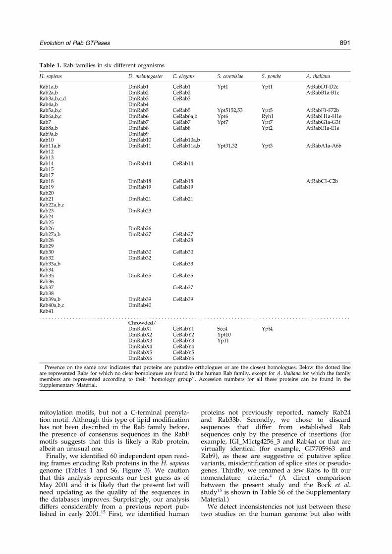
Table 1. Rab families in six different organisms
H. sapiens D. melanogaster C. elegans S. cerevisiae S. pombe A. thaliana
Rab1a,b DmRab1 CeRab1 Ypt1 Ypt1 AtRabD1-D2cRab2a,b DmRab2 CeRab2 AtRabB1a-B1cRab3a,b,c,d DmRab3 CeRab3Rab4a,b DmRab4Rab5a,b,c DmRab5 CeRab5 Ypt5152,53 Ypt5 AtRabF1-F72bRab6a,b,c DmRab6 CeRab6a,b Ypt6 Ryh1 AtRabH1a-H1eRab7 DmRab7 CeRab7 Ypt7 Ypt7 AtRabG1a-G3fRab8a,b DmRab8 CeRab8 Ypt2 AtRabE1a-E1eRab9a,b DmRab9Rab10 DmRab10 CeRab10a,bRab11a,b DmRab11 CeRab11a,b Ypt31,32 Ypt3 AtRabA1a-A6bRab12Rab13Rab14 DmRab14 CeRab14Rab15Rab17Rab18 DmRab18 CeRab18 AtRabC1-C2bRab19 DmRab19 CeRab19Rab20Rab21 DmRab21 CeRab21Rab22a,b,cRab23 DmRab23Rab24Rab25Rab26 DmRab26Rab27a,b DmRab27 CeRab27Rab28 CeRab28Rab29Rab30 DmRab30 CeRab30Rab32 DmRab32Rab33a,b CeRab33Rab34Rab35 DmRab35 CeRab35Rab36Rab37 CeRab37Rab38Rab39a,b DmRab39 CeRab39Rab40a,b,c DmRab40Rab41. . . . . . . . . . . . . . . . . . . . . . . . . . . . . . . . . . . . . . . . . . . . . . . . . . . . . . . . . . . . . . . . . . . . . . . . . . . . . . . . . . . . . . . . . . . . . . . . . . . . . .
Chrowded/DmRabX1 CeRabY1 Sec4 Ypt4DmRabX2 CeRabY2 Ypt10DmRabX3 CeRabY3 Yp11DmRabX4 CeRabY4DmRabX5 CeRabY5DmRabX6 CeRabY6
Presence on the same row indicates that proteins are putative orthologues or are the closest homologues. Below the dotted lineare represented Rabs for which no clear bomologues are found in the human Rab family, except for A. thaliana for which the familymembers are represented according to their ``homology group''. Accession numbers for all these proteins can be found in theSupplementary Material.
Evolution of Rab GTPases 891
mitoylation motifs, but not a C-terminal prenyla-tion motif. Although this type of lipid modi®cationhas not been described in the Rab family before,the presence of consensus sequences in the RabFmotifs suggests that this is likely a Rab protein,albeit an unusual one.
Finally, we identi®ed 60 independent open read-ing frames encoding Rab proteins in the H. sapiensgenome (Tables 1 and S6, Figure 3). We cautionthat this analysis represents our best guess as ofMay 2001 and it is likely that the present list willneed updating as the quality of the sequences inthe databases improves. Surprisingly, our analysisdiffers considerably from a previous report pub-lished in early 2001.15 First, we identi®ed human
proteins not previously reported, namely Rab24and Rab33b. Secondly, we chose to discardsequences that differ from established Rabsequences only by the presence of insertions (forexample, IGI_M1ctg4256_3 and Rab4a) or that arevirtually identical (for example, GI7705963 andRab9), as these are suggestive of putative splicevariants, misidenti®cation of splice sites or pseudo-genes. Thirdly, we renamed a few Rabs to ®t ournomenclature criteria.4 (A direct comparisonbetween the present study and the Bock et al.study15 is shown in Table S6 of the SupplementaryMaterial.)
We detect inconsistencies not just between thesetwo studies on the human genome but also with
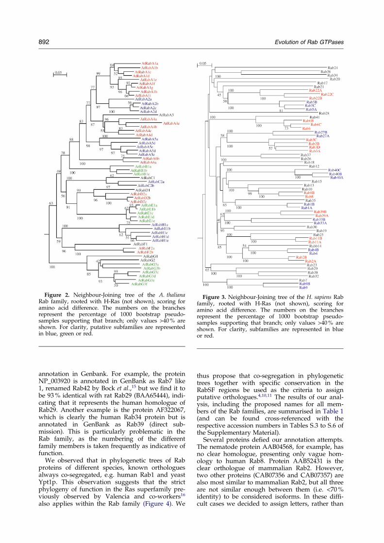
Figure 2. Neighbour-Joining tree of the A. thalianaRab family, rooted with H-Ras (not shown), scoring foramino acid difference. The numbers on the branchesrepresent the percentage of 1000 bootstrap pseudo-samples supporting that branch; only values >40 % areshown. For clarity, putative subfamilies are representedin blue, green or red.
Figure 3. Neighbour-Joining tree of the H. sapiens Rabfamily, rooted with H-Ras (not shown), scoring foramino acid difference. The numbers on the branchesrepresent the percentage of 1000 bootstrap pseudo-samples supporting that branch; only values >40 % areshown. For clarity, subfamilies are represented in blueor red.
892 Evolution of Rab GTPases
annotation in Genbank. For example, the proteinNP_003920 is annotated in GenBank as Rab7 like1, renamed Rab42 by Bock et al.,15 but we ®nd it tobe 93 % identical with rat Rab29 (BAA65444), indi-cating that it represents the human homologue ofRab29. Another example is the protein AF322067,which is clearly the human Rab34 protein but isannotated in GenBank as Rab39 (direct sub-mission). This is particularly problematic in theRab family, as the numbering of the differentfamily members is taken frequently as indicative offunction.
We observed that in phylogenetic trees of Rabproteins of different species, known orthologuesalways co-segregated, e.g. human Rab1 and yeastYpt1p. This observation suggests that the strictphylogeny of function in the Ras superfamily pre-viously observed by Valencia and co-workers16
also applies within the Rab family (Figure 4). We
thus propose that co-segregation in phylogenetictrees together with speci®c conservation in theRabSF regions be used as the criteria to assignputative orthologues.4,10,11 The results of our anal-ysis, including the proposed names for all mem-bers of the Rab families, are summarised in Table 1(and can be found cross-referenced with therespective accession numbers in Tables S.3 to S.6 ofthe Supplementary Material).
Several proteins de®ed our annotation attempts.The nematode protein AAB04568, for example, hasno clear homologue, presenting only vague hom-ology to human Rab8. Protein AAB52431 is theclear orthologue of mammalian Rab2. However,two other proteins (CAB07356 and CAB07357) arealso most similar to mammalian Rab2, but all threeare not similar enough between them (i.e. <70 %identity) to be considered isoforms. In these dif®-cult cases we decided to assign letters, rather than
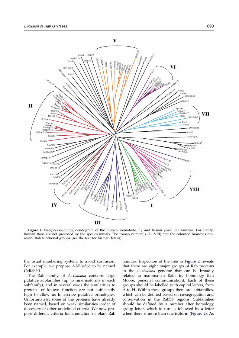
Figure 4. Neighbour-Joining dendogram of the human, nematode, ¯y and ®ssion yeast Rab families. For clarity,human Rabs are not preceded by the species initials. The roman numerals (I - VIII) and the coloured branches rep-resent Rab functional groups (see the text for further details).
Evolution of Rab GTPases 893
the usual numbering system, to avoid confusion.For example, we propose AAB04568 to be namedCeRabY1.
The Rab family of A. thaliana contains largeputative subfamilies (up to nine isoforms in eachsubfamily), and in several cases the similarities toproteins of known function are not suf®cientlyhigh to allow us to ascribe putative orthologies.Unfortunately, some of the proteins have alreadybeen named, based on weak similarities, order ofdiscovery or other unde®ned criteria. We now pro-pose different criteria for annotation of plant Rab
families. Inspection of the tree in Figure 2 revealsthat there are eight major groups of Rab proteinsin the A. thaliana genome that can be broadlyrelated to mammalian Rabs by homology (IanMoore, personal communication). Each of thesegroups should be labelled with capital letters, fromA to H. Within these groups there are subfamilies,which can be de®ned based on co-segregation andconservation in the RabSF regions. Subfamiliesshould be de®ned by a number after homologygroup letter, which in turn is followed by a letterwhen there is more than one isoform (Figure 2). As

894 Evolution of Rab GTPases
Rabs should be preceded by the species initials(e.g. AtRab), protein BAB09078 for example shouldbe called AtRabA6a, which should be read asArabidopsis thaliana Rab, group A, subtype 6,isoform a.
Rab families across evolution
Of all available Rab families, the ®ssion yeast(S. pombe) presents the smallest number of Rabproteins (Table 2). In this organism, all the Rabsproduce a detectable phenotype when the corre-sponding gene is disrupted, ranging from lethality(Ypt1p, Ypt2p, Ypt3p), temperature-sensitivegrowth (Rhy1p), fragmentation, size increase orreduction in number of vacuoles (Ypt7p andYpt4p,respectively) and vesicle accumulation (Ypt5p).13,17
Furthermore, all S. pombe Rabs, with the exceptionof Ypt4p, are conserved in all other organisms.
The evolutionarily divergent budding yeast(S. cerevisiae), on the other hand, displays anincreased number of Rab genes, many of whichproduce no phenotype when disrupted (Ypt31,Ypt32, Ypt51, Ypt52, Ypt53, Ypt10, Ypt11). Thisincreased number is in part due to the appearanceof subfamilies, i.e. functionally redundant isoforms(Ypt51, Ypt52,Ypt53 and Ypt31, Ypt32), but alsodue to the appearance of Rab proteins withoutclear functional or sequence homology to knownRab proteins in other organisms (Ypt10, Ypt11, andSec4). The genome of the budding yeast containsseveral duplicated chromosomal regions that couldunderlie the appearance of subfamilies, which isconsistent with the non-essentiality of the differentisoforms.18 ± 20
The human Rab family is the largest of all Rabfamilies studied here and re¯ects the increasedfamily size that accompanies multicellularity.Many of the Rab proteins have close homologuesand form subfamilies. If we consider, in a simplis-tic view, that each subfamily corresponds to onefunction, then 39 functions are required in mam-mals. A signi®cant number of these functions mayinvolve specialised, tissue-speci®c traf®cking path-ways, as many Rabs are not expressed ubiqui-tously.
The nematode and the ¯y Rab families contain anumber of Rabs intermediate between those ofyeasts and mammals. Interestingly, we observe theexistence of subfamilies in the nematode, but not
Table 2. Structure of the Rab family in six organisms
Species Cell number
S. pombe 1S. cerevisiae 1C. elegans �1 � 103
D. melanogaster �1 � 109
H. sapiens �1 � 1013
A. thaliana -
The listing of the family members and their accession numbers c
in the ¯y Rab family, consistent with the recentgenomic duplications observed in the nematode.21
Analysis of these genomes suggests that there isnot a linear increase in the number of Rab proteinswith the number of cells, as the nematode is madeof less than a thousand cells and the fruit ¯y con-tains more than three billion, and both organismhave a similar number of Rab proteins. This is trueeven if we discard putative redundant Rabs (iso-forms, i.e. Rabs forming subfamilies), assumingthey represent a single function or sub-class10 (26Rab functions in C. elegans compared with 29 inD. melanogaster).
The Rab family of A. thaliana is quite differentfrom the other Rab families considered here. Itexhibits large subfamilies, which can be groupedin terms of homology to a small set of animalRabs: Rab1, Rab2, Rab5, Rab6, Rab7, Rab8,Rab11 and Rab18. Surprisingly, the Rab11-likegroup in A. thaliana contains 26 proteins, but thesigni®cance of this fact is unclear. This Rabfamily organisation seems to be found in otherplant Rab families (Ian Moore, personal com-munication). It seems obvious to suggest thatRab proteins in plants followed an evolutionarypathway different from that taken by animals orfungi. The rationalisation of this observations,however, will have to wait for more functionalinformation on the different plant Rab proteins,as very little is known at present.
So what is the minimum number of Rab proteinsrequired in a eukaryotic cell? One possibility isseven, as this is the number of Rabs found in the®ssion yeast.13 Multicellularity and cellular special-isation may require more Rab proteins, possiblythose corresponding to the conserved Rab proteinsbetween the animal genomes considered here(Table 1). Such candidate Rabs include Rab3 andRab27, two examples of tissue-restricted Rabs withspecialised functions. The future availability ofmore sequenced genomes will allow a more accu-rate de®nition of the basic vesicular transport stepsrequired in a multicellular organism.
Conserved interactions with general regulatorsand effectors
We recently proposed in mammalian Rabs thatthe RabF motifs, clustering in and around the puta-tive switch regions, determine the interaction with
Rab number Subfamilies
7 011 229 329 0560 11557 12
an be found in the Supplementary Material.

Evolution of Rab GTPases 895
Rab-speci®c general regulators such as Rab escortproteins (REP) and GDP dissociation inhibitors(RabGDI). We sought to determine whether thismode of interaction with general regulators is con-served across evolution. To do so, we asked if thesame regions are conserved in the different Rabfamiles considered in this study. We calculatedpHMMs and generated model sequences for eachorganism Rab family. Upon alignment, weobserved that the same regions are conserved in allorganisms and that there is no organism-speci®cconsensus (Figure 5). Thus, the RabF motifs seemto be a feature conserved in evolution and mayindicate a conserved mode of interaction betweenRabs and general regulators. Recent work showingthat similar positions in yeast REP and RabGDImediate interactions with Rabs further supportsthis possibility.22
We worried that this observation could havebeen biased by the Rab identi®cation strategy fol-lowed, as the presence of RabF motifs was one ofthe criteria used. However, this was by no meansthe only criterion. We BLASTed each individual``putative'' Rab sequence against the non-redun-dant and organism-speci®c databases to con®rmthe similarity with other Rabs, and to look forfurther members of the Rab family. Also, wechecked every sequence against pHMM of othersmall GTPase families (Ras, Rho, Arf, Ran, Gem).
We proposed previously the existence of Rabsubfamily (RabSF) regions in mammalian Rabs,possibly involved in determining binding speci-®city to effectors. These RabSF regions are con-served across species.10 Consistently, Rabs fromevolutionarily distant organisms exhibit functionalcomplementation. For example, yeast Ypt1 del-etions or temperature sensitive mutations can becomplemented by small GTPases from Volvox car-teri, Chlamydomonas reinhardtii,23 Brassica napus24
and Mus musculus,25 and ypt6 null mutants can becomplemented by a small GTPase from A. thali-ana.26
Taken together, the absence of organism-speci®cconsensus, the conservation of RabF regions, thecross-species functional complementation and theconservation of RabSF regions make a strong argu-ment for a highly conserved mechanism of effectorand general regulator recognition, likely to be pre-sent at the point of divergence from other smallGTPases. It also suggests that this family origi-nated by a single divergence event and that theseinteraction mechanisms represent a major con-straint to the evolution of Rab proteins. Further-more, the conservation of this effector recognitionmechanism is indicative of effector conservation,an assumption that is supported by some recentevidence. While exchange factors for the Rabfamily form a very divergent class of proteins,there is a striking conservation of these proteinsacross evolution for known orthologues in the fewknown cases. For example, the mammalian andnematode exchange factors for Rab3 (Rab3GEPand Aex3) are highly conserved,27,28 and so are theexchange factors for Ypt51 and Rab5 (Vps9 andRabex-5).29,30
Prenylation and targeting motifs
Rab proteins contain one or two C-terminalcysteine residues that undergo post-translationalprenyl modi®cation.31 These cysteine residues arearranged in a variety of prenylation motifs. SomeRabs (such as HsRab8 and HsRab23) have a singlecysteine residue, fourth from the C terminus, some-times within a CAAX box (C, cys; A, aliphatic; X,any), a motif commonly observed in the Ras andRho families.32 However, most Rabs have twocysteine residues arranged in different double-cysteine prenylation motifs (e.g. XXCC, XCXC,CCXX, CCXXX, XCCX), both of which are modi-®ed by geranylgeranyl moieties. Unlike Ras andRho proteins, the prenylation motif in Rabs doesnot determine which prenyl transferase (and conse-quently which type of prenyl moiety) will modifythe C-terminal cysteine residues. All Rabs appearto be substrates for a unique enzyme, Rab Geranyl-
Figure 5. Partial alignment ofmodel sequences for each family,calculated from pHMM describingthe multiple sequence alignment ofthe complete Rab family in eachorganism. Positions in uppercaseoccur at p > 0.5. The PM/G motifsare highlighted in green, the RabFmotifs in red boxes or red charac-ters when the position is oftenoccupied by a conservative substi-tution, and the RabSF regions inyellow boxes. Black bars above thealignment represent the switchregions.

896 Evolution of Rab GTPases
geranyl Transferase (RGGT), following the bindingto REP, a general regulator of Rabs.
It is conceivable that the diversity of Rab preny-lation motifs arises from lack of functional con-strains other than those imposed by thegeranylgeranylation reaction mechanism. When wecompared the prenylation motifs in Rabs from allorganisms, we observed that the number ofcysteine residues is frequently conserved, andin many cases the topology of the prenylationmotif is retained (e.g. Ypt1(CC)! Rab1(CC),Ypt6(CXC)! Rab6(CXC)). Conservation of thesetopologies indicates constraints to evolution poss-ibly due to the requirement for carboxyl-methyl-ation, even though the functional signi®cance ofRab carboxyl-methylation affecting Rabs ending inCXC but not in CC is not understood.33,34 Further-more, we observed conservation of the number ofcysteine residues available for prenylation. Thissuggests that the number of prenyl groups (one ortwo) may be functionally important, possiblyrevealing the existence of two distinct membrane-association mechanisms or perhaps different mem-brane targeting strategies.
We observed some unusual Rab proteins in thisregard. For example, protein BAB32953 in A. thali-ana exhibits putative N-terminal myristoylationand palmitoylation motifs, but no C-terminal pre-nylation motifs, and these motifs are also found insimilar plant proteins, indicating a conserved fea-ture. This type of lipid modi®cation is novel withinthe Rab family. Another peculiar Rab protein isRab24, which is thought to be cytosolic.35 Theseunusual cases may represent recent evolutions ofthe Rab family where motifs not normally presentin this family are recruited to provide for newfunctions.
We caution that sequencing errors or artefactscomplicate annotation attempts. For example, sev-eral Rab proteins in D. melanogaster, C. elegans andA. thaliana do not exhibit C-terminal prenylationmotifs. However, their putative orthologues inother organisms do exhibit prenylation motifs,suggesting either bad quality sequencing orde®cient gene identi®cation algorithms.
Rab functional groups
Phylogenetic trees for all the sequences con-sidered here, reconstructed using the Neighbour-Joining method, revealed a clear phylogeny offunction, as opposed to a phylogeny of species(Figures 1-4). In other words, Rab proteins of simi-lar function in different organisms always co-segre-gate. As mentioned above, this represents anextension of the strict phylogeny of function pre-viously observed in the yeast Ras superfamily.16,36
Within the clades representing each putative Rab``function'', we observed a phylogeny of species,with proteins segregating according to organismprovenience (Figure 4).
Figures 1-3 show the trees calculated for eachorganism Rab family. We noted that some proteins
always co-segregate, even if they do not conformto the criteria de®ning isoforms.4 For example,members of the Rab1 sub-family always segregatewith Rab35. Based on the tree topology, we canidentify eight possible groups of co-segregatingproteins (Figure 4). The proteins in each of thesegroups are more similar at the amino acid levelthan any two random Rab proteins, suggesting ahigher-order organisation in the Rab family,above the subfamily level. This higher-order organ-isation may represent a shared ancestry betweenco-segregating proteins, functional relatedness orboth.
To test the hypothesis that this co-segregation of``unrelated'' proteins represents co-segregation offunctional properties, we sought to identify pat-terns of function and/or cellular localisation ineach group indicated in Figure 4. In group V,which includes the sub-families 5 and 22, we noteda pattern of subcellular localisation and possibly offunction. Rab5a has been studied extensively, itlocalises to early endosomes and clathrin-coatedvesicles, and regulates endosome budding andfusion.2,37 ± 41 Rab22a localises to endosomes andthe plasma membrane. Over-expression of Rab22aresults in the formation of abnormal endosomalstructures, which is suggestive of a role in endocy-tosis.42 Rab21 also segregates with the subfamilies5 and 22, albeit showing less sequence relatedness.Rab21 seems to be speci®c for polarised cells,where it localises to apical vesicles and shows par-tial localisation to an endosomal compartment,suggesting that it may be functionally related toRab5.43 Interestingly, other Rab proteins thatbroadly segregate with Rab5 isoforms also displayan endosomal localisation, namely Rab1744,45
and Rab20,46 but not Rab24, which is reportedlycytosolic.35
In group III we observed a pattern of subcellularlocalisation to secretory granules. Rab37 hasrecently been identi®ed and localised to secretorygranules in mast cells,47 and Rab26 has been loca-lised to secretory granules in pancreatic acinarcells.48 Rab27a was the subject of recent work byseveral groups and found to localise to secretorygranules (lytic granules) of cytotoxic T-lympho-cytes (CTL). Defects in the RAB27A gene in Griscel-li disease lead to haemophagocytic syndrome dueto loss of CTL activity.49,50 In melanocytes, Rab27associates with melanosomes, lysosome-like pig-ment-containing organelles destined forsecretion.51 ± 53 Rab27 appears to recruit myosinVato regulate the transport of melanosomes to thecell periphery prior to secretion.51 ± 53 Rab3 isoformsco-segregate with other members of this group inthe human and ¯y Rab tree, but not in the nema-tode Rab family tree, which is suggestive of a moredistant relationship. Members of the Rab3 subfam-ily have been implicated in regulated secretoryevents such as neurotransmitter release and insulinsecretion, and associate with secretory granulessuch as synaptic vesicles,54 ± 60 thus exhibiting a

Figure 6. Representation of the coding exons (greyboxes) of available ``functional group members'',mapped to a cartoon representing a generalised Rabprotein sequence, aligned by the conserved PM and Gmotifs (black boxes).
Evolution of Rab GTPases 897
similar type of cellular localisation as the othermembers of this group.
Group VII includes Rab7 and Rab9 isoforms.Both proteins show overlapping localisation to lateendosomes.61 ± 63
In group II, there is no apparent pattern ofsimilar subcellular localisation. For example, Rab2localises to the endoplasmic reticulum (ER) andGolgi,64 and Rab4 to endosomes and plasma mem-brane.65 Nevertheless, there may be a pattern offunctional similarity in this group. Rab11 and 25are proposed to be involved in recycling of pro-teins through the recycling endosome,66,67 andRab4 is proposed to be involved in endocytic recy-cling.68 No functional data are available for Rab14,but Rab2 has recently been proposed to regulate arecycling step in the retrograde Golgi-ER trans-port.69
In conclusion, we suggest that there is a recogni-sable pattern of subcellular localisation and poss-ibly of function, which supports the hypothesis ofa phylogeny of function applying between the sub-family and the family level.
We next questioned the origin of the sequencerelatedness, and functional/localisation similarityunderlying the groups shown in Figure 4. The sim-plest explanation is that the phylogeny of functionalso represents the evolutionary history of the Rabfamily, and that members of one given branchhave a shared ancestry. For example, Rab5 andRab22 would share the same ancestor, and thisancestral protein would also be the ancestor of thebudding yeast Ypt5.
This hypothesis is based on the phylogeneticreconstruction methods. However, these methodscan be biased to functional similarities in highlyconserved protein families, therefore, differentlines of evidence need to be obtained to substanti-ate it. Supporting this hypothesis is the observationthat there is differential conservation in discreteregions such as the PM/G and RabF motifs insome cases. For example, human group II proteinsRab25, Rab14, and the sub-families Rab11, Rab4and Rab2, all share identical IGVEF sequence atthe RabF1 motif, while human group III proteinsRab3 and Rab27 isoforms, Rab26 and Rab37 dis-play the conserved sequence VGIDF.
In order to complement this analysis, wesearched all available genomic structures of humanRab genes for an indication of shared ancestry. Ifproteins from one branch indeed arose from a com-mon ancestor, we expected to ®nd similarities inexon organisation. We retrieved available intron-exon boundaries in Rab genes from GenBank usingMapView. In many cases the sequencing data arestill of low quality, resulting in incomplete or nouseful information at all. Using a limited numberof genes, we observed many common features ingenomic structures within Rab functional groups.The intron-exon boundaries are either absolutelyconserved, or are close within a maximum of ®vecodons (Figure 6). Some Rabs within each groupshare highly similar genomic structures. For
example, Rab11 and Rab25 are almost identical,while Rab4 and Rab14 are distinguished by theappearance of an intron in Rab14 splitting in twothe ®fth exon in Rab4. Within subfamilies, genomicstructures tend to be highly conserved if not identi-cal (data not shown), a fact already noted byothers.70 ± 73
Based on these observations, we cannot discreditthe hypothesis that co-segregating Rab proteinsshare a common ancestry. Consequently, the Rabfamily trees not only represent a separation accord-ing to function and may re¯ect the evolutionaryhistory of this family of proteins. A more compre-hensive analysis of gene structures of Rab genes indifferent organisms is required to provide clearerevidence for shared ancestry.
Conclusions
We have identi®ed and annotated complete Rabfamilies in all eukaryotic organisms that had theirgenome substantially sequenced in May 2001. Wepropose here objective criteria for annotation ofanimal and plant Rab families on the basis of rec-ognition of putative orthologies.
Our analysis suggests that interactions betweenRab proteins and their general regulators andspeci®c effectors is conserved across evolution, asthe sequence determinants of this interactions(RabF motifs and RabSF regions) are conserved inall Rab families studied here.
We addressed the evolution of the Rab familyand observed a higher-order organisation withinthe Rab family corresponding to Rab proteins,which co-segregate in phylogenetic trees. Rabswithin these groups exhibit similar function and/or cellular localisation and related genomic struc-tures. It is tempting to speculate that early ineukaryotic evolution a minimum number of Rab
898 Evolution of Rab GTPases
proteins provided the ``ancestral'' Rab regulatoryactivities. Organism specialisation and multicellu-larity drove the multiplication of Rab family mem-bers from the initial set of ``ancestral Rabs''. Thesenovel Rabs appear to have maintained one or moreproperties that de®ned their ancestry, allowing usto group Rab proteins according to their ancestry,i.e. according to their putative ``ancestral Rab func-tion''. Thus, we propose that these related func-tions/cellular localisations form an intermediatelevel of classi®cation between family and subfam-ily, better described as ``Rab functional groups''.One interesting and testable possibility is that thisorganisation level could have predictive value tosuggest a function, localisation or interactions witheffectors of a given Rab protein. A possible Rab27effector, melanophilin was identi®ed recently andshown to be similar to Rabphilin-3a, a Rab3a effec-tor.74 This raises the possibility that members ofone functional group will interact with a family ofconserved effectors and suggest parallel evolutionbetween Rabs and their effectors. We expect morefunctional groups to be de®ned as more functionalinformation becomes available.
The minimal set of Rab proteins has beenequated with the essential yeast Rab proteins.13
These minimal Rab properties may representlocalisation to a given cellular compartment,interaction with classes of related effectors/regu-lators, speci®c GTPase characteristics or a combi-nation of these. A better understanding of thisissue is essential to fully understand the natureof the ``ancestral Rab functions'', the way thatthey evolved to provide regulators for increas-ingly complex organisms, and to ascribe generalfunctions to novel Rab sequences based solelyon their segregation pattern in phylogenetictrees. Furthermore, the understanding of theproperties shared by groups of co-segregatingRabs identi®ed here will allow informative corre-lations between group-speci®c sequence conser-vation and localisation/function of Rab proteins.
Materials and Methods
We retrieved protein sequences of known Rab familiesfrom GenBank. To identify the complete Rab families inH. sapiens, D. melanogaster, C. elegans and A. thaliana, wedownloaded the latest releases of the calculated openreading frames of each organism from the public data-bases. We then used a pro®le hidden Markov model(pHMM) calculated from the alignment of the mamma-lian Rab sequences presented previously4 to query eachdatabase using the software HMMER 2.1.1 found athmmer.wustl.edu.75
All the positive hits were then inspected visually,compared to pHMMs representing other small GTPasefamilies and individually BLASTed against the non-redundant database in GenBank to assert if they wereindeed Rab proteins, and to ensure that no sequence wasmissed from our analysis due to a possible bias createdby the query sequences.
Protein sequences were aligned using the CLUSTALW 1.8076 multiple sequence alignment program with
default parameters. Phylogenetic trees were recon-structed by the distance method of Neighbour-Joining,77
scoring for observed amino acid difference and werealways bootstrapped with 1000 replicates,78 using thesoftware Phylo_Win.79 Genomic structures wereobtained from GenBank, via the interface MapView.
Acknowledgements
We thank Ian Moore for helpful discussions concern-ing the plant Rab families and members of our labora-tory for stimulating ideas. This work was supported,in part, by the Wellcome Trust. J.P.-L. is a student of``Programa Gulbenkian de Doutoramento em Biologia eMedicina''.
References
1. Chavrier, P. & Goud, B. (1999). The role of ARF andRab GTPases in membrane transport. Curr. Opin.Cell Biol. 11, 466-475.
2. Zerial, M. & McBride, H. M. (2001). Rab proteins asmembrane organisers. Nature Rev. Mol. Cell Biol. 2,107-119.
3. Stenmark, H. & Olkkonen, V. M. (2001). The RabGTPase family. Genome Biol. 2, R3007.1-R3007.7.
4. Pereira-Leal, J. B. & Seabra, M. C. (2000). The mam-malian Rab family of small GTPases: de®nition offamily and subfamily sequence motifs suggests amechanism for functional speci®city in the Rassuperfamily. J. Mol. Biol. 301, 1077-1087.
5. Merithew, E., Hatherly, S., Dumas, J. J., Lawe, D.,Heller-Harrison, R. & Lambright, D. (2001). Struc-tural plasticity of an invariant hydrophobic triad inthe switch regions of Rab GTPases is a determinantof effector recognition. J. Biol. Chem. 276, 13982-13988.
6. Dumas, J. J., Zhu, Z., Connolly, J. L. & Lambright,D. G. (1999). Structural basis of activation and GTPhydrolysis in Rab proteins. Structure, 7, 413-423.
7. White, M. A., Nicolette, C., Minden, A., Polverino,A., Van Aeist, L., Marin, M. & Wigler, M. H. (1995).Multiple ras functions can contribute to mammaliancell transformation. Cell, 80, 533-541.
8. Vetter, I. R., Linnemann, T., Wohlgemuth, S., Geyer,M., Kalbitzer, H. R., Herrmann, C. & Wittinghofer,A. (1999). Structural and biochemical analysis ofRas-effector signaling via RalGDS. FEBS Letters, 451,175-180.
9. Nassar, N., Horn, G., Herrmann, C., Scherer, A.,McCormick, F. & Wittinghofer, A. (1995). The 2.2 AÊ
crystal structure of the Ras-binding domain of theserine/threonine kinase c-Raf1 in complex withRap1A and a GTP analogue. Nature, 375, 554-560.
10. Moore, I., Schell, J. & Palme, K. (1995). Subclass-speci®c sequence motifs identi®ed in Rab GTPases.Trends Biochem. Sci. 20, 10-12.
11. Ostermeier, C. & Brunger, A. T. (1999). Structuralbasis of Rab effector speci®city: crystal structure ofthe small G protein Rab3A complexed with theeffector domain of rabbphilin-3A. Cell, 96, 363-374.
12. Armstrong, J. (2000). How do Rab proteins functionin membrane traf®c? Int. J. Biochem. Cell Biol. 32,303-307.

Evolution of Rab GTPases 899
13. Lazar, T., Gotte, M. & Gallwitz, D. (1997). Vesiculartransport: how many Ypt/Rab-GTPases make aeukaryotic cell? Trends Biochem. Sci. 22, 468-472.
14. Armstrong, J. (2000). Membrane traf®c between gen-omes. Genome Biol. 1, 104.1-104.4.
15. Bock, J., Matern, H., Peden, A. & Scheller, R. H.(2001). A genomic prespective on membrane com-partment organization. Nature, 409, 839-841.
16. Valencia, A., Chardin, P., Wittinghofer, A. & Sander,C. (1991). The ras protein family: evolutionary treeand role of conserved amino acids. Biochemistry, 30,4637-4648.
17. Olkkonen, V. M. & Stenmark, H. (1997). Role of RabGTPases in membrane traf®c. Int. Rev. Cytol. 176,1-85.
18. Seoighe, C. & Wolfe, K. (1998). Extent of genomicrearrangement after genome duplication in yeast.Proc. Natl Acad. Sci. USA, 95, 4447-4452.
19. Wolfe, K. & Shields, D. (1997). Molecular evidencefor an ancient duplication of the entire yeast gen-ome. Nature, 387, 708-713.
20. Achaz, G., Coissac, E., Viari, A. & Netter, P. (2000).Analysis of intrachromosomal duplications in yeastSaccharomyces cerevisiae: a possible model for theirorigin. Mol. Biol. Evol. 17, 1268-1275.
21. Friedman, R. & Hughes, A. (2001). Gene duplicationand the structure of the eukaryotic genomes. GenomeRes. 11, 373-381.
22. Alory, C. & Balch, W. E. (2000). Molecular basis forRab prenylation. J. Cell Biol. 150, 89-103.
23. Fabry, S., Steigerwald, R., Berknlau, C., Dietmaier,W. & Schmidt, R. A. (1995). Structure-function anal-ysis of small G proteins from volvox and Chlamido-monas by complementation of Saccharomycescerevisiae YPT/SEC mutations. Mol. Gen. Genet. 247,265-274.
24. Palme, K., Diefenthal, T. & Moore, I. (1993). The yptgene family from maize and Arabidopsis: structuraland functional analysis. J. Exp. Bot. 44, 183-195.
25. Haubruck, H., Prange, R., Vorgias, C. & Gallwitz, D.(1989). The ras-related mouse ypt1 protein can func-tionally replace the YPT1 gene product in yeast.EMBO J. 8, 1427-1432.
26. Bednarek, S., Reynolds, T., Schroeder, M.,Grabowski, R., Hengst, L., Gallwitz, D. & Raikhel,N. (1994). A small GTP-binding protein fromArabidopsis thaliana functionally complements theyeast ypt6 null mutant. Plant Physiol. 104, 591-596.
27. Iwasaki, K., Staunton, J., Saifee, O., Nonet, M. &Thomas, J. H. (1997). aex-3 encodes a novel regula-tor of presynaptic activity in C. elegans. Neuron, 18,613-622.
28. Wada, M., Nakanishi, H., Satoh, A., Hirano, H.,Obaishi, H., Matsuura, Y. & Takai, Y. (1997). Iso-lation and characterization of a GDP/GTP exchangeprotein speci®c for the Rab3 subfamily small Gproteins. J. Biol. Chem. 272, 3875-3878.
29. Hama, H., Tall, G. G. & Horazdovsky, B. F. (1999).Vps9p is a guanine nucleotide exchange factorinvolved in vesicle-mediated vacuolar protein trans-port. J. Biol. Chem. 274, 15284-15291.
30. Horiuchi, H., Lippe, R., McBride, H. M., Rubino, M.,Woodman, P., Stenmark, H. et al. (1997). A novelRab5 GDP/GTP exchange factor complexed toRabaptin-5 links nucleotide exchange to effectorrecruitment and function. Cell, 90, 1149-1159.
31. Seabra, M. C. (2000). Biochemistry of Rab geranyl-geranyl transferase. In The Enzymes (Tamanoi, F. &
Sigman, D. S., eds), vol. 21, pp. 131-154, AcademicPress, San Diego.
32. Casey, P. J. & Seabra, M. C. (1996). Protein prenyl-transferases. J. Biol. Chem. 271, 5289-5292.
33. Smeland, T. E., Seabra, M. C., Goldstein, J. L. &Brown, M. S. (1994). Geranylgeranylated Rab pro-teins terminating in Cys-Ala-Cys, but not Cys-Cys,are carboxyl-methylated by bovine brain membranesin vitro. Proc. Natl Acad. Sci. USA, 91, 10712-10716.
34. Bergo, M. O., Gordon, K., Ambroziak, P., Otto, J. C.,Casey, P. J.,Gomes, A. Q. et al. (2001). Isoprenylcys-teine carboxyl methyltransferase de®ciency in mice.J. Biol. Chem. 276, 5841-5845.
35. Erdman, R. A., Shellenberger, K. E., Overmeyer, J. H.& Maltese, W. A. (2000). Rab24 is an atypical mem-ber of the Rab GTPase family. De®cient GTPaseactivity, GDP dissociation inhibitor interaction, andprenylation of Rab24 expressed in cultured cells.J. Biol. Chem. 275, 3848-3856.
36. Garcia-Ranea, J. A. & Valencia, A. (1998). Distri-bution and functional diversi®cation of the rassuperfamily in Saccharomyces cerevisiae. FEBS Letters,434, 219-225.
37. Bucci, C., Parton, R. G., Mather, I. H., Stunnenberg,H., Simons, K., Ho¯ack, B. & Zerial, M. (1992). Thesmall GTPase rab5 functions as a regulatory factorin the early endocytic pathway. Cell, 70, 715-728.
38. Barbieri, M. A., Hoffenberg, S., Roberts, R.,Mukhopadhyay, A., Pomrehn, A., Dickey, B. F. &Stahl, P. D. (1998). Evidence for a symmetricalrequirement for Rab5-GTP in in vitro endosome-endosome fusion. J. Biol. Chem. 273, 25850-25855.
39. Roberts, R. L., Barbieri, M. A., Pryse, K. M., Chua,M., Morisaki, J. H. & Stahl, P. D. (1999). Endosomefusion in living cells overexpressing GFP-rab5. J. CellSci. 112, 3667-3675.
40. Rybin, V., Ullrich, O., Rubino, M., Alexandrov, K.,Simon, I., Seabra, M. C. et al. (1996). GTPase activityof Rab5 acts as a timer for endocytic membranefusion. Nature, 383, 266-269.
41. Stenmark, H., Parton, R., Steele-Mortimer, O.,Lutcke, A., Gruenberg, J. & Zerial, M. (1994). Inhi-bition of Rab5 GTPase activity stimulates membranefusion in endocytosis. EMBO J. 13, 1287-1296.
42. Olkkonen, V. M., Dupree, P., Killisch, I., Lutcke, A.,Zerial, M. & Simons, K. (1993). Molecular cloningand subcellular localization of three GTP-bindingproteins of the rab subfamily. J. Cell Sci. 106, 1249-1261.
43. Opdam, F. J., Kamps, G., Croes, H. J., vanBokhoven, H., Ginsel, L. A. & Fransen, J. A. (2000).Expression of Rab small GTPases in epithelialCaco-2 cells: Rab21 is apically located GTP-bindingprotein in polarised intestinal epithelial cells. Eur. J.Cell Biol. 79, 308-318.
44. Hunziker, W. & Peters, P. J. (1998). Rab17 localizesto recycling endosomes and regulates receptor-mediated transcytosis in epithelial cells. J. Biol.Chem. 273, 15734-15741.
45. Zacchi, P., Stenmark, H., Parton, R. G., Orioli, D.,Lim, F., Giner, A. et al. (1998). Rab17 regulates mem-brane traf®cking through apical recycling endo-somes in polarized epithelial cells. J. Cell Biol. 140,1039-1053.
46. Lutcke, A., Parton, R., Murphy, C., Olkkonen, V. M.,Dupree, P., Valencia, A. et al. (1994). Cloning andsubcellular localization of novel rab proteins revealspolarized and cell type speci®c expression. J. CellSci. 107, 3437-3438.
900 Evolution of Rab GTPases
47. Masuda, E. S., Luo, Y., Young, C., Shen, M., Rossi,A. B., Huang, B. C. et al. (2000). Rab37 is a novelmast cell speci®c GTPase localized to secretory gran-ules. FEBS Letters, 470, 61-64.
48. Yoshie, S., Imai, A., Nashida, T. & Shimomura, H.(2000). Expression, characterization, and localizationof Rab26, a low molecular weight GTP-binding pro-tein, in the rat parotid gland. Histochem. Cell Biol.113, 259-263.
49. Haddad, E., Wu, X., Hammer, J., III & Henkart, P.(2001). Defective granule exocytosis in Rab27a-de®cient lymphocytes from ashen mice. J. Cell Biol.152, 835-842.
50. Stinchcombe, J. C., Barral, D. C., Mules, E. H.,Booth, S., Hume, A. N., Machesky, L. M. et al.(2001). Rab27a is required for regulated secretion incytotoxic t lymphocytes. J. Cell Biol. 152, 825-834.
51. Hume, A. N., Collinson, L. M., Rapak, A., Gomes,A. Q., Hopkins, C. R. & Seabra, M. C. (2001).Rab27a regulates the peripheral distribution of mela-nosomes in melanocytes. J. Cell Biol. 152, 795-808.
52. Wu, X., Rao, K., Bowers, M., Copeland, N., Jenkins,N. & Hammer, J. (2001). Rab27a enables myosinVa-dependent melanosomes capture. J. Cell Sci. 114,1091-1100.
53. Bahadoran, P., Aberdam, R., Mantoux, F., Busca, R.,Bille, K., Yalman, N. et al. (2001). Rab27a. A key tomelanosome transport in human melanocytes. J. CellBiol. 152, 843-850.
54. Iezzi, M., Escher, G., Meda, P., Charollais, A.,Baldini, G., Darchen, F. et al. (1999). Subcellular dis-tribution and function of Rab3A, B, C, and D iso-forms in insulin-secreting cells. Mol. Endocrinol. 13,202-212.
55. Tuvim, M. J., Adachi, R., Chocano, J. F., Moore,R. H., Lampert, R. M., Zera, E., Romero, E. et al.(1999). Rab3D, a small GTPase, is localized on mastcell secretory granules and translocates to theplasma membrane upon exocytosis. Am. J. Respir.Cell Mol. Biol. 20, 79-89.
56. Al-Matubsi, H. Y., Breed, W., Jenkin, G. &Fairclough, R. J. (1999). Co-localization of Rab3Band oxytocin to electron dense granules of the sheepcorpus luteum during the estrous cycle. Anat. Rec.254, 214-221.
57. Raffaniello, R. D., Lin, J., Schwimmer, R. & Ojakian,G. K. (1999). Expression and localization of Rab3Din rat parotid gland. Biochim. Biophys. Acta, 1450,352-363.
58. Fischer, V. M., Mignery, G. A., Baumert, M., Perin,M. S., Hanson, T. J., Burger, P. M. (1990). rab3 is asmall GTP-binding protein exclusively localized tosynaptic vesicles. Proc. Natl Acad. Sci. USA, 87, 1988-1992.
59. Darchen, F., Zahraoui, A., Hammel, F., Monteils,M. P., Tavitian, A. & Scherman, D. (1990). Associ-ation of the GTP-binding protein Rab3A with bovineadrenal chromaf®n granules. Proc. Natl Acad. Sci.USA, 87, 5692-5696.
60. Oishi, H., Sasaki, T., Nagano, F., Ikeda, W., Ohya,T., Wada, M. et al. (1998). Localization of the Rab3small G protein regulators in nerve terminals andtheir involvement in Ca2 �-dependent exocytosis.J. Biol. Chem. 273, 34580-34585.
61. Feng, Y., Press, B. & Wandinger-Ness, A. (1995).Rab7: an important regulator of late endocytic mem-brane traf®c. J. Cell Biol. 131, 1435-1452.
62. Lombardi, D., Soldati, T., Riederer, M. A., Goda, Y.,Zerial, M. & Pfeffer, S. R. (1993). Rab9 functions in
transport between late endosomes and the transGolgi network. EMBO J. 12, 677-682.
63. Meresse, S., Gorvel, J. P. & Chavrier, P. (1995). TheRab7 GTPase resides on a vesicular compartmentconnected to lysosomes. J. Cell Sci. 108, 3349-3358.
64. Tisdale, E. J., Bourne, J. R., Khosravi-Far, R., Der,C. J. & Balch, W. E. (1992). GTP-binding mutants ofrab1 and rab2 are potent inhibitors of vesiculartransport from the endoplasmic reticulum to theGolgi complex. J. Cell Biol. 119, 749-761.
65. Sonnichsen, B., De Renzis, S., Nielsen, E., Rietdorf, J.& Zerial, M. (2000). Distinct membrane domains onendosomes in the recycling pathway visualized bymulticolor imaging of Rab4, Rab5, and Rab11. J. CellBiol. 149, 901-914.
66. Casanova, J. E., Wang, X., Kumar, R., Bhartur, S. G.,Navarre, J., Woodrum, J. E. et al. (1999). Associationof Rab25 and Rab11a with the apical recycling sys-tem of polarized Madin-Darby canine kidney cells.Mol. Biol. Cell, 10, 47-61.
67. Ullrich, O., Reinsch, S., Urbe, S., Zerial, M. &Parton, R. G. (1996). Rab11 regulates recyclingthrough the pericentriolar recycling endosome. J. CellBiol. 135, 913-924.
68. van der Sluijs, P., Hull, M., Webster, P., Male, P.,Goud, B. & Mellman, I. (1992). The small GTP-bind-ing protein rab4 controls an early sorting event onthe endocytic pathway. Cell, 70, 729-740.
69. Tisdale, E. J. (1999). A Rab2 mutant with impairedGTPase activity stimulates vesicle formation frompre-Golgi intermediates. Mol. Biol. Cell, 10, 1837-1849.
70. Adachi, R., Nigam, R., Tuvim, M. J., DeMayo, F. &Dickey, B. F. (2000). Genomic organization, chromo-somal localization, and expression of the murineRAB3D gene. Biochem. Biophys. Res. Commun. 273,877-883.
71. Bhartur, S. G., Calhoun, B. C., Woodrum, J.,Kurkjian, J., Iyer, S., Lai, F. & Goldenring, J. R.(2000). Genomic structure of murine Rab11 familymembers. Biochem. Biophys. Res. Commun. 269, 611-617.
72. Ramalho, J. S., Tolmachova, T., Hume, A.,McGuigan, A., Gregory-Evans, C., Huxley, C. &Seabra, M. C. (2001). Chromosomal mapping, genestructure and characterization of the human andmurine RAB27B gene. BMC Genet. 2, 2.
73. Tolmachova, T., Ramalho, J. S., Anant, J. S., Schultz,R. A., Huxley, C. M. & Seabra, M. C. (1999). Clon-ing, mapping and characterization of the humanRAB27A gene. Gene, 239, 109-116.
74. Matesic, L. E., Yip, R., Reuss, A. E., Swing, D. A.,O'Sullivan, T. N., Fletcher, C. F. et al. (2001).Mutations in Mlph, encoding a member of the Rabeffector family, cause the melanosome transportdefects observed in leaden mice. Proc. Natl Acad. Sci.USA, 98, 10238-10243.
75. Eddy, S. R. (1998). Pro®le hidden Markov models.Bioinformatics, 14, 755-763.
76. Thompson, J. D., Higgins, D. G. & Gibson, T. J.(1994). CLUSTAL W: improving the sensitivity ofprogressive multiple sequence alignment throughsequence weighting, position-speci®c gap penaltiesand weight matrix choice. Nucl. Acids Res. 22, 4673-4680.
77. Saitou, N. & Nei, M. (1987). The neighbour-joiningmethod: a new method to reconstruct phylogenetictrees. Mol. Biol. Evol. 4, 406-425.
Evolution of Rab GTPases 901
78. Felsenstein, J. (1985). Con®dence limits on phylo-genies: an approach using the bootstrap. Evolution,39, 783-791.
79. Galtier, N., Gouy, M. & Gautier, C. (1996). SeaViewand Phylo_Win, two graphic tools for sequencealignment and molecular phylogeny. Comput. Applic.Biosci. 12, 543-548.
Edited by J. Thornton
(Received 11 June 2001; received in revised form 4September 2001; accepted 5 September 2001)
http://www.academicpress.com/jmb
Supplementary Material comprising six Tables isavailable on IDEAL