evod/vo: the origins of bmp signalling in the...
TRANSCRIPT

The evolutionary conservation of genes involved in patterning the primary body axes has revolutionized the fields of both developmental and evolutionary biol-ogy, and has spawned the field of evo–devo. One of the most fertile areas of evo–devo has been the analysis of neural induction, during which the embryonic ecto-derm is partitioned into neural and epidermal domains. Current studies have raised several important ques-tions, including whether a condensed central nervous system (CNS) arose only once or multiple times during evolution and whether patterning of the CNS by bone morphogenetic proteins (BMPs) in different branches of the phylogenetic tree reflects a conserved ancestral mechanism (homology) or parallel evolution (conver-gent evolution). Answers to these questions are essential for reconstructing the nature of the nervous system in the common ancestor of bilateral organisms and, more generally, for translating molecular similarities between existing organisms to morphological homologies in the body plans of ancestors. Several technological innova-tions have aided recent experimental advances, such as imaging multiple gene expression patterns and compar-ing genome sequences and homologous gene expression patterns in an ever increasing array of organisms from different branches of evolution. In this Review, we dis-cuss the key issues pertaining to the role of BMP signal-ling in establishing pattern within the developing CNS and its evolutionary implications.
Recent phylogenetic analyses suggest that bilaterian organisms can be subdivided into three main branches: the Ecdysozoa, moulting invertebrates such as arthro-pods and nematodes; the Lophotrochozoa, non-moulting
invertebrates such as molluscs and annelid worms; and Deuterostomata, including chordates, hemichordates, and echinoderms1. Among the 37 extant phyla populating these three branches there are many examples of organ-isms with condensed ventral nerve chords that are par-titioned into two or three primary tracts2. This recurring theme argues for evolutionary conservation of the trunk CNS across these lineages. However, there are also scat-tered examples of bilaterian species with a diffuse CNS, which raises the question of which of the two forms is the most derived or the most primitive state, and when the two different modes of nervous system patterning are likely to have arisen. In principle, it should be possible to distinguish between these alternatives by comparing fossil records for various lineages. However, in practice, owing to incomplete fossil data, these questions demand comparative genetic and molecular studies.
Two of the best studied examples of comparative molecular anatomy are the mutually exclusive expres-sion of BMPs and their antagonists in the epidermal and neural ectoderm, respectively, and the conserved relative expression domains of neural identity genes that subse-quently subdivide the nervous system along the dorsal–ventral (D/V) axis. There are striking parallels in how the BMP signalling pathway controls D/V patterning and establishes the embryonic neural territory in a vari-ety of organisms3–5. BMP signalling is also involved in the aforementioned subdivision of the neuroectoderm of vertebrates and invertebrates6, and similar expression and regulatory relationships have been found in a primitive polychaete annelid7. These parallels strongly suggest that a conserved genetic system controls neural induction and
Section of Cell and Developmental Biology, University of California, San Diego, La Jolla, California 92093‑0349, USA.e‑mails: [email protected]; [email protected]:10.1038/nrg2417Published online 5 August 2008
HemichordatesFrom the Greek hemi (half) and from the Latin chorda (cord). Marine worm-like animals that can be slow burrowers (acorn worms, for example) or sessile (pterobranchs, for example). The Hemichordata phylum is closely related to Echinodermata and Chordata phyla; together they constitute the Deuterostomata superphylum.
EchinodermsFrom the Greek ekhinos (spiny) and derma (skin). Marine deuterostome animals that include sea stars, sea cucumbers and sea urchins. They possess bilateral symmetry during larval stages, but in adult life they become radially symmetrical.
EvoD/Vo: the origins of BMP signalling in the neuroectodermClaudia Mieko Mizutani and Ethan Bier
Abstract | The genetic systems controlling body axis formation trace back as far as the ancestor of diploblasts (corals, hydra, and jellyfish) and triploblasts (bilaterians). Comparative molecular studies, often referred to as evo–devo, provide powerful tools for elucidating the origins of mechanisms for establishing the dorsal–ventral and anterior–posterior axes in bilaterians and reveal differences in the evolutionary pressures acting upon tissue patterning. In this Review, we focus on the origins of nervous system patterning and discuss recent comparative genetic studies; these indicate the existence of an ancient molecular mechanism underlying nervous system organization that was probably already present in the bilaterian ancestor.
Nature Reviews Genetics | AOP, published online 5 August 2008; doi:10.1038/nrg2417 R E V I E W S
NATuRE REVIEwS | genetics ADVANCE ONLINE PuBLICATION | 1

patterning, and also that the role of BMP4 signalling in this process (and the role of the Drosophila melanogaster homologue Decapentaplegic, DPP) traces back to a common bilaterian ancestor with an organized nervous system. Given these recent findings, we argue that the diffuse nervous systems found in a broad array of organ-isms probably are derived secondary simplifications, rather than the ancestral bilaterian state.
In this Review, we first discuss the evidence for a conserved genetic system controlling neural induc-tion and its subsequent role in neural patterning. we also compare neural patterning in different organisms, including hemichordates. These organisms have a dif-fuse nerve net, which we hypothesize secondarily lost the ability to pattern the neuroectoderm in response to BMPs. Finally, we propose a hypothesis that might help reconcile the various findings and that provides a start-ing point for comparisons of the regulatory systems that control patterning along the anterior–posterior (A/P) and D/V axes.
St-Hilaire’s hypothesisIn 1822, the prominent comparative anatomist Geoffrey St-Hilaire noted that the general organization of the body plan was virtually identical in vertebrates and invertebrates, except that they were inverted along the D/V axis with respect to each other8 (BOX 1). The verte-brate heart, for instance, lies along the ventral side, but is located dorsally in invertebrates. Similarly, the nerv-ous system lies dorsally in vertebrates, but ventrally in invertebrates (FIG. 1). As would be expected if the D/V axis was inverted in vertebrates relative to invertebrates, BMP4 and DPP are expressed in inverse patterns in these organisms where they act to define the epider-mal ectoderm. Conversely, secreted BMP antagonists, such as Short gastrulation (SOG) in D. melanogaster and the vertebrate homologue chordin (CHD), are expressed in patterns complementary to DPP (in D. melanogaster) and BMP4 (in vertebrates) and function to promote genesis of the nervous system9–11. Although St-Hilaire’s proposal was initially rejected, it was
Nature Reviews | Genetics
Arthropod Vertebrate
• Whole-body inversion• Mouth moved ventrally or a new mouth is formed on the ventral side
a b
• Trunk rotated 180° relative to the head
• Head nervous system only in ancestor• Independent evolution of trunk CNS on opposite sides in arthropods versus vertebrates
c
d
Box 1 | Inversion of the dorsal–ventral axis in vertebrates?
A unique feature of the nerve chord in vertebrates is that it forms dorsally, whereas it is located ventrally in invertebrates. As the heart and direction of fluid flow are also reversed in vertebrates versus invertebrates, Geoffrey St-Hilaire suggested that the dorsal–ventral (D/V) axes might have been inverted during evolution. The reversed relative expression patterns of components of the bone morphogenetic protein (BMP) signalling system, such as BMPs, Short gastrulation (SOG), chordin (CHD), Tolloid (TLD), xolloid (XLD) and Twisted gastrulation (TSG), are consistent with an early developmental inversion of the D/V axis. An important evolutionary implication of the D/V inversion hypothesis is that both the vertebrate and invertebrate lineages are likely to have inherited a central nervous system (CNS) from a common ancestor, which was condensed to one side of the body and was subsequently inverted dorsoventrally in the vertebrate lineage (see a,b: blue indicates the ventral nervous system defective (vnd) domain in the fly and NK transcription factor related (Nkx) in vertebrates; green indicates intermediate neuroblasts defective (ind) domain in the fly and genomic screen homeobox (Gsh) in vertebrates; red indicates muscle segment homeobox (msh, also known as Drop) domain in the fly and MSH homeobox (Msx) in vertebrates).
It should be noted that the assignment ‘ventral’ and ‘dorsal’ is somewhat arbitrary, as the ventral side of both vertebrates and invertebrates is defined by the location of the mouth orifice. Otherwise, one could imagine that D/V inversion was a trivial consequence of a vertebrate ancestor just evolving to swim upside down. A necessary element of the D/V inversion hypothesis is that there was a concomitant shift of the mouth to the opposite side in a vertebrate ancestor, which might have involved the formation of a new oral opening ventrally87 (b). Another possibility to account for the relative organization of the trunk versus mouth is that there was a 180° rotation of the trunk relative to the head (c). This hypothesis has the advantage of providing an explanation for an otherwise puzzling feature of the vertebrate nervous systems, which is that many sensory systems decussate (that is, project to the opposite side of the brain). An alternative hypothesis for the relative location of the mouth is that the nervous system evolved independently in vertebrate and invertebrate lineages from a more primitive and diffuse net of nerve cells76,88–90, and only later became centralized on opposite sides, either dorsally in vertebrates or ventrally in invertebrates (d). As mentioned above, based on the known similarities in gene expression patterns across phylogenetic lineages, we argue that the common bilaterian ancestor had an organized centralized nerve chord, thus favouring a D/V inversion model. The figure is modified, with permission, from REF. 91 (2008) Elsevier Science.
R E V I E W S
2 | ADVANCE ONLINE PuBLICATION www.nature.com/reviews/genetics

resurrected almost 200 years later, in the light of con-served gene expression patterns and regulatory rela-tionships between DPP and SOG, BMP4 and CHD, and other pathway components during neural induction in flies and vertebrates12–17.
An important element of the D/V inversion hypoth-esis is to show that the similar final patterns of neural gene expression in vertebrates and invertebrates are not just the result of fortuitous evolutionary conver-gence, but that this developmental process is broadly shared across different organisms. As we discuss below, there is a growing body of evidence supporting the hypothesis that the developmental process is conserved across a range of organisms and that the BMP signal-ling pathway, which is responsible for organizing gene expression in the neuroectoderm, might once have controlled patterning along the entire D/V axis of the ectoderm.
The conserved BMP signalling pathwayThe BMP signalling pathway consists of both intracellu-lar and extracellular components that are conserved and that exert similar functions from fruitflies to humans18. Extracellular factors interact to establish the availability
of ligand in graded patterns throughout tissues, whereas the intracellular signalling transduction components regulate target gene expression (BOX 2).
In the early embryo, the first step in neural devel-opment is subdivision of the ectoderm into neural and epidermal domains (FIG. 2a). This process is typically referred to as neural induction4. Subsequently, the neu-ral ectoderm is partitioned into three non-overlapping D/V stripes that express different homeobox transcrip-tion factors known as neural identity genes19 (FIG. 2b,c). The resulting tripartite neuroectoderm gives rise to three primary rows of neuroblasts that differentiate into distinct neuronal progeny. These two steps of neural development seem to be highly conserved in all three bilaterian branches and depend on two distinct modes of BMP signalling.
All-or-none role of BMPs in neural inductionDuring the early phase of neural induction, the primary role of BMP signalling is to divert cells in epidermal regions from adopting the neural fate. This all-or-none role of BMP signalling is achieved by repressing neural gene expression and activating ectodermal genes9–11. In the neural ectoderm, BMP antagonists such as SOG
Nature Reviews | Genetics
Epidermis
Neuroepithelium
Dorsal
Ventral
Dorsal
Ventral
Mesoderm
Ventral midline
a Vertebrates
b Flies
Ventral midline
Neuroblasts
Denticles
EpidermisDorsalepidermis
Epidermis
Neuraltube
Notochord
Somites
Notochord
Ventral midlineof neural tubeMesoderm
Neuroectoderm
Dorsal midline of embryo
Non-neural ectoderm
Mesoderm
NeuroepitheliumVentralepithelium
Figure 1 | neurulation in flies and vertebrates. a | Cross-sectional diagram of neurulation in vertebrates. As this process proceeds, the mesoderm (red) becomes partitioned into the notochord and somites, and the original dorsal midline of the embryo becomes the ventral midline of the neural tube, thereby inverting the dorsal–ventral (D/V) orientation of cells in the neural tube with respect to the body axis. b | Cross-sectional diagram of the Drosophila melanogaster embryo as it gastrulates. The ventral mesoderm (red) invaginates, resulting in the joining of the two lateral neuroectodermal domains along the future ventral midline of the embryo. Following mesoderm invagination, neuroblasts delaminate from the neuroectoderm to form the central nervous system. This mechanism for generating neuroblasts preserves their D/V positions with respect to the overall body axis. Top and bottom panels on the right indicate the final positions of D/V domains of neural gene expression in the neural tube and the fly neuroectoderm. Blue indicates ventral nervous system defective (VND) in flies and NK2 transcription factor-related (NKX2.1) in vertebrates; green indicates intermediate neuroblasts defective (IND) in flies and genomic screen homeobox (GSH) and paired box 6 (PAX6) in vertebrates; red indicates muscle segment homeobox (MSH) in flies and MSH homeobox (MSX) in vertebrates. The figure is modified, with permission, from REF. 91 (2008) Elsevier Science.
R E V I E W S
NATuRE REVIEwS | genetics ADVANCE ONLINE PuBLICATION | 3

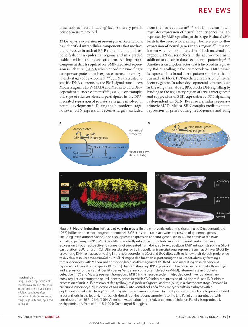
and CHD bind to BMPs and prevent them from gaining access to their receptors, thereby allowing these cells to adopt the default neural fate. It is clear that this process is highly conserved at the molecular level because SOG can act as a neural inducer in vertebrate embryos15,17,20 (FIG. 2d) and CHD can block BMP signalling in flies15,20,21.
BMPs accumulate to high levels in future epidermal regions, whereas BMP antagonists are either expressed by the cells of the neural ectoderm (for example, SOG in D. melanogaster) or they diffuse into the neural ecto-derm from the adjacent cells (such as CHD, which is produced by dorsal mesoderm cells in vertebrates)5,22. In flies, diffusion of SOG towards the dorsal region is thought to form a reciprocal BMP activity gradient in the epidermal ectoderm (BOX 3). An intricate extracel-lular system, which includes additional antagonists and proteases that degrade SOG and thereby free BMPs from inhibitory complexes (BOX 2), results in graded receptor activation5.
High level of BMP signalling in the dorsal epidermal ectoderm has two effects: it represses expression of all neural genes and activates expression of epidermal genes in a dose-dependent fashion (BOXES 2,3). A key gene that is activated by high levels of BMP signal-ling is dpp itself 23 (FIG. 2a). The combination of DPP diffusion and autoactivation can result in the invasive spread of DPP signalling into the neighbouring neur-oectoderm. However, this potentially invasive positive feedback cycle is blocked in the neuroectoderm by the orthologous BMP antagonists SOG and CHD in flies and vertebrates, respectively, as well as by noggin and DAN (differential screening-selected gene aberrative in neuroblastoma) family members in vertebrates16,23–26. Acting in parallel with extracellular antagonists, BMP signalling is also blocked at the transcriptional level in the D. melanogaster neuroectoderm by the repressor Brinker (BRK)27. By preventing high-level BMP sig-nalling from repressing the expression of neural genes,
Nature Reviews | Genetics
Cell membrane
MAD
SOG
p-MAD
Degradation byTLD protease
Medea I-MADBRK
BRK
SHN
Epidermal genes
Neural genes
Nucleus
TKV
PUT
TKV SAX
PUT
DPP / SCWDPP / DPP
Box 2 | The conserved BMP signalling pathway
In Drosophila melanogaster, there are six diffusible extracellular ligands belonging to the transforming growth factor-β (TGF-β) superfamily of growth factors, including the bone morphogenetic protein (BMP) ligands Decapentaplegic (DPP), Screw (SCW) and Glass-bottom-boat (GBB, not shown)92–94, whereas in vertebrates there are thirty TGF-β ligands, including BMP2 and BMP4, which are the orthologues of DPP (reviewed in REF. 18). BMP ligands are secreted by expressing cells in the form of homodimers (for example, DPP–DPP) or heterodimers (such as DPP–SCW), and activate specific combinations of tetrameric receptor complexes formed by type I and type II serine-threonine kinase receptors95. In the figure, the names of pathway components are given for D. melanogaster; the human orthologues are given here in parentheses. DPP dimers (BMP2 or BMP4) bind to heterotetrameric receptors consisting of the two Thick veins (TKV) (BMP receptor 1A, BMPR1A, or BMPR1B) type-I chains and two Punt (PUT) (activin receptor type II, ACTRII) type II chains. By contrast, DPP–SCW heterodimers signal through one TKV type I chain, one Saxophone (SAX) (activin receptor-like kinase 1, ALK1 and ALK2) type I chain, and two PUT type II chains95–98. The type II chains of the activated receptor phosphorylate the type I chains, which in turn phosphorylate and activate Mothers against DPP (MAD) (SMAD1, SMAD5, SMAD8 and SMAD9) to generate phospho-MAD (p-MAD)99,100. p-MAD forms a complex with the cooperating-MAD (co-MAD) Medea (SMAD4), and enters the nucleus to alter gene expression. Formation of the p-MAD–Medea complex can be inhibited by inhibitory-MAD (I-MAD) such as Daughters against DPP (DAD, not shown) (SMAD6 and SMAD7), which negatively regulate signalling by either targeting active receptors or at the level of transcription18. Once in the nucleus, high levels of the p-MAD–Medea complex activate expression of epidermal genes dorsally. p-MAD and Medea can also form a trimeric complex with Schnurri (SHN), which efficiently represses expression of neural genes in the lateral neuroectoderm where DPP levels are much lower than dorsally. The levels of MAD and Medea that are required to repress target genes are much less than those required to activate epidermal genes. Another transcription factor that regulates BMP signalling is Brinker (BRK), which binds to sequences that overlap those of p-MAD and blocks the response to BMP signalling27,101–103.
In addition to the intracellular regulatory control of BMP signalling, there is tight control of the distribution and availability of the ligands in the extracellular space. The BMP antagonist Short gastrulation (SOG) binds to DPP–SCW heterodimers and prevents the ligand from gaining access to receptors. SOG can also bind DPP homodimers when in a trimeric complex with Twisted gastrulation (TSG, not shown). The metalloprotease Tolloid (TLD) can cleave SOG, releasing DPP and allowing it to signal; TLD can also process SOG into forms that have broader BMP inhibitory activities (for example, that inhibit DPP directly21).
R E V I E W S
4 | ADVANCE ONLINE PuBLICATION www.nature.com/reviews/genetics
Imaginal discSingle layer of epithelial cells that forms a sac-like structure in the larvae and gives rise to adult appendages after metamorphosis (for example, wings, legs, antenna, eyes and genitalia).
these various ‘neural inducing’ factors thereby permit neurogenesis to proceed.
BMPs repress expression of neural genes. Recent work has identified intracellular components that mediate the repressive branch of BMP signalling in an all-or-none fashion in epidermal regions and in a graded fashion within the neuroectoderm. An important component that is required for BMP-mediated repres-sion is Schnurri (SHN), which encodes a zinc-finger co-repressor protein that is expressed across the embryo in early stages of development28–30. SHN is recruited to specific DNA elements by the BMP signal transducers Mothers against DPP (MAD) and Medea to bind DPP-dependent silencer elements31,32 (BOX 2). For example, this type of silencer element participates in the DPP-mediated repression of gooseberry, a gene involved in neural development32. During the blastoderm stage, however, SHN expression becomes largely excluded
from the neuroectoderm28–30 so it is not clear how it regulates expression of neural identity genes that are repressed by BMP signalling at this stage. Reduced SHN levels in the neuroectoderm might be necessary to allow expression of neural genes in this region28,33. It is not known whether loss of function of both maternal and zygotic SHN causes defects in the neuroectoderm in addition to defects in dorsal ectodermal patterning28–30. Another transcription factor that is involved in regulat-ing BMP signalling in the neuroectoderm is BRK, which is expressed in a broad lateral pattern similar to that of sog and can block DPP-mediated repression of neural identity genes6. In other developmental contexts, such as the wing imaginal disc, BRK blocks DPP signalling by binding to the regulatory region of DPP-target genes34, and suppression of BRK expression by DPP signalling is dependent on SHN. Because a similar repressive trimeric MAD–Medea–SHN complex mediates potent repression of genes during neurogenesis and wing
Nature Reviews | Genetics
Non-neural genesNeural genesDPP
Neuronal genesMSH
VND
a b
Non-neuralectoderm
Neuroectoderm(default state)
Autoactivates
DPP
SOG
SkinNeurogenesis
Blocks DPP autoactivationSHN?
BRK Permits neurogenesis
IND
dpp
msh
ind
vnd
Secondary tail
Secondary axis
Uninjected control
c d
Figure 2 | neural induction in flies and vertebrates. a | In the embryonic epidermis, signalling by Decapentaplegic (DPP) in flies or bone morphogenetic protein 4 (BMP4) in vertebrates activates expression of epidermal genes, including itself (autoactivation), and also represses expression of neural genes (see BOX 2 for details of the BMP signalling pathway). DPP (BMP4) can diffuse ventrally into the neuroectoderm, where it would induce its own expression through autoactivation were it not prevented from doing so by extracellular BMP antagonists such as Short gastrulation (SOG; chordin (CHD) in vertebrates) or by intracellular transcriptional repressors such as Brinker (BRK). By preventing DPP from autoactivating in the neuroectoderm, SOG and BRK allow cells to follow their default preference to develop as neuroectoderm. Schnurri (SHN) might also function in patterning the neuroectoderm by forming a trimeric complex with Medea and phosphorylated Mothers against DPP (MAD) and mediating dose-dependent repression of neural target genes (BOX 2). b | Diagram showing DPP expression in the dorsal ectoderm of a fly embryo and expression of the neural identity genes Ventral nervous system defective (VND), Intermediate neuroblasts defective (IND) and Muscle segment homeobox (MSH) in the neuroectoderm. Also depicted is ventral dominant cross-regulation among the neural identity genes in which VND inhibits expression of ind and msh, and IND inhibits expression of msh. c | Expression of dpp (yellow), msh (red), ind (green) and vnd (blue) in a blastoderm stage Drosophila melanogaster embryo. d | Injection of sog mRNA into ventral cells of a frog embryo results in embryos with a duplicated neural axis. Drosophila melanogaster gene names are shown in the figure; vertebrate homologues are listed in parenthesis in the legend. In all panels dorsal is at the top and anterior is to the left. Panel c in reproduced, with permission, from REF. 126 (2004) American Association for the Advancement of Science. Panel d is reproduced, with permission, from REF. 17 (1995) Company of Biologists.
R E V I E W S
NATuRE REVIEwS | genetics ADVANCE ONLINE PuBLICATION | 5
development32, it is possible that a general mechanism underlies BMP-mediated activation versus repression of target gene expression.
Dose-dependent neuroectodermal patterningThere is evidence that low levels of BMPs can diffuse into the neural domain and contribute to the subsequent subdivision of that region into three abutting territories6. In contrast to the total repression of all neural genes observed in the non-neural ectoderm, in which BMP levels are high, the much lower BMP levels that diffuse into the neighbouring neuroectoderm repress neural genes in a dose-dependent fashion. Patterning arises as a consequence of neural genes that are more sensitive to BMP repression being expressed only in ventral-most regions of the neuroectoderm, far from the source of BMPs, whereas genes that are less sensitive to BMP repression are expressed closer to the epidermal source more dorsally.
The neuroectoderm of flies and vertebrates is subdi-vided into three non-overlapping domains, which express specific homeobox transcription factors: the domain nearest the epidermis expresses muscle segment homeobox
(msh, also known as Drop) in the fly and MSH homeobox 1 (Msx1), Msx2, and Msx3 in vertebrates; the intermediate domain expresses intermediate defective neuroblasts (ind) in the fly and genomic screen homeobox 1 (Gsh1) and Gsh2 in vertebrates; and the cells adjacent to the CNS midline express ventral nervous system defective (vnd) in the fly and NK2 transcription factor-related (Nkx2.2) and/or NK6 homeobox 1 (Nkx6.1) in vertebrates19 (FIG. 2b,c). The three fly genes (msh, ind and vnd) are referred to as neural identity genes because they are required to establish cell fates of neuroblasts in each of the domains, which divide to produce distinct neuronal cell lineages35–41. Cells expressing msh or Msx form the border between the neuroectoderm and the ectoderm, and are exposed to the highest levels of BMP signalling, whereas cells expressing ind or Gsh and vnd or Nkx are located progressively further away from the BMP source and receive correspondingly lower levels of BMPs (FIG. 2b,c). It is important to note that although low levels of BMPs can repress neural gene expression, they are insufficient to activate epidermal genes in the neuroectoderm, because much higher levels of BMP signalling are required to activate than to repress target gene expression23.
Nature Reviews | Genetics
Hig
hLo
w
Neuroectoderm Epidermalectoderm
Amnioserosa
Rela
tive
BMP
activ
ity
vnd ind msh race
Ventral Dorsal
SOG DPP
High BMPHigh BMP
SOG
BMP Low BMPLow BMPBMP TLDTLD
b
a
Ant
erio
r
Post
erio
r
Ventral
Dorsal
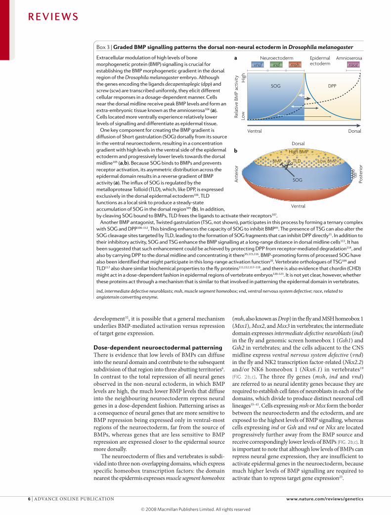
Box 3 | Graded BMP signalling patterns the dorsal non-neural ectoderm in Drosophila melanogaster
Extracellular modulation of high levels of bone morphogenetic protein (BMP) signalling is crucial for establishing the BMP morphogenetic gradient in the dorsal region of the Drosophila melanogaster embryo. Although the genes encoding the ligands decapentaplegic (dpp) and screw (scw) are transcribed uniformly, they elicit different cellular responses in a dosage-dependent manner. Cells near the dorsal midline receive peak BMP levels and form an extra-embryonic tissue known as the amnioserosa104 (a). Cells located more ventrally experience relatively lower levels of signalling and differentiate as epidermal tissue.
One key component for creating the BMP gradient is diffusion of Short gastrulation (SOG) dorsally from its source in the ventral neuroectoderm, resulting in a concentration gradient with high levels in the ventral side of the epidermal ectoderm and progressively lower levels towards the dorsal midline105 (a,b). Because SOG binds to BMPs and prevents receptor activation, its asymmetric distribution across the epidermal domain results in a reverse gradient of BMP activity (a). The influx of SOG is regulated by the metalloprotease Tolloid (TLD), which, like DPP, is expressed exclusively in the dorsal epidermal ectoderm106. TLD functions as a local sink to produce a steady-state accumulation of SOG in the dorsal region105 (b). In addition, by cleaving SOG bound to BMPs, TLD frees the ligands to activate their receptors107.
Another BMP antagonist, Twisted gastrulation (TSG, not shown), participates in this process by forming a ternary complex with SOG and DPP108–112. This binding enhances the capacity of SOG to inhibit BMP95. The presence of TSG can also alter the SOG cleavage sites targeted by TLD, leading to the formation of SOG fragments that can inhibit DPP directly21. In addition to their inhibitory activity, SOG and TSG enhance the BMP signalling at a long-range distance in dorsal midline cells113. It has been suggested that such enhancement could be achieved by protecting DPP from receptor-mediated degradation114, and also by carrying DPP to the dorsal midline and concentrating it there95,115,116. BMP-promoting forms of processed SOG have also been identified that might participate in this long-range activation function20. Vertebrate orthologues of TSG109 and TLD117 also share similar biochemical properties to the fly proteins111,112,117–119, and there is also evidence that chordin (CHD) might act in a dose-dependent fashion in epidermal regions of vertebrate embryos120–123. It is not yet clear, however, whether these proteins act through a mechanism that is similar to that involved in patterning the epidermal domain in vertebrates.
ind, intermediate defective neuroblasts; msh, muscle segment homeobox; vnd, ventral nervous system defective; race, related to angiotensin converting enzyme.
R E V I E W S
6 | ADVANCE ONLINE PuBLICATION www.nature.com/reviews/genetics

The broad range in cellular sensitivity to high versus low levels of BMP signalling enables the two branches of BMP signalling to be used in different regions of the embryo (FIG. 2b; BOX 3). By restricting the activation and repression branches of the BMP pathway to dorsal ver-sus ventral domains, respectively, this single signalling system can establish multiple cell fates along the entire embryonic D/V axis (BOX 3a).
Threshold-dependent repression of neural gene expres-sion in flies. when BMP signalling is locally compro-mised within the D. melanogaster neuroectoderm, the borders of neural-identity-gene expression domains shift dorsally (FIG. 3a,b), revealing an important role for BMP signalling in establishing pattern in the neuroectoderm6. BMPs exert their strongest influence on genes nearest to the dorsal ectodermal source of secretion. Thus, the border between msh and ind expression, which is located closest to the BMP source, shifts dorsally several cell diameters resulting in an expansion of ind expression into the normal msh domain, whereas the border between vnd and ind expression, located 10–12 cell diameters from the ectoderm, shifts only by one or two cells. This concerted shift in gene expression is the result of two processes: dose-dependent repression of ind versus msh by low-level BMP signalling; and ‘ventral dominant’ cross-regulatory inhibition among neural identity genes33,42, in which more ventrally expressed transcription factors repress the expression of more dorsal genes (FIG. 3b).
Normally, BMPs act in concert with other pattern-ing systems to establish borders between neural identity genes (BOX 4). One example is the Dorsal morphogenetic gradient in flies, which predetermines the primary D/V territories. However, under experimental conditions, BMPs alone can also generate neural patterning, albeit not as precisely as in wild-type embryos. Experiments performed in embryos, in which the graded DPP sig-nalling was uncoupled from patterning mediated by the Dorsal gradient, revealed that less BMP signalling is required to inhibit expression of ind than of msh6. Threshold-dependent repression of ind and msh results in ind expression being excluded from cells near the DPP source. A consequence of ind expression being
eliminated from dorsal-most cells of the neuroectoderm by BMP signalling is to relieve the ventral dominant repression of msh by IND, which results in the apparent activation of msh in those cells. This double-negative mechanism effectively segregates cells into adjacent non-overlapping domains of neural gene expression.
Similar patterning mechanisms in flies and vertebrates. In vertebrates, a wealth of evidence indicates that BMPs function as dorsal morphogens in the neural tube, defin-ing distinct cell fates by different levels of signalling6,43–51. The conserved expression of neural identity genes rela-tive to the BMP source in flies and vertebrates raises the possibility that BMPs function by similar mechanisms to regulate neural genes in these two species. However, the prevailing view in vertebrates has been that high levels of BMPs promote the expression of genes such as Msx1 in dorsal regions of the neural tube, whereas lower levels of BMPs activate the expression of intermediate neural genes45,48. This model is opposite to the observed neural repressive role of BMPs in flies and is based pri-marily on experiments in which increasing levels of BMP expression leads to a ventral expansion of the expression domain of Msx genes and a concomitant reduction of intermediate cell fates.
These apparently contrasting modes of regulatory function of BMPs in vertebrates versus flies might be deceiving, as nearly all the vertebrate data could be equally well explained by a double-negative mechanism similar to that described above for D. melanogaster. Thus, the expansion of Msx gene expression might not reflect direct activation by BMP signalling, but rather might result indirectly from inhibition of the expression of a Msx gene repressor. Consistent with this hypothesis, studies in zebrafish suggest that high levels of BMP sig-nalling, corresponding to those acting in the epidermis, can suppress Msx1 and Msx2 expression50, paralleling the situation observed in flies. In addition, BMP signal-ling can repress the expression of intermediate genes, such as developing brain homeobox 1 (Dbx1), Dbx2 and paired box 6 (Pax6)52–55. To distinguish between these two models, it will be crucial to determine whether cross-regulation among vertebrate neural identity genes
Nature Reviews | Genetics
a
st2–BRK
b st2–BRK
msh
ind
vnd
Figure 3 | Patterning the neuroectoderm in flies. The two panels illustrate the design (a) and consequences (b) of inhibiting bone morphogenetic protein (BMP) signalling in a localized pattern in the Drosophila melanogaster embryo by expressing the repressor Brinker (BRK) in a narrow stripe of cells (vertical grey bar in a) under the control of the even-skipped stripe-2 (st2) promoter. Indicated in horizontal stripes are expression domains of three neural identity genes: ventral nervous system defective (vnd), intermediate neuroblasts defective (ind) and muscle segment homeobox (msh) after BRK misexpression (b). Note the significant dorsal shift in the dorsal border of the ind expression domain and a smaller shift in the vnd–ind border. Normal borders are indicated with arrows. Image in panel b is reproduced from REF. 6.
R E V I E W S
NATuRE REVIEwS | genetics ADVANCE ONLINE PuBLICATION | 7
follows a ventral dominant pattern as has been demon-strated in D. melanogaster. It will also be important to test whether BMP-dependent repression of neural genes is a general mechanism operating across vertebrates.
Another parallel with fruitflies is the fact that BMP signalling in vertebrates is sufficient to pattern the neur-oectoderm when this tissue is isolated from other pattern-ing cues. In apolar chick neural-plate explants that have been adjusted to generate uniform ventralized cell fates, addition of BMPs can cause a ventral-to-dorsal shift in the expression of neural genes6. Moreover, there is evidence that BMPs can pattern the entire D/V axis of the neural tube in the complete absence of the ventral sonic hedge-hog (SHH) patterning system56 (see below and BOX 4).
Similar BMP-mediated patterning has also been observed in primitive polychaete annelids7. The
sufficiency of BMP signalling for creating neural pattern and its highly conserved activity in the three primary phylogenetic lineages of Metazoa provides compelling evidence that the bilaterian ancestor already used this signalling pathway to subdivide a highly organized nerv-ous system, which was inherited in many descendent branches. These findings are difficult to reconcile with the alternative model that organized condensed nerve chords evolved independently in these three lineages, as would be the case if a diffuse nervous system were the primitive state in their most recent common ancestor.
Ventral patterning cues in the neuroectodermIn conjunction with BMP gradients emanating dorsally, ventral patterning cues also help to establish neuronal fates. In D. melanogaster there is a ventral-to-dorsal
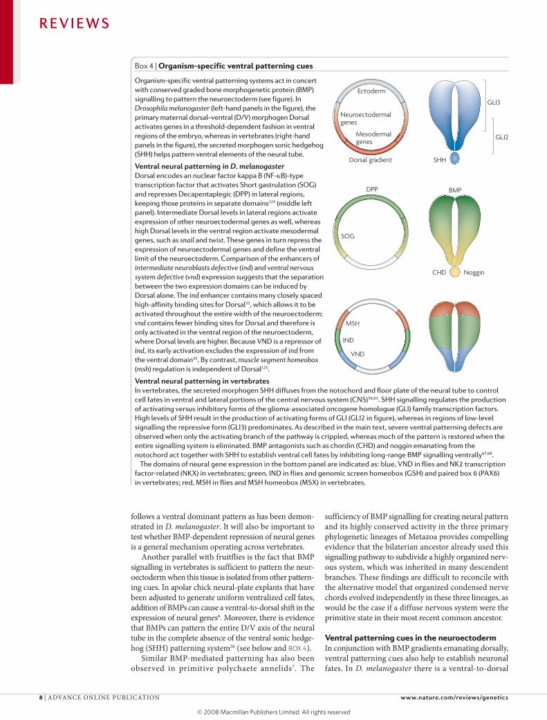
Box 4 | Organism-specific ventral patterning cues
Organism-specific ventral patterning systems act in concert with conserved graded bone morphogenetic protein (BMP) signalling to pattern the neuroectoderm (see figure). In Drosophila melanogaster (left-hand panels in the figure), the primary maternal dorsal–ventral (D/V) morphogen Dorsal activates genes in a threshold-dependent fashion in ventral regions of the embryo, whereas in vertebrates (right-hand panels in the figure), the secreted morphogen sonic hedgehog (SHH) helps pattern ventral elements of the neural tube.
Ventral neural patterning in D. melanogasterDorsal encodes an nuclear factor kappa B (NF-κB)-type transcription factor that activates Short gastrulation (SOG) and represses Decapentaplegic (DPP) in lateral regions, keeping those proteins in separate domains124 (middle left panel). Intermediate Dorsal levels in lateral regions activate expression of other neuroectodermal genes as well, whereas high Dorsal levels in the ventral region activate mesodermal genes, such as snail and twist. These genes in turn repress the expression of neuroectodermal genes and define the ventral limit of the neuroectoderm. Comparison of the enhancers of intermediate neuroblasts defective (ind) and ventral nervous system defective (vnd) expression suggests that the separation between the two expression domains can be induced by Dorsal alone. The ind enhancer contains many closely spaced high-affinity binding sites for Dorsal33, which allows it to be activated throughout the entire width of the neuroectoderm; vnd contains fewer binding sites for Dorsal and therefore is only activated in the ventral region of the neuroectoderm, where Dorsal levels are higher. Because VND is a repressor of ind, its early activation excludes the expression of ind from the ventral domain42. By contrast, muscle segment homeobox (msh) regulation is independent of Dorsal125.
Ventral neural patterning in vertebratesIn vertebrates, the secreted morphogen SHH diffuses from the notochord and floor plate of the neural tube to control cell fates in ventral and lateral portions of the central nervous system (CNS)56,61. SHH signalling regulates the production of activating versus inhibitory forms of the glioma-associated oncogene homologue (GLI) family transcription factors. High levels of SHH result in the production of activating forms of GLI (GLI2 in figure), whereas in regions of low-level signalling the repressive form (GLI3) predominates. As described in the main text, severe ventral patterning defects are observed when only the activating branch of the pathway is crippled, whereas much of the pattern is restored when the entire signalling system is eliminated. BMP antagonists such as chordin (CHD) and noggin emanating from the notochord act together with SHH to establish ventral cell fates by inhibiting long-range BMP signalling ventrally67,68.
The domains of neural gene expression in the bottom panel are indicated as: blue, VND in flies and NK2 transcription factor-related (NKX) in vertebrates; green, IND in flies and genomic screen homeobox (GSH) and paired box 6 (PAX6) in vertebrates; red, MSH in flies and MSH homeobox (MSX) in vertebrates.
Nature Reviews | Genetics
Ectoderm
Mesodermal genes
Neuroectodermal genes
Noggin
BMP
GLI2
GLI3
MSH
IND
VND
SOG
DPP
Dorsal gradient
CHD
SHH
R E V I E W S
8 | ADVANCE ONLINE PuBLICATION www.nature.com/reviews/genetics
gradient of the Dorsal morphogen, whereas in verte-brates SHH diffuses dorsally from the ventral noto-chord and floor plate of the neural tube (BOX 4). The opposing gradients of BMPs and ventral morphogens might more reliably create sharp boundaries of gene expression within a large field of cells than a single gra-dient. The recruitment of these additional cues seems to be species specific, as neither nuclear factor kappa B (NF-κB)-related homologues of Dorsal in vertebrates nor Hedgehog in flies participate in D/V patterning of the nervous system. It is also possible that one of these path-ways was ancestral and was lost from the other lineage. For example, it has been reported that a hedgehog homo-logue is expressed along the ventral midline in a mollusc embryo57. However, it remains to be determined whether this signalling pathway is involved in neuroectoderm patterning in these invertebrate organisms.
DPP and Dorsal cooperate in flies. In flies, the Dorsal and DPP gradients act together during neuroectodermal patterning. when a gradient of BMP signalling is pro-duced in isolation from the Dorsal gradient, the resulting separation of ind and msh expression is not nearly as sharp as the endogenous mutually exclusive expression of these genes6. There are two possible mechanisms by which the Dorsal gradient might help sharpen borders in the neuroectoderm. First, as mentioned above, Dorsal directly activates ventral and lateral genes in a threshold-dependent fashion6,42 (BOX 4). Second, Dorsal dynami-cally regulates expression of the BMP antagonists SOG and BRK58,59, which initially are expressed throughout the entire neuroectoderm, but then fade dorsally as the maternal Dorsal gradient collapses during late blasto-derm stages14,27. The graded distribution of these BMP antagonists might refine the BMP gradient formed within the neuroectoderm, thereby providing more precision to cellular responses.
The role of SHH in neural tube patterning. Evidence in vertebrate systems suggest that dorsally produced BMPs alone can generate pattern across most of the D/V axis in the neural tube, although under normal circumstances ventrally produced SHH also contributes to this process. SHH is secreted from the notochord and floor plate and regulates the expression of class I genes (intermediate genes such as Pax6, iroquois related homeobox 3 (Irx3), Dbx1, Dbx2 and Pax7) and class II genes (ventral genes such as Nkx2.2, oligodendrocyte lineage transcription factor 2 (Olig2), Nkx6.1 and Nkx6.2)60,61. In mutant mice lacking SHH function, there is a ventral expansion of intermediate fates and a concomitant loss of ventral cell fates. A similar loss of ventral fates is observed in mutants for the glioma-associated oncogene homologue 2 (GLI2) repressor, which mediates SHH signalling in ventral regions of the neural tube, whereas loss of function of GLI3 alters intermediate cell fates (BOX 4). Surprisingly, expression of most ventral markers that are lost or greatly reduced in Shh single mutants (except for NKX2.2) can be restored in Shh;Gli3 double mutants, albeit with less regularity than in wild-type individuals56,62–66. This finding indicates that the main
role of SHH signalling is to block the repressive activ-ity of GLI3, and that most ventral neural patterning can be elaborated in the absence of the SHH gradient. Consistent with this hypothesis, BMP mutants exhibit defects spanning the entire D/V axis of the neural tube43,48 and BMP antagonists expressed ventrally in the notochord act synergistically with SHH67,68, suggesting that inhibition of BMPs near the midline normally has an important role in ventral patterning.
These various observations strongly suggest that the BMP gradient effectively reaches the ventral-most regions of the neural tube to provide cell-fate cues. The ability of BMPs to pattern over such large distances and the conserved roles they have in neural patterning sug-gest that this pathway might once have been sufficient to pattern the entire D/V axis. The proposal that a sin-gle morphogen could pattern the entire D/V axis of the nervous system is in agreement with speculation that the bilaterian ancestor living in Precambrian times was small69,70. As organisms grew markedly in size during the Cambrian period, additional ventral cues might have been recruited independently in vertebrate and invertebrate lineages to create more robust and reliable patterning in regions far from the dorsal source of BMPs.
Neural patterning in other organismsAs argued above, the similarity of neuronal gene expression patterns and their regulation by the highly conserved BMP signalling pathway in flies (BOX 2) and vertebrates (FIG. 4) strongly suggests that this process was inherited from a common ancestor. An alternative pos-sibility is that similar patterns of gene expression evolved independently in the vertebrate and Drosophila lineages, which is consistent with the lack of clear fossil data for a bilaterian ancestor with a condensed nervous system. One way to distinguish between conserved versus convergent evolutionary processes is to examine pat-tern formation in a broad variety of organisms (FIG. 4a). Although analysis of neurogenesis across phylogeny is still in its nascent phase, interesting parallels can already be gleaned from studies in diploblasts, hemichordates, arthropods and annelids (FIG. 4b–d).
The antiquity of the BMP and SOG/CHD system is revealed by the localized expression of these genes in embryos of diploblasts, such as corals71,72 (FIG. 4b), jellyfish73 and the sea anemone74,75, along an axis that is orthogonal to the longitudinal body axis, which expresses nested patterns of homeotic (Hox) genes. It is not clear, however, whether there is a necessary link between the localized expression of BMP and SOG/CHD genes and neural development. Recent analysis of the hemichordate Saccoglossus kowalevskii76 has shown that, although BMP and SOG/CHD are expressed in opposing domains, misexpression of BMPs does not suppress neuronal development and neural iden-tity genes are not expressed in a restricted fashion along the D/V axis as they are in flies or vertebrates. However, these organisms are nearly rotationally sym-metrical so it is difficult to establish dorsoventrality in the hemichordate ectoderm, which consists of a nearly uniform epidermis with a diffuse net of nerve cells.
R E V I E W S
NATuRE REVIEwS | genetics ADVANCE ONLINE PuBLICATION | 9

Nature Reviews | Genetics
b BMP4 c
Ecdysozoa Lophotrochozoa
NematodaArthropodaCnidaria Chordate Tunicate Hemichordate
Echinoderm
Bilateralia
Triploblasts
Deuterostomata
Diploblasts
a
e
d CHD
BMP4
?
?
BMPs BMPs BMPs BMPs
Fly Vertebrate Annelid Hemichordate
msh
ind/ey
vnd
sim
Msx1Dlx
Pax3/7
dlx
pax3/7
dbx
nkx6
Dbx1/2
Nkx6.2Nkx6.1
Gsh/Pax6
Nkx2.2
Sim
msx msx
gsh/pax6
nkx2.2
sim
Sensory neurons
Motor neurons
Serotonergic neurons
Midline cells
DPP
SOG
R E V I E W S
10 | ADVANCE ONLINE PuBLICATION www.nature.com/reviews/genetics

It is clear that the neural suppressive activity of BMPs and their inhibition by SOG/CHD in the neur-oectoderm in flies is shared with other arthropods, such as spiders77 and beetles78. One model that was missing from the phylogenetic comparisons until recently was analysis of organisms from the third main branch of bilaterians, namely the Lophotrochozoa. This branch includes annelids, molluscs and brachiopods (FIG. 4a). Such data has now been obtained for a polychaete anne-lid7, which is thought to have retained many primitive characteristics that were present in the bilaterian ances-tor. Consistent with studies in flies and vertebrates, BMPs also differentially regulate expression of neural genes in this primitive annelid. Remarkably, functional studies verified that the progeny of neurons arising from different D/V positions have similar patterns of connectivity, similar expression levels of neurotrans-mitters, and similar physiological properties in anne-lids and vertebrates7. Indeed, the conservation in gene expression patterns and cell types between the annelid worm and vertebrates is even greater than between Drosophila and vertebrates. For example, motor neu-rons arise only from a ventral domain in both verte-brates and annelids, whereas flies also produce some motor neurons from lateral and dorsal rows of neu-roblasts. These detailed similarities in a third branch of the bilaterian lineage strongly suggest that the ancestor not only had a condensed nerve chord with defined cell types arising from stereotyped positions, but also used distinct thresholds of BMP signalling to establish those cell fates79. The simplest explanation to account for absence of this pattern in S. kowalevskii is that the response to the BMP and SOG/CHD patterning sys-tem was lost in that lineage, consistent with the nearly radially symmetrical organization of the hemichordate body plan, which would gain no obvious benefit from restricting neurons to one side of the body.
Together, the existing comparative molecular data are most consistent with a bilaterian ancestor with
a condensed nerve chord and at least three primary subdivisions. This view is also in agreement with the ubiquity of two or three ventral nerve chords in extant organisms (FIG. 4e). According to this view, the morpho-type of a simple dispersed CNS, which is also observed broadly across phylogenies, is probably the result of frequent secondary loss of the ancestral organized body plan. This interpretation is supported by the high rate of gene loss during evolution, which has become appreciated through comprehensive comparisons of fully sequenced genomes3,80.
Regulatory treadmilling: a hypothesis for the conserva-tion of A/P and D/V patterning genes. One of the most striking findings in developmental biology in the past two decades has been the evolutionary conservation of basic patterning mechanisms that define the body plan. These mechanisms are exemplified by Hox genes that specify cell fates along the A/P axis81 and the com-plementary function of the BMP and SOG/CHD genes to establish conserved gene expression along the D/V axis9–11. It has generally been assumed that the con-served protein-coding sequences of genes that function early in genetic hierarchies, such as the Hox or neural identity genes, reflects the conserved function of these genes in the regulation of shared sets of downstream effector genes.
Alternative hypotheses regarding the function of higher order regulatory factors are also possible. One could imagine, for example, that Hox genes define only an abstract positional code that is interpreted differ-ently in morphologically diverse organisms82. This view is particularly compelling in the case of the A/P axis, which has undergone extensive morphological trans-formation during evolution with different structures forming in different positions along the body in one taxon compared with another. A potential difficulty with this idea is that it does not immediately account for the high degree of evolutionary conservation of the patterning genes. If the downstream targets differ, what constrains changes so effectively in the regulatory factor?
One possible explanation for the strong conserva-tion of higher order regulatory factors such as the Hox genes, which might define only abstract positional codes, is that in any given organism they control large numbers of genes, even though these sets of targets differ across phylogenies. According to this model, change in such a regulatory factor is constrained during evolution simply because, at any given point during evolution, the regulator controls many targets. The identity of downstream target genes could turn over during evolution so that some ancestral target genes lose their regulatory dependence on the regula-tory factor and others newly acquire such regulation. Although, with time, the constituents of the control-led set of genes would change, large numbers of target genes would nonetheless always be under the control of the regulatory factor in any organism (FIG. 5a). we refer to this process of target-gene turn-over as ‘regulatory treadmilling’.
Figure 4 | BMP patterning in diverse organisms. a | Evolutionary tree showing the three major branches of Metazoa (Ecdysozoa, lophotrochozoa, and Deuterostomata) as well as their relationship to diploblasts (Cnidaria). b,c,d | RNA in situ hybridizations in embryos showing opposing Decapentaplegic (DPP; bone morphogenetic protein, BMP, in vertebrates) and Short gastrulation (SOG; chordin, CHD, in vertebrates) expression in: a diploblast (coral) embryo (b; BMP4 expression in blue, CHD not shown); a fly embryo (c; DPP expression in blue and SOG in orange); and in frog embryos (d). e | Relative gene expression patterns in flies, vertebrates, polychaete annelids and hemichordates. The position of expression domains of neural genes between vertebrates and annelids is more similar to each other than between flies and vertebrates, although they all share a basic conserved arrangement of neural domains. By contrast, most of the nervous system patterning along the dorsal–ventral (D/V) axis might have been lost in the hemichordate. Dbx, developing brain homeobox; Dlx, distal-less homeobox; ey, eyeless; Gsh, genomic screen homeobox; ind, intermediate neuroblasts defective; msh, muscle segment homeobox; Msx, muscle segment homeobox; Nkx, NK transcription factor-related; Pax, paired box; sim, single-minded; vnd, ventral nervous system defective. Panel b is reproduced, with permission, from REF. 71 (2002) National Academy of Sciences, USA. Panel c is reproduced, with permission from REF. 14 (1995) Cold Spring Harbor laboratory Press. CHD image in panel d is reproduced, with permission, from REF. 127 (1996) Company of Biologists. BMP4 image in panel d is reproduced, with permission, from REF. 128 (1995) Elsevier Science.
◀
R E V I E W S
NATuRE REVIEwS | genetics ADVANCE ONLINE PuBLICATION | 11
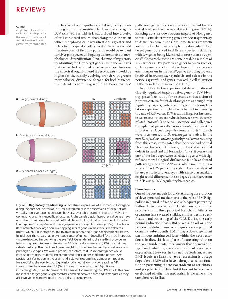
The crux of our hypothesis is that regulatory tread-milling occurs at a considerably slower pace along the D/V axis (FIG. 5c), which is subdivided into a series of well conserved tissues, than along the A/P axis, in which morphological diversification is greater and is less tied to specific cell types (FIG. 5a,b). we would therefore predict that two patterns would be evident for divergent species undergoing different rates of mor-phological diversification. First, the rate of regulatory treadmilling for Hox target genes along the A/P axis (defined as the fraction of target genes shared between the ancestral organism and it descendents) would be higher for the rapidly evolving branch with greater morphological divergence. Second, for both branches, the rate of treadmilling would be lower for D/V
patterning genes functioning at an equivalent hierar-chical level, such as the neural identity genes (FIG. 5c). Existing data on downstream targets of Hox genes versus tissue-determining genes are too fragmentary to draw firm conclusions, but some trends are worth analysing further. For example, the diversity of Hox target genes observed in different species is striking, with few genes being identified in more than one spe-cies82. Conversely, there are some notable examples of similarities in D/V patterning genes between species, such as genes encoding related types of ion channels and transporters in the heart83, genes encoding proteins involved in transmitter synthesis and release in the nervous system84, and genes involved in cell migration in the mesoderm (reviewed in REF. 85).
In addition to the experimental determination of directly regulated targets of Hox genes or D/V iden-tity genes (see REF. 82 for an excellent discussion of rigorous criteria for establishing genes as being direct regulatory targets), interspecific germline transplan-tation experiments might also be helpful in assessing the rate of A/P versus D/V treadmilling. For instance, in an attempt to create hybrids between two distantly related Drosophila species, Lawrence and colleagues transplanted germ cells from Drosophila rajasekari into sterile D. melanogaster female hosts86, which were then crossed to D. melanogaster males. In the rare D. rajasekari–melanogaster hybrid larvae resulting from this cross, it was noted that the cuticle had normal D/V morphological structures, but showed substantial defects in head and tail formation. Thus, it seems that one of the first departures in related species with sig-nificant morphological differences is to have altered patterning along the A/P axis, while maintaining a very similar D/V patterning system. Future analysis of interspecific hybrid embryos with molecular markers might reveal differences in the degree of conservation in A/P versus D/V regulatory hierarchies.
ConclusionsOne of the best models for understanding the evolution of developmental mechanisms is the role of BMP sig-nalling in neural induction and subsequent patterning within the neuroectoderm. Detailed analysis of these processes in the three principal branches of bilaterian organisms has revealed striking similarities in speci-fication and patterning of the CNS. During the early neural-induction phase, BMPs act in an all-or-none fashion to inhibit neural gene expression in epidermal domains. Subsequently, BMPs play a dose-dependent part in determining cell fates within the neuroecto-derm. In flies, this later phase of patterning relies on the same fundamental mechanism that operates dur-ing neural induction, namely repression of neural gene expression. However, in the neuroectoderm, where BMP levels are limiting, gene repression is dosage dependent. BMPs also have a dosage-sensitive func-tion in patterning the neuroectoderm of vertebrates and polychaete annelids, but it has not been clearly established whether the mechanism is the same as the one observed in flies.
Nature Reviews | Genetics
b Pax6 (eye and brain cell types)
c Nkx (ventral neuronal cell types)
a Hox (segmental identity) Fly Vertebrate
Eye genes
Figure 5 | Regulatory treadmilling. a | localized expression of a Homeotic (Hox) gene along the anterior–posterior (A/P) axis (left) results in the expression of large sets of virtually non-overlapping genes in flies versus vertebrates (right) that are involved in generating organism-specific structures. Right panels depict hypothetical gene arrays with Hox target genes indicated by filled circles. b | localized expression of the paired box 6 gene (Pax 6; eyeless and twin-of-eyeless in Drosophila melanogaster) in the brain (left) activates two large non-overlapping sets of genes in flies versus vertebrates (right), which, like Hox genes, are involved in generating organism-specific structures. In addition, there is a smaller overlapping set of genes induced by PAX6 in both species that are involved in specifying the eye field. Genes defining the eye field provide an interesting predicted exception to the A/P versus dorsal–ventral (D/V) treadmilling rate dichotomy. This module of genes might turn over less frequently, as in the case of primary tissue types. We would predict, therefore, that PAX6 target genes would consist of a rapidly treadmilling component (those genes mediating general A/P positional information in the brain) and a slower treadmilling component required for specifying the eye field. c | Expression of a neural identity gene such as NK transcription factor-related 2.2 (Nkx2.2; ventral nervous system defective in D. melanogaster) in a subdomain of the neuroectoderm along the D/V axis. In this case, most of the target genes expressed are common between flies and vertebrate as they are involved in specifying conserved cell and tissue types.
CuticleA rigid layer of sclerotized chitin and cuticular proteins that covers the insect larval and adult epidermis and constitutes the exoskeleton.
R E V I E W S
12 | ADVANCE ONLINE PuBLICATION www.nature.com/reviews/genetics

Although current day flies and vertebrates use distinct ventral patterning cues in addition to dorsally derived BMPs, we propose a model, developed on the basis of the common elements of gene regulation observed in flies, vertebrates and annelids, in which BMPs had an ancestral function in patterning the full D/V axis of the neuroectoderm. The model is based in part on the
premise that high-order regulatory genes control similar sets of downstream target genes in corresponding cell types in diverse organisms. we further suggest that this conserved regulation of target genes might be a more salient property of D/V patterning genes, which define conserved cell types, than of A/P patterning genes, which might define a more abstract positional code.
1. Dunn, C. W. et al. Broad phylogenomic sampling improves resolution of the animal tree of life. Nature 452, 745–749 (2008).
2. Valentine, J. W. On The Origin of Phyla (University of Chicago Press, Chicago, 2004).
3. De Robertis, E. M. Evo–devo: variations on ancestral themes. Cell 132, 185–195 (2008).
4. De Robertis, E. M. & Kuroda, H. Dorsal–ventral patterning and neural induction in Xenopus embryos. Annu. Rev. Cell Dev. Biol. 20, 285–308 (2004).
5. O’Connor, M. B., Umulis, D., Othmer, H. G. & Blair, S. S. Shaping BMP morphogen gradients in the Drosophila embryo and pupal wing. Development 133, 183–193 (2006).
6. Mizutani, C. M., Meyer, N., Roelink, H. & Bier, E. Threshold-dependent BMP-mediated repression: a model for a conserved mechanism that patterns the neuroectoderm. PLoS Biology 4, e313 (2006).Shows that BMPs act in a dose-dependent fashion to repress the expression of neural genes in dorsal and lateral regions of the D. melanogaster embryo; it also proposes that this might be a conserved mechanism for neural patterning.
7. Denes, A. S. et al. Molecular architecture of annelid nerve cord supports common origin of nervous system centralization in Bilateria. Cell 129, 277–288 (2007).Reveals remarkable similarities in the D/V organization of cell markers and cell types in the CNS of annelid worms and vertebrates.
8. Geoffroy St‑Hilaire, E. Considérations générales sur la vertèbre (Translation: General considerations on vertebrates). Mém. Mus. Hist. Nat. 9, 89–119 (1822).In this paper, St-Hilaire suggests that the D/V axis in invertebrates is inverted with respect to that of vertebrates.
9. Bier, E. Anti-neural-inhibition: a conserved mechanism for neural induction. Cell 89, 681–684 (1997).
10. De Robertis, E. M. & Sasai, Y. A common plan for dorsoventral patterning in Bilateria. Nature 380, 37–40 (1996).
11. Ferguson, E. L. Conservation of dorsal–ventral patterning in arthropods and chordates. Curr. Opin. Genet. Dev. 6, 424–431 (1996).
12. Arendt, D. & Nubler-Jung, K. Inversion of dorsoventral axis? Nature 371, 26 (1994).Resuscitates the argument of St-Hilaire (reference 8) and A. Dohrn (reference 97) regarding a common origins of the D/V axis in vertebrates and invertebrates.
13. Francois, V. & Bier, E. Xenopus chordin and Drosophila short gastrulation genes encode homologous proteins functioning in dorsal–ventral axis formation. Cell 80, 19–20 (1995).
14. Francois, V., Solloway, M., O’Neill, J. W., Emery, J. & Bier, E. Dorsal–ventral patterning of the Drosophila embryo depends on a putative negative growth factor encoded by the short gastrulation gene. Genes Dev. 8, 2602–2616 (1994).
15. Holley, S. A. et al. A conserved system for dorsal–ventral patterning in insects and vertebrates involving sog and chordin. Nature 376, 249–253 (1995).
16. Sasai, Y. et al. Xenopus chordin: a novel dorsalizing factor activated by organizer-specific homeobox genes. Cell 79, 779–790 (1994).
17. Schmidt, J., Francois, V., Bier, E. & Kimelman, D. Drosophila short gastrulation induces an ectopic axis in Xenopus: evidence for conserved mechanisms of dorsal–ventral patterning. Development 121, 4319–4328 (1995).
18. Schmierer, B. & Hill, C. S. TGFbeta-SMAD signal transduction: molecular specificity and functional flexibility. Nature Rev. Mol. Cell Biol. 8, 970–982 (2007).
19. Cornell, R. A. & Ohlen, T. V. vnd/Nkx, ind/Gsh, and msh/Msx: conserved regulators of dorsoventral neural patterning? Curr. Opin. Neurobiol. 10, 63–71 (2000).
20. Yu, K. et al. Cysteine repeat domains and adjacent sequences determine distinct BMP modulatory activities of the Drosophila Sog protein. Genetics 166, 1323–1336 (2004).
21. Yu, K. et al. Processing of the Drosophila Sog protein creates a novel BMP inhibitory activity. Development 127, 2143–2154 (2000).
22. De Robertis, E. M. Spemann’s organizer and self-regulation in amphibian embryos. Nature Rev. Mol. Cell Biol. 7, 296–302 (2006).
23. Biehs, B., Francois, V. & Bier, E. The Drosophila short gastrulation gene prevents Dpp from autoactivating and suppressing neurogenesis in the neuroectoderm. Genes Dev. 10, 2922–2934 (1996).Shows that the BMP antagonist SOG prevents BMPs from repressing neural gene expression in D. melanogaster.
24. Piccolo, S., Sasai, Y., Lu, B. & De Robertis, E. M. Dorsoventral patterning in Xenopus: inhibition of ventral signals by direct binding of Chordin to BMP-4. Cell 86, 589–598 (1996).
25. Lamb, T. M. et al. Neural induction by the secreted polypeptide noggin. Science 262, 713–718 (1993).
26. Smith, W. C., McKendry, R., Ribisi, S. Jr & Harland, R. M. A nodal-related gene defines a physical and functional domain within the Spemann organizer. Cell 82, 37–46 (1995).
27. Jazwinska, A., Rushlow, C. & Roth, S. The role of brinker in mediating the graded response to Dpp in early Drosophila embryos. Development 126, 3323–3334 (1999).
28. Arora, K. et al. The Drosophila schnurri gene acts in the Dpp/TGF beta signaling pathway and encodes a transcription factor homologous to the human MBP family. Cell 81, 781–790 (1995).
29. Grieder, N. C., Nellen, D., Burke, R., Basler, K. & Affolter, M. schnurri is required for Drosophila Dpp signaling and encodes a zinc finger protein similar to the mammalian transcription factor PRDII-BF1. Cell 81, 791–800 (1995).
30. Staehling-Hampton, K., Laughon, A. S. & Hoffmann, F. M. A Drosophila protein related to the human zinc finger transcription factor PRDII/MBPI/HIV-EP1 is required for Dpp signaling. Development 121, 3393–3403 (1995).
31. Muller, B., Hartmann, B., Pyrowolakis, G., Affolter, M. & Basler, K. Conversion of an extracellular Dpp/BMP morphogen gradient into an inverse transcriptional gradient. Cell 113, 221–233 (2003).
32. Pyrowolakis, G., Hartmann, B., Muller, B., Basler, K. & Affolter, M. A simple molecular complex mediates widespread BMP-induced repression during Drosophila development. Dev. Cell 7, 229–240 (2004).
33. Stathopoulos, A. & Levine, M. Localized repressors delineate the neurogenic ectoderm in the early Drosophila embryo. Dev. Biol. 280, 482–493 (2005).
34. Affolter, M. & Basler, K. The Decapentaplegic morphogen gradient: from pattern formation to growth regulation. Nature Rev. Genet. 8, 663–674 (2007).
35. Briscoe, J., Pierani, A., Jessell, T. M. & Ericson, J. A homeodomain protein code specifies progenitor cell identity and neuronal fate in the ventral neural tube. Cell 101, 435–445 (2000).
36. Chu, H., Parras, C., White, K. & Jimenez, F. Formation and specification of ventral neuroblasts is controlled by vnd in Drosophila neurogenesis. Genes Dev. 12, 3613–3624 (1998).
37. Isshiki, T., Takeichi, M. & Nose, A. The role of the msh homeobox gene during Drosophila neurogenesis: implication for the dorsoventral specification of the neuroectoderm. Development 124, 3099–3109 (1997).
38. Jimenez, F. et al. vnd, a gene required for early neurogenesis of Drosophila, encodes a homeodomain protein. EMBO J. 14, 3487–3495 (1995).
39. McDonald, J. A. et al. Dorsoventral patterning in the Drosophila central nervous system: the vnd homeobox gene specifies ventral column identity. Genes Dev. 12, 3603–3612 (1998).
40. Skeath, J. B., Panganiban, G. F. & Carroll, S. B. The ventral nervous system defective gene controls proneural gene expression at two distinct steps during neuroblast formation in Drosophila. Development 120, 1517–1524 (1994).
41. Weiss, J. B. et al. Dorsoventral patterning in the Drosophila central nervous system: the intermediate neuroblasts defective homeobox gene specifies intermediate column identity. Genes Dev. 12, 3591–3602 (1998).
42. Cowden, J. & Levine, M. Ventral dominance governs sequential patterns of gene expression across the dorsal–ventral axis of the neuroectoderm in the Drosophila embryo. Dev. Biol. 262, 335–349 (2003).Provides evidence that neural identity genes act in a hierarchical repressive cascade in which more ventrally expressed transcription factors repress the expression of more dorsal genes.
43. Barth, K. A. et al. Bmp activity establishes a gradient of positional information throughout the entire neural plate. Development 126, 4977–4987 (1999).Shows that BMPs can act over long distances to pattern the D/V axis of the zebrafish neural tube and that high-level signalling in the epidermis can inhibit expression of dorsal markers such as Msx genes.
44. LaBonne, C. & Bronner-Fraser, M. Neural crest induction in Xenopus: evidence for a two-signal model. Development 125, 2403–2414 (1998).
45. Lee, K. J. & Jessell, T. M. The specification of dorsal cell fates in the vertebrate central nervous system. Annu. Rev. Neurosci. 22, 261–294 (1999).
46. Marchant, L., Linker, C., Ruiz, P., Guerrero, N. & Mayor, R. The inductive properties of mesoderm suggest that the neural crest cells are specified by a BMP gradient. Dev. Biol. 198, 319–329 (1998).
47. Neave, B., Holder, N. & Patient, R. A graded response to BMP-4 spatially coordinates patterning of the mesoderm and ectoderm in the zebrafish. Mech. Dev. 62, 183–195 (1997).
48. Nguyen, V. H. et al. Dorsal and intermediate neuronal cell types of the spinal cord are established by a BMP signaling pathway. Development 127, 1209–1220 (2000).
49. Timmer, J. R., Wang, C. & Niswander, L. BMP signaling patterns the dorsal and intermediate neural tube via regulation of homeobox and helix-loop-helix transcription factors. Development 129, 2459–2472 (2002).
50. Tribulo, C., Aybar, M. J., Nguyen, V. H., Mullins, M. C. & Mayor, R. Regulation of Msx genes by a Bmp gradient is essential for neural crest specification. Development 130, 6441–6452 (2003).
51. Wilson, P. A., Lagna, G., Suzuki, A. & Hemmati-Brivanlou, A. Concentration-dependent patterning of the Xenopus ectoderm by BMP4 and its signal transducer Smad1. Development 124, 3177–3184 (1997).
52. Furuta, Y., Piston, D. W. & Hogan, B. L. Bone morphogenetic proteins (BMPs) as regulators of dorsal forebrain development. Development 124, 2203–2212 (1997).
53. Golden, J. A. et al. Ectopic bone morphogenetic proteins 5 and 4 in the chicken forebrain lead to cyclopia and holoprosencephaly. Proc. Natl Acad. Sci. USA 96, 2439–2444 (1999).
R E V I E W S
NATuRE REVIEwS | genetics ADVANCE ONLINE PuBLICATION | 13
54. Hartley, K. O., Hardcastle, Z., Friday, R. V., Amaya, E. & Papalopulu, N. Transgenic Xenopus embryos reveal that anterior neural development requires continued suppression of BMP signaling after gastrulation. Dev. Biol. 238, 168–184 (2001).
55. Pierani, A., Brenner-Morton, S., Chiang, C. & Jessell, T. M. A sonic hedgehog-independent, retinoid-activated pathway of neurogenesis in the ventral spinal cord. Cell 97, 903–915 (1999).
56. Jacob, J. & Briscoe, J. Gli proteins and the control of spinal-cord patterning. EMBO Rep. 4, 761–765 (2003).
57. Nederbragt, A. J., van Loon, A. E. & Dictus, W. J. Evolutionary biology: hedgehog crosses the snail’s midline. Nature 417, 811–812 (2002).
58. Markstein, M. et al. A regulatory code for neurogenic gene expression in the Drosophila embryo. Development 131, 2387–2394 (2004).
59. Stathopoulos, A., Van Drenth, M., Erives, A., Markstein, M. & Levine, M. Whole-genome analysis of dorsal–ventral patterning in the Drosophila embryo. Cell 111, 687–701 (2002).
60. Gomez-Skarmeta, J. L., Campuzano, S. & Modolell, J. Half a century of neural prepatterning: the story of a few bristles and many genes. Nature Rev. Neurosci. 4, 587–598 (2003).
61. Ruiz i Altaba, A., Nguyen, V. & Palma, V. The emergent design of the neural tube: prepattern, SHH morphogen and GLI code. Curr. Opin. Genet. Dev. 13, 513–521 (2003).
62. Bai, C. B., Stephen, D. & Joyner, A. L. All mouse ventral spinal cord patterning by hedgehog is Gli dependent and involves an activator function of Gli3. Dev. Cell 6, 103–115 (2004).
63. Lei, Q., Zelman, A. K., Kuang, E., Li, S. & Matise, M. P. Transduction of graded Hedgehog signaling by a combination of Gli2 and Gli3 activator functions in the developing spinal cord. Development 131, 3593–3604 (2004).
64. Litingtung, Y. & Chiang, C. Specification of ventral neuron types is mediated by an antagonistic interaction between Shh and Gli3. Nature Neurosci. 3, 979–985 (2000).
65. Persson, M. et al. Dorsal–ventral patterning of the spinal cord requires Gli3 transcriptional repressor activity. Genes Dev. 16, 2865–2878 (2002).
66. Wijgerde, M., McMahon, J. A., Rule, M. & McMahon, A. P. A direct requirement for Hedgehog signaling for normal specification of all ventral progenitor domains in the presumptive mammalian spinal cord. Genes Dev. 16, 2849–2864 (2002).
67. Liem, K. F. Jr, Jessell, T. M. & Briscoe, J. Regulation of the neural patterning activity of sonic hedgehog by secreted BMP inhibitors expressed by notochord and somites. Development 127, 4855–4866 (2000).Shows that the BMP antagonist CHD, which is secreted by the ventrally located notochord, acts in concert with SHH to promote ventral cell fates, revealing the long-range action of dorsally produced BMPs.
68. McMahon, J. A. et al. Noggin-mediated antagonism of BMP signaling is required for growth and patterning of the neural tube and somite. Genes Dev. 12, 1438–1452 (1998).
69. Conway Morris, S. Early metazoan evolution: reconciling paleontology and molecular biology. Am. Zool. 38, 867–877 (1998).
70. Conway-Morris, S. The Cambrian ‘explosion’ of metazoans and molecular biology: would Darwin be satisfied? Int. J. Dev. Biol. 47, 505–515 (2003).
71. Hayward, D. C. et al. Localized expression of a dpp/BMP2/4 ortholog in a coral embryo. Proc. Natl Acad. Sci. USA 99, 8106–8111 (2002).
72. Samuel, G., Miller, D. & Saint, R. Conservation of a DPP/BMP signaling pathway in the nonbilateral cnidarian Acropora millepora. Evol. Dev. 3, 241–250 (2001).
73. Reber-Muller, S. et al. BMP2/4 and BMP5–8 in jellyfish development and transdifferentiation. Int. J. Dev. Biol. 50, 377–384 (2006).
74. Rentzsch, F. et al. Asymmetric expression of the BMP antagonists chordin and gremlin in the sea anemone Nematostella vectensis: implications for the evolution of axial patterning. Dev. Biol. 296, 375–387 (2006).
75. Finnerty, J. R., Pang, K., Burton, P., Paulson, D. & Martindale, M. Q. Origins of bilateral symmetry: Hox and Dpp expression in a sea anemone. Science 304, 1335–1337 (2004).
76. Lowe, C. J. et al. Dorsoventral patterning in hemichordates: insights into early chordate evolution. PLoS Biol. 4, e291 (2006).Examines the expression and function of BMPs in hemichordate embryos; it finds that although BMPs and SOG/CHD are expressed in opposing domains along the D/V axis, BMPs do not repress the formation of diffusely distributed neurons in the nearly rotationally symmetrical ectoderm.
77. Akiyama-Oda, Y. & Oda, H. Axis specification in the spider embryo: dpp is required for radial-to-axial symmetry transformation and sog for ventral patterning. Development 133, 2347–2357 (2006).
78. van der Zee, M., Stockhammer, O., von Levetzow, C., Nunes da Fonseca, R. & Roth, S. Sog/Chordin is required for ventral-to-dorsal Dpp/BMP transport and head formation in a short germ insect. Proc. Natl Acad. Sci. USA 103, 16307–16312 (2006).
79. Arendt, D., Denes, A. S., Jekely, G. & Tessmar-Raible, K. The evolution of nervous system centralization. Philos. Trans. R. Soc. Lond., B, Biol. Sci. 363, 1523–1528 (2008).
80. Raible, F. & Arendt, D. Metazoan evolution: some animals are more equal than others. Curr. Biol. 14, R106–R108 (2004).
81. Lohmann, I. & McGinnis, W. Hox genes: it’s all a matter of context. Curr. Biol. 12, R514–R516 (2002).
82. Pearson, J. C., Lemons, D. & McGinnis, W. Modulating Hox gene functions during animal body patterning. Nature Rev. Genet. 6, 893–904 (2005).A review of the role of Hox genes in patterning the A/P axis. It suggests that they act by defining abstract positional codes.
83. Ocorr, K. et al. Genetic control of heart function and aging in Drosophila. Trends Cardiovasc. Med. 17, 177–182 (2007).
84. Wu, M. N. & Bellen, H. J. Genetic dissection of synaptic transmission in Drosophila. Curr. Opin. Neurobiol. 7, 624–630 (1997).
85. Bier, E. Drosophila, the golden bug, emerges as a tool in human genetics. Nature Rev. Genet. 6, 9–23 (2005).
86. Lawrence, P. A., Ashburner, M. & Johnston, P. An attempt to hybridize Drosophila species using pole cell transplantation. Genetics 134, 1145–1148 (1993).
87. Dohrn, A. Der Ursprung der Wirbelthiere und das Princip des Functionwecshels (Translation: The Origin of Vertebrates and the Principle of Successions of Functions) (Wilhelm Engelman, Leipzig, 1875).Dohrn re-examines the relationship between the D/V axes of vertebrate and invertebrate embryos and suggests that the invertebrate body plan is the ancestral state and that a new ventral oral opening might have formed during this evolutionary process.
88. Chang, T., Mazotta, J., Dumstrei, K., Dumitrescu, A. & Hartenstein, V. Dpp and Hh signaling in the Drosophila embryonic eye field. Development 128, 4691–4704 (2001).
89. Holland, N. D. Early central nervous system evolution: an era of skin brains? Nature Rev. Neurosci. 4, 617–627 (2003).
90. Lowe, C. J. Molecular genetic insights into deuterostome evolution from the direct-developing hemichordate Saccoglossus kowalevskii. Philos. Trans. R. Soc. Lond., B, Biol. Sci. 363, 1569–1578 (2008).
91. Mizutani, C. M. & Bier, E. in The New Encyclopedia of Neuroscience (ed. L. Squire) (Elsevier, 2008) (in the press).
92. Arora, K., Levine, M. S. & O’Connor, M. B. The screw gene encodes a ubiquitously expressed member of the TGF-beta family required for specification of dorsal cell fates in the Drosophila embryo. Genes Dev. 8, 2588–2601 (1994).
93. Padgett, R. W., St Johnston, R. D. & Gelbart, W. M. A transcript from a Drosophila pattern gene predicts a protein homologous to the transforming growth factor-beta family. Nature 325, 81–84 (1987).
94. Wharton, K. A., Thomsen, G. H. & Gelbart, W. M. Drosophila 60A gene, another transforming growth factor beta family member, is closely related to human bone morphogenetic proteins. Proc. Natl Acad. Sci. USA 88, 9214–9218 (1991).
95. Shimmi, O., Umulis, D., Othmer, H. & O’Connor, M. B. Facilitated transport of a Dpp/Scw heterodimer by Sog/Tsg leads to robust patterning of the Drosophila blastoderm embryo. Cell 120, 873–886 (2005).
96. Brummel, T. J. et al. Characterization and relationship of Dpp receptors encoded by the saxophone and thick veins genes in Drosophila. Cell 78, 251–261 (1994).
97. Letsou, A. et al. Drosophila Dpp signaling is mediated by the punt gene product: a dual ligand-binding type II receptor of the TGF beta receptor family. Cell 80, 899–908 (1995).
98. Penton, A. et al. Identification of two bone morphogenetic protein type I receptors in Drosophila and evidence that Brk25D is a Decapentaplegic receptor. Cell 78, 239–250 (1994).
99. Sekelsky, J. J., Newfeld, S. J., Raftery, L. A., Chartoff, E. H. & Gelbart, W. M. Genetic characterization and cloning of mothers against dpp, a gene required for decapentaplegic function in Drosophila melanogaster. Genetics 139, 1347–1358 (1995).
100. Raftery, L. A., Twombly, V., Wharton, K. & Gelbart, W. M. Genetic screens to identify elements of the decapentaplegic signaling pathway in Drosophila. Genetics 139, 241–254 (1995).
101. Winter, S. E. & Campbell, G. Repression of Dpp targets in the Drosophila wing by Brinker. Development 131, 6071–6081 (2004).
102. Rushlow, C., Colosimo, P. F., Lin, M. C., Xu, M. & Kirov, N. Transcriptional regulation of the Drosophila gene zen by competing Smad and Brinker inputs. Genes Dev. 15, 340–351 (2001).
103. Kirkpatrick, H., Johnson, K. & Laughon, A. Repression of dpp targets by binding of brinker to mad sites. J. Biol. Chem. 276, 18216–18222 (2001).
104. Sutherland, D. J., Li, M., Liu, X. Q., Stefancsik, R. & Raftery, L. A. Stepwise formation of a SMAD activity gradient during dorsal–ventral patterning of the Drosophila embryo. Development 130, 5705–5716 (2003).
105. Srinivasan, S., Rashka, K. E. & Bier, E. Creation of a Sog morphogen gradient in the Drosophila embryo. Dev. Cell 2, 91–101 (2002).
106. Shimell, M. J., Ferguson, E. L., Childs, S. R. & O’Connor, M. B. The Drosophila dorsal–ventral patterning gene tolloid is related to human bone morphogenetic protein 1. Cell 67, 469–481 (1991).
107. Marques, G. et al. Production of a DPP activity gradient in the early Drosophila embryo through the opposing actions of the SOG and TLD proteins. Cell 91, 417–426 (1997).
108. Mason, E. D., Konrad, K. D., Webb, C. D. & Marsh, J. L. Dorsal midline fate in Drosophila embryos requires twisted gastrulation, a gene encoding a secreted protein related to human connective tissue growth factor. Genes Dev. 8, 1489–1501 (1994).
109. Oelgeschlager, M., Larrain, J., Geissert, D. & De Robertis, E. M. The evolutionarily conserved BMP-binding protein Twisted gastrulation promotes BMP signalling. Nature 405, 757–763 (2000).
110. Oelgeschlager, M. et al. The pro-BMP activity of Twisted gastrulation is independent of BMP binding. Development 130, 4047–4056 (2003).
111. Ross, J. J. et al. Twisted gastrulation is a conserved extracellular BMP antagonist. Nature 410, 479–483 (2001).
112. Scott, I. C. et al. Homologues of Twisted gastrulation are extracellular cofactors in antagonism of BMP signalling. Nature 410, 475–478 (2001).
113. Ashe, H. L. & Levine, M. Local inhibition and long-range enhancement of Dpp signal transduction by Sog. Nature 398, 427–431 (1999).
114. Mizutani, C. M. et al. Formation of the BMP activity gradient in the Drosophila embryo. Dev. Cell 8, 915–924 (2005).
115. Eldar, A. et al. Robustness of the BMP morphogen gradient in Drosophila embryonic patterning. Nature 419, 304–308 (2002).
116. Wang, Y. C. & Ferguson, E. L. Spatial bistability of Dpp-receptor interactions during Drosophila dorsal–ventral patterning. Nature 434, 229–234 (2005).
R E V I E W S
14 | ADVANCE ONLINE PuBLICATION www.nature.com/reviews/genetics
117. Piccolo, S. et al. Cleavage of Chordin by Xolloid metalloprotease suggests a role for proteolytic processing in the regulation of Spemann organizer activity. Cell 91, 407–416 (1997).
118. Chang, C. et al. Twisted gastrulation can function as a BMP antagonist. Nature 410, 483–487 (2001).
119. Larrain, J. et al. Proteolytic cleavage of Chordin as a switch for the dual activities of Twisted gastrulation in BMP signaling. Development 128, 4439–4447 (2001).
120. Blitz, I. L., Shimmi, O., Wunnenberg-Stapleton, K., O’Connor, M. B. & Cho, K. W. Is chordin a long-range- or short-range-acting factor? Roles for BMP1-related metalloproteases in chordin and BMP4 autofeedback loop regulation. Dev. Biol. 223, 120–138 (2000).
121. Hama, J. & Weinstein, D. C. Is Chordin a morphogen? Bioessays 23, 121–124 (2001).
122. Hammerschmidt, M., Serbedzija, G. N. & McMahon, A. P. Genetic analysis of dorsoventral pattern formation in the zebrafish: requirement of a BMP-like ventralizing activity and its dorsal repressor. Genes Dev. 10, 2452–2461 (1996).
123. Ben-Zvi, D., Shilo, B. Z., Fainsod, A. & Barkai, N. Scaling of the BMP activation gradient in Xenopus embryos. Nature 453 1205–1211 (2008).
124. Stathopoulos, A. & Levine, M. Whole-genome expression profiles identify gene batteries in Drosophila. Dev. Cell 3, 464–465 (2002).
125. von Ohlen, T. & Doe, C. Q. Convergence of dorsal, dpp, and egfr signaling pathways subdivides the Drosophila neuroectoderm into three dorsal–ventral columns. Dev. Biol. 224, 362–372 (2000).
126. Kosman, D. et al. Multiplex detection of RNA expression in Drosophila embryos. Science 305, 846 (2004).
127. Schmidt, J. E., von Dassow, G. & Kimelman, D. Regulation of dorsal–ventral patterning: the ventralizing effects of the novel Xenopus homeobox gene Vox. Development 122, 1711–1721 (1996).
128. Schmidt, J. E., Suzuki, A., Ueno, N. & Kimelman, D. Localized BMP-4 mediates dorsal/ventral patterning in the early Xenopus embryo. Dev. Biol. 169, 37–50 (1995).
AcknowledgementsThanks to E. Ball and D. Hayward of the Australian National University, Canberra, for supplying the image in figure 4b. We would like to thank W. McGinnis, S. Wasserman, R. Bodmer and R. Sousa-Neves, and members of the Bier laboratory for comments on the manuscript.
DATABASESUniProtKB: http://www.uniprot.orgBMP4 | CHDFlyBase: http://flybase.orgBRK | DPP | MAD | Medea | SHN | SOG
FURTHER INFORMATIONBier laboratory homepage: http://superfly.ucsd.eduHomophila: human disease gene cognates in Drosophila: http://superfly.ucsd.edu/homophilaThe Interactive Fly: http://www.sdbonline.org/fly/aimain/1aahome.htmSociety for Developmental Biology research links to other organisms: http://www.sdbonline.org/index.php?option=content&task=view&id=14&Itemid=19
All links ARe ActiVe in the online Pdf
R E V I E W S
NATuRE REVIEwS | genetics ADVANCE ONLINE PuBLICATION | 15