evidence from human myectomy samples that … · 1 evidence from human myectomy samples that mybpc3...
TRANSCRIPT

1
Evidence from human myectomy samples that MYBPC3 mutations cause hypertrophic
cardiomyopathy through haploinsufficiency.
S. Marston1, O. Copeland
1, A. Jacques
1, K. Livesey
2, V. Tsang
3, P.M. Elliott
3, W.J.
McKenna3, , S. Jalilzadeh
4, S. Carballo
4, C. Redwood
4, H. Watkins
4
1Cardiovascular Science, NHLI, Imperial College London, London, UK;
2Department of Clinical Genetics, Churchill Hospital, Oxford, UK;
3Institute of Cardiovascular Science, University College, London, UK;
4Department of Cardiovascular Medicine, University of Oxford, Oxford, UK.
Address for correspondence:
Prof. Hugh Watkins, Department of Cardiovascular Medicine, University of Oxford,
Level 6 West Wing, John Radcliffe Hospital, Oxford OX3 9DU, United Kingdom.

2
Most sarcomere gene mutations that cause hypertrophic cardiomyopathy are missense
alleles that encode dominant negative proteins. The potential exceptions are mutations
in the MYBPC3 gene (encoding cardiac myosin-binding protein C, MyBP-C), which
frequently encode truncated proteins, suggesting that they may act as null alleles
resulting in haploinsufficiency. We compared left ventricular muscle from patients
undergoing surgical myectomy with samples from donor hearts. Nine of 37 myectomy
samples had mutations in MYBPC3: two missense alleles (Glu258Lys, Arg502Trp) and
seven premature terminations. No specific truncated MyBP-C peptides were detected in
whole muscle homogenates of HCM tissue. However, the overall level of MyBP-C in
myofibrils was significantly reduced (p<0.0005) in tissue containing either a truncation
or missense MyBP-C mutation: 0.76±0.03 compared with 1.00±0.05 in donor and
1.01±0.06 in non-MYBPC3 mutant myectomies. The absence of any detectable truncated
MyBP-C argues against its incorporation in the myofibre and any dominant negative
effect. In contrast, the lowered relative level of full length protein argues strongly for
haploinsufficiency as the disease mechanism for both truncation and missense MYBPC3
mutations.
3
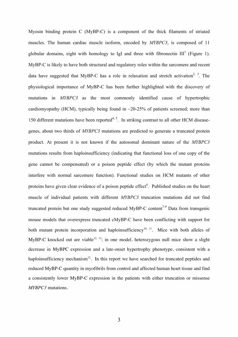
Myosin binding protein C (MyBP-C) is a component of the thick filaments of striated
muscles. The human cardiac muscle isoform, encoded by MYBPC3, is composed of 11
globular domains, eight with homology to IgI and three with fibronectin III1 (Figure 1).
MyBP-C is likely to have both structural and regulatory roles within the sarcomere and recent
data have suggested that MyBP-C has a role in relaxation and stretch activation2, 3
. The
physiological importance of MyBP-C has been further highlighted with the discovery of
mutations in MYBPC3 as the most commonly identified cause of hypertrophic
cardiomyopathy (HCM), typically being found in ~20-25% of patients screened; more than
150 different mutations have been reported4, 5
. In striking contrast to all other HCM disease-
genes, about two thirds of MYBPC3 mutations are predicted to generate a truncated protein
product. At present it is not known if the autosomal dominant nature of the MYBPC3
mutations results from haploinsufficiency (indicating that functional loss of one copy of the
gene cannot be compensated) or a poison peptide effect (by which the mutant proteins
interfere with normal sarcomere function). Functional studies on HCM mutants of other
proteins have given clear evidence of a poison peptide effect6. Published studies on the heart
muscle of individual patients with different MYBPC3 truncation mutations did not find
truncated protein but one study suggested reduced MyBP-C content7-9
Data from transgenic
mouse models that overexpress truncated cMyBP-C have been conflicting with support for
both mutant protein incorporation and haploinsufficiency10, 11. Mice with both alleles of
MyBP-C knocked out are viable12, 13; in one model, heterozygous null mice show a slight
decrease in MyBPC expression and a late-onset hypertrophy phenotype, consistent with a
haploinsufficiency mechanism12. In this report we have searched for truncated peptides and
reduced MyBP-C quantity in myofibrils from control and affected human heart tissue and find
a consistently lower MyBP-C expression in the patients with either truncation or missense
MYBPC3 mutations.

4
Materials and Methods
We obtained human heart muscle from donor hearts and interventicular septum from HCM
patients at surgical myectomy. Genotyping and mRNA analysis was by standard methods.
MyBP-C protein was detected in muscle homogenates and myofibrillar fractions using an
antibody specific to the N-terminal region of MyBP-C and the MyBP-C content was
quantified relative to the actin content using an anti-actin antibody. Details of the methods,
and clinical details, are given in full in the online supplement.
Results
We screened for MYBPC3 mutations in a series of left ventricular septum samples
from HCM patients undergoing septal myectomy to relieve left ventricular outflow tract
obstruction. In nine of the 39 patients, mutations in MYBPC3, with convincing evidence that
they were responsible for HCM, were identified (Figure 1). Two carried previously described
missense alleles Glu258Lys (sample code M10) and Arg502Trp (MA); seven had premature
terminations, truncating in domains C3 (same mutation present in M8, MI, MT, predicted
molecular weight 52 kDa), C5 (M9, 90 kDa), C7 (M15, 97kDa; M25, 114 kDa), and C10
(M6, 140 kDa).
Immunoblots were carried out on whole tissue homogenates from the myectomy
samples using an antibody specific to the N-terminus (C0-C2) of MyBP-C (see Online Figure
1). At moderate loading (2!g tissue), MyBP-C was detected as a single band (Figure 1B). We
did not observe any bands corresponding to the expected truncated protein in M6, M8, M9,
M15, M25, MI or MT at moderate (Figure 1B) or high (Online Figure 2) loading. Loading
tests indicated that the antibody could detect a concentration of less than 3% of the main
bands.
The quantity of MyBP-C in myofibrils was determined in myectomy samples and
compared with non-failing donor heart muscle (Figure 2). The quantity of MyBP-C relative

5
to actin was consistent between donor heart samples and the mean ratio was used to normalise
all of the data. An MyBP-C/actin content significantly lower than donor was found in every
myectomy sample containing a MYBPC3 mutation, including the two missense mutations
(Figure 2B and Online Figure 3, Table 2). The mean MyBP-C/actin ratio in myofibrils of all
samples with MYBPC3 mutations was 24±3% lower than donor tissue but the ratio was
unaltered in myectomy samples that did not have a MYBPC3 mutation (myectomy/donor=
1.01±0.06).
In order to examine whether reduced amounts of mutant message contributed to the
lower total MyBP-C protein content, wild type/mutant MYBPC3 mRNA ratios were measured
in four of the samples using a real time PCR assay with allele-specific primers (Figure 2C). A
moderate decrease in the relative abundance of the mutant transcript in comparison with the
wild type was found in three samples, including the missense mutation sample M10.

6
Discussion
We have been able to systematically assess the effect of both truncation and missense
HCM-causing MYBPC3 mutations in human heart muscle by studying a series of 9 samples
obtained from patients undergoing surgical myectomy in comparison with donor heart and
myectomy samples without a MYBPC3 mutation. In samples with MYBPC3 truncation
mutations we show that no truncated MyBP-C proteins are detectable, either incorporated (as
determined by analysis of myofibril fractions) or indeed unincorporated into the sarcomere
(from analysis of homogenates). In analysis of all heart samples of patients bearing MYBPC3
mutations, we find a 24% lower MyBP-C content, thus arguing strongly for
haploinsufficiency as the disease mechanism for both truncation and missense mutations. This
agrees with, and extends, certain earlier observations7-9
. For the truncation mutants, the
measured modest reductions in mRNA (Figure 2C) cannot account for the undetectable levels
of mutant MyBP-C protein and thus degradation of the truncated protein is likely, possibly via
the ubiquitin-proteasome system as earlier proposed14
. The presence of normal MyBP-C
mRNA from the remaining wild type allele is apparently not sufficient to yield a full
complement of MyBP-C protein; this is in contrast to some other contractile proteins, for
example a heterozygous !-tropomyosin knockout mouse has the normal level of protein in the
heart15
. Our surprising finding that missense mutations can cause MyBP-C haploinsufficiency
may also be explained by proteolysis of the mutant protein (as reported for one
heterologously expressed MYBPC3 missense mutant16
) although a modest reduction in mutant
mRNA (as suggested by the M10 data in Figure 2C) could also account for the reduced full
length protein.
Although haploinsufficiency has not been observed with mutations in other HCM
genes, we propose that it accounts for the pathogenic effect of MYBPC3 mutations.
Functional studies, in which MyBP-C has been partially extracted from fiber preparations to a

7
similar extent to the reduction observed in myectomy tissue, suggest that the observed
reduction in protein content is sufficient to have a significant effect on contractility17, 18
. The
depletion of MyBP-C protein in different samples is not equivelent and we suggest this may
contribute to the observed spectrum of disease severity.

8
Acknowledgements
We thank the British Heart Foundation, the Oxford NIHR Comprehensive Biomedical
Research Centre and FP6 EUGeneHeart programme LSHM-CT-2005-018833 for funding,
Samantha Harris (UCSD) for the MyBP-C antibody and Cris Dos Remedios (Sydney) for the
donor heart muscle. The contribution of Professor McKenna, Dr Elliott and Mr Tsang was in
part funded by the Department of Health's NIHR Biomedical Research Centres scheme.

9
Disclosures
None
10
Figure Legends
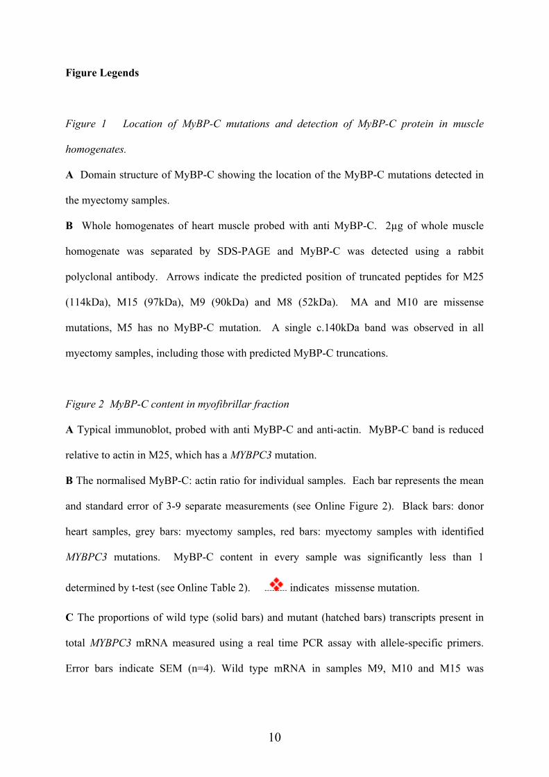
Figure 1 Location of MyBP-C mutations and detection of MyBP-C protein in muscle
homogenates.
A Domain structure of MyBP-C showing the location of the MyBP-C mutations detected in
the myectomy samples.
B Whole homogenates of heart muscle probed with anti MyBP-C. 2!g of whole muscle
homogenate was separated by SDS-PAGE and MyBP-C was detected using a rabbit
polyclonal antibody. Arrows indicate the predicted position of truncated peptides for M25
(114kDa), M15 (97kDa), M9 (90kDa) and M8 (52kDa). MA and M10 are missense
mutations, M5 has no MyBP-C mutation. A single c.140kDa band was observed in all
myectomy samples, including those with predicted MyBP-C truncations.
Figure 2 MyBP-C content in myofibrillar fraction
A Typical immunoblot, probed with anti MyBP-C and anti-actin. MyBP-C band is reduced
relative to actin in M25, which has a MYBPC3 mutation.
B The normalised MyBP-C: actin ratio for individual samples. Each bar represents the mean
and standard error of 3-9 separate measurements (see Online Figure 2). Black bars: donor
heart samples, grey bars: myectomy samples, red bars: myectomy samples with identified
MYBPC3 mutations. MyBP-C content in every sample was significantly less than 1
determined by t-test (see Online Table 2). indicates missense mutation.
C The proportions of wild type (solid bars) and mutant (hatched bars) transcripts present in
total MYBPC3 mRNA measured using a real time PCR assay with allele-specific primers.
Error bars indicate SEM (n=4). Wild type mRNA in samples M9, M10 and M15 was

11
significantly greater than mutant MYBPC3 mRNA (* p<0.05) whereas the difference in the
M6 sample was not significant (ns).
12
References
1. Flashman E, Redwood C, Moolman-Smook J, Watkins H. Cardiac
myosin binding protein C: its role in physiology and disease. Circ Res.
2004;94(10):1279-1289.
2. Pohlmann L, Kroger I, Vignier N, Schlossarek S, Kramer E, Coirault C,
Sultan KR, El-Armouche A, Winegrad S, Eschenhagen T, Carrier L.
Cardiac myosin-binding protein C is required for complete relaxation in
intact myocytes. Circ Res. 2007;101(9):928-938.
3. Stelzer JE, Dunning SB, Moss RL. Ablation of cardiac myosin-binding
protein-C accelerates stretch activation in murine skinned myocardium.
Circ Res. 2006;98(9):1212-1218.
4. Watkins H, Conner D, Thierfelder L, Jarcho JA, MacRae C, McKenna
WJ, Maron BJ, Seidman JG, Seidman CE. Mutations in the cardiac
myosin binding protein-C gene on chromosome 11 cause familial
hypertrophic cardiomyopathy. Nature Genetics. 1995;11(4):434-437.
5. Bonne G, Carrier L, Bercovici J, Cruaud C, Richard P, Hainque B, Gautel
M, Labeit S, James M, Beckmann J, Weissenbach J, Vosberg HP,
Fiszman M, Komajda M, Schwartz K. Cardiac myosin binding protein-C
gene splice acceptor site mutation is associated with familial hypertrophic
cardiomyopathy. Nature Genetics. 1995;11(4):438-440.
6. Redwood CS, Moolman-Smook JC, Watkins H. Properties of mutant
contractile proteins that cause hypertrophic cardiomyopathy. Cardiovasc
Res. 1999;44(1):20-36.
7. Rottbauer W, Gautel M, Zehelein J, Labeit S, Franz WM, Fischer C,
Vollrath B, Mall G, Dietz R, Kubler W, Katus HA. Novel splice donor
site mutation in the cardiac myosin-binding protein-C gene in familial
hypertrophic cardiomyopathy. Characterization Of cardiac transcript and
protein. J Clin Invest. 1997;100(2):475-482.
8. Moolman JA, Reith S, Uhl K, Bailey S, Gautel M, Jeschke B, Fischer C,
Ochs J, McKenna WJ, Klues H, Vosberg HP. A newly created splice
donor site in exon 25 of the MyBP-C gene is responsible for inherited
hypertrophic cardiomyopathy with incomplete disease penetrance.
Circulation. 2000;101(12):1396-1402.
9. van Dijk SJ, Dooijes D, dos Remedios C, Michels M, Lamers JM,
Winegrad S, Schlossarek S, Carrier L, ten Cate FJ, Stienen GJ, van der
Velden J. Cardiac myosin-binding protein C mutations and hypertrophic
cardiomyopathy: haploinsufficiency, deranged phosphorylation, and
cardiomyocyte dysfunction. Circulation. 2009;119(11):1473-1483.
10. Yang Q, Sanbe A, Osinska H, Hewett TE, Klevitsky R, Robbins J. A
mouse model of myosin binding protein C human familial hypertrophic
cardiomyopathy. J Clin Invest. 1998;102(7):1292-1300.

13
11. Yang Q, Sanbe A, Osinska H, Hewett TE, Klevitsky R, Robbins J. In vivo
modeling of myosin binding protein C familial hypertrophic
cardiomyopathy. Circ Res. 1999;85(9):841-847.
12. Carrier L, Knoll R, Vignier N, Keller DI, Bausero P, Prudhon B, Isnard R,
Ambroisine ML, Fiszman M, Ross J, Jr., Schwartz K, Chien KR.
Asymmetric septal hypertrophy in heterozygous cMyBP-C null mice.
Cardiovasc Res. 2004;63(2):293-304.
13. Harris SP, Bartley CR, Hacker TA, McDonald KS, Douglas PS, Greaser
ML, Powers PA, Moss RL. Hypertrophic cardiomyopathy in cardiac
myosin binding protein-C knockout mice. Circ Res. 2002;90(5):594-601.
14. Sarikas A, Carrier L, Schenke C, Doll D, Flavigny J, Lindenberg KS,
Eschenhagen T, Zolk O. Impairment of the ubiquitin-proteasome system
by truncated cardiac myosin binding protein C mutants. Cardiovasc Res.
2005;66(1):33-44.
15. Blanchard EM, Iizuka K, Christe M, Conner DA, Geisterfer-Lowrance A,
Schoen FJ, Maughan DW, Seidman CE, Seidman JG. Targeted ablation
of the murine alpha-tropomyosin gene. Circ Res. 1997;81(6):1005-1010.
16. Bahrudin U, Morisaki H, Morisaki T, Ninomiya H, Higaki K, Nanba E,
Igawa O, Takashima S, Mizuta E, Miake J, Yamamoto Y, Shirayoshi Y,
Kitakaze M, Carrier L, Hisatome I. Ubiquitin-proteasome system
impairment caused by a missense cardiac myosin-binding protein C
mutation and associated with cardiac dysfunction in hypertrophic
cardiomyopathy. Journal of molecular biology. 2008;384(4):896-907.
17. Hofmann PA, Hartzell HC, Moss RL. Alterations in Ca2+ sensitive
tension due to partial extraction of C-protein from rat skinned cardiac
myocytes and rabbit skeletal muscle fibers. J Gen Physiol.
1991;97(6):1141-1163.
18. Kulikovskaya I, McClellan G, Levine R, Winegrad S. Effect of extraction
of myosin binding protein C on contractility of rat heart. Am J Physiol
Heart Circ Physiol. 2003;285(2):H857-865.

M6
Arg1271stop1-1270 amino acids
C0 C1 C2 C3 C4 C5 C6 C7 C8 C9 C10C0 C1 C2 C3 C4 C5 C6 C7 C8 C9 C10
M10
Glu258LysMA
Arg502Trp
M8, MI, MT intron17 donor site A>T+4
1-485 amino acids
M9
InsG2374 1-791 + 40 novel amino acids
M15
T>A 2604, delC 26051-868 + 13 nonsense amino acids
M6
Arg1271stop1-1270 amino acids
M25
delCT 2864/5954 + 94 nonsense amino acids
IgI-like domain
Fn3-like domain
phosphorylatableLAGGGRRIS loop
A B
Marston et al., Figure 1
M25 MA M15 M10 M9 M8 M6 M5

MYBP-C
ACTIN
0
0.2
0.4
0.6
0.8
1
1.2
1.4
N6 N8 N9 N10 N11 M1 M2 M3 M4 M5 M6 M7 M8 M9 M10 M11 M15 M25 M26 MA
mean HOCM
* ** *
BA
D8 M26 M25
D6 D8 D9 D10 D11
NormalizedMyBP-Ccontent
C
Figure 3
0
0.5
1
1.5
2
myectomy donor MYBPC3
mutation
No
rma
lize
d M
yB
P-C
/Actin
1.01±0.06 1.00±0.05 0.76±0.04
p=0.96
p=0.0005
0
0.2
0.4
0.6
0.8
1
1.2
1.4
D6 D8 D9 D10 D11 M1 M2 M3 M4 M5 M7 M11M26 M6 M8 M9 M10M15M25 MA MIMT
mean H
OC
M ! !
Donor Myectomy Myectomy ( no MYBPC3 mutation) with MYBPC3 mutation
A
B
C
Marston et al., Figure 2
MyB
P-C
conte
nt re
lative to d
onor
truncation mutations missensemutation
ns * * *
0
0.2
0.4
0.6
0.8
M6 M9 M15 M10
wild typemutant
Pro
port
ion o
f to
tal MYBPC3 m
RN
A
Evidence from human myectomy samples that MYBPC3 mutations cause hypertrophic
cardiomyopathy through haploinsufficiency.
S. Marston, O. Copeland, A. Jacques, K. Livesey, V. Tsang, P.M. Elliott, W.J. McKenna, S.
Jalilzadeh, S. Carballo, C. Redwood, H. Watkins
SUPPLEMENTARY MATERIAL: METHODS
Collection and Storage of Human Myocardium
Hypertrophic Cardiomyopathy Human myocardial samples were obtained from patients with
hypertrophic cardiomyopathy undergoing surgical septal myectomy for relief of left
ventricular outflow tract obstruction. The samples were frozen in liquid nitrogen and stored
for later use. Local ethical approval was obtained from University College London Hospitals
and Royal Brompton and Harefield ethics committees for collection of tissue samples. The
HCM patients clinical phenotypes were characterised by obtaining detailed clinical histories
and examinations. All patients had cardiac investigations including, 12-lead ECG, chest X-
ray, holter monitor, cardiopulmonary exercise test, two-dimensional transthoracic
echocardiography, transoesophageal echocardiography, cardiac catheterisation and coronary
angiography ( Online Table 1) 1, 2
.
Non-Failing donor heart muscle. Tissue samples were supplied by Prof. C Dos Remedios,
University of Sydney, Australia. Ethical approval was obtained from The Brompton,
Harefield & NHLI, London and St Vincent’s Hospital, Sydney. The investigation conformed
with the principles outlined in the Declaration of Helsinki. Non-failing heart tissue (donor)
was obtained from donor hearts where no suitable transplant recipient was found. These were
obtained from patients with no history of cardiac disease, a normal cardiac examination,
normal ECG and normal ventricular function on echocardiography within 24 hours of heart
explantation. Myocardium was immediately frozen in liquid nitrogen and stored for later
analysis. Clinical and functional characteristics of troponin from these samples has been
previously reported 3.

Genotyping of HCM Patients
Blood samples were collected and genetic analysis performed on the genomic DNA extracted.
Local ethical approval was obtained for collection of blood samples; mutation screening of
coding regions and splice sites of the MYBPC3 gene (exon 1-34) was undertaken using 'Hi-res
Melting' analysis (LightScanner) and bi-directional fluorescent sequencing using big dye
terminators and ABI3730.
Preparation of whole muscle homogenates and myofibrillar fraction from human heart
muscle
Human heart samples (50mg) were removed from liquid nitrogen and immediately pulverised
in a lN2 cooled percussion mortar. Pulverised human heart muscle was manually homogenised
in 1.5ml of a wash buffer containing 5mM NaH2PO4, 5mM Na2HPO4 pH 7.0, 0.1M NaCl,
5mM MgCl2, 0.5mM EGTA, 0.1% Triton X-100 and 5mM DTT with 2µg/ml each of the
protease inhibitors E64, chymostatin, leupeptin and pepstatin A. This whole homogenate was
used to assay for truncated peptides. For myofibrils the homogenate was then centrifuged at
16,500xg for 5 minutes and the supernatant discarded. The wash-homogenisation-
sedimentaion step was repeated three more times until the pellet was pale yellow. The
myofibrillar pellet was then dissolved in SDS-gel solution for analysis by SDS-PAGE.
Western blotting.
The MyBP-C content in whole muscle homogenates and in the myofibrillar fraction was
measured in western blots of SDS-PAGE. Gel-electrophoresed proteins were transferred to
nitrocellulose membrane (Hybond ECL, Amersham Pharmacia Biotech, RPN 303D) with a
Hoeffer semi-dry electroblotter (Semi-phor TE70) at 6mA and 200 mV for 2 hours at 4°C.
Membranes were blocked for 1 hour at room temperature in blocking buffer [1% dried milk
powder, 0.1% Tween 20 and 1 x phosphate buffered saline (137 mM NaCl, 27 mM KCL, 4.3
mM Na2HPO4.7H2O, 1.4 mM KH2PO4, pH 7.3)]. Blots were incubated overnight in rabbit
polyclonal antibody to MyBP-C at 1/20,000 (gift from Samantha Harris 4) normalised to
tissue quantity by including a rabbit polyclonal anti-actin antibody (Sigma A2066, 1/2000).
MyBP-C and actin were detected using anti-rabbit HRP and ECL and visualised with a cooled
CCD camera. 12-bit TIFF images were analysed with GeneQuant software (Syngene). Tests
with recombinant MyBP-C fragments showed that the antibody cross-reacted specifically
with the N-terminal region (C0-C2, see Online Figure 1).

mRNA quantitation
RNA was extracted from 30 mg of frozen myectomy tissue using the RNeasy Fibrous Tissue
kit (Qiagen) according to manufacturers’ instructions. Reverse transcription was carried out
using oligo dT primers. The following Taqman primers specific for the wild type (FAM
labelled) and mutant (YAK labelled) allele for samples M6, M9, M10 and M15 were
synthesised:
M6: 5’-CGCCTGGAGGTGCGAGTGCC (wild type)
5’-CGCCTGGAGGTGTGAGTGCC (mutant)
M9: 5’-CCTGCAGTACAGTTGGGAGCCGC (wild type)
5’-CTGCACAGTACAGGTTGGGAGCCGC (mutant)
M10: 5’-CTCACTGTCCACGAGGCCTGGGCA (wild type)
5’-CTCACTGTCCACAAGGCCTGGGCA (mutant)
M15 5’-TCGCTGGGGGGACCGATAGGC (wild type)
5’-GGTTCGCTGGGGGTCCGATAGGC (mutant)
qPCR carried out using a Roche 480 Lightcycler. The difference in the number of cycles
between wild type and mutant to reach threshold (!Ct method) was used to calculate the ratio
of wild type to mutant mRNA with the assumption that the efficiencies of the wild and mutant
reactions were identical.

SUPPLEMENTARY MATERIAL: RESULTS
Online Figure 1
Demonstration of specificity of the MyBP-C antibody for the N-terminal domains C0-C2
upstream of all predicted chain terminations.

Online Figure 2
Western blot of 15!g of whole muscle homogenate separated by SDS-PAGE and probed with
antibody to MyBP-C. Bands at 95, 60 and 45kDa represent non-specific labelling of
myofibrillar proteins or breakdown of MyBP-C and were the same in all samples. Arrows
indicate the predicted position of truncated peptides in M25, M15, M9 and M8.
M25 MA M15 M10 M9 M8 M6 M5
13
5
95
60
45
114
97 90
52

Online Figure 3
Complete dataset of MyBP-C content determinations in myectomy samples.
Means and standard errors of these data are plotted in Figure 2C. Red asterisk indicates
missense mutations.
0
0.2
0.4
0.6
0.8
1
1.2
M6 M9 M10 MI MT M8 M15 M25 MA
M6M9M10MIMTM8M15M25MA
MyB
P-C
co
nte
nt
rela
tive
to
do
no
r
! !

Online Table 2
Statistical analysis of data.
MyBP-C content is significantly less than the donor control (=1, t-test) and in most cases
significantly greater than 0.5.
Sample Quantity
relative to
NF ± sem
n P single group v.s. 0.5 P single group v.s. 1.0
M6 0.77±0.05 9 0.0006 0.002
M8 0.68±0.07 5 .07 .01
M9 0.81±0.05 5 0.004 0.028
M10 0.85±0.03 9 <0.0001 0.002
M15 0.65±0.04 9 <.0001 .006
M25 0.65±0.05 3 .088 .02
MA 0.82±0.05 7 .0004 .01
MI 0.83±0.04 8 <0.0001 0.003
MT 0.74±0.03 8 0.0002 0.0001

Online Figure 4
Determination of the transcripts produced by the Glu258Lys mutation
An earlier report found that the Glu258Lys mutation has an effect on splicing in that two
transcripts were shown to be generated by this mutation in peripheral blood lymphocyte
cDNA, one full length bearing the expected missense mutation and the other in which exon 6
is skipped5. |In order to test whether the Glu258Lys mutation affects splicing in the heart,
cDNA was amplified by PCR using the MYBPC3 primers 5’-
ACTGCAGAACATATGATTGGCCTCTTCGTGATGCGG and 5’-
GCTGGAGGTGGTCGGCGGATCAGTGATAGCCAT; this was predicted to give a 417bp
product for the normally spliced transcript and a 249bp product if exon 6 were skipped. Our
data show no evidence of the 249bp product suggesting that this mutation generates only a
full length transcript including the Glu258 mutation in the myocardium.

REFERENCES
1. Jacques A, Briceno N, Messer A, Gallon C, Jalizadeh S, Garcia E, Kikonda-Kanda G,
Goddard J, Harding S, Watkins H, Tsang V, McKenna W, Marston S. The molecular
phenotype of human cardiac myosin associated with hypertrophic obstructive
cardiomyopathy. Cardiovasc Res. 2008;79:481-491.
2. Jacques A, Copeland O, Messer A, Gallon C, King C, McKenna W, Tsang V, Marston
S. Myosin binding protein C phosphorylation in normal, hypertrophic and failing
human heart muscle. J Mol Cell Cardiol. 2008;45:209-216.
3. Messer AE, Jacques AM, Marston SB. Troponin phosphorylation and regulatory
function in human heart muscle: Dephosphorylation of Ser23/24 on troponin I could
account for the contractile defect in end-stage heart failure. J Mol Cell Cardiol.
2007;42:247-259.
4. Harris SP, Bartley CR, Hacker TA, McDonald KS, Douglas PS, Greaser ML, Powers
PA, Moss RL. Hypertrophic cardiomyopathy in cardiac myosin binding protein-C
knockout mice. Circ Res. 2002;90:594-601.
5. Andersen PS, Havndrup O, Bundgaard H, Larsen LA, Vuust J, Pedersen AK, Kjeldsen
K, Christiansen M. Genetic and phenotypic characterization of mutations in myosin-
binding protein C (MYBPC3) in 81 families with familial hypertrophic
cardiomyopathy: total or partial haploinsufficiency.Eur J Hum Genet. 2004 ;12:673-7.