evidence for transovarial transmission of tomato yellow
TRANSCRIPT

Evidence for Transovarial Transmission of Tomato Yellow Leaf Curl Virusby Its Vector, the Whitefly Bemisia tabaci
Murad Ghanim, Shai Morin, Muhammad Zeidan, and Henryk Czosnek1
Department of Field Crops and Genetics, and the Otto Warburg Center for Biotechnology in Agriculture, Faculty of Agricultural,Food and Environmental Quality Sciences, The Hebrew University of Jerusalem, Rehovot 76100, P.O. Box 12, Israel
Received August 12, 1997; returned to author for revision September 17, 1997; accepted November 9, 1997
The whitefly Bemisia tabaci is the only vector of the tomato yellow leaf curl geminivirus (TYLCV). The insect transmits thevirus in a persistent-circulative manner. TYLCV DNA was detected by polymerase chain reaction and by Southern blothybridization in progeny (eggs, first and second instars, adults) of single viruliferous whiteflies that developed on eggplantor on cotton (two TYLCV nonhost plants). Furthermore, TYLCV DNA was present in the progeny of insects that had acquiredthe virus through the egg. The adult progeny of the viruliferous insects and their own progeny were able to infect tomato testplants, producing typical disease symptoms. Ovaries and maturing eggs of viruliferous insects contained viral DNA, as dideggs laid by viruliferous insects maintained on an artificial diet. Eggs laid by nonviruliferous whiteflies on cotton plantspreviously caged with viruliferous insects did not acquire viral DNA from the plant. Hence, TYLCV can be transmitted throughthe egg for at least two generations. In the absence of an available plant host, the whitefly may serve as a reservoir of thevirus between growing seasons. © 1998 Academic Press
INTRODUCTION
Tomato yellow leaf curl virus (TYLCV) is the namegiven to a group of geminiviruses transmitted by thewhitefly Bemisia tabaci (Cohen and Harpaz, 1964;Czosnek et al., 1988a). TYLCV causes extensive dam-age to tomato crops in many tropical and subtropicalregions worldwide (Czosnek and Laterrot, 1997). Thegenome of TYLCV is either monopartite (Mediterra-nean isolates) or bipartite (Thailand isolate) (for reviewsee Pico et al., 1996; Czosnek and Laterrot, 1997).While the acquisition and transmission of TYLCV by B.tabaci (also denominated Bemisia argentifolii) havebeen studied in some detail (Cohen and Nitzany, 1966;Zeidan and Czosnek, 1991; Mehta et al., 1994; Caciagliet al., 1995; Caciagli and Bosco, 1997; Rubinstein andCzosnek, 1997), the interactions between this gemini-virus (as well as others) and its insect vector are stillpoorly understood. Similarly to other whitefly-transmit-ted geminiviruses (Duffus, 1987), B. tabaci transmitsTYLCV in a persistent-circulative manner (Cohen andNitzany, 1966; Rubinstein and Czosnek, 1997). Thenucleic acid of the virus remains associated with theinsect for its entire adult life; this long-term associa-tion affects the transmission efficiency, longevity, andfecundity of the insect, features reminiscent of aninsect pathogen (Rubinstein and Czosnek, 1997).Hence, the relationship between TYLCV and B. tabaci
seems to be much subtler than the previous assump-tions, depicting the passage of the virus in the insectas neutral. In this communication we investigatewhether TYLCV is transmitted by its insect vectorthrough the egg.
Transovarial transmission of a plant virus by itsinsect vector was first described by Fukushi (1933),who showed that rice dwarf virus was transmitted forseveral generations through the egg of the leafhoppervector Nephotettix apicalis. The ability of the progenyof viruliferous insects reared on immune plants toinfect susceptible hosts pointed to transovarial pas-sage of the virus. Since then, transovarial passage ofseveral viruses in their leafhopper/planthopper (Grylls,1954; Black, 1953), and aphid (Sylvester, 1969) vectorshas been described (for review see Matthews, 1991).Transovarial transmission has been often associatedwith replication of the virus in its vector (Fukushi, 1935;Black, 1950; Duffus, 1963; Miyamoto and Miyamoto,1966) and with deleterious effects on the insect host(Sylvester, 1969, 1973; Sylvester and Richardson, 1969).Usually, the virus was transmitted to some, but not toall progeny.
Until now, all plant viruses that have been shown tobe transmitted to progeny were RNA viruses. Gemini-viruses have not been considered to be transmittedtransovarially to progeny (Harrison, 1985; Lazarowitz,1992). Using polymerase chain reaction (PCR), South-ern blot hybridization, and transmission tests, wepresent evidence that TYLCV can be transmitted to the
1 To whom correspondence and reprint requests should be ad-dressed. Fax: 972 8 9468265. E-mail: [email protected].
VIROLOGY 240, 295–303 (1998)ARTICLE NO. VY978937
0042-6822/98 $25.00Copyright © 1998 by Academic PressAll rights of reproduction in any form reserved.
295

progeny of viruliferous insects for at least two gener-ations.
RESULTS
PCR analysis of progeny of viruliferous whiteflies
Whiteflies develop from an egg, through fournymphal instars, into an adult. Figure 1A shows thedevelopmental time course under our experimentalconditions. The association of TYLCV DNA with devel-oping eggs of viruliferous insects was investigated.Viruliferous whiteflies were caged on eggplants andon cotton plants (about 20 insects per plant) and ontwo TYLCV nonhosts (Cohen and Nitzany, 1966; Al-Musa, 1982). The adults were collected after 5 daysand the eggs were allowed to develop. Samples fromeach developmental stage (Fig. 1A) were collected atrandom. Total DNA was extracted from groups of 10viruliferous females, 50 of their eggs, 20 crawlers, and20 adult progeny. In parallel, DNA was extracted fromthe same number of nonviruliferous females, theireggs, crawlers, and adult progeny and from an in-fected tomato leaflet. All samples were subjected to
PCR. Plasmid pTYH19 that contains a cloned copy ofthe TYLCV genome was used as a positive control.Figure 1B shows that the ;410-bp TYLCV DNA frag-ment that was amplified from plasmid pTYH19, frominfected tomato, and from viruliferous whiteflies wasalso amplified from the progeny of the viruliferousfemales (eggs, crawlers, and adults), but not fromthose of nonviruliferous insects. These results suggestthat TYLCV is transmitted to the progeny of viruliferouswhiteflies through the egg.
The offspring of a single viruliferous whitefly containTYLCV DNA
The question of whether TYLCV is present in all prog-eny of a single viruliferous insect was addressed. Viru-liferous whiteflies were allowed to lay eggs on eggplants(one insect per plant). After 5 days, the insects werecollected and eggs were allowed to develop. In accor-dance with the developmental time scale of Fig. 1A,eggs, crawlers, pupae, and adults were collected in sucha fashion that all the individuals at a given developmen-tal stage were progeny of a single insect. DNA extractedfrom each individual was subjected to PCR amplification.Figure 2 shows the ;410-bp viral DNA fragment ampli-fied from 16 of the 23 eggs laid by a single viruliferouswhitefly. Twenty-five of the 30 crawlers issued from an-other viruliferous insect contained viral DNA as did 7 ofthe 13 pupae and 15 of the 16 adults issued from a thirdand a fourth insect. Hence, TYLCV was transmitted tosome, but not to all, progeny of viruliferous whiteflies.
This analysis was extended to the progeny of nineviruliferous whiteflies. Viruliferous insects were allowedto lay eggs on eggplants and on cotton plants (one insectper plant). Eggs, first and second instars, and adultswere collected in such a fashion that all the individuals ata given developmental stage were progeny of three dif-ferent insects. The progeny from a single insect werekept separated from those of the other insects. DNA fromeach individual was subjected to PCR amplification, asdescribed in the legend to Fig. 2. The results summa-rized in Table 1 show that TYLCV DNA was amplifiedfrom some, but not from all progeny. This proportionvaried from insect to insect. TYLCV DNA was present inall of the developmental stages of the whitefly, from eggto adult. Adult progeny of three additional viruliferousinsects were used to inoculate tomato test plants (seebelow).
The second-generation progeny of a singleviruliferous whitefly contain TYLCV DNA
The passage of TYLCV through the egg from the first tothe second generation of viruliferous whiteflies progenywas investigated using the methods described above.Eggs laid by whiteflies that had acquired TYLCV frominfected tomato were allowed to develop on cotton. The
FIG. 1. Analysis of viruliferous whiteflies and their offspring by PCR.(A) Development stages of the whitefly Bemisia tabaci and their dura-tion, under our experimental conditions. The first instar is the crawlerstage; the fourth instar is the pupal stage. (B) Amplification of TYLCVDNA from groups of 50 eggs, 20 crawlers and 20 adults, from offspringof viruliferous whiteflies, and from the same number of progeny ofnonviruliferous insects; infected tomato plants and plasmid pTYH19 arepositive controls. The reaction products were submitted to agarose gelelectrophoresis and stained with ethidium bromide. Thick arrow, am-plified TYLCV DNA; thin arrow, primers.
296 GHANIM ET AL.

adult offspring, 1 to 5 days after emergence, were cagedwith cotton plants, one insect per plant. After 5 days, theinsects were collected and analyzed by PCR for thepresence of TYLCV DNA. Only the progeny of thoseinsects that contained viral DNA were further analyzed.Eggs, first and second instars, and adults were collectedas described above. The progeny from a single insectwere kept separated from those of the other insects.DNA extracted from each individual was subjected toPCR amplification and the products were analyzed asdescribed in the legend to Fig. 2. The results obtainedwith progeny of nine insects are summarized in Table 1.
TYLCV DNA was present in all of the developmentalstages of the progeny of whiteflies that have acquired thevirus through the egg. The proportion of individuals con-taining viral DNA varied from one insect to another andwas similar to that found in the first generation progenyof viruliferous insects. These results showed that TYLCVwas transmitted through the egg for two generations.Adult progeny of three insects, second generation prog-eny, were used to inoculate tomato test plants.
Southern blot analysis of DNA extracted from groupsof eggs, crawlers, and adults, offspring of the first andthe second generation of viruliferous whiteflies, indi-
FIG. 2. PCR amplification of TYLCV DNA in the developing progeny of individual viruliferous whiteflies. All the individuals collected at eachdevelopmental stage are offspring of a single whitefly. The reaction products were submitted to agarose gel electrophoresis, blotted, and hybridizedwith the virus DNA probe.
297TRANSMISSION OF TYLCV BY Bemisia tabaci
cated that they all contained a single detectable TYLCVDNA species, comigrating with the virus genomic DNAfrom viruliferous whiteflies (Fig. 3).
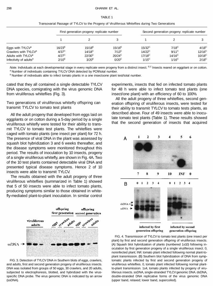
Two generations of viruliferous whitefly offspring cantransmit TYLCV to tomato test plants
All the adult progeny that developed from eggs laid oneggplants or on cotton during a 5-day period by a singleviruliferous whitefly were tested for their ability to trans-mit TYLCV to tomato test plants. The whiteflies werecaged with tomato plants (one insect per plant) for 72 h.The presence of viral DNA in the plant was assessed bysquash blot hybridization 3 and 6 weeks thereafter, andthe disease symptoms were monitored throughout thisperiod. The results of inoculation by 10 insects, progenyof a single viruliferous whitefly, are shown in Fig. 4A. Twoof the 10 test plants contained detectable viral DNA andpresented typical disease symptoms. Hence 2 of 10insects were able to transmit TYLCV.
The results obtained with the adult progeny of threeviruliferous whiteflies (summarized in Table 1) showedthat 5 of 50 insects were able to infect tomato plants,producing symptoms similar to those obtained in white-fly-mediated plant-to-plant inoculation. In similar control
experiments, insects that fed on infected tomato plantsfor 48 h were able to infect tomato test plants (oneinsect/one plant) with an efficiency of 60 to 100%.
All the adult progeny of three whiteflies, second gen-eration offspring of viruliferous insects, were tested fortheir ability to transmit TYLCV to tomato tests plants, asdescribed above. Four of 49 insects were able to inocu-late tomato test plants (Table 1). These results showedthat the second generation of insects that acquired
TABLE 1
Transovarial Passage of TYLCV to the Progeny of Viruliferous Whiteflies during Two Generations
First generation progeny: replicate number Second generation progeny: replicate number
1 2 3 1 2 3
Eggs with TYLCVa 16/23E 15/18E 15/16E 15/32C 7/18C 4/18C
Crawlers with TYLCVa 4/37C 14/18C 7/13C 14/22C 9/11C 12/16C
Adults with TYLCVa 4/27C 22/30C 20/24C 17/18C 14/16C 10/18C
Infectivity of adultsb 2/10E 3/20E 0/20C 1/15C 1/16C 2/18C
Note. Individuals at each developmental stage in every replicate were progeny from a distinct insect. E,C Insects reared on eggplant or on cotton.a Number of individuals containing TYLCV DNA detected by PCR/total number.b Number of individuals able to infect tomato plants in a one insect–one plant test/total number.
FIG. 3. Detection of TYLCV DNA in Southern blots of eggs, crawlers,and adults, first and second generation progeny of viruliferous insects.DNA was isolated from groups of 50 eggs, 30 crawlers, and 20 adults,subjected to electrophoresis, blotted, and hybridized with the virus-specific DNA probe. The virus genomic DNA is indicated by an arrow(ssDNA).
FIG. 4. Transmission of TYLCV to tomato test plants (one insect perplant) by first and second generation offspring of viruliferous insects.(A) Squash blot hybridization of plants (numbered 1–10) following in-oculation by first generation progeny of a single viruliferous insect. 0,noninfected plant; INF, tomato plant infected following normal plant-to-plant transmission. (B) Southern blot hybridization of DNA from symp-tomatic plants infected by first and second generation progeny ofviruliferous whiteflies. 0, tomato plant infected following normal plant-to-plant transmission. 1–4, tomato plants infected by progeny of viru-liferous insects. ssDNA, single-stranded TYLCV genomic DNA. dsDNA,double-stranded DNA replicative forms of the virus genomic DNA(upper band, relaxed; lower band, supercoiled).
298 GHANIM ET AL.

TYLCV through the egg was able to transmit the virus toplants with efficiency similar to that of the first genera-tion.
The Southern blot analysis shown in Fig. 4B indicatedthat the symptomatic tomato plants inoculated by theprogeny of viruliferous insects and their second gener-ation offspring contained TYLCV-associated viral DNAforms similar to those present in naturally infected toma-toes.
Developing eggs, instars, and adults could notacquire TYLCV from eggplant or cotton on whichviruliferous whiteflies laid their eggs
We had to exclude the possibility that eggs acquiredTYLCV DNA from the plants on which they developed.During the 5-day oviposition, it is likely that the insectinjected virus while phloem feeding on cotton or egg-plant. Although these plants are immune to TYLCV (Co-hen and Nitzany, 1966; Al-Musa, 1982), it is still possiblethat the virus was present in undetectable amounts (rep-licating or not) in the cells on which the developing eggswere feeding. As a result, the freshly laid eggs may haveacquired the virus from the few inoculated cells via thepedicel, although it penetrates only the leaf epidermis(Pollard, 1955). TYLCV may also have been acquired at alater developmental stage, by crawlers that are known tofeed on phloem (Pollard, 1955) and by adults.
Experiments were conducted (mimicking the experi-mental conditions described above) to verify that prog-eny of viruliferous whiteflies, at any developmental stage,could not acquire TYLCV from the plant on which eggswere laid. One viruliferous insect was caged on each offive eggplants and five cotton plants, using a 2 cm diam-eter leaf cage. After 10 days, the adult as well as all eggsand instars were collected and discarded. Nonvirulifer-ous insects were caged for 5 days on the same plants(one insect per plant), at the same location, using leafcages. All the crawlers that developed from the eggs andthat were analyzed for the presence of TYLCV DNA byPCR did not contain TYLCV DNA, indicating that eggsand crawlers did not acquired TYLCV DNA from theplants. Five cotton plants and five eggplants were cagedfor 10 days with about 200 viruliferous whiteflies each.The viruliferous insects were discarded, and nonvirulif-erous ones were caged with these plants (200 insectsper plant) for 48 h. These whiteflies were then collectedand caged on six tomato plants for 48 h (50 insects perplant, three plants with insects from eggplants and threewith insects from cotton). Observation of plants for symp-toms and hybridization of plant extracts with virus-spe-cific DNA probes showed that none of the tomato plantswas inoculated by these insects. Therefore adult prog-eny of viruliferous whiteflies could not have acquired(sufficient amounts of) viral DNA from eggplants or cot-ton plants to inoculate tomato test plants.
Eggs laid by viruliferous insects on artificial mediumcontain TYLCV DNA
In order to avoid possible contamination of eggs withTYLCV from the plants, insects were allowed to lay eggson artificial medium. After a 5-day ovoposition period, theeggs were collected individually. Total DNA from eachegg was extracted and subjected to PCR. In one case, 23of the 25 eggs laid by an insect contained TYLCV DNA;in another case, 16 of the 17 eggs laid by another insectcontained viral DNA. Hence, the eggs were laid whilealready containing viral DNA.
Ovaries and developing eggs of viruliferous insectscontain TYLCV DNA
The experiments described above referred to devel-oping eggs, after oviposition. Viruliferous insects weredissected to verify that the ovaries and the matureeggs in the ovary contained viral DNA. The organswere washed until no viral DNA could be amplified byPCR from the wash. Samples from ovaries and frommature eggs were observed in the scanning electronmicroscope (Figs. 5A and 5B). DNA prepared fromindividual ovaries and from single maturing eggs wassubjected to PCR, and the products were analyzed asdescribed above. The results presented in Fig. 5Cshow that viral DNA was present in the ovaries and inthe mature eggs of viruliferous insects. Washing theeggs with mineral oil to avoid possible contaminationwith hemolymph did not eliminate the viral DNA.
DISCUSSION
In this study, we have examined whether TYLCV canbe transmitted to the progeny of viruliferous whitefliesthrough the egg. The rationale of this question wasbased on observations regarding the survival of the virusbetween growing seasons of tomato: (1) The virus is nottransmitted through seeds of infected plants; (2) there isno obvious alternative host to tomato that has beenshown to be the likely reservoir of the virus betweenseasons; (3) infection of tomatoes starts almost immedi-ately after planting even when the insect population isnot at its peak (Cohen et al., 1988). Therefore, we havepostulated that the whitefly serves as a source for thevirus, which is passed from generation to generationthrough the egg.
Besides the molecular biology techniques that werenot available at that time, we have followed method-ologies used to demonstrate transovarial passage of anumber of plant viruses by their leafhopper, planthop-per, or aphid vector (Black, 1950, 1953; Grylls, 1954;Duffus, 1963; Sylvester, 1969; Sylvester and Richard-son, 1969, 1970). We have shown here by PCR ampli-fication, by Southern blot hybridization, and by trans-mission tests that indeed TYLCV can be found in
299TRANSMISSION OF TYLCV BY Bemisia tabaci

progeny of a single viruliferous insect for at least twogenerations, demonstrating that TYLCV can be trans-mitted through the egg.
In experiments aimed at detecting transovarial trans-mission, it is essential to eliminate the possibility that theprogeny acquire the virus from plants on which they arereared. Inoculation and transmission tests using white-flies have shown that cotton and eggplant are TYLCVnonhosts (Cohen and Nitzany, 1966; Al-Musa, 1982). Re-gardless, we had to exclude the possibility that eggs,crawlers, or adult whiteflies acquired the virus from theeggplant and cotton on which viruliferous insects fedduring oviposition. Ideally, the eggs laid by viruliferouswhiteflies and by their offspring should have been trans-ferred immediately to new plants to avoid possible con-tamination. Unfortunately whitefly eggs did not developafter transplantation on eggplant, cotton, or tomato.Therefore, we tested the possibility that eggs, crawlers,or adults could acquire (and later transmit) TYLCV fromthe eggplant and cotton plant on which viruliferous in-sects had fed. Eggplants and cotton plants on which asingle viruliferous insect had laid its eggs did not containTYLCV DNA in amounts detectable by PCR. Egg andcrawler progeny of nonviruliferous insects were unableto acquire virus from such plants. Moreover, nonvirulif-erous adults were unable to acquire (or later transmit)the virus from cotton and eggplants previously cagedwith a large number of viruliferous insects. We have triedto use artificial media to achieve development of theprogeny of viruliferous insects as an alternative to non-host plants. We found that eggs laid and maintained onartificial diet also contained TYLCV DNA. On the diet we
used, eggs developed until the second instar stage only,preventing us from assessing the ability of the adultinsect to transmit the virus to tomato test plants. Themost successful diet described allowed development tofourth instar only; complete development to the adultstage has not been achieved (Jancovich et al., 1997).
The way in which TYLCV penetrates the whitefly re-productive system is unknown. TYLCV is transmitted bythe whitefly B. tabaci in a persistent-circulative manner.Similarly to other geminiviruses, TYLCV is acquired whilefeeding and passes through the alimentary canal and themidgut, enters the hemolymph, and finally reaches thesalivary glands from which it is transmitted to the plant(Harris et al., 1995). Dissection and analysis of the repro-ductive system of viruliferous whiteflies showed thatboth the ovaries and the maturing eggs contained TYLCVDNA. Observations on the maturation process of eggs inthe ovaries suggest a possible role of insect endosym-bionts in virus transovarial transmission. Whitefly endo-symbionts (as well as endosymbionts from leafhoppers,aphids, and mealybugs) are passed from generation togeneration only via the egg. Endosymbionts are incorpo-rated during egg maturation, penetrating via an aperturein the membrane (Costa et al., 1996a). It is possible, butnot proven, that TYLCV penetrates the egg at this stage.Indeed, electron microscope observation indicated thatrice dwarf virus enters the egg on the membrane surfaceof one type of endosymbiont of the mycetome of theleafhopper vector Nephotettix cincticeps (Nasu, 1965). Inwhiteflies, unidentified virus-like particles have been ob-served abutting the nuclear membrane in ovarian tissueand in mycetocyte cells (Costa et al., 1996b), suggesting
FIG. 5. Presence of TYLCV in ovaries and in developing eggs of viruliferous insects. (A and B) Ovaries were dissected from viruliferous whitefliesand observed in the scanning electron microscope. (A) Ovaries with immature eggs. (B) Ovaries with a mature egg. (C) PCR amplification of TYLCVDNA from two individual ovaries and from a single egg. The products were analyzed by agarose gel electrophoresis, stained with ethidium bromide(top), blotted, and hybridized with a virus-specific probe (bottom). Plasmid pTYH19 was a positive control.
300 GHANIM ET AL.
that virus may invade B. tabaci eggs via the endosymbi-ont. Our recent findings demonstrating that TYLCV cap-sid protein contains a functional nuclear localization sig-nal that allows the protein to penetrate insect cell nuclei(Kunik et al., 1997) may be relevant to the mechanism bywhich the virus finds its way into maturating whiteflyeggs.
Invasion of the whitefly reproduction system andtransmission of TYLCV to progeny is one of the manyfacets of the interaction of the virus with its vector.Following acquisition after emergence, the amount ofviral DNA remained approximately constant during the30- to 40-day-long adult life of the insect, whereas thecapsid protein vanished after about 10 days and thetransmission efficiency decreased from 100 to 10%(Rubinstein and Czosnek, 1997). These findings implythat most of the virus, considered as an infectiousentity, leaves the transmission pathway progressively.A decrease of approximately 20% in the life expect-ancy of the insect and a reduction of 50% in thenumber of eggs laid accompanied the long-time asso-ciation of TYLCV with the whitefly (Rubinstein andCzosnek, 1997). The decrease in fertility may be aresult of TYLCV invading the insect reproduction tis-sues and penetrating (and perhaps aborting part of)the maturing eggs. Of the plant viruses transmittedthrough the egg, some invade many insect tissues andorgans (Sylvester and Richardson, 1970), replicate intheir insect vector (Sylvester, 1969; Sylvester and Ri-chardson, 1969), and cause a decrease in the longev-ity of their insect host (Sylvester, 1973).
In our study, we have used mainly PCR technologyto detect the presence of TYLCV DNA in offspring ofviruliferous whiteflies. The large proportion of individ-uals that contained TYLCV DNA contrasted with thesmall proportion of insects that were able to transmitthe virus in biological tests (Table 1). Hence, most ofthe insects contained virus below the amount neededto infect tomato plants. In each generation, up to 20%of the offspring were able to infect tomato plants,compared with 60 to 100% of the insects that acquiredthe virus from infected plants. These figures providean estimation of the rate at which inoculativity of aninsect population diminishes from generation to gen-eration, in the absence of an external virus source andprovided that there is no input of exogenous virulifer-ous insects.
The finding that TYLCV can be transmitted from insectto insect through the egg is of importance in the epide-miology of the TYLCV disease. In the absence of a hostplant, the whitefly may constitute the major host of thevirus between growing seasons and may serve as areservoir immediately available when tomato plants areplanted in the open field.
MATERIALS AND METHODS
Maintenance of virus cultures, whiteflies, and plants
Cultures of the Israeli isolate of TYLCV (Navot et al.,1991) were maintained in tomato plants (Lycopersiconesculentum, cv. Daniella). B. tabaci of the B biotype(Cohen, 1993) were reared on cotton plants (Gossypiumhirsutum, cv. Akala) grown in insect-proof wooden cagesat 24–27°C, as previously described (Zeidan and Czos-nek, 1991).
Acquisition of TYLCV by adult insects, oviposition,and transmission of TYLCV by adult progeny
All experiments were conducted in insect-proofwooden cages kept at 24–27°C in an insect-proof growthchamber; leaf cages were used when necessary. TYLCVwas acquired by whiteflies, 5–8 days after emergence,after being caged for 48 h with the youngest true leaf ofinfected tomato plants. The viruliferous insects werecaged with either cotton plants (G. hirsutum, cv. Akala) oreggplants (Solanum melongena, cv. Hishtil) at their twoto four leaf stage for a 5-day oviposition period. Inocula-tion of TYLCV by adult progeny of viruliferous insectswas achieved by caging a single insect with the young-est true leaf of a tomato seedling at its two to four leafstage.
Sampling whiteflies at different development stages
Viruliferous whiteflies were caged with eggplants andwith cotton plants (one insect per plant, 20 plants). Theinsects were collected individually after 5 days. All theeggs laid by a single whitefly were collected from someof the plants, while the eggs were allowed to develop onthe remaining plants. In accordance with the develop-mental time scale of Fig. 1A, the crawlers were collectedfrom a second group of plants, the pupae from a thirdgroup, and the adults from a fourth group of plants. In thismanner, all the individuals collected at a given develop-mental stage from a distinct plant were progeny of asingle viruliferous whitefly.
Preparation of insect DNA and amplification of TYLCVDNA using the PCR
Individual eggs, instars, or adults were homogenizedin 30, 40, or 100 ml (respectively) of a solution comprising100 mg/ml proteinase K, 0.45% Triton X-100, 0.45% Tween20, and 1 M Tris–HCl, pH 8.0. The mixture was incubatedat 55°C for 1 h followed by 10 min at 100°C and 5 min at0°C. The mixture was cleared by a 5-min centrifugationat 10,000 g. The supernatant (2 ml) was used for the PCR(50 ml). It was sometimes necessary to dilute the super-natant (1/10 to 1/1000) to get efficient amplification.
A ;410-bp TYLCV DNA fragment was amplified usingtwo primers deduced from the nucleotide (nt) sequenceof the genome of TYLCV from Israel (Navot et al., 1991):
301TRANSMISSION OF TYLCV BY Bemisia tabaci

V61 (nt 61–80, viral strand, 59ATACTTGGACACCTAAT-GGC39) and C473 (nt 473–457, complementary strand,59AGTCACGGGCCCTTACA39). Oligonucleotides werepurchased from Biotechnology General (Rehovot, Israel).The cycling protocol (using a Techne PHC-2 thermocy-cler) was as follows: initial denaturation for 3 min at95°C, annealing of primers for 1 min at 45°C, addition of1 unit of TaqI polymerase, extension for 2 min at 72°C,and denaturation for 1 min at 94°C; subsequent cycleswere: 1 min at 45°C, 2 min at 72°C, and 1 min at 94°C;after 30 cycles, the reaction was terminated by a 10 minincubation at 72°C (Navot et al., 1992). The PCR productswere subjected to electrophoresis in a 1% agarose geland were photographed. The amplified virus DNA wasidentified after blotting and hybridization with radiola-beled plasmid pTYH19 containing a full-length copy ofthe TYLCV genome (Navot et al., 1991). Autoradiographywas for 1 to 5 h.
Detection of TYLCV DNA in plants and in insects bysquash blot and Southern blot hybridization
Squashes of tomato leaves were prepared and hybrid-ized with a radiolabeled DNA probe as described previ-ously (Navot et al., 1989). Total DNA extracted from tomatoplants and from insects (egg, instar, and adult) was hybrid-ized with radiolabeled plasmid pTYH19 as described (Czos-nek et al., 1988b; Zeidan and Czosnek, 1991).
Dissection of ovaries and maturing eggs of B. tabaciand observation in the scanning electron microscope
Insects were kept on the microscope cylindrical mountusing double-sided adhesive tape. The abdomen wasseparated from the thorax and pressing the tip of theabdomen expelled its content. The internal organs werewashed several times with water using a Pasteur pipettewith a narrow tip, and the ovaries were separated fromthe other organs. For PCR analysis, DNA was extractedas described above. For microscope observation, thetissues were processed essentially as described by Na-tion (1983), omitting the treatment with hexamethyldisi-lazane. Ovaries were fixed by immersing the tissues in0.2% glutaraldehyde, 4% paraformaldehyde in PBS for 5min. The tissues were then flushed two to three timeswith sterile distilled water and allowed to air dry. Thesamples were observed in a JOEL 5410 LV scanningelectron microscope at low vacuum.
Oviposition on artificial medium
Viruliferous insects were placed in a 5-ml glass vial(one insect per vial). The vial was covered with a layer ofKimwipes held in place with a layer of stretched Parafilmmembrane (the paper allowed the eggs to stick). About0.5 ml LB medium containing 15% sucrose was depos-ited on the membrane and covered with a second layerof stretched Parafilm membrane.
ACKNOWLEDGMENTS
This work was supported by Grant 95-168 from The US-Israel Bina-tional Science Foundation. M.Z. was recipient of The Golda Meir Fel-lowship Fund.
REFERENCES
Al-Musa, A. (1982). Incidence, economic importance, and control oftomato yellow leaf curl in Jordan. Plant Dis. 66, 561–563.
Black, L. M. (1950). A plant virus that multiplies in its insect vector.Nature 166, 852–853.
Black, L. M. (1953). Occasional transmission of some plant virusesthrough the eggs of their insect vectors. Phytopathology 43, 9–10.
Caciagli, P., Bosco, D., and Al-Bitar, L. (1995). Relationships of theSardinian isolate of tomato yellow leaf curl geminivirus with itswhitefly vector Bemisia tabaci Gen. Eur. J. Plant Pathol. 101, 163–170.
Caciagli, P., and Bosco, D. (1997). Quantitation over time of tomatoyellow leaf curl geminivirus DNA in its whitefly vector. Phytopathol-ogy 87, 610–613.
Cohen, S., and Harpaz, I. (1964). Periodic, rather than continual, acqui-sition of a new tomato virus by its vector, the tobacco whitefly(Bemisia tabaci Gennadius). Ent. Exp. Appl. 7, 155–166.
Cohen, S., and Nitzany, F. E. (1966). Transmission and host range of thetomato yellow leaf curl virus. Phytopathology 56, 1127–1131.
Cohen, S., Kern, J., Harpaz, I., and Ben-Joseph, R. (1988). Epidemiolog-ical studies of the tomato yellow leaf curl virus (TYLCV) in the JordanValley, Israel. Phytoparasitica 16, 259–270.
Cohen, S. (1993). Sweet potato whitefly biotypes and their connectionwith squash silver leaf. Phytoparasitica 21, 174.
Costa, H. S., Toscano, N. C., and Henneberry, T. J. (1996a). Mycetocyteinclusion in the oocytes of Bemisia argentifollii (Homoptera: Aleyro-didae). Ann. Entomol. Soc. Am. 89, 694–699.
Costa, H. S., Westcot, D. M., Ullman, D. E., Rodell, R. C., Brown, J. K., andJohnson, M. W. (1996b). Virus-like particles in the mycetocyte of thesweetpotato whitefly, Bemisia tabaci (Homoptera, Aleyrodidae). J. In-vert. Pathol. 67, 183–186.
Czosnek, H., Ber, R., Antignus, Y., Cohen, S., Navot, N., and Zamir, D.(1988a). Isolation of the tomato yellow leaf curl virus—A geminivirus.Phytopathology 78, 508–512.
Czosnek, H., Ber, R., Navot, N., Zamir, D., Antignus, Y., and Cohen, S.(1988b). Detection of tomato yellow leaf curl virus in lysates of plantsand insects by hybridization with a viral DNA probe. Plant Dis. 72,949–951.
Czosnek, H., and Laterrot, H. (1997). A worldwide survey of tomatoyellow leaf curl viruses. Arch. Virol. 142, 1391–1406.
Duffus, J. E. (1963). Possible multiplication in the aphid vector ofsowthistle yellow vein virus, a virus with an extremely long insectlatent period. Virology 21, 194–202.
Duffus, J. E. (1987). Whitefly transmission of plant viruses. In ‘‘CurrentTopics in Vector Research’’ (K. F. Harris, Ed.), Vol. 4, pp. 73–91.Springer-Verlag, Berlin.
Fukushi, T. (1933). Transmission of the virus through the eggs of aninsect vector. Proc. Imp. Acad. (Tokyo) 9, 457–460.
Fukushi, T. (1935). Multiplication of virus in its vector. Proc. Imp. Acad.(Tokyo) 11, 801–303.
Grylls, N. E. (1954). Rugose leaf curl—A new virus disease transovari-ally transmitted by the leafhopper Austroagallia torrida. Australian.J. Biol. Sci. 7, 47–58.
Harris, K. F., Pesic-Van Esbroeck, Z., and Duffus, J. E. (1995). Anatomy ofa virus vector. In ‘‘Bemisia 1995: Taxonomy, Biology, Damage, Controland Management’’ (D. Gerling and R. Mayer, Eds.), pp. 289–318.Intercept, Buckinghamshire, UK.
Harrison, B. D. (1985). Advances in geminivirus research. Annu. Rev.Phytopathol. 23, 55–82.
Jancovich, J. K., Davidson, E. W., Lavine, M., and Hendrix, D. L. (1997).Feeding chamber and diet for culture of nymphal Bemisia argentifolii(Homoptera: Aleyrodidae). J. Econom. Entomol. 90, 628–633.
302 GHANIM ET AL.

Kunik, T., Palanichelvam, K., Czosnek, H., Citovsky, V., and Gafni, Y.(1997). Nuclear import of a geminivirus capsid protein in plant andinsect cells: Implications for the viral nuclear entry. Plant J. [In press.]
Lazarowitz, S. G. (1992). Geminiviruses: Genome structure and genefunction. Crit. Rev. Plant Sci. 11, 32–349.
Matthews, R. E. F. (1991). Relationships between plant viruses andinvertebrates. In ‘‘Plant Virology,’’ pp. 520–561. Academic Press, NY.
Mehta, P., Wyman, J. A., Nakhla, M. K., and Maxwell, D. P. (1994).Transmission of tomato yellow leaf curl geminivirus by Bemisiatabaci (Homoptera: Aleyrodidae). J. Econom. Entomol. 87, 1291–1297.
Miyamoto, S., and Miyamoto, Y. (1966). Notes on aphid-transmission ofpotato leafroll virus. Sci. Rept. Hyogo Univ. Agr. 7, 51–56.
Nasu, S. (1965). Electron microscopic studies on transovarial passageof rice dwarf virus. Jpn. J. Appl. Entomol. Zool. 9, 225–237.
Nation, J. L. (1983). A new method using hexamethyldisilazane forpreparation of soft insect tissues for scanning electron microscope.Stain Technology 58, 347–351.
Navot, N., Ber, R., and Czosnek, H. (1989). Rapid detection of tomatoyellow leaf curl virus in squashes of plant and insect vectors. Phy-topathology 79, 562–568.
Navot, N., Pichersky, E., Zeidan, M., Zamir, D., and Czosnek, H. (1991).Tomato yellow leaf curl virus: A whitefly-transmitted geminivirus witha single genomic component. Virology 185, 151–161.
Navot, N., Zeidan, M., Pichersky, E., Zamir, D., and Czosnek, H. (1992).Use of the polymerase reaction to amplify tomato yellow leaf curl
virus DNA from infected plants and viruliferous whiteflies. Phyto-pathology 82, 1199–1202.
Pico, B., Diez, M. J., and Nuez, F. (1996). Viral diseases causing thegreatest economic losses to the tomato crop. II. The tomato yellowleaf curl virus—A review. Sci. Hort. 67, 151–196.
Pollard, D. G. (1955). Feeding habits of the cotton whitefly. Ann. Appl.Biol. 43, 664–671.
Rubinstein, G., and Czosnek, H. (1997). Long-term association of tomatoyellow leaf curl virus (TYLCV) with its whitefly vector Bemisia tabaci:Effect on the insect transmission capacity, longevity and fecundity.J. Gen. Virol. 78, 2683–2689.
Sylvester, E. S. (1969). Evidence of transovarial passage of thesowthistle yellow vein virus in the aphid Hyperomyzus lactucae.Virology 38, 440–446.
Sylvester, E. S., and Richardson, J. (1969). Additional evidence of mul-tiplication of the sowthistle yellow vein virus in an aphid vector—Serial passage. Virology 37, 26–31.
Sylvester, E. S., and Richardson, J. (1970). Infection of Hyperomyzuslactucae by sowthistle yellow vein virus. Virology 42, 1023–1042.
Sylvester, E. S. (1973). Reduction of excretion, reproduction, and sur-vival in Hyperomyzus lactucae fed on plants infected with isolates ofsowthistle yellow vein virus. Virology 56, 632–635.
Zeidan, M., and Czosnek, H. (1991). Acquisition of tomato yellow leafcurl virus by the whitefly Bemisia tabaci. J. Gen. Virol. 72, 2607–2614.
303TRANSMISSION OF TYLCV BY Bemisia tabaci