eventration of the diaphragm - thorax.bmj.comthorax.bmj.com/content/thoraxjnl/14/4/311.full.pdf ·...
TRANSCRIPT

Thorax (1959), 14, 311.
EVENTRATION OF THE DIAPHRAGM ;/BY
POUL CHRISTENSENFrorn the Surgical Departments D and R of the University Hospital (Rigshospitalet), Copenhiagenz
(RECEIVED FOR PUBLICATION SEPTEMBER 3, 1959)
Eventration of the diaphragm was formerlyconsidered very rare. The condition does notoccur as often as hernia of the diaphragm, butthoracic departments come across several casesyearly.
Until 1954 more than 300 cases had beenpublished, but only a few operated on (Laxdal,McDougall, and Mellin, 1954). In theSouthampton and Portsmouth mass radiographystudy only 32 cases of eventration were foundduring five years of examinations comprising412,000 persons (Chin and Lynn, 1956).Statistics are also available from the mass radio-graphy units of the county of Vejle, Denmark(Slottved, 1958). In 1950-1 and in 1953 a totalof 107,778 persons was examined after a generalinvitation to the population of the county, and 38cases were found, i.e., the frequency was fourtimes as great as in the above-mentioned studyfrom England. In 1950 the population of thecounty of Vejle was 201,041.The first case of eventration was described by
Petit, published in 1774 in a posthumous editionof his work. Korns (1921) and later on Reed andBorden (1935) collected all cases and criticallyreviewed the literature until 1921 and 1935respectively. Only two cases have so far beenpublished in Denmark (Ottosen, 1924; Andersen,1954).
DEFINITIONEventration of the diaphragm is generally
regarded as a condition in which the left orthe right leaf of the diaphragm has ascendedabnormally high into the chest. In rare cases bothleaves are elevated. The elevated leaf may appearalmost normal or show degeneration of varyingdegree; it may even be a thin, translucent mem-brane without muscular fibres. The eventrationmay be total or partial, the partial eventrationoften being difficult to distinguish from a hernia.As a point of difference between defects andhernias of the diaphragm, Tondury (1937) requiredthat a layer of muscular tissue be present or a
2A
layer representing one, so that even the thinnestleaf had to consist of three layers. (In most ofthe cases published microscopic study of sectionshas been lacking.) The size of the elevated areadoes not seem to be used as a criterion of thecondition. Authors of articles about hernias ofthe diaphragm usually include eventration as aspecial group (Halonen, Perasalo, and Viikari,1952; Meyer, 1950; Rinsma, 1956).Even in the extensive cases the visible
degeneration is often limited to the central partof the diaphragm and the narrow peripheralportion looks normal; in several cases theresponse to stimulation of the phrenic nerve wasnormal. Many authors maintain that elevation ofthe diaphragm resulting from a paralysis of thephrenic nerve should never be called "true'"eventration, which they consider to be ofcongenital origin only (for example, Reich, 1926;Chin and Lynn, 1956). If so, the above-mentionedfrequency of the condition has to be reducedconsiderably. We, as most other authors, use theterm " eventration " whatever the origin of thecondition.
In the majority of cases the eventration is foundon the left side, as are hernias of the diaphragm.Reed and Borden (1935) stated that in 165 casesof the 183 cases collected from the literatureeventration appeared on the left side. Nylanderand Elfving (1951) are of the opinion thatcases of total eventration appear mainly on theleft side and partial ones mainly on the right. Thelatter are placed by the same authors in threegroups according to their site and partly accordingto their presumed origin. Ordinarily the condi-tion is said to be more common in men than inwomen, but the ratio stated by the various authorsis very inconstant. Reed and Borden found theratio to be 4: 3. The difference may be fictitiousand may be explained by the usually heavier workof men, which may cause the symptoms to appearto such an extent that examination and treatmentare wanted more often than in women.
on 13 July 2018 by guest. Protected by copyright.
http://thorax.bmj.com
/T
horax: first published as 10.1136/thx.14.4.311 on 1 Decem
ber 1959. Dow
nloaded from

POUL CHRISTENSEN
AETIOLOGY
Eventration may be congenital or acquired,although there has formerly been much discussionabout the existence of the acquired form.Gruber (1953) maintains that the acquired formsare the most frequent. Some of the numeroushypotheses of the origin of the condition are givenbelow.The congenital form is possibly due to
disturbance of the complicated development ofthe diaphragm from damaged or missing genes(Halonen and others, 1952; Gruber, 1914;Rinsma, 1956; Meyer, 1950). Several cases havebeen reported in which eventration was associatedwith other abnormalities such as hare lip, cleftpalate, hypospadia, aplasia of the lung, hypoplasiaof the aorta, transposition of the abdominalcontents, etc. The condition has been seen in thefoetus. Among others, Mertins (1952) reportedinherited anomalies of the diaphragm, includingeventration.
Injury to the phrenic nerve during delivery ismentioned among the causes of the acquiredforms of eventration. Bingham (1954) reportedtwo cases in newborn infants who had beendelivered as breech presentations. Both infantswere operated on. In one of them, 6 weeks oldat the time of operation, the diaphragm lookednormal, but microscopy showed the muscle fibresto be degenerate. They were swollen andhyalinized and had lost their pattern of striations.Five months after operation the affected leaf ofthe diaphragm still showed paradoxical move-ments on screening, but seven months afteroperation, though still slightly elevated, it wasmoving synchronously, a sign of restitution of thephrenic nerve. According to Bingham it ispossible that in certain cases an incompleterecovery of the phrenic nerve or the diaphragmis all that can be expected. This may explain thefact that the thin central part of the elevation,often seen in cases of eventration in later life,does not contract, while the peripheral partcontracts normally on stimulation of the phrenicnerve. (The same appearance and reaction of thediaphragm are identical to the condition, which iscalled by some authors the " true " eventration ofcongenital origin.) Tyson and Bowman (1933)and Greenebaum and Harper (1946) havedescribed diaphragmatic paralysis of infantsdiagnosed within a few days after delivery inwhom the diaphragm recovered its function lateron. Cavrot and Richard (1956) write thatdiaphragmatic paralysis in the newborn is seenunexpectedly often as a result of rough obstetrical
handling. In 75% it is combined with someparalysis of the brachial plexus; and it is generallyoverlooked. They consider eventration anothercondition, and that it is of congenital origin.Kwerch (1949) has reported a case of eventrationin an infant who died at 3 months of age fromvolvulus of the stomach. At necropsy the leftphrenic nerves looked normal, but microscopyshowed segmental medullary atrophy and increaseof the endoneurium.Among others, Fel:x (1953) stated that eventra-
tion rarely arose from cutting the phrenic nerve,as in cases of phrenic exairesis. This does notseem to be in conformity with the general experi-ence and may be explained by the accessoryphrenic nerve having been left uncut. Anaccessory nerve is considered to be present in75% of persons, whereas formerly it wasconsidered to be present in 30'S only (Kelley,1950).Morrison (1923) in one case found the phrenic
nerve to be involved in a mass of fibrous tissue, inanother the phrenic nerve was invaded by atumour.According to Jansen (1931) and Strauss (1933),
the phrenic nerve is the only motor nerve of thediaphragm, but there is some cross-innervation.The branches from the intercostal nerves are saidto be purely sensory. The tone of the diaphragmis maintained by the sympathetic nervous system(Moran Campbell, 1958).Reed and Borden (1935) stated that eventration
of the diaphragm had been seen in connexion withmeasles, typhoid, diphtheria, rheumatic fever,pulmonary tuberculosis, spondylitis, pneumonia,fracture or luxation of the spinal column,alcoholic neuritis, mediastinitis, Huntington'schorea, subphrenic abscess, and pericarditis.Caughey and Gray (1954) have published threecases of elevation of the diaphragm among 25cases of dystrophia myotonica. Pasteur (1890)reported cases during an epidemic of diphtheria.Freedman (1950) has published cases associatedwith pneumonia and Abeles and Leiner (1944)with poliomyelitis.During the epidemic of poliomyelitis in
Copenhagen in 1952, 1,235 patients who hadparalytic, and 1,006 patients who had non-paralytic, poliomyelitis were admitted to onehospital alone in five months. In 333 cases thelesion threatened life on account of insufficientrespiration of varying degree; 39 of the patientsdeveloped right-sided, 20 left-sided, and 72bilateral paralysis of the diaphragm (Lassen,1956). Fifty-two surviving adults have been
312
on 13 July 2018 by guest. Protected by copyright.
http://thorax.bmj.com
/T
horax: first published as 10.1136/thx.14.4.311 on 1 Decem
ber 1959. Dow
nloaded from

EVENTRATION OF THE DIAPHRAGM
submitted to follow-up examinations, and sevencases of permanent eventration were found, fourright-sided, two left-sided, and one bilateral(Sotrup, 1958).
Inflammatory conditions in the abdomenmay also produce eventration (Harley, 1949),presumably because the terminal branches of thephrenic nerve fan out on the lower side of thediaphragm.
In some cases trauma has been mentioned asthe cause of eventration, almost exclusively of theleft leaf; but most investigators do not accepttrauma as a cause. Wood and Wood (1931) havereported a temporary high elevation of the leftleaf of the diaphragm after injury to the chestwall. They considered the effect to be reflex.
Constipation, gas, and obesity have beenconsidered able to produce an eventration. Itseems more likely that these conditions may turnsilent cases into symptom-giving ones by pushingthe lax diaphragm higher into the chest, and in thecase of obesity by exposing the heart and lungs toincreased work.
SYMPTOMSIn their review of 183 cases from the literature
Reed and Borden stated that nearly all cases ofeventration were connected with symptoms thatmight be respiratory, dyspeptic, or circulatory;whereas several later authors were of the opinionthat few of the cases gave symptoms, Laxdal andothers (1954) estimated that about 50% of thecases were silent.
In the order of frequency the symptomsare dyspeptic, respiratory, and cardiac. Thedyspeptic symptoms are pain in the abdomen,nausea, vomiting, belching, etc., and are due tothe displacement of the abdominal contents. The"cascade" type of bilocular stomach often metwith has rotated and been pushed upwards withthe greater curvature lying adjacent to the lowersurface of the elevated leaf of the diaphragm(Fig. 3), thus giving the appearance of an invertedfish hook (Rosenfeld, 1944). In some cases theinversion of the stomach may cause a twiEtedbend of the oesophagus at the cardia or ofthe pyloric part leading to dilatation of theoesophagus or retention in the stomach respec-tively.The respiratory symptoms of dyspnoea, pain
in the chest, cough, and cyanosis are due tothe reduced lung space and the not infrequentlyassociated atelectasis due to compression ofthe basal part of the lung. In the newbornwith paralysis of the diaphragm without elevation
the main symptoms may be attacks of cyanosis orpolypnoea (Cavrot and Richard, 1956).The cardiac symptoms of palpitations, tachy-
cardia, and extrasystole are probably caused bythe displacement of the mediastinum or themediastinal flutter often seen. Reed and Bordenmention that the symptom of pain in the chest,often associated with pain down the left side ofthe arm, may lead one to suspect angina involvingthe coronary arteries, but no such condition wasreported in any of the necropsy observations inthe literature. One of our patients who had thesymptoms mentioned died from a coronaryocclusion.
DIAGNOSISThe diagnosis is almost invariably made by
radiographs. In the distinct cases they show anunbroken, curved line representing the elevateddiaphragm (Fig. 1). On the lateral film thedifference of level between the normal and theelevated leaf may be still more pronounced thanit appears from the frontal film, because the topof the curve of a normal leaf lies more anteriorlythan that of an elevated leaf, so that the realreduction of the lung space is bigger than a frontalfilm suggests (Fig. 2). A gastric bubble of air isoften seen below the diaphragm, and lateral tothat a colonic bubble. Radiographs of the chestshould be supplied with films of the stomach, bothin the erect position and in the position ofTrendelenburg, and sometimes with films of thecolon. The degree of dislocation of the stomachis thus revealed, and the procedure may help indiagnosing the condition if the radiographs of thethorax are dense. Fluoroscopic examination ofthe chest to observe the movements of thediaphragm and the mediastinum may be of value,especially when one is to estimate if an operationwill better the condition in elderly persons withsuspected stiffness of the tissues. In our opinionscreening may often yield better informationthan do lung function tests. On the affected sidethe movements of the diaphragm may be normal,diminished, absent, or paradoxical. Sniffingduring a fluoroscopy may reveal paradoxicalmovements of a diaphragm which seems to havesynchronous, diminished movements or to beimmobile. There may be a displacement of themediastinum to the opposite side, rotation of theheart, and mediastinal glutter.
In the newborn the elevation is as a rule high.When only a temporary paralysis is present, theposition of the diaphragm is often normal, butit moves paradoxically so that the diagnosis isapparent on screening.
313
on 13 July 2018 by guest. Protected by copyright.
http://thorax.bmj.com
/T
horax: first published as 10.1136/thx.14.4.311 on 1 Decem
ber 1959. Dow
nloaded from

POUL CHRISTENSEN
FIG. 1.-A radiograph showing a pronounced left-sided eventrationof the diaphragm. The picture demonstrates the unbroken,curved line of the diaphragm. Below the left diaphragm theair bubbles in the stomach and in the colon can be seen. Thearrow points to the triangle, the sides of which are formed bythese three structures. When present the triangle is patho-gnomonic of an eventration. Unfortunately in most cases thecondition cannot be so readily recognized.
The diagnosis is more difficult to make when abasal atelectasis of the lung is present. It is notalways possible to determine whether theatelectasis is produced by compression of anelevated diaphragm or by some other condition(for instance, tumour or pneumonia). Negativefindings by bronchoscopy, bronchography, andexamination of the sputum do not exclude otherdiseases, apart from the fact that, if present, aneventration may be secondary to these diseases.The diagnosis may also be difficult to make in
the case of partial eventration especially on theright side, because the elevated dome is often filledby protrusion of the liver simulating a tumour. Inthese instances the condition is as a rule firstdiscovered at operation.Of the diseases from which partial eventration
must be differentiated hernia of the diaphragm isby far the most frequent. From the literature itappears that in most cases of partial eventrationoperation has been carried out on the diagnosisof hernia of the diaphragm. The curved line ofa dome seen on a film of the thorax may be thewall of the stomach. It may even reach such asize that it is mistaken for a total eventration.A radiograph of the stomach after ingestion of abariurn meal may reveal a lace groove of thestomach corresponding to the defect in thediaphragm, which itself is often difficult torecognize on accoutnt of density of the film.
FIG. 2.-On the lateral radiograph the difference of level between tilenormal and the elevated leaf may be moie pronounced than inthe postero-anterior film, as the top of the curve of a normalleaf lies anterior to that of an elevated leaf.
Pneumoperitoneum may give valuable informa-tion when there are no adhesions in theabdominal cavity. Formerly many authors warnedagainst this procedure, considering it risky.Among others, Nylander and Elfving (1951) andEvans and Simpson (1950) have successfully usedthe method in differentiating between partialeventration and hernia.Of conditions other than hernia that are to be
differentiated from eventration, most authorsmention cysts and tumours of the diaphragm.These conditions are rare. From 1868 until 1950only 35 cases of primary "tumours " of thediaphragm, including cysts, have been described(Samson and Childress, 1950). Pleurisy withfluid, neoplasm, atelectasis, cyst of the lung, cystof the liver, and cyst of the pericardium are alsomentioned.
MATERIAL
From 1953 until 1958, 17 cases of eventrationhave been admitted to our thoracic departmentsand one case to Dronning Louises B0rnehospital,but operated on by our staff. The diagnoses underwhich the patients were admitted were asfollows:
Hernia of the diaphragm ..Suspected tumour of the lung ..Dermoid cyst of the lung ..Tumour or cyst of the lung ..Eventration of the diaphragm
53118
314
on 13 July 2018 by guest. Protected by copyright.
http://thorax.bmj.com
/T
horax: first published as 10.1136/thx.14.4.311 on 1 Decem
ber 1959. Dow
nloaded from
EVENTRATION OF THE DIAPHRAGM
The patients were aged as follows:Below 1 year of age .From 10 to 20 years of age . .
20 , 30..,, 30 , 40.40 50.50,, 60.60 , 70.
2 cases
0
6,.44 ,,
Three of the patients were women and 15 were
men. Fourteen cases were left-sided (12 total andtwo partial) and four right-sided (two total andtwo partial). Eight of the patients had respiratorycomplaints alone, seven had both respiratory anddyspeptic symptoms, while three had no symptoms.In 10 pain in the chest or epigastrium was presentand in six cases palpitations (one of these patientshad coronary attacks and died three years laterfrom a coronary occlusion). The two children,aged 9 and 11 months, were admitted to hospitalon account of repeated attacks of pneumonia.Twelve of the 18 patients were operated on. Of
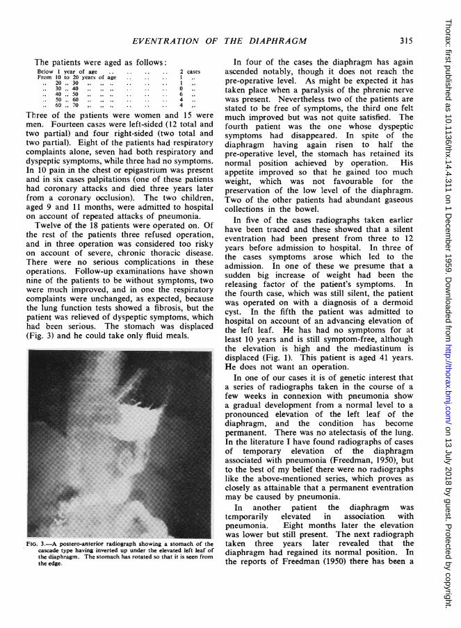
the rest of the patients three refused operation,and in three operation was considered too riskyon account of severe, chronic thoracic disease.There were no serious complications in theseoperations. Follow-up examinations have shownnine of the patients to be without symptoms, twowere much improved, and in one the respiratorycomplaints were unchanged, as expected, becausethe lung function tests showed a fibrosis, but thepatient was relieved of dyspeptic symptoms, whichhad been serious. The stomach was displaced(Fig. 3) and he could take only fluid meals.
FIG. 3.-A postero-anterior radiograph showing a stomach of thecascade type having inverted up under the elevated left leaf ofthe diaphragm. The stomach has rotated so that it is seen fromthe edge.
In four of the cases the diaphragm has againascended notably, though it does not reach thepre-operative level. As might be expected it hastaken place when a paralysis of the phrenic nervewas present. Nevertheless two of the patients arestated to be free of symptoms, the third one feltmuch improved but was not quite satisfied. Thefourth patient was the one whose dyspepticsymptoms had disappeared. In spite of thediaphragm having again risen to half thepre-operative level, the stomach has retained itsnormal position achieved by operation. Hisappetite improved so that he gained too muchweight, which was not favourable for thepreservation of the low level of the diaphragm.Two of the other patients had abundant gaseouscollections in the bowel.
In five of the cases radiographs taken earlierhave been traced and these showed that a silenteventration had been present from three to 12years before admission to hospital. In three ofthe cases symptoms arose which led to theadmission. In one of these we presume that asudden big increase of weight had been thereleasing factor of the patient's symptoms. Inthe fourth case, which was still silent, the patientwas operated on with a diagnosis of a dermoidcyst. In the fifth the patient was admitted tohospital on account of an advancing elevation ofthe left leaf. He has had no symptoms for atleast 10 years and is still symptom-free, althoughthe elevation is high and the mediastinum isdisplaced (Fig. 1). This patient is aged 41 years.He does not want an operation.
In one of our cases it is of genetic interest thata series of radiographs taken in the course of afew weeks in connexion with pneumonia showa gradual development from a normal level to apronounced elevation of the left leaf of thediaphragm, and the condition has becomepermanent. There was no atelectasis of the lung.In the literature I have found radiographs of casesof temporary elevation of the diaphragmassociated with pneumonia (Freedman, 1950), butto the best of my belief there were no radiographslike the above-mentioned series, which proves asclosely as attainable that a permanent eventrationmay be caused by pneumonia.
In another patient the diaphragm wastemporarily elevated in association withpneumonia. Eight months later the elevationwas lower but still present. The next radiographtaken three years later revealed that thediaphragm had regained its normal position. Inthe reports of Freedman (1950) there has been a
315
on 13 July 2018 by guest. Protected by copyright.
http://thorax.bmj.com
/T
horax: first published as 10.1136/thx.14.4.311 on 1 Decem
ber 1959. Dow
nloaded from

POUL CHRISTENSEN
latent period up to five weeks after the onset ofpneumonia before an elevation developed. Thetemporary elevations lasted from a few weeks toseveral months.Three other of our patients had suffered from
pneumonia once or several times on the same sideas that on which a permanent eventration waslater found. We were not able to determinewhether the pneumonia was primary or secondaryto the eventration.Three of the patients had had severe diphtheria
in childhood. In two tracheotomy was performed.No information as to paralysis was available. Ithas not been possible in these cases to provea connexion between the diphtheria and theeventration found several years later.
In one of our patients the course of symptoms,examinations, and operations indicated that*eventration might have been due to syphilis. Thepatient had temporary paretic symptoms of thelimbs and speech 13 years earlier. His chest hadbeen controlled regularly by screening for 10years, and the latest screening six months beforeadmission to our hospital showed a normalposition of the diaphragm. Examinations revealedno active syphilis. The Wassermann reaction wasof grade 2 and the Kahn of grade 2. There wasno reaction of the spinal fluid. As his formerparetic symptoms had disappeared after specifictreatment, the eventration was suspected to be ofsyphilitic origin. It was determined to observe thepatient for three months and to give penicillin inthe meantime. On the patient's second admissionto hospital the elevation of the diaphragm wasonly a little lower, but he felt better. On screen-ing, the formerly immobile left leaf was nowmoving synchronously with the right leaf, althoughthe movements were diminished. The pulmonaryfunction tests showed a slight improvement.Three months earlier electro-stimulation of thephrenic nerve in the neck and directly on thechest at the level of the diaphragm did not giveany contraction of the left leaf, whereas anelectro-stimulation of the right leaf gave a normalcontraction. At operation stimulation of thenerve showed a clear but sluggish contraction ofthe diaphragm and stimulation to the diaphragmitself showed a fast and vigorous contraction.Electro-stimulation was not applied imme-diately before operation, as the patient consideredit unpleasant. The basal atelectasis had remainedunchanged, and, though pre-operatively the mostlikely diagnosis was eventration with atelectasisof the lung from compression, an operation waspreferred to exclude a tumour as the origin of theatelectasis. 'We feel that the evidence is in favour
of considering this a case of eventration ofsyphilitic origin. The phrenic nerve may have beeninterrupted at the site where the nerve crosses thearch of the aorta, caused by syphilitic inflamma-tion of that vessel. The apparent temporarycharacter may be due to the intensive treatmentwith penicillin.Of further interest in the same case is the history
of a severe injury to the left chest wall 10 yearsearlier, confining the patient to bed for a coupleof weeks. Had there been no screening shortlyafter the incident and once yearly during the next10 years, the trauma might erroneously have beenconsidered the cause of the eventration.
PULMONARY FUNCTION TESTS
Ordinary spirometry and bronchospirometryhave not been important. When the results of thetests were abnormal, the respiratory symptoms ofthe patient were so pronounced that they alonedemanded an opzration. On the other hand,when the function tests showed the presence ofpathological changes in the lung such as fibrosis,we could not improve the condition of the patientif his complaints were respiratory alone. In onecase the pulmonary function tests, indicatingfibrosis, and the respiratory complaints wereunchanged after the operation, and for some timethe subjective symptoms were worse. However,the patient was operated upon mainly on accountof the dyspepsia that resulted from the displace-ment of the stomach. He was relieved ofdyspeptic symptoms.
It may be difficult to tell if changes found bythe pulmonary function tests in connexion with aneventration are such that the condition cannot beimproved by operation, among other thingsbecause the functions of the heart and the lungare so intimately connected. Thus in cases ofdoubt we operate if symptoms are pronounced.The fatal course in one of our cases seems to
justify this opinion. The patient presented ahighly elevated diaphragm with the mostpronounced respiratory symptoms we have comeacross in this condition. He was 66 years oldand overweight. Bronchospirometry showed analmost absent oxygen uptake in the affected lungand spirometry indicated emphysema of bothlungs. The patient refused operation. Fivemonths later he was admitted to another hospitalbecause of respiratory comp!aints that hadprogressively increased during the foregoing 14days. He now had to sit up in bed when sleeping.His visual power had simultaneously diminished
316
on 13 July 2018 by guest. Protected by copyright.
http://thorax.bmj.com
/T
horax: first published as 10.1136/thx.14.4.311 on 1 Decem
ber 1959. Dow
nloaded from

EVENTRATION OF TH-E DIAPHRAGM
and the vessels of the eyes were found to becyanotic. He died four days after admissionto hospital with symptoms of anoxaemia andhypercapnoea. A radiograph showed displace-ment of the mediastinum causing some density ofthe unaffected lung. The electrocardiogram wasnormal.Although there would be rigidity of the lung
in such a case, screening showed a pronouncedmovement of the mediastinum to the unaffectedside during inspiration, indicating a mobilemediastinum, so an operation would diminish thedisplacement of the mediastinum, and tighteningof the affected leaf of the diaphragm at a lowerlevel would give a better opportunity of increasingthe respiration by " mediastinal ventilation."
Sarnoff, Gaensler, and Maloney (1950) andSarnoff, Sarnoff, and Whittenberger (1951) haveshown by electro-stimulation of one phrenic nervein the neck of normal persons that it is possibleto maintain ventilation of the contralateral sidealmost as great as the ventilation of the lung onthe side stimulated. The contralateral respirationis carried out by " mediastinal ventilation."
In a work on analysis of lung function in someintrathoracic diseases, Svanberg (1956) stated thefo!lowing about eventration of the diaphragm[sic]:
"A question is whether this condition indicatesoperation. As a rule these patients have only slightsymptoms, and the average age in the presentmaterial was as high as 63 years. From a functionalpoint of view it must be questioned whether suchtherapy is justified, since it is not certain that fixa-tion of the diaphragm at a lower level, whichdecreases the possibility of the so-called mediastinalventilation, will improve ventilation of the affectedlung."9This statement is not in conformity with our
experience. In three of our patients in whomthe diaphragm had ascended notably after theoperation, the lung function tests showed improve-ment. Bronchospirometry showed normal oralmost normal oxygen uptake in the affected lung.Two were relieved of respiratory symptoms, andone was improved.Although the symptoms are improved, they are
not as good as may be suggested by oxygen uptakeshown by bronchospirometry. The values are toohigh. The leaf of the diaphragm operated on isimmobile or moves slightly paradoxically, and themediastinum moves to the unaffected side duringinspiration. The affected lung is thus betterventilated, but the unaffected lung is almostcorrespondingly less ventilated in operatedpersons.
It appears from the above that tightening ofthe diaphragm at a lower level does improveventilation in the affected lung.
INDICATIONS FOR OPERATIONIt is generally believed that an undiagnosed
eventration has been the cause of death in somecases of dyspnoea, cyanosis, and dyspepsia in thenewborn. In some the condition had beendiagnosed, but until the end of the last decadesurgeons dared not operate on these infants. Of13 cases reported in the literature five of thepatients died (Bingham, 1954). Bisgard (1947)performed the first operation on an infant. Latera number of infants were successfully operatedon. Although some of the cases of eventration aredue to a paralysis of the phrenic nerve duringdelivery and may be temporary only, it maynevertheless be necessary to operate. Thecondition may threaten life so that one dares notawait a possible regression, as the patient maybecome beyond surgical care. The symptoms arein most cases severe, because the affected leaf ofthe diaphragm is high and the mediastinum ispushed towards the opposite side. On the otherhand, if during fits of dyspnoea and cyanosis theseinfants respond to conservative treatment, such asoxygen and upright posture, and they can managewithout permanent oxygen, it may be consideredwise to postpone surgery for some months asadvised by Laxdal and others (1954).Newborn children with paralysis of the
diaphragm and without elevation should beobserved. When lying down they are placedon the affected side, which improves ventilation.Oxygen may be necessary.
Eventration rarely causes symptoms in personsbetween 1 and 40 years of age. Thereafter thenumber of cases increases. No statistics areavailable stating how many remain silent. Besidesthe height of the elevation this depends onwhether the patient is doing heavy work (intra-abdominal pressure), and whether the affected leafis being pressed further upwards by increasedintra-abdominal pressure on account of obesity,constipation, or gaseous collections; and whethera displacement of the mediastinum is pronounced.Furthermore, the changes of the lung and heartwith advancing years and the increasing stiffnessof the thoracic cage play a role.
In five of our cases a silent eventration hadbeen diagnosed from three to 12 years before thepatients were admitted to hospital. In threesymptoms developed, whereas the fourth is stillfree of symptoms, although eventration is
317
on 13 July 2018 by guest. Protected by copyright.
http://thorax.bmj.com
/T
horax: first published as 10.1136/thx.14.4.311 on 1 Decem
ber 1959. Dow
nloaded from

POUL CHRISTENSEN
pronounced (Fig. 1), but the elevation has beenincreasing during these 10 years, and symptomswill probably arise eventually. The patient is now41 years of age. The course of these cases favoursour opinion that it is advisable to operate evenon silent cases if the elevation is fairly pronouncedand especially when the mediastinum is distinctlydisplaced. It is difficult for patients to see thesense of an operation when they feel well; butthe result will be better the younger the patient.
Patients with a less pronounced eventrationought to be checked up regularly, and, if thecondition progresses, operation should beperformed.When the symptoms are pronounced the
question is more simple. Several of our patientshave been handicapped on account of dyspnoea.Some have even been unable to do their workand they have wanted an operation. Whenjudging the indications for operation in elderlypersons with presumed changes in the lungtissues, we find screening an important help.
It seems as if the dyspeptic symptoms make thepatient more ready to accept operation than dothe respiratory symptoms alone. The symptomsare mainly due to the stomach having rotatedand ascended, so that the greater curvaturelies adjacent to the under surface of theelevated diaphragm. The stomach is oftenlax and lengthened and may be folded sothat food has difficulty in passing through it.The pain may disappear at once when the patientlies down on his left side, thus facilitating theonward flow. Sometimes the patient is notrelieved of his pain until he has vomited. Inthe literature examples of a "false" stenosis ofthe cardiac end of the oesophagus have been seen,causing dilatation of the oesophagus, and also atthe pylorus, causing retention in the stomach. Byoperation the abnormal site of the stomach iscorrected, and the dyspeptic symptoms disappear.When combined with a basal atelectasis of the
lung the condition also requires operation, evenif the symptoms are negligible. There is a riskof pneumonia and lung abscess in these cases, anda tumour cannot be excluded with certainty. Theeventration may be secondary.With the increasing frequency by which people
in Denmark are submitted to routine x-ray
mass examinations, it will be possible to find more
people with a recently acquired elevation of thediaphragm. Three of our patients had aneventration acquired within one year of admissionto hospital. Observation may ordinarily beadvisable and when possible specific treatment
given. In consideration of the rapidly appearingatrophy from inactivity, it may be justifiable tokeep the diaphragm active by electro-stimulation.A prolonged treatment may be suggested, lasting10 minutes daily. The electrodes are placed onthe back and the front of the thorax at the site ofthe diaphragm.Apart from cases in newborn infants a fatal
course associated with eventration of thediaphragm has been reported following rupture ofthe affected leaf during delivery (Blackford andBooth, 1932), during lifting of a heavy object(Hicks and Harding, 1944), and following volvulusof the stomach. The ligaments of the stomachwere long, permitting it to turn round under theelevated diaphragm (Kwerch, 1949).
TREATMENTThere has been some discussion whether the
abdominal, the thoraco-abdominal, or thoracicapproach should be preferred (State, 1949;Butsch and Leahy, 1950). Surgeons preferringthe abdominal approach maintain that it is easierto avoid catching abdominal structures whensuturing the thin diaphragm. Most surgeonshave chosen the thoracic approach, which offers thebest facilities to reconstruct a normally situateddiaphragm and which yields the best chance toinspect the thoracic structures.We have used the technique which seems to be
most common. After an oval, radlal excision thediaphragm has been sutured in two or three layersby non-absorbable material. Some authors haveused tantalum mesh or fascia lata grafts asreinforcement (Sanford, 1953 ; Monahan, 1951).
Post-operatively the patients must avoid heavywork, overweight, constipation, and gaseouscollections in the bowel. The same instructionsare given to patients who have not beenoperated on, who are also told to take small mealsand to eat slowly.
SUMMARYThe main features of eventration of the
diaphragm are reviewed, and the indications foroperation practised at the University Hospital,Copenhagen, are mentioned.The value of lung function tests is discussed.The present material consists of 18 cases.
Twelve were operated on. Three refusedoperation. In three others operation wasconsidered too risky.At the follow-up examination nine of the 12
patients operated upon were free of symptoms.
318
on 13 July 2018 by guest. Protected by copyright.
http://thorax.bmj.com
/T
horax: first published as 10.1136/thx.14.4.311 on 1 Decem
ber 1959. Dow
nloaded from

EVENTRATION OF THE DIAPHRAGM
Two were much improved. In one of the patientsthe respiratory complaints were unchanged, but hewas relieved of dyspeptic distress.No serious complications arose post-operatively.
My thanks are due to Professor E. Husfeldt,Professor of Surgery at the University Hospital ofCopenhagen, for his helpful suggestions and criticismsof this paper.
REFERENCESAbeles, H., and Leiner, G. C. (1944). Amer. J. Roentgenol., 51, 572.Andersen, lb (1954). Nord. Med., 52, 1446.Bingham, J. A. W. (1954). Thorax, 9, 248.Bisgard, J. D. (1947). J. thorac. Surg., 16, 484.Blackford, L. M., and Booth, W. T. (1932). J. Amer. med. Ass., 98,
883.Butsch, W. L., and Leahy, L. J. (1950). J. thorac. Surg., 20, 968.Campbell, E. J. Moran (1958). The Respiratory Muscles and the
Mechanics of Breathing. Lloyd-Luke, London.Caughey, J. E., and Gray, W. G. (1954). Thorax, 9, 67.Cavrot, E., and Richard, J. (1956). Bull. Soc. roy. belge Gynec.
Obstet., 6, 26.Chin, E. F., and Lynn, R. B. (1956). J. thorac. Surg., 32, 6.Evans, C. J., and Simpson, J. A. (1950). Thorax, 5, 343.Felix, W. (1953). Bruns' Beitr. klin. Chir., 186, 1.
Freedman, B. (1950). Thorax, 5, 169.Greenebaum, J. V., and Harper, F. G. (1946). J. Pediat., 28, 483.Gruber, G. B. (1914). Beitr. path. Anat., 59, 491. Quoted by Felix.
(1953). Bruns' Beitr. klin. Chir., 186, 129.Halonen, P. I., Perasalo, O., and Viikari, S. J. (1952). Ann. Chir.
Gynaec. Fenn., 41, Suppl. 4.Harley, H. R. S. (1949). Thorax, 4, 1.Hicks, F. G., and Harding, E. W. (1944). J. Canad. med. Serv.,1, 129.Jansen, J. (1931). Ned. T. Geneesk., 75, 827.
Kelley, W. 0. (1950). J. thorac. Surg., 19, 923.Korns, H. M. (1921). Arch. intern. Med., 28, 192. Quoted by Reed
and Borden.Kwerch, H. (1949). Ost. Z. Kinderheilk., 4, 35.Lassen, H. C. A. (1956). Management of Life-Threatening Polio-
myelitis. Livingstone, Edinburgh.Laxdal, 0. E., McDougall, H., and Mellin, G. W. (1954). New Engl.
J. Med., 250, 401.Mertins, H. (1952). Zbl. Gynak., 74, 951, quoted by Gruber (1953).Meyer, H. W. (1950). J. thorac. Surg., 20, 235.Monahan, D. T. (1951). New Engi. J. Med., 244,475. Quoted by
Sanford.Morrison, J. M. W. (1923). Arch. Radiol. Electrother., 28, 72.
Quoted by Reed and Borden.Nylander, P. E. A., and Elfving, G. (1951). Ann. Chir. Gynaec. Fenn.,
40, 1.Ottosen, D. (1924). Hospitalstidende, 67, 465.Pasteur, W. (1890). Amer. J. med. Sci., 100, 242.Petit, J. L. (1774). Traites des maladies chirurgicals, et des opera-
tions qui leur conviennent, Paris 1774, T. -. Didot jeune, vol.2, p. 226. Quoted by Reed and Borden.
Reed, J. A., and Borden, D. L. (1935). Arch. Surg. (Chicago), 31, 30.Reich, L. (1926). Klin. Wsdhr., 5, 1470.Rinsma, S. G. (1956). Thesis, Hernia Diaphragmatica, Univ.
Groningen, Holland.Rosenfeld, D. H. (1944). Amer. J. Roentgenol., 52, 607.Samson, P. C., and Childress, M. E. (1950). J. thorac. Surg., 20,901.Sanford, M. C. (1953). Ibid., 25, 422.Sarnoff, S. J., Gaensler, E. A., and Maloney, J. V. (1950). Ibid., 19,
929.Sarnoff, L. C., and Whittenberger, J. L. (1951). Surg. Gynec.Obstet., 93, 190.
Slottved, Axel (1958). Personal communication.Sotrup, T. (1958). Personal communication.State, D. (1949). Surgery, 25, 461.Strauss, L. H. (1933). Z. ges. exp. Med., 86, 244.Svanberg, L. (1956). Acta chir. scand., 111, 169.Tondury, G. (1937). Schweiz. med. Wschr., 18, 142.Tyson, R. M., and Bowman, J. E. (1933). Amer. J. Dis. Child.,
46, 30. Quoted by Bingham.Wood, F. G., and Wood, W. B. (1931). Lancet, 2, 392.
319
on 13 July 2018 by guest. Protected by copyright.
http://thorax.bmj.com
/T
horax: first published as 10.1136/thx.14.4.311 on 1 Decem
ber 1959. Dow
nloaded from