evaluation of the these results provide new ... - nano-portal...
TRANSCRIPT

Tie
toa ty
östä
FINNISH INSTITUTE OF OCCUPATIONAL HEALTH
Topeliuksenkatu 41 a A, 00250 Helsinkiwww.ttl.fi
ISBN 978-952-261-319-6 (paperback)ISBN 978-952-261-320-2 (PDF)
Elina RydmanJulia CatalánPenny Nymark Jaana PalomäkiHannu NorppaHarri AleniusJoonas KoivistoHenrik WolffKaarle HämeriLea Pylkkänen
Evalu
atio
n o
f the h
ealth
effe
cts of ca
rbo
n n
an
otu
bes
The development, production and technological applications of carbon nanotube are rapidly growing, due to the unique characteristics of these fibers. Consequently, an increase is also expected in human exposure to such materials. However, little is still known about the safety of the multiple sorts of carbon nanotubes.
Recent studies have suggested that some types of multi-walled carbon nanotubes (MWCNTs) have similar effects as asbestos. This report shows that rigid, long and needle-like MWCNTs induce inflammation and DNA damage in the lungs and in cultured cells, while flexible, long and tangled MWCNTs do not. It appears that the rigidity of MWCNTs is a key feature in triggering a specific inflammatory reaction and in causing cellular alterations involved in cancer formation.
These results provide new information on the adverse effects of MWCNTs and are useful in assessing which forms of MWC-NTs require regulatory attention and special safety measures in occupational settings.
Hilkka JärventausSatu SuhonenKirsi SiivolaTimo TuomiMerja JärveläEsa VanhalaJenni RantalaMinnamari VippolaKai Savolainen
Evaluation of the health effects of carbon nanotubes
FINAL REPORT ON PROJECT NUMBER 109137 OF THE FINNISH WORK ENVIRONMENT FUND

Evaluation of the health effects of carbon nanotubes
FINAL REPORT ON PROJECT NUMBER 109137 OF THE FINNISH WORK ENVIRONMENT FUND
Elina Rydman, Julia Catalán, Penny Nymark, Jaana Palomäki, Hannu
Norppa, Harri Alenius, Joonas Koivisto, Henrik Wolff, Kaarle Hämeri, Lea
Pylkkänen, Hilkka Järventaus, Satu Suhonen, Kirsi Siivola, Timo Tuomi,
Merja Järvelä, Esa Vanhala, Jenni Rantala, Minnamari Vippola, Kai
Savolainen
Finnish Institute of Occupational Health
Helsinki 2013

Evaluation of the health effects of carbon nanotubes
Finnish Institute of Occupational Health
Nanosafety Research Centre
Topeliuksenkatu 41 a A
00250 Helsinki
www.ttl.fi
Editors: Elina Rydman (née Rossi), Julia Catalán, Penny Nymark, Jaana Palomäki, Hannu
Norppa
Photos: Minnamari Vippola (nanomaterials in Fig. 1), Esa Vanhala (asbestos in Fig. 1),
Joonas Koivisto (Fig. 2, right panel in Fig. 3), Elina Rydman (left panel in Fig. 3, Figs 14
and 18), Jukka Sund (Fig. 4)
Cover: Albert Hall Finland Oy Ltd
© 2013 Authors and Finnish Institute of Occupational Health
This publication has been accomplished with the support of the Finnish Work Environment
Fund (Project No. 109137; “Hiilinanoputkien terveysvaikutusten arviointi”)
Even partial copying of this work without permission is prohibited (Copyright law 404/61).
ISBN 978-952-261-319-6 (paperback)
ISBN 978-952-261-320-2 (PDF)
Juvenes Print, Tampere 2013

Evaluation of the health effects of carbon nanotubes
SUMMARY Carbon nanotubes are among the most important nanomaterials and their production and
industrial use are rapidly growing. Long and rigid carbon nanotubes have been described
to have similar adverse effects as asbestos. The purpose of the present project was to
assess how carbon nanotubes induce pulmonary inflammation and fibrosis and if these
phenomena involve genotoxic alterations which may be important in the formation of
malignant tumors. In addition, complementary studies were performed in vitro to further
understand the cellular effects of carbon nanotubes. Mice were exposed to two types
(needle-like and tangled) of long, multi-walled carbon nanotubes (MWCNTs) by pharyn-
geal aspiration (10-200 µg/mouse) and inhalation (8 mg/m³) for 4 h or 4 days (4 h/day).
Immunotoxic and genotoxic effects in the lungs were studied by using e.g. different
molecular biological, histological, and cytogenetic methods and electron and light
microscopy. Cultured human macrophages –cells of first line immune defense – were
exposed to various types of carbon nanomaterials and crocidolite asbestos, and
immunotoxic effects were examined by e.g. assessing the expression of selected
inflammatory cytokines and chemokines and by inhibiting some of them. Human bronchial
epithelial cells were exposed in vitro to MWCNTs, and cytotoxic and genotoxic effects were
assessed by fluorescence microscopy. Long, needle-like MWCNTs caused inflammation in
mice and in cultured macrophages. In the lungs, the effect was seen as a clear increase of
inflammatory cells, certain cytokines and chemokines; the inflammatory reaction was
stronger than seen with crocidolite asbestos. IL-1β and the inflammasome complex
appeared to play a central role in the inflammatory process. The type and extent of the
inflammatory effect depended on the route of exposure. Needle-like MWCNTs induced a
much clearer inflammation when given by inhalation than by pharyngeal aspiration. The
inflammation caused by inhaled MWCNTs greatly resembled allergic asthma, which is an
unusual finding. Needle-like MWCTs also increased DNA damage in lung cells both after
pharyngeal aspiration and inhalation exposure. In the inhalation experiments, a clear
genotoxic effect was seen in broncho-alveolar lavage cells consisting mostly of
macrophages. The genotoxic effect of MWCNTs was local – no alterations were seen in
blood cells. Long, needle-like MWCNTs caused DNA damage also in cultures of bronchial
epithelial cells. Long, tangled MWCNTs were not genotoxic in mice and induced only a
marginal increase in DNA damage in vitro. Our studies agree with the idea that long,
needle-like MWCNTs are hazardous, showing effects that have not previously been
described such as asthma-like inflammation and DNA damage in the lungs. The rigidity of
long, needle-like MWCNTs is probably a central characteristic determining their health
effects. Rigid, needle-like MWCNTs with a diameter of >50 nm caused a strong
inflammation and were genotoxic, while thinner (diameter ~ 8-15 nm), tangled MWCNTs
did not have similar effects. It appears that long, needle-like MWCNTs should be handled
with special caution.

Evaluation of the health effects of carbon nanotubes
TIIVISTELMÄ
Hiilinanoputket ovat tärkeimpiä nanomateriaaleja, ja niiden tuotanto ja teknologinen käyt-
tö on kasvanut nopeasti. Pitkillä ja jäykillä hiilinanoputkikuiduilla on havaittu samoja hait-
tavaikutuksia kuin asbestilla. Tämän hankkeen tavoitteena oli selvittää, miten hiilinano-
putkien aiheuttama keuhkokudoksen tulehdus ja soluvälifibroosi syntyvät ja liittyykö näi-
hin ilmiöihin genotoksisia muutoksia, joilla katsotaan olevan merkitystä pahanlaatuisten
kasvainten synnyssä. Eläinkokeista saatuja tuloksia täydennettiin ja selvennettiin soluvil-
jelmissä tehdyillä tutkimuksilla. Hiiriä altistettiin kahden tyyppisille (neulamaisille ja
taipuisille), pitkille, moniseinäisille hiilinanoputkille ja krokidoliitti-asbestille aspiraatio-tek-
niikalla (10-200 µg/hiiri) sekä hengitysteitse (8 mg/m³) 4 tunnin tai 4 päivän (4 h/päivä)
ajan. Immunotoksisia ja genotoksisia vaikutuksia keuhkoissa tutkittiin mm. käyttäen eri-
laisia molekyylibiologisia, histologisia, ja sytogeneettisiä menetelmiä sekä elektroni- ja va-
lomikroskopiaa. Soluviljelmissä ihmisen makrofageja, etulinjan immuunipuolustussoluja,
altistettiin erilaisille hiilinanomateriaaleille ja krokidoliitti-asbestille, ja immunotoksisia vai-
kutuksia tutkittiin mm. selvittämällä tulehdusta kuvaavien välittäjäaineiden ilmentymistä
ja estämällä eräitä niistä. Ihmisen viljeltyjä keuhkoepiteelisoluja altistettiin hiilinanoput-
kille, ja solutoksisia sekä genotoksisia vaikutuksia tutkittiin fluoresenssimikroskoopilla. Pit-
kät, neulamaiset hiilinanoputket aiheuttivat tulehdusta sekä hiirissä että soluviljelmissä.
Keuhkoissa vaikutus näkyi tulehdussolujen ja tulehdusta ilmentävien välittäjäaineiden sel-
keänä lisääntymisenä, ja tulehdusreaktio oli voimakkaampi kuin krokidoliitti-asbestilla. Vä-
littäjäaine IL-1β ja inflammasomi-kompleksi näyttivät olevan tulehdusmekanismissa kes-
keisessä asemassa. Tulehdusreaktion laatu ja voimakkuus riippuivat altistustavasta. Neu-
lamaiset hiilinanoputket aiheuttivat huomattavasti vakavamman tulehduksen hengitys-
ilman kautta kuin aspiraatio-menetelmällä annettuina. Hengitettyinä neulamaisten hiilina-
noputkien aikaansaama tulehdus muistutti suuresti allergista astmaa, mikä on poikkeuk-
sellista. Neulamaiset hiilinanoputket aiheuttivat myös DNA-vaurioita keuhkosoluissa sekä
aspiraatio-tekniikalla annosteltuina että hengitettyinä. Selvä perimämyrkyllinen vaikutus
nähtiin hengitysilman kautta tapahtuneessa altistuksessa keuhkon huuhtelunäytteen so-
luissa (pääosin makrofageja). Hiilinanoputkien genotoksinen vaikutus oli paikallinen –
muutoksia ei nähty verisoluissa. Pitkät, neulamaiset hiilinanoputket aiheuttivat DNA-vau-
rioita myös epiteelisolujen viljelmissä. Pitkät, taipuisat hiilinanoputket eivät olleet genotok-
sisia hiirillä, ja viljellyissä keuhkoputkiepiteelisoluissakin niiden vaikutus oli lievä. Tutki-
muksemme vahvisti käsitystä pitkien ja neulamaisten hiilinanoputkien vaarallisuudesta ja
toivat esiin aivan uusia vaikutuksia kuten astmaa muistuttavan tulehduksen syntyminen
ja DNA-vauriot keuhkoissa. Pitkien hiilinanoputkien jäykkyys on ilmeisesti keskeinen tekijä
niiden vaikutusten kannalta. Yli 50 nm paksut, jäykät, neulamaiset hiilinanoputket aiheut-
tivat voimakkaan tulehduksen ja olivat genotoksisia, mutta ohuemmilla (8-15 nm) ja
taipuisilla hiilinanoputkilla ei juuri ollut vaikutuksia. Näyttää siltä, että pitkiä, neulamaisia
hiilinanoputkia tulisi käsitellä erityistä varovaisuutta noudattaen.

Evaluation of the health effects of carbon nanotubes
1
TABLE OF CONTENTS 1 INTRODUCTION ......................................................................................... 3
1.1 Importance of engineered nanomaterials for economy and society ................... 3
1.2 Carbon nanomaterials ................................................................................. 4
1.3 Inflammatory and carcinogenic effects of carbon nanomaterials ....................... 5
1.4 Genotoxic effects of carbon nanomaterials ..................................................... 6
2 AIMS OF THE STUDY .................................................................................. 8
3 MATERIALS AND METHODS ....................................................................... 9
3.1 Carbon nanotubes and their characterisation .................................................. 9
3.2 In vitro studies ......................................................................................... 12
3.2.1 Immunotoxicological studies ...................................................................... 12
3.2.2 Genotoxicological studies ........................................................................... 14
3.3 In vivo studies .......................................................................................... 18
3.3.1 Immunotoxicological studies ...................................................................... 20
3.3.2 Genotoxicological studies ........................................................................... 23
4 4 RESULTS ............................................................................................... 26
4.1 Immunotoxicology .................................................................................... 26
4.1.1 In vitro .................................................................................................... 26
4.1.2 In vivo ..................................................................................................... 30
4.2 Genotoxicology ......................................................................................... 40
4.2.1 In vitro .................................................................................................... 40
4.2.2 In vivo ..................................................................................................... 42
5 CONCLUSIONS AND DISCUSSION .......................................................... 48
6 DISSEMINATION OF KNOWLEDGE .......................................................... 51
7 REFERENCES ............................................................................................ 52

Evaluation of the health effects of carbon nanotubes
2

Evaluation of the health effects of carbon nanotubes
3
1 INTRODUCTION
1.1 Importance of engineered nanomaterials for economy and society
Engineered nanomaterials (ENM) have been defined as having at least one dimen-
sion ≤100 nm. In general, ENMs can be categorised into carbon-based materials,
such as fullerenes and carbon nanotubes, other organic materials (e.g. nanocellu-
lose, synthetic polymers), and inorganic nanoparticles, including nanosized metal
oxides (zinc oxide, iron oxide, titanium dioxide, and cerium oxide, etc), metals
(gold, silver and iron) and quantum dots (cadmium sulfide and cadmium selenide).
ENMs have attracted a great deal of attention during recent years, due to their
many technologically interesting properties. The unique properties of ENM and their
applications have given birth to large technological and economic growth, and future
expectations for industries using materials at nano-scale. Nanotechnologies utilizing
ENM are envisaged to become the cornerstone for a number of industrial sectors,
such as micro-electronics, materials, paper, textile, energy, and cosmetics, which
are all capable of incorporating some nano-scale-enabled properties into their
goods, with an estimated annual turnover of ENM-based products of 3 trillion US
dollars (Roco et al., 2010) by 2020.
ENM can be found in more than 800 consumer products (Woodrow Wilson Inter-
national Centre for Scholars, 2011), including electronic components, cosmetics,
cigarette filters, antimicrobial and stain-resistant fabrics and sprays, sunscreens,
cleaning products, ski waxes, different surfaces requiring antimicrobial properties,
and self-cleaning windows.
Nanotechnology applications are very likely to contribute positively to the quality of
life through the production of durable and light materials, cleaner energy, and inex-
pensive clean water production, as well as by enabling several beneficial medical
applications, especially smart drugs (Adlakha-Hutcheon et al., 2009). Additionally,
great environmental benefits are predicted from nanotechnology related applications
because of the savings in raw materials, the consumption of natural resources, and
a reduced environmental pollution (Kuhlbusch et al., 2009).

Evaluation of the health effects of carbon nanotubes
4
It should be noted, however, that some of the properties that make ENM so unique
and beneficial for technological applications may also endanger human health
through the potential induction of cytotoxic effects, inflammation, and even cancer.
These features include a large surface area to mass ratio, increased surface
reactivity, altered physico-chemical properties such as changes in melting point or
solubility, electrical conductivity, or changes e.g. in the crystalline structure of the
materials (Maynard and Aitken, 2007; Elder, 2009).
1.2 Carbon nanomaterials
Carbon nanotubes, fullerenes, and mesoporous carbon structures constitute a new
class of carbon nanomaterials with properties that differ significantly from other
forms of carbon such as graphite and diamond. The ability to customize synthesized
nanotubes by attached functional groups or to assemble fullerene clusters into
three-dimensional arrays has opened up new avenues to design high surface area
catalyst supports and materials with high photochemical and electrochemical activi-
ty. Carbon nanotubes are also the strongest and stiffest materials yet discovered in
terms of tensile strength and elastic modulus, respectively. Some of the applications
utilizing carbon nanotubes and fullerenes include semiconductors, controlled drug
delivery/release, batteries, data storage, waste recycling, and thermal protection
(De Volder et al., 2013).
Fullerenes are spherical, caged molecules with carbon atoms located at the corner
of a polyhedral structure consisting of pentagons and hexagons. The best known
and most stable fullerene is C60. The discovery of fullerenes by laser vaporization
technique resulted in awarding the 1996 Nobel Prize in Chemistry to Curl, Kroto,
and Smalley.
Conventional carbon nanotubes (CNTs) are made of seamless cylinders of hexa-
gonal carbon networks and are synthesized as single-wall (SWCNTs) or multiwall
carbon nanotubes (MWCNTs). Electric field alignment is a powerful technique that
has been shown to orient carbon nanotubes along a particular direction during the
nanotube growth process. In addition, carbon nanotubes can be assembled as linear
bundles in suspension, by applying a DC electric field. Individual nanotubes have
extensively been studied for application in field emission devices.

Evaluation of the health effects of carbon nanotubes
5
Carbon nanobuds form a material which combines two previously discovered
nanomaterials: CNTs and fullerenes (Nasibulin et al., 2007). In this new material,
fullerenes are covalently bonded to the outer sidewalls of the underlying nanotube.
Consequently, nanobuds exhibit properties of both CNTs and fullerenes. The
characteristics of nanobuds suggest that they may possess advantageous properties
compared with SWCNTs or fullerenes alone or in their non-bonded configurations.
1.3 Inflammatory and carcinogenic effects of carbon nanomaterials
Although the health effects of various carbon nanomaterials are poorly known at the
moment, existing evidence suggests that exposure to certain MWCNTs has the capacity to
induce severe adverse effects in rodent models. This underlines the need for further
research and great caution before introducing such products into the market. The needle-
like shape of certain CNTs has been compared with asbestos, raising concern that the
widespread use of such CNTs may lead to pleural fibrosis or mesothelioma (cancer of the
lining of the lung) which are mostly caused by exposure to asbestos.
It was recently reported that exposing the mesothelial lining of the body cavity of mice, as
a surrogate for the mesothelial lining of the chest cavity, to long MWCNTs results in
asbestos-like inflammatory behaviour. This included the formation of inflammatory lesions
known as granulomas (Poland et al., 2008). Moreover, Takagi et al. (2008) exposed a
tumor-prone p53+/- mouse strain via single intraperitoneal injection to crocidolite
asbestos or MWCNTs and observed that the ability of MWCNTs to induce mesotheliomas in
this mouse model markedly exceeded that of crocidolite asbestos. The induction of
mesothelioma by MWCNTs was subsequently shown to be dose-dependent (Takagi et al.,
2012). Another study by Sakamoto et al. (2009) reported that single intra-scrotal dose of
MWCNTs in Fisher rats had a much higher potential to induce mesotheliomas than a
comparable dose of crocidolite asbestos. Ryman-Rasmussen (2009) showed that MWCNTs
reach the subpleura in mice after inhalation exposure. Subpleural fibrosis unique to this
form of nanotubes increased after 2 and 6 weeks following inhalation - none of these
effects was seen in mice that inhaled non-fibrous carbon black.
These observations merit immediate attention and need to be confirmed by other studies
as they have a very remarkable impact on the risk assessment of these unique

Evaluation of the health effects of carbon nanotubes
6
nanomaterials. Moreover, the molecular mechanisms of observed pathologic phenomena
need to be elucidated.
1.4 Genotoxic effects of carbon nanomaterials
Damage to DNA is one of the most significant human health hazards, since it results in
mutations, chromosome alterations, and increased genetic instability which are associated
with cancer development (Bonassi et al., 2010; Kisin et al., 2011). Most known human
carcinogens are genotoxic (Waters et al., 2010). Carcinogenesis by carbon nanomaterials
may also involve genotoxic processes. In principle, the possible genotoxicity of nano-
materials may result from primary or secondary mechanisms (Schins and Knaapen,
2007). Primary genotoxicity refers to the elicitation of genetic damage in the absence of
inflammation, either by a direct interaction with genomic DNA or associated components
that determine its integrity, or indirectly through the enhanced production of reactive
oxygen species (ROS) by cellular constituents in response to their interaction with
particles or through the depletion of antioxidants within the cell (Donaldson et al., 2010).
Secondary genotoxicity is probably also an oxidative stress-driven response, but in this
case the oxidants are considered to be derived from inflammatory leucocytes recruited to
the site of particle deposition (Donaldson et al., 2010).
It has been suggested that the apparent clastogenic (chromosome-breaking) capacity of
various fibrous materials, such as asbestos, CNTs, and carbon nanofibers, is linked to the
presence of fiber-associated iron, which would initiate ROS generation via a Fenton
reaction (Kisin et al., 2011; Catalán et al., 2011). Fibrous materials have also been
reported to exhibit aneugenic effects (induction of numerical chromosome alterations).
CNTs (Muller et al., 2008, Sargent et al., 2010, 2012), carbon nanofibers (Kisin et al.,
2011), and crocidolite asbestos (Yegles et al., 1995; Dopp et al., 1997) induced chro-
mosomal aneuploidy and disturbed the mitotic spindle. The similarities of SWCNTs with
microtubules has been suggested to make it possible for thin nanotubes to be
incorporated into cellular structures including the mitotic spindle, which could result in the
disruption of the centrosome and microtubules (Sargent et al., 2010, 2012), whereas
physical interference with the spindle might occur with larger fibers such as asbestos
(Cortez and Machado-Santelli, 2008). In both cases, the inhibition of the separation of
dividing cells induces multi-polar mitotic spindles, which results in errors of chromosome
number (Sargent et al., 2010, 2012). The length of the fibers could differentially affect

Evaluation of the health effects of carbon nanotubes
7
their aneugenic capacity. In fact, long asbestos fibers were more genotoxic and
carcinogenic than shorter fibers (Cortez and Machado-Santelli, 2008). Similarly, high-
aspect-ratio MWCNTs exhibited higher toxicity than low-aspect-ratio MWCNTs (Kim et al.,
2011).

Evaluation of the health effects of carbon nanotubes
8
2 AIMS OF THE STUDY
The aims of this study were to evaluate whether CNTs cause inflammatory and genotoxic
effects.
The specific goals of the final project were to investigate:
Inflammatory reactions in the lungs of mice following CNT exposure. This
research was expected to produce new information on inflammatory changes caused
by CNTs in comparison with asbestos exposure. In addition, correlation between in
vivo and in vitro approaches was investigated to gain insight on inflammatory
mechanisms elicited by CNTs.
Genotoxicity of CNT exposure to mouse lungs. This research focused on
assessing possible DNA damage and chromosomal changes caused by CNTs. In
addition, correlation between in vivo and in vitro results was investigated to shed
light on the genotoxic mechanisms of CNTs.
The original project proposal additionally included plans to study (a) the importance of
inflammation in CNT-induced cancer and (b) exposure to CNTs at workplaces. Due to cuts
in the budget of the project, this research was not included in the final project. However,
occupational exposure to nanomaterials (including carbon nanotubes) is studied in
another, on-going project (No. 112132) supported by the Finnish Work Environment
Fund.

Evaluation of the health effects of carbon nanotubes
9
3 MATERIALS AND METHODS
3.1 Carbon nanotubes and their characterisation
Four different carbon nanomaterials and asbestos were selected for the in vitro -
experiments (see also Table 1):
1. Carbon black (Average size 14 nm; Printex 90®, Evonik Industries)
2. Short MWCNTs (Outer diameter, OD, 5-20 nm, length 1- >10 μm;
Baytubes C 150 HP, Bayer Material Science)
3. Long tangled MWCNTs (OD 8-15 nm, length 10-50 µm; MWCNTs 8-15
OD, CheapTubes Inc©)
4. Long needle-like MWCNTs (OD >50 nm, length 13 µm; Mitsui-7, Mitsui
& Co.)
5. Crocidolite asbestos (Average diameter 180 nm, length 4.6 μm;
Pneumoconiosis Research Centre)
The size and morphology of the nanomaterials were characterised by scanning (SEM) and
transmission (TEM) electron microscopy (Zeiss ULTRAplus FEG-SEM, Carl Zeiss NTS
GmbH, Germany and FEI Quanta 200F SEM FEI Company, The Netherlands and Jeol JEM
2010 TEM, Jeol Ltd., Japan) and their composition by energy dispersive spectroscopy
(EDSThermoNoran Vantage, Thermo Scientific, the Netherlands attached to Jeol JEM 2010
TEM) (Table 1 and Fig. 1).
Two MWCNTs of different appearances were chosen to be tested in vivo.
1. Long tangled MWCNTs (outside diameter 8-15 nm, length 10-50 µm;
CheapTubes Inc©)
2. Long needle-like MWCNTs (outside diameter >50 nm, length 13 µm;
Mitsui-7; Mitsui & Co.)

Evaluation of the health effects of carbon nanotubes
10
Table 1. Characteristics of the nanomaterials and crodicolite asbestos used (see also Fig.
1).
Variable Short MWCNTs Long tangled
MWCNTs
Long needle-like
MWCNTs
Carbon black Asbestos
Trade name Baytubes C150 HP
MWCNT 8-15 nm Mitsui MWCNT-7 Printex 90® Crocidolite
asbestos
Manufacturer Bayer Material
Science
Cheaptubes, Inc. Mitsui & Co. Ltd Evonik
Industries
Pneumoco-
niosis
Research
Centre
Characteristics
of primary fibres
or particles
(provided by
manufacturer)
OD 2-20 nm
Length 1->10 µm
OD 8-15 nm
Length 10-50 µm
SSA 233 m2/g
OD >50 nm
Length ~13 µm
Average size
14 nm
SSA 300 m2/g
ø 180 nm
Length
4.6 µm
Composition
measured by
TEM + EDS,
average of 5
measurements
Carbon content
>99 % (w/w)
Residual catalyst
metals: Co
<0.2 % (w/w)
Carbon content
>99 % (w/w)
Residual catalyst
metals: Co, Fe, Ni
<0.5 % (w/w)
Carbon content
>99 % (w/w)
Residual catalyst
metals: < 0.1 %
(w/w; detection
limit)
Carbon content
~ 100 % (w/w)
Not applicable
Compositional analysis shown is the average of five separate analyses by transmission electron microscopy (TEM) and energy dispersive X-ray spectroscopy (EDS). MWCNTs, multi-walled carbon nanotubes; OD, outer diameter; SSA, specific surface area.
In addition, the nanomaterials were compared to crocidolite asbestos (PRC, South-Africa)
as a positive control. These materials were chosen in light of existing knowledge on the
induction of mesothelioma and inflammation by MWCNTs (Poland et al., 2009; Takagi et
al., 2008; Sakamoto et al., 2009). An important point was also the fact that MWCNTs
have presently far greater industrial importance than SWCNTs, which makes it more likely
for employees to be exposed to MWCNTs than SWCNTs.

Evaluation of the health effects of carbon nanotubes
11
Figure 1. The morphology of the test materials, as studied by scanning electron microscopy (SEM; upper part of each micrograph pair) and transmission electron microscopy (TEM; lower part of each micrograph pair). A , short multiwalled carbon nanotubes (MWCNTs). B, long tangled MWCNTs. C, long needle-like MWCNTs. D, carbon black. E, crocidolite asbestos.
A B C
D E

Evaluation of the health effects of carbon nanotubes
12
3.2 In vitro studies
3.2.1 Immunotoxicological studies
In vitro immunotoxicological studies were performed using human monocyte-
derived primary macrophages. Primary macrophages were exposed to different
carbon nanomaterials (carbon black, short MWCNTs, long tangled MWCNTs, long
needle-like MWCNTs) and crocidolite asbestos at two different concentrations (10
and 100 μg/ml). The characteristics of the materials used are represented in Table 1
and Fig. 2. The secretion of important pro-inflammatory cytokines IL-1α and IL-1β
after exposure to the carbon nanomaterials and asbestos was studied by the ELISA
assay. To investigate whether an important molecular complex, NLRP3 inflamma-
some, previously associated with particulate exposure (e.g. asbestos, silica) is acti-
vated, macrophages were left untreated or pre-treated with bacterial lipopoly-
saccharide (LPS, 100 ng/ml) before the exposure. All studies were performed using
a 6-h exposure, and the secretion of pro-inflammatory cytokines caused by long
needle-like MWCNTs was also studied after 3-h and 9-h exposures to find out the
dynamics of cytokine secretion.
Cells: Peripheral blood mononuclear cells (PBMCs) from healthy blood donors
(Finnish Red Cross Blood Transfusion Service, Helsinki, Finland) were isolated from
buffy coats by low-speed density gradient centrifugation on Ficoll-Paque Plus
(Amersham Biosciences, Uppsala, Sweden). The monocytes were resuspended in
RPMI-1640 (Invitrogen, Paisley, UK) with supplemental 1 % penicillin-streptomycin
(PEST; Invitrogen, Paisley, UK) and 1 % L-glutamine (Ultraglutamine®; Invitrogen,
Paisley, UK). After 45 min of attachment on 6- or 12-well-plates, non-adherent cells
were washed away with Dulbecco's phosphate buffered saline without Ca2+ and Mg2+
(DPBS; Lonza, Basel, Switzerland). The adherent monocytes were cultured in se-
rum-free macrophage medium (Macrophage-SFM; Invitrogen, Paisley, UK) supple-
mented with granulocyte-macrophage colony-stimulating factor (GM-CSF; Bio-
Source, Camarillo, CA, USA) and PEST. The cells were cultured for 7 days in the res-
pective medium to allow for differentiation of the macrophages before exposure to
the nanomaterials.
Dispersion preparation for in vitro experiments: Nanomaterial suspensions for
the experiments were prepared by weighing the materials into glass tubes and

Evaluation of the health effects of carbon nanotubes
13
diluting them to a stock dispersion of 1 000 μg/ml with 2 % fetal bovine serum
(FBS) in phosphate buffered saline (PBS) which was sonicated for 20 min at 30 °C.
The stock dispersion was further serially diluted to 100 and 10 μg/ml final
concentrations in serum-free macrophage medium and sonicated for 20 min at 30
°C just before cell exposures. Old media was carefully removed and replaced with
new media containing the final concentrations of nanomaterials.
Electron microscopy: PBMCs were isolated and purified as described above. After
7 days of differentiation, the cells were primed for 2 h with LPS and exposed to
different carbon nanomaterials and asbestos. After the exposure, the cells were
washed twice with DPBS, fixed with 2.5 % glutaraldehyde in 0.1 M phosphate buf-
fer, and removed from the plate by scraping. The cells were post-fixed in 1 % osmi-
um tetroxide, dehydrated and embedded in Epon LX-112 (Ladd Research, Williston,
VT, USA). Thin sections were collected on uncoated copper grids, stained with ura-
nyl acetate and lead citrate and then examined with a transmission electron micro-
scope operated at an acceleration voltage of 80 KV (JEM-1220, Jeol Ltd., Japan).
Reagents: Bacterial LPS (Escherichia coli 0111:B4, Sigma-Aldrich, Germany) was
used at a concentration of 100 ng/ml. Pharmacological inhibitors used in the experi-
ments were cathepsin B inhibitor Ca-047-Me (10 μM; Calbiochem, Germany) and
P2X7 receptor inhibitor AZ11645373 (1 μM; Sigma-Aldrich). When inhibitors were
used, they were added to the wells 1 h prior to exposure to the materials.
Small interfering RNA assays: After 6 days of cell culture in 12-well plates,
macrophages were transfected with 200 nM non-targeting control small interfering
RNA (siRNA, AllStars Negative Control siRNA, Qiagen, CA, US), 50 nM of four
different NLRP3 siRNAs (Hs_CIAS1_6, Hs_CIAS1_9; Hs_CIAS1_10, Hs_CIAS1_11;
Qiagen) or 100 nM of two different P2X7 siRNAs (Hs_P2RX7_1, Hs_P2RX7_2;
Qiagen) using the HiPerFect Transfection Reagent (Qiagen) according to the manu-
facturer’s instruction. After 4 h of incubation with siRNAs, cell culture media was
removed and 500 μl of fresh media was added to the wells. On day 7, appropriate
wells were primed for 2 h with 100 ng/ml of LPS, and the cells were left untreated
or treated with 100 μg/ml of long needle-like MWCNTs or asbestos. After the
exposure period, cell culture supernatants were collected.

Evaluation of the health effects of carbon nanotubes
14
Western blotting and ELISA: Processing and secretion of IL-1β, cathepsin B and
ASC were analysed by Western blot performed by using concentrated cell superna-
tants. Cell culture supernatants (6 ml) were concentrated by Amicon UItra-15 –cen-
trifugal filter devices (Millipore, MA, US) according to the manufacturer’s instruc-
tions. After the concentration, 30 µl from 240 µl of each supernatant was separated
on 12 % SDS-PAGE at 200 V and transferred onto Immobilon-P Transfer Membranes
(Millipore, MA, US) by the Isophor electrotransfer apparatus PowerPac Basic (Bio-
Rad Laboratories) at 4 ºC and 100 V for 1 h. The membranes were blocked in PBS
containing 5 % non-fat milk for 30 min after which they were incubated at 4 ºC
overnight with primary antibodies. After this, the membranes were incubated at
room temperature for 1 h with the appropriate HRP-conjugated secondary antibo-
dies (Dako A/S, Denmark). Finally, proteins were visualised by the Image Quant
LAS 4000 mini quantitative imager (GE Healthcare, CT, US). Anti-IL-1β antibody has
previously been described (Palomäki et al., 2011), anti-Cathepsin B antibody was
purchased from Calbiochem and anti-ASC antibody from Millipore. Both human IL-
1α MAXTM Deluxe and IL-1β Eli-pair were purchased from Diaclone (Besançor Cedex,
France) and human IL-18 ELISA from Bender MedSystems (Bender MedSystems,
Austria). All ELISAs were performed according to the manufacturer's instructions.
Statistical analyses: Each macrophage sample represented a pool of separately
stimulated cells from three different blood donors. ELISA results were combined
from values obtained in three different stimulations, and Western blot results were
representative of three independent, but similarly performed experiments unless
otherwise mentioned. Data were analysed using GraphPad Prism 4 Software (Graph-
Pad Software Inc., San Diego, CA, USA). An unpaired t-test or Mann-Whitney U-test
was used to compare the differences between the groups. A P-value of <0.05 was
considered to be statistically significant. In ELISA figures, data were expressed as
means ±SD.
3.2.2 Genotoxicological studies
Dispersion preparation for in vitro experiments: The materials were dispersed
in BEGM cell culture medium (Clonetics, Walkerwille, MD, USA) supplemented with
0.6 mg/ml of BSA (bovine serum albumin) and subjected to ultrasonication
(Elmasonic, Singen, Germany) for 20 min at 37 kHz prior to addition to the cell

Evaluation of the health effects of carbon nanotubes
15
cultures. Both the stock dispersions and the serially diluted final dispersions were
sonicated.
Cell culture: Transformed human bronchial epithelial BEAS 2B cells, exhibiting an
epithelial phenotype (Reddel et al., 1988) were obtained from the American Type Culture
Collection through LGC Promochem AB (Borås, Sweden). The BEAS 2B cells were grown
in serum-free BEGM medium at 37 ºC in a humidified atmosphere of 5 % CO2. For assays
on cytotoxicity and DNA damage (the comet assay), 20 000 log-phase BEAS 2B cells were
plated on each well of a 24-well plate (Nunc, Roskilde, Denmark; culture area 1.9
cm2/well, 1 ml culture medium per well) two days prior to exposure. For the micronucleus
(MN) assay, 250 000 cells were grown on T25 culture flasks (Nunc, Roskilde, Denmark;
culture area 25 cm2/ flask, 5 ml culture medium per flask) for three days prior to
exposure, i.e. until semiconfluency.
Cytotoxicity: Semiconfluent cells on 24-well plates were exposed to 500 µl per well of
ultrasonicated dispersions of carbon nanomaterials for 4, 24 and 48 h at doses 5, 10, 50,
80, 100, 200, 250, 300 and 350 µg/cm2 (corresponding to 9.5, 19, 95, 152, 190, 380,
475, 570 and 665 µg/ml). Untreated controls were included at each time point. All the
treatments were done in duplicate and the experiments were repeated twice.
Cytotoxicity was measured after collecting the cells by trypsination. In the Trypan blue
dye exclusion technique, the number of living (unstained) cells was determined under a
phase-contrast microscope. In the CellTiter-Glo® Luminescent Cell Viability Assay
(Promega, Madison, USA), the number of viable cells was based on the quantification of
ATP which signals the presence of metabolically active cells. Cell number was expressed
as the percentage of viable cells in the treated cultures in comparison with the control
cultures. These assays reflect all treatment-related effects (necrosis, cell cycle delay and
apoptosis) that reduce the number of viable cells.
Comet assay: The single cell gel electrophoresis (comet) assay was used to study DNA
strand breaks and alkaline labile sites in BEAS 2B cells after nanomaterial exposures.
Semiconfluent cultures on 24-well plates were exposed (500 µl per well) for 4 and 24 h to
six doses of the carbon nanomaterials: 5, 10, 50, 100, 200 and 250 µg/cm2 (corre-
sponding to 19, 38, 190, 380, 760 and 950 µg/ml, respectively). The doses were chosen
according to the Trypan blue cytotoxicity assay. Untreated controls and positive controls
(20 mM hydrogen peroxide, Riedel-de Haen, Seelze, Germany) were included in all series.

Evaluation of the health effects of carbon nanotubes
16
The comet assay was performed at alkaline conditions (pH > 13) as described previously
(Nygren et al., 2004). Briefly, after the exposure the cells were trypsinised and
centrifuged at 1100 rpm for 5 min. Ten to thirty thousand cells were resuspended in 75 µl
molten (37 °C) 0.5 % low-melting-point agarose (LMPA; Merck, Darmstadt, Germany).
The resuspended cells in agarose were applied to dry microscope slides (Menzel Gmbh,
Braunschweig Germany), pre-coated with 1 % normal-melting agarose (BDH Electran
VWR international Ltd., Lutterworth, UK), and the agar was allowed to solidify for 10 min.
The slides were thereafter immersed in cold lysing solution (2.5 M NaCl, 100 mM EDTA,
10 mM Tris, 1 % Triton X-100) for at least 1 h at 4 °C, after which they were transferred
to an electrophoresis tank containing freshly made electrophoresis buffer (1 mM EDTA,
300 mM NaOH; pH> 13), where they were kept for 20 min at room temperature to allow
DNA unwinding. Electrophoresis was performed in the same buffer at room temperature
for 15 min at 24 V and 300 mA (0.8 V/cm). The slides were then neutralized three times
with 0.4 M Tris buffer (pH 7.5), air-dried, and fixed in methanol. DNA was stained with
ethidium bromide (2 µg/ml) in water for 5 min.
The slides were coded, and one scorer performed the comet analysis using a fluorescence
microscope (Axioplan 2, Zeiss, Jena, Germany) and an interactive automated comet
counter (Komet 5.5, Kinetic Imaging Ltd., Liverpool, UK). The percentage of DNA in the
comet tail from 100 cells per replicate was used as a measure of the amount of DNA
damage. In each experiment, two replicates per dose were included, and the experiment
was repeated twice.
Micronucleus assay: Semiconfluent cells in T25 flasks were exposed for 48 h to five
doses of long tangled MWCNTs (Cheaptubes): 5, 10, 50, 100 and 200 µg/cm2 (corre-
sponding to 25, 50, 250, 500 and 1000 µg/ml) and to five doses of long needle-like
MWCNTs (Mitsui-7): 2.5, 5, 10, 20 and 40 µg/cm2 (corresponding to 12.5, 25, 50, 100
and 200 µg/ml). The doses were chosen based on the Trypan blue cytotoxicity assay.
Cytochalasin B (Cyt-B; 9 µg/ml; Sigma-Aldrich Chemie, Steinheim, Germany) was added
to the cell cultures after 6 h of exposure to induce binucleation of dividing cells. Untreated
controls and positive controls receiving 150 ng/ml mitomycin C (MMC; Sigma-Aldrich,
Steinheim, Germany) were also included.
After the exposure, the cells were trypsinised for 20 min, PBS containing 10 % FBS was
added, and the cells were centrifuged at 1,100 rpm for 5 min. The supernatant was

Evaluation of the health effects of carbon nanotubes
17
removed, and PBS was added to the cell suspension. After centrifugation and removal of
the supernatant, the cells were incubated in 5 ml of hypotonic solution (50 % RPMI) for
<2 min. The cells were again centrifuged and first fixed in 3:1 methanol-acetic acid and
then in 97 % methanol - 3 % acetic acid. The cells were spread on microscopy slides and
left to dry overnight. The slides were stained with acridine orange (32 μg/ml in Sørensen
buffer, pH 6.8) for 1 min and rinsed in Sørensen buffer for 3 x 3 min. Finally, the slides
were stained with 4',6-diamidino-2-phenylindole (DAPI, 5 µg/ml) for 5 min, rinsed in tap
water and allowed to dry. The stained and fixed slides were kept protected from light at 4
ºC until analysis.
The slides were coded, and the frequency of micronucleated cells in 2000 binucleate cells
(1000 cells/repeat) were analysed by one scorer using an Axioplan 2E Universal
microscope (Zeiss, Jena, Germany). Binucleate cells were identified with 40× magni-
fication using a green/red (FITC/TRITC) double filter. MN in the cells were verified with a
DAPI filter to ensure DNA content.
Cytokinesis block proliferation index (CBPI; Surrallés et al., 1995) was calculated from
200 cells per culture as follows: CBPI = [(No. mononucleate cells) + 2(No. binucleate
cells) + 3(No. multinucleate cells)]/(Total No. cells).
Statistics: Two-way or one-way analyses of variance (ANOVA) were applied, respec-
tively, to examine whether the percentage of DNA in tail (Comet) or the frequency of
micronucleated cells and the CBPI values were statistically significantly affected by the in
vitro exposure to the CNTs in comparison with the untreated control cultures. Since
differences among experiments have previously been reported for the Comet assay,
"experiment" was included as a second factor (in addition to nanoparticle dose) in the
ANOVAs, to reduce the residual variability of the model. Tukey's test was applied for an a
posteriori comparison of the means.
For all the assays, linear regression analysis was applied to examine whether a linear
dose-response could be observed. The difference between the positive control and the
untreated control was assessed by a two-sample t-test. Differences were interpreted to be
significant if the p-value was <0.05. All statistical analyses were performed with the
Statistix for Windows 2.0 program (Tallahassee, USA).

Evaluation of the health effects of carbon nanotubes
18
3.3 In vivo studies
Animals: Female C57Bl/6 mice (7-8 weeks old) were purchased from Scanbur AB
(Sollentuna, Sweden) and quarantined for one week. The mice were housed in groups of
four in stainless steel cages bedded with aspen chip and were provided standard mouse
chow diet (Altromin No. 1314 FORTI, Altromin Spezialfutter GmbH & Co., Germany) and
tap water ad libitum. The environment of the animal room was carefully controlled, with a
12-h dark/light cycle, temperature of 20-21 °C, and relative humidity of 40-45 %.
The experiments were performed in agreement with the European Convention for the
Protection of Vertebrate Animals Used for Experimental and Other Scientific Purposes
(Strasbourg March 18, 1986, adopted in Finland May 31, 1990). The study was approved
by the Animal Experiment Board and the State Provincial Office of Southern Finland.
Inhalation: The fibrous materials were aerosolized with a fluid bed aerosol generator
(TSI FBAG 3400A) which produced constant aerosol into the exposure chamber. The
aerosol in the chamber was monitored using a variety of equipment: mass concentration
with weighted filter capsules, number concentration with a condensation particle counter,
and size distribution with an optical particle sizer.
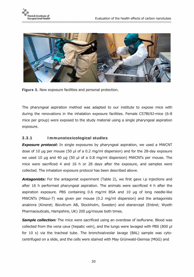
After performing the first inhalation study (Fig. 2) with fibrous materials, we came to the
conclusion that our facilities need upgrading to ensure the safety of the researchers. This
lead to extensive planning and renovations in our exposure facilities. The generator and
the exposure chamber were encapsulated (Fig. 3). The spread of the study material was
restrained as well as possible. We also developed strict working procedures and applied
the best possible personal protection for our workers. The facilities were ready for use
during the last few months of the project. Planning and executing the needed changes
was a valuable learning experience which was used as an example of a model solution for
possible ENM exposures.
In the summer of 2011, we performed our first study in the new facilities. We exposed 8
mice to long needle-like MWCNTs for 4 hours during one day and during 4 consecutive
days. The exposure concentration was kept constant at ~8 mg/m³. Samples were
collected from the mice 24 h after the end of the exposure for immunotoxicological studies
in the 1-day experiment and for both immunotoxicological and genotoxicological analyses,
using the same animals, in the 4-day experiment.

Evaluation of the health effects of carbon nanotubes
19
Figure 2. Old exposure facilities (left) and a schematic drawing of the fluidized bed
aerosol generator (right).
Pharyngeal aspiration: Pharyngeal aspiration is a safe and reliable method to use for
exposure of dusting, dangerous or very expensive materials. The study material was
suspended in PBS containing 0.6 mg/ml BSA and sonicated for 20 min. The mice were
anesthetized with vaporized 4.5 % isoflurane and suspended by their incisors on a thin
wire on a custom made mouse support at approximately 66 degrees angle. A cold-light
source was placed against their throat to provide optimal illumination of the trachea. The
tongue was pulled out using blunted forceps and pressed down using a small spatula to
prevent the mouse from swallowing. 50 µl of the particulate suspension was delivered
onto the vocal folds under visual control using an extended pipette tip (Finntip 200 Ext).
Immediately after delivery the mouse nostrils were covered enforcing the mouse to
inspire the instilled suspension.
Evaluation of the health effects of carbon nanotubes
20
Figure 3. New exposure facilities and personal protection.
The pharyngeal aspiration method was adapted to our institute to expose mice with
during the renovations in the inhalation exposure facilities. Female C57Bl/6J-mice (6-8
mice per group) were exposed to the study material using a single pharyngeal aspiration
exposure.
3.3.1 Immunotoxicological studies
Exposure protocol: In single exposures by pharyngeal aspiration, we used a MWCNT
dose of 10 µg per mouse (50 µl of a 0.2 mg/ml dispersion) and for the 28-day exposure
we used 10 µg and 40 µg (50 µl of a 0.8 mg/ml dispersion) MWCNTs per mouse. The
mice were sacrificed 4 and 16 h or 28 days after the exposure, and samples were
collected. The inhalation exposure protocol has been described above.
Antagonists: For the antagonist experiment (Table 2), we first gave i.p injections and
after 16 h performed pharyngeal aspiration. The animals were sacrificed 4 h after the
aspiration exposure. PBS containing 0.6 mg/ml BSA and 10 µg of long needle-like
MWCNTs (Mitsui-7) was given per mouse (0.2 mg/ml dispersion) and the antagonists
anakinra (Kineret; Biovitrum AB, Stockholm, Sweden) and etanercept (Enbrel; Wyeth
Pharmaceuticals, Hampshire, UK) 200 µg/mouse both times.
Sample collection: The mice were sacrificed using an overdose of isoflurane. Blood was
collected from the vena cava (hepatic vein), and the lungs were lavaged with PBS (800 µl
for 10 s) via the tracheal tube. The bronchoalveolar lavage (BAL) sample was cyto-
centrifuged on a slide, and the cells were stained with May Grünwald-Giemsa (MGG) and

Evaluation of the health effects of carbon nanotubes
21
counted under light microscopy. The BAL supernatant was stored at -70 ºC for cytokine
analysis, and the remaining cells were fixed in ethanol (1:2). The mouse chest was
opened, and half of the left pulmonary lobe was removed, quick-frozen and kept at -70 ºC
for later RNA isolation. A slice of the lungs and part of BAL cells were collected from the
mice, fixed with glutaraldehyde and then prepared for electron microscopy. The rest of the
lungs were formalin-fixed, embedded in paraffin, cut, affixed on slides, and stained with
hematoxylin and eosin (H&E), periodic acid-Schiff (PAS), and Herovici's (HERO) solutions.
Table 2. Treatment groups included in the antagonist experiment with long needle-like multiwalled carbon nanotubes (MWCNTs; 10 µg/mouse). Etanercept (200 µg/mouse) was used as an antagonist of tumor necrosis factor alpha (TNF-α) and anakinra (200 µg/mouse) as an antagonist of interleukin 1 beta (IL-1β).
Intraperitoneal injection Pharyngeal aspiration
PBS/BSA PBS/BSA
PBS/BSA PBS/BSA + long needle-like MWCNTs
PBS/BSA + etanercept PBS/BSA + long needle-like MWCNTs + etanercept
PBS/BSA + anakinra PBS/BSA + long needle-like MWCNTs + anakinra
PBS/BSA + etanercept
+ anakinra
PBS/BSA + long needle-like MWCNTs + etanercept
+ anakinra
BSA/PBS, bovine serum albumin (0.6 mg/ml) in phosphate-buffered saline.
RNA isolation from the lung tissues: The lung samples were homogenized in a
FastPrep FP120 (BIO 101, Thermo Savant, Waltham, Mass. USA) -machine and RNA was
extracted using the FastRNA Pro Green Kit (Qbiogene/ MP Biomedicals, Illkirch, France)
and its instructions. The quantity and purity of extracted RNA was determined by
NanoDrop spectrophotometer (ND-1000, Wilmington, Delaware USA). Isolated total RNA
was dissolved in DEPC water and stored at -70ºC.
cDNA synthesis: cDNA was synthesized from 1 µg of total RNA in a 25 µl reaction using
MultiScribe Reverse Transcriptase and random primers (The High-Capacity cDNA Archive
Kit, Applied Biosystems, Foster City, CA) using the manufacturer's protocol. The synthesis
was performed in a 2720 Thermal Cycler (Applied Biosystems, Carlsbad, California, USA )
starting with 25 °C for 10 minutes and continuing with 37 °C for 120 minutes.

Evaluation of the health effects of carbon nanotubes
22
Polymerase chain reaction (PCR) amplification: PCR primers and probes were
ordered as pre-developed assay reagents from Applied Biosystems. The real-time
quantitative PCR was performed in a 96-well optical reaction plate with Relative
Quantification 7500 Fast System (7500 Fast Real-Time PCR system, Applied Biosystems)
using the manufacturer's instructions. Amplifications were done in 11 µl reaction volume
containing 20 ng cDNA and TaqMan universal PCR master mix and primers provided by
Applied Biosystems. Endogenous 18S was used as the housekeeping gene.
ELISA: Mouse ELISAs (eBioscience, San Diego, CA, US) were performed according to
manufacturer's instructions. An ELISA plate absorbance reader (Multiskan MS,
Labsystems, Titertek Multiscan, Eflab, Turku, Finland) was used to read the results.
Luminex: For analysis of proteins in BAL fluid supernatants we used a Milliplex Mouse
Cytokine/Chemokine Immunoassay (Millipore Corporation, Billerica, MA) according to the
manufacturers' protocol. 3 % bovine serum albumin (BSA; Sigma-Aldrich, St Louis, MO)
in PBS was added at a concentration of 0.5 % to samples, controls and standards to
ensure sufficient protein amounts for the assay. Assay was performed using Luminex
xMAP Technology (Bio-Plex 200 System, BioRad, Hercules, CA).
Electron microscopy: Samples were fixed in 2.5 % glutaraldehyde and postfixed in 1 %
osmium tetroxide, dehydrated and embedded in LX-112 (Ladd Research, Williston, VT,
USA). Thin sections were collected on uncoated copper grids, stained with uranyl acetate
and lead citrate and then examined with a transmission electron microscope operated at
100 KV (JEM-1220, Jeol Ltd., Tokyo, Japan).
Fibrosis: Lung fibrosis was analysed using Sircol Collagen Assay kit following the manu-
facturer's instructions. The HERO stained slides were analysed for possible fibrotic cells
using light microscopy.
Statistical analyses: Data were analysed using GraphPad Prism 5 Software
(GraphPad Software Inc., San Diego, CA, USA). An unpaired t-test or Mann-Whitney
U-test was used to compare the differences between the groups. A P-value of <0.05
was considered to be statistically significant.

Evaluation of the health effects of carbon nanotubes
23
3.3.2 Genotoxicological studies
Exposure protocol: In each experiment performed by pharyngeal aspiration, six mice
per group were exposed to a single dose of one of the following materials: a) first
experiment: 1, 10 and 40 µg/mouse (respective dispersions: 0.02, 0.2 and 0.8 mg/ml) of
long needle-like MWCNTs (Mitsui-7), b) second experiment: 50, 100 and 200 µg/mouse
(respective dispersions: 1, 2 or 4 mg/ml) of long needle-like MWCNTs, and c) third
experiment: 10, 40, 100 and 200 µg/mouse (respective dispersions: 0.2, 0.8, 2, and 4
mg/ml) of long tangled MWCNTs (Cheaptubes). In addition, in each experiment the
negative control mice (n= 6) were exposed to 50 µl of the solvent alone (PBS with 6 %
bovine serum albumin), whereas the positive control group (n = 6) received a single dose
of 1 mg/mouse (20 mg/ml dispersion) of tungsten carbide-cobalt mixture (WC-Co; kindly
provided by Dr. Lison, Université catholique de Louvain, Belgium).
The inhalation exposure protocol has been described above. As a positive control, we
included a group of 8 mice simultaneously exposed to a single dose of WC-Co (1
mg/mouse) by pharyngeal aspiration, as described in the previous paragraph and to
Mitomycin C (MMC, 2 mg/kg) by intraperitoneal injection.
Sample collection: The mice were sacrificed 24 h after the exposure using an overdose
of isoflurane, and the samples were collected as previously described in the immuno-
toxicological section, with small modifications. Briefly, blood was collected from the vena
cava in an insuline syringe containing 0.04 ml EDTA, to prevent coagulation, placed into a
tube and stored on ice. To obtain the BAL cells, the lungs were lavaged with 800 µl of PBS
(BAL sample for the determination of cell composition), and then infused six times with
0.8 ml sterile 0.15 M NaCl through the trachea (BAL sample for the comet analyses). The
first BAL sample was cytocentrifuged on a slide, and the cells were stained using MGG and
counted under light microscopy. The second BAL fluid was stored on ice until centri-
fugation at 400 x g for 5 min. The mouse chest was opened and the lungs were removed
and placed, on ice, into a petri dish containing 0.15 M NaCl. The left lung (or the right lung
in the inhalation experiment) was minced in chilled mincing solution (Hank’s balanced salt
solution with 20 mM EDTA) and mechanically dispersed into a single cell suspension by
using a cell strain (40 µm Ø). Then, the cell suspension was collected, divided in two
aliquots and stored on ice until centrifugation at 400 ×g for 5 min. In the pharyngeal
aspiration experiments, the caudal lobule of the right lung was formalin-fixed, embedded
in paraffin, cut, affixed on slides and stained with H&E, PAS and HERO solutions. The rest

Evaluation of the health effects of carbon nanotubes
24
of the right lung was quick-frozen and stored at -70 ºC for further possible analyses. From
some few animals, the medium lobule of the right lung was fixed with glutaraldehyde and
prepared for electron microscopy, as described in the immunological studies section. In
addition, in the inhalation experiment, the femurs of the mice were collected to extract
bone marrow cells.
Comet assay on BAL and lung suspensions: The comet assay was performed in
alkaline conditions (pH > 13) as described above. The percentage of DNA in the comet tail
from 100 cells per animal (two replicates, 50 cells each) was used as a measure of the
amount of DNA damage.
γ-H2AX assay on peripheral blood mononuclear cells and lung cell suspensions:
Peripheral blood was incubated in ice-cold lysis solution (0.154 M NH4Cl, 0.01 M KHCO3,
0.09 mM EDTA, pH 7.3) on ice for 10 min to remove erythrocytes. After centrifugation at
4000 rpm, 4 min at 4 °C, the cell pellet was twice resuspended in 10 ml of ice-cold lysis
solution for 10 min and centrifuged as above. The cell pellet was suspended in 2 ml of cold
PBS and stored on ice until processing onto slides.
Lung cell suspension was incubated in ice-cold 96 % ethyl alcohol, mixed carefully and
washed twice with PBS. After the last centrifugation, the cell pellet was suspended in 0.5
ml of cold PBS and stored on ice until processing onto slides.
Blood and lung suspensions were directly spun onto polylysine slides with a cytocentrifuge
(Shandon Cytospin 2, Astmoor, UK). The slides were then fixed with freshly prepared 4 %
paraformaldehyde at 4 °C for 25 min. The cells were rinsed three times in PBS and made
permeable by incubation in 0.5 % Triton X-100 in PBS for 5 min at room temperature.
Nonspecific antibody binding was blocked by incubation in 5 % foetal calf serum in PBS for
1 h. The cells were then incubated with 1:50 dilution of Phospho-Histone H2A.X (Ser139)
(20E3) Rabbit mAb (Alexa fluor 488 conjugated) (Cell Signaling) in 1 % BSA in PBS. After
overnight incubation at 4 °C, the slides were washed three times with PBS and counter-
stained with 4,6-diamidino-2-phenylindole (DAPI, 1 µg/ml) for 5 min. Then, the slides
were rinsed in tap water and allowed to dry. Immediately before analysis, the slides were
mounted in Prolong Gold antifade reagent (Invitrogen) and mounted with a cover slip. The
slides were coded, and the frequency of cells with more than four distinct foci in the
nucleus (positive cells) in 1000 mononucleate cells per animal and per tissue was scored
by one microscopist using an Axioplan 2E Universal microscope (Zeiss, Jena, Germany).

Evaluation of the health effects of carbon nanotubes
25
The presence of γ-H2AX-foci is a measure of DNA double-strand breaks.γ-H2AX is the
phosphorylated form of histone 2AX. H2AX becomes phosphorylated on serine 139 as a
reaction on DNA double-strand breaks.
Automated micronucleus assay in bone marrow erythrocytes: Bone marrow
extraction and preparation of the slides for automated MN analysis were done primarily as
previously described by Romagna et al. (1989). Briefly, one femur was removed from
each mouse, cut at the proximal end of the bone and flushed with a foetal bovine serum
mix (FBS in 25 mM EDTA) to collect bone marrow cells in suspension. The suspension was
loaded into Poly-prep chromatography columns (0.8 x 4 cm) that contained a 30-µm pore
size filter bed (Bio-Rad Laboratories, Hemel Hempstead, UK) and had been pre-filled with
a cellulose suspension (a 1:1 mix of type 50 cellulose and α-cellulose, both from Sigma) in
Hanks’ balanced salts solution. After allowing the suspension to drain into a centrifuge
tube, marrow was concentrated by centrifugation, and slides were prepared by cytocentri-
fuge (Shandon Cytospin 2; 1400 rpm, high acceleration for 7 min), air-dried and fixed in
methanol for 10 min. The slides were stained with MGG in Sørensen buffer (pH 6.8-7.0)
for 20 min at room temperature, washed twice in fresh buffer, air-dried and covered with
Entellan (Merck, Germany ) and cover slips. Two thousand PCEs were scored per sample
for the frequency of MNPCEs and 1000 erythrocytes per sample to determine the percent-
ages of PCEs and normochromatic erythrocytes (NCEs). Automated scoring was per-
formed by the MetaSystems Metafer Metacyte image system (MetaSystems, Altlussheim,
Germany).
Statistics: A hierarchic ANOVA, where the animal factor was hierarchical to dose factor,
was used to analyse the percentage of DNA in the tail of the comets, both in BAL and lung
cells. One-way ANOVA was applied to examine the frequency of positive γ-H2AX cells,
both in PBMCs and lung cells. 'A posteriori' comparison among the means of doses was
done by Tukey's test. The unpaired two-sample t-test was applied to determine whether
the exposure to the positive control, WC-Co, induced a statistically significant difference as
compared with the corresponding untreated control groups for the frequency of positive γ-
H2AX cells, both in PBMCs and lung cells. Finally, the dose-response relationship for all the
end-points analysed was investigated by linear regression analysis. The differences were
interpreted to be significant if P was below 0.05. The statistical analyses were performed
by Harvey WR (1987) and Statistix for Windows 2.0 programmes.

Evaluation of the health effects of carbon nanotubes
26
4 RESULTS
4.1 Immunotoxicology
4.1.1 In vitro
Transmission electron microscopy (TEM) was utilised to study, whether long needle-
like MWCNTs; long tangled MWCNTs and asbestos are taken up by the primary mac-
rophages. TEM images showed that all materials studied had intracellular localiza-
tion in macrophages after a 6-h exposure (100 μg/ml; Fig. 4). All materials were
observed free inside the cells, but not in vacuoles or in the nucleus of the macro-
phages. These results demonstrated no differences among the materials in uptake
by primary macrophages.
Figure 4. Transmission electron microscopic images of lipopolysaccharide-primed human primary macrophages exposed for 6 h (100 μg/ml) to long needle-like MWCNTs (left panel), long tangled MWCNTs (middle panel) and asbestos (right panel). The squares show fibers inside the cell. Measure bar is 5 µm.
Exposure of human macrophages to carbon nanomaterials or asbestos without LPS
priming did not induce any IL-1α or IL-1β secretion from the macrophages (data not
shown). However, IL-1α secretion was strongly induced, when LPS-primed primary
macrophages were exposed to long needle-like MWCNTs (data not shown). In
contrast, other types of carbon nanomaterials and asbestos were weak inducers of
IL-1α secretion. The pro-IL-1β is not continuously expressed in the cytoplasm and
its transcription is known to be activated after stimulation of the Toll-like receptor,

Evaluation of the health effects of carbon nanotubes
27
for example by bacterial LPS. However, a second signal is required for inflamma-
some complex formation and the cleavage of IL-1β into its active form (Martinon et
al. 2002). In contrast to unprimed macrophages, LPS-primed human macrophages
secreted IL-1β after exposure to carbon nanomaterials or asbestos.
In the comparison of the different materials, the long needle-like MWCNTs induced
more IL-1β secretion than the other carbon nanomaterials or even asbestos (Fig. 5).
Western blotting analysis confirmed that macrophages release mature IL-1β after
exposure to long needle-like MWCNTs and asbestos but not after exposure to long
tangled MWCNTs or short MWCNTs (Fig. 5). These data suggest that long needle-
like MWCNTs are able to activate IL-1β secretion from human primary macrophages
in an even more profound manner than asbestos fibers.
Figure 5. Long needle-like carbon nanotubes induce IL-1β secretion from lipopoly-saccharide (LPS) -primed human primary macrophages. LPS-primed macrophages were exposed to carbon black, short MWCNTs; long tangled MWCNTs; long needle-like MWCNTs, and asbestos (100 µg/ml) for 6 h, cell culture supernatants were harvested and analysed for IL-1β ELISA. Secretion of cleaved IL-1β cytokine was confirmed by Western blotting analysis: The cell culture supernatants were concen-trated, and IL-1β expression was analyzed by Western blotting with anti-IL-1β antibodies. All values are means ± SD from three independent analyses. **, P < 0.01 and ***, P < 0.001.

Evaluation of the health effects of carbon nanotubes
28
To clarify whether the NLRP3 inflammasome had been activated in response to the
long needle-like MWCNTs, we performed gene silencing with NLRP3 targeting small
interfering RNA in human macrophages. The NLRP3 siRNA treatment clearly de-
creased IL-1β secretion from macrophages after long needle-like MWCNTs and
asbestos exposures (Fig. 6A, B)
Figure 6. Needle-like multi-walled carbon nanotubes (MWCNTs) and asbestos acti-vate NLRP3 inflammasome. Macrophages were transfected with the non-targeting siRNA control (NT-i) or the NLRP3 siRNAs (NLRP3-i) as described in materials and methods. LPS-primed human macrophages were stimulated with (A.) long needle-like MWCNTs and (B.) asbestos (100 µg/ml) for 6 h, cell culture supernatants were collected, and IL-1β ELISA was performed. The values are percentages of measured protein concentrations of two independent analyses where control siRNA = 100 %.
It is known that the extracellular ATP gating cation channel P2X7 is an important
upstream activator of the NLRP3 inflammasome (Kahlenberg et al., 2004, Pelegrin &
Suprenant, 2006, Petrelli et al., 2007, Riteau et al., 2010). The P2X7 receptor allows
cations to pass through the cell membrane, e.g. K+ efflux, and this is known to be
associated to the activation of the NLRP3 inflammasome (Gross et al., 2009). In an
attempt to understand the role of P2X7 receptor in the secretion of IL-1β evoked by
long needle-like MWCNTs and asbestos, we used both pharmacological blockade and

Evaluation of the health effects of carbon nanotubes
29
siRNA induced inhibition of the P2X7 receptor. The P2X7 receptor inhibition clearly
decreased IL-1β secretion from human primary macrophages (Fig. 7), suggesting
that the P2X7 receptor is an important molecule upstream of the NLRP3 inflamma-
some after exposure of cells to long needle-like MWCNTs and asbestos (Fig. 7B, C).
These results demonstrate that the stimulation of the P2X7 receptor is essential for
the NLRP3 inflammasome activation triggered by rigid, needle-like materials.
Figure 7. P2X7 receptor activation is an upstream signal for NLRP3 inflammasome activation. Lipopolysaccharide (LPS) -primed human monocyte-derived macro-phages were treated with long needle-like MWCNTs or asbestos (100 μg/ml) in the absence or presence of P2X7 inhibitor (1 μM). Cell culture supernatants were harvested after 6 h of exposure, and IL-1β ELISA was performed.
All findings from the in vitro immunotoxicology studies have been published (Palo-
mäki et al., 2011) and will be included in a PhD thesis.

Evaluation of the health effects of carbon nanotubes
30
4.1.2 In vivo
Acute pharyngeal aspiration exposure: A single pharyngeal aspiration exposure to
long needle-like MWCNTs, long tangled MWCNTs, and asbestos all resulted in a significant
influx of neutrophils into the lungs of mice (Fig. 8). Pulmonary neutrophilia is a sign of
Figure 8. Percent of neutrophils of total bronchoalveolar lavage cells counted under light microscopy after exposure to PBS/BSA, long tangled MWCNTs (cheaptube), long needle-like MWCNTs (mitsui), and asbestos. The bars represent mean ± SE; *P 0.05, **P 0.01 and ***P < 0.001 significantly different from control; Mann-Whitney U test.
inflammation. Out of these three materials, long needle-like MWCNTs elicited the
fastest and highest reaction reaching close to 20 % of neutrophils among BAL cells
in four hours and almost 40 % in 16 h. We also looked at various inflammatory
markers from the lungs of mice in the form of mRNA and proteins. Based on the
results we obtained from the in vitro experiments, IL-1β and TNF-α were of interest.
Indeed, with long needle-like MWCNTs we saw a huge increase in IL-1β 4 h after the
aspiration exposure (Fig. 9). TNF-α, on the other hand, was significantly elevated
only with asbestos exposure (Fig. 9).
PBS/BSA 4h 16h 4h 16h 4h 16h0
10
20
30
40
50
*
**
****** cheaptube
mitsui
asbestos
% o
f n
eutr
op
hils
in B
AL

Evaluation of the health effects of carbon nanotubes
31
Figure 9. mRNA-expression in lung tissue by proinflammatory cytokines IL-1β and TNF-α. Results are shown as relative quantity (RQ) for mice exposed to PBS/BSA, long tangled MWCNTs (cheaptube), long needle-like MWCNTs (mitsui) and asbestos and sacrificed 4 and 16 h after the exposure. All values are presented as means ± SD. *** P < 0.001, significantly different from control; Mann-Whitney U test.
Another interesting finding was the elevation of CXCL5, a neutrophil-attracting chemokine
(Fig. 10). After four hours of exposure to long needle-like MWCNTs, we observed a huge
increase in CXCL5 expression. Also with asbestos the levels were significantly increased.
This correlated with pulmonary neutrophilia seen with both long needle-like MWCNTs and
asbestos exposure (Fig. 10).
PBS/BSA 4h 16h 4h 16h 4h 16h0
100000
200000
300000 ***
IL-1beta mRNA
cheaptubemitsuiasbestos
RQ
TNF-alpha mRNA
PBS/BSA 4h 16h 4h 16h 4h 16h0
5000
10000
15000 ***
cheaptubemitsui
asbestosRQ

Evaluation of the health effects of carbon nanotubes
32
Figure 10. mRNA-expression in the lung tissue of neutrophil attracting chemokine CXCL5. Results are shown as relative quantity (RQ) for PBS/BSA, long tangled MWCNTs (cheaptube), long needle-like MWCNTs (mitsui), and asbestos exposed mice sacrificed 4 and 16 h after the exposure. All values are presented as means ± SD where ***P < 0.001 001 significantly different from control; Mann-Whitney U test.
On protein level, we found interesting results with CXCL9, CXCL1 and CCL3. CXCL9 was
increased significantly after exposure to long needle-like MWCNTs but not with long
tangled MWCNTs or asbestos (Fig. 11). CXCL9 is a T-cell chemoattractant which is known
Figure 11. Protein expression of T-cell chemoattractant CXCL9 in bronchoalveolar lavage. Results are shown as relative quantity (RQ) for PBS/BSA, long tangled MWCNTs (cheaptube), long needle-like MWCNTs (mitsui), and asbestos exposed mice sacrificed 4 and 16 h after the exposure. All values are presented as means ± SD where **P 0.01 and ***P < 0.001, significantly different from control; Mann-Whitney U test.
PBS/BSA 4h 16h 4h 16h 4h 16h0
50000
100000
150000
200000 ******
***
CXCL5 mRNA
cheaptubemitsuiasbestos
RQ
CXCL9 protein
PBS/BSA 4h 16h 4h 16h 4h 16h0
50
100
150
200
250 ***
**
cheaptube
mitsuiasbestos
pg
/ml

Evaluation of the health effects of carbon nanotubes
33
to be induced by IFN-γ. This correlated with increased amount of lymphocytes seen only
in mice exposed to long needle-like MWCNTs (data not shown).
CXCL1 is expressed by macrophages, neutrophils and epithelial cells and has neutrophil
chemoattractant activity. Accordingly, it was seen at high levels after four hours after all
exposures (Fig. 12). At 16 h, CXCL1 was still elevated for long needle-like MWCNTs.
Neutrophilia seems to be a common reaction to particles which can be seen with all
materials, its degree reflecting the harmfulness of the material.
Figure 12. Protein expression of CXCL1, a neutrophil chemoattractant, in broncho-alveolar lavage. Results are shown as pg/ml for PBS/BSA, long tangled MWCNTs (cheaptube), long needle-like MWCNTs (mitsui), and asbestos exposed mice sacrificed 4 and 16 h after the exposure. All values are presented as means ± SD. **P 0.01 and ***P < 0.001, significantly different from control; Mann-Whitney U test.
Another protein that was expressed considerably more with long needle-like MWCNTs
than the other materials was CCL3 (Fig. 13). CCL3 is known as macrophage inflammatory
protein-1α (MIP-1α) and it is a cytokine that is involved in the acute inflammatory state in
the recruitment and activation of polymorphonuclear leukocytes i.e. neutrophils and
eosinophils.
Chronic pharyngeal aspiration exposure: A single pharyngeal aspiration exposure to
long needle-like MWCNTs with sampling after 28 days resulted in pulmonary granulomas
filled with CNTs and mucus-producing goblet cells (Fig. 14).
CXCL1 protein
PBS/BSA 4h 16h 4h 16h 4h 16h0
500
1000
1500
2000 ***
**
******
cheaptubemitsuiasbestos
pg
/ml

Evaluation of the health effects of carbon nanotubes
34
Figure 13. Protein expression of CCL3, an acute inflammatory protein, in bronchoalveolar lavage. Results are shown in relative pg/ml for PBS/BSA, long tangled MWCNTs (cheaptube), long needle-like MWCNTs (mitsui), and asbestos exposed mice sacrificed 4 and 16 h after the exposure. All values are presented as means ± SD. **P 0.01 and ***P < 0.001, significantly different from control; Mann-Whitney U test.
Figure 14. Periodic acid-Schiff (PAS) -stained mouse lung tissue, where mucus-producing goblet cells can be seen in red colour around the bronchioles. Black clumps of long needle-like multiwall carbon nanotubes are inside and surrounded by granulomas.
CCL3 protein
PBS/BSA 4h 16h 4h 16h 4h 16h0
50
100
150
200
******
*
cheaptubemitsuiasbestos
pg
/ml

Evaluation of the health effects of carbon nanotubes
35
Inhibition using antagonists: To find out whether TNF-alpha and IL-1β indeed had a
significant part in the inflammation process, we used etanercept and anakinra to block
TNF-alpha and IL-1β, respectively.
Etanercept and Anakinra are drugs developed to treat autoimmune diseases and
rheumatoid arthritis, respectively. Etanercept acts as a TNF inhibitor by binding to TNF-
alpha. Anakinra blocks the biological activity of IL-1 by competitively inhibiting the binding
of IL-1 to the interleukin-1 type receptor.
Both antagonists given separately and together resulted in a significant decrease in
neutrophilia (Fig. 15) and mRNAs of IL-1β and TNF-α (Fig. 16) which were expressed after
the single pharyngeal aspiration exposure to long needle-like MWCNTs.
Figure 15. The effect of a single pharyngeal aspiration exposure on neutrophil infiltration to bronchoalveolar lavage fluid calculated from May–Grünwald–Giemsa (MGG)-stained cytospin slides with light microscopy (x40). Results are shown as cells per high power field (HPF) for PBS/BSA and long needle-like multi-walled carbon nanotubes (mitsui) exposed mice treated with antagonists of TNF-α (etanersepti), IL-1β (anakinra) or both (E+A). The bars represent mean ± SE; * P 0.05 and ** P 0.01, significantly different from control; Mann-Whitney U test.
neutrophils
Etanersepti Anakinra E+A0
20
40
60 ***
mitsui 200ug/mlPBS/BSA
cells
/HP
F

Evaluation of the health effects of carbon nanotubes
36
Figure 16. mRNA-expression in the lung tissue of proinflammatory cytokines IL-1β and TNF-α. Results are shown in relative quantity (RQ) for mice exposed to PBS/BSA and long needle-like multiwall carbon (mitsui) and treated with antagonists of TNF-alpha (etanersepti), IL-1β (anakinra) or both (e+a). All values are presented as means ± SE. ***P < 0.001 significantly different from control; Mann-Whitney U test.
Acute inhalation exposure to long needle-like multi-walled carbon nanotubes:
Inhalation exposure to long needle-like MWCNTs resulted in a significant increase in
inflammatory cells, i.e. neutrophils, eosinophils and lymphocytes, in the BAL of the mice
(Fig. 17). Neutrophils are usually the first cells to respond to inflammation and they
migrate to the site in the acute phase. They are the hallmark of acute inflammation and
an essential part of the innate immune system. Eosinophils and lymphocytes, on the other
hand, are usually associated with allergic or atopic diseases and the adaptive immune
response.
IL-1beta mRNA
etanercept anakinra e+a0.0
200000.0
400000.0
600000.0
800000.0* *
****
PBS/BSA
mitsui 200 g/ml
RQ
TNF-alpha mRNA
etanercept anakinra e+a0
10000
20000
30000
40000
50000**
***
PBS/BSA
mitsui 200 g/ml
RQ

Evaluation of the health effects of carbon nanotubes
37
Figure 17. Effect of inhalation exposure to long needle-like multi-walled carbon nanotubes (MWCNTs; mitsui) on infiltration of macrophages, neutrophils, eosino-phils, and lymphocytes to bronchoalveolar lavage fluid calculated from May–Grün-wald–Giemsa (MGG)-stained cytospin slides with light microscopy (x40). Results are shown as cells per high power field (HPF) for control mice and mice exposed to long needle-like MWCNTs for 4 h or 4 h on four consecutive days. The bars represent mean ± SE; * P 0.05, ** P 0.01 and ***P < 0.001, significantly different from unexposed control; Mann-Whitney U test.
In addition to a flood of inflammatory cells in the BAL, we also encountered some foreign-
body giant cells (Fig. 18). They are collections of fused macrophages. These cells are
generated in response to large foreign bodies and are particularly common with particles
that cause chronic inflammation and the foreign body response.
macrophages
contro
l
exp 4
h
exp 4
*4h
0
50
100
150**
**
cells
/HP
F
neutrophils
contro
l
exp 4
h
exp
4*4h
0
2
4
6
8
10
**
*****
cells
/HP
Feosinophils
contro
l
exp
4h
exp
4*4h
0
20
40
60
*
*****
cells
/HP
F
lymfocytes
cont
rol
exp
4h
exp
4*4h
0
1
2
3
4
5 *****
cells
/HP
F

Evaluation of the health effects of carbon nanotubes
38
Figure 18. May–Grünwald–Giemsa-stained cytospin slide under light microscopy showing a neutrophil and eosinophils together with long needle-like multiwall carbon nanotubes between a macrophage and a foreign-body giant cell. Bar 25 µm.
IL-1β was significantly expressed on protein level after inhalation of long needle-like
MWCNTs (Fig. 19). On mRNA level, IL-13 and CCL17 were significantly upregulated (Fig.
19). IL-13 is involved with allergic lung diseases and is an important factor in inducing
airway hyperresponsiveness, goblet cell metaplasia and mucus hypersecretion. In
addition, it also induces secretion of chemokines that recruit allergic effector cells to the
lungs. CCL17 specifically binds and induces chemotaxis in T cells.

htlaeh eht fo noitaulavE effects of carbon nanotubes
39
Figure 19. Expression of proinflammatory protein IL-1β in bronchoalveolar lavage and lung tissue mRNA expression of IL-13 and CCL17. Results are shown in pg/ml and relative quantity (RQ) for control mice and mice exposed to long needle-like multi-walled carbon nanotubes for 4 h or for 4 h on four consecutive days. The bars represent mean ± SE; * P � 0.05, ** P � 0.01 and ***P < 0.001, significantly different from unexposed control; Mann-Whitney U test.

Evaluation of the health effects of carbon nanotubes
40
4.2 Genotoxicology
4.2.1 In vitro
Cytotoxicity: Long needle-like MWCNTs (Mitsui-7) induced a clear dose-dependent
decrease in the number of BEAS 2B cells after all treatment times (4 h, 24 h and 48 h), as
measured by the Trypan blue assay (Fig. 20A,). Cytotoxicity exceeding 50 % occurred
approximately at 200 µg/cm2 for all time points. The luminescent cell viability assay
(measuring ATP) showed similar results, although >50% cytotoxicity occurred already at
about 80 µg/cm2 at each time point (Fig. 20B).
Long needle-like CNTs, Trypan blue
0
100
150
200
2504 h24 h
48 h
10 50 80 100
200
50% viab
A
250
300
350
Dose (µg/cm2)
No
. via
ble
ce
lls (
% o
f co
ntr
ol)
Long needle-like CNTs, ATP
0
100
1504 h24 h
48 h
10 50 80 100
200
50% viab
B
250
300
350
Dose (µg/cm2)
RL
U (
% o
f co
ntr
ol)
Long tangled CNTs, Trypan blue
0
100
150
2004 h24 h48 h
10 50 80 100
200
50% viab
C
250
300
350
Dose (µg/cm2)
No
. via
ble
ce
lls (
% o
f co
ntr
ol)
Long tangled CNTs, ATP
0
100
150
2004 h24 h
48 h
10 50 80 100
200
50% viab
D
250
300
350
Dose (µg/cm2)
RL
U (
% o
f co
ntr
ol)
Figure 20. The number of viable cells (control = 100 %; Trypan blue exclusion) or ATP level (luminescent cell viability assay; RLU, relative luminescence units) after treatment with the multi-walled carbon nanotubes studied. The 50 % limit of viable cell number is marked with a dotted line.
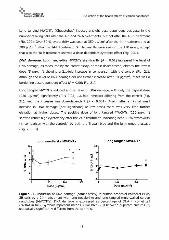
Evaluation of the health effects of carbon nanotubes
41
Long tangled MWCNTs (Cheaptubes) induced a slight dose-dependent decrease in the
number of living cells after the 4-h and 24-h treatments, but not after the 48-h treatment
(Fig. 20C). Over 50 % cytotoxicity was seen at 350 µg/cm2 after the 4-h treatment and at
200 µg/cm2 after the 24-h treatment. Similar results were seen in the ATP assay, except
that also the 48-h treatment showed a dose-dependent cytotoxic effect (Fig. 20D).
DNA damage: Long needle-like MWCNTs significantly (P < 0.01) increased the level of
DNA damage, as measured by the comet assay, at most doses tested, already the lowest
dose (5 µg/cm2) showing a 2.1-fold increase in comparison with the control (Fig. 21).
Although the level of DNA damage did not further increase after 10 µg/cm2, there was a
borderline dose-dependent effect (P = 0.06; Fig. 21).
Long tangled MWCNTs induced a lower level of DNA damage, with only the highest dose
(250 µg/cm2) significantly (P < 0.05; 1.6-fold increase) differing from the control (Fig.
21); yet, the increase was dose-dependent (P < 0.001). Again, after an initial small
increase in DNA damage (not significant) at low doses there was very little further
elevation at higher doses. The positive dose of long tangled MWCNTs (250 µg/cm2)
showed rather high cytotoxicity after the 24 h treatment, indicating near 50 % cytotoxicity
(in comparison with the controls) by both the Trypan blue and the luminometric assays
(Fig. 20C, D).
Long needle-like MWCNTs
0 100 200 3000
2
4
6
8
10
** * *
Dose (µg/cm²)
% D
NA
in t
ail
Long tangled MWCNTs
0 100 2000
2
4
6
8
10
*
Dose (µg/cm²)
% D
NA
in t
ail
Figure 21. Induction of DNA damage (comet assay) in human bronchial epithelial BEAS 2B cells by a 24-h treatment with long needle-like and long tangled multi-walled carbon nanotubes (MWCNTs). DNA damage is expressed as percentage of DNA in comet tail (%DNA in tail). Symbols represent means, error bars SEM between duplicate cultures. *, statistically significantly different from the controls.

Evaluation of the health effects of carbon nanotubes
42
Micronuclei and CBPI: Neither long needle-like MWCNTs nor long tangled MWCNTs
were able to induce micronuclei in BEAS 2B cells (Fig. 22). However, there was a signifi-
cant negative dose-response for the long needle-like MWCNTs (P < 0.01) and a similar
trend for the long tangled MWCNTs. The nanomaterials covered the slides at the highest
concentrations and made the analysis impossible for 40 µg/cm2 and higher doses of long
needle-like MWCNTs and for doses over 200 µg/cm2 of long tangled MWCNTs. CBPI
decreased dose-dependently (P < 0.01) following exposure to both of the MWCNTs (Fig.
22), indicating an induction of cell cycle delay.
Figure 22. Cytokinesis-block micronucleus assay with long needle-like and long tangled multi-walled carbon nanotubes (MWCNTs) in BEAS 2B cells. 1000 binucleate cells scored per culture (2000 per experimental point) for the No. of micronucleated binucleate cells (MNC; columns) and 200 cells (400 per experimental point) for cyto-kinesis-block proliferation index (CBPI; symbols). Columns and symbols represent means, error bars SEM between duplicate cultures.
4.2.2 In vivo
Pharyngeal aspiration. Fig. 23 shows the results of the Comet assay on BAL cells and
lung cells 24 h after pharyngeal aspiration exposure to long needle-like and long tangled
MWCNTs. A significant, 3.5-fold increase (P = 0.002) in the percentage of DNA in tail in
the lung cells, compared with vehicle-treated animals, was found at the highest dose (200
µg) of long needle-like MWCNTs, with a very clear positive dose-response (P < 0.0001).
Long tangled MWCNTs caused no significant increase in DNA damage in BAL or lung cells -
in fact there was a significant negative dose-response (P < 0.05) in both cell types. The
positive control, WC-Co (1 mg/mouse), increased the percentage of DNA in tail (in com-
parison with the controls) by 1.5-1.8-fold in the lungs and 1.7-2.8-fold in BAL cells.

Evaluation of the health effects of carbon nanotubes
43
Long needle-like MWCNTs, 1st experiment
0 10 20 30 400
10
20
30
40Lung cells
BAL cells
Dose (g/animal)
% D
NA
in t
ail
Long needle-like MWCNTs, 2nd experiment
0 50 100 150 2000
10
20
30
40 Lung cells
BAL cells
*
Dose (g/animal)
% D
NA
in t
ail
Long tangled MWCNTs
0 50 100 150 2000
10
20
30
40
BAL cells
Lung cells
Dose (g/animal)
% D
NA
in t
ail
Figure 23. Induction of DNA damage (comet assay) in bronchoalveolar lavage (BAL) cells and lung cells 24 h after pharyngeal aspiration of long needle-like and long tangled multi-walled carbon nanotubes (MWCNTs). Note that the 1st experiment (upper panel) included a lower dose range (10-40 µg/animal) than the other experiments (middle and lower panels; 50-200 µg/animal). Symbols represent means of 6 animals per group, error bars are SEM. The asterisk indicates a significant effect (as compared with the control group) of long needle-like MWCNTs on DNA damage in lung cells; there was also a significant positive dose-response (linear regression analysis; P < 0.0001). The positive control, tungsten carbide-cobalt mixture produced a 1.5-1.8-fold (lung cells) and a 1.7-2.8-fold (BAL) increase in % DNA in tail in comparison with the controls.

Evaluation of the health effects of carbon nanotubes
44
The frequencies of γ-H2AX positive cells, indicative of DNA double-strand break repair,
among PBMCs and lung cells obtained from the pharyngeal aspiration experiment are
shown in Table 3. Long needle-like MWCNTs did not significantly increase the frequency of
γ-H2AX positive cells at any of the tested doses (1-200 µg/mouse) in peripheral blood or
in lungs. No significant linear dose-response relationship could be found in the first
experiment where lower doses (1-40 µg/mouse) were applied. However, a linear dose-
response of borderline significance (P = 0.059) was seen in lung cells but not in PBMNs in
the second experiment where higher doses were tested (50-200 µg/mouse). Long tangled
MWCNTs showed no significant increase in the level of DNA damage at any of the tested
doses (1-200 µg/mouse) in either tissue. No significant linear dose response could either
be found in PBMNs. In lung cells, a significant negative dose response (P = 0.021) was
seen. WC-Co, used as a positive control for the comet assay in BAL and lung cells, did not
significantly increase the frequency of γ-H2AX positive cells in lung cells or PBMCs. In
general, the baseline level of γ-H2AX positive cells was 6-8 times higher in PBMCs than in
lung cells (Table 3).
Inhalation exposure. Results from the inhalation exposure to long needle-like MWCNTs
(~8 mg/m3 for 4 days, 4 h/day) are shown in Figs 24-26. DNA damage, as measured by
the percentage of DNA in comet tail (Fig. 24) was significantly increased (P < 0.05) by the
MWCNT exposure, both in BAL and lung cells. DNA damage was induced particularly in
BAL cells where a 6-fold higher was seen in comparison with the control group. The
positive control treatment, WC-Co (1 mg/mouse), induced 1.2-fold and 2.1-fold increases
in the percentage of DNA in tail in lung cells and BAL cells, respectively.
Long needle-like MWCNTs did not induce systemic genotoxic effects in blood leukocytes,
as measured by the presence of γ-H2AX foci (Fig. 25), or in bone marrow, as determined
by the micronucleus assay (Fig. 26). The positive control Mitomycin C (2 mg/kg
intraperitoneally) was clearly positive in both assays.

Evaluation of the health effects of carbon nanotubes
45
Table 3. Frequencies of γ-H2AX positive cells among peripheral blood mononuclear leukocytes and lung cells of mice 24 h after pharyngeal aspiration to long needle-like and long tangled multi-walled carbon nanotubes (MWCNTs).
Exposure dose (µg/ani-mal)
Long needle-like MWCNTs Long tangled MWCNTs
Experiment 1 (low doses) Experiment 2 (high doses)
No.
animalsa
Mean (SD) No. γ-H2AX positive cells among 1000
lung cells
Mean (SD) No. γ-H2AX positive cells among 1000 blood leuko-
cytes
No.
animalsa
Mean (SD) No. γ-H2AX positive cells among 1000 lung cellsb
Mean (SD) No. γ-H2AX positive cells among 1000 blood leuko-
cytes
No.
animalsa
Mean (SD) No. γ-H2AX positive cells among 1000 lung cellsc
Mean (SD) No.γ-H2AX
positive cells among 1000 blood leuko-
cytes
0 5 2.5 (0.6) 15.4 (6.3) 6 4.5 (3.7) 28.0 (8.3) 6 3.0 (2.2) 24.2 (16.1)
1 4 3.0 (3.2) 23.0 (16.3)
10 5 4.6 (1.5) 22.6 (25.6) 6 6.5 (4.1) 27.8 (21.6)
40 5 4.2 (4.6) 15.6 (7.2) 6 4.3 (2.4) 13.3 (3.3)
50 6 3.3 (1.7) 21.3 (16.4)
100 6 5.6 (3.3) 22.3 (10.4) 6 0.8 (0.7) 20.3 (9.4)
200 6 6.8 (2.2) 16.5 (8.4) 5 1.6 (2.6) 17.0 (7.0)
WC-Co (1 mg/ animal)
3 7.3 (4.2) 16.0 (6.2) 6 2.3 (1.7) 18.2 (6.8) 4 1.5 (1.3) 19.0 (13.9)
a 1000 cells were scored per animal and tissue. b Borderline significant positive dose response (P = 0.059), linear regression analysis. c Significant negative dose response (P = 0.021), linear regression analysis.

Evaluation of the health effects of carbon nanotubes
46
Comet assay
0
10
20
30 Negative control Long needle-like MWCNTsPositive control
BAL cells Lung cells
*
* *
% D
NA
in t
ail
Figure 24. Mean (+SEM) percentage of DNA in tail (comet assay) in bronchoalveolar lavage (BAL) cells and lung cells of mice exposed to long needle-like multi-walled carbon nanotubes (MWCNTs) by inhalation (~8 mg/m3 for 4 days, 4 h/day) or to tungsten carbide-cobalt mixture (pharyngeal aspiration, 1 mg/mouse; positive control). Asterisks indicate a significant difference (P < 0.05) in comparison with the negative control group.
-H2AX assay
0
50
100
150
Negative control
Long needle-like MWCNTs
Positive control
*
No
.-
H2A
X-p
osi
tive
cel
lsin
100
0 le
uko
cyte
s
Figure 25. Frequency of γ-H2AX-positive cells in 1000 peripheral blood mononuclear leukocytes of mice (2000 cells scored per animal) exposed to long needle-like multi-walled carbon nanotubes (MWCNTs) by inhalation (~8 mg/m3 for 4 days, 4 h/day) or to mitomycin C (2 mg/kg i.p.; positive control; sampling 24 h after treatment). Columns represent means, error bars SEM. Asterisks indicate a significant difference (P < 0.05) in comparison with the negative control group.

Evaluation of the health effects of carbon nanotubes
47
Micronucleus assay
0
50
100
150
*
Negativecontrol
Positivecontrol
Long needle-like MWCNTs
No
. m
icro
nu
clea
ted
PC
Es
in 2
000
PC
Es
Figure 26. Frequency of micronucleated polychromatic erythrocytes (PCEs) among 2000 PCEs in bone marrow of mice exposed to long needle-like multi-walled carbon nanotubes (MWCNTs) by inhalation (~ 8 mg/m3 for 4 days, 4 h/day) or to mitomycin C (2 mg/kg i.p.; positive control; sampling 24 h after treatment). Columns represent means, error bars SEM. Asterisks indicate a significant difference (P < 0.05) in comparison with the negative control group.

Evaluation of the health effects of carbon nanotubes
48
5 CONCLUSIONS AND DISCUSSION
Long needle-like MWCNTs induced inflammation in both cultured cells and mice. In the
lungs, we saw a clear increase in inflammatory cells and proinflammatory cytokines and
chemokines. The inflammatory response was stronger with long needle-like carbon
nanotubes than with crocidolite asbestos. The cytokine IL-1β and the inflammasome
complex appeared to be in a central position in the mechanism leading to inflammation.
Neutrophils were the key effector cells in mice exposed by aspiration and eosinophils in
mice exposed by inhalation.
The inflammation produced by inhaled long needle-like MWCNTs greatly resembled
allergic asthma, which is most exceptional. Such a reaction caused by particle exposure
alone is unseen in mice without sensitisation. Especially the induction of IL-13 and
eosinophils was surprising.
In vivo, the features and severity of the inflammation depended on the type of exposure.
Long needle-like MWCNTs invoked a much stronger inflammation when administered by
inhalation as an aerosol than by pharyngeal aspiration as dispersion. This may have been
due to proteins covering the dispersed particles, while inhaled particles reached the lungs
bare and without a protein corona. Protein corona may dampen the particle-cell reactions
by changing the appearance of MWCNTs as seen by the cells. Another factor possibly
influencing the differential response is the dose rate. In pharyngeal aspiration, MWCNTs
were delivered as a single bolus, whereas more gradual exposure, 4 h/day for 4 days, was
applied in the inhalation experiment.
Both long needle-like and long tangled MWCNTs induced a dose-dependent increase in
DNA damage in cultured human bronchial epithelial BEAS 2B cells. While long-needle-like
MWCNTs significantly increased the level of DNA damage already at the lowest dose
tested, long tangled MWCNTs produced a significant increase in DNA damage only at a
50-fold higher, cytotoxic dose. Therefore, the effect of long tangled MWCNTs could only be
considered borderline positive. Neither of the MWCNTs was able to induce chromosome
damage in vitro, as judged by the micronucleus assay.
In vivo in mice, long needle-like MWCNTs were able to increase DNA damage in lung cells
after exposure by inhalation and pharyngeal aspiration. It was remarkable that the DNA-
damaging effect of long needle-like MWCNTs was clearer in vivo than in vitro. The weak

Evaluation of the health effects of carbon nanotubes
49
DNA-damaging potential of long tangled MWCNTs seen at the highest dose in vitro could
not be reproduced in vivo. These findings suggest limited predictivity for the in vitro DNA
damage assay with respect to the production of in vivo DNA damage. It is, however,
unclear if the mechanisms of DNA damage induction by long, needle-like MWCNTs were
entirely the same in vitro and in vivo. Although oxidative stress may have played a role in
DNA damage induction by MWCNTs in both conditions, secondary genotoxic effects
associated with inflammation - which cannot fully be produced in vitro - may have been
important in vivo.
Exposure to long needle-like MWCNTs produced an increase in lung cell DNA damage
concurrently with the pulmonary influx of inflammatory cells and activation of various
proinflammatory cytokines and chemokines. At this stage, it is still too early to tell if these
two phenomena just occurred at the same time or if the genotoxic effect actually was
secondary to inflammation. If the DNA damage was associated with reactive oxygen
species produced by inflammatory cells such as neutrophils, a rather strong increase in
these cells was needed, because the slight pulmonary neutrophilia brought about by
pharyngeal aspiration of long tangled MWCNTs did not result in DNA damage.
In BAL cells, long needle-like MWCNTs induced a high increase in DNA damage after
inhalation exposure but had no effect after pharyngeal aspiration. This may suggest that
macrophages, which constitute the majority of BAL cells, are more sensitive to the
genotoxicity of long needle-like MWCNTs after exposure by inhalation than pharyngeal
aspiration. The pharyngeal aspiration used delivered a high dose at a single administration
of MWCNT dispersion, while the total dose was lower and dose rate much slower in the 4-
day, 4 h/day inhalation exposure to MWCNT aerosol. It is presently unclear how these
differences could explain the results. The high acute exposure by the pharyngeal
aspiration may have resulted in a quick overload of macrophages with particles followed
by their replacement with fresh cells less exposed to MWCNTs. Alternatively, the
pulmonary eosinophilia induced by the inhalation but not by the pharyngeal exposure may
have contributed by a secondary genotoxic effect. It is also possible that albumin, used in
dispersing MWCNTs in the pharyngeal aspiration study, affected macrophage reaction
towards MWCNTs.
Long needle-like MWCNTs did not induce MN in bone marrow of mice after inhalation
exposure. This finding agreed with the negative results obtained with the MN assay in
Evaluation of the health effects of carbon nanotubes
50
vitro. MWCNTs neither produced γ-H2AX foci, indicative of DNA double strand breaks, in
mouse lung cells or PBMNs. These results suggest that long needle-like MWCNTs do not
have local or systemic clastogenic (chromosome-breaking) or aneugenic effects under the
exposure conditions studied.
In conclusion, our study underlined the special toxicity of long and needle-like MWCNTs,
revealing completely new effects such as asthma-like inflammation and DNA damage in
the lungs. Our findings suggest that the rigidity of long carbon nanotubes is a central
characteristic with respect to their harmful effects. Long needle-like MWCNTs, more than
50 nm thick, produced a strong inflammation and were genotoxic, while thinner (8-15
nm) and flexible tangled MWCNTs, which tend to form agglomerates, had hardly any
effects. Further mechanistic understanding of the inflammatory and genotoxic effects of
MWCNTs is urgently needed. Therefore, we have continued our investigations deeper into
the mechanisms of the exceptional inflammatory response observed and into the possible
connections between inflammation and genotoxic effects. We are interested in finding out
(a) what cell types are involved in launching the extraordinary inflammatory reaction, (b)
why especially long and needle-like carbon nanotubes are particularly toxic, and (c) if
oxidative stress and inflammation could explain, and by which mechanism, the concurrent
genotoxic effects.
Our findings provide new information on the adverse effects of MWCNTs and are useful in
assessing which forms of MWCNTs require regulatory attention and special safety
measures in occupational settings.

Evaluation of the health effects of carbon nanotubes
51
6 DISSEMINATION OF KNOWLEDGE
Besides the present report, our results will be published in international and domestic
scientific literature. One such publication has already appeared (Palomäki et al., 2011)
and others are in preparation. The findings will also be presented in international and
domestic scientific conferences, symposia and workshops, in committees dealing with the
safety of nanomaterials, and within the Nanosafety Cluster of the European Commission.
The research carried out in this project will form a part of two doctoral theses which are in
preparation.
During this project, we improved our practices in work with nanomaterials, including a
complete renewal of the exposure facilities and safety precautions. We determined the
special requirements that handling of nanomaterials sets for the working space, venti-
lation, and personal protection. Comprehensive alterations were introduced to ascertain
safety. Detailed instructions for safe work with nanomaterials were prepared and
communicated to the staff. The outcome of this process was utilized in defining a model
solution applicable to this type of MWCNT exposure. Action plans for dealing with
occupational exposure to nanomaterials are further being developed in another project
(Finnish Work Environment Fund, No. 112132).
The mechanisms behind the immunotoxic effects of MWCNTs described in this report were
further characterized in a subsequent project supported by the Finnish Work Environment
Fund (No. 110168; see Sund et al., 2013). Another project, concentrating on the
genotoxic mechanisms of nanomaterials (including MWCNTs), is presently in progress
(Finnish Work Environment Fund, No. 112248).

Evaluation of the health effects of carbon nanotubes
52
7 REFERENCES
Adlakha-Hutcheon, G., Khaydarov, R., Korenstein, R., Varma, R., Vaseashta, A., Stamm, H., et al. Nanomaterials, nanotechnology: applications, consumer products, and benefits. In: Linkov, I., Steevens, J., (eds), Nanomaterials: risks and benefits. Dordrecht: Springer, 2009, p. 195–207.
Bonassi, S.; Norppa, H.; Ceppi, M.; Strömberg, U.; Vermeulen, R.; Znaor, A.; Cebulska-Wasilewska, A.; Fabianova, E.; Fucic, A.; Gundy, S.; Hansteen, I.-L.; Knudsen, L.E.; Lazutka, J.; Rossner, P.; Sram, R.J.; Boffetta, P. Chromosomal aberration frequency in lymphocytes predicts the risk of cancer: results from a pooled cohort study of 22.358 subjects in 11 countries. Carcinogenesis 2008, 29:1178-1183.
Catalán, J., Järventaus, H., Vippola, M., Savolainen, K., Norppa, H. Induction of chromo-somal aberrations by carbon nanotubes and titanium dioxide nanoparticles in human lymphocytes in vitro. Nanotoxicology 2012, 6:825-836.
Cortez, B.A., Machado-Santelli, G.M. Chrysotile effects on human lung cell carcinoma in culture: 3-D reconstruction and DNA quantification by image analysis. BMC Cancer 2008, 8:181.
De Volder, M.F., Tawfick, S.H., Baughman, R.H., Hart, A.J. Carbon nanotubes: present and future commercial applications. Science 2013, 339:535-539.
Donaldson, K., Poland, C.A., Schins, R.P. Possible genotoxic mechanisms of nanoparticles: criteria for improved test strategies. Nanotoxicology 2010, 4:414-420.
Dopp, E., Schuler, M., Schiffmann, D., Eastmond, D.A. Induction of micronuclei, hyperdiploidy and chromosomal breakage affecting the centric/pericentric regions of chromosomes 1 and 9 in human amniotic fluid cells after treatment with asbestos and ceramic fibers. Mutat. Res. 1997, 377:77-87.
Elder, A. Nanotoxicology: How do nanotubes suppress T cells? Nature Nanotechnol. 2009, 4:409-410.
Gross, O., Poeck, H., Bscheider, M., Dostert, C., Hannesschlager, N., Endres, S., Hartmann, G., Tardivel, A., Schweighoffer, E., Tybulewicz, V. et al. Syk kinase signalling couples to the Nlrp3 inflammasome for anti-fungal host defense. Nature 2009, 459:433-436.
Kahlenberg, J. M.; Dubyak, G. R., Mechanisms of caspase-1 activation by P2X7 receptor-mediated K+ release. Am. J. Physiol. Cell Physiol. 2004, 286:C1100-1108.
Kim, J.S., Lee, K., Lee, Y.H., Cho, H.S., Kim, K.H., Choi, K.H., Lee, S.H., Song, K.S., Kang, C.S., Yu, I.J. Aspect ratio has no effect on genotoxicity of multi-wall carbon nanotubes. Arch. Toxicol. 2011, 5:775-786.

Evaluation of the health effects of carbon nanotubes
53
Kisin, E.R., Murray, A.R., Sargent, L., Lowry, D., Chirila, M., Siegrist, K.J., Schwegler-Berry, D., Leonard, S., Castranova, V., Fadeel, B., Kagan, V.E., Shvedova, A.A. Genotoxicity of carbon nanofibers: are they potentially more or less dangerous than carbon nanotubes or asbestos? Toxicol. Appl. Pharmacol. 2011, 252:1-10.
Kuhlbusch, T.A.J., Fissan, H., Asbach, C. Nanotechnologies and environmental risks: measurement technologies and strategies. In: Linkov, I., Steevens, J., (eds), Nanomaterials: Risks and Benefits. Dordrecht: Springer; 2009, p. 233–243.
Martinon, F., Burns, K., Tschopp, J. The inflammasome: a molecular platform triggering activation of inflammatory caspases and processing of proIL-beta. Mol. Cell 2002, 10:417-426.
Maynard, A.D., Aitken, R.J. Assessing exposure to airborne nanomaterials: current abilities and future requirements. Nanotoxicology 2007, 1:26–41.
Muller, J., Decordier, I., Hoet, P.H., Lombaert, N., Thomassen, L., Huaux, F., Lison, D., Kirsch-Volders, M. Clastogenic and aneugenic effects of multi-wall carbon nanotubes in epithelial cells. Carcinogenesis 2008, 29:427-433.
Nasibulin, A.G., Pikhitsa, P.V., Jiang, H., Brown, D.P., Krasheninnikov, A.V., Anisimov, A.S., Queipo, P., Moisala, A., Gonzalez, D., Lientschnig, G., Hassanien, A., Shandakov, S.D., Lolli, G., Resasco, D.E., Choi, M., Tománek. D., Kauppinen, E,I. A novel hybrid carbon material. Nature Nanotechnol. 2007, 2:156-161.
Nygren, J., S. Suhonen, S., Norppa, H., Linnainmaa, K. DNA damage in bronchial epithelial and mesothelial cells with and without associated crocidolite asbestos fibers. Environ. Molec. Mutag. 2004, 44:477-482.
Palomäki, J., Välimäki, E., Sund, J., Vippola, M., Clausen, P.A., Jensen, K.A., Savolainen K., Matikainen, S., Alenius, H. Long, needle-like carbon nanotubes and asbestos activate the NLRP3 inflammasome through a similar mechanism. ACS Nano 2011, 5:6861-6870.
Pelegrin, P, Surprenant, A. Pannexin-1 mediates large pore formation and inter-leukin-1beta release by the ATP-gated P2X7 receptor. Embo J. 2006, 25:5071-5082.
Petrilli, V., Papin, S., Dostert, C., Mayor, A., Martinon, F., Tschopp, J. Activation of the NALP3 inflammasome is triggered by low intracellular potassium concentration. Cell Death Differ. 2007, 14:1583-1589.
Poland, C. A., Duffin, R., Kinloch, I., Maynard, A., Wallace, W. A., Seaton, A., Stone, V., Brown, S., Macnee, W., Donaldson, K. Carbon nanotubes introduced into the abdominal cavity of mice show asbestos-like pathogenicity in a pilot study. Nature Nanotechnol. 2008, 3:423-428.
Reddel, R.R., Ke, Y., Gerwin, B.I., McMenamin, M.G., Lechner, J.F., Su, R.T., Brash, D.E., Park, J.B., Rhim, J.S., Harris, C.C. Transformation of human bronchial epithelial cells by infection with SV40 or adenovirus-12 SV40 hybrid virus, or transfection via strontium phosphate coprecipitation with a plasmid containing SV40 early region genes. Cancer Res. 1988, 48:1904-1909.

Evaluation of the health effects of carbon nanotubes
54
Riteau, N., Gasse, P., Fauconnier, L., Gombault, A., Couegnat, M., Fick, L., Kanello-poulos, J., Quesniaux, V. F., Marchand-Adam, S., Crestani, B. et al. Extracellular ATP is a danger signal activating P2X7 receptor in lung inflammation and fibrosis. Am. J. Respir. Crit. Care Med. 2010, 182:774-783.
Roco, M.C., Mirkin, C.A., Hersam, M.C (eds). WTEC Panel Report on Nanotechnology Research Directions for Societal Needs in 2020. Retrospective and Outlook. World Technology Evaluation Center, Science Policy Reports. September 30, 2010. Springer, 2010.
Romagna, F. Series: 'current issues in mutagenesis and carcinogenesis'.12. Improved method of preparing bone marrow micronucleus assay slides. Mutat. Res. 1988, 206: 307-309.
Ryman-Rasmussen, J.P., Cesta, M.F., Brody, A.R., Shipley-Phillips, J.K., Everitt, J.I., Tewksbury, E.W., Moss, O.R., Wong, B.A., Dodd, D.E., Andersen, M.E., Bonner, J.C. Inhaled carbon nanotubes reach the subpleural tissue in mice. Nature Nanotechnol. 2009, 4:747-751.
Sakamoto, Y., Nakae, D., Fukumori, N., Tayama, K., Maekawa, A., Imai, K., Hirose, A., Nishimura, T., Ohashi, N., Ogata, A. Induction of mesothelioma by a single intrascrotal administration of multi-wall carbon nanotube in intact male Fischer 344 rats. J. Toxicol. Sci. 2009, 34:65–76.
Sargent, L.M., Reynolds S.H., Castranova, V. Potential pulmonary effects of engineered carbon nanotubes: in vitro genotoxic effects. Nanotoxicology 2010, 4:396-408.
Sargent, L.M., Hubbs, A.F., Young, S.H., Kashon, M.L., Dinu, C.Z., Salisbury, J.L., Benkovic, S.A., Lowry, D.T., Murray, A.R., Kisin, E.R., Siegrist, K.J., Battelli, L., Mastovich, J., Sturgeon, J.L., Bunker, K.L., Shvedova, A.A. and Reynolds, S.H. Single-walled carbon nanotube-induced mitotic disruption. Mutat. Res. 2012, 745:28-37.
Savolainen, K., Pylkkänen, L., Norppa, H., Falck, G., Lindberg, H., Tuomi, T., Vip-pola, M., Alenius, H., Hämeri, K., Koivisto, J., Brouwer, J., Mark, D., Bard, D., Berges, M., Jankowska, E., Posniak, M., Farmer, P., Singh, R., Krombach, F., Bihari, P., Kasper, G., Seipenbusch, M. Nanotechnologies, engineered nanomaterials and occupational health. Safety Sci. 2010, 48:957-963.
Schins, R.P., Knaapen, A.M. Genotoxicity of poorly soluble particles. Inhal. Toxicol. 2007, 19 Suppl 1:189-198.
Sund J, Palomäki J, Ilves M, Rydman E, Savinko T, Koivisto J, Vippola M, Greco D, Wolff H, Pylkkänen L, Savolainen K, Puustinen A, Alenius H. Hiilinanoputkien aiheut-tamien terveysvaikutusten karakterisointi systeemitoksikologian avulla. Loppura-portti Työsuojelurahaston hankkeesta Nro 110168. Työterveyslaitos 2013. Tietoa Työstä.
Surrallés, J., Xamena, N., Creus, A., Catalán, J., Norppa, H., Marcos, R. Induction of micronuclei by five pyrethroid insecticides in whole-blood and isolated human lymphocyte cultures. Mutat. Res. 1995, 341:169–184.

Evaluation of the health effects of carbon nanotubes
55
Takagi, A., Hirose, A., Nishimura, T., Fukumori, N., Ogata, A., Ohashi, N., Kitajima, S., Kanno, J. Induction of mesothelioma in p53+/- mouse by intraperitoneal application of multi-wall carbon nanotube. J. Toxicol. Sci. 2008, 33:105-116.
Takagi, A., Hirose, A., Futakuchi, M., Tsuda, H., Kanno J. Dose-dependent mesothelioma induction by intraperitoneal administration of multi-wall carbon nanotubes in p53 heterozygous mice. Cancer Sci. 2012, 103:1001-1004.
Waters, M.D., Jackson, M., Lea, I. Characterizing and predicting carcinogenicity and mode of action using conventional and toxicogenomics methods. Mutat. Res. 2010, 705:184-200.
Woodrow Wilson International Center for Scholars (2011) Project on Emerging Nanotechnologies. Consumer Products. An inventory of nanotechnology-based consumer products currently on the market. www.nanotechproject.org/inventories/consumer.
Yegles, M., Saint-Etienne, L., Renier, A., Janson, X., Jaurand, M.C. Induction of metaphase and anaphase/telophase abnormalities by asbestos fibers in rat pleural mesothelial cells in vitro. Am. J. Respir. Cell Mol. Biol. 1993, 9:186-191.

Tie
toa ty
östä
FINNISH INSTITUTE OF OCCUPATIONAL HEALTH
Topeliuksenkatu 41 a A, 00250 Helsinkiwww.ttl.fi
ISBN 978-952-261-319-6 (paperback)ISBN 978-952-261-320-2 (PDF)
Elina RydmanJulia CatalánPenny Nymark Jaana PalomäkiHannu NorppaHarri AleniusJoonas KoivistoHenrik WolffKaarle HämeriLea Pylkkänen
Evalu
atio
n o
f the h
ealth
effe
cts of ca
rbo
n n
an
otu
bes
The development, production and technological applications of carbon nanotube are rapidly growing, due to the unique characteristics of these fibers. Consequently, an increase is also expected in human exposure to such materials. However, little is still known about the safety of the multiple sorts of carbon nanotubes.
Recent studies have suggested that some types of multi-walled carbon nanotubes (MWCNTs) have similar effects as asbestos. This report shows that rigid, long and needle-like MWCNTs induce inflammation and DNA damage in the lungs and in cultured cells, while flexible, long and tangled MWCNTs do not. It appears that the rigidity of MWCNTs is a key feature in triggering a specific inflammatory reaction and in causing cellular alterations involved in cancer formation.
These results provide new information on the adverse effects of MWCNTs and are useful in assessing which forms of MWC-NTs require regulatory attention and special safety measures in occupational settings.
Hilkka JärventausSatu SuhonenKirsi SiivolaTimo TuomiMerja JärveläEsa VanhalaJenni RantalaMinnamari VippolaKai Savolainen
Evaluation of the health effects of carbon nanotubes
FINAL REPORT ON PROJECT NUMBER 109137 OF THE FINNISH WORK ENVIRONMENT FUND