evaluation of the effects on reproduction, recommendation for classificationphenytoin+pro.pdf ·...
TRANSCRIPT

PhenytoinEvaluation of the effects on reproduction, recommendation for classification
To: the State Secretary of Social Affairs en EmploymentNo. 2018/15, The Hague, June 27, 2018

contentscontentsSamenvatting 3
Executive summary 5
01 Scope 61.1 Background 7
1.2 Committee and procedure 7
1.3 Labelling for lactation 8
1.4 Data 8
1.5 Presentation of conclusions 9
1.6 Final remark 9
02 Phenytoin 102.1 Properties 11
2.2 Context 11
2.3 Human studies 12
2.4 Animal studies 33
2.5 Conclusions 48
References 54
Annexes 68A Animal fertility and developmental toxicity studies 69
B Abbreviations 79
The Committee 80
Health Council of the Netherlands | No. 2018/15
Contents Phenytoin | page 2 of 81

samenvattingOp verzoek van de minister van Sociale Zaken
en Werkgelegenheid (SZW) heeft de
Gezondheidsraad het effect beoordeeld dat het
geneesmiddel fenytoïne heeft op de
voortplanting.
Dit advies is opgesteld door de commissie
Classificatie reproductietoxische stoffen – hierna
aangeduid als de commissie – een
subcommissie van de vaste commissie
Gezondheid en beroepsmatige blootstelling aan
stoffen (GBBS).
De Gezondheidsraad heeft een vaste rol bij de
bescherming van werknemers tegen mogelijke
schadelijke effecten van stoffen waar zij tijdens
hun werk mee in aanraking kunnen komen.
Meer informatie over die rol staat op www.
gezondheidsraad.nl.
Gebruik van fenytoïneFenytoïne is een synthetisch geneesmiddel. Het
wordt voornamelijk gebruikt tegen epileptische
aanvallen, maar soms ook bij
hartritmestoornissen. Mensen die werkzaam zijn
in de farmaceutische industrie, in apotheken of
in ziekenhuizen kunnen tijdens hun werk in
aanraking komen met fenytoïne.
Classificeren naar bewijskracht voor schadelijk effectBij de beoordeling van effecten op de
voortplanting kijkt de commissie zowel naar de
effecten op de fertiliteit (vruchtbaarheid) van
mannen en vrouwen als naar de effecten op de
ontwikkeling van het nageslacht. Daarnaast
worden de effecten op de lactatie (hoeveelheid
en kwaliteit van moedermelk) beoordeeld en de
effecten via de moedermelk op de zuigeling.
De commissie beoordeelt of er aanwijzingen zijn
dat de stof een schadelijk effect kan hebben. Als
dergelijke aanwijzingen bestaan stelt ze voor om
de stof in te delen in een bepaalde
gevarencategorie, die aangeeft hoe sterk de
bewijskracht is voor het schadelijke effect van
de stof. Op basis van dat voorstel kan de
minister van SZW besluiten om de stof al dan
niet als reproductietoxische stof aan te merken.
De indeling in gevarencategorieën is gebaseerd
op EU-verordening (EG) 1272/2008.
Advies aan de staatssecretarisOp grond van de beschikbare wetenschappelijke
gegevens stelt de commissie voor om fenytoïne
alleen voor effecten op de ontwikkeling in te
delen in een gevarencategorie. Over de effecten
Health Council of the Netherlands | No. 2018/15
Samenvatting Phenytoin | page 3 of 81

op de vruchtbaarheid en op of via lactatie zijn
onvoldoende geschikte gegevens beschikbaar.
Classificatievoorstel commissie voor fenytoïne
• voor effecten op de fertiliteit adviseert de
commissie fenytoïne niet in te delen in een
gevarencategorie wegens onvoldoende
geschikte gegevens.
• voor effecten op de ontwikkeling adviseert de
commissie fenytoïne in te delen in categorie
1B (stoffen waarvan verondersteld wordt dat
zij toxisch zijn voor de menselijke
voortplanting) en te etiketteren met H360D
(kan het ongeboren kind schaden).
• voor effecten op of via lactatie adviseert de
commissie om fenytoïne niet te etiketteren
wegens onvoldoende geschikte gegevens.
Health Council of the Netherlands | No. 2018/15
Samenvatting Phenytoin | page 4 of 81

executive summaryAt the request of the Minister of Social Affairs
and Employment, the Health Council of the
Netherlands evaluated the effects of phenytoin
on reproduction. This advisory report has been
drafted by the Subcommittee on the
Classification of Reproduction Toxic Substances
of the Dutch Expert Committee on Occupational
Safety (DECOS) of the Health Council, hereafter
called the Committee. The Health Council has a
permanent task in assessing the hazard of
substances to which man can be occupationally
exposed. More information about this task can
be found at www.gezondheidsraad.nl.
Use of phenytoinPhenytoin is a synthetic anticonvulsant, which
can be used in the treatment of most types of
seizure disorders and status epilepticus.
Occasionally, phenytoin is used as an
antiarrhythmic drug. Workers can be
occupationally exposed to phenytoin in the
pharmaceutical industry, in pharmacies or in
hospitals.
Classification according to strength of evidence for toxic effectFor assessing the effects on reproduction, the
Committee evaluates the effects on male and
female fertility and on the development of the
offspring. Moreover, the Committee considers
the effects of a substance on lactation and on
the offspring via lactation.
If there are data indicating hazardous properties,
the Committee recommends classification in a
category based on the strength of the evidence.
Based on that proposal, the Minister of Social
Affairs and Employment can decide whether to
classify the substance as toxic to reproduction.
The classification is performed according to
EU-regulation (EC) 1272/2008.
Recommendations to the State SecretaryBased on the available scientific data, the
Committee recommends to classify phenytoin
only for effects on offspring development.
There are insufficient data for classification
with regard to effects on paternal and
maternal fertility and effects on or via
lactation.
The Committee’s classification proposal for
phenytoin:
• For fertility, the Committee recommends not
classifying phenytoin due to a lack of
appropriate data.
• For developmental toxicity, the Committee
recommends to classify phenytoin in category
1B (presumed human reproductive toxicant)
and to label it H360D (may damage the
unborn child).
• For effects on or via lactation, the Committee
recommends not labelling phenytoin due to a
lack of appropriate data.
Health Council of the Netherlands | No. 2018/15
Executive summary Phenytoin | page 5 of 81

01 scope
Health Council of the Netherlands | No. 2018/15
chapter 01 | Scope Phenytoin | page 6 of 81

Classification concerning fertility (F/f) and offspring development (D/d)a:Category 1 Known or presumed human reproductive toxicant (H360(F/D)).Category 1A Known human reproductive toxicant .Category 1B Presumed human reproductive toxicant.Category 2 Suspected human reproductive toxicant (H361(f/d)).No classification for effects on fertility or development.Classification for lactation
Effects on or via lactation (H362).No labelling for lactation.
Hazard statement codes:H360FH360DH361fH361dH360FDH361fdH360FdH360DfH362
May damage fertility.May damage the unborn child.Suspected of damaging fertility.Suspected of damaging the unborn child.May damage fertility. May damage the unborn child.Suspected of damaging fertility. Suspected of damaging the unborn child.May damage fertility. Suspected of damaging the unborn child.May damage the unborn child. Suspected of damaging fertility.May cause harm to breast-fed children.
a Substances in category 1 are assigned F or D, substances in category 2 f or d.
The classification and labelling of substances is performed according to
the guidelines of the European Union (Regulation (EC)1272/2008). The
classification of compounds is the result of an integrated assessment of
the nature of all parental and developmental effects observed, their
specificity and adversity, and the dosages at which the various effects
occur. The guideline necessarily leaves room for interpretation, dependent
on the specific data set under consideration. In the process of using the
regulation, the Committee has agreed upon a number of additional
considerations.
Health Council of the Netherlands | No. 2018/15
chapter 01 | Scope
1.1 BackgroundAs a result of the Dutch regulation on registration of compounds toxic to
reproduction that came into force on 1 April 1995, the Minister of Social
Affairs and Employment requested the Health Council of the Netherlands
to classify compounds toxic to reproduction. This classification is
performed by the Health Council’s Subcommittee on the Classification of
Reproduction Toxic Substances of the Dutch Expert Committee on
Occupational Safety (DECOS). The classification is performed according
to European Union Regulation (EC) 1272/2008 on classification, labelling
and packaging (CLP) of substances and mixtures. The CLP regulation is
based on the Globally Harmonised System of Classification and Labelling
of Chemicals (GHS). The Subcommittee’s advice on the classification will
be applied by the Ministry of Social Affairs and Employment to extend the
existing list of compounds classified as reproductive toxicant (category 1A
and 1B and 2) or compound with effects on or via lactation.
1.2 Committee and procedureThis document contains the recommendations for classification of
phenytoin by the Health Council’s Subcommittee on the Classification of
Reproduction Toxic Substances, hereafter called the Committee. The
members of the Committee are listed on the last page of this report.
The classification is based on the evaluation of published human and
animal studies concerning adverse effects with respect to fertility and
offspring development as well as adverse effects on or via lactation.
Phenytoin | page 7 of 81

Additional considerations to Regulation (EC) 1272/2008If there is sufficient evidence to establish a causal relationship between human exposure to the substance and impaired fertility or subsequent developmental toxic effects in the offspring, the compound will be classified in category 1A, irrespective of the general toxic effects (see Regulation (EC) 1272/2008, 3.7.2.2.1.).Adverse effects in a reproductive study, reported without information on the parental or maternal toxicity, may lead to a classification other than category 1B, when the effects occur at dose levels which cause severe toxicity in general toxicity studies.Clear adverse reproductive effects will not be disregarded on the basis of reversibility per se.The Committee does not only use guideline studies (studies performed according to OECDa standard protocols) for the classification of compounds, but non-guideline studies are taken into consideration as well.
a Organisation for Economic Cooperation and Development
Regarding fertility, the Committee takes into account data on
parameters related to fertility, such as seminal fluid volume and
spermatozoa concentration, that are related to male fertility. The
Committee excludes publications containing only data on sex hormone
levels from the assessment, because the relationship between these
hormone levels and functional fertility (ability to conceive children) is too
uncertain.
In 2017, the President of the Health Council released a draft of the
report for public review. The Committee has taken the comments
received into account in deciding on the final version of the report.
These comments, and the reply by the Committee, can be found on the
website of the Health Council.
1.3 Labelling for lactationThe recommendation for classifying substances for effects on or via
lactation is also based on Regulation (EC) 1272/2008. The criteria
define that substances which are absorbed by women and have been
shown to interfere with lactation or which may be present (including
metabolites) in breast milk in amounts sufficient to cause concern for
the health of a breastfed child, shall be classified and labelled. Unlike
the classification of substances for fertility and developmental effects,
which is based on hazard identification only (largely independent of
dosage), the labeling for effects on or via lactation is based on risk
characterization and therefore, it also includes consideration of the level
of exposure of the breastfed child.
Consequently, a substance should be labelled for effects on or via
lactation when it is likely that the substance would be present in breast
milk at potentially toxic levels. The Committee considers a concentration
of a compound as potentially toxic to the breastfed child when this
concentration leads to exceeding the exposure limit for children, or, if
that level is unknown, the exposure limit for the general population, e.g.
the acceptable daily intake (ADI).
1.4 DataLiterature searches were conducted in the on-line databases Current
Contents and Medline, from 1966 up to May 2011 and by searches on
internet; updates were performed in TOXNET, the last search was
Health Council of the Netherlands | No. 2018/15
chapter 01 | Scope Phenytoin | page 8 of 81

performed in July 2017. Literature was selected primarily on the basis of
the text of the abstracts. Publications cited in the selected articles, but
not selected during the primary search, were reviewed if considered
appropriate. In addition, handbooks and a collection of most recent
reviews were consulted as well as several websites regarding
(publications on) toxicology and health. References are divided in
literature cited and literature consulted, but not cited.
The Committee describes both human and animal studies in the text.
The animal data are described in more detail in Annex A as well. Of
each study, the quality of the study design (performed according to
internationally acknowledged guidelines) and the quality of
documentation is considered.
In the assessment of the potential adverse effects of phenytoin on
reproduction, the Committee also used data on adverse effects related
to its application as a therapeutic agent.
1.5 Presentation of conclusionsThe classification is given with key effects, species and references
specified. In case a substance is not classified as toxic to reproduction,
one of two reasons is given:
• Lack of appropriate data precludes assessment of the compound for
reproductive toxicity.
• Sufficient data show that no classification for toxicity to reproduction is
indicated.
1.6 Final remarkThe classification of compounds is based on hazard evaluation (Niesink
et al., 1995)1 only, which is one of a series of elements guiding the risk
evaluation process. The Committee emphasizes that for derivation of
health based occupational exposure limits these classifications should
be placed in a wider context. For a comprehensive risk evaluation,
hazard evaluation should be combined with dose-response
assessment, human risk characterization, human exposure assessment
and recommendations of other organizations.
Health Council of the Netherlands | No. 2018/15
chapter 01 | Scope Phenytoin | page 9 of 81

02 phenytoin
Health Council of the Netherlands | No. 2018/15
chapter 02 | Phenytoin Phenytoin | page 10 of 81

2.1 Properties
Name : PhenytoinCAS-no : 57-41-0Synonyms : Diphenylhydantoin; Fenitoina; Phenantoinum; Phenytoinum;
5,5-Diphenylhydantoin; 5,5-Diphenylimidazoline-2,4-dioneTrade names : Dantoin, Dilantin, Diphenlyn, Phenyltoin, Divulsan, Novo-diphenyl, Phentoin
sodium, Denyl sodium, Dilantin sodium, Diphentoin, Diphenylan sodium, Kessodanten, Elsanutin, Phentoin, Di-Hydan, Phenhydan
Structural formula
: Phenytoin
Use : Phenytoin is a commonly prescribed synthetic anti-epileptic, which can be used in the treatment of most types of seizure disorders and status epilepticus. Occasionally, phenytoin is used as an antiarrhythmic drug. It is incidentally prescribed off-label for treatment of skin disorders. Phenytoin may be administered orally or intravenously at doses of 300 – 600 mg/day. The therapeutic plasma level is 10-20 μg/ml.
Occupational exposure
: Can occur in the pharmaceutical industry, in pharmacies or in hospitals.
Mol weight : 252.268 g/molChemical formula
: C15H12N2O2
General toxicity
: Phenytoin can cause a wide range of adverse idiosyncratic effects such as cosmetic changes, blood dyscrasias, Stevens-Johnson syndrome and hepatotoxicity. The most common signs of toxicity of phenytoin are referable to the central nervous system and are usually dose-related. At plasma drug concentrations higher than 20 μg/ml drowsiness, dysarthria, tremor, ataxia and cognitive difficulties were reported (Brodie, 1996).2
Mechanism : The motor cortex appears to be the primary site of action where spread of seizure activity is inhibited. Phenytoin tends to stabilize the threshold against hyperexcitability possibly by promoting sodium efflux from neurons. This includes the reduction of posttetanic potentiation at synapses. Loss of posttetanic potentiation prevents cortical seizure foci from detonating adjacent cortical areas. Phenytoin can reduce the maximal activity of brain stem centres responsible for the tonic phase of tonic-clonic (grand mal) seizures.
Kinetics : Oral bioavailability of phenytoin is high (90%). Phenytoin is widely distributed throughout the body and more than 90% of the circulating drug is protein-bound, mainly to albumin. The half-life is dose-dependent and has an average of 20 to 30 hours for a therapeutic dose. Phenytoin is metabolized in the liver by cytochrome P450-2C9 and -2C19. Most of the drug is excreted in the bile as inactive metabolites which are then reabsorbed from the intestinal tract and excreted in the urine. Urinary excretion of phenytoin and its metabolites occurs partly with glomerular filtration but more importantly by tubular secretion. Phenytoin accumulates in fat tissue.
Data from HSDB3, unless otherwise noted
2.2 ContextEpilepsy is the most common neurological disorder in women of
reproductive age. Women with epilepsy are at risk of reproductive
dysfunction, through ovulatory failure for instance (Morrell et al., 2002;
Bauer et al., 2008).4,5 Approximately eight in 1,000 pregnancies occur
among women with epilepsy part of whom are taking antiepileptic drugs
(Fairgrieve et al., 2000).6 Phenytoin is a classic antiepileptic drug used
therapeutically as early as 1938.
In many human studies, the duration of exposure to phenytoin and the
dose levels used are not mentioned. Furthermore, (pregnant) epileptic
women are very often treated with a combination of antiepileptic drugs,
making an evaluation of the effect of phenytoin on reproductive outcomes
Health Council of the Netherlands | No. 2018/15
chapter 02 | Phenytoin Phenytoin | page 11 of 81

more difficult. The assessment in this report is restricted to phenytoin
exposure given as monotherapy.
2.3 Human studies
2.3.1 Fertility studies
Male fertilityHerzog et al. (2006) compared serum levels of neuroactive steroids
among men with epilepsy who took various antiepileptic drugs, untreated
men with epilepsy and non-epileptic controls. Twenty-five men with
epilepsy who had been taking phenytoin only, ten men who had untreated
epilepsy and 25 non-epileptic controls were included in the study. In
addition to serum analysis, sexual interest and function were assessed
with a questionnaire. The phenytoin group had lower sexual interest and
function levels (p<0.001), lower serum dehydroepiandrosterone sulfate
(p<0.001), bioactive testosterone (p<0.001) and bioactive androstanediol
(p<0.05) levels and a lower ratio of bioactive androstanediol to bioactive
estradiol (p<0.05) compared to non-epileptic controls. The bioactive
estradiol concentration was not different from the control group. The
untreated men with epilepsy had lower dehydroepiandrosterone sulfate
and bioactive androstanediol levels (both p<0.01) and a lower ratio of
bioactive androstanediol to bioactive estradiol (p<0.05) compared to
non-epileptic controls.7
Taneja et al. (1994) evaluated the effect of seizures and phenytoin
monotherapy on the male reproductive system by studying seminal fluid.
Three groups of men were included in this study: 42 epileptic men treated
with phenytoin, 13 untreated epileptic men and 28 control men
(non-epileptic and untreated). After collection of semen, a minimum
number of 500-1,000 mature sperm cells was scored for morphologic
abnormalities of head, neck, and tail. Serum samples of treated and
untreated patients were analyzed for testosterone, luteinizing hormone
(LH) and follicle-stimulating hormone (FSH), hormones influencing testis
function. The treated and untreated epileptic patients showed no
difference in total seminal fluid volume, spermatozoa concentration, total
sperm count, or number of morphologically abnormal sperm. Both treated
and untreated patients showed lower seminal fluid volumes (p<0.05) and
spermatozoa concentrations (p<0.001) than the control group. In addition,
the percentages of abnormal sperm were increased in untreated patients
(15.39±6.87; p<0.005) and in phenytoin-treated patients (12.77±7.17, not
statistically significant) as compared to controls (9.47±4.04). Testosterone
levels were lower than normal (<13 nM) in 4/9 untreated patients and 5/12
phenytoin-treated patients. LH levels were increased (>9.8 IU/L) in 1/3
untreated patients and 2/6 phenytoin-treated patients. According to the
investigators, the results of this study are indicative for an indirect effect of
epilepsy on the male reproductive function rather than a direct effect of
phenytoin.8
Health Council of the Netherlands | No. 2018/15
chapter 02 | Phenytoin Phenytoin | page 12 of 81

Chen et al. (1992) investigated the effects of phenytoin on sperm motility.
Six epileptic patients treated with phenytoin monotherapy for at least five
years (serum levels varied from 7.2 to 13.4 μg/ml) were observed for
effects on sperm motility immediately after semen had liquefied or after a
2-h pre-incubation at 37ºC (to detect effects on the duration of activity).
Forty-five healthy volunteers served as control group. Sperm motility was
decreased in all treated patients when motility was measured immediately
after semen liquefication (22.9±11.7) or after a 2-h pre-incubation at 37ºC
(13.5±8.7) compared to healthy donors (42.6±12.5 and 35.2±7.0
respectively). In addition, the duration of sperm activity of epileptic
patients was shorter than that of the controls (p<0.05). The effect on
sperm motility showed no correlation with duration of therapy, serum
phenytoin levels or seizure types.9
The in vitro experiment published by Chen et al. in 19929 but originally
reported by Shen and Chen (1990) showed reduced sperm motility upon
2-h pre-incubation with phenytoin concentrations 1.6, 4.2 and 5.0 mM,
compared to incubation medium (all p<0.01). The inhibitory effect on
sperm motility appeared to be dependent on both duration of
pre-incubation and the concentration of the drug.10
Hong et al. (1982) also measured the effects of phenytoin on human
sperm motility in vitro. Using concentrations as high as 2mM, phenytoin
did not have an effect on sperm motility.11
Female fertilityMorrell et al. (2005) investigated sexual dysfunction, sex steroid hormone
abnormalities, and depression in women with epilepsy treated with
antiepileptic drug monotherapy. Subjects were women with epilepsy
receiving an antiepileptic drug in monotherapy for at least six months prior
to study participation, as well as women without epilepsy not receiving
antiepileptic drugs who served as controls. Thirteen epileptic women
receiving phenytoin were included in the study, as well as thirteen
nonepileptic controls. All subjects were between the ages of eighteen and
40, cycling, and at least four years postmenarche. Women with epilepsy
were recruited from the patient populations of two U.S. Epilepsy Centres.
Nonepileptic controls were recruited from friends and family of the
participants. Subjects provided medication history and completed a
reproductive history form and a seizure history form. Sexual function was
determined by the subject’s response to sexual inventories examining
sexual experience and attitude. The subjects also completed an internally
developed questionnaire examining sexual desire and arousal to
determine whether a sexual dysfunction was present as defined by the
Diagnostic and Statistical Manual of Diseases Classification (4th Edition).
Sexual dysfunction and sexual anxiety were higher and sexual arousal
was lower than in controls (p<0.05).12
Health Council of the Netherlands | No. 2018/15
chapter 02 | Phenytoin Phenytoin | page 13 of 81

2.3.2 Developmental toxicity studiesTreatment with phenytoin during pregnancy has been associated with a
distinct set of malformations called the foetal hydantoin syndrome
(Hanson and Smith, 1975; Smith‘s Recognizable Patterns of Human
Malformations, 2014).13,14 Among these developmental disturbances are
abnormal facial features, anomalies of the distal phalanges, pre- and
postnatal growth deficiency, as well as mental performance deficiency.
Other defects reported less frequently include cleft lip and/or palate,
cardiovascular anomalies, renal defects, positional limb deformities and
diaphragmatic hernias. Clinicians generally consider phenytoin as a
teratogen. A drawback of most of the epidemiological studies this
conclusion is based upon, is that they concern pregnant women receiving
combinations of phenytoin and other antiepileptic drugs (polytherapy). The
Committee assesses the strength of the epidemiological evidence for the
purpose of classification focused on data regarding monotherapy.
Structural defects
Surveillance systems
Puhó et al. (2007) used the Hungarian Case-Control Surveillance of
Congenital Abnormalities to investigate the developmental toxicity of all
drugs used in at least 0.5% of pregnant women who later delivered
children with orofacial clefts. A total of 1,975 cases with orofacial clefts,
38,151 population-based controls (without birth defects) and 20,868
malformed controls with other defects were included. There were 1,374
cases of isolated cleft lip with or without cleft palate. Maternal phenytoin
treatment was recorded by gestational month. Phenytoin use during the
second and third months of pregnancy was evaluated. Phenytoin use in
gestational months before and after the above period was not evaluated,
However, if pregnant women took phenytoin in the first gestational month
and continued to use it in the second, third and/or fourth months, these
treatments were evaluated as well. The odds ratios (ORs) with 95%
confidence intervals (CIs) were adjusted for maternal age and
employment status, parity and acute maternal disease in the second and/
or third months of pregnancy. Polytherapy was not mentioned as exclusion
criterion. An increased risk of isolated cleft lip with or without cleft palate
was shown for phenytoin (OR=3.0; 95% CI 1.5-5.8, compared to
population controls, and OR=4.4; 95% CI 2.1-9.1, compared to malformed
controls). The ORs for the comparison with population controls differed
between mothers who used folic acid during the critical period (OR=2.3;
95% CI 0.7-7.7) and mothers who did not (OR=3.6; 95% CI 1.6-8.0).15
Arpino et al.. (2000) used an international database on congenital
malformations and drug exposure (MADRE) to assess the teratogenic
effects of five antiepileptic drugs, one of them being phenytoin. Among
8,005 cases of congenital malformations, 24 infants had been exposed to
phenytoin monotherapy during the first trimester of pregnancy. Cases
were defined as infants presenting with a specific malformation, and
Health Council of the Netherlands | No. 2018/15
chapter 02 | Phenytoin Phenytoin | page 14 of 81
controls as infants presenting with any other birth defect. Infants exposed
to drugs other than antiepileptic drugs were classified as nonexposed.
ORs with 95% CIs were estimated for each of the nine contributing
registries separately and combined. An association between phenytoin
and a congenital malformation, i.e. spina bifida, was observed in only one
registry (two exposed cases; OR=62.33; 95% CI 4.9-785.9).16
Prospective studies
Vajda et al. (2012) analysed prospectively collected data from the
Australian Pregnancy Register to provide information on the relative
teratogenicity of antiepileptic drugs. The database contained pregnancy
outcomes from 1,317 women with epilepsy. Information on the duration of
the prenatal exposure of the children was not provided. Whether the
exclusion criteria were those of EURAP, was not mentioned. Excluded
were pregnancies that ended in spontaneous abortions or chromosomal
or genetic abnormalities, those in which the women had treatment
changes in the first trimester, and those involving other diseases or
treatment that could affect foetal outcome. The prevalence of
malformations associated with phenytoin monotherapy was 1/35 (2.9%).
Their prevalence in the untreated population was 6/106 (5.2%).17
Hernández-Diaz et al. (2012) examined the offspring of women who
enrolled in the North American AED Pregnancy Registry between 1997
and 2011. The risk of major malformations was calculated among infants
exposed to specific antiepileptic drugs in monotherapy during the first
trimester of pregnancy and compared to an unexposed group. Women
were eligible for analysis if they had a liveborn infant, a stillborn infant, or
a pregnancy terminated because of a foetal abnormality. They were
ineligible if they had a spontaneous abortion, withdrew from the registry, or
were lost to follow-up. Internal controls were pregnant women not taking
an antiepileptic drug and without epilepsy who had been recruited among
the friends and relatives of antiepileptic drug-exposed participants since
2003. The phenytoin-exposed group consisted of 416, the control group of
442 subjects. In addition, an external control group of 206,224 infants was
captured by a surveillance system at Brigham and Women’s Hospital in
Boston using the same inclusion and exclusion criteria for outcome
definition. Twelve phenytoin-exposed infants had major malformations
(2.9%; 95% CI 1.5-5.0) and five unexposed internal controls (1.1%; 95%
CI 0.4-2.6), which led to a relative risk (RR) of 2.6 (95% CI 0.9-7.4).
Comparison with the external controls gave a similar result.18
Thomas et al. (2008) examined the prevalence of cardiac malformations
and its association with antiepileptic drugs in a prospective study
evaluating several drugs as mono- or polytherapy, based on the Indian
Kerala Registry of Epilepsy and Pregnancy. The evaluation concerned
740 pregnant women with epilepsy enrolled between April 1998 and
December 2004. Thirty-one infants were born from mothers treated with
phenytoin monotherapy during the first trimester of pregnancy. At three
Health Council of the Netherlands | No. 2018/15
chapter 02 | Phenytoin Phenytoin | page 15 of 81

months of age, a cardiologist carried out a clinical examination and
echocardiography on all live-born babies. None of the babies showed
cardiac malformations.19
Thomas et al. (2017) estimated the RR of major congenital malformations
in the offspring of 2,454 pregnant women with epilepsy enrolled in the
Kerala registry from 1 April 1998 until 31 December 2013. They evaluated
the effects of prenatal exposure to various antiepileptic drugs as mono- or
polytherapy. One hundred and six children were born to mothers who
received phenytoin monotherapy. Two control groups were included:
women with epilepsy not taking any antiepileptic drugs (n=252) and
women without epilepsy in the first trimester of pregnancy and not using
antiepileptic drugs, from an antenatal clinic (n=319). Their socioeconomic
backgrounds were similar. The RR of major congenital malformations in
the children exposed to phenytoin prenatally was not increased, compared
to the offspring of women with epilepsy not using antiepileptic drugs
(RR=1.0; 95% CI 0.0-2.5), or the offspring of unexposed healthy women
(RR=1.6; 95% CI 0.6-4.3).20
Meador et al. (2006) investigated the foetal outcome after phenytoin
intake during pregnancy in a prospective study across 25 epilepsy
centres, aimed at comparison of four commonly used antiepileptic drugs.
In total, 354 mother/child pairs were included in the study. Most mothers
(81%) were seizure-free during pregnancy; only 3% had more than five
convulsions during their pregnancy. Fifty-six children were exposed to
phenytoin monotherapy during the first trimester of pregnancy. In 3.6%
(2/56) foetal death was observed and in 7.1% (4/56) congenital
malformations were seen. These congenital malformations were observed
in the brain (agenesis of corpus callosum), heart (ventricular septal defect)
and genital region (hydronephrosis with extrarenal pelvis and
undescended testicle). Controls were not included in this study.21
Morrow et al. (2006) studied the risk of major congenital malformation
from in utero exposure to antiepileptic drugs. Prospective data collected
by the UK Epilepsy and Pregnancy Register during the period 1996-2005
were analysed. Included were 82 children born to women taking phenytoin
as monotherapy and 239 children born to women with epilepsy but not
taking any antiepileptic drug, who served as controls. The investigators
adjusted the data for the age at delivery, parity of the mother, family
history of major congenital malformations, periconceptional folic acid
exposure, and sex of the infant. The phenytoin-exposed children did not
show a higher percentage of malformations than the controls. 22
Holmes et al. (2001) investigated the presence of major malformations,
signs of hypoplasia of the midface and fingers, microcephaly and small
body size in children born to women with epilepsy taking antiepileptic
drugs at five American maternity hospitals from 1986 to 1993. Women
were excluded if they did not speak English, had a multiple-gestation
Health Council of the Netherlands | No. 2018/15
chapter 02 | Phenytoin Phenytoin | page 16 of 81

pregnancy, or had another potentially teratogenic factor, such as type 1
diabetes mellitus. They completed questionnaires to provide demographic
and medical data. Included were infants exposed to antiepileptic drugs
(n=316), as well as infants not exposed to antiepileptic drugs whose
mother reported having had epilepsy (n=98). For each of these children,
a control was recruited from the ten infants born closest in time to him or
her (n=508). In 87 cases the exposed infants were born to mothers on
phenytoin monotherapy. The infants of mothers with a history of epilepsy
who had not taken antiepileptic drugs did not show more abnormalities
than the control infants, neither in term of individual outcomes, nor overall.
Compared to the controls, the phenytoin-exposed infants had a higher
frequency of at least one of the following abnormalities: major
malformations, microcephaly, growth retardation, midface hypoplasia and
hypoplasia of the fingers (OR=2.8; 95% CI 1.1-8.8).23
Wide et al. (2000) assessed minor anomalies in infants exposed to
antiepileptic drugs in utero in a population-based follow-up study of
children born to women with epilepsy, treated during pregnancy, and
enrolled from 1985 to 1995. Twenty-two women received phenytoin
monotherapy at a mean dose of 274 mg/day (range 75-450). They took
the drug throughout pregnancy. Twenty-one exposed children were
examined for minor facial, digital, genital, skin and other anomalies. The
control group consisted of 87 unexposed children, born in the same
hospital within two days of the birth of a phenytoin-exposed child and
matched for gestational age, gender and mode of delivery. Four controls
were lost to follow-up. At the age of nine months, children exposed to
phenytoin in utero and unexposed children showed facial anomalies in
2/21 vs 6/83 cases, digital anomalies in 2/21 vs 8/83, genital anomalies in
0/21 vs 0/83, skin anomalies in 4/21 vs 9/83, and other anomalies in
0/21 vs 2/83 cases, respectively).24
Kaneko et al. (1999) identified the major risk factors for the occurrence of
congenital malformations, the relative teratogenic risks of several
antiepileptic drugs and advisable ranges of antiepileptic drug doses to
reduce the risk of congenital malformations in offspring in a prospective
study. A total of 423 children were exposed to phenytoin in utero during at
least the first trimester at daily doses of 50 mg/day (3.0 μg/ml drug level).
A group of 98 children born to epileptic women not taking any antiepileptic
drugs during gestation served as control. The children were examined for
congenital malformations at birth, at five days and at one month of age.
Three controls had malformations (3.1%). The prevalence of
malformations in offspring exposed to phenytoin monotherapy (n=132)
was 9.1% (OR=3.2; 95% CI 0.9-11.5 (CI calculated by the Committee)).
The types of malformation were not reported.25
Canger et al. (1999) analysed the prevalence of malformations among
infants of mothers with epilepsy treated with antiepileptic drugs during
pregnancy. The first pregnancies of 517 women were studied
Health Council of the Netherlands | No. 2018/15
chapter 02 | Phenytoin Phenytoin | page 17 of 81

prospectively from December 1977 to 1996. Fifty-eight pregnancies that
had ended in early spontaneous (n=38) or early elected (n=20) abortions
were excluded from the analyses. Seven of the remaining pregnancies
were lost to follow-up. Another eight ended in a therapeutic abortion due
to a malformation. Thirty-one of the remaining 444 pregnancies were in
women receiving phenytoin monotherapy. No information on duration of
treatment was given and no control group was included. The children born
to three of the women taking phenytoin as monotherapy, at 200 or 375
mg/day, had malformations: one child had a club foot, one a hip
dislocation, and one an umbilical hernia.26
Samrén et al. (1997) pooled and reanalyzed data from five prospective
studies in Europe to quantify the risk of congenital malformations following
intrauterine antiepileptic drug exposure in mono- and polytherapy. Data
were available for 1,221 children of which 141 were exposed to phenytoin
in utero. A control group of 158 children of unexposed pregnancies among
non-epileptic women from Germany was included. The results showed an
overall increased risk of major congenital abnormalities in children of
mothers with epilepsy treated with anti-epileptic drugs during pregnancy
as compared with children of healthy controls. The number of malformed
children after phenytoin monotherapy was 9/141 (6%), compared to
12/158 (8%) among the nonexposed controls. When the analyses were
restricted to the exposed and unexposed pregnancies from Germany,
matched for maternal age, parity, social class, smoking habits and
previous abortions, an OR of 2.2 (95% CI 0.7-6.7) was observed, with
5 congenital malformations occurring among 33 phenytoin-exposed
pregnancies. The malformations observed among phenytoin-exposed
infants were inguinal hernia, heart defects, cleft lip with or without cleft
palate, pre- or postaxial polydactyly, hypospadias, microcephaly, and
megacolon.27
Koch et al. (1992) reviewed data on congenital malformations obtained in
a collaborative study on epilepsy, antiepileptic drugs and pregnancy
outcome. The aim of the study was to clarify the role of antiepileptic drugs
versus genetic predisposition and other confounding factors in the major
and minor anomalies observed at birth and at one and four years of age.
Pregnant women with epilepsy treated with phenytoin monotherapy
(n=24), women with epilepsy without treatment (n=25) and women whose
partners had epilepsy (n=22) were included in the study and matched with
healthy controls on socioeconomic status and maternal age, previous
abortions, parity, and smoking habits during the third trimester of
pregnancy. The duration of the prenatal exposure to phenytoin of the
offspring was not reported. Two major malformations, a heart defect and a
megacolon occurred in offspring of mothers with phenytoin monotherapy.
Specific minor anomalies more frequent in children exposed to phenytoin
than in controls at one year of age were hypoplasia of the nails (n=6,
p<0.001) and hypoplasia of the phalanges (n=7, p<0.001). These
Health Council of the Netherlands | No. 2018/15
chapter 02 | Phenytoin Phenytoin | page 18 of 81

anomalies were less frequent at four years of age and in children of
untreated mothers with epilepsy and fathers with epilepsy.28
Oguni et al. (1992) investigated the prevalence of abnormal pregnancy
outcomes in the offspring of 103 epileptic women who had 115
pregnancies and were followed during pregnancy between 1982 and
1989. Ninety pregnancies had sufficient follow-up of the pregnancy
outcomes. Twelve women were included with more than one pregnancy
each. Therapeutic abortions were excluded except when the reason for
abortion was a foetal abnormality. Five epileptic women not receiving any
anticonvulsants served as controls. Twenty epileptic women received
phenytoin monotherapy during the first trimester of pregnancy. Their
pregnancies led to spontaneous abortion in two cases, but not to any
babies with major malformations. There were no spontaneous abortions in
the control group, nor babies with malformations.29
Gaily et al. (1988) reported the outcome of a prospective study in which
121 children of epileptic mothers were examined at 5.5 years of age. A
control group of healthy children was included (n=105). Selection criteria
for inclusion in the control group were absence of maternal epilepsy or
other chronic disorder in the mother, absence of intrauterine drug
exposure, gestational period at least 37 weeks, and no major perinatal
illness or complication. Eighty-two of the 121 children of epileptic mothers
had been exposed to phenytoin in utero (46 to monotherapy and 36 to
polytherapy). The presence of nine minor craniofacial and digital features
was examined clinically. The effects of phenytoin mono- and polytherapy
were not analysed separately. The in utero exposure duration was not
reported. Maternal phenytoin levels were measured. Moderate to high
(>40 μmol/L) phenytoin levels in the first 20 weeks of pregnancy were
associated with hypertelorism (RR=6.3; 95% CI 1.5-26.8), nail hypoplasia
(RR=6.2; 95% CI 1.9-20.0), three or more dermal arches (RR=3.9; 95% CI
1.0-15.7), distal phalangeal hypoplasia (RR=11.9; 95% CI 3.7-38.8) and
digital hypoplasia combineda (RR=10.6; 95% CI 1.8-40.2).30
Gaily (1990) studied the phalangeal and metacarpal bone length in
children from epileptic mothers (n=111) and in control children of
nonepileptic mothers (n=96) as a follow-up. Gaily used radiological
measurement of the degree of distal digital hypoplasia, a more discrete
and objective method than clinical examination. The mean age of both
groups at the time of the examination was 5.5 years (range 5.2-5.8 years).
Seventy-six of the children born to epileptic mothers had been exposed in
utero to phenytoin, 21 to other anti-epileptic drugs and 14 had not been
exposed to any anti-epileptic drug. Maternal phenytoin levels were
measured at least once during the first 20 weeks of pregnancy. Phenytoin
exposure during this period was associated with an elevated prevalence
of radiologically defined distal phalangeal hypoplasia of mainly the second
a Nail/distal phalangeal hypoplasia and ≥ 3 dermal arches combined
Health Council of the Netherlands | No. 2018/15
chapter 02 | Phenytoin Phenytoin | page 19 of 81

and fifth digits (11% versus 1% among controls and not phenytoin-
exposed children, p<0.01). The eight children exposed to the highest
phenytoin levels (>40 µmol/L) showed the most prominent effects. Only
44 of the 76 women treated with phenytoin received no other antiepileptic
drugs. Separate results for this monotherapy group were not reported.31
D’Souza et al. (1990) studied the outcome among 22 children born to
mothers treated with phenytoin as monotherapy during pregnancy in a
prospective study including 61 epileptic women. Maternal phenytoin levels
were 10-25 mg/L. A control group, matched for maternal age, parity and
social class, without any medical complications and not taking any drugs
regularly (n=62), was included. Twelve of the 22 children exposed to
phenytoin in utero throughout pregnancy had congenital anomalies versus
none of the controls. The congenital anomalies observed were: epicanthic
folds, extra digits, heart disease, dislocation of hip, hypoplastic nails, distal
phalangeal hypoplasia, hypospadias, ptosis, craniosynostosis and Down’s
syndrome. One child that had been exposed to phenytoin during the first
trimester of pregnancy only showed no anomalies.32
Kelly et al. (1984) investigated the prevalence of abnormalities in the
offspring until two years of age in a prospective study among mothers
from low socioeconomic status with high frequency of tonic-clonic
generalized seizures and the use of anticonvulsants. The duration of the
in utero exposure of the children was not reported. Of the 41 children
exposed to phenytoin monotherapy in utero, 15 were diagnosed with distal
digital hypoplasia (in most cases in hands and feet) and five with minor
craniofacial abnormalities. One case of microcephaly and one with major
abnormality were also observed. A control group was not described.33
As part of the study, hand radiographs were obtained on 51 children born
to epileptic mothers and phalanges and metacarpals were measured. The
results of these analyses were reported separately (Kelly, 1984).34 In 20
pregnancies phenytoin was taken during pregnancy at doses of 200-600
mg/day. Seven of the 20 children (35%) had distal digital hypoplasia.
Follow-up studies of pharmacovigilance centre data
In a follow-up study of women reported to the Motherisk Program
described by Gladstone et al. (1992), children exposed to phenytoin in
utero were investigated for major and minor anomalies. The study
included 16 women with epilepsy who had taken phenytoin during the first
trimester of pregnancy. Fifteen of them had taken phenytoin as
monotherapy and one had taken a second anti-epileptic drug. The study
included a matched control group (n=16) of women exposed to other
drugs without medical complications. Abnormalities observed among
phenytoin-exposed offspring were minor anomalies (3/16; flattened nasal
bridge, nail hypoplasia, or absence of the distal phalanx in the right index
finger), adrenal haemorrhage (1/16) and developmental delay (1/16). One
elective abortion and one neonatal death, but no malformations occurred
in the control group. Statistics were not reported.35
Health Council of the Netherlands | No. 2018/15
chapter 02 | Phenytoin Phenytoin | page 20 of 81

In another study among women reported to the Motherisk Program,
Scolnik et al. (1994) investigated children exposed in utero to phenytoin
monotherapy for the presence of major malformations. Thirty-four women
exposed to phenytoin were matched with controls exposed to other drugs.
These women came in for counselling after exposure to other drugs.
Other matching criteria were age, gravidity, parity, and socioeconomic
class. Two phenytoin-exposed infants showed major malformations, one
of them having cleft palate and hypospadias, the other demonstrating
meningomyelocele and hydrocephalus. None of the control children
showed major malformations.36 It is unclear whether this study includes
the women reported about by Gladstone et al.35
In a third follow-up study of women reported to the Motherisk Program,
Nulman et al. (1997), investigated the effects of phenytoin monotherapy in
the treatment of maternal epilepsy and separated them from those of
epilepsy itself. The study comprised a group of pregnant women with
epilepsy treated with phenytoin (n=29), a group of pregnant epileptic
women not treated with phenytoin (n=9) and a group of pregnant women
treated with phenytoin for conditions other than epilepsy (n=5), all
matched to women who came in for counselling after exposure to other
drugs (n=34). Other matching criteria were age, gravidity, parity, and
socioeconomic class. The phenytoin-using women took the medicine
throughout pregnancy. Three out of 34 children exposed to phenytoin in
utero (born from epileptic and non-epileptic mothers) and two out of 34
controls had congenital malformations. The malformations in the exposed
children were clubfoot, hypospadias and missing distal phalanx of the right
index finger. Minor anomalies, in particular high forehead, frontal bossing,
malar hypoplasia, toe-finger-nail dysplasia and epicanthal folds, were
observed more frequently among exposed children compared to controls
(overall RR= 2.1, p<0.01). 37 It is unclear whether this study includes the
women reported about by Gladstone et al.35
Retrospective cohort studies
Adab et al. (2004) performed a retrospective cohort study in 249 children
aged six months to sixteen years and exposed to antiepileptic drugs in
utero. An unexposed control group consisted of 101 children, 83 of them
aged six years or older (10.5±3.2 years) and eighteen up to five years of
age (3.3±0.9 years). A clinician conducted semi-structured interviews of
the epileptic mothers to ascertain information about their epilepsy and
relevant pregnancy. Clinical records were used to confirm this information.
The interviews were also used to collect data for each child on early
development, behavioural problems, schooling, additional educational
needs, and the need for additonal therapy including speech therapy. The
children were examined for dysmorphic features, defined as cosmetic
variations without disability, and major malformations, defined as structural
abnormalities requiring medical or surgical intervention to prevent
disability, using the EUROCAT guidelines. Twenty-one children aged six
years or older (10.7±3.5 years) and five children up to five years of age
Health Council of the Netherlands | No. 2018/15
chapter 02 | Phenytoin Phenytoin | page 21 of 81

(2.6±1.4 years) were exposed to phenytoin monotherapy in utero. The
duration of the prenatal exposure was not reported. The frequencies of
dysmorphic features and major malformations were either incompletely or
not reported. The only results reported were that phenytoin-exposed
children aged six years or older did not have more dysmorphic features
than the controls (no statistical details mentioned). The other data from
the study are reported in the section Functional and cognitive effects. 38
Orup et al. (2003) identified skeletal deviations in children after prenatal
exposure to the anticonvulsant phenytoin. The study comprised 28
children of which seven children were exposed to phenytoin as
monotherapy. The exposure lasted the entire nine months of pregnancy.
The children exposed to phenytoin monotherapy were examined at five to
14 years of age. Craniofacial structures, hand-wrist structures and dental
maturity were evaluated. Normative data from several sources served as
reference populations for comparative purposes. Deviations in drug-
exposed children as compared to controls were decreased lengths of the
maxilla and mandible, several dental changes, decreased face height and
decreased nasal bone length and bone angle (p<0.01 to p<0.0001).
Deviations, especially in the maxilla, persisted with age (unclear whether
this observation applies to phenytoin monotherapy) 39
Dean et al. (2002) examined the neonatal and later childhood morbidity in
children exposed to antiepileptic drugs prenatally. They investigated the
frequencies of neonatal withdrawal, congenital malformations, childhood
medical problems, developmental delay and behavioural disorders in the
offspring of 149 women who took antiepileptic drugs during pregnancy
and delivered between 1976 and 2000. They were identified through
review of hospital records, or recruited from the antenatal clinic and
postnatal wards. A structured interview including pregnancy histories and
standardised assessment of all available offspring was carried out by a
trained research nurse. Thirty-eight siblings of exposed cases were not
exposed to antiepileptic drugs in utero. They served as controls. In 16
cases the mother had epilepsy but took no treatment and in 22 cases the
child was born before epilepsy developed. Twenty-four children that had
been exposed to phenytoin as single antiepileptic drug were identified.
They had a mean age of 11 years at the time of study. The mean age of
the controls was 15 years. The phenytoin-exposed children demonstrated
increased frequencies of minor congenital malformations and either major
congenital malformation or developmental delay, as compared to the
controls (p<0.05). The neurodevelopment and behaviour findings are
reported in the section Functional and cognitive effects.40
Lu et al. (2000) investigated whether digit anomalies, associated with in
utero exposure to antiepileptic drugs, could be better identified using
radiographs and dermatoglyphics rather than relying only on visual
inspection. Forty-six antiepileptic-exposed individuals were examined of
which 13 children were exposed to phenytoin monotherapy. An unexposed
Health Council of the Netherlands | No. 2018/15
chapter 02 | Phenytoin Phenytoin | page 22 of 81

control group (n=75) was included. This group consisted of (1) children
recruited from the same sources as the phenytoin group, (2) children of
families enrolled in a large health maintenance organization and (3)
children whose previous radiographs were used for a subjective analysis
of the qualitative changes in comparison to those in antiepileptic drug-
exposed children and who were not growth retarded, and did not have
either a serious medical disorder or exposure in utero to anti-epileptic
drugs. The age range was only reported for monotherapy-exposed and
polytherapy-exposed children combined. The evaluation included:
physical examination of the fingers, nail size, dermal ridge patterns,
measurements of phalanges and metacarpals, qualitative assessment of
radiographs and interrelationships. Information as to the duration of the
prenatal exposure to phenytoin was not provided. Nail size was not
decreased in phenytoin-exposed children compared to controls (60 out of
the 75, selection criteria not specified). Three or more arched fingerprint
patterns in ten fingers occurred at a frequency of 14.4% in children
exposed to phenytoin monotherapy as compared to 1.3% in controls,
p<0.0005). Data on other digit changes were not reported for the group
exposed to phenytoin monotherapy.41
Samrén et al. (1999) performed a large retrospective cohort study to
assess the risk of major congenital abnormalities associated with
antiepileptic drugs. The study comprised offspring of women with epilepsy,
with or without antiepileptic drug use during pregnancy. Nine hundred
twenty-one women were using one or multiple antiepileptic drugs during at
least the first trimester of pregnancy (1,411 children were exposed in
utero). One thousand nine hundred fifty-five nonexposed control women
(2000 children) were included in this study. Matching criteria were age and
parity of the mother, and sex, birth year and hospital of delivery of the
child. One-hundred fifty-one women were treated with phenytoin as
monotherapy. One child (1%) had major congenital abnormalities (type of
defect was not mentioned). Statistical analysis showed no excess risk (RR
0.5; 95% CI 0.1-3.4).42
Meta-analyses
Weston et al. (2016) carried out a meta-analysis to assess the effects of
prenatal exposure to antiepileptic drugs on the prevalence of congenital
malformations in the child. They included prospective cohort studies,
cohort studies set within pregnancy registries and randomised controlled
trials. Participants were women with epilepsy taking antiepileptic drugs;
the two control groups were women without epilepsy and women with
epilepsy who were not taking antiepileptic drugs during pregnancy. The
primary outcome was the presence of a major congenital malformation.
Secondary outcomes included specific types of major congenital
malformations. Thirty-one studies, on a variety of antiepileptic drugs,
contributed to the meta-analysis. Children born to epileptic women
receiving phenytoin monotherapy were at an increased risk of congenital
malformation compared with children born to women without epilepsy (five
Health Council of the Netherlands | No. 2018/15
chapter 02 | Phenytoin Phenytoin | page 23 of 81

studies; n=477 vs 987; RR 2.38; 95% CI 1.12-5.03) and to women with
untreated epilepsy (15 studies; n=640 vs 1,256; RR=2.40; 95% CI 1.42-
4.08). The malformations noted were neural tube, cardiac, oro-facial/
craniofacial and skeletal/limb defects.43
Veroniki et al. (2017) conducted a systematic review and network meta-
analysis of congenital malformations and prenatal outcomes of
pregnancies of women receiving antiepileptic drugs for any indication.
Included were monotherapy and polytherapy. The comparators were
placebo, no antiepileptic treatment (women not exposed to any
antiepileptic drug, but with the same indications for use), or other
antiepileptic drugs alone or in combination. The primary outcomes were
the prevalence of overall and specific types of major congenital
malformation. When studies also reported on major congenital
malformation cases that were diagnosed prenatally and resulted in
elective terminations, these were included in the congenital malformation
analysis. For specific types of congenital malformation, the six most
frequently occurring in the literature were selected, namely cardiac defect,
cleft lip/palate, club foot, hypospadias, inguinal hernia and undescended
testes (boys only). The secondary outcomes of interest were foetal loss,
prenatal growth retardation, preterm birth, and minor congenital
malformations. The investigators included 92 cohort studies, three case-
control studies, and one randomised clinical trial. Phenytoin monotherapy
was associated with a higher risk of developing major congenital
malformations than control (OR=1.67; 95% CI 1.30-2.17). It was also
associated with two types of specific malformation: cleft lip/palate
(OR=3.11; 95% CI 1.31-7.72), and club foot (OR=2.73; 95% CI 1.13-6.18).
Phenytoin monotherapy was not associated with an increased risk of
cardiac malformation, hypospadias, inguinal hernia, or undescended
testes, nor with an increased risk of foetal loss, prenatal growth
retardation, preterm birth, or any minor congenital malformations.44
Growth
Prospective studies
Arulmozhi et al. (2006) evaluated the physical growth and psychomotor
development of infants exposed to antiepileptic drugs in utero in a
prospective study of 30 pregnant women. Eighteen women exposed to
phenytoin monotherapy (200-300 mg/day) throughout pregnancy were
included in this study. A control group (n=30) was included, matched for
socioeconomic status, education of the parents and nutritional status of
the mothers. The mothers were interviewed regarding the feeding pattern
of the child – whether breastfed or started on supplementary feeds and
intercurrent illness if any was noted. The physical growth of the babies
was examined at birth and at the 1st, 6th, and 12th month of age. It was
assessed by measuring the body weight, head circumference, length and
anterior fontanelle. The body weight, head circumference and length of
the case and control babies did not differ at birth and at one month of age.
Health Council of the Netherlands | No. 2018/15
chapter 02 | Phenytoin Phenytoin | page 24 of 81

At six and 12 months of age, the case babies had a lower body weight
and a smaller head circumference, and they were shorter than the
controls (p<0.01 or p<0.001). Phenytoin monotherapy was associated with
a larger area of the anterior fontanelle at birth and at 12 months of age
(p<0.001). The data on psychomotor development of the babies are
reported in the section Functional and cognitive effects.45
Gaily and Granström (1989) investigated the delay of early postnatal
physical growth and intelligence in drug-exposed children of epileptic
mothers. Forty-eight women on phenytoin monotherapy during pregnancy
(duration not reported) and 103 control children were included in this
study. The control children were born to non-epileptic mothers at the same
hospital during the study period. The sampling criteria for the control
children were gestation of at least 37 weeks, no major pre- or perinatal
complication, and no drug exposure. Crown-heel length and weight was
routinely measured up to 18 months of age. There was no evidence of
intrauterine phenytoin exposure causing growth retardation, indicated by
length or weight increments in the first postnatal month. The intelligence
findings are reported in the section Functional and cognitive effects.46
Hiilesmaa et al. (1981) studied foetal growth retardation in babies born to
133 epileptic mothers on antiepileptic medication. After delivery, a control
pair (mother and child) matched for maternal age, parity, social class, and
foetal sex was selected for each epileptic woman. Fifty-five women
received phenytoin monotherapy, but its duration was not mentioned.
Phenytoin exposure did not cause any change in offspring body weight,
body length, or head circumference compared to unexposed controls.47
Retrospective cohort studies
Artama et al. (2013) examined the effect of antiepileptic drug use during
pregnancy on perinatal health in offspring in a nationwide, retrospective
cohort study in Finland. This register-based study was based on all
pregnancies ending in birth in Finland between 1996 and 2008. The
outcomes included preterm birth, low birth weight, weight for gestational
age, low Apgar score, need for respiratory treatment, perinatal death and
infant death. The data concern 751,139 singleton births. In cases with
conflicting or missing information, the register data were confirmed and
supplemented with information from the maternity hospital records.
Maternal age at delivery, parity, maternal residence district, socioeconomic
status and offspring major congenital anomalies were included in the
analyses as potential confounding factors. Twenty-six infants were born to
epileptic women receiving phenytoin monotherapy one month prior to and/
or during pregnancy. The controls were 721,948 women without epilepsy
or antiepileptic drug use and their singleton offspring. All 26 children were
born alive, 21 of them full term. The OR for preterm birth) was 1.85 (95%
CI 0.43-7.88), for low birth weight 1.33 (0.18-9.92), for large-for-
gestational-age 3.23 (0.96-10.83). The outcome small-for-gestational-age
was not assessed. None of the 21 infants in the phenytoin-exposed group
Health Council of the Netherlands | No. 2018/15
chapter 02 | Phenytoin Phenytoin | page 25 of 81

that were born full term had a low Apgar score or neonatal respiratory
problems needing treatment.48
Functional and cognitive effects
Prospective studies
Thomas et al. (2008) investigated the motor and mental development of
infants exposed to antiepileptic drugs in utero. They evaluated the
offspring of 395 women enrolled in the Indian Kerala Registry of Epilepsy
and Pregnancy between 1998 and 2004. Twenty-nine infants from
mothers receiving phenytoin monotherapy at any time during pregnancy
were included in the study. Thirty-two infants born to epileptic mothers not
prenatally exposed to any antiepileptic drug served as controls. The
Mental Developmental Quotient and Motor Developmental Quotient of the
infants were measured at 12 months of age. The Mental Development
Quotient of the phenytoin-exposed infants was 90.3 (95% CI 77.3-103.3)
and that of the controls 92.3 (95% CI 81.4-103.2). The Motor
Developmental Quotients of these groups were 100 (95% CI 91.6-108.4)
and 94.7 (95% CI 84.9-104.5), respectively.49
Gopinath et al. (2015) further followed up this cohort when the children
were between ten and 12 years of age. They studied IQ, attention,
memory and other neuropsychological functions in the 190 children
completing the study and whose mothers enrolled between April 1998 and
December 2001. The investigators selected 149 age- and sex-matched
controls from among the children attending two nearby schools in order to
avoid any socioeconomic bias. Eleven children exposed to phenytoin
prenatally were included in the study. Only the full scale IQ (measured
with the Wechsler Intelligence Scale for Children, fourth edition) was
reported for this group separately. The full scale IQ of the phenytoin-
exposed children was 82.6±13.5 (mean±SD) and that of the controls
80.7±13.7.50
Meador et al. (2011) examined dose-related effects of foetal antiepileptic
drug exposure on verbal and non-verbal cognitive measures at the age of
three years as part of a prospective multi-centre study. A control group
was not included in the study and the duration of the foetal exposure to
the drugs was not mentioned. Forty-one children were exposed to
phenytoin monotherapy during pregnancy at an average daily dose of 401
mg (range 67-750). Their cognitive performance was measured using the
Differential Ability Scales, Preschool Language Scale (fourth edition),
Peabody Picture Vocabulary Test (fourth edition) and Developmental Test
of Visual-Motor Integration (fifth edition). Testing was conducted at 36-45
months of age; standardized scores were calculated. The verbal index
was 95.9 (95% CI 91.0-100.8), the non-verbal index 102.0 (95% CI 96.9-
107.2). Partial Pearson correlation for verbal and non-verbal indices to
pregnancy average standardized dose, controlling for maternal IQ, were
0.09 and -0.20, respectively. No dose-related effects were seen.51
Health Council of the Netherlands | No. 2018/15
chapter 02 | Phenytoin Phenytoin | page 26 of 81

Arulmozhi et al. (2006) evaluated the physical growth and psychomotor
development of infants exposed to antiepileptic drugs in utero in a
prospective study of 30 pregnant women. Eighteen women exposed to
phenytoin monotherapy (200-300 mg/day) throughout pregnancy were
included in this study. A control group (n=30) was included, matched for
socioeconomic status, education of the parents and nutritional status of
the mothers. The mothers were interviewed regarding the feeding pattern
of the child – whether breastfed or started on supplementary feeds and
intercurrent illness if any was noted. The data on the physical growth of
the babies are reported in the section Growth. The psychomotor
development of the babies was examined at the 2nd, 6th, and 12th month of
age. It was assessed using the Griffiths scales and babies were examined
for locomotor score, reaching behaviour and personal-social development.
Phenytoin monotherapy was associated with a delayed locomotor
development at the 2nd, 6th and 12th month, indicated by a negative impact
on sitting progression (compared to controls, p<0.001). The reaching
behaviour and the personal-social scores of the phenytoin-exposed
babies did not differ from those of the controls.45
Wide et al. (2000) assessed the psychomotor development in infants
exposed to antiepileptic drugs in utero in a population-based follow-up
study of children born to women with epilepsy, treated during pregnancy,
and enrolled from 1985 to 1995. Twenty-two women received phenytoin
monotherapy at a mean dose of 274 mg/day (range 75-450). They took
the drug throughout pregnancy. Twenty-one exposed children were tested
with the Griffiths test for psychomotor development, which consisted of the
subsets: gross motor function, personal and social behaviour, hearing and
speech, eye and hand coordination, and performance. The control group
consisted of 87 unexposed children, born in the same hospital within two
days of the birth of a phenytoin-exposed child and matched for gestational
age, gender and mode of delivery. At the age of nine months children
exposed to phenytoin in utero showed a mean total score in the Griffiths
test that was similar to that of unexposed children (346 versus 344; 95%
CI for difference of the mean: -7.1 to 11.3).24
Wide et al. (2002) also assessed the psychomotor development of the
children at the preschool age of 4.5-5 years. Follow-up at this age was
started in 1992. At that time, the children born in 1985-1986 were too old
to be included. Sixteen women were exposed to phenytoin monotherapy
at a mean dose of 253 mg/day (range 75-450), throughout pregnancy.
Fifteen exposed children were tested with the Griffiths test for
psychomotor development, which consisted of the subsets: locomotor
function, personal and social behaviour, hearing and speech, eye and
hand coordination, performance, and practical reasoning. The control
group consisted of 66 unexposed children, matched for gestational age,
gender and mode of delivery, recruited within ±2 days of the birth of a
phenytoin-exposed child. At the age of 4.5-5 years children exposed to
phenytoin in utero showed a reduction in mean scores for locomotor
Health Council of the Netherlands | No. 2018/15
chapter 02 | Phenytoin Phenytoin | page 27 of 81

development compared with unexposed children (98 versus 106; 95% CI
for difference of the mean: -14.0 to -0.4). The performance in the other
subsets of the test was not affected.52
Gaily and Granström (1989) investigated delay of early postnatal physical
growth and intelligence in drug-exposed children of epileptic mothers.
Forty-eight women on phenytoin monotherapy during pregnancy (duration
not reported) and 103 control children were included in this study. The
control children were born to non-epileptic mothers at the same hospital
during the study period. The sampling criteria for the control children were
gestation of at least 37 weeks, no major pre- or perinatal complication,
and no drug exposure. The data on physical growth of the children are
reported in the section Growth. Intelligence was assessed at 5.5 years by
a verbal (Wechsler Preschool and Primary Scale of Intelligence) and a
non-verbal (Leiter International Performance Scale) method. There was
no evidence of intrauterine phenytoin exposure affecting intelligence.46
Gaily et al. (1988) investigated the intellectual performance of 148 children
of epileptic mothers enrolled in a prospective study during pregnancy, and
of 105 control children. Control selection criteria were absence of maternal
epilepsy or other chronic disorder in the mother, absence of intrauterine
drug exposure, gestational period at least 37 weeks, and no major
perinatal illness or complication. One hundred twenty-nine out of the 148
children born to epileptic mothers had been exposed to antiepileptic drugs
during the first 20 weeks of pregnancy. One hundred twenty-one of the
148 children were examined together with the control group at the age of
5.5 years. The intelligence of 117 exposed and 104 control children was
assessed by both verbal (Wechsler Preschool and Primary Scale of
Intelligence) and nonverbal (Leiter International Performance Scale)
methods. Of the exposed children, 103 had been exposed in utero to
phenytoin, 54 of which to this drug as monotherapy. The results of children
exposed in utero to mono- or polytherapy were not analysed separately.
The intelligence scores of the treated and control groups did not differ.53
Follow-up study of pharmacovigilance centre data
Scolnik et al. (1994) investigated the neurodevelopment of children
exposed in utero to phenytoin monotherapy in a study among women
reported to the Motherisk Program. Thirty-four women exposed to
phenytoin were matched with controls. These women came in for
counselling after exposure to other drugs. Other matching criteria were
age, gravidity, parity, and socioeconomic class. All 34 women were treated
with phenytoin during the first trimester; 29 women took phenytoin
throughout pregnancy. Complete physical and neurologic examination of
the children was performed, followed by neurobehavioral testing of both
mothers and offspring. Children between 18 and 30 months of age were
tested by means of the Bayley scales of infant development (1969) and
children beyond that age with the McCarthy scales (1972). All children
were also tested with the Reynell developmental language scales (1977).
Health Council of the Netherlands | No. 2018/15
chapter 02 | Phenytoin Phenytoin | page 28 of 81

Children exposed to phenytoin had lower (mean±SD) scores of global IQ
(103±25.2) than their controls (113.4±13.1) (p<0.05). Additionally, the
children exposed to phenytoin scored lower on the Reynell developmental
language scales for verbal comprehension (0.2±1.6 versus control value
of 1.1±0.95) and expressive language (-0.47±1.2 versus control value of
0.2±0.96) (p<0.05).36
Retrospective cohort studies
Forsberg et al. (2011) studied children’s school grades at age sixteen in
order to evaluate long-term effects on neurodevelopment in children born
to women with epilepsy during pregnancy. The Patient Register, the
Medical Birth Register, and a local study at a Stockholm hospital were
used to identify women with epilepsy in Sweden who had given birth
between 1973 and 1986. Exposed children were compared to all other
children born in Sweden between 1973 and 1986. The analysis was
adjusted for child’s year of birth, maternal age, parity and maternal
education level. Among the 1,070 children in this study, 429 had been
exposed to anticonvulsants in polytherapy and 316 to phenytoin
monotherapy. The duration of the prenatal exposure was not reported.
The ORs with 95% CIs for ‘not passed’ and ‘passed in excellence’ in
sports, mathematics, English and Swedish were measured. The ORs for
‘not passed’ were 1.00 (95% CI 0.68-1.47); 1.13 (0.81-1.54); 1.16 (0.81-
1.66) and 1.17 (0.81-1.69) for sports, mathematics, English and Swedish,
respectively, for the children exposed to phenytoin monotherapy in utero.
The ORs for ‘passed with excellence’ were 0.76 (0.59-1.07); 0.82 (0.62-
1.07); 0.92 (0.70-1.20) and 0.95 (0.73-1.24) for the four subjects. There
was no increased risk of leaving compulsory school without a final grade
(OR=1.19 (0.79-1.80).54
Adab et al. (2004) reported similar findings in a retrospective cohort study
performed in 249 children aged six months to sixteen years and exposed
to antiepileptic drugs in utero. An unexposed control group consisted of
101 children, 83 of them aged six years or older (10.5±3.2 years) and
eighteen up to five years of age (3.3±0.9 years). A clinician conducted
semi-structured interviews of the epileptic mothers to ascertain information
about their epilepsy and relevant pregnancy. Clinical records were used to
confirm this information. Twenty-one children aged six years or older
(10.7±3.5 years) and five children up to five years of age (2.6±1.4 years)
were exposed to phenytoin monotherapy in utero. The duration of the
prenatal exposure was not reported. The 21 phenytoin-exposed children
aged six or older were tested for verbal IQ, performance IQ and fullscale
IQ using the Wechsler IntelligenceTest for Children III. Eighty of the 83
unexposed children over 6 years of age served as controls. Verbal IQ in
the phenytoin group (mean 98.5; 95% CI 90.6-106.4) was not different
from that in unexposed control children from epileptic mothers (90.9; 95%
CI 87.2-94.6). Neither were performance IQ (mean 97.1; 95% CI 91.7-
102.6 vs 90.2; 95% CI 86.1-93.0) and full scale IQ (mean 97.6; 95% CI
90.3-105.0 vs 89.5; 95% CI 85.5-93.4). Educational problems (indicated
Health Council of the Netherlands | No. 2018/15
chapter 02 | Phenytoin Phenytoin | page 29 of 81

by the presence of additional educational needs, that were not specified
any further) were assessed in children aged four and above. These
problems occurred in one (4.5%) of the phenytoin-exposed children and in
seven (8%) of the unexposed children. The OR for additional educational
needs was 0.98 (95% CI 0.36-2.68).38
Vinten et al. (2009) analysed the behaviour of children aged between six
and sixteen from the above cohort who had undergone a full
neuropsychological assessment and whose mothers had completed the
Vineland Adaptive Behavior Scales (VABS). Women and their children
were excluded from the study if they had a progressive neurological
deficit, a major learning difficulty, or symptomatic generalized epilepsy.
A total of 150 mothers completed the VABS regarding 242 children. The
mothers were also asked to complete the Parenting Stress Index (PSI).
The VABS measures communication, daily living skills and socialization
skills, while the PSI assesses the behavioural and temperament qualities
of the child. Twenty children were exposed to phenytoin monotherapy in
utero, whereas 80 children were not exposed to any antiepileptic drugs in
utero, although their mothers had a diagnosis of epilepsy. The data were
adjusted for maternal and child IQ. The exposed children showed no
statistically significant effects in the VABS compared to the controls. PSI
data were available from 13 phenytoin cases and 52 controls. The
percentages of exposed children versus controls falling within the highest
stress ranges for the domains of the PSI were: for distractibility 7.7% vs
19.2%, for adaptability 7.7% vs 13.5%, for reinforces-parent 30.8% vs
48.1%, for being demanding 7.7% vs 26.9%, for mood 46.2% vs 42.3%,
for acceptability 0% vs 25.0%, and for total domains 15.4% vs 25.0%.55
Dean et al. (2002) examined the neonatal and later childhood morbidity in
children exposed to antiepileptic drugs prenatally. They investigated the
frequencies of neonatal withdrawal, congenital malformations, childhood
medical problems, developmental delay and behaviour disorders in the
offspring of 149 women who took antiepileptic drugs during pregnancy
and delivered between 1976 and 2000. They were identified through
review of hospital records, or recruited from the antenatal clinic and
postnatal wards. A structured interview including pregnancy histories and
standardised assessment of all available offspring was carried out by a
trained research nurse. Thirty-eight siblings of exposed cases were not
exposed to antiepileptic drugs in utero. They served as controls. In sixteen
cases, the mother had epilepsy but took no treatment and in 22 cases the
child was born before epilepsy developed. Twenty-four children that had
been exposed to phenytoin as single antiepileptic drug were identified.
They had a mean age of 11 years at the time of the study. The mean age
of the controls was 15 years. The phenytoin-exposed children did not
have a higher frequency of speech delay, motor delay, or behaviour
disorders. The structural defect findings are reported in the section
Structural defects.40
Health Council of the Netherlands | No. 2018/15
chapter 02 | Phenytoin Phenytoin | page 30 of 81
Meta-analysis
Veroniki et al. (2017) conducted a systematic review and network meta-
analysis to compare the safety of antiepileptic drugs for neurodevelopment
of infants exposed in utero and/or during breast feeding. Randomized
clinical trials, quasi-randomised clinical trials and observational studies
were eligible. Included studies assessed infants or children ≤ 12 years of
age whose mothers used antiepileptic drugs during pregnancy and/or
while breast feeding. Both monotherapy and polytherapy antiepileptic
drugs were eligible. Placebo, no antiepileptic drug treatment, or other
antiepileptic drugs alone or in combination were considered as
comparators. The primary neurological outcomes were cognitive
developmental delay and autism/dyspraxia, and the secondary outcomes
included attention-deficit hyperactivity disorder, language delay, neonatal
seizures, psychomotor developmental delay and social impairment. The
investigators included 29 cohort studies (5,100 patients). Phenytoin
monotherapy during pregnancy and/or breast feeding was not associated
with cognitive developmental delay, autism/dyspraxia, neonatal seizure,
psychomotor developmental delay, or attention-deficit hyperactivity
disorder in the child. Adequate data on its effects on language delay and
social impairment were not available.56
2.3.3 Lactation
Effects via lactationMeador et al. (2010) investigated the effect of breastfeeding during
phenytoin monotherapy on subsequent cognitive abilities in children in a
prospective multicentre observational study. They used the Differential
Ability Scales method to measure the children’s cognitive abilities. A
nonexposed control group was not included. Mothers with an IQ below 70
were excluded to avoid floor effects and because maternal IQ was
considered the major predictor of child IQ in population studies. Other
exclusion criteria included positive syphilis or HIV serology, progressive
cerebral disease, other major diseases (e.g., diabetes), exposure to
teratogenic agents other than antiepileptic drugs, poor antiepileptic drug
compliance, drug abuse in the prior year, or drug abuse sequelae. At the
age of three years the IQ of children breastfed (n=17) and non-breastfed
(n=23) was measured. The mean IQ for breastfed children, 91 (95% CI
84-98), was reported not to differ from the mean IQ for non-breastfed
children, 99 (95% CI 93-105).57
Meador et al. (2014) also investigated the effect of breastfeeding during
phenytoin monotherapy on subsequent cognitive abilities in children after
a period of three more years. All mothers in the study continued taking the
drug after delivery. At the age of six the IQ of children breastfed (n=17)
and non-breastfed (n=20) was measured using the same method. The
Health Council of the Netherlands | No. 2018/15
chapter 02 | Phenytoin Phenytoin | page 31 of 81

former had a mean IQ of 104 (95% CI 99-110), the latter a mean IQ of 108
(95% CI 103-113), values that were reported not to differ.58
Transfer via lactationShimoyama et al. (1998) determined the concentrations of phenytoin in
breast milk and plasma samples from five lactating women. These women
had been treated with 100 (n=1), or 300 mg (n=5) phenytoin/kg bw/d. The
investigators reported an average milk to maternal plasma ratio of 0.289,
with ratios ranging from 0.13 to 0.52 (13-52%).
The concentrations in breast milk ranged between 0.41 and 1.30 µg/ml.
The authors calculated that a 4-kg infant drinking 1L of milk daily would
ingest approximately 0.1-0.33 mg phenytoin/kg bw/d when the standard
dose was administered.59
Fleishaker et al. (1987) analysed the phenytoin concentration in breast
milk and plasma samples from four lactating women. They reported a milk
to plasma concentration ratio of 0.34±0.09 (34±9%).60
Steen et al. (1982) examined the milk to plasma concentration ratios of six
women receiving phenytoin at a daily dose of 200, 300 or 400 mg. They
also analysed the plasma concentrations of their infants. The study was
carried out between one and three months after delivery. The mean
plasma phenytoin concentrations of the six mothers ranged from 3.2 to
19.8 µg/ml. The milk to plasma concentration ratios ranged from 6 to 18%.
The mean plasma concentrations in two of the infants were 0.18 and
0.12 µg/ml. They were below the detection level in the remaining four.
Assuming a total daily milk intake of 160 ml/kg bw, the investigators
calculated that the daily dose to the infant would vary between 0.03 to
0.47 mg/kg bw.61
Kaneko et al. (1979) analysed the concentrations of phenytoin in serum
and breast milk of nine phenytoin-treated women with epilepsy at
postnatal days 3-32. The dose of phenytoin they received was not
reported, but the concentration in their serum was 4.5±1.4 µg/ml and the
concentration in their milk 0.8±0.3 µg/ml. The milk to serum ratio was
18±6%.62
Rane et al. (1974) analysed the transfer of phenytoin from mother to child
in seven pairs of mothers taking phenytoin therapy and their newborn
children. The mothers took 200-500 mg/day, with or without additional
medicines. Their plasma concentrations ranged from 0.7 to 15 µg/ml
phenytoin at 0.03-3.6 hours before delivery. The first observed plasma
concentrations in the newborn infants were between 0.43 and 13 µg/ml
(blood samples taken 0 to 48 hours after birth). The amount of breast milk
given to the infants was reported. The concentration of phenytoin in the
milk was measured and reported in only one case. The breast milk of that
mother contained 0.26-0.38 µg/ml, and the corresponding milk to maternal
plasma ratio was 45%. The plasma levels of the infants were measured
Health Council of the Netherlands | No. 2018/15
chapter 02 | Phenytoin Phenytoin | page 32 of 81

daily during the first week after birth. The data show that the plasma
levels of the infants decreased during nursing. The half-life of phenytoin
in plasma was determined in five cases and varied between 6.6 and
34.0 hours.63
Mirkin (1971) investigated the milk to plasma ratio in two women treated
chronically with phenytoin at 300 mg/day. Three measurements were done
during the period of one to 33 days after birth. The plasma concentrations
of one subject were 2.5 to 3.2 µg/ml, of the other 5.4 to 8.4 µg/ml. The
average concentrations in their milk were 1.5 and 1.7 µg/ml, respectively.
The women had average milk to plasma ratios of 51% (range 43-59) and
27% (range 15-42), respectively (ranges calculated by the Committee, no
consistent increasing or decreasing trend).64
2.4 Animal studies In Tables 1 and 2 (Annex A), fertility studies and developmental studies
performed in animals are summarized.
2.4.1 Fertility studies
Male fertility
Rats
Shetty (2007) treated male Wistar rats (9-13 weeks old; n=5 per group)
intraperitoneally with 3.5, 5.5 or 7.0 mg phenytoin/day (i.e. approximately
14-28 mg/kg/day) on five consecutive days. Concurrent negative controls
(n=5) were treated intraperitoneally with 0.5 ml water/day. Information on
general toxicity was not provided. Epididymal sperm morphology,
evaluated on days 14 and 35 after the last treatment, showed no
differences in the numbers of normal and abnormal spermatozoa between
rats treated with phenytoin and controls.65
Cohn et al. (1982) administered 20 mg phenytoin/kg bw/day by
subcutaneous injection to male Wistar rats (n=11) during three months,
starting at weaning, postnatal day (PND) 21. The control group (n=10)
received saline subcutaneously (volume not reported). Necropsy was
performed two days after the last injection. There were no signs of general
toxicity (growth was the only endpoint reported). Fertility rate was
determined at the end of treatment by caging each male with two
untreated females for five days. The males were considered fertile if at
least one of the females became pregnant. Fertility was not affected by
phenytoin (phenytoin: 5/10 fertile; control: 7/11). Relative reproductive
Health Council of the Netherlands | No. 2018/15
chapter 02 | Phenytoin Phenytoin | page 33 of 81

organ weights (testis, seminal vesicle, prostate, coagulating gland,
epididymis) and epididymal sperm content and motility were not affected
either.66
Cohn et al. (1978) treated male albino rats (age and strain not reported; 5
or 7 rats per group) subcutaneously with 5, 10 or 20 mg phenytoin/day
(corresponding to about 20, 40 and 80 mg/kg bw/day, respectively) on 70
consecutive days. The study included a control group of five rats
(treatment not reported). Mean phenytoin concentrations measured in
tissues from four male rats given 20 mg phenytoin/day subcutaneously for
12 days were 10.9 µg/ml (serum), 1.9 µg/g (testis), 2.15 µg/g (epididymis),
3.1 µg/g (coagulating gland) and 2.34 µg/g (prostate). Phenytoin
concentration in post-coital uterine fluid was below the limit of detection
(uterine fluid was collected from seven proestrous female rats mated with
males treated with 20 mg phenytoin/day subcutaneously for seven days).
Phenytoin treated rats showed extensive cutaneous necrosis at the
injection sites (no other information on general toxicity was given). Fertility
rate, determined at the end of treatment by caging each male with two
untreated females for five days, was reduced at the highest dose level
(high dose phenytoin 2/5; control 7/7; p<0.05). Relative reproductive
organ weights (testis, seminal vesicle, prostate, coagulating gland,
epididymis), epididymal sperm content and motility, Leydig cell count and
blood testosterone concentration were not affected by exposure to
phenytoin.67
Female fertility
Mice
Roberts et al. (1991) treated two strains of female mice (C57BL/6 and A/J;
n=10-13 per group) with phenytoin sodium salt by gastric intubation (0, 40,
65 or 105 mg/kg bw/day) every 48 hours, starting at least one week prior
to breeding and continuing throughout pregnancy. Controls (n=10 for
C57BL/6, n=12 for A/J) received 5 ml/kg bw/day of the vehicle (water
adjusted with NaOH to pH 11). On gestation day (GD) 10-12 (vaginal plug
= GD0), blood was obtained by tail-tip bleeding for determination of dam
phenytoin pharmacokinetics. On GD18, dams were sacrificed. Maximal
phenytoin serum concentrations were reached 4-8 hr after dosing and
approximated 23 (low-dose), 30 (mid-dose) and 33 (high-dose) µg/ml in
C57 mice and 15 (low-dose) and 25 (mid-dose) µg/ml in A/J mice. Serum
phenytoin remained above the human therapeutic concentration (5 µg/ml)
for most of the interdose interval. Mortality occurred in nonpregnant C57
females at 65 and 105 mg/kg bw/day (1/11 and 5/12, respectively
(p<0.05)). It was preceded by weight loss and tremors. Mortality did not
occur in nonpregnant A/J dams. The proportion of mated females that
became pregnant was decreased at the highest dose in C57 mice
(phenytoin 2/12; control 8/10, p<0.01). It was decreased dose-
dependently in A/J mice (phenytoin 9/12, 5/11 and 0/13 at 40, 60 and
105 mg/kg bw/day, respectively; control 10/12), but the decrease was only
statistically significant at the highest dose (p<0.01). The proportion of
Health Council of the Netherlands | No. 2018/15
chapter 02 | Phenytoin Phenytoin | page 34 of 81

females that mated, total number of implantations per litter and number of
viable implantations per litter were not affected by exposure to
phenytoin.68
2.4.2 Developmental toxicity studies
Structural defects
Gavage
Rats
Rowland et al. (1990) daily administered phenytoin (2,2-diphenylhydantoin
sodium salt) to pregnant Sprague-Dawley rats (n=4-9 per group) by
gavage on GD8 to GD17. Doses of 0, 150, 375, 750, 1125, and 1500 mg/
kg bw/day phenytoin were administered. Reproductive outcomes were
determined at GD20.
After the oral dosing, a dose-dependent increase of maternal toxicity was
observed, which was characterized by impaired motor function, decreased
maternal weight gain (p<0.05), hepatic necrosis and gastritis. Maternal
deaths occurred between GD10 and GD17 in the groups treated with
1,125 mg/kg/day (2/8 dams dead) and 1,500 mg/kg/day (3/4 dams dead).
The reproductive outcomes of the remaining dams in these dose groups
were affected. An increase in embryonic loss occurred at 1125 mg/kg/day:
all foetuses were resorbed in two out of six litters; 11/14 foetuses were
dead in one out of six litters. Additionally, all foetuses of the one dam
examined after 1500 mg/kg phenytoin per day were resorbed. A dose-
dependent intrauterine growth retardation was observed, which was
characterized by decreases in foetal weight, crown-rump length and
ossification of the appendicular and axial skeleton (compared to controls,
all p<0.05). An increased percentage of malformed foetuses per litter was
observed at 750 and 1,125 mg/kg/day doses (compared to controls,
p<0.05). Visceral malformations included cardiovascular malformations
(mainly absent aortic arches), urogenital anomalies (mainly
hydronephrosis and ectopic kidneys) and craniofacial malformations
(mainly highly arched palates often associated with a thin, tapered snout).
Skeletal malformations included hemivertebrae and fused vertebrae in the
cervical and thoracic regions.69
Zengel et al. (1989) evaluated the effect of prenatal phenytoin on the
postnatal growth and craniofacial morphology in mature rats. Pregnant
females were treated with 1,000 mg/kg bw phenytoin (suspended in 1%
solution of carboxymethylcellulose) by gavage on days 9, 11 and 13 of
gestation. Controls received an equivalent volume of the vehicle on the
same days. Group size and maternal toxicity were not described. After
delivery, pup weight was recorded frequently till postnatal day (PND) 135,
when pups were sacrificed and skeletons were prepared for analysis.
Average litter size after delivery was comparable, but a higher pup
mortality was observed in phenytoin-treated animals (94% compared to
Health Council of the Netherlands | No. 2018/15
chapter 02 | Phenytoin Phenytoin | page 35 of 81

1% in controls). At the time of weaning on PND25 all viable pups from the
phenytoin-treated dams which were left, were used for the examinations.
Non-viable pups were not further examined. Six male and six female pups
of the control group were randomly selected from five control litters. After
weaning, body weight accumulation was reduced in the offspring of
treated females (compared to controls, p<0.01, 0.02 or 0.05, at several
time points from PND 36 on). Male pups were more affected than female
pups. All pups from treated females showed less protrusion of the eyes,
absent or rudimentary lacrimal bones and presence of nasolacrimal
canals. Examination of the skeletons revealed reduced body length
(compared to controls, p<0.05) and changes in craniofacial morphology in
both male and female pups from phenytoin-treated females. The
craniofacial changes included reduced cranial length (p<0.005),
bizygomatic width (p<0.005), facial height (p<0.005) and maxtransfrontal
width (p<0.01).70
Lorente et al. (1981) treated pregnant CD rats with phenytoin by gavage
at doses of 700, 800 or 1,000 mg/kg bw/d, either on single or multiple
days of gestation in the periods GD9-11, GD9-13, GD10-12, GD10-14,
GD11-13, GD12-14 or GD13-15. With these high-dosage regimens, the
authors aimed to produce the greatest number of anomalies with the
lowest level of maternal mortality. The multiple-dose regimen of 1,000 mg/
kg bw/d on GD9-13 was chosen, because it produced a relatively low
maternal mortality, but the longest exposure of the foetus to phenytoin.
However, the maternal mortality or toxicity was not specified. Furthermore,
dam group size was not reported.
Rats exposed in utero to 1,000 mg/kg bw/d on GD9-13 exhibited foetal
onset growth retardation, abnormalities of the craniofacial region and axial
skeleton. In addition, the exposed offspring had lower body weights than
controls at all foetal ages examined (GD14.5-18) and at birth (all p<0.05).
They also had a shortened snout and high-arched, irregular palate, and
delays in skeletal maturation, demonstrated by reduced bone length and
ossified bone length at birth (both p<0.05). No consistent information was
provided on other dosage regimens, but retarded palatal growth was also
observed at a 15% incidence at single day administration of 700-1,000
mg/kg bw.71
Kim et al. (2012) treated pregnant Sprague-Dawley rats with phenytoin by
gavage at doses of 0, 50, 150, or 300 mg/kg bw/day (11 animals per dose
group) from GD6 through GD15. They investigated the dose-response
effects of phenytoin on pregnant dams and embryo-foetal development as
well as the relationship between maternal and developmental toxicity. At
300 mg/kg bw/day, various signs of maternal toxicity were observed:
increased clinical changes such as ataxia and seizure, suppressed body
weight and body weight gain (p<0.01), decreased food intake (p<0.01),
decreased absolute weights of lung, spleen, heart, and brain and
increased relative weights of adrenal glands, kidneys, brain, ovary and
liver as compared to the control group (p<0.01 or p<0.05, depending on
Health Council of the Netherlands | No. 2018/15
chapter 02 | Phenytoin Phenytoin | page 36 of 81
the organ). At 150 mg/kg bw/day, maternal toxicity signs similar to those at
300 mg/kg were present (p<0.01), though less strongly: suppressed body
weight and body weight gain, decreased food intake, a decreased
absolute weight of the heart and increased relative weights of adrenal
glands and brain. No treatment-related maternal effects were observed at
50 mg/kg bw/day.
At 300 mg/kg bw/day, developmental toxicity, including decreased foetal
and placental weights (p<0.01), an increased incidence of morphological
alterations (p<0.01) and a delay in foetal ossification occurred (p<0.01 or
p<0.05, depending on the parameter), as compared to the controls.
Developmental toxicity was less severe at 150 mg/kg bw/day than at 300
mg/kg bw/day. It was restricted to a decreased placental weight (p<0.01)
and an increased incidence of visceral and skeletal alterations (p<0.01).
No treatment-related developmental effects were observed at 50 mg/kg
bw/day.72
Mice
Eluma et al. (1984) administered phenytoin to pregnant CD 1 mice at
levels of 0, 50, 75 and 125 mg/kg bw/d by gavage during GD8-10,
GD11-13, GD14-16 or GD8-16 (2 mice/group/time point; six mice per
control group). Information on maternal toxicity was not presented. Foetal
weight showed a window-dependent and dose-related decrease in all
treatment groups (p-values not mentioned). In treated offspring, a high
incidence of cleft palate was noted, and a lower incidence of foetal death,
cleft lips, haematomas, hydroencephaly and exencephaly (compared to
controls, p-values ranged from <0.001 to <0.0001). These lesions were
however, not specified per time point and dose level.73
Fritz et al. (1976) treated mated (Tif/MAG) mice (n=30/group) with 0, 15,
50, 100 or 170 mg phenytoin/kg bw/d during GD6-15 by gavage. Offspring
was subjected to visceral (one third) and skeletal examination (two thirds).
Ten dams in the high-dose group died. In the lower dose groups, there
was no maternal mortality. At the next higher dose of 100 mg/kg food
intake was diminished. The reactions of the dams in the lower dose
groups (15 and 50 mg/kg) did not differ from those of the controls. No
further information on maternal toxicity was given. Early embryonic death
occurred at 100 and 170 mg/kg bw/d, and mean foetal weight was
reduced in these groups, as compared to the controls (p<0.01). There was
a dose-related increase in cleft palate at 15, 50, 100 and 170 mg/kg bw/d
(0.3%, 2%, 5.2% and 9.3%, respectively, versus 0.13% in historical control
data from 500 mice), which was statistically significant from 50 mg/kg
bw/d onwards (compared to controls, p<0.01). Skeletal examination
showed incomplete ossification of the fore-limbs at 170 mg/kg bw/d
(p<0.01), but no malformations.74
Roberts et al. (1991) treated two strains of female mice (C57BL/6 and A/J;
n=10-13/group) with phenytoin sodium salt by gastric intubation (0, 40, 65
or 105 mg/kg bw/day) every 48 hours, starting at least 1 week prior to
Health Council of the Netherlands | No. 2018/15
chapter 02 | Phenytoin Phenytoin | page 37 of 81

breeding and continuing throughout pregnancy. Controls (n=10 for C57,
n=12 for A/J) received 5 ml/kg bw/day of the vehicle (water adjusted with
NaOH to pH 11). On GD10-12 (vaginal plug = GD0), blood was obtained
by tail-tip bleeding for determination of dam phenytoin pharmacokinetics.
On GD18, dams were sacrificed. Gestational weight gain, gravid uterus
weight and foetal weight were determined. Foetuses were examined for
visceral and skeletal abnormalities. These endpoints could not be
examined in A/J mice at 105 mg/kg because there were no pregnant
females at this dose. Maximal phenytoin serum concentrations were
reached 4-8 hr after dosing and approximated 23 (low-dose), 30
(mid-dose) and 33 (high-dose) µg/ml in C57 mice and 15 (low-dose) and
25 (mid-dose) µg/ml in A/J mice. Serum phenytoin remained above the
human therapeutic concentration (5 µg/ml) for most of the interdose
interval. Mortality occurred in nonpregnant C57 females at 65 and 105
mg/kg bw/day (1/11 and 5/12, respectively) and was preceded by weight
loss and tremors. Gestational weight gain and gravid uterus weight were
dose-dependently decreased in C57 mice (compared to controls,
statistically significant by a linear trend analysis, p<0.05). Postimplantation
loss was decreased in A/J mice given 65 mg/kg bw/day (p<0.05). Foetal
weight was only lower at 65 mg/kg in C57 (p<0.05). There was decreased
ossification of the sternebrae (in C57 at 65 mg/kg, p<0.05) and of the
thoracic centra, metacarpals and metatarsals (in A/J at 65 mg/kg, p<0.05).
The frequencies of hydroencephaly (at 65 mg/kg) and open eyelid (at 40
and 65 mg/kg) were increased in A/J mice, and the frequency of cardiac
calcium deposit (inside the apex of the left ventricle) was increased (at 40
mg/kg) in C57 mice (all p<0.05).68
Cats
Khera (1979) treated pregnant cats (n=13-14 per group) with 0, 1 or 2 mg
phenytoin sodium salt/kg bw/d by gavage during GD10-22, and
necropsied them on GD 43. Foetuses were examined for external, visceral
and skeletal malformations.
Maternal body weights were decreased at 2 mg/kg bw/d on GD20 and
GD30 (compared to controls, p<0.05), but normal at GD40 and GD43.
Maternal body weights were normal at 1 mg/kg bw/d at all time points. The
incidences of abortion, non-pregnancy, live or dead foetuses and mean
foetal weights were within control range. The number of resorptions was
higher in the 2 mg/kg bw/d group than in controls (p<0.05). There were no
treatment-related anomalies in live foetuses. A high incidence of
malformations however, was noted in test foetuses from four aborting cats
(on GD31-37): one control cat, two cats given 1 mg/kg bw/d and one cat
given 2 mg/kg bw/d. None of the seven foetuses from the control cat were
anomalous, whereas all eleven foetuses from the two cats given 1 mg/kg
bw/d had cleft palate and open eyelids and four out of the ten foetuses
from the cat given 2 mg/kg bw/d had umbilical hernia and one had open
eyelids.75
Health Council of the Netherlands | No. 2018/15
chapter 02 | Phenytoin Phenytoin | page 38 of 81

Water intake
Mice
Finnell et al. (1989) exposed three strains of virgin female mice (SWV,
LM/Bc and C57BL/6J) to phenytoin (0, 10, 20, 40, or 60 mg/kg bw) orally
via the drinking water for a 15-day period (before mating) and throughout
gestation until GD18. Phenytoin was administered before mating in order
to reach a steady state concentration of phenytoin, which was within the
desired therapeutic range for a mouse (within 2.5 and 12.5 μg/ml plasma).
No information on maternal toxicity was reported. A dose-dependent
decrease in foetal weight was observed in all strains (compared to
controls, p<0.05). Additionally, the incidence of offspring with one or more
congenital abnormality was enhanced with increasing doses of phenytoin
in all mice strains (p<0.05). These included both skeletal defects (mainly a
pattern of multiple malformations and ossification delays in supraoccipital
bones, sternebrae, distal phalanges and midfacial bones) and soft tissue
defects (mainly dilated or immaturely developed cerebral ventricles and
renal defects, digital, cardiac and ocular anomalies). A correlation was
observed between the risk of offspring with abnormalities and plasma
phenytoin (p<0.05).76
Feed
Mice
Hansen and Billings (1986) treated pregnant A/J mice with phenytoin via
the diet prior to and throughout gestation (daily dose of 0, 60 or 75 mg/kg)
to investigate the effects of chronic exposure to phenytoin. On GD18 or
-19, mice were sacrificed and foetuses were examined. Blood samples
were collected from phenytoin-treated dams and their phenytoin
concentrations were determined. These concentrations were 9.2±0.9
µg/ml of plasma at 60 mg/kg body weight and 13.0±0.9 µg/ml at 75 mg/kg.
Maternal toxicity, indicated by food consumption, weight gain rate,
sedation and ataxia was not observed after phenytoin administration in the
diet (data not shown). A decrease in foetal weight was observed at both
doses (compared to controls, p<0.05) and a decrease in the number of
implantation sites in the highest dose group (p<0.05). No increase in the
incidence of clefts was found.77
Intraperitoneal injection
Rats
Soysal (2011) treated female Wistar albino rats (n=7) on GD8-10 with 25
mg/kg phenytoin diluted with serum physiologic intraperitoneally. A control
group (n=6) was included. On GD20, foetuses were isolated by
Caesarean section. Maternal toxicity was not reported upon. Forty
Health Council of the Netherlands | No. 2018/15
chapter 02 | Phenytoin Phenytoin | page 39 of 81

foetuses of the control group and 42 foetuses of the phenytoin-treated
group were examined for bone and cartilage defects. Mean length (2.75±
0.29 cm) and weight (3.04±0.42 g) of foetuses from drug-treated animals
were different from the control group (length 3.21±0.27 cm and weight
3.51±0.35 g; p<0.001). Ossification of the skull bones in drug-treated
foetuses was deteriorated (increased costal separation in 10/42 foetuses
of drug-treated mothers) and shape malformations in the ribs were
observed.78
Shapiro et al. (1987) treated female Sprague-Dawley rats with 10, 50 or
100 mg phenytoin sodium salt/kg body weight/day intraperitoneally from
GD17 through day 7 post partum (PP). Vehicle controls received 0.93 ml
water (pH 11.6)/kg bw/day. On the basis of previous studies 100 mg/kg bw
was assumed to be within the human therapeutic range. Information on
general toxicity in the dams was not provided. At 100 mg/kg about half of
the dams lost all their foetuses, the other half delivered two days later than
the other groups and had normal litter sizes. Offspring of dams treated
with 50 or 100 mg/kg had decreased body weights from birth throughout
their life (compared to controls, p<0.05). Male offspring of the 10 and 50
mg/kg groups showed decreased relative testis weights at day 8 PP
(p<0.002 and p<0.02, respectively), but not at days 28 or 120 PP (no data
for the 100 mg/kg group). At day 120 PP, relative seminal weight was
decreased in the 50 and 100 mg/kg groups (p<0.02 and p<0.001,
respectively). Serum levels of the hormones testosterone and
dihydrotestosterone in male offspring were not affected at days 8, 28 or
120 PP. Levels of the hormone androstenedione in male offspring
exposed in utero to 50 mg/kg were increased at day 8 PP (p<0.02), and
decreased at day 28 PP (p<0.02). Estrous cyclicity (vaginal smears from
about 13-week old offspring) and weights of the uterus and ovaries (at
about 16 weeks PP) were examined in female offspring. These endpoints
were not affected by prenatal exposure to phenytoin. 79
Mice
Hansen and Hodes (1983) administered phenytoin to groups of 25
pregnant ICR mice at doses of 0, 50, 75 or 100 mg/kg bw/d by
intraperitoneal injection on GD10-12. The highest dose was lethal to the
dams and omitted from the study. Maternal toxicity (measures not
specified) was not observed at doses of 50 and 75 mg/kg bw/d. Mean
foetal weight and crown rump length showed a dose-related decrease in
both test groups (compared to controls, p<0.01 or p<0.05), while
transumbilical distance was decreased in the 75 mg/kg bw/d group only
(p<0.01). The incidence of malformed foetuses was increased at 75 mg/kg
bw/d (p<0.05), with orofacial anomalies most frequently observed
(p<0.01). Other malformations included ectopic kidneys, cryptorchydism
and cardiac defects, but no data were presented. Skeletal defects were
not found in any group. A dose of 75 mg/kg bw/d was also administered to
groups of 19 or 20 pregnant A/J, C57BL/6J, B6AF1, AB6F1, (B6A)F2 and
C3H/He mice on GD10-12. Fifteen or 20 pregnant animals of the same
Health Council of the Netherlands | No. 2018/15
chapter 02 | Phenytoin Phenytoin | page 40 of 81

strain received vehicle and served as controls. A higher number of
resorptions was observed in two strains, a higher number of malformed
foetuses in three strains, a higher number of orofacial anomalies in two
strains and a lower foetal weight in three strains treated with phenytoin as
compared to control animals (p<0.01 or p<0.05).80
Hansen and Billings (1986) treated pregnant A/J mice with phenytoin by
intraperitoneal injection on GD10 (0, 60 or 75 mg/kg) to investigate the
effects of acute exposure to phenytoin. The plasma phenytoin levels of the
phenytoin-treated dams were determined on GD12. These levels were
5.8±1.2 µg/ml of plasma at 60 mg/kg body weight and 17.0±1.0 µg/ml at
75 mg/kg. On day 18 or 19 of gestation, mice were sacrificed and foetuses
were examined. Phenytoin administered by intraperitoneal injection
resulted in sedation and ataxia for approximately 24 hours in the high-
dose dams. A dose-related increase in resorptions was observed
(compared to controls, p<0.05) and nearly all of the surviving foetuses had
a morphological abnormality, mainly cleft lip and palate (p<0.05). No effect
was observed on foetal weight.77
Intravenous injection
Rats
Rowland et al. (1990) administered phenytoin (Dilantin, phenytoin sodium
injection) to pregnant Sprague-Dawley rats (n= 4-9 per group) by
intravenous injection on GD8 to GD17. Doses of 0, 25, 50, 75, and 100
mg/kg bw/day were administered. Reproductive outcomes were
determined at GD20. After the intravenous dosing, a dose-dependent
increase in maternal toxicity was observed, which was characterized by
mild to severe imbalance and ataxia in the 50 and 75 mg/kg groups. Due
to excessive ataxia in the 100 mg/kg group, this dose was discontinued.
No decreases in maternal weight gain were evident after intravenous
dosing. Decreases in foetal weights and crown-rump lengths and
increases in foetal malformations (mainly craniofacial malformations, i.e.
thin, tapered snouts and arched palates) occurred at 75 mg/kg phenytoin
(compared to controls, p<0.05).
Pharmacokinetic sampling on GD8-9 and GD16-17 revealed an increase
in plasma phenytoin exposure between the beginning and the end of the
treatment period.69
Functional and cognitive effects
Gavage
Rats
Mowery et al. (2008) dosed female Sprague-Dawley rats twice daily with 0
or 50 mg phenytoin/kg bw/d by gavage from ten days before mating,
throughout pregnancy and the three week preweaning period. Maternal
plasma levels on the last day of dosing approximated the low level of
Health Council of the Netherlands | No. 2018/15
chapter 02 | Phenytoin Phenytoin | page 41 of 81

human therapeutic concentrations (10-20 µg/ml). Information on maternal
toxicity was not provided. Behavioural testing was conducted with 80-90
days old female offspring (test group: n=31 from 11 litters; controls: n=22
from 10 litters). Female rats developmentally exposed to phenytoin
showed increased performance in simple associative learning tasks
(appetitive-to-aversive transfer paradigm; compared to controls, p<0.05),
but displayed impaired performance in a higher-order learning and
memory task (avoidance conditioning, p<0.05).81
Schilling et al. (1999) dosed pregnant Sprague-Dawley rats (10 controls,
15 treated dams) with 0 or 200 mg phenytoin/kg bw/d by gavage during
GD7-18. They tested the effects on learning and memory, as well as on
circling behaviour in water. Maternal body weight was decreased in the
treatment group during GD14-18 (compared to controls, p<0.05).
Offspring mortality was increased in the treatment group during the first
week(p<0.01). Per litter, two male offspring were tested, one exhibiting
circling in a straight water channel and one in a circling tank. Circling in a
water channel occurred in 53% of the exposed offspring. Circling was,
however, never seen in the control group. Litter characteristics showed no
differences between the groups. Preweaning body weights were not
affected. Postweaning body weights were decreased in the treatment
group (p<0.05 or p<0.01).
Noncircling phenytoin-exposed offspring demonstrated impaired reference
memory-based spatial learning in the Morris water maze (p<0.05). Circling
offspring demonstrated impaired reference memory based spatial
learning, cued (visible) platform learning, spatial discrimination and
working memory-based learning (all p<0.01).82
McCartney et al. (1999) dosed pregnant Sprague-Dawley rats (≥20 dams
per group) with 0, 50, 100 or 150 mg phenytoin sodium salt/kg bw/d by
gavage during GD7-18. Examinations were conducted in all pups
(preweaning) or in 1 pup/sex/litter (postweaning). Various developmental
and behavioural indices were evaluated. Maternal weight gain showed a
dose related reduction in all treatment groups (compared to controls,
p<0.05), and other signs of maternal toxicity, i.e. increased incidences of
chromodacryorrhea, lacrimation and circling, were noted in the 100 and
150 mg/kg bw groups (data not reported). Accelerations in developmental
landmarks and preweaning behaviour (eye opening, incisor eruption,
negative geotaxis and olfactory orientation) and delays in air righting were
noted among all offspring treatment groups (p<0.05). Locomotor activity
was increased in high-dose pups (at PND21) and adults (at PND62)
(p<0.05). Dose-related performance deficits were noted in a water maze
assay (learning/memory impairments, p<0.01) and in auditory startle
responses (p<0.05 in at least one dose group). For a number of these
endpoints, male and female rats showed different sensitivity.
Offspring brain weights also showed sex differences in sensitivity, that
depended on age. Hind brain weights were reduced in male pups at 150
Health Council of the Netherlands | No. 2018/15
chapter 02 | Phenytoin Phenytoin | page 42 of 81

mg/kg bw/d (p<0.05). Forebrain and whole brain weights were reduced in
adult females at 100 and 150 mg/kg bw/d (p<0.05).83
Tsutsumi et al. (1998) dosed pregnant Sprague-Dawley rats (7-10 dams
per group) by gavage with 0, 50 or 100 mg phenytoin/kg bw/d by gavage
during GD7-18, and subjected male offspring (generally 10-15 males/
group) to a reflex test and several learning/memory tests. No information
on maternal toxicity was provided. The completion of the negative
geotaxis (reflex) test was delayed in both treatment groups (compared to
controls, p<0.001). No effects of phenytoin were detected in a figure-eight-
maze, a Biel water maze, or a Morris maze test. In a radial maze test, the
total number of choices was higher (p<0.05), whereas the number of
correct choices was lower in the high-dose group (p<0.05). The
percentage of correct choices in the high-dose group was also lower in a
delayed nonmatching-to-sample test (T-shaped maze, p<0.05). Offspring
whole brain weights were decreased in the high dose group in week 6
(p<0.05), whereas their body weights were unaffected. The brain weights
had returned to normal at 16 weeks of age.84
Vorhees et al. (1995) dosed pregnant Sprague-Dawley rats (5-6 dams per
group) with 0 or 100 mg phenytoin sodium salt/kg bw/d in corn oil by
gavage during GD7-18. Maternal weight gain, gestation length, pups per
litter and sex ratio within litters were not affected. Phenytoin offspring
(number/group not indicated) were subdivided according to circling
behaviour and tested at approximately 50 days of age. Phenytoin offspring
committed more errors and had longer latencies to find the goal than
controls in the Cincinnati (water) Maze (compared to controls; circling
animals: p<0.01; noncircling animals: p<0.05). The effects in circling
animals were larger than those in noncircling animals.85
Pizzi et al. (1992) dosed pregnant Sprague-Dawley rats (10-11 dams per
group) with 0, 100 or 200 mg phenytoin sodium salt/kg bw/d by gavage
during GD9-18.
Phenytoin administration resulted in a dose-related decrease in maternal
weight gain (compared to controls, both doses p<0.001). Maternal plasma
levels in the low- and high-dose groups were 11.5 and 26.2 mga
phenytoin/ml. Offspring had lower birth weights and body weights at
PND30 (high dose group males and females: p<0.001; low dose group
males p<0.05). This was accompanied with increased mortality (41% and
61% in the low- and high-dose group, respectively). Phenytoin-exposed
offspring frequently showed chromodacryorrhea. Pups exposed in utero to
100 mg/kg bw/d showed an increase in pivoting locomotor activity on
PND7 and 9 (p<0.005). Due to mortality, the 200 mg/kg bw/d group was
not examined for this activity. Phenytoin-exposed offspring developed an
abnormal spontaneous circling behaviour (12 and 33% in the low- and
high-dose group, respectively, compared to 0% of the controls). As adults,
a The Committee assumes that µg rather than mg was meant.
Health Council of the Netherlands | No. 2018/15
chapter 02 | Phenytoin Phenytoin | page 43 of 81

the animals exposed to 200 mg/kg bw/d showed increases in locomotor
activity measures (at PND45: p<0.05; at PND66: p<0.05).86
Weisenburger et al. (1990) dosed pregnant Sprague-Dawley rats (10-13
litters/group remained) by gavage with 0, 100 or 200 mg phenytoin/kg
bw/d by gavage during GD7-18. Where possible, six pups/sex/litter were
retained for testing. Maternal serum concentrations on GD18 were 15.1
and 20.9 µg/ml for low- and high-dose, respectively. Maternal weight gain
was reduced in both treatment groups (compared to controls, p<0.05).
Pup mortality was increased at 200 mg/kg bw/d at birth (p<0.01), PND7
(p<0.01) and PND28 (p<0.05). Preweaning and postweaning offspring
body weights were reduced at 200 mg/kg bw/d (p<0.05). Exposed
offspring showed dose-related increases in circling behaviour (both doses:
p<0.01), preweaning locomotor activity (both doses: p<0.05), errors in a
complex water maze (p<0.01) and impaired performance in a radial-arm
maze (both circlers and noncirclers at each dose p<0.01).87
Vorhees (1987) dosed pregnant Sprague-Dawley rats (13-20 litters/group)
by gavage with 0, 100, 150 or 200 mg phenytoin/kg bw/d by gavage
during GD7-18. These doses resulted in maternal plasma levels on GD18
(4h after dosing) of about 10, 20 and 24 µg/ml, in the low-, mid- and high-
dose group, respectively (with little decline up to 24h). The adult/
embryonic plasma ratio in five individual rats of the mid- and high-dose
group at 24h post-dosing on GD18 was between 1.3 and 2.4. Maternal
weights were decreased in the high-dose group during gestation
(compared to controls; GD18 (p<0.01) and GD20 (p<0.05)). Litter
numbers were not affected by prenatal exposure to phenytoin . Offspring
mortality was increased in the high-dose group during PND1-21 (p<0.01).
Offspring of both sexes (number/group not indicated) was tested prior to
weaning (for activity only) and afterwards. The highest dose of phenytoin
produced increased activity (in various tests of activity, p<0.01), delayed
dynamic righting development (p<0.01), impaired Biel multiple-T water
maze learning (p<0.01), Y-maze avoidance learning (p<0.05), and
inhibited tactile startle responses (p<0.05). The two lower doses of
phenytoin generally showed a dose-effect relationship on most measures.
Phenytoin-exposed offspring showed a dose-related increase in circling
behaviour (p<0.05 or p<0.01). Offspring showed no effects on postnatal
growth, total brain weight, or brain protein content as adults (at 79-84 days
of age).88
Ruiz et al. (1987) dosed Sprague-Dawley rats with 0 or 50 mg phenytoin/
kg bw/d by gavage from 7 days before fertilization, throughout pregnancy.
Pups were also given the drug by gavage, but the dosing regimen was not
reported. Information on maternal toxicity was not given either. Neuronal
structure in the somatosensory cortex at 30 days of postnatal
development was investigated in six offspring/group. The cortical layers III
and IV in phenytoin-treated rats and controls showed no major
differences. However, the total cell density in layer V was higher in treated
Health Council of the Netherlands | No. 2018/15
chapter 02 | Phenytoin Phenytoin | page 44 of 81

animals (compared to controls, p<0.0001). Numbers and lengths of apical
and basilar dendrites were decreased (p<0.05 to p<0.0001). The degree
of neuronal maturation at the level of collateral apical dendrites of
pyramidal cells was diminished in the surface layers of the cortex, while
poor development of the basilar dendrites was also seen in the deeper
layers of the sensory cortex of the treated animals. The pattern of
phosphorylation of cytoskeletal proteins was changed in the treated
animals.89
Elmazar and Sullivan (1981) dosed pregnant Wistar rats (10 (first study)-
20 (repeat study) litters/group) with 0 or 100 mg phenytoin/kg bw/d by
gastric intubation during GD7-19. Previously, 100 mg/kg had been shown
to produce a steady-state situation by GD19, with a blood level of 6.7±1.8
µg/ml, 24 hours after dosing, being within the human therapeutic range.
The maternal body weight decreased up to day 10 and then increased to
about 300 g at day 19. Due to paired feeding, the body weights of the
control dams followed the same pattern. After parturition, the offspring was
culled to six-eight/litter and reared by fostering or cross fostering. Survival
and body weights of the pups from treated mothers were lower than in
control pups in two experiments at two days of age (p<0.05), in one of the
experiments at 21 days of age (p<0.01) and at 90 days of age (p<0.05),
as compared to controls. Both of these effects could be reduced by cross
fostering. A number of neurological effects were seen in the phenytoin
group; there was a delay of up to fifteen days in the development of the
dynamic righting reflex (no statistics reported), a decreased ability of
offspring to stay on a rotating rod (p<0.05), and a decrease ability to walk
along elevated parallel rods (p<0.01). There were no changes in
development of physical landmarks, or in the development of crawling and
walking activities at 9-21 days of age, and no changes were seen in a
head-dipping test or in a conditioned avoidance test (shuttle box) at 26-34
days of age. There was a decrease in absolute brain weight of the treated
group at age three days (p<0.05 or <0.001), which in males remained
lower than the controls even at 90 days (p<0.001). There was no
difference in cerebellar DNA content. 90
Vorhees (1983) administered phenytoin or vehicle to pregnant Sprague-
Dawley rats by gavage and investigated the effects of different dose
regimens in three experiments. Structural effects and postnatal functioning
were investigated on GD20 or postnatally. The first experiment served to
determine the highest dose for assessing postnatal functional
teratogenesis in experiments 2 and 3. In this dose range-finding study with
five or six rats per group 200 mg/kg bw/day was chosen as the highest
dose to be used in the other experiments. This choice was based on
maternal toxicity (reduced maternal weight; compared to controls, p<0.01)
and the fraction of resorbed or dead foetuses (not statistically different
from controls).
In the second experiment, doses of 0, 5, 50 or 200 mg/kg bw/day were
administered on GD7-18 (group size not reported). Maternal weight was
Health Council of the Netherlands | No. 2018/15
chapter 02 | Phenytoin Phenytoin | page 45 of 81

decreased at 200 mg/kg bw/day on GD14 and GD18 (p<0.05), and during
lactation (p<0.01). Length of gestation, external malformations, number of
offspring delivered and sex ratio within litters were not affected. Offspring
mortality was increased at 50 and 200 mg/kg bw/day on PND0 (both
p<0.0001) and at 200 mg/kg bw/day on PND 21 (p<0.001). The mortality
had returned to normal on PND22-70. Before weaning (PND1-21), no
reduction in offspring body weight occurred in any dose group compared
to controls (data not shown). After weaning (PND22-70), offspring body
weight was reduced: the 200 mg/kg group was 10.8% lighter than the
controls on PND42 (p<0.001) and 7.2% lighter on PND70 (p<0.05). No
treatment effects were found on lower incisor eruption, eye opening or
vaginal patency development. In the preweaning period no treatment
effects were found on negative geotaxis, olfactory orientation, figure-8
activity or neonatal T-maze behaviour. Treatment effects were found in
tests of righting (data not shown), pivoting and startle (200 mg/kg;
compared to controls, both p<0.001), as well as swimming (200 mg/kg,
at various ages, p<0.05). No treatment effects were observed on
postweaning M-maze behaviour, passive avoidance or spontaneous
alternation. Phenytoin affected postweaning figure-8 activity and Biel
water maze learning (200 mg/kg bw/day, p<0.05 to p<0001).
In the third experiment, doses of 0 or 200 mg/kg bw/ day were given on
GD7-10, 11-14 or 15-18 (group size not reported). Maternal weight was
not affected. Length of gestation, external malformations, number of
offspring delivered and sex ratio within litters were not affected either.
Offspring mortality was increased during the preweaning period after
treatment at GD7-10 (p<0.001) and GD15-18 (p<0.01). It was also
increased postweaning after treatment at GD11-14 (p<0.01). No treatment
effects were found on incisor eruption, eye opening or vaginal patency
development. In the preweaning period no treatment effects were found
on righting, negative geotaxis, olfactory orientation, figure-8 activity or
neonatal T-maze behaviour. Phenytoin increased preweaning pivoting
locomotion and delayed swimming development in the groups exposed at
GD11-14 or GD15-18 (p<0.05 or <001), but it did not affect startle. No
treatment effects were observed on postweaning M-maze behaviour or
spontaneous alternation. Phenytoin also increased postweaning figure 8
ambulation (p<0.01) and water maze errors (p<0.001) and impaired
passive avoidance retention in the GD11-14 exposure group (p<0.05).91
Mice, newborn
The neonatal period in mice corresponds to the period of development of
the central nervous system in the third trimester of human pregnancy.
Therefore, the following experiments with neonatal mice are relevant for
assessing the effects of phenytoin on the developing central nervous
system.
Ogura et al. (2002) dosed newborn C57BL/6 mice (13-15 males/ group)
with 0 or 35 mg phenytoin/kg bw/d by gavage during PND5-14. Plasma
level of phenytoin was 20 ± 2.8 µg/ml, 3h after the last dose. Brain
Health Council of the Netherlands | No. 2018/15
chapter 02 | Phenytoin Phenytoin | page 46 of 81

concentration of phenytoin was 1.6 times higher (31.9±10.3 µg/ml). Some
treated pups showed acute behavioural detoriation, including anorexia,
hyperactivity and motor coordination deficits. Mortality (38%) and weight
loss was noted in the treated pups, but at 56 days of age the body weights
of treated mice had returned to control values. Brainstem weight, cerebral
weight, cerebellar weight and total brain weight were reduced in treated
56 days old mice (p<0.01). Histopathological examination showed that
phenytoin treatment interferes with the development of granule cells in the
hippocampus and the cerebellum, and with the dendritic development of
Purkinje cells. Treated mice were impaired in the acquisition of a hidden
platform task in a water maze test (compared to controls, p<0.001), and
committed more errors during the learning process in a radial arm maze
than controls (p<0.01). 92
Ohmori et al. (1999) administered phenytoin (0 or 35 mg/kg bw/d) to
newborn Jcl:ICR mice (13-16/sex/group) by gavage during PND2-4.
Plasma level of phenytoin on the third day of administration was 17.7
µg/ml; in the brain, the level was higher (31.4 µg/g). Mortality was >30% in
males and females of the treatment group versus none in controls. Weight
loss, anorexia, motor hypoactivity and incoordination was noted in treated
pups at PND4 (data not shown). Their body weight recovered to normal at
PND56. Motor performance of the treated mice in a rotating rod was
impaired (compared to controls, p<0.05). Their spontaneous locomotor
activity, detected by movements in a box, was impaired as well (p<0.01).
Total brain weight, cerebral weight and cerebellar weight were reduced in
treated mice (p<0.01). Phenytoin induced neurotoxic damage in the
developing cerebellum; it induced cell death of external granule cells and
inhibited migration of granule cells in the cerebella, and affected Purkinje
cell differentiation. 93
Hatta et al.(1999) administered phenytoin to newborn Jcl:ICR mice
(number per group not indicated) at levels of 0, 10, 17.5, 25 or 35 mg/kg
bw/d by gavage on PND2-4. Mortality rate in the high-dose group was 8%
in either sex. Pup body weights (on PND5-PND21) did not differ from
those in controls. Mice treated with 25 or 35 mg/kg bw/d showed
decreased locomotor abilities and righting reflex on PND5 (compared to
controls, p<0.05 or <0.01). Total brain weight, cerebral weight and
cerebellar weight were decreased in pups treated with 25 or 35 mg/kg
bw/d (p<0.05 or <0.01).94
Monkeys
Phillips and Lockard (1993) dosed four adult female monkeys (Macaca
fascicularis) with phenytoin via stomach catheter from one month before
mating throughout gestation. The animals were dosed twice daily at levels
providing plasma concentrations between 4-12 µg/ml phenytoin (the initial
dose was 20 mg/kg bw). The infants were investigated for
hyperexcitability. Control groups consisted of monkeys treated with
stiripentol (n=5), or phenytoin plus stiripentol (n=4), because stiripentol
Health Council of the Netherlands | No. 2018/15
chapter 02 | Phenytoin Phenytoin | page 47 of 81

has been shown to reduce the incidence of phenytoin-induced congenital
malformations in mice. Information on maternal toxicity was not given.
After birth, infants were transferred to a nursery for testing. They were
tested at an age of about two weeks to 45 days (exact times were
corrected for gestational age, i.e., age based on conception date rather
than birth date). Infants exposed to phenytoin showed hyperexcitability
(jerking, screeching, refusing to attend to stimuli, lack of visual orientation)
during cognitive testing when compared to stiripentol (p<0.05), not
compared to phenytoin plus stiripentol.95
Intraperitoneal injection
Rats
Wolansky & Azcurra (2005) studied motor and learning disorders in
offspring (n= 28-32/group) of female Sprague Dawley rats treated
intraperitoneally with 0 or 30 mg phenytoin sodium salt/kg bw/d during
GD13-18. Maternal body weights and behaviour, and gestation time were
not affected by the treatment (data not shown). Pup body weight was not
affected either (data not shown). Circling velocities in a circular maze and
spatial error rates for direction of circling were increased in the phenytoin
group at all time points investigated (PND40, PND80 and PND150;
compared to controls, p<0.05). This study shows that gestational
exposure to phenytoin results in long-term functional disorders.96
2.4.3 LactationNo relevant animal studies on effects of phenytoin on or via lactation were
available.
2.5 Conclusions
2.5.1 Fertility No data are available on the effects of phenytoin on functional fertility
(ability to have children) of men or women. There are only some data on
changes in parameters related to fertility. The data regarding men concern
the effects of phenytoin on sperm characteristics and sexual interest and
function. The data regarding women are limited to effects on sexual
function and arousal.
Sexual interest and function and serum levels of neuroactive hormones in
men were investigated in one study.7 They were shown to be lower in men
with epilepsy treated with phenytoin.
Sperm characteristics were investigated in men with epilepsy treated with
phenytoin by two research groups.8,9 These groups demonstrated a variety
of changes: a lower seminal fluid volume, a decreased sperm
concentration, an increased percentage of abnormal sperm, a decreased
sperm motility and a decreased duration of sperm activity. However, an
effect of epilepsy cannot be excluded, since similar effects were observed
in both untreated and phenytoin-treated epileptic patients. Sperm
characteristics were also investigated after in vitro incubation of sperm
Health Council of the Netherlands | No. 2018/15
chapter 02 | Phenytoin Phenytoin | page 48 of 81

with phenytoin.10,11 These studies produced contradictory findings.
Together, the data do not clearly indicate an adverse effect of phenytoin
exposure on male fertility.
Sexual dysfunction, anxiety and arousal were examined in women with
epilepsy treated with phenytoin.12 The investigators observed decreased
sexual function, increased anxiety and reduced arousal. An effect of
epilepsy cannot be excluded, because phenytoin-treated epileptic women
were compared with non/epileptic controls.Together, the data do not
clearly indicate an adverse effect of phenytoin exposure on female fertility.
Overall, the human data are not sufficient for classifying phenytoin for
effects on fertility.
In animal studies the effect of phenytoin on fertility was investigated in
male rats and female mice. In studies performed in male rats, no effects
were seen on fertility at dose levels up to 30 mg/kg/day.65,66 At higher dose
levels of 80 mg/kg/day, close to the therapeutic range, an affected fertility
rate was reported, but no effects on reproductive organs, sperm
morphology and testosterone levels.67
At dose levels above 60 mg/kg/day pregnancy rate in mice was affected,
whereas no effect was seen on mating rate and number of implantations.68
This could have been a nonspecific effect of these dose levels, because
maternal mortality was increased.
Together, the animal evidence is insufficient for classifying phenytoin for
effects on fertility.
In conclusion, the human data do not clearly indicate that male or female
fertility is affected by phenytoin. The same holds true for the animal data.
Taken together, the human and animal studies do not provide sufficient
evidence for classification. Therefore, the Committee recommends not
classifying phenytoin for effects on fertility due to a lack of appropriate
human data and data in experimental animals.
2.5.2 Development
Developmental effects in humansThe Committee has assessed the strength of the epidemiological
evidence. It is clear that a large number of epidemiological studies has
been performed to examine the developmental effects of phenytoin. Many
of these studies have flaws, however. Only a subset concerns phenytoin
monotherapy, and is relatively well-designed and well-reported upon. In
these studies, various effects belonging to the foetal hydantoin syndrome
were found after prenatal exposure to phenytoin. These effects comprise
major and minor structural defects, as well as functional and cognitive
ones. Structural defects belonging to the foetal hydantoin syndrome were
observed in a surveillance system study by Arpino et al. (2000)16, in
prospective studies by D’Souza et al. (1990)32, Koch et al. (1992)28,
Holmes et al. (2001)23 and Hernández-Diaz et al. (2012)18, in retrospective
studies by Orup et al. (2003)39 and Lu et al. (2000)41, and Dean et al.
Health Council of the Netherlands | No. 2018/15
chapter 02 | Phenytoin Phenytoin | page 49 of 81

(2002)40, as well as in follow-up studies of pharmacovigilance centre data
(Gladstone et al. (1992)35, Scolnik et al. (1994)36, Nulman et al. (1997)37).
However, overlap of datasets in the last three studies cannot be ruled
out.35-37 Among the major malformations observed are spina bifida, cleft
lip/palate, hypospadias, extra digits, heart disease, hip dislocation and
megacolon. Minor malformations found include a high forehead, frontal
bossing, dental changes and nail and finger anomalies.
Functional and cognitive effects of the foetal hydantoin syndrome were
shown in three studies, by Arulmozhi et al. (2006)45, Scolnik et al.(1994)36
and Wide et al.(2002)52. These effects include delayed locomotor
development and a lower IQ. There is supportive evidence from a small IQ
study, demonstrating that verbal IQ is lower than non-verbal IQ in children
exposed prenatally.51
Although the studies mentioned above are the most informative ones, they
also have some weaknesses. Examples are the nature of the control
groups and the size of the studies. The main or only control groups in the
prospective studies of D’Souza et al. (1990)32, Koch et al. (1992)28,
Nulman et al. (1997)37, Holmes et al. (2001)23 and Dean et al. (2002)40
consisted of children born to epileptic women not taking phenytoin. In the
other studies, the control groups consisted of children born to
non-epileptic women not taking phenytoin. Therefore, an effect of epilepsy
in those studies cannot be excluded. The studies of D’Souza et al.
(1990)32, Koch et al. (1992)28 and Nulman et al. (1997)37, on the other
hand, are small. Statistical analysis of the data was only possible in the
latter.
The results of many other studies suggest that taking phenytoin during
pregnancy may lead to structural, functional and cognitive effects of the
foetal hydantoin syndrome in the offspring. These studies have major
shortcomings, however, including combined exposure with other anti-
epileptic drugs, lacking information as to co-exposure to such drugs,
absence of a control group, a small number of exposed women and
incomplete reporting, of statistics for example. Due to these limitations,
the findings cannot be ascribed to prenatal phenytoin exposure with
certainty. Available data suggest that exposure to phenytoin can cause
developmental effects like distal phalangeal and nail hypoplasia and
hypertelorism. Many of the studies on minor congenital anomalies,
functional and cognitive effects have an additional flaw. Genetic factors
play a role in these outcomes. However, the parents often have not, or
only partly been examined for the same traits as the children. This also
raises doubt as to prenatal phenytoin exposure being the cause of the
effects observed.
Two well-performed, large studies provided no evidence for an excess risk
of developmental effects in children exposed to phenytoin in utero. These
are the retrospective cohort studies by Forsberg et al. (2011)54 and by
Samrén et al. (1999)42.
In addition to the many primary studies, three meta-analyses of the effects
of maternal antiepileptic drug use on offspring development have been
Health Council of the Netherlands | No. 2018/15
chapter 02 | Phenytoin Phenytoin | page 50 of 81

reported: one on congenital malformations, one on congenital
malformations and pregnancy outcomes, and one on neurological
development.43,44,56 The results of these meta-analyses are in line with the
results of the primary studies, with the advantage of being based on larger
numbers. Meta-analyses have their own drawbacks, however. An
important one is that they cannot correct for the weaknesses of the
original studies included.
Overall, there is a large amount of epidemiological data suggesting that
phenytoin is a developmental toxicant and clinicians generally consider
phenytoin a teratogen. Indeed, multiple studies show effects in terms of
one or more features of the foetal hydantoin syndrome. All these studies
have shortcomings however, minor or major ones. The Committee
considers the quality of the human data insufficient to base the
classification of phenytoin primarily on the human data and classify
phenytoin as a known human developmental toxicant.
Developmental effects in animalsMany animal studies with phenytoin have been carried out, mainly
involving oral or intraperitoneal administration to pregnant rats or mice.
These studies show a variety of prenatal and postnatal effects on
development.
The Committee considers the oral studies to be the most relevant ones for
assessing the safety of occupational (or other) exposure to phenytoin
during gestation for offspring development. Fritz et al. (1976)74 reported
structural defects, i.e. cleft palate and diminished limb ossification, in
offspring of mice exposed orally. Hansen & Billings (1986)77 reported
decreased foetal growth. Furthermore, Vorhees et al. (1995)85, Elmazar
and Sullivan (1981)90 and Vorhees (1983)91 demonstrated a variety of
functional effects in offspring of rats exposed orally. Among these effects
are impaired motor coordination and abnormal circling behaviour in water
(more errors and longer time needed to find the goal). In all of these
studies, maternal toxicity was absent.
Three studies in newborn mice also provide evidence for phenytoin
causing functional developmental changes. These studies, by Ogura et al.
(2002)92, Ohmori et al. (1999)93 and Hatta et al. (1999)94, contribute such
evidence, because the murine neuronal system is still developing shortly
after birth and known to be sensitive to developmental toxicity at this
stage. The investigations with newborn mice extend the range of
functional effects demonstrated to decreased motor coordination,
hyperactivity, hypoactivity and impaired locomotor activity. These effects
were accompanied by lower weights of brain, cerebrum and cerebellum.
Additional evidence comes from two studies with intraperitoneal
administration of phenytoin to pregnant mice.77,80 The effects reported in
the offspring were affected growth and occurrence of structural defects, in
the absence of maternal toxicity. The effects observed were decreased
foetal weight and crown rump length, decreased transumbilical distance, a
higher number of foetal deaths and malformed foetuses, and increased
incidence of orofacial anomalies (cleft lip and palate).
Health Council of the Netherlands | No. 2018/15
chapter 02 | Phenytoin Phenytoin | page 51 of 81

The levels of phenytoin in plasma were measured in some of the studies
mentioned above.77,90,92,93 The concentrations found were in the range of
human therapeutic levels (10-20 µg/mL).97,98
In the remaining animal studies maternal toxicity was observed, or
information on its presence or absence was lacking. Therefore, the effects
found in those studies, that are similar to those described above, might be
due to phenytoin, but cannot be ascribed to it with certainty.
Together, the animal studies provide sufficient evidence that prenatal
exposure to phenytoin can cause growth retardation, as well as
morphological and behavioural abnormalities. Some effects found are
identical to those observed in humans, the main one being cleft lip and
palate. Many endpoints that can be investigated in animal tests differ from
those examined in humans. Taking this fact into account, the structural
and functional developmental effects in animals are similar to those in
humans.
Overall conclusion on developmentThe major effects of prenatal exposure to phenytoin in animals are similar
to those in humans, i.e. growth retardation and abnormalities in structural
and functional development. Additionally, the serum levels producing
these effects in animals were within the commonly accepted therapeutic
range for humans. Overall, the Committee is of the opinion that the
available human and animal data are sufficient to recommend
classification of phenytoin in category 1B (presumed human reproductive
toxicant) and to label phenytoin with H360D (may damage the unborn
child).
2.5.3 LactationThere are only human data regarding the effects of phenytoin on or via
lactation.
Effects via lactationThe analyses of effects via lactation were limited to IQ in breastfed
children of women taking phenytoin. The studies did not show an adverse
effect.57,58
Transfer via lactation and related risk of developmental toxicityThe majority of the human data concerns the transfer from maternal
plasma to breast milk.59-64 The milk to maternal plasma ratio reported
ranges from six to 59 percent. The high upper value of this range is a
cause for concern. All transfer data concern women with epilepsy taking
phenytoin breastfeeding their infants. The breastfeeding in this clinical
situation is often regarded as beneficial for the infant as it reduces
withdrawal symptoms in the child. During breast feeding, the infant
gradually gains weight and, as a consequence, the dose of phenytoin
received (in mg/kg bw) is gradually decreasing. The high upper value of
the milk to maternal plasma range suggests that breastfed infants of
epileptic women taking phenytoin receive a dose postnatally that might
Health Council of the Netherlands | No. 2018/15
chapter 02 | Phenytoin Phenytoin | page 52 of 81

cause developmental toxicity. There are no data regarding the phenytoin
plasma level of women only occupationally exposed to phenytoin. This
level will probably be much lower than that of epileptic women taking
phenytoin. Consequently, withdrawal symptoms in their children, if any,
would presumably be lighter and more seldom.
One research group calculated that breast feeding leads to a daily
phenytoin dose to the infant between 0.03 and 0.47 mg/kg bw.61 Assuming
a worst-case scenario, this dose range is still tenfold lower than the
therapeutic dose range for children (5-10 mg/kg bw).
As stated above, information on the phenytoin plasma level of women
exposed to phenytoin occupationally only, is not available. Therefore, it is
not possible to assess the risk of developmental toxicity these women
mediate through breastfeeding.
Overall conclusion on lactationOverall, the Committee is of the opinion that, for the effects on or via
lactation, phenytoin should not be labelled due to a lack of appropriate
data.
2.5.4 Proposed classification and labelling
Proposed classification for fertilityFor fertility, the Committee recommends not classifying phenytoin due to a
lack of appropriate data.
Proposed classification for developmental toxicityFor developmental toxicity, the Committee recommends to classify
phenytoin in category 1B (presumed human reproductive toxicant) and to
label it H360D (may damage the unborn child).
Proposed labelling for effects on or via lactationFor effects on or via lactation, the Committee recommends not labelling
phenytoin due to a lack of appropriate data.
Health Council of the Netherlands | No. 2018/15
chapter 02 | Phenytoin Phenytoin | page 53 of 81

references
Health Council of the Netherlands | No. 2018/15
References Phenytoin | page 54 of 81

1 Niesink R, de Vries J, Hoolinger M. Toxicology principles and
applications. Boca Raton, FL, USA: CRC Press; 1995. 2 Brodie MJ, Dichter MA. Antiepileptic drugs. New England Journal of
Medicine 1996; 334(3): 168-75.3 US National Library of Medicine (editor). Phenytoin. In: Hazardous
Substances Data Bank (HSDB). http://toxnet.nlm.nih.gov/cgi-bin/sis/
htmlgen?HSDB. Consulted: 14-06-2018.4 Bauer J, Cooper-Mahkorn D. Chapter 7 Reproductive Dysfunction in
Women with Epilepsy: Menstrual Cycle Abnormalities, Fertility, and
Polycystic Ovary Syndrome. Epilepsy in women: the scientific basis for
clinical management. International Review of Neurobiology 83: 135-55.
Academic Press; 2008. 5 Morrell MJ, Giudice L, Flynn KL, Seale CG, Paulson AJ, Done S, et al.
Predictors of ovulatory failure in women with epilepsy. Annals of
Neurology 2002; 52(6): 704-11.6 Fairgrieve SD, Jackson M, Jonas P, Walshaw D, White K, Montgomery
TL, et al. Population based, prospective study of the care of women
with epilepsy in pregnancy. BMJ 2000; 321(7262): 674-5.7 Herzog AG, Drislane FW, Schomer DL, Pennell PB, Bromfield EB,
Dworetzky BA, et al. Differential effects of antiepileptic drugs on
neuroactive steroids in men with epilepsy. Epilepsia 2006; 47(11):
1945-8.8 Taneja N, Kucheria K, Jain S, Maheshwari MC. Effect of phenytoin on
semen. Epilepsia 1994; 35(1): 136-40.
9 Chen SS, Shen MR, Chen TJ, Lai SL. Effects of antiepileptic drugs on
sperm motility of normal controls and epileptic patients with long-term
therapy. Epilepsia 1992; 33(1): 149-53.10 Shen MR, Chen SS. The inhibitory effect of anticonvulsants on human
sperm motility: measured with a trans-membrane migration method.
Kao-Hsiung i Hsueh Ko Hsueh Tsa Chih [Kaohsiung Journal of Medical
Sciences] 1990; 6(6): 295-301.11 Hong CY, Chaput de Saintonge DM, Turner P. Effects of
chlorpromazine and other drugs acting on the central nervous system
on human sperm motility. European Journal of Clinical Pharmacology
1982; 22(5): 413-6.12 Morrell MJ, Flynn KL, Done S, Flaster E, Kalayjian L, Pack AM. Sexual
dysfunction, sex steroid hormone abnormalities, and depression in
women with epilepsy treated with antiepileptic drugs. Epilepsy Behav
2005; 6(3): 360-5.13 Fetal hydantoin syndrome. In: Jones,K.L., Jones,M.C., del Campo,M.
(Eds.) Smith’s Recognizable Patterns of Human Malformations, 7th
Edition: Amsterdam, The Netherlands: Elsevier; 2014, pp. 734-5. 14 Hanson JW, Smith DW. The fetal hydantoin syndrome. Journal of
Pediatrics 1975; 87(2): 285-90.15 Puhó EH, Szunyogh M, Metneki J, Czeizel AE. Drug treatment during
pregnancy and isolated orofacial clefts in Hungary. Cleft Palate-
Craniofacial Journal 2007; 44(2): 194-202.16 Arpino C, Brescianini S, Robert E, Castilla EE, Cocchi G, Cornel MC,
Health Council of the Netherlands | No. 2018/15
References Phenytoin | page 55 of 81

et al. Teratogenic effects of antiepileptic drugs: use of an International
Database on Malformations and Drug Exposure (MADRE). Epilepsia
2000; 41(11): 1436-43.17 Vajda FJ, Graham J, Roten A, Lander CM, O’Brien TJ, Eadie M.
Teratogenicity of the newer antiepileptic drugs - the Australian
experience. J Clin Neurosci 2012; 19(1): 57-9.18 Hernández-Díaz S, Smith CR, Shen A, Mittendorf R, Hauser WA, Yerby
M, et al. Comparative safety of antiepileptic drugs during pregnancy.
Neurology 2012; 78(21): 1692-9.19 Thomas SV, Ajaykumar B, Sindhu K, Francis E, Namboodiri N,
Sivasankaran S, et al. Cardiac malformations are increased in infants
of mothers with epilepsy. Pediatric Cardiology 2008; 29(3): 604-8.20 Thomas SV, Jose M, Divakaran S, Sankara Sarma P. Malformation risk
of antiepileptic drug exposure during pregnancy in women with
epilepsy: Results from a pregnancy registry in South India. Epilepsia
2017; 58(2): 274-81.21 Meador KJ, Baker GA, Finnell RH, Kalayjian LA, Liporace JD, Loring
DW, et al. In utero antiepileptic drug exposure: fetal death and
malformations. Neurology 2006; 67(3): 407-12.22 Morrow J, Russell A, Guthrie E, Parsons L, Robertson I, Waddell R,
et al. Malformation risks of antiepileptic drugs in pregnancy: a
prospective study from the UK Epilepsy and Pregnancy Register.
J Neurol Neurosurg Psychiatry 2006; 77(2): 193-8.23 Holmes LB, Harvey EA, Coull BA, Huntington KB, Khoshbin S, Hayes
AM, et al. The Teratogenicity of Anticonvulsant Drugs. New England
Journal of Medicine 2001; 344(15): 1132-8.24 Wide K, Winbladh B, Tomson T, Sars-Zimmer K, Berggren E.
Psychomotor development and minor anomalies in children exposed to
antiepileptic drugs in utero: a prospective population-based study.
Developmental Medicine & Child Neurology 2000; 42(2): 87-92.25 Kaneko S, Battino D, Andermann E, Wada K, Kan R, Takeda A, et al.
Congenital malformations due to antiepileptic drugs. Epilepsy Research
1999; 33: 145-58.26 Canger R, Battino D, Canevini MP, Fumarola C, Guidolin L, Vignoli A,
et al. Malformations in Offspring of Women with Epilepsy: A Prospective
Study. Epilepsia 1999; 40(9): 1231-6.27 Samren EB, van Duijn CM, Koch S, Hiilesmaa VK, Klepel H, Bardy AH,
et al. Maternal use of antiepileptic drugs and the risk of major
congenital malformations: a joint European prospective study of human
teratogenesis associated with maternal epilepsy. Epilepsia 1997; 38(9):
981-90.28 Koch S, Losche G, Jager-Roman E, Jakob S, Rating D, Deichl A, et al.
Major and minor birth malformations and antiepileptic drugs. Neurology
1992; 42(Suppl 5): 83-8.29 Oguni M, Dansky L, Andermann E, Sherwin A, Andermann F. Improved
pregnancy outcome in epileptic women in the last decade: Relationship
to maternal anticonvulsant therapy. Brain and Development 1992;
14(6): 371-80.
Health Council of the Netherlands | No. 2018/15
References Phenytoin | page 56 of 81

30 Gaily E, Granstrom ML, Hiilesmaa V, Bardy A. Minor anomalies in
offspring of epileptic mothers. Journal of Pediatrics 1988; 112(4): 520-9.31 Gaily E. Distal phalangeal hypoplasia in children with prenatal
phenytoin exposure: results of a controlled anthropometric study.
American Journal of Medical Genetics 1990; 35(4): 574-8.32 D’Souza SW, Robertson IG, Donnai D, Mawer G. Fetal phenytoin
exposure, hypoplastic nails, and jitteriness. Archives of Disease in
Childhood 1990; 65: 320-4.33 Kelly TE, Edwards P, Rein M, Miller JQ, Dreifuss FE. Teratogenicity of
anticonvulsant drugs. II: A prospective study. American Journal of
Medical Genetics 1984; 19: 435-43.34 Kelly TE. Teratogenicity of anticonvulsant drugs. III: Radiographic hand
analysis of children exposed in utero to diphenylhydantoin. American
Journal of Medical Genetics 1984; 19: 445-50.35 Gladstone DJ, Bologa M, Maguire C, Pastuszak A, Koren G. Course of
pregnancy and fetal outcome following maternal exposure to
carbamazepine and phenytoin: a prospective study. Reproductive
Toxicology 1992; 6(3): 257-61.36 Scolnik D, Nulman I, Rovet J, Gladstone D, Czuchta D, Gardner HA,
et al. Neurodevelopment of children exposed in utero to phenytoin and
carbamazepine monotherapy.[Erratum in JAMA 1994; 271(22): 1745].
JAMA 1994; 271(10): 767-70.37 Nulman I, Scolnik D, Chitayat D, Farkas LD, Koren G. Findings in
children exposed in utero to phenytoin and carbamazepine
monotherapy: independent effects of epilepsy and medications.
American Journal of Medical Genetics 1997; 68(1): 18-24.38 Adab N, Kini U, Vinten J, Ayres J, Baker G, Clayton-Smith J, et al. The
longer term outcome of children born to mothers with epilepsy. Journal
of Neurology, Neurosurgery & Psychiatry 2004; 75(11): 1575-83.39 Orup HI, Jr., Holmes LB, Keith DA, Coull BA. Craniofacial skeletal
deviations following in utero exposure to the anticonvulsant phenytoin:
monotherapy and polytherapy. Orthodontics & Craniofacial Research
2003; 6(1): 2-19.40 Dean JCS, Hailey H, Moore SJ, Lloyd DJ, Turnpenny PD, Little J. Long
term health and neurodevelopment in children exposed to antiepileptic
drugs before birth. Journal of Medical Genetics 2002; 39(4): 251-9.41 Lu MC, Sammel MD, Cleveland RH, Ryan LM, Holmes LB. Digit effects
produced by prenatal exposure to antiepileptic drugs. Teratology 2000;
61(4): 277-83.42 Samrén EB, van Duijn CM, Christiaens GC, Hofman A, Lindhout D.
Antiepileptic drug regimens and major congenital abnormalities in the
offspring. Annals of Neurology 1999; 46(5): 739-46.43 Weston J, Bromley R, Jackson CF, Adab N, Clayton-Smith J,
Greenhalgh J, et al. Monotherapy treatment of epilepsy in pregnancy:
congenital malformation outcomes in the child. Cochrane Database
Syst Rev 2016; 11: CD010224.44 Veroniki AA, Cogo E, Rios P, Straus SE, Finkelstein Y, Kealey R, et al.
Comparative safety of anti-epileptic drugs during pregnancy: a
Health Council of the Netherlands | No. 2018/15
References Phenytoin | page 57 of 81

systematic review and network meta-analysis of congenital
malformations and prenatal outcomes. BMC Med 2017; 15(1): 95.45 Arulmozhi T, Dhanaraj M, Rangaraj R, Vengatesan A. Physical growth
and psychomotor development of infants exposed to antiepileptic drugs
in utero. Neurology India 2006; 54(1): 42-6.46 Gaily E, Granstrom ML. A transient retardation of early postnatal growth
in drug-exposed children of epileptic mothers. Epilepsy Research 1989;
4(2): 147-55.47 Hiilesmaa VK, Teramo K, Granstrom ML, Bardy AH. Fetal head growth
retardation associated with maternal antiepileptic drugs. Lancet 1981;
2(8239): 165-7.48 Artama M, Gissler M, Malm H, Ritvanen A. Effects of Maternal Epilepsy
and Antiepileptic Drug Use during Pregnancy on Perinatal Health in
Offspring: Nationwide, Retrospective Cohort Study in Finland. Drug
Safety 2013; 36(5): 359-69.49 Thomas SV, Ajaykumar B, Sindhu K, Nair MK, George B, Sarma PS.
Motor and mental development of infants exposed to antiepileptic drugs
in utero. Epilepsy Behav 2008; 13(1): 229-36.50 Gopinath N, Muneer AK, Unnikrishnan S, Varma RP, Thomas SV.
Children (10-12 years age) of women with epilepsy have lower
intelligence, attention and memory: Observations from a prospective
cohort case control study. Epilepsy Res 2015; 117: 58-62.51 Meador KJ, Baker GA, Browning N, Cohen MJ, Clayton-Smith J,
Kalayjian LA, et al. Foetal antiepileptic drug exposure and verbal
versus non-verbal abilities at three years of age. Brain 2011; 134(2):
396-404.52 Wide K, Henning E, Tomson T, Winbladh B. Psychomotor development
in preschool children exposed to antiepileptic drugs in utero. Acta
Paediatrica 2002; 91(4): 409-14.53 Gaily E, Kantola-Sorsa E, Granstrom ML. Intelligence of children of
epileptic mothers. J Pediatr 1988; 113(4): 677-84.54 Forsberg L, Wide K, Källén B. School performance at age 16 in children
exposed to antiepileptic drugs in utero-A population-based study.
Epilepsia 2011; 52(2): 364-9.55 Vinten J, Bromley RL, Taylor J, Adab N, Kini U, Baker GA, et al. The
behavioral consequences of exposure to antiepileptic drugs in utero.
Epilepsy Behav 2009; 14(1): 197-201.56 Veroniki AA, Rios P, Cogo E, Straus SE, Finkelstein Y, Kealey R, et al.
Comparative safety of antiepileptic drugs for neurological development
in children exposed during pregnancy and breast feeding: a systematic
review and network meta-analysis. BMJ Open 2017; 7(7): e017248.57 Meador KJ, Baker GA, Browning N, Clayton-Smith J, Combs-Cantrell
DT, Cohen M, et al. Effects of breastfeeding in children of women
taking antiepileptic drugs. Neurology 2010; 75(22): 1954-60.58 Meador KJ, Baker GA, Browning N, Cohen MJ, Bromley RL, Clayton-
Smith J, et al. Breastfeeding in children of women taking antiepileptic
drugs: cognitive outcomes at age 6 years. JAMA Pediatr 2014; 168(8):
729-36.
Health Council of the Netherlands | No. 2018/15
References Phenytoin | page 58 of 81

59 Shimoyama R, Ohkubo T, Sugawara K, Ogasawara T, Ozaki T, Kagiya
A, et al. Monitoring of phenytoin in human breast milk, maternal plasma
and cord blood plasma by solid-phase extraction and liquid
chromatography. Journal of Pharmaceutical & Biomedical Analysis
1998; 17: 863-9.60 Fleishaker JC, Desai N, McNamara PJ. Factors affecting the milk-to-
plasma drug concentration ratio in lactating women: Physical
interactions with protein and fat. Journal of Pharmaceutical Sciences
1987; 76(3): 189-93.61 Steen B, Rane A, Lonnerholm G, Falk O, Elwin CE, Sjoqvist F.
Phenytoin excretion in human breast milk and plasma levels in nursed
infants. Ther Drug Monit 1982; 4(4): 331-4.62 Kaneko S, Sato T, Suzuki K. The levels of anticonvulsants in breast
milk. Br J Clin Pharmacol 1979; 7(6): 624-7.63 Rane A, Garle M, Borga O, Sjoqvist F. Plasma disappearance of
transplacentally transferred diphenylhydantoin in the newborn studied
by mass fragmentography. Clin Pharmacol Ther 1974; 15(1): 39-45.64 Mirkin BL. Diphenylhydantoin: placental transport, fetal localization,
neonatal metabolism, and possible teratogenic effects. J Pediatr 1971;
78(2): 329-37.65 Shetty AJ. The effect of gabapentin and phenytoin on sperm-
morphology in Wistar rats. Reproductive Biology 2007; 7(3): 247-51.66 Cohn DF, Homonnai ZT, Paz GF. The effect of anticonvulsant drugs on
the development of male rats and their fertility. Journal of Neurology,
Neurosurgery & Psychiatry 1982; 45(9): 844-6.67 Cohn DF, Axelrod T, Homonnai ZT, Paz G, Streifler M, Kraicer PF.
Effect of diphenylhydantoin on the reproductive function of the male rat.
Journal of Neurology, Neurosurgery & Psychiatry 1978; 41(9): 858-60.68 Roberts LG, Laborde JB, Slikker W, Jr. Phenytoin teratogenicity and
midgestational pharmacokinetics in mice. Teratology 1991; 44(5):
497-505.69 Rowland JR, Binkerd PE, Hendrickx AG. Developmental toxicity and
pharmacokinetics of oral and intravenous phenytoin in the rat.
Reproductive Toxicology 1990; 4(3): 191-202.70 Zengel AE, Keith DA, Tassinari MS. Prenatal exposure to phenytoin
and its effect on postnatal growth and craniofacial proportion in the rat.
Journal of Craniofacial Genetics & Developmental Biology 1989; 9(2):
147-60.71 Lorente CA, Tassinari MS, Keith DA. The effects of phenytoin on rat
development: an animal model system for fetal hydantoin syndrome.
Teratology 1981; 24(2): 169-80.72 Kim SH, Lee IC, Baek HS, Lim JH, Moon C, Shin DH, et al. Dose-
response effects of diphenylhydantoin on pregnant dams and embryo-
fetal development in rats. Birth Defects Res B Dev Reprod Toxicol
2012; 95(5): 337-45.73 Eluma FO, Sucheston ME, Hayes TG, Paulson RB. Teratogenic effects
of dosage levels and time of administration of carbamazepine, sodium
valproate, and diphenylhydantoin on craniofacial development in the
Health Council of the Netherlands | No. 2018/15
References Phenytoin | page 59 of 81

CD-1 mouse fetus. Journal of Craniofacial Genetics & Developmental
Biology 1984; 4(3): 191-210.74 Fritz H, Muller D, Hess R. Comparative study of the teratogenicity of
phenobarbitone, diphenylhydantoin and carbamazepine in mice.
Toxicology 1976; 6(3): 323-30.75 Khera KS. A teratogenicity study on hydroxyurea and
diphenylhydantoin in cats. Teratology 1979; 20: 447-52.76 Finnell RH, Abbott LC, Taylor SM. The fetal hydantoin syndrome:
answers from a mouse model. Reproductive Toxicology 1989; 3(2):
127-33.77 Hansen DK, Billings RE. Effect of route of administration on phenytoin
teratogenicity in A/J mice. Journal of Craniofacial Genetics &
Developmental Biology 1986; 6(2):131-8. 78 Soysal H, Unur E, Düzler A, Karaca Ö, Ekinci N. Effects of
intraperitoneal administration of the phenytoin on the skeletal system of
rat fetus. Seizure 2011; 20(3): 187-93.79 Shapiro BH, Babalola GO. Developmental profile of serum androgens
and estrous cyclicity of male and female rats exposed, perinatally, to
maternally administered phenytoin. Toxicology Letters 1987; 39:
165-75.80 Hansen DK, Hodes ME. Comparative teratogenicity of phenytoin
among several inbred strains of mice. Teratology 1983; 28(2): 175-9.81 Mowery TM, McDowell AL, Garraghty PE. Chronic developmental
exposure to phenytoin has long-term behavioral consequences.
International Journal of Developmental Neuroscience 2008; 26(5):
401-7.82 Schilling MA, Inman SL, Morford LL, Moran MS, Vorhees CV. Prenatal
phenytoin exposure and spatial navigation in offspring: effects on
reference and working memory and on discrimination learning.
Neurotoxicology & Teratology 1999; 21(5): 567-78.83 McCartney MA, Scinto PL, Wang SS, Altan S. Developmental effects of
phenytoin may differ depending on sex of offspring. Neurotoxicology &
Teratology 1999; 21(2): 119-28.84 Tsutsumi S, Akaike M, Ohno H, Kato N. Learning/memory impairments
in rat offspring prenatally exposed to phenytoin. Neurotoxicology &
Teratology 1998; 20(2): 123-32.85 Vorhees CV, Acuff-Smith KD, Schilling MA, Moran MS. Prenatal
exposure to sodium phenytoin in rats induces complex maze learning
deficits comparable to those induced by exposure to phenytoin acid at
half the dose. Neurotoxicology & Teratology 1995; 17(6): 627-32.86 Pizzi WJ, Jersey RM. Effects of prenatal diphenylhydantoin treatment
on reproductive outcome, development, and behavior in rats.
Neurotoxicology & Teratology 1992; 14(2): 111-7.87 Weisenburger WP, Minck DR, Acuff KD, Vorhees CV. Dose-response
effects of prenatal phenytoin exposure in rats: effects on early
locomotion, maze learning, and memory as a function of phenytoin-
induced circling behavior. Neurotoxicology & Teratology 1990; 12(2):
145-52.
Health Council of the Netherlands | No. 2018/15
References Phenytoin | page 60 of 81

88 Vorhees CV. Fetal hydantoin syndrome in rats: dose-effect relationships
of prenatal phenytoin on postnatal development and behavior.
Teratology 1987; 35(3): 287-303.89 Ruiz G, Flores OG, Gonzalez-Plaza R, Inestrosa NC. Effect of
phenytoin on cytoskeletal protein phosphorylation and neuronal
structure in the rat sensory cortex. Journal of Neuroscience Research
1987; 18(3): 466-72.90 Elmazar MM, Sullivan FM. Effect of prenatal phenytoin administration
on postnatal development of the rat: a behavioral teratology study.
Teratology 1981; 24(2): 115-24.91 Vorhees CV. Fetal anticonvulsant syndrome in rats: dose- and period-
response relationships of prenatal diphenylhydantoin, trimethadione
and phenobarbital exposure on the structural and functional
development of the offspring. J Pharmacol Exp Ther 1983; 227(2):
274-87.92 Ogura H, Yasuda M, Nakamura S, Yamashita H, Mikoshiba K, Ohmori
H. Neurotoxic damage of granule cells in the dentate gyrus and the
cerebellum and cognitive deficit following neonatal administration of
phenytoin in mice. Journal of Neuropathology & Experimental
Neurology 2002; 61(11): 956-67.93 Ohmori H, Ogura H, Yasuda M, Nakamura S, Hatta T, Kawano K, et al.
Developmental neurotoxicity of phenytoin on granule cells and Purkinje
cells in mouse cerebellum. Journal of Neurochemistry 1999; 72(4):
1497-506.
94 Hatta T, Ohmori H, Murakami T, Takano M, Yamashita K, Yasuda M.
Neurotoxic effects of phenytoin on postnatal mouse brain development
following neonatal administration. Neurotoxicology & Teratology 1999;
21(1): 21-8.95 Phillips NK, Lockard JS. Phenytoin and/or stiripentol in pregnancy:
infant monkey hyperexcitability. Epilepsia 1993; 34(6): 1117-22.96 Wolansky MJ, Azcurra JM. Permanent motor activity and learning
disorders induced by exposure to phenytoin during gestation and early
infancy in the rat. Neurotoxicology & Teratology 2005; 27(2): 299-310.97 Hawcutt DB, Sampath S, Timmis A, Newland V, Newland P, Appleton R.
Serum phenytoin concentrations in paediatric patients following
intravenous loading. Archives of Disease in Childhood 2011; 96(9):
883-4.98 Koren G, Brand N, Halkin H, Dany S, Shahar E, Barzilay Z. Kinetics of
intravenous phenytoin in children. Pediatr Pharmacol (New York) 1984;
4(1): 31-8.
Literature consulted but not cited- Adams J, Vorhees CV, Middaugh LD. Developmental neurotoxicity of
anticonvulsants: human and animal evidence on phenytoin.
Neurotoxicology & Teratology; 1990: 12(3): 203-14.- Albengres E, Tillement JP. Phenytoin in pregnancy: a review of the
reported risks. Biological Research in Pregnancy & Perinatology 1983:
4(2): 71-4.
Health Council of the Netherlands | No. 2018/15
References Phenytoin | page 61 of 81

- Al Bunyan Mrcp M, Abo-Talib Mrcog Z. Outcome of pregnancies in
epileptic women: a study in Saudi Arabia. Seizure 1999; 8(1): 26-9.- American Academy of Neurology and American Epilepsy Society:
Quality Standards Subcommittee and Therapeutics and Technology
Assessment Subcommittee. Practice parameter update: Management
issues for women with epilepsy. Focus on pregnacy (an evidence-
based review): Vitamin K, folic acid, blood levels, and breastfeeding.
Neurology; 2009: 73: 142-9.- American Academy of Pediatrics: Committee on drugs. The transfer of
drugs and other chemicals into human milk. Pediatrics; 1994: 93:
137-50.- Artama M, Auvinen A, Raudaskoski T, Isojarvi I, Isojarvi J. Antiepileptic
drug use of women with epilepsy and congenital malformations in
offspring. Neurology 2005; 64(11): 1874-78.- Bag S, Behari M, Ahuja GK, Karmarkar MG. Pregnancy and epilepsy.
Journal of Neurology 1989; 236(5): 311-3.- Bar-Oz B, Nulman I, Koren G, Ito S. Anticonvulsants and breast
feeding: a critical review. Paediatric Drugs 2000; 2(2): 113-26.- Battino D, Kaneko S, Andermann E, Avanzini G, Canevini MP, Canger
R, et al. Intrauterine growth in the offspring of epileptic women: a
prospective multicenter study. Epilepsy Research 1999; 36(1): 53-60.- Belmont-Gomez A, Garza MS, Mancio-Chassin O, Ibarra PJ.
Pharmacokinetic changes of carbamazepine and phenytoin
postpartum and its excretion in maternal milk. Proc.West.Pharmacol.
Soc. 1998; 41: 272.- Bertollini R, Kallen B, Mastroiacovo P and Robert E. Anticonvulsant
drugs in monotherapy. Effect on the fetus. Eur J Epidemiol 1987; 3(2):
164-71.- Cassidy SL, Rose GP, Hend RW. Lack of behavioural teratogenic effect
of phenytoin in rats. Toxicology Letters 1984; 22(1): 39-46.- Chodirker BN, Chudley AE, Reed MH and Persaud TV. Possible
prenatal hydantoin effect in a child born to a nonepileptic mother.
American Journal of Medical Genetics 1987; 27(2): 373-8.- Crawford PM. Managing epilepsy in women of childbearing age. Drug
Safety 2009: 32(4): 293-307.- Dansky LV, Andermann E, Andermann F. Marriage and fertility in
epileptic patients. Epilepsia 1980: 21(3): 261-71.- Dansky LV, Andermann E, Rosenblatt D, Sherwin AL, Andermann F.
Anticonvulsants, folate levels, and pregnancy outcome: A prospective
study. Annals of Neurology 1987; 21(2): 176-82.- Davanzo R, Dal BS, Bua J, Copertino M, Zanelli E, Matarazzo L.
Antiepileptic drugs and breastfeeding. Ital J Pediatr 2013; 39: 50.- DeVore GR, Woodbury DM. Phenytoin: an evaluation of several
potential teratogenic mechanisms. Epilepsia 1977: 18(3): 387-96.- Dıaz-Romero RM, Garza-Morales S, Mayén-Molina DG, Ibarra-Puig J,
Avila-Rosas H. Facial Anthropometric Measurements in Offspring of
Epileptic Mothers. Archives of Medical Research 1999; 30(3): 186-9.- Dravet C, Julian C, Legras C, Magaudda A, Guerrini R, Genton P et al.
Health Council of the Netherlands | No. 2018/15
References Phenytoin | page 62 of 81

Epilepsy, antiepileptic drugs, and malformations in children of women
with epilepsy: a French prospective cohort study. Neurology 1992: 42(4
Suppl 5): 75-82.- Eadie MJ. Antiepileptic drugs as human teratogens. [Review] [90 refs].
Expert Opinion on Drug Safety 2008: 7(2): 195-209.- Eroğlu E, Gökçh Z, Bek S, Ulaş UH, Odabaşi Z. Pregnancy and
teratogenicity of antiepileptic drugs. Acta neurol belg 2008; 108: 53-7.- Fedrick J. Epilepsy and pregnancy: a report from the Oxford Record
Linkage Study. Br Med J 1973; 2: 442-8.- Finnell RH. Phenytoin-induced teratogenesis: a mouse model. Science
1981; 211(4481): 483-4.- Finnell RH and Chernoff GF. Mouse fetal hydantoin syndrome: effects
of maternal seizures. Epilepsia 1982; 23(4): 423-9.- Finnell RH and Chernoff GF. Variable patterns of malformation in the
mouse fetal hydantoin syndrome. Am J Med Genet 1984; 19(3):
463-71.- Fortinguerra F, Clavenna A, Bonati M. Psychotropic drug use during
breastfeeding: A review of the evidence. Pediatrics 2009; 124(4):
e547-56.- Fitzgerald KE. Use of phenytoin in pregnancy for epileptic seizure
prevention: a case report. Journal of Midwifery & Women’s Health
2004; 49(2): 145-7.- Fröscher W, Herrmann R, Niesen M, Bülau P, Penin H, Hildenbrand G.
The course of pregnancy and teratogenicity of antiepileptic agents in 66
patients with epilepsy. Schweiz Arch Neurol Psychiatr (1985) 1991;
142(5): 389-407.- Hägg S, Spigset O. Anticonvulsant use during lactation. Drug Safety
2000; 22(6): 425-440.- Hanson JW, Myrianthopoulos NC, Harvey MA and Smith DW. Risks to
the offspring of women treated with hydantoin anticonvulsants, with
emphasis on the fetal hydantoin syndrome. J Pediatr 1976; 89(4):
662-8.- Harden CL. Antiepileptic drug teratogenesis: what are the risks for
congenital malformations and adverse cognitive outcomes?.
International Review of Neurobiology 2008: 83: 205-13.- Harden CL, Meador KJ, Pennell PB, Hauser WA, Gronseth GS, French
JA et al. Management issues for women with epilepsy - focus on
pregnancy (an evidence-based review): II. Teratogenesis and perinatal
outcomes: Report of the Quality Standards Subcommittee and
Therapeutics and Technology Subcommittee of the American Academy
of Neurology and the American Epilepsy Society. Epilepsia 2009; 50(5):
1237-46.- Harden CL, Pennell PB, Koppel BS, Hovinga CA, Gidal B, Meador KJ
et al. Management issues for women with epilepsy - focus on
pregnancy (an evidence-based review): III. Vitamin K, folic acid, blood
levels, and breast-feeding: Report of the Quality Standards
Subcommittee and Therapeutics and Technology Assessment
Subcommittee of the American Academy of Neurology and the
Health Council of the Netherlands | No. 2018/15
References Phenytoin | page 63 of 81

American Epilepsy Society. Epilepsia.2009; 50(5): 1247-55.- Harden CL. Pregnancy and epilepsy. CONTINUUM: Lifelong Learning
in Neurology 2014; 20(1): 60-79.- Herzog AG, Drislane FW, Schomer DL, Pennell PB, Bromfield EB,
Dworetzky BA, et al. Differential effects of antiepileptic drugs on sexual
function and hormones in men with epilepsy. Neurology 2005; 65(7):
1016-20.- Hill RM, Verniaud WM, Horning MG, McCulley LB and Morgan NF.
Infants exposed in utero to antiepileptic drugs. A prospective study. Am
J Dis Child 1974; 127(5): 645-53.- Holmes LB. Teratogen-induced limb defects. Review. American Journal
of Medical Genetics 2002; 112(3): 297-303.- Kaaja E, Kaaja R, Hiilesmaa V. Major malformations in offspring of
women with epilepsy. Neurology 2003; 60(4): 575-9.- Kallen B, Robert E, Mastroiacovo P, Martinez-Frias ML, Castilla EE,
Cocchi G. Anticonvulsant drugs and malformations is there a drug
specificity? European Journal of Epidemiology 1989; 5(1): 31-6.- Kim JS, Kondratyev A, Tomita Y, Gale K. Neurodevelopmental impact of
antiepileptic drugs and seizures in the immature brain.[Erratum appears
in Epilepsia. 2007 Dec; 48(12): 2379]. Epilepsia 2007; 48 Suppl 5:
19-26.- Kjaer D, Horvath-Puho E, Christensen J, Vestergaard M, Czeizel AE,
Sorensen HT et al. Use of phenytoin, phenobarbital, or diazepam
during pregnancy and risk of congenital abnormalities: a case-time-
control study. Pharmacoepidemiology & Drug Safety 2007; 16(2):
181-8.- Kjaer D, Horvath-Puho E, Christensen J, Vestergaard M, Czeizel AE,
Sorensen HT, et al. Antiepileptic drug use, folic acid supplementation,
and congenital abnormalities: a population-based case-control study.
BJOG 2008; 115(1): 98-103.- Lewis DP, Van D, Stumbo PJ, Berg MJ. Drug and environmental factors
associated with adverse pregnancy outcomes. Part I: Antiepileptic
drugs, contraceptives, smoking, and folate. Review. Annals of
Pharmacotherapy 1998; 32(7-8): 802-17.- Lindhout D, Meinardi H, Meijer JW, Nau H. Antiepileptic drugs and
teratogenesis in two consecutive cohorts: changes in prescription policy
paralleled by changes in pattern of malformations. Neurology 1992;
42(4 Suppl 5): 94-110.- Lossius MI, Tauboll E, Mowinckel P, Morkrid L, Gjerstad L. Reversible
effects of antiepileptic drugs on reproductive endocrine function in men
and women with epilepsy--a prospective randomized double-blind
withdrawal study. Epilepsia 2007; 48(10): 1875-1882.- Loughnan P, Vance J, Gold H. Phenytoin teratogenicity in man. The
Lancet; 1973: 301(7794): 70-2.- Makatsori A, Michal D, Eduard U, Bakos J, Jezova D. Neuroendocrine
changes in adult female rats prenatally exposed to phenytoin.[Erratum
by J Bakos in Neurotoxicol Teratol. 2006; 28(4): 526]. Neurotoxicology
& Teratology 2005;.27(3): 509-14.
Health Council of the Netherlands | No. 2018/15
References Phenytoin | page 64 of 81

- Malone FD, D’Alton ME. Drugs in pregnancy: anticonvulsants. [Review]
[41 refs]. Seminars in Perinatology 1997; 21(2): 114-23.- Mawer G, Briggs M, Baker GA, Bromley R, Coyle H, Eatock J, et al.
Pregnancy with epilepsy: obstetric and neonatal outcome of a
controlled study. Seizure 2010; 19(2): 112-9.- Meador KJ, Baker GA, Browning N, Clayton-Smith J, Combs-Cantrell
DT, Cohen M et al. Cognitive function at 3 years of age after fetal
exposure to antiepileptic drugs. New England Journal of Medicine
2009; 360(16): 1597-605.- Meador KJ, Baker GA, Browning N, Cohen MJ, Bromley RL, Clayton-
Smith J, et al. Fetal antiepileptic drug exposure and cognitive outcomes
at age 6 years (NEAD study): a prospective observational study. The
Lancet Neurology 2013; 12(3): 244-52.- Meadow SR. Anticonvulsant drugs and congenital abnormalities.
Lancet 1968; 2: 1296.- Minck DR, Erway LC and Vorhees CV. Preliminary findings of a
reduction of otoconia in the inner ear of adult rats prenatally exposed to
phenytoin. Neurotoxicol Teratol 1989; 11(3): 307-11.- Monson RR, Rosenberg L, Hartz S, Shapiro S, Heinonen OP, Slone D.
Diphenylhydantoin and selected congenital malformations. New
England Journal of Medicine 1973; 289(20): 1049-52.- Moore SJ, Turnpenny P, Quinn A, Glover S, Lloyd DJ, Montgomery T,
et al. A clinical study of 57 children with fetal anticonvulsant syndromes.
Journal of Medical Genetics 2000; 37(7): 489-97.
- Morel CF, Duncan AM, Desilets V. A fragile site at 10q23 (FRA10A) in a
phenytoin-exposed fetus: a case report and review of the literature.
Prenatal Diagnosis 2005; 25(4): 318-21.- Mullenix P, Tassinari MS, Keith DA. Behavioral outcome after prenatal
exposure to phenytoin in rats. Teratology 1983; 27(2): 149-57.- Murasaki O, Yoshitake K, Tachiki H, Nakane Y, Kaneko S.
Reexamination of the Teratological Effect of Antiepileptic Drugs.
Psychiatry and Clinical Neurosciences 1988; 42(3): 592-3.- Murray JC, Hill RM, Hegemier S and Hurwitz RL. Lymphoblastic
lymphoma following prenatal exposure to phenytoin. Journal of
Pediatric Hematology/Oncology 1996; 18(2): 241-3.- Nakane Y, Okuma T, Takahashi R, Sato Y, Wada T, Sato T, et al. Multi-
institutional study on the teratogenicity and fetal toxicity of antiepileptic
drugs: a report of a collaborative study group in Japan. Epilepsia 1980;
21(6): 663-80;- Nicolai J, Vles JS, Aldenkamp AP. Neurodevelopmental delay in
children exposed to antiepileptic drugs in utero: a critical review
directed at structural study-bias. [Review] [128 refs]. Journal of the
Neurological Sciences 2008; 271(1-2): 1-14.- Nilsson MF, Ritchie H, Webster WS. The effect on rat embryonic heart
rate of Na+, K+, and Ca2+ channel blockers, and the human teratogen
phenytoin, changes with gestational age. Birth Defects Res B Dev
Reprod Toxicol 2013; 98(5): 416-27.- Ohmori H, Kobayashi T, Yasuda M. Neurotoxicity of phenytoin
Health Council of the Netherlands | No. 2018/15
References Phenytoin | page 65 of 81

administered to newborn mice on developing cerebellum.[Erratum
appears in Neurotoxicol Teratol 1992 Sep-Oct;14(5):373].
Neurotoxicology & Teratology 1992; 14(3): 159-65.- Ono T, Moshe SL, Galanopoulou AS. Carisbamate acutely suppresses
spasms in a rat model of symptomatic infantile spasms. Epilepsia 2011;
52(9): 1678-84.- Ornoy A. Neuroteratogens in man: an overview with special emphasis
on the teratogenicity of antiepileptic drugs in pregnancy. Reproductive
Toxicology 2006; 22(2): 214-26.- Patsalos PN, Wiggins RC. Brain maturation following administration of
phenobarbital, phenytoin, and sodium valproate to developing rats or to
their dams: effects on synthesis of brain myelin and other subcellular
membrane proteins. Journal of Neurochemistry 1982; 39(4): 915-23.- Paulson GW, Paulson RB. Teratogenic effects of anticonvulsants.
Archives of Neurology 1981; 38(3): 140-3.- Pennell PB, Hovinga CA. Antiepileptic drug therapy in pregnancy I:
gestation-induced effects on AED pharmacokinetics. International
Review of Neurobiology 2008; 83: 227-40.- Roman IC and Caratzali A. Effects of anticonvulsant drugs on
chromosomes. Br Med J 1971; 4(5781): 234.- Quinn DL. Influence of diphenylhydantoin on spontaneous release of
ovulating hormone in the adult rat. Proceedings of the Society for
Experimental Biology & Medicine 1965; 119(4): 982-5.- Speidel B, Meadow SR. Maternal epilepsy and abnormalities of the
fetus and newborn. Lancet 1972; 2: 839-43.- Steegers-Theunissen RPM, Renier WO, Borm GF, Thomas CMG,
Merkus HMWM, de Coul DAWO, et al. Factors influencing the risk of
abnormal pregnancy outcome in epileptic women: A multi-centre
prospective study. Epilepsy Research 1994; 18(3): 261-9.- Sukumaran SC, Sarma PS, Thomas SV. Polytherapy increases the risk
of infertility in women with epilepsy. Neurology 2010; 75(15): 1351-5.- Tachibana T, Terada Y, Fukunishi K, Tanimura T. Estimated magnitude
of behavioral effects of phenytoin in rats and its reproducibility: a
collaborative behavioral teratology study in Japan. Physiology &
Behavior 1996; 60(3): 941-52.- Theunissen PT, Robinson JF, Pennings JL, van Herwijnen MH,
Kleinjans JC, Piersma AH. Compound-specific effects of diverse
neurodevelopmental toxicants on global gene expression in the neural
embryonic stem cell test (ESTn). Toxicol Appl Pharmacol 2012; 262(3):
330-40.- Thomas SV, Sukumaran S, Lukose N, George A, Sarma PS.
Intellectual and language functions in children of mothers with epilepsy.
Epilepsia 2007; 48(12): 2234-40.- Vajda FJ, O’Brien TJ, Graham J, Lander CM, Eadie MJ. Associations
between particular types of fetal malformation and antiepileptic drug
exposure in utero. Acta Neurol Scand 2013; 128(4): 228-34.- Van Dyke DC, Hodge SE, Heide F, Hill LR. Family studies in fetal
phenytoin exposure. Journal of Pediatrics 1988; 113(2): 301-6.
Health Council of the Netherlands | No. 2018/15
References Phenytoin | page 66 of 81

- Vanoverloop D, Schnell RR, Harvey EA, Holmes LB. The effects of
prenatal exposure to phenytoin and other anticonvulsants on
intellectual function at 4 to 8 years of age. Neurotoxicology & Teratology
1992; 14(5): 329-35.- Verrotti A, Loiacono G, Laus M, Coppola G, Chiarelli F, Tiboni GM.
Hormonal and reproductive disturbances in epileptic male patients:
emerging issues. Reprod Toxicol 2011; 31(4): 519-27.- Victor A, Lundberg PO, Johansson ED. Induction of sex hormone
binding globulin by phenytoin. Br Med J 1977; 2(6092): 934-5.- Vorhees CV. Developmental effects of anticonvulsants. Neurotoxicology
1986; 7(2): 235-44.- Vorhees CV. Maze learning in rats: a comparison of performance in two
water mazes in progeny prenatally exposed to different doses of
phenytoin. Neurotoxicology & Teratology 1987; 9(3): 235-41.- Vorhees CV, Minck DR. Long-term effects of prenatal phenytoin
exposure on offspring behavior in rats. Neurotoxicology & Teratology
1989; 11(3): 295-305.- Waters CH, Belai Y, Gott PS, Shen P, De Giorgio CM. Outcomes of
pregnancy associated with antiepileptic drugs. Archives of Neurology
1994; 51(3): 250-3.- Webber MP, Hauser WA, Ottman R, Annegers JF. Fertility in persons
with epilepsy: 1935-1974. Epilepsia 1986; 27(6): 746-52.- Weigt S, Huebler N, Strecker R, Braunbeck T, Broschard TH. Zebrafish
(Danio rerio) embryos as a model for testing proteratogens. Toxicology
2011; 281(1-3): 25-36.- Werler MM, Ahrens KA, Bosco JLF, Mitchell AA, Anderka MT, Gilboa
SM, et al. Use of Antiepileptic Medications in Pregnancy in Relation to
Risks of Birth Defects. Annals of Epidemiology 2011; 21(11): 842-50.- Wester U, Brandberg G, Larsson M, Lönnerholm T, Annerén G.
Chondrodysplasia punctata (CDP) with features of the tibia-metacarpal
type and maternal phenytoin treatment during pregnancy. Prenatal
Diagnosis; 2002: 22(8): 663-8.- Wide K, Winbladh B, Källén B. Major malformations in infants exposed
to antiepileptic drugs in utero, with emphasis on carbamazepine and
valproic acid: a nation‐wide, population‐based register study. Acta
Paediatrica 2004; 93(2): 174-6.- Yalcinkaya C, Tuysuz B, Somay G and Cenani A. Polydactyly and fetal
hydantoin syndrome: an additional component of the syndrome?
Clinical Genetics 1997; 51(5): 343-5.- Yang TS, Chi CC, Tsai CJ and Chang MJ. Diphenylhydantoin
teratogenicity in man. Obstetrics & Gynecology 1978; 52(6): 682-4.
Health Council of the Netherlands | No. 2018/15
References Phenytoin | page 67 of 81

annexes
Health Council of the Netherlands | No. 2018/15
Annexes Phenytoin | page 68 of 81

A animal fertility and developmental toxicity studies Table 1. In vivo fertility studies
Reference Species Experimental period/design Dose and route General toxicity Effects on reproductive organs/ effects on reproduction
Male fertility
Shetty, 2007 Wistar rat(5/group)
Epididymal sperm morphology was evaluated at 14 and 35 days after a 5-day treatment period.
0, 3.5, 5.5 or 7.0 mg phenytoin/ rat/d, i.p.
No information. No effects on epididymal sperm morphology.
Cohn et al., 1982
Wistar rat (10 or 11/group)
Fertility rate, reproductive organ weighs and sperm content and motility were evaluated after a 3-month treatment period (first dose at 3 wk PP).
0 or 20 mg phenytoin/kg bw/d, s.c.
Growth not affected. No effects on fertility rate, relative reproductive organ weights, epididymal sperm content or motility.
Cohn et al., 1978
Albino rat (5 or 7/group)
Fertility rate, reproductive organ weights, epididymal sperm content and motility, Leydig cell count and blood testosterone were evaluated after a 70-day treatment period (age at start not reported).
0, 5, 10 or 20 mg phenytoin/rat/d, s.c.(~ 20, 40 or 80 mg/kg bw/d).
Extensive cutaneous necrosis at injection site.
Fertility rate decreased at 20 mg/rat (p<0.05).Other endpoints not affected.
Female fertility
Roberts et al., 1991
C57BL/6J and A/J mice (10-13/group)
Female mice were dosed every 48 hr from ≥1 week prior to mating until sacrifice on GD18. Fertility endpoints comprised % of females that mated, fecundity index, total number of implantations per litter and number of live implantations per litter.
0, 40, 65 or 105 mg phenytoin sodium salt/kg bw/day, p.o. (gastric intubation).
Mortality in non-pregnant C57 females (1/11 at 65 mg/kg, 5/12 at 105 mg/kg, p<0.05) preceded by weight loss and tremors.
Fecundity index (% of mated females that became pregnant) decreased at 105 mg/kg in C57 mice (p<0.01), and dose-dependently in A/J mice, although only statistically significant at the highest dose (p<0.01).Other fertility endpoints not affected.
Table 2. In vivo developmental studies
Reference Species Experimental period/design Dose and route General toxicity Developmental toxicityStructural defects
Rowland et al., 1990
Sprague-Dawley rats (n=4-9/group).
Pregnant females were daily exposed orally at GD8-17. reproductive outcomes were determined at GD20.
p.o.: 0, 150, 375, 750, 1,125, or 1,500 mg/kg bw/day.
Maternal death, impaired motor function, decreased maternal weight gain at 1,125 and 1,500 mg/kg.
• Embryolethality at 1,125 and 1,500 mg/kg.• Intrauterine growth retardation, associated with
reductions in foetal weight at 375-1,135 mg/kg (p<0.05), crown-rump length at 750 and 1,125 mg/kg (p<0.05) and appendicular and axial skeleton ossification.
• Craniofacial, cardiovascular and urogenital malformations at 750 and 1,125 mg/kg (p<0.05).
Health Council of the Netherlands | No. 2018/15
Annexes Phenytoin | page 69 of 81
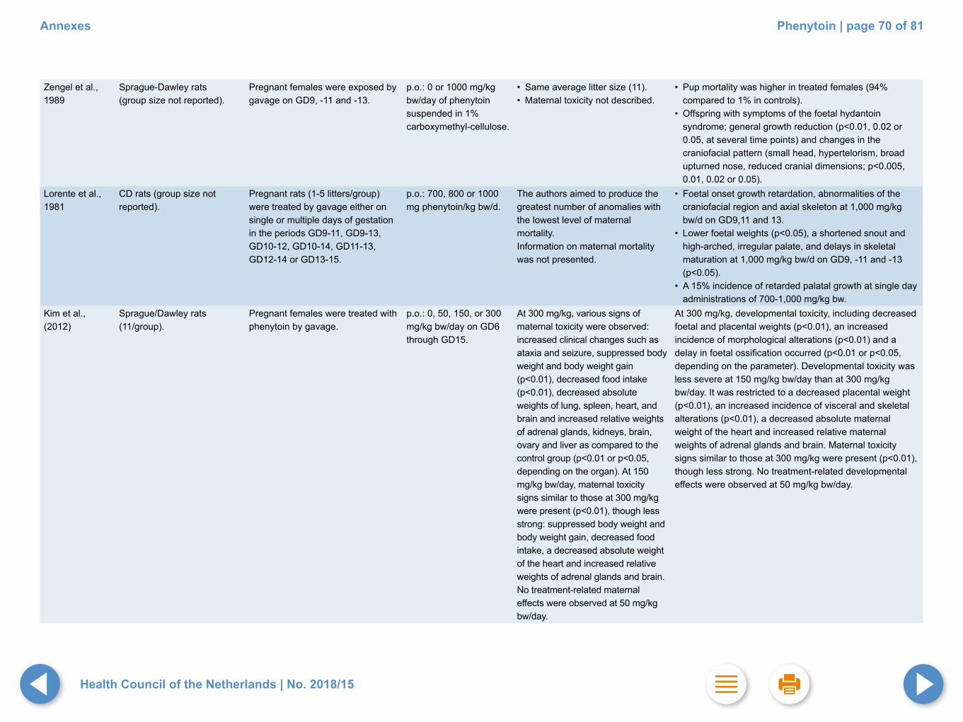
Zengel et al., 1989
Sprague-Dawley rats (group size not reported).
Pregnant females were exposed by gavage on GD9, -11 and -13.
p.o.: 0 or 1000 mg/kg bw/day of phenytoin suspended in 1% carboxymethyl-cellulose.
• Same average litter size (11).• Maternal toxicity not described.
• Pup mortality was higher in treated females (94% compared to 1% in controls).
• Offspring with symptoms of the foetal hydantoin syndrome; general growth reduction (p<0.01, 0.02 or 0.05, at several time points) and changes in the craniofacial pattern (small head, hypertelorism, broad upturned nose, reduced cranial dimensions; p<0.005, 0.01, 0.02 or 0.05).
Lorente et al., 1981
CD rats (group size not reported).
Pregnant rats (1-5 litters/group) were treated by gavage either on single or multiple days of gestation in the periods GD9-11, GD9-13, GD10-12, GD10-14, GD11-13, GD12-14 or GD13-15.
p.o.: 700, 800 or 1000 mg phenytoin/kg bw/d.
The authors aimed to produce the greatest number of anomalies with the lowest level of maternal mortality.Information on maternal mortality was not presented.
• Foetal onset growth retardation, abnormalities of the craniofacial region and axial skeleton at 1,000 mg/kg bw/d on GD9,11 and 13.
• Lower foetal weights (p<0.05), a shortened snout and high-arched, irregular palate, and delays in skeletal maturation at 1,000 mg/kg bw/d on GD9, -11 and -13 (p<0.05).
• A 15% incidence of retarded palatal growth at single day administrations of 700-1,000 mg/kg bw.
Kim et al., (2012)
Sprague/Dawley rats (11/group).
Pregnant females were treated with phenytoin by gavage.
p.o.: 0, 50, 150, or 300 mg/kg bw/day on GD6 through GD15.
At 300 mg/kg, various signs of maternal toxicity were observed: increased clinical changes such as ataxia and seizure, suppressed body weight and body weight gain (p<0.01), decreased food intake (p<0.01), decreased absolute weights of lung, spleen, heart, and brain and increased relative weights of adrenal glands, kidneys, brain, ovary and liver as compared to the control group (p<0.01 or p<0.05, depending on the organ). At 150 mg/kg bw/day, maternal toxicity signs similar to those at 300 mg/kg were present (p<0.01), though less strong: suppressed body weight and body weight gain, decreased food intake, a decreased absolute weight of the heart and increased relative weights of adrenal glands and brain. No treatment-related maternal effects were observed at 50 mg/kg bw/day.
At 300 mg/kg, developmental toxicity, including decreased foetal and placental weights (p<0.01), an increased incidence of morphological alterations (p<0.01) and a delay in foetal ossification occurred (p<0.01 or p<0.05, depending on the parameter). Developmental toxicity was less severe at 150 mg/kg bw/day than at 300 mg/kg bw/day. It was restricted to a decreased placental weight (p<0.01), an increased incidence of visceral and skeletal alterations (p<0.01), a decreased absolute maternal weight of the heart and increased relative maternal weights of adrenal glands and brain. Maternal toxicity signs similar to those at 300 mg/kg were present (p<0.01), though less strong. No treatment-related developmental effects were observed at 50 mg/kg bw/day.
Health Council of the Netherlands | No. 2018/15
Annexes Phenytoin | page 70 of 81

Eluma et al., 1984
CD 1 mice (2 mice/group/ time point; 6 mice per control group).
Pregnant mice were treated by gavage during GD8-10, GD11-13, GD14-16 or GD8-16.
p.o.: 0, 50, 75 and 125 mg phenytoin/kg bw/d.
No information. • Time-dependent and dose-related decrease in foetal weights in all treatment groups (p-values not mentioned).
• High incidence of cleft palate, and a lower incidence of foetal death, cleft lips, haematomas, hydroencephaly and exencephaly (p-values ranged from <0.001 to <0.0001) Lesions were not specified per time point and dose level.
Fritz et al., 1976
(Tif/MAG) mice (n=30/group).
Mated mice were treated by gavage during GD6-15. Offspring was subjected to visceral (one third) and skeletal examination (two thirds).
p.o.: 0, 15, 50, 100 or 170 mg phenytoin/kg bw/d.
• Ten high-dose dams died. • No maternal mortality in the lower
dose groups.• Food intake diminished at
100 mg/kg.• No effect on mortality or food
intake at 15 or 50 mg/kg.
• Early embryonic death at 100 and 170 mg/kg.• Reduced mean foetal weight at 100 and 170 mg/kg
(p<0.01).• Dose-related increase in cleft palate at 15, 50, 100 and
170 mg/kg bw/d (0.3%, 2%, 5.2% and 9.3%, respectively, versus 0.13% in historical control data from 500 mice) (statistically significant from 50 mg/kg bw/d onwards, p<0.01).
• Incomplete ossification of the fore-limbs at 170 mg/kg bw/d (p<0.01), but no malformations.
Roberts et al. 1991
C57BL/6J and A/J mice (10-13/group).
Female mice were dosed every 48 hr from ≥1 week prior to mating until sacrifice on GD18. Gestational weight gain, gravid uterus weight, foetal weight and visceral and skeletal observations were recorded.
p.o.: 0, 40, 65 or 105 mg phenytoin sodium salt/kg bw/day, p.o. (gastric intubation).
Mortality in non-pregnant C57 females (1/11 at 65 mg/kg, 5/12 at 105 mg/kg), preceded by weight loss and tremors.
• Decreased post-implantation loss in A/J mice at 65 mg/kg (p<0.05).
• Dose-dependent decrease (≥40 mg/kg) gestational weight gain and gravid uterus weight in C57 (linear trend analysis p<0.05).
• Decreased foetal weight in C57 (at 65 mg/kg, p<0.05).• Decreased ossification in both strains at 65 mg/kg.• Increased frequency of hydroencephaly (65 mg/kg) and
open eyelid (40 and 65 mg/kg) in A/J, cardiac calcium deposit (40 mg/kg) in C57 mice (all p<0.05).
Khera, 1979 Cats (n=13-14/group). Mated cats were treated by gavage during GD10-22, and necropsied on GD 43. Foetuses were examined for external, visceral and skeletal malformations.
p.o.: 0, 1 or 2 mg phenytoin sodium salt/kg bw/d.
Decreased maternal body weights at 2 mg/kg bw/d on GD20 and GD30 (p<0.05), normal body weights at 1 mg/kg bw/d.
• No effects on abortion, non-pregnancy, live or dead foetuses or mean foetal weights.
• Increased number of resorptions at 2 mg/kg bw/d (p<0.05).
• No anomalies in live foetuses, but a high incidence of malformations in cats aborting on GD31-37; (all 11 foetuses from two cats given 1 mg/kg bw/d had cleft palate and open eyelids and of the 10 foetuses from one cat given 2 mg/kg bw/d, four had umbilical hernia and one had open eyelids).
Health Council of the Netherlands | No. 2018/15
Annexes Phenytoin | page 71 of 81

Finnell et al., 1989
SWV, LM/Bc, and C57BL/6J mice (10-15 litters/treatment).
Virgin mice daily exposed orally via drinking water 15-days before mating and throughout gestation (until GD18).
p.o.: 0, 10, 20, 40, or 60 mg phenytoin/kg bw.
Not reported. • Foetal weight was dose-dependently decreased in all strains (p<0.05).
• The incidence of offspring with one or more congenital abnormalities was increased dose-dependently in all mice strains (p<0.05).
• Skeletal defects included mainly a pattern of multiple malformations and ossification delays in supraoccipital bones, sternebrae, distal phalanges and midfacial bones.
• Soft tissue defects included mainly dilated or immaturely developed cerebral ventricles and renal defects, digital, cardiac and ocular anomalies.
• Correlation between risk of abnormalities and plasma phenytoin (p<0.05).
Hansen and Billings, 1986
A/J mice. Pregnant females were exposed orally via the feed daily, prior to and during gestation.
p.o.: 0, 60 or 75 mg/kg phenytoin.
No maternal toxicity, indicated by food consumption, weight gain rate, sedation and ataxia (data not shown).
• Decreased foetal weight at both doses (p<0.05)• Decreased number of implantation sites in the highest
dose group (p<0.05).
Soysal et al., 2011
Wistar albino rats (test group: n=42 foetuses from 7 litters, control group: n=40 foetuses from 6 litters).
Pregnant females were treated i.p. on GD 8, 9 and 10. Foetuses were isolated on GD 20 and examined for bone and cartilage defects.
i.p. 25 mg/kg phenytoin diluted with serum physiologic.
Not reported. • Mean length (2.75±0.29 cm) and weight (3.04±0.42 g) of foetuses from drug-treated animals were different from the control group (length 3.21±0.27 cm and weight 3.51±0.35 g; p<0.001).
• Increased costal separation in 10/42 foetuses from drug treated animals.
• Shape malformations in the ribs of foetuses from drug-treated animals.
Shapiro et al., 1987
Sprague-Dawley rat. Dams were dosed from GD17 through day 7 PP. Weights of testis and seminal vesicle, and serum hormone levels (andro-stenedione, testosterone, dihydrotestosterone) were evaluated in male offspring of 8, 28 or 120 days of age.
0, 10, 50 or 100 mg phenytoin sodium salt/kg bw/day, i.p.
No information. • Decreased body weights from birth throughout life at 50 or 100 mg/kg (p<0.05).
• Decreased relative testis weights at day 8 PP in male offspring at 10 and 50 mg/kg (p<0.002 and p<0.02, respectively), but not at days 28 or 120 PP. No data for the 100 mg/kg group.
• Relative seminal weight was decreased at day 120 PP at 50 and 100 mg/kg (p<0.02 and p<0.001, respectively).
• Serum levels testosterone and dihydrotestosterone in male offspring were not affected at days 8, 28 or 120 PP. Levels of androstenedione at 50 mg/kg were increased at day 8 PP (p<0.02), and decreased at day 28 PP (p<0.02).
• Estrous cyclicity (vaginal smears from about 13-week old offspring) and weights of the uterus and ovaries (at about 16 weeks PP) in female offspring not affected.
Health Council of the Netherlands | No. 2018/15
Annexes Phenytoin | page 72 of 81

Hansen & Billings, 1986
A/J mice. Pregnant females were i.p. injected with phenytoin on GD10.
i.p.: 0, 60 or 75 mg/kg phenytoin.
sedation and ataxia (high-dose group).
• A dose-related increase in resorptions (p<0.05)• Nearly all surviving foetuses had an abnormality, mainly
cleft lip and palate (p<0.05).Hansen & Hodes, 1983
ICR mice(25/group).
C57BL/6J, B6AF1, AB6F1, (B6A)F2 and C3H/He mice (15, 19 or 20 animals per group).
Pregnant mice were treated by i.p. injection on GD10-12.
i.p.: 0, 50, 75 or 100 mg phenytoin/kg bw/d.
i.p.: 0 or 75 mg/kg bw/d.
• The high-dose was lethal to the dams and omitted from the study.
• No maternal toxicity at 50 and 75 mg/kg bw/d (measures not specified).
• Dose-related decrease in mean foetal weight and crown rump length in both test groups (p<0.01 or p<0.05).
• Increased transumbilical distance at 75 mg/kg bw/d (p<0.01).
• Increased incidence of malformed foetuses at 75 mg/kg bw/d (p<0.05), with orofacial anomalies most frequently observed (p<0.01).
• Other malformations included ectopic kidneys, cryptorchydism and cardiac defects, but no data were presented..
• No skeletal defects in any group.
A higher number of resorptions was observed in two strains, a higher number of malformed foetuses in three strains, a higher number of orofacial anomalies in two strains and a lower foetal weight in three strains treated with phenytoin as compared to control animals (p<0.01 or p<0.05).
Rowland et al., 1990
Sprague-Dawley rats (n=4-9/group).
Pregnant females were daily exposed intravenously at GD8-17. Reproductive outcomes were determined at GD20.
i.v.: 0, 25, 50, 75, or 100 mg/kg bw/day.
No decreased weight gain, mild to severe imbalance and ataxia at 50and75 mg/kg, excessive ataxia at 100mg/kg (100 mg/kg therefore discontinued).
• Embryolethality• Intrauterine growth retardation, associated with
reductions in foetal weight and crown-rump length (p<0.05).
• Craniofacial malformations (p<0.05).Functional and cognitive defects
Mowery et al., 2008
Sprague Dawley rats(test group: n=31 from 11 litters; controls: n=22 from 10 litters).
Female rats were dosed by gavage twice daily from 10 days before mating, throughout pregnancy and the three week pre-weaning period. Behavioural testing was conducted with 80-90 days old female offspring.
p.o.: 0 or 50 mg phenytoin/kg bw/d.Maternal plasma levels on the last day of dosing approximated the low level of human therapeutic concentrations (10-20 µg/ml).
• No information on maternal toxicity.
• Increased performance in simple associative learning tasks (appetitive-to-aversive transfer paradigm, p<0.05).
• Impaired performance in a higher-order learning and memory task (avoidance conditioning, p<0.05).
Health Council of the Netherlands | No. 2018/15
Annexes Phenytoin | page 73 of 81

Schilling et al., 1999
Sprague Dawley rats (10 (controls) or 15 (treated dams)).
Pregnant Sprague Dawley rats were dosed by gavage during GD7-18. Per litter, 2 male offspring were tested in the Morris water maze, one exhibiting circling in a straight water channel and one in a circling tank.
p.o.: 0 or 200 mg phenytoin/kg bw/d.
• Decreased maternal body weight during GD14-18 (p<0.05).
• Increased offspring mortality during the first week (p<0.01).
• Preweaning body weights not affected.
• Decreased post weaning body weights (p<0.05, or <0.01).
• Circling in 53% of the exposed offspring; no circling in the control group.
• Impaired reference memory-based spatial learning in the Morris water maze in noncircling offspring (p<0.01).
• Impaired reference memory based spatial learning, impaired cued (visible) platform learning (p<0.01), impaired spatial discrimination (p<0.01) and impaired working memory-based learning (p<0.01) in circling offspring.
McCartney et al.,1999.
Sprague Dawley rats (≥20 dams/group). Examinations were conducted in all pups (pre-weaning) or in 1 pup/sex/ litter (post-weaning).
Pregnant Sprague Dawley rats (≥20 dams/group) were dosed by gavage during GD 7-18.
p.o.: 0, 50, 100 or 150 mg phenytoin sodium salt/kg bw/d.
• Dose-related reduction in maternal weight gain in all treatment groups (p<0.05).
• Increased incidences of chromodacryorrhea, lacrimation and circling at 100 and 150 mg/kg bw/d (data not reported).
• Accelerations in developmental landmarks and preweaning behaviour (eye opening, incisor eruption, negative geotaxis and olfactory orientation) in pups of all treatment groups (p<0.05).
• Delays in air righting in pups of all offspring treatment groups (p<0.05).
• Increased locomotor activity in high-dose pups (at PND21) and adults (at PND62) (p<0.05).
• Dose-related performance deficits in a water maze assay (learning/ memory impairments, p<0.01) and in auditory startle responses (p<0.05 in at least one dose group).
• Reduced hind brain weights in male pups at 150 mg/kg bw/d (p<0.05).
• Reduced forebrain and whole brain weights in adult females at 100 and 150 mg/kg bw/d (p<0.05).
Tsutsumi et al., 1998
Sprague Dawley rats (7-10 dams/group) Examinations were conducted in (generally) 10-15 offspring males/group.
Pregnant rats were dosed by gavage during GD 7-18, and male offspring was subjected to a reflex test and several learning/memory tests.
p.o.: 0, 50 or 100 mg phenytoin/kg bw/d.
• No information on maternal toxicity.
• Delayed time completion of a negative geotaxis (reflex) test in both treatment groups (p<0.001).
• No effects in a figure-eight-maze, a Biel water maze, or a Morris maze test.
• High total number of choices (p<0.05).and low number of correct choices (p<0.05).in the high-dose group in radial maze test.
• Low number of correct choices in the high-dose group in delayed nonmatching-to-sample test (T-shaped maze, p<0.05.).
• Decreased offspring whole brain weights in high dose group in wk 6 (p<0.05); body weigths unaffected. Offspring brain weight had returned to normal at 16 weeks.
Health Council of the Netherlands | No. 2018/15
Annexes Phenytoin | page 74 of 81

Vorhees et al., 1995
Sprague Dawley rats (5-6 dams/group) (Offspring number tested was not indicated).
Pregnant rats were dosed by gavage during GD7-18.Offspring was tested at approximately 50 days of age.
p.o.: 0 or 100 mg phenytoin sodium salt/kg bw/d in corn oil.
• Maternal weight gain, gestation length, pups per litter and sex ratio within litters were not affected.
• More errors and longer latencies to find the goal in the Cincinnati (water) Maze (circling animals p<0.01), noncircling animals p<0.05).
• The effect in circling animals was larger than in noncircling animals.
Pizzi et al., 1992
Sprague Dawley rats (10-11 dams/group).
Pregnant rats were dosed by gavage during GD9-18.
p.o.: 0, 100 or 200 mg phenytoin sodium salt/kg bw/d. (On GD18, maternal plasma levels in the low- and high-dose group were 11.5 and 26.2 mg¹ phenytoin/ml).
¹ The Committee assumes that µg rather than mg was meant.
• Dose-related decrease in maternal weight gain (both doses p<0.001).
• Lower birth weights and body weights at PND30 (high dose group males and females p<0.001; low dose group males p<0.05)., and increased offspring mortality (41% and 61% in the lowand high-dose group, respectively).
• Exposed offspring frequently showed chromodacryorrhea.
• Increased pivoting locomotor activity on PND7 and 9 at 100 mg/kg bw/d (due to mortality, the 200 mg/kg bw/d group was not examined).
• Abnormal spontaneous circling behaviour (2 and 33% in the low- and high-dose group, respectively).
• Increases in locomotor activity measures in adult rats that had been exposed to 200 mg/kg bw/d (at PND45: p<0.05; at PND66: p<0.05).
Weisenburger et al., 1990
Sprague Dawley rats (10-13 litters/group).Where possible, six offspring rats/sex/litter were retained for testing.
Pregnant rats were dosed by gavage during GD7-18.
p.o.: 0, 100 or 200 mg phenytoin/kg bw/d. Maternal serum concen-trations on GD18 were 15.1 and 20.9 µg/ml for low- and high-dose, respectively which was stated to be in the range of human therapeutic ranges (10-25 µg/ml).
• Reduced maternal weight gain in both treatment groups (p<0.05).
• Increased pup mortality at 200 mg/kg bw/d at birth (p<0.01), PND7 (p<0.01).and PND28 (p<0.05).
• Reduced preweaning and postweaning offspring body weights at 200 mg/kg bw/d (p<0.05).
• Dose-related increases in circling behaviour (both doses: p<0.01), preweaning locomotor activity (both doses: p<0.05), errors in a complex water maze and impaired performance in a radial-arm maze (both circlers and noncirclers at each dose: p<0.01).
Vorhees, 1987 Sprague Dawley rats (13-20 litters/group) (Offspring number tested per group was not indicated).
Pregnant rats (13-20 litters/group) were dosed by gavage during GD7-18.Offspring of both sexes was tested prior to weaning (for activity only) and afterwards.
p.o.: 0, 100, 150 or 200 mg phenytoin/kg bw/d• These levels resulted
in maternal plasma levels on GD 18 (4h after dosing) of about 10, 20 and 24 µg/ml, in the low-, mid- and high-dose group, respectively (with little decline up to 24h).
• Decreased maternal weights in the high-dose group during gestation (at GD18 p<0.01, at GD20 p<0.05).
• 23% of the litters in the high-dose group were lost (versus 7% in controls, not statistically significant).
• Increased offspring mortality in the high-dose group during PND1-21 (p<0.01).
• Dose-related increase in circling behaviour (p<0.05 or <0.01).
• The highest dose produced: - increased activity (in various tests of activity, p<0.01), - delayed dynamic righting development (p<0.01), - impaired Biels (multiple-T water) maze learning (p<0.01), - Y-maze (avoidance) learning (p<0.05), - inhibited tactile startle responses (p<0.05).
• The two lower doses showed a dose-effect relationship on most measures.
• No effects on postnatal growth, total brain weight, or brain protein content in adulthood (at 79-84 days of age).
Health Council of the Netherlands | No. 2018/15
Annexes Phenytoin | page 75 of 81

Ruiz et al., 1987
Sprague Dawley rats(6 offspring/group).
Rats were dosed by gavage, from 7 days before fertilization, throughout pregnancy, while pups were also given the drug by gavage. The dosing regimen was not reported.Neuronal structure in the somatosensory cortex at 30 days of postnatal development was investigated.
p.o.: 0 or 50 mg phenytoin/kg bw/d.
• No information on maternal toxicity.
• No major differences in cortical layers III and IV.• Total cell density in layer V higher (p<0.0001). • Numbers and lengths of apical and basal dendrites
decreased (p<0.05 to p<0.0001)• Diminished degree of neuronal maturation at the level of
collateral apical dendrites of pyamidal cells in the surface layers of the cortex.
• Poor development of the basilar dendrites in the deeper layers of the sensory cortex.
• Altered pattern of phosphorylation of cytoskeletal proteins.Elmazar & Sullivan, 1981
Wistar rats (10 (first study) - 20 (repeat study) litters/group).
Pregnant rats were dosed by gastric intubation during GD7-19.After parturition, the offspring was culled to six-eight/litter and reared by fostering or cross fostering.
p.o.: 0 or 100 mg phenytoin/kg bw/d.
• Decreased body weights of the treated dams up to GD10.
• Due to paired feeding, the body weights of the control dams followed the same pattern.
• Lower survival and decreased body weights in two experiments at PND2 (p<0.05), in one of them at PND21 (p<0.01).and PND90 (p<0.05).
• Delay of up to 15 days in the development of the dynamic righting reflex (no statistics reported).
• Decreased ability of offspring to stay on a rotating rod (p<0.05).
• Decreased ability to walk along elevated parallel rods (p<0.01).
• No changes in development of physical landmarks, or in the development of crawling and walking activities at 9-21 days of age, and no changes in a head-dipping test or in a conditioned avoidance test (shuttle box) at 26-34 days of age.
• Decreased absolute brain weight at 3 d (p<0.05 or <0.001), which remained until 90 d (p<0.001).
• No differences in cerebellar DNA content.Vorhees, 1983 Sprague-Dawley rats
(number not reported).Pregnant rats received phenytoin or vehicle by gavage. Structural effects and postnatal functioning were investigated on GD20 or postnatally. The first experiment served to determine the highest dose for assessing postnatal functional teratogenesis in experiments 2 and 3. In this dose range-finding study 200 mg/kg bw/ day was chosen as the highest dose to be used in the other experiments. This choice was based on maternal toxicity (reduced maternal weight, p<0.01) and thefraction of resorbed or dead
Health Council of the Netherlands | No. 2018/15
Annexes Phenytoin | page 76 of 81

foetuses (not statistically different from controls).
Experiment 2: 0, 5, 50 or 200 mg/kg bw/day p.o. on GD7-18.
Experiment 3: 0 or 200 mg/kg bw/ day p.o. on GD7-10, 11-14 or 15-18.
• Decreased maternal body weight at 200 mg/kg bw/day on GD14 and -18 (p<0.05), and during lactation (p<0.01).
• Length of gestation, external malformations, number of offspring delivered and sex ratio within litters not affected.
• Offspring mortality increased at 50 and 200 mg/kg bw/day on PND0 (both p<0.0001) and at 200 mg/kg bw/day on PND 21 (p<0.001). Mortality returned to normal on PND22-70.
• Before weaning (PND1-21), no reduction in offspring body weight in any dose group (data not shown).
• After weaning (PND22-70), offspring body weight was reduced: 200 mg/kg group 10.8% lighter than controls on PND42 (p<0.001) and 7.2% lighter on PND70 (p<0.05).
• Maternal weight not affected.• Length of gestation, external
malformations, number of offspring delivered and sex ratio within litters not affected.
• Offspring mortality increased preweaning after treatment at GD7-10 (p<0.001) and GD15-18 (p<0.01).
• Offspring mortality increased postweaning after treatment at GD11-14 (p<0.01).
• No effects on lower incisor eruption, eye opening or vaginal patency development.
• No effects on preweaning negative geotaxis, olfactory orientation, figure-8 activity or neonatal T-maze behaviour.
• Effects on preweaning righting (data not shown), pivoting (200 mg/kg, p<0.001), startle (200 mg/kg, p<0.001), and swimming (200 mg/kg, at various ages, p<0.05).
• No effects on postweaning M-maze behaviour, passive avoidance or spontaneous alternation.
• Postweaning figure-8 activity and Biel water maze learning affected (200 mg/kg bw/day, p<0.05 to p<0001).
• No effects on incisor eruption, eye opening or vaginal patency development.
• No effects on preweaning righting, negative geotaxis, olfactory orientation, figure-8 activity or neonatal T-maze behaviour.
• Increased preweaning pivoting locomotion and delayed swimming development in the groups exposed at GD11-14 or GD15-18 (p<0.05 or <001); startle not affected.
• No effects on postweaning M-maze behaviour or spontaneous alternation.
• Increased postweaning figure 8 ambulation (p<0.01) and water maze errors (p<0.001) and impaired passive avoidance retention in the GD11-14 exposure group (p<0.05).
Health Council of the Netherlands | No. 2018/15
Annexes Phenytoin | page 77 of 81

Ogura et al., 2002
C57BL/6 mice (13-15 males/ group).
Newborn mice were dosed by gavage during PND5-14.
p.o.: 0 or 35 mg phenytoin/kg bw/d - Plasma level of phenytoin was 20±2.8 µg/ml, 3h after the last dose. • Brain concentration of
phenytoin was 1.6 times higher (31.9±10.3 µg/ml).
• In some pups: acute behavioural detoriation, including anorexia, hyperactivity and motor coordination deficits.
• Mortality (38%).• Weight loss (at 56 days, the body
weights of treated mice had returned to control values).
• Reduced brainstem weight, cerebral weight, cerebellar weight and total brain weight in 56 days old mice (p<0.01).
• Interference with the development of granule cells in the hippocampus and the cerebellum, and with the dendritic development of Purkinje cells.
• Impaired acquisition of a hidden platform task in a water maze test (p<0.001).
• Increased errors during learning in a radial arm maze (p<0.01).
Ohmori et al., 1999
Jcl:ICR mice (13-16/sex/group).
Phenytoin was administered by gavage to newborn Jcl:ICR mice during PND2-4.
p.o.: 0 or 35 mg phenytoin/kg bw/dPlasma level of phenytoin on the third day of administration was 17.7 µg/ml. In the brain the level was higher (31.4 µg/g).
• Mortality >30% in males and females.
• Weight loss, anorexia, motor hypoactivity and incoordination at PND4 (data not shown). Body weight recovered to normal at PND56.
• Impaired motor performance in a rotating rod (p<0.05).• Impaired spontaneous locomotor activity (p<0.01).• Reduced total brain weight, cerebral weight and
cerebellar weight (p<0.01).• Neurotoxic damage in the developing cerebellum (cell
death of external granule cells, inhibited migration of granule cells and impaired Purkinje cell differentiation).
Hatta et al., 1999
Jcl:ICR mice (n not indicated).
Newborn mice were dosed by gavage on PND2-4.
p.o.: 0, 10, 17.5, 25 or 35 mg phenytoin/kg bw/d. Dose levels were stated to correspond to therapeutic plasma levels in humans.
• 8% mortality rate in the high-dose group in either sex.
• No effects on pup body weights (PND5-PND21).
• Decreased locomotor abilities and righting reflex at 25 or 35 mg/kg bw/d, on PND5 (p<0.05 or <0.01).
• Decreased total brain weight, cerebral weight and cerebellar weight at 25 or 35 mg/kg bw/d (p<0.05 or <0.01).
Phillips and Lockard, 1993
Monkeys (Macaca fascicularis).(Four adult females were dosed; control groups consisted of monkeys treated with stiripenol (n=5), or phenytoin plus stiripenol (n=4), because stiripenol has been shown to reduce the incidence of phenytoin-induced congenital malformations in mice.)
Adult female monkeys (Macaca fascicularis) were dosed via stomach catheter from 1 month before mating throughout gestation. After birth, the infants were transferred to a nursery for testing. They were tested at an age of about 2 weeks to 45 days (exact times were corrected for gestational age, i.e., age based on conception date rather than birth date).
By stomach catheter at levels providing plasma concentrations between 4-12 µg phenytoin/ml (initial dose was 20 mg/kg bw).
• No information on maternal toxicity.
• Infants showed hyperexcitability (jerking, screeching, refusing to attend to stimuli, lack of visual orientation) during cognitive testing when compared to stiripenol (p<0.05), not to phenytoin plus stiripenol.
Health Council of the Netherlands | No. 2018/15
Annexes Phenytoin | page 78 of 81

Wolansky & Azcurra, 2005
Sprague Dawley rats (n= 28-32 offspring/group).
Pregnant females were treated during GD 13-18. Motor and learning disorders of offspring were examined in a circular maze at PND40, PND80 and PND150.
i.p.: 0 or 30 mg phenytoin sodium salt/kg bw/d.
• Maternal body weights and pup body weights not affected (data not shown).
• Maternal behaviour not affected (data not shown).
• Gestation time not affected.
• Increased circling velocities in a circular maze at all time points (p<0.05).
• Increased spatial error rates for direction of circling at all time points (p<0.05).
B abbreviationsbw body weight
CI confidence interval
d day
GD gestation day
h hours
i.m. intramuscular
i.p. intraperitoneal
i.v. intravenous
n number
NOEL no observed effect level
NOAEL no observed adverse effect level
OR odds ratio of an effect in exposed population versus non exposed
population
PND postnatal day
PP post partum day
p.o. oral
RR relative risk
s.c. subcutaneous
Health Council of the Netherlands | No. 2018/15
Annexes Phenytoin | page 79 of 81

the CommitteeThis advisory report has been prepared by the Subcommittee on the
Classification of Substances Toxic to Reproduction, a permanent
subcommittee of the Health Council of the Netherlands.
The first draft of this report was prepared by M.M. Tegelenbosch-
Schouten, M. Sc, in cooperation with Dr. M.J.W. van den Hoven,
Dr. D. Jonker and Dr. B.A.R. Lina, Department Toxicology and Applied
Pharmacology, TNO Quality of Life, Zeist.
Chair• D Lindhout, Emeritus professor of Medical Genetics; paediatrician (not
practising), clinical geneticist; University Medical Center Utrecht
Members• N Roeleveld, Reproductive epidemiologist; Radboud university medical
center, Nijmegen
• JG Theuns-van Vliet, Reproductive toxicologist, Triskelion BV, Zeist
• TGM Vrijkotte, Epidemiologist, AMC, Amsterdam
• ECM Tonk, Regulatory toxicologist, Charles River Laboratories Den
Bosch BV (since 1 January 2018)
• DH Waalkens-Berendsen, Reproductive toxicologist; Zeist (until 15
September 2017)
• PJJM Weterings, Toxicologist; Weterings Consultancy BV, Rosmalen
Structurally consulted expert• AH Piersma, Professor of reproductive and developmental toxicology,
Utrecht University; National Institute of Public Health and the
Environment, Bilthoven
Scientific secretary• PW van Vliet, Health Council of the Netherlands, Den Haag
Health Council of the Netherlands | No. 2018/15
The Committee Phenytoin | page 80 of 81

This publiation can be downloaded from www.healthcouncil.nl.
Preferred citation:Health Council of the Netherlands. Phenytoin. Evaluation of the effects on reproduction, recommendation for classification. The Hague: Health Council of the Netherlands, 2018; publication no. 2018/15.
All rights reserved
The Health Council of the Netherlands, established in 1902, is an independent scientific advisory body. Its remit is “to advise the government and
Parliament on the current level of knowledge with respect to public health issues and health (services) research...” (Section 22, Health Act).
The Health Council receives most requests for advice from the Ministers of Health, Welfare and Sport, Infrastructure and Water Management, Social
Affairs and Employment, and Agriculture, Nature and Food Quality. The Council can publish advisory reports on its own initiative. It usually does this in
order to ask attention for developments or trends that are thought to be relevant to government policy.
Most Health Council reports are prepared by multidisciplinary committees of Dutch or, sometimes, foreign experts, appointed in a personal capacity.
The reports are available to the public.
Health Council of the Netherlands | No. 2018/15