evaluation of renography by monte carlo simulations
TRANSCRIPT

Medical Radiation Physics, Clinical Sciences Lund, Lund University
Michael Ljungberg
Evaluation of renography by Monte Carlo simulations
Användarmöte Nuklearmedicin 15 nov, 2011

Medical Radiation Physics, Clinical Sciences Lund, Lund University
Quality Control of procedures
Physical Phantoms
Commonly used in general SPECT research.
Cylindrcal and elliptical and more realistic antropomorphic phantoms available.

Medical Radiation Physics, Clinical Sciences Lund, Lund University
Kidney phantoms
Static phantom with kidneys available
Not possible to simulate dynamic processes
Limited number of organs
•Liver •Kidney •Tumors in the liver
Medical Radiation Physics, Clinical Sciences Lund, Lund University
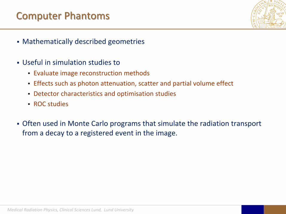
Computer Phantoms
Mathematically described geometries
Useful in simulation studies to • Evaluate image reconstruction methods • Effects such as photon attenuation, scatter and partial volume effect • Detector characteristics and optimisation studies • ROC studies
Often used in Monte Carlo programs that simulate the radiation transport
from a decay to a registered event in the image.

Medical Radiation Physics, Clinical Sciences Lund, Lund University
Pixar’s Transformable Model
The 4D XCAT Phantoms
Based on state-of-the-art computer graphics techniques
• Organs modeled using NURBS and subdivision surfaces, tools widely used in current games and movies
Utilize Pixar’s Universal Man and Woman Idea
• Transformable base anatomies upon which to model different types of people (inside and out)
Courtesy Paul Segars, Duke University, NC, USA
Medical Radiation Physics, Clinical Sciences Lund, Lund University
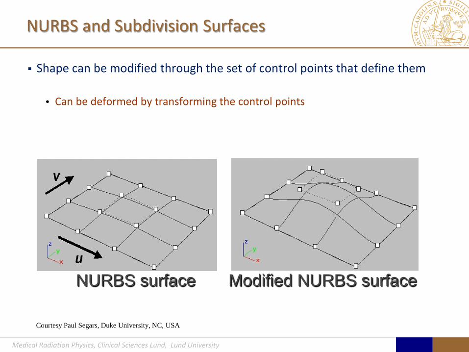
NURBS and Subdivision Surfaces
Shape can be modified through the set of control points that define them
• Can be deformed by transforming the control points
NURBS surface Modified NURBS surface
Courtesy Paul Segars, Duke University, NC, USA

Medical Radiation Physics, Clinical Sciences Lund, Lund University
Visible Female Anatomical Images Male Anatomy Female Anatomy
Segmented Organs From Imaging Data
Visible Male Anatomical Images
Fit 3D NURBS and Subdivision Surfaces to Segmented Organs
Development of the XCAT Male and Female Base Anatomies
Courtesy Paul Segars, Duke University, NC, USA

Medical Radiation Physics, Clinical Sciences Lund, Lund University
Male Abdomen Respiratory Motion
Chest
Female Abdomen Right Left
Head
Phantom Anatomy
Courtesy Paul Segars, Duke University, NC, USA

Medical Radiation Physics, Clinical Sciences Lund, Lund University
4D Beating Heart Model
RCA LCX
RV
RA
LA
LV
Aorta
LAD
Courtesy Paul Segars, Duke University, NC, USA

Medical Radiation Physics, Clinical Sciences Lund, Lund University
Segmented Branches Extended Branches
Mathematical Extension of the Segmented Coronary Artery Tree
Courtesy Paul Segars, Duke University, NC, USA

Medical Radiation Physics, Clinical Sciences Lund, Lund University
Plaques and Perfusion Defects
Myocardial SPECT
Plaque Imaging
Ant. Projection
SA LA
plaque defect
• Plaques modeled as NURBS surfaces on the interior of the arteries
• Perfusion defects modeled as wedges in the LV myocardium
Beating Heart Model Coronary Artery Disease (CAD)
Courtesy Paul Segars, Duke University, NC, USA

Medical Radiation Physics, Clinical Sciences Lund, Lund University
Interactive IDL Program
Motion vectors for the lung
* *
4D Respiratory Model
• Based on respiratory-gated CT data
• IDL program to mark and track points on the respiratory structures
Courtesy Paul Segars, Duke University, NC, USA

Medical Radiation Physics, Clinical Sciences Lund, Lund University
Gated Chest CT
Lung with Spherical Tumor
w/o resp. motion w/ resp. motion
Contrast ~0.5
Effect of Motion on the Lung Tumor Imaging (SPECT)
Modeling of Tumors
Courtesy Paul Segars, Duke University, NC, USA

Medical Radiation Physics, Clinical Sciences Lund, Lund University
Effect of Anatomy on Imaging
Nuclear Medicine procedures are made on a population with large variations Methods can be studied on a population of computer phantoms in a controlled way May catch problems that a measurement or a simulations using a single phantom can not find
Male Female
Courtesy Paul Segars, Duke University, NC, USA

Medical Radiation Physics, Clinical Sciences Lund, Lund University
Monte Carlo Calculation
A method to simulate the radiation transport Main applications are in Dosimetry and Medical Imaging

Medical Radiation Physics, Clinical Sciences Lund, Lund University
SIMIND Monte Carlo program

Medical Radiation Physics, Clinical Sciences Lund, Lund University
Scintillation Camera Simulation
SIMIND is a Monte Carlo program that simulates a scintillation camera system.
Useful to evaluate imaging parameters. A. No patient motion and perfect camera resolution. B. Patient respiration and heart beating. C. Normal system resolution and patient movements D. Photon attenuation added E. Photon attenuation and scatter F. Realistic noise level added.
A B C D E F

Medical Radiation Physics, Clinical Sciences Lund, Lund University
EQUALIS Project
Modified to simulate Dynamic Scintigraphy
Activity in the lidney compartment and in other organs in the XCAT phantom can be described by a time-activity curve
Hospital-specific parameters • Time sequences, typical for clinical acquisitions • Camera type • Collimator • Activity • Acquisition time
Medical Radiation Physics, Clinical Sciences Lund, Lund University
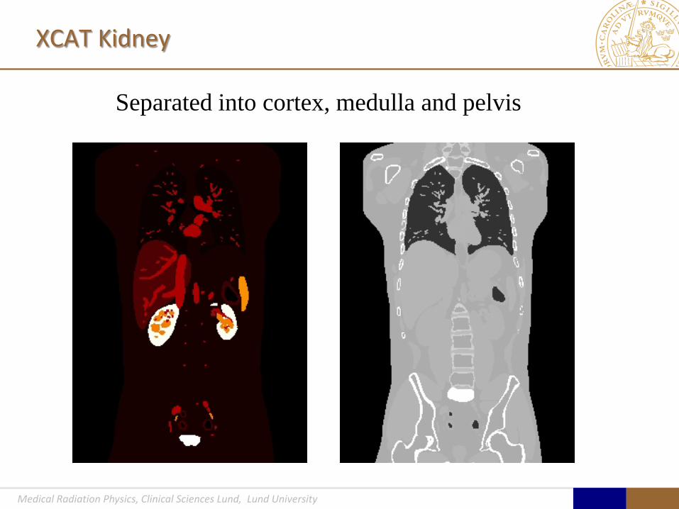
XCAT Kidney
Separated into cortex, medulla and pelvis

Medical Radiation Physics, Clinical Sciences Lund, Lund University
Kidney kinetics – version 0.0
Kinetics is defined for • Left cortex, medulla and pelvis • Left cortex, medulla and pelvis • Ureter and bladder • Liver, spleen,……
Normal and abnormal kinetic
Possible to include anatomical patient variations

Medical Radiation Physics, Clinical Sciences Lund, Lund University
Kidney kinetics
-5.00
0.00
5.00
10.00
15.00
20.00
25.00
0 100 200 300 400 500 600 700 800 900
Cortex-L
Medulla-L
Pelvis-L
Cortex-R
Medulla-R
Pelvis-R
Bladder
ureter
liver
spleen

Medical Radiation Physics, Clinical Sciences Lund, Lund University
Example of a Monte Carlo simulated renography study

Medical Radiation Physics, Clinical Sciences Lund, Lund University
Import to Xeleris possible

Medical Radiation Physics, Clinical Sciences Lund, Lund University
Image Transfer
Image data from simulations converted to Interfile or DICOM Download data from a web page and import into the clinical system Evaluated on site using daily clinical routine procedure Results are compared to truth
Future developments • More cases • More patients • Perhaps a better kidney model • ……..