evaluation of deep learning to augment image-guided
TRANSCRIPT

Original Investigation | Health Informatics
Evaluation of Deep Learning to Augment Image-Guided Radiotherapyfor Head and Neck and Prostate CancersOzan Oktay, PhD; Jay Nanavati, MS; Anton Schwaighofer, PhD; David Carter, PhD; Melissa Bristow, MS; Ryutaro Tanno, PhD; Rajesh Jena, MD; Gill Barnett, MD;David Noble, MD; Yvonne Rimmer, MD; Ben Glocker, PhD; Kenton O’Hara, PhD; Christopher Bishop, PhD; Javier Alvarez-Valle, MS; Aditya Nori, PhD
Abstract
IMPORTANCE Personalized radiotherapy planning depends on high-quality delineation of targettumors and surrounding organs at risk (OARs). This process puts additional time burdens ononcologists and introduces variability among both experts and institutions.
OBJECTIVE To explore clinically acceptable autocontouring solutions that can be integrated intoexisting workflows and used in different domains of radiotherapy.
DESIGN, SETTING, AND PARTICIPANTS This quality improvement study used a multicenterimaging data set comprising 519 pelvic and 242 head and neck computed tomography (CT) scansfrom 8 distinct clinical sites and patients diagnosed either with prostate or head and neck cancer. Thescans were acquired as part of treatment dose planning from patients who received intensity-modulated radiation therapy between October 2013 and February 2020. Fifteen different OARswere manually annotated by expert readers and radiation oncologists. The models were trained on asubset of the data set to automatically delineate OARs and evaluated on both internal and externaldata sets. Data analysis was conducted October 2019 to September 2020.
MAIN OUTCOMES AND MEASURES The autocontouring solution was evaluated on external datasets, and its accuracy was quantified with volumetric agreement and surface distance measures. Mod-els were benchmarked against expert annotations in an interobserver variability (IOV) study. Clinicalutility was evaluated by measuring time spent on manual corrections and annotations from scratch.
RESULTS A total of 519 participants’ (519 [100%] men; 390 [75%] aged 62-75 years) pelvic CTimages and 242 participants’ (184 [76%] men; 194 [80%] aged 50-73 years) head and neck CTimages were included. The models achieved levels of clinical accuracy within the bounds of expertIOV for 13 of 15 structures (eg, left femur, κ = 0.982; brainstem, κ = 0.806) and performedconsistently well across both external and internal data sets (eg, mean [SD] Dice score for left femur,internal vs external data sets: 98.52% [0.50] vs 98.04% [1.02]; P = .04). The correction time ofautogenerated contours on 10 head and neck and 10 prostate scans was measured as a mean of 4.98(95% CI, 4.44-5.52) min/scan and 3.40 (95% CI, 1.60-5.20) min/scan, respectively, to ensureclinically accepted accuracy. Manual segmentation of the head and neck took a mean 86.75 (95% CI,75.21-92.29) min/scan for an expert reader and 73.25 (95% CI, 68.68-77.82) min/scan for a radiationoncologist. The autogenerated contours represented a 93% reduction in time.
CONCLUSIONS AND RELEVANCE In this study, the models achieved levels of clinical accuracy withinexpert IOV while reducing manual contouring time and performing consistently well across previouslyunseen heterogeneous data sets. With the availability of open-source libraries and reliable perfor-mance, this creates significant opportunities for the transformation of radiation treatment planning.
JAMA Network Open. 2020;3(11):e2027426.
Corrected on December 9, 2020. doi:10.1001/jamanetworkopen.2020.27426
Key PointsQuestion Can machine learning models
achieve clinically acceptable accuracy
in image segmentation tasks in
radiotherapy planning and reduce
overall contouring time?
Findings This quality improvement
study was conducted on a set of 242
head and neck and 519 pelvic computed
tomography scans acquired for
radiotherapy planning at 8 distinct
clinical sites with heterogeneous
population groups and image acquisition
settings. The proposed technology
achieved levels of accuracy within
interexpert variability; statistical
agreement was observed for 13 of 15
structures while reducing the
annotation time by a mean of 93%
per scan.
Meaning The study findings highlight
the opportunity for widespread
adoption of autosegmentation models
in radiotherapy workflows to reduce
overall contouring and planning time.
+ Supplemental content
Author affiliations and article information arelisted at the end of this article.
Open Access. This is an open access article distributed under the terms of the CC-BY-NC-ND License.
JAMA Network Open. 2020;3(11):e2027426. doi:10.1001/jamanetworkopen.2020.27426 (Reprinted) November 30, 2020 1/11
Downloaded From: https://jamanetwork.com/ on 12/30/2021

Introduction
Each year, more than half a million patients are diagnosed with cancer and receive radiotherapyeither alone or in combination with surgery.1,2 Intensity-modulated radiation therapy has become akey component of contemporary cancer treatment because of reduced treatment-induced toxiceffects, with 40% of successfully cured patients undergoing some form of radiotherapy.3
Development of personalized radiation treatment plans that match a patient’s unique anatomicalconfiguration of tumor and organs at risk (OARs) is a multistep process starting with the acquisitionof cross-sectional images and the segmentation of relevant anatomical volumes within the imagesthrough to dose calculation and subsequent delivery of radiation to the patient.
The segmentation of the images represents a significant rate-limiting factor within thistreatment workflow. Currently, this task is performed manually by an oncologist using speciallydesigned software to draw contours around the regions of interest. While the task demandsconsiderable clinical judgement, it is also laborious and repetitive, with contoured volumes needingto be constructed slice by slice across entire cross-sectional volumes. Consequently, it is an extremelytime-consuming process, often taking up to several hours per patient.4 It can create delays in theworkflow that may be detrimental to patient outcomes, but it also comes with an increasing financialburden to the hospital. As such, there is a significant motivation to provide automated orsemiautomated support to reduce overall segmentation time for process.
In addition to long contouring times, there are challenges that derive from a dependency oncomputed tomography (CT) scans as primary reference images for tumor and healthy tissueanatomy. The inherent limitation of CT images in terms of image contrast on soft tissues makessegmentation challenging, and there remains uncertainty in the exact extent of tumor and normaltissues. This introduces a further key challenge for manual contouring; it is well documented thatthere is as a source of interoperator variability (IOV) in segmentation.5-11 Such variability can affectsubsequent dose calculations, with the potential for poorer patient outcomes.12 Likewise, it presentsa concern in the context of clinical trials carried out across multiple hospital sites. In addition to timesavings, automating contouring would offer potential for greater standardization.
There has been significant investment to establish autosegmentation techniques that aim toreduce time and variability. Recent efforts are exploring machine learning (ML) methods forautosegmentation of CT scans in radiotherapy.13-16 While they achieve reasonable accuracy withinthe same-site data sets on which they are trained and evaluated, model performance is oftencompromised when deployed across other hospital sites. Such approaches can be further limited inadaptability to different clinical domains of radiotherapy. Restricting these algorithms to a singlebodily region or a single hospital site with specific acquisition protocols limits the value andapplicability of these approaches in real-world clinical contexts. Furthermore, integration of suchtools into existing hospital workflows is often not considered. To address these limitations, wepresent a generic segmentation solution for both prostate and head and neck cancer treatmentplanning and demonstrate how it can be integrated into existing workflows.
Methods
Ethical Review of StudyAll data sets were licensed under an agreement with the clinical sites involved and received afavorable opinion from the research ethics committee from the East of England–Essex researchethics committee and the Health Research Authority. Under the agreements between the parties,the clinical sites agreed to obtain all consents, permissions, and approvals. This study followed theStandards for Quality Improvement Reporting Excellence (SQUIRE) reporting guideline.
The proposed segmentation method is based on a state-of-the-art convolutional neuralnetwork (CNN) model, and the same methodology is applied to both prostate and head and neck
JAMA Network Open | Health Informatics Deep Learning to Augment Image-Guided Radiotherapy for Head and Neck and Prostate Cancers
JAMA Network Open. 2020;3(11):e2027426. doi:10.1001/jamanetworkopen.2020.27426 (Reprinted) November 30, 2020 2/11
Downloaded From: https://jamanetwork.com/ on 12/30/2021
imaging data sets. It uses a variant of the 3-dimensional (3D) U-Net model17 to generate contours ofthe OARs from raw 3D CT images (eAppendix 2 in the Supplement).
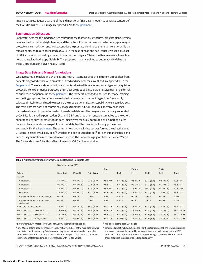
Segmentation ObjectivesFor prostate cancer, the model focuses contouring the following 6 structures: prostate gland, seminalvesicles, bladder, left and right femurs, and the rectum. For the purposes of radiotherapy planning inprostate cancer, radiation oncologists consider the prostate gland to be the target volume, while theremaining structures are delineated as OARs. In the case of head and neck cancer, we used a subsetof OAR structures defined by a panel of radiation oncologists,18 based on their relevance to routinehead and neck radiotherapy (Table 1). The proposed model is trained to automatically delineatethese 9 structures on a given head CT scan.
Image Data Sets and Manual AnnotationsWe aggregated 519 pelvic and 242 head and neck CT scans acquired at 8 different clinical sites frompatients diagnosed either with prostate or head and neck cancer, as outlined in eAppendix 1 in theSupplement. The scans show variation across sites due to differences in scanner type and acquisitionprotocols. For experimental purposes, the images are grouped into 2 disjoint sets: main and external,as outlined in eAppendix 1 in the Supplement. The former is intended to be used for model trainingand testing purposes; the latter is an excluded data set composed of images from 3 randomlyselected clinical sites and used to measure the model’s generalization capability to unseen data sets.The main data set does not contain any images from these 3 excluded sites, thereby enabling amasked evaluation to be performed on the external data set. The images were manually annotatedby 2 clinically trained expert readers (R.J. and G.B.) and a radiation oncologist masked to the others’annotations; as such, all structures in each image were manually contoured by 1 expert and laterreviewed by a separate oncologist. For further details of the manual contouring process, seeeAppendix 1 in the Supplement. The external head and neck data set was formed by using the headCT scans released by Nikolov et al,15 which is an open-source data set19 for benchmarking head andneck CT segmentation models and was acquired in The Cancer Imaging Archive Cetuximab20 andThe Cancer Genome Atlas Head-Neck Squamous Cell Carcinoma studies.
Table 1. Autosegmentation Performance on 3 Head and Neck Data Sets
Data set
Dice score, mean (SD)
Brainstem Mandible Spinal cord
Globe Parotid SMG
Left Right Left Right Left RightIOV-10a
Annotator 1 89.3 (4.2) 98.6 (1.0) 92.9 (1.5) 96.4 (0.9) 96.5 (1.1) 92.7 (3.5) 92.7 (3.5) 92.3 (3.4) 92.3 (2.6)
Annotator 2 91.8 (2.0) 98.5 (0.5) 91.8 (2.3) 95.6 (1.3) 96.7 (1.1) 91.1 (4.3) 91.2 (3.7) 91.3 (4.7) 91.3 (5.4)
Annotator 3 89.6 (2.7) 96.9 (1.0) 81.9 (7.3) 96.5 (0.8) 95.7 (1.0) 88.2 (3.8) 90.1 (2.8) 91.6 (2.8) 90.3 (8.0)
Ensemble 88.5 (2.0) 97.0 (1.0) 87.7 (3.6) 94.8 (1.0) 94.5 (1.9) 88.5 (2.3) 87.8 (4.1) 87.0 (2.9) 85.1 (5.3)
Agreement between annotators, κ 0.831 0.971 0.836 0.927 0.939 0.838 0.845 0.848 0.836
Agreement between annotatorsand model
0.806 0.966 0.844 0.917 0.931 0.852 0.825 0.803 0.794
Main data set, ensembleb 85.0 (3.7) 95.7 (2.3) 84.0 (3.8) 92.9 (1.6) 93.1 (1.5) 87.9 (3.8) 87.8 (4.3) 87.5 (2.3) 86.7 (3.5)
External data set, ensemblec 84.9 (6.8) 93.8 (2.5) 80.3 (7.7) 92.7 (3.6) 93.3 (1.4) 84.3 (4.6) 84.5 (4.3) 83.3 (9.1) 78.2 (21.1)
External data set,c Nikolov et al15 79.1 (9.6) 93.8 (1.6) 80.0 (7.8) 91.5 (2.1) 92.1 (1.9) 83.2 (5.4) 84.0 (3.7) 80.3 (7.8) 76.0 (16.5)
External data set, radiographerc 89.5 (2.2) 93.9 (2.3) 84.0 (4.8) 92.9 (1.9) 93.0 (1.7) 86.7 (3.5) 87.0 (3.1) 83.3 (19.7) 74.9 (30.2)
Abbreviations: IOV, interobserver variability; SMG, submandibular glands.a IOV-10 data set included 10 images. In the IOV study, a subset of the main data set was
annotated multiple times by 2 radiation oncologists and a trained reader. Later, theproposed model was compared against each human expert. The statistical agreementbetween annotators and model were measured with Fleiss κ values.
b Main data set included 20 images.c External data set included 26 images. For the external data set, the reference ground
truth contours were delineated by an expert head and neck oncologist, and IOVbetween clinical experts was measured by comparing the reference contours withthose produced by an experienced radiographer.15
JAMA Network Open | Health Informatics Deep Learning to Augment Image-Guided Radiotherapy for Head and Neck and Prostate Cancers
JAMA Network Open. 2020;3(11):e2027426. doi:10.1001/jamanetworkopen.2020.27426 (Reprinted) November 30, 2020 3/11
Downloaded From: https://jamanetwork.com/ on 12/30/2021

Evaluation MetricsTo evaluate model performance we used the Dice coefficient21 as a similarity metric, which quantifiesthe correspondence between pairs of volumetric segmentations for the same structure. Perfectlyoverlapping structures result in a Dice score of 100.00%, while a Dice score of 0.00% correspondsto complete lack of overlap. In addition to this, we measured the overlap between pairs of contoursusing Hausdorff and mean surface-to-surface distance metrics (in mm). The metrics are visuallypresented and described further in eAppendix 3 in the Supplement.
Statistical AnalysisAn ensemble of CNN models were trained with different training and validation set splits from maindata set while leaving out a fixed disjoint testing set (see eAppendix 2 in the Supplement for details).The agreement between contours generated by the model and expert readers was measuredstatistically with the Cohen and Fleiss κ22 for single and multiple annotators, respectively. For eachstructure, an agreement score was computed on foreground pixels defined by a binary mask. This isintended to avoid a possible bias due to a large number of background pixels. Similarly, Bland-Altman plots23 were generated to visualize the level of agreement on a patient level (eAppendix 3 inthe Supplement). The performance differences observed between the main and external sites wasstatistically tested with the Mann-Whitney test.24 The same model training setup was also deployedon the main head CT data set to train a head and neck model that can delineate OARs in the contextof head and neck radiotherapy(Table 1). Figure 1 shows qualitative assessment of contours predictedwith the proposed models. Additionally, to identify any gross contouring mistakes, thesegmentations were also compared in terms of geometric surface distances.
In a second set of experiments to test the generalization to data sets from unseen clinical sites,the previously trained pelvic and head and neck CT models were tested on their correspondingexternal data set (external), which was comprised of images acquired at 3 particular clinical sites thatwere excluded from the training and validation data sets (main). With this experiment, the aim wasto assess the generalization of the trained models to unseen CT acquisition protocols andpatient groups.
All statistical analyses were conducted using Python version 3.7.3 (Python SoftwareFoundation), with scikit-learn package version 0.21.1 for the Cohen-Fleiss κ and scipy package version1.3.1 for the Mann-Whitney test. Statistical significance was set at P < .01 for null hypothesis testingand κ > 0.75 for the agreement analysis. All tests were 2-tailed.
Results
A total of 519 participants’ (519 [100%] men; 390 [75%] aged 62-75 years) pelvic CT images and 242participants’ (184 [76%] men; 194 [80%] aged 50-73 years) head and neck CT images were included.The prostate segmentation results (Table 2) show that the autogenerated organ delineations(ensemble) for prostate scans were consistent with the contours produced by clinical experts, withsurface errors being within the acceptable error bound (eg, left femur, κ = 0.982). Results wereconsistent with head and neck segmentation results (eg, brainstem, κ = 0.806) (Table 1). Similarly, invalidations on external data sets (Table 1 and Table 2), the model performed consistently well in bothradiotherapy domains across multiple sites (eg, mean [SD] Dice score for left femur, internal vsexternal data sets: 98.52% [0.50] vs 98.04% [1.02]; P = .04), only with a slight performance drop onsegmenting the submandibular glands due to low tissue contrast. Our observations of thesegmentation errors tended to occur in the superior and inferior extent of tubular structures and inthe interface between adjacent organs. However, we have not observed any inconsistencies that, ifnot corrected, could lead to significant errors in a treatment plan, as evidenced by the surfacedistance results. This is because the proposed postprocessing method does not allow inconsistenciesat a distance from the anatomical structure by design.
JAMA Network Open | Health Informatics Deep Learning to Augment Image-Guided Radiotherapy for Head and Neck and Prostate Cancers
JAMA Network Open. 2020;3(11):e2027426. doi:10.1001/jamanetworkopen.2020.27426 (Reprinted) November 30, 2020 4/11
Downloaded From: https://jamanetwork.com/ on 12/30/2021

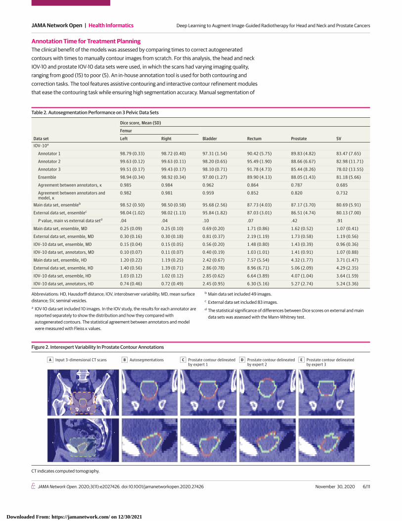
IOV AnalysisAn acceptable measure of performance is expected to be within the bounds of IOV found in humanexperts.5,7 The IOV Dice scores and surface distances between 3 experts contouring 10 test imagesfor each radiotherapy domain are provided in Table 1 and Table 2. For 14 of 15 structures, statisticalagreement (ie, κ > 0.75) was observed between autogenerated contours and expert annotations.The reference contours were determined by applying a majority voting scheme using all 3annotators. At least 2 experts must have agreed to imply that a structure is in fact present. For all thestructures except SMGs, the similarity scores with ground truth achieved the criteria of being on-parwith levels of expert IOV in contouring, as indicated by the κ values and Bland-Altman plots(eAppendix 3 in the Supplement) collected for the agreement analysis. Here we can see that for moreclearly defined structures with high contrast, such as the bladder and femurs, there is reasonablyhigh consistency across the experts (κ > 0.96). But for lower contrast and deformable features, suchas the prostate gland, seminal vesicles, and SMGs, we see a higher rate of variability because theorgan boundaries are typically unclear in the presence of such adverse conditions (Figure 2 andTable 2). A similar pattern of performance difference is seen on the contours generated by the model,where the same test images are segmented and compared qualitatively with the same referencecontours (Figure 2).
Figure 1. Qualitative Evaluation of Expert and Autogenerated Contours on Head and Neck Computed Tomography Scans
Main data setA
Main data setC External data setD
External data setB
JAMA Network Open | Health Informatics Deep Learning to Augment Image-Guided Radiotherapy for Head and Neck and Prostate Cancers
JAMA Network Open. 2020;3(11):e2027426. doi:10.1001/jamanetworkopen.2020.27426 (Reprinted) November 30, 2020 5/11
Downloaded From: https://jamanetwork.com/ on 12/30/2021
Annotation Time for Treatment PlanningThe clinical benefit of the models was assessed by comparing times to correct autogeneratedcontours with times to manually contour images from scratch. For this analysis, the head and neckIOV-10 and prostate IOV-10 data sets were used, in which the scans had varying imaging quality,ranging from good (15) to poor (5). An in-house annotation tool is used for both contouring andcorrection tasks. The tool features assistive contouring and interactive contour refinement modulesthat ease the contouring task while ensuring high segmentation accuracy. Manual segmentation of
Table 2. Autosegmentation Performance on 3 Pelvic Data Sets
Data set
Dice score, Mean (SD)
Femur
Bladder Rectum Prostate SVLeft RightIOV-10a
Annotator 1 98.79 (0.33) 98.72 (0.40) 97.31 (1.54) 90.42 (5.75) 89.83 (4.82) 83.47 (7.65)
Annotator 2 99.63 (0.12) 99.63 (0.11) 98.20 (0.65) 95.49 (1.90) 88.66 (6.67) 82.98 (11.71)
Annotator 3 99.51 (0.17) 99.43 (0.17) 98.10 (0.71) 91.78 (4.73) 85.44 (8.26) 78.02 (13.55)
Ensemble 98.94 (0.34) 98.92 (0.34) 97.00 (1.27) 89.90 (4.13) 88.05 (1.43) 81.18 (5.66)
Agreement between annotators, κ 0.985 0.984 0.962 0.864 0.787 0.685
Agreement between annotators andmodel, κ
0.982 0.981 0.959 0.852 0.820 0.732
Main data set, ensembleb 98.52 (0.50) 98.50 (0.58) 95.68 (2.56) 87.73 (4.03) 87.17 (3.70) 80.69 (5.91)
External data set, ensemblec 98.04 (1.02) 98.02 (1.13) 95.84 (1.82) 87.03 (3.01) 86.51 (4.74) 80.13 (7.00)
P value, main vs external data setd .04 .04 .10 .07 .42 .91
Main data set, ensemble, MD 0.25 (0.09) 0.25 (0.10) 0.69 (0.20) 1.71 (0.86) 1.62 (0.52) 1.07 (0.41)
External data set, ensemble, MD 0.30 (0.16) 0.30 (0.18) 0.81 (0.37) 2.19 (1.19) 1.73 (0.58) 1.19 (0.56)
IOV-10 data set, ensemble, MD 0.15 (0.04) 0.15 (0.05) 0.56 (0.20) 1.48 (0.80) 1.43 (0.39) 0.96 (0.36)
IOV-10 data set, annotators, MD 0.10 (0.07) 0.11 (0.07) 0.40 (0.19) 1.03 (1.01) 1.41 (0.91) 1.07 (0.88)
Main data set, ensemble, HD 1.20 (0.22) 1.19 (0.25) 2.42 (0.67) 7.57 (5.54) 4.32 (1.77) 3.71 (1.47)
External data set, ensemble, HD 1.40 (0.56) 1.39 (0.71) 2.86 (0.78) 8.96 (6.71) 5.06 (2.09) 4.29 (2.35)
IOV-10 data set, ensemble, HD 1.03 (0.12) 1.02 (0.12) 2.85 (0.62) 6.64 (3.89) 4.07 (1.04) 3.64 (1.59)
IOV-10 data set, annotators, HD 0.74 (0.46) 0.72 (0.49) 2.45 (0.95) 6.30 (5.16) 5.27 (2.74) 5.24 (3.36)
Abbreviations: HD, Hausdorff distance; IOV, interobserver variability; MD, mean surfacedistance; SV, seminal vesicles.a IOV-10 data set included 10 images. In the IOV study, the results for each annotator are
reported separately to show the distribution and how they compared withautogenerated contours. The statistical agreement between annotators and modelwere measured with Fleiss κ values.
b Main data set included 49 images.c External data set included 83 images.d The statistical significance of differences between Dice scores on external and main
data sets was assessed with the Mann-Whitney test.
Figure 2. Interexpert Variability In Prostate Contour Annotations
Input 3-dimensional CT scansA AutosegmentationsB Prostate contour delineatedby expert 1
C Prostate contour delineatedby expert 2
D Prostate contour delineatedby expert 3
E
CT indicates computed tomography.
JAMA Network Open | Health Informatics Deep Learning to Augment Image-Guided Radiotherapy for Head and Neck and Prostate Cancers
JAMA Network Open. 2020;3(11):e2027426. doi:10.1001/jamanetworkopen.2020.27426 (Reprinted) November 30, 2020 6/11
Downloaded From: https://jamanetwork.com/ on 12/30/2021

the head and neck scans for the same 9 OARs took a mean of 86.75 (95% CI, 75.25-98.29) min/scanfor an expert reader and 73.25 (95% CI, 68.68-77.82) min/scan for a radiation oncologist. For thesame scans, the review and correction time of autocontours was measured as 4.98 (95% CI, 4.44-5.52) min/scan for head and neck scans and 3.40 (95% CI, 1.60-5.20) min/scan for prostate scans,which are inspected and updated (if necessary) by the oncologist to ensure clinical accuracy requiredfor treatment planning. This represented a mean 93% reduction in time. Among all 20 scans, theslowest correction time per scan was measured as 7.05 minutes because of low imaging quality. Amean inference time of 23 (95% CI, 20-26) seconds was taken to segment target and all OARforeground pixels in full input CT scan.
Discussion
Several frameworks have been proposed for autosegmentation of head and neck 15,25 and pelvicorgans.13,16,26 In 2 studies,13,16 the authors describe an approach for prostate and OAR segmentation,where organ localization is performed prior to segmentation. Their algorithm was validated on a dataset of 88 CT scans. A similar cascaded autosegmentation approach was proposed in Wang et al26 todelineate OARs in head and neck CT scans; this study was conducted by training (33 scans) andevaluating (15 scans) using the public data set released in an autosegmentation challenge.27
There have been efforts to show the potential clinical use cases of ML solutions for automaticOAR contouring. In contrast to previous work, in which evaluations were performed on small sets ofhomogenous images, we evaluated how ML solutions could lead to generalized performance across(1) different radiotherapy domains and (2) data sets from multiple sites. We aimed to demonstratethe robustness and generalizability of these solutions. More importantly, we found that integratingthese models into clinical workflows could reduce the time required to prepare dose plans fortreatment.
The models demonstrated performance generalizability across diverse acquisition settingswhile achieving good levels of agreement with expert contours. This could facilitate easierdeployment in new clinical sites. Of further importance for any practical adoption of this technologyacross large scale health care systems is the ability to work across diverse clinical domains. We haveshown how our approach, without any substantial changes, can enable the training of models indiverse radiotherapy domains, as demonstrated through applications in prostate and head and neckcancer. This is especially significant given the distinct imaging challenges associated with thesedifferent domains.
Practical adoption in clinical contexts is enhanced by incorporating the presented models intothe existing workflow of radiation oncologists (Figure 3). The illustrated system has beenimplemented and evaluated by clinical experts working at Cambridge University Hospitals. In thisworkflow, CT scans are acquired from patients as they attend preparations for radiotherapytreatment. These scans are initially stored at the hospital’s image database and later securelytransferred via the gateway to the autosegmentation platform in the cloud after anonymizing them.Once the segmentation process is completed, resultant files are uploaded back to the hospital’simage database, creating a seamless clinical workflow in which clinicians can review and refinecontours in their existing contouring and planning tools.
Bringing these ML tools to the point where they can be meaningfully adopted in clinical practicerequires a level of clinical accuracy commensurate with expert observers. While the models haveperformed well in this regard, in instances where the model performed poorly, the opportunity tomanually correct the segmentations remains a necessary component of the presented workflow. Thepresented workflow enables oncologists to use their existing clinical systems for review and editing,which makes this technology more accessible across clinics because the existing workflows aremaintained. At the same time, clinicians can inspect and edit contours in minutes rather than hours.Such time savings are significant even when considered only in absolute terms.
JAMA Network Open | Health Informatics Deep Learning to Augment Image-Guided Radiotherapy for Head and Neck and Prostate Cancers
JAMA Network Open. 2020;3(11):e2027426. doi:10.1001/jamanetworkopen.2020.27426 (Reprinted) November 30, 2020 7/11
Downloaded From: https://jamanetwork.com/ on 12/30/2021

The source code used in this study is made publicly available.28 This creates an opportunity foroncology centers to use this technology to train and deploy new models using their own data sets. Inthis way, users can include other normal tissue structures in the autocontouring pipeline, includingcochlear and oral-cavity structures in head and neck cancer treatments. The availability of new publicdata sets and sharing across clinics is an important milestone in improving the performance ofmodels and making them accessible. Similarly, image quality (IQ) assurance29 is essential for reliableuse of models. IQ assessment should be performed prior to model deployment30 both at acquisitionand processing time to filter out images with metal artifacts. Training models on a diverse set of datasets, as performed in this study, is an effective way to cope with low-contrast (eg, cone-beam CT)and high-noise images. External data set validation is also essential to measure such impacts; forinstance, the images from the external head and neck data set used in this study contained severebeam-hardening artifacts.
More adaptive forms of radiotherapy, in which anatomy is resegmented and the dose planreoptimized for each fraction, are regarded as a more ideal way to deliver treatment,31 which hasbeen challenging to adopt due to its heavy resource demand.32 In that regard, the presentedtechnology can enable continuous resegmentation and adaptive reoptimization of therapy to beadopted at scale. For instance, in the cases of hypofractionated regimens or emergency treatments,extension of these models to resegment anatomy on scans would have significant clinical utility tosave time and allow patients to progress to treatment more quickly. Integration with technologiessuch as The Magnetic Resonance Linear Accelerator,33 used for simultaneous imaging and dosedelivery, could also potentially offer more adaptive forms of treatment to pinpoint the location oftumors at the time of treatment.
LimitationsThis study has limitations. The data sets used in the IOV and annotation time experiments are smallerthan the remaining evaluations presented in this study. For further statistical significance, theseexperiments shall be repeated with larger data sets with varying imaging quality. Additionally, surfaceand Dice metrics used in model evaluation do not always correlate with time savings in manualcontouring process.15,34 This necessitates the design of new metrics that quantify segmentationerrors by taking into account the cost of required user interaction to correct them.
Figure 3. Integration of the Proposed Segmentation Models Into Radiotherapy Planning Workflow
Segmentationservice
Radiomics portal
Anonymized 3D-CT scan
Autogenerated organ contours
Quality control andground truth labeling
Segmentation model trainingand deployment pipeline
Dataset curation
Train models on Azure ML
Results analysisin the radiomics portal
Automatic deploymentto segmentation service
Image acquisition
Image store/PACs
Hospital
Refinement tool
InnerEye GatewayAnonymization
Cloud computing
JAMA Network Open | Health Informatics Deep Learning to Augment Image-Guided Radiotherapy for Head and Neck and Prostate Cancers
JAMA Network Open. 2020;3(11):e2027426. doi:10.1001/jamanetworkopen.2020.27426 (Reprinted) November 30, 2020 8/11
Downloaded From: https://jamanetwork.com/ on 12/30/2021

Conclusions
This study found that ML-based autosegmentation reduces contouring time while yielding clinicallyvalid structural contours on heterogeneous data sets for both prostate and head and neckradiotherapy planning. This is evidenced in evaluations on external data sets and IOV experimentsconducted on a multisite data set. Overall, the approach contributes to the practical challenges ofscalable adoption across health care systems through off-the-shelf extensibility across hospital sitesand applicability across multiple cancer domains. Future ML studies validating the applicability ofthe proposed technology on other radiotherapy domains and larger data sets will be valuable forwider adoption of ML solutions in health care systems.
ARTICLE INFORMATIONAccepted for Publication: October 1, 2020.
Published: November 30, 2020. doi:10.1001/jamanetworkopen.2020.27426
Correction: This article was corrected on December 9, 2020, to fix an error in the Abstract.
Open Access: This is an open access article distributed under the terms of the CC-BY-NC-ND License. © 2020 OktayO et al. JAMA Network Open.
Corresponding Author: Ozan Oktay, PhD, Health Intelligence, Microsoft Research, 21 Station Rd, Cambridge, CB12FB United Kingdom ([email protected]).
Author Affiliations: Health Intelligence, Microsoft Research, Cambridge, United Kingdom (Oktay, Nanavati,Schwaighofer, Carter, Bristow, Tanno, Jena, Barnett, Glocker, O’Hara, Bishop, Alvarez-Valle, Nori); Department ofOncology, Cambridge University Hospitals NHS Foundation Trust, United Kingdom (Noble, Rimmer); now withEdinburgh Cancer Centre, Western General Hospital, Edinburgh, United Kingdom (Noble).
Author Contributions: Dr Oktay had full access to all of the data in the study and takes responsibility for theintegrity of the data and the accuracy of the data analysis.
Concept and design: Oktay, Nanavati, Schwaighofer, Tanno, Jena, Noble, Glocker, Bishop, Alvarez-Valle, Nori.
Acquisition, analysis, or interpretation of data: Oktay, Nanavati, Schwaighofer, Carter, Bristow, Jena, Barnett,Noble, Rimmer, Glocker, O'Hara, Alvarez-Valle.
Drafting of the manuscript: Oktay, Nanavati, Schwaighofer, Bristow, Jena, Glocker, O'Hara, Alvarez-Valle, Nori.
Critical revision of the manuscript for important intellectual content: Oktay, Schwaighofer, Carter, Tanno, Jena,Barnett, Noble, Rimmer, Glocker, O'Hara, Bishop, Alvarez-Valle, Nori.
Statistical analysis: Oktay, Nanavati, Schwaighofer, Carter.
Obtained funding: Nori.
Administrative, technical, or material support: Oktay, Schwaighofer, Bristow, Jena, Noble, Bishop, Alvarez-Valle, Nori.
Supervision: Oktay, Schwaighofer, Jena, Barnett, Rimmer, Glocker, Alvarez-Valle, Nori.
Conflict of Interest Disclosures: Dr Jena reported receiving personal fees from Microsoft during the conduct ofthe study. Dr Noble reported receiving grants from Cancer Research UK and personal fees from MicrosoftResearch, Cambridge, during the conduct of the study. No other disclosures were reported.
Funding/Support: The research work reported in the manuscript was self-funded by Microsoft ResearchCambridge.
Role of the Funder/Sponsor: The funder had no role in the design and conduct of the study; collection,management, analysis, and interpretation of the data; preparation, review, or approval of the manuscript; anddecision to submit the manuscript for publication.
REFERENCES1. Pan HY, Haffty BG, Falit BP, et al. Supply and demand for radiation oncology in the United States: updatedprojections for 2015 to 2025. Int J Radiat Oncol Biol Phys. 2016;96(3):493-500. doi:10.1016/j.ijrobp.2016.02.064
2. Sklan A, Collingridge D. Treating head and neck cancer: for better or for worse? Lancet Oncol. 2017;18(5):570-571. doi:10.1016/S1470-2045(17)30269-3
3. Barnett GC, West CM, Dunning AM, et al. Normal tissue reactions to radiotherapy: towards tailoring treatmentdose by genotype. Nat Rev Cancer. 2009;9(2):134-142. doi:10.1038/nrc2587
JAMA Network Open | Health Informatics Deep Learning to Augment Image-Guided Radiotherapy for Head and Neck and Prostate Cancers
JAMA Network Open. 2020;3(11):e2027426. doi:10.1001/jamanetworkopen.2020.27426 (Reprinted) November 30, 2020 9/11
Downloaded From: https://jamanetwork.com/ on 12/30/2021

4. Vorwerk H, Zink K, Schiller R, et al. Protection of quality and innovation in radiation oncology: the prospectivemulticenter trial the German Society of Radiation Oncology (DEGRO-QUIRO study). Strahlenther Onkol. 2014;190(5):433-443. doi:10.1007/s00066-014-0634-0
5. Cazzaniga LF, Marinoni MA, Bossi A, et al. Interphysician variability in defining the planning target volume in theirradiation of prostate and seminal vesicles. Radiother Oncol. 1998;47(3):293-296. doi:10.1016/S0167-8140(98)00028-0
6. Cooper JS, Mukherji SK, Toledano AY, et al. An evaluation of the variability of tumor-shape definition derived byexperienced observers from CT images of supraglottic carcinomas (ACRIN protocol 6658). Int J Radiat Oncol BiolPhys. 2007;67(4):972-975. doi:10.1016/j.ijrobp.2006.10.029
7. Dubois DF, Prestidge BR, Hotchkiss LA, Prete JJ, Bice WS Jr. Intraobserver and interobserver variability of MRimaging- and CT-derived prostate volumes after transperineal interstitial permanent prostate brachytherapy.Radiology. 1998;207(3):785-789. doi:10.1148/radiology.207.3.9609905
8. Fiorino C, Reni M, Bolognesi A, Cattaneo GM, Calandrino R. Intra- and inter-observer variability in contouringprostate and seminal vesicles: implications for conformal treatment planning. Radiother Oncol. 1998;47(3):285-292. doi:10.1016/S0167-8140(98)00021-8
9. Fotina I, Lütgendorf-Caucig C, Stock M, Pötter R, Georg D. Critical discussion of evaluation parameters for inter-observer variability in target definition for radiation therapy. Strahlenther Onkol. 2012;188(2):160-167. doi:10.1007/s00066-011-0027-6
10. Gardner SJ, Wen N, Kim J, et al. Contouring variability of human- and deformable-generated contours inradiotherapy for prostate cancer. Phys Med Biol. 2015;60(11):4429-4447. doi:10.1088/0031-9155/60/11/4429
11. Valicenti RK, Sweet JW, Hauck WW, et al. Variation of clinical target volume definition in three-dimensionalconformal radiation therapy for prostate cancer. Int J Radiat Oncol Biol Phys. 1999;44(4):931-935. doi:10.1016/s0360-3016(99)00090-5
12. Ohri N, Shen X, Dicker AP, Doyle LA, Harrison AS, Showalter TN. Radiotherapy protocol deviations and clinicaloutcomes: a meta-analysis of cooperative group clinical trials. J Natl Cancer Inst. 2013;105(6):387-393. doi:10.1093/jnci/djt001
13. Balagopal A, Kazemifar S, Nguyen D, et al. Fully automated organ segmentation in male pelvic CT images. PhysMed Biol. 2018;63(24):245015. doi:10.1088/1361-6560/aaf11c
14. Lou B, Doken S, Zhuang T, et al. An image-based deep learning framework for individualising radiotherapydose. Lancet Digit Health. 2019;1(3):e136-e147. doi:10.1016/S2589-7500(19)30058-5
15. Nikolov S, Blackwell S, Zverovitch A, et al. Deep learning to achieve clinically applicable segmentation of headand neck anatomy for radiotherapy. arXiv. Preprint published online September 12, 2018. Accessed October 29,2020. https://arxiv.org/pdf/ 1809.04430.pdf
16. Wang S, He K, Nie D, Zhou S, Gao Y, Shen D. CT male pelvic organ segmentation using fully convolutionalnetworks with boundary sensitive representation. Med Image Anal. 2019;54:168-178. doi:10.1016/j.media.2019.03.003
17. Ronneberger O, Fischer P, Brox T. U-net: convolutional networks for biomedical image segmentation. In: NavabN, Hornegger J, Wells W, Frangi A, eds. Medical Image Computing and Computer-Assisted Intervention—MICCAI2015. Lecture Notes in Computer Science, vol 9351. Springer; 2015. doi:10.1007/978-3-319-24574-4_28
18. Brouwer CL, Steenbakkers RJ, Bourhis J, et al. CT-based delineation of organs at risk in the head and neckregion: DAHANCA, EORTC, GORTEC, HKNPCSG, NCIC CTG, NCRI, NRG Oncology and TROG consensus guidelines.Radiother Oncol. 2015;117(1):83-90. doi:10.1016/j.radonc.2015.07.041
19. TCIA CT scan dataset. Accessed October 29, 2020. https://github.com/deepmind/tcia-ct-scan-dataset
20. Cancer Imaging Archive. Head-neck cetuximab. Published June 3, 2020. Accessed October 29, 2020. https://wiki.cancerimagingarchive.net/display/Public/Head-Neck+Cetuximab
21. Dice LR. Measures of the amount of ecologic association between species. In: Ecology. 1945;26(3):297–302.doi:10.2307/1932409
22. Lin L, Hedayat AS, Wu W. Statistical Tools for Measuring Agreement. Springer Science & Business Media,2012."https://doi.org/10.1007/978-1-4614-0562-7"
23. Bland JM, Altman DG. Statistical methods for assessing agreement between two methods of clinicalmeasurement. Lancet. 1986;1(8476):307-310. doi:10.1016/S0140-6736(86)90837-8
24. Mann HB, Whitney DR. On a test of whether one of two random variables is stochastically larger than theother. Ann Math Stat. 1947;18(1):50-60. doi:10.1214/aoms/1177730491
JAMA Network Open | Health Informatics Deep Learning to Augment Image-Guided Radiotherapy for Head and Neck and Prostate Cancers
JAMA Network Open. 2020;3(11):e2027426. doi:10.1001/jamanetworkopen.2020.27426 (Reprinted) November 30, 2020 10/11
Downloaded From: https://jamanetwork.com/ on 12/30/2021

25. Zhu W, Huang Y, Zeng L, et al. AnatomyNet: deep learning for fast and fully automated whole-volumesegmentation of head and neck anatomy. Med Phys. 2019;46(2):576-589. doi:10.1002/mp.13300
26. Yueyue Wang et al Organ at risk segmentation in head and neck CT images using a two-stage segmentationframework based on 3D U-net. IEEE Access. 2019;7:144591–144602. doi:10.1109/ACCESS.2019.2944958
27. Raudaschl PF, Zaffino P, Sharp GC, et al. Evaluation of segmentation methods on head and neck CT: auto-segmentation challenge 2015. Med Phys. 2017;44(5):2020-2036. doi:10.1002/mp.12197
28. InnerEye Deep Learning. Accessed October 29, 2020. https://github.com/microsoft/InnerEye-DeepLearning/
29. Barrett JF, Keat N. Artifacts in CT: recognition and avoidance. Radiographics. 2004;24(6):1679-1691. doi:10.1148/rg.246045065
30. Tarroni G, Oktay O, Bai W, et al. Learning-based quality control for cardiac MR IMAGES. IEEE Trans MedImaging. 2019;38(5):1127-1138. doi:10.1109/TMI.2018.2878509
31. Sonke JJ, Aznar M, Rasch C. Adaptive radiotherapy for anatomical changes. Semin Radiat Oncol. 2019;29(3):245-257. doi:10.1016/j.semradonc.2019.02.007
32. Heukelom J, Fuller CD. Head and neck cancer adaptive radiation therapy (ART): conceptual considerations forthe informed clinician. Semin Radiat Oncol. 2019;29(3):258-273. doi:10.1016/j.semradonc.2019.02.008
33. Lagendijk JJW, Raaymakers BW, van Vulpen M. The magnetic resonance imaging-linac system. Semin RadiatOncol. 2014;24(3):207-209. doi:10.1016/j.semradonc.2014.02.009
34. Valenzuela W, Ferguson SJ, Ignasiak D, et al. FISICO: fast image segmentation correction. PLoS One. 2016;11(5):e0156035. doi:10.1371/journal.pone.0156035
35. Acosta, O., Dowling, J., Drean, G. et al Multi-atlas-based segmentation of pelvic structures from CT scans forplanning in prostate cancer radiotherapy. In: El-Baz AS, Saba L, Suri JS, eds. Abdomen and Thoracic Imaging.Springer, 2014:623–656. doi:10.1007/978-1-4614-8498-1_24
SUPPLEMENT.eAppendix 1. Author Contributions and Data SeteAppendix 2. Supplementary MethodseAppendix 3. Supplementary MaterialeReferences.
JAMA Network Open | Health Informatics Deep Learning to Augment Image-Guided Radiotherapy for Head and Neck and Prostate Cancers
JAMA Network Open. 2020;3(11):e2027426. doi:10.1001/jamanetworkopen.2020.27426 (Reprinted) November 30, 2020 11/11
Downloaded From: https://jamanetwork.com/ on 12/30/2021