evaluation of anti microbial activity of osyris lanceolata
TRANSCRIPT

Evaluation of Anti microbial activity of Osyris lanceolata (East African Sandalwood)
Edna Akinyi Ooko
(Bsc. Hons) JKUAT
A thesis submitted in partial fulfilment for the Degree of Master of Science in Biochemistry at Jomo Kenyatta University of Agriculture and Technology.
2009

2
DECLARATION
This thesis is my original work and has not been presented for any degree in any other
university.
Signed:.................................................. Date:............................................................
EDNA AKINYI OOKO
This thesis has been submitted for examination with our approval as university supervisors:
Signed:...................................................... Date:........................................................
Dr. Peter Lomo
JKUAT, KENYA
Signed:.................................................. Date:................................................................
Dr. David Odee
KEFRI, KENYA
Signed:............................................... Date:....................................................................
Prof. Ahmed Hassanali
ICIPE, KENYA

3
DEDICATION
This work is dedicated to my God for seeing me through it all, my parents for their constant
support and my husband for encouraging me to its completion.

4
ACKNOWLEDGEMENT
I would like to acknowledge my supervisors Dr. Peter Lomo, Prof. Ahmed Hassanali and Dr.
David Odee for their guidance and leadership throughout my research work. Their
invaluable comments and advises have gone a long way to make this work what it is.
I thank KEFRI for supporting this study by financing it and offering laboratory space. I would
also like to acknowledge the support of KEFRI Kitui in my data collection and specially thank
Dr. Ben Muok and Mr. Osore for theri leadership and assistance in facilitating my data
collection in this area.
I thank KEMRI Center for Respiratory Disease Research for assisting me carry out the
bioassay studies and special thanks to Dr. Christine Bii and Tom Ouko.
I thank ICIPE for allowing me use their facilities to run the chromatograms and would like to
acknowledge the assistance of Mr. Nyandat and Mr. Angira of the chemistry laboratory in
ICIPE.

5
TABLE OF CONTENTS
Declaration..................................................................................................................................i
Dedication..................................................................................................................................ii
Acknowledgement....................................................................................................................iii
Table of contents.......................................................................................................................iv
List of tables.............................................................................................................................vii
List of figures...........................................................................................................................viii
Abbreviations............................................................................................................................ix
Abstract......................................................................................................................................x
1.0 INTRODUCTION
1.1 Background of plant.................................................................................................1
1.1.1 Geography.......................................................................................................1
1.1.2 Biology.............................................................................................................2
1.1.3 Uses.................................................................................................................5
1.2 Aspects of anti microbial activity and its importance..............................................6
1.3 Problem statement..................................................................................................7
1.4 Justification of study................................................................................................8
1.5 Objectives.................................................................................................................9

6
1.5.1 General objectives..................................................................................9
1.5.2 Specific objectives...................................................................................9
2.0 LITERATURE REVIEW
2.1 Use of Medicinal plants..........................................................................................10
2.2 Medicinal plants with specific anti microbial activity.............................................12
2.3 Medicinal plants of the dryland areas....................................................................14
2.4 Conservation of medicinal plants through research..............................................15
3.0 MATERIALS AND METHODS
3.1 Sample site.............................................................................................................17
3.2 Collection and preparation of samples..................................................................18
3.3 Extraction...............................................................................................................18
3.4 Anti microbial testing.............................................................................................21
3.4.1 Preparation of Nutrient Agar andgrowing of Bacteria cultures............22
3.4.2 Preparation of Sabourand Dextrose Agar media and growing of fungal
spores...................................................................................................22
3.4.3 Primary screening of the plant extracts................................................23
3.4.4 Secondary screening of the plant extracts against microbes that
showed susceptibility...........................................................................23

7
3.5 HPLC profile determination of the plant extracts..................................................24
3.6 Data analysis...........................................................................................................25
4.0 RESULTS
4.1 Extraction...........................................................................................................................26
4.2 Bioassay.............................................................................................................................28
4.3 High Performance Liquid chromatography........................................................................31
5.0 DISCUSSION
5.1 Extraction and percentage yield........................................................................................38
5.2 Bioassay.............................................................................................................................39
5.3 Chromatographic analysis..................................................................................................41
CONCLUSION AND RECOMMENDATIONS
6.1 Conclusion..........................................................................................................................44
6.2 Recommendations.............................................................................................................45
REFERENCES......................................................................................................................47
8
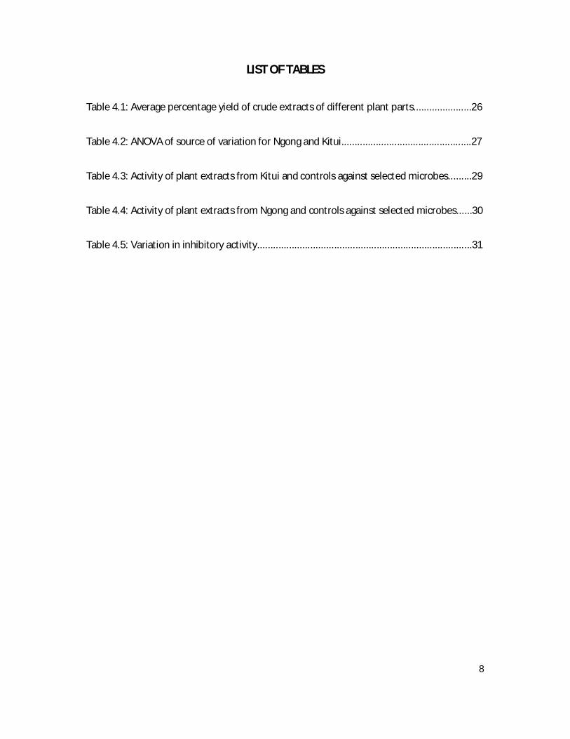
LIST OF TABLES
Table 4.1: Average percentage yield of crude extracts of different plant parts......................26
Table 4.2: ANOVA of source of variation for Ngong and Kitui.................................................27
Table 4.3: Activity of plant extracts from Kitui and controls against selected microbes.........29
Table 4.4: Activity of plant extracts from Ngong and controls against selected microbes......30
Table 4.5: Variation in inhibitory activity.................................................................................31

9
LIST OF FIGURES
Figure 1.1 Osyris lanceolata growing in rocky areas..................................................................1
Figure 1.2 A closer view of the shrub growing between rocks..................................................2
Figure 1.3 The shrub..................................................................................................................3
Figure 1.4 The canopy of Osyris lanceolata...............................................................................3
Figure 1.5 Leaf arrangement......................................................................................................3
Figure 1.6 Multi stemmed..........................................................................................................4
Figure 1.7 Barks..........................................................................................................................4
Figure 1.8 The Flowers...............................................................................................................5
Figure 1.9 The fruit in the plant and the collected fruits...........................................................5
Figure 3.1 Kibiko area of Ngong...............................................................................................17
Figure 3.2 A scheme of sequential extraction..........................................................................21
Figure 4.1 Bioassay results.......................................................................................................28
Figure 4.2 Chromatograms of hexane extract.........................................................................33
Figure 4.3 Chromatograms of dichloromethane extract.........................................................34
Figure 4.4 Chromatograms of aqueous methanol extract.......................................................35
Figure 4.5 Chromatograms of water extract............................................................................37

10
ABBREVIATIONS
ANOVA – Analysis of Variance
ATCC – American Type Culture Collection
CFU – Colony forming Unit
CHL - Chloroamphenical
Clin. - Clinical
CRDR – Center for Respiratory Research
GEN - Gentamycine
HPLC – High Performance Liquid Chromatography
ICIPE – International Center for Insect Physiology and Ecology
KAN - Kanamycin
KEFRI – Kenya Forestry Research Institutue
KEMRI – Kenya Medical Research Institute
MIC – Minimum Inhibitory Concentration
MTT - Methyltrityl
Etc – Et Cetera

11
ABSTRACT
Osyris lanceolata (East African Sandalwood) is an evergreen shrub to small tree (1 – 6m) in
the family Santalacea. The species has a relatively wide ecological distribution occurring in
Eastern and Southern Africa. In Kenya, it grows in Coast, Eastern, Rift valley, Nyanza and
Western provinces.
The aim of this work was to investigate anti microbial activity of the plant. The plant parts
used were the roots, stem and stem bark. Polar and non-polar extracts were obtained from
each plant part and these were used to screen for anti microbial activity and to obtain HPLC
profiles.
Using the disc diffusion technique the extracts were screened against five bacteria and three
fungi and the results showed that aqueous methanol extract and water extract had activity
against Staphylococuss aureus. There was no significance difference in the activity of these
extracts on the microorganism (p>0.05). The minimum inhibitory concentrations (MIC) value
was a range between 294 – 301 µg/ml. The HPLC profiles displayed very large peak ares of
polar constituents in aqueous methanol and water extracts and moderate peak areas of non-
polar constituents. While the hexane and dichloromethane extracts displayed only
moderate peak areas of non-polar constituents.

12
CHAPTER 1
INTRODUCTION
1.1 Background of plant.
1.1.1 Geography
Osyris lanceolata is a dryland species with spatial distribution in Kenya and Tanzania.
It occurs as isolated individuals, in close association with other woody species, and does
not occur communally in large numbers. This could be attributed to its slow growth and
host preference. It is found as a shrub on wooded rocky ridges or outcrops and on
mountain slopes and as a tree in gorges, loofs and forest margins. It is common on
rocky ridges in the Magaliesberg, North America (Palmer and Pitman, 1972) and in
rocky sites where the original vegetation has been cleared (Beentje, 1994) as shown in
Figure 1.1. It is also in margins of dry forests, evergreen bushland, grassland,thicket at
900 – 2550 m altitude (Pooley, 1993). In Kenya the species has been recorded in
Oloitokitok, Amboseli, Makueni, Taveta, Chyulu hills, Narok, Mbeer
Fig 1.1 The shrub growing between rocks

13
1.1.2 Biology
Osyris lanceolata is a slender shrub or samll evergreen tree from 1 to 6 m tall as shown
in Fig 1.2. It is a, multi-stemmed, spreading tree with a round to irregular canopy
refered to in Fig 1.3. The dark branches and blue-green leaves contrast with one
another. It is probably a partial root parasite, growing on the roots of other plants (Van
Wyk and Van Wyk, 1997) and utilizing the root systems of these hosts, but does not
produce its own chlorphyll (Thomas and Grant, 2002). As a result it is usually
intimately associated with shrubs of other woody species. In Kenya it is known to have
association with several species within the following genera: Acacia, Albizia, Rapanea,
Bridellia, Cordia and Teclea.
Figure 1.2 The shrub Figure 1.3 Canopy of Osyris lanceolata
The leaves are alternately arranged (fig 1.4). They are small, simple, thick, leathery,
rigid and tough and characteristically point upwards. They vary in size from 30-45 ×
10-25 mm (Schmidt et al., 2002) but their length may vary from 13-50 mm long
(Pooley, 1993). They are lance-shaped or sometimes egg-shaped. The apex is broadly
tapering to rounded with a fine, sharp tip. The base is broadly tapering. The leaf
margin is entire and rolled. These hairless leaves are grey-green or blue green, smooth
with a waxy bloom that can be rubbed off (Palmer and Pitman, 1972). The leaves often
have an orange coloured margin (Thomas and Grant, 2002). The petiole attachment to
the stem forms ridges running down the stem (Coates, 1977).

14
Figure 1.4 Leaf arrangement
Osyris lanceolata is usually multi-stemmed Figure 1.5 with the bark dark brown to
blackish in color (Palmer and Pitman 1972) or grey and smooth (Van Wyke and Van
wyk, 1997) Figure 1.6. The branches are erect, angular and rigid and the branchlets are
greenish-blue and angular or square in section (Pooley, 1993). The twigs at the top of
the canopy spread out from a single point (Thomas and Grant, 2002) and the twigs and
leaves point upwards.
Fig 1.5 Multi stemmed Fig 1.6 Barks
Its flowers are unisexual and are in the form of short auxiliary panicles with small
bracteoles. The tiny flowers are yellowish-greenish; they are borne in the axils of the
leaves in short clusters of 2-3 flowers. The very small, greenish-yellow flowers are on
long slender stalks (Schmidt et al., 2002) this is shown in figure 1.7. All floral parts are
in fours with stamens attached to the base of the fleshy perianth lobes. Male and female
flowers are separate (Thomas and Grant, 2002), borne on different trees (Palmer and
Pitman, 1972). Flowering time appears to be poorly documented with different authors

15
giving very different flowering periods: from March to August or even later (Coates,
1977), October to February (Pooley, 1993) and September to February (Schmidt et al.,
2002).
Fig 1.7 The Flowers
The fruit is a small one seeded drupe (Palmer and Pitman, 1972), about 15 × 10 mm in
size. These fleshy, egg-shaped fruits are green at first, turning yellow and becoming
bright red to purple-black when ripe and are crowned with a persistent calyx (Schmidt
et al., 2002) as shown in Figure 1.8. The fruits ripen between May and September
(Pooley, 1993; Schmidt et al., 2002).
Fig 1.8 The fruit in the plant and the collected fruit
1.1.3 Uses
Its fruit is edible but very bitter causing some dryness in the salivary glands. The roots
give a strong red dye used to treat fibres for basketry. Among the Kamba community in
Kitui, the wood is used to smoke milk containers to obtain an excellent aroma. The

16
wood is also a substitute for Asian sandalwood. At industrial level, the wood and bark
oils are reportedly used to extract highly valued perfumes and other cosmetics
(Mohamed and Musya, 2005). It is also used as an ingredient for quality lotions and
rare soaps. The plant is highly valued by the local communities for its medicinal
properties, providing income generation to the herbalists(Pamplona and Rogers, 2000).
Locals are reported to use its bark powder to heal wounds. It is also reported to treat
stomach aches, tonsils, diarrhoea, ulcers, snakebites and rashes (Mohamed and Musya,
2005). The species has been of little importance, until recently when it was captured in
the limelight due to its overexploitation to meet the international demand for its
perfumery and medicinal products in the treatment of hepatitis. The nature of it
exploitation raises concern on its survival in the wild as it involves uprooting of the
whole tree (destructuve harvesting).
1.2 Aspects of antimicrobial activity and its importance
An antimicrobial is a substance that kills or inhibits the growth of microbes such as
bacteria, fungi, or viruses. Antimicrobial drugs either kill microbes or prevent the
growth of microbes. A wide range of chemical and natural compounds are used as
antimicrobials. Organic acids are widely used as antimicrobials in food products, e.g.
lactic acid, citric acid and their salts. Traditional healers have long used plants to
prevent or cure infectious disease. Many of these plants have been investigated
scientifically for antimicrobial activity and a large number of plant products have been
shown to inhibit the growth of pathogenic microorganisms (Eloff, 1998). A number of
these agents appear to have structures and modes of action that are distinct from those
of the antibiotics in current use, suggesting that cross-resistance with agents already in

17
use may be minimal (Eloff, 1998). So, it is important to study plants and plant products
for activity against resistant bacteria.
The standard disk diffusion or Kirby Bauer test is used in clinical laboratories
worldwide (Lemos et al., 1992). Since it was first described over 30 years ago, it has
been refined to allow accurate and reproducible testing of most bacterial pathogens. It
is relatively inexpensive, versatile, and easy to perform. In addition, it requires media,
reagents, equipment and supplies that are readily accessible to most clinical
laboratories. Instruments have recently become available that examine and measure
zones and store zone measurements in electronic files. These have allowed high
volume laboratories to perform disk diffusion tests more efficiently. Results generated
from disk diffusion tests are qualitative (e.g. susceptible, intermediate, and resistant).
Qualitative results are sufficient for many types of infections but are often not optimal
for guidance in management of serious bacterial infections. In addition, the disk
diffusion test cannot always reliably identify subtle decreases in susceptibility. For
some bacteria, such as glycopeptide- intermediate Staphylococcus aureus and
penicillin- intermediate or resistant Streptococcus pneumoniae, MIC methods are
recommended to test them. Provided that clinical laboratory scientists perform the disk
diffusion test according to standard recommendations, recognize its limitations, and
communicate results effectively, it is a valuable test to consider for many bacterial
pathogens (Hindler, 1999).
1.3 Problem statement
Osyris lanceolata is reported to be used as a medicinal plant to treat stomach upsets
especially in young children in the local areas of Kitui and Kajiado. It is also used to
treat snake bites and skin rashes as a paste is made and applied to the skin

18
areas(Mohamed and Musya, 2005). This study is interested in obtaining scientific
evidence of inhibitory activity of the plant extracts on the microorganisms that cause the
above mentioned ailments and other common microorganisms. This is because the
evidence of local use of Osyris lanceolata extracts to treat disease and the active
ingredients in the plant are not known. The study intends to answer the following
question do Osyris lanceolata extracts have inhibitory activity against selected
microorganisms?
1.4 Justification of the study
Since 2004, Osyris lanceolata has been one of the most highly valued and extremly
over exploited medicinal plants in Kenya. The plant is much sought by businessmen
from Tanzania, South Africa and their collaborators in Kenya. The species is poached
even from security tight national parks in the southern drylands of Kenya. Apparently
150 tons of logs of the plant per month are being exported to India by a company in
Mumbai(Mohamed and Musya, 2005). This shows that the plant has a high economic
value and there is need to tap on this resource and protect it. The findings from the
study will be used to sensitize the government to conserve the plant and prohibit its
overexploitation. Detailed research involving chemical analysis is one of the
sustainable management options for Osyris lanceolata. Results of the study may also
assist in partnership with pharmacetical companies to provide an ingredient for the
making of antibiotics. The results will also give us the evidence supporting the folklore
knowledge of the use of O. Lanceolata to treat ailments locally. The plant also grows
naturally in the arid and semi-arid areas which receive a mean annual rainfall of 500 –
700 mm per year. In these dryland areas the main land uses are subsistence dryland
farming, agropastrolism, national park/reserves and isolated horticulture irrigation. This
research is also expected to provide an alternative economic activity of farming of

19
Osyris lanceolata in large scale to provide raw material for pharmaceutical industries.
At the community level this will increase their income and improve livelihood.
1.5 Objectives
1.5.1 General Objective
To evaluate the antimicrobial activity of Osyris lanceolata.
1.5.2 Specific Objectives
1. To obtain organic and aqueous extracts from the plant
2. To determine anti microbial activity of the plant extracts against selected
microorganisms.
3. To determine the HPLC profiles of the extracts.

20
CHAPTER 2
LITERATURE REVIEW
2.1 Use of Medicinal plants
Medicinal plants contain physiologically active principles that over the years have been
exploited in traditional medicine for the treatment of various ailments(Sokmen et al.,
1999; Kelmanson et al., 2000; Sirinivansal et al., 2001). Pharmaceutical industries
have produced a number of new antibiotics in the last three decades, but resistance to
these drugs by microorganisms has increased. In general bacteria have the genetic
ability to transmit and acquire resistance to drugs, which are utilized as therpeutic
agents. For years plants have been valuble sources of natural products for maintaining
human health. More intensive studies have been carried out with plant extracts to
determine their potential therapeutic properties in the last decade. According to World
Health Organization and Santos et al., (1995) medicinal plants would be the best source
to obtain a variety of drugs. About 80% of populations from developed countries use
traditional herbal or plant based medicine. Therefore such plants should be
investigated to better understand their properties, safety and efficacy (Elof, 1998).
The use of plant extracts with known anti microbial properties can be of great
significance in therapeutic treatments. In the last few years, a number of studies have
been conducted in different countries to prove such efficacy (Ikran and Inamul 1984;
Almagboul et al., 1985; Kubo et al., 1993; Shapoval et al., 1994; Artizzu et al., 1995;
Izzo et al., 1995). Many plants have been used because of their anti microbial traits,
which are due to compounds synthesized during the secondary metabolism of the plant.
Examples of the compounds are phenol compounds, which are part of the essential oils
(Jasen et al., 1987) as well as tannins (Saxen et al., 1994).
21
The anti microbial properties of plants have been investigated by a number of
researchers worldwide. In Argentina, a research tested 122 known plant species used
for therapeutic treatments (Anesin and Prez, 1993). It was documented that among the
compunds extracted from these plants, twelve inhibited the growth of Staphylococcus
aureus, ten inhibited Escherichia coli and four inhibiited Aspergillus niger. The anti
microbial properties of compounds obtained from Parthenum argentatum Gray
(Asteraceae) against Candida albicans, Torulopsis, Hansemula, Klebsiella pneumoniae
and Pseudomonous aeruginosa was detected (Martinez et al., 1994 and Martinez et al.,
1996). A more detailed study on anti microbial compounds was also done evaluating
extracts from 120 plant species from 28 differeent families (Santos et al., 1990). It was
documented that 81 extracts obtained from 58 plants were active against S. aureus and
five extracts from four other plants inhibited the growth of P. aeruginosa. Another
study (Lemos et al., 1992) detailed the anti bacterial and anti fungal activity of essential
oils obtained from Croton triangularis (Asteraceae) leaves. The investigation of anti
microbial activity as well as cell toxcicity of extracts from 30 plant species against five
bacterial species and two fungi species was studied (Nascimento et al., 1990). It was
concluded that ethanol extracts from 70% of the plants were toxic to cells and only one
of the species of Combretum duarteanum showed anti microbial activity. The toxicity
of extracts form Arthemus sativa, which is known to have anti microbial activity, has
been studied (Carralho et al., 1988). The anti microbial activity of Mikania triangularis
L. (Asteraceae) also known as “thin leaf guaco” was tested against five genera of
bacteria and three genera of yeast and it showed activity against Bacillus cereus, E. coli,
P. aeruginosa, S. aureus and S. epidemidis (Cruz et al., 1996).

22
2.2 Medicinal plants with specific anti microbial activity
Escherichia coli:
Enterohaemorrhagic Escherichia coli have increasingly emerged as pathogens that
cause significant human diseases, including diarrhoea (Pai et al., 1998), haemorrhagic
colitis (Riley, 1987), and occasionally complications such as haemolytic-uremic
syndrome and thrombocytopenic purpura (Scotland et al., 1998; Griffin and Tauxe,
1991; Karmali, 1992).
Extracts from Acacia catechu L. (Fabaceae), Psidium guajava L. (Myrtaceae), Punica
granatum L. (Punicaceae), Quercus infectoria Olivier (Fabaceae), Uncaria gambir
Hunter (Rubiaceae), and Walsura robusta Roxb. (Walsuronoid) have been ahown to
have activity against all strains of Escherichia coli O157:H7. Aqueous extract of
Holarrhena antidysenterica and Uncaria gambir did not have anti bacterial effect while
ethanolic extract produced zones of inhibition. In addition, ethanolic extract of
Holarrhens antidysenterica also produced large zones of inhibition (11-13
mm)(Supayang et al., 2004).
Staphhylococcus aureus:
Busera simaruba L. (Burseraceae), Haematoxylon brasiletto Karst (Fabaceae),
Calophyllum brasiliense Cambess (Guttiferae), and Mammea americana L.
(Guttifereae) showed high activity against Stapphylococcus aureus. Bursera simaruba
is a widely distributed tree in the tropical area in Mexico and is also well known for its
applications as water decoctions or poultices made from the leaves against bacteria
related diseases. Haematoxylum brasiletto, a tree distributed in dry tropical forests, had
red heartwood. This morphological feature of red heartwood could probably be related
to several medical applications, due to an association with blood or heart diseases.

23
Haematoxylum brasiletto is also known in certain localities as a febrifuge (Mullika et
al., 2005).
The extracts of Senna alata L. (Fabaceaea), Eupatorium odoratum L. (Asteraceaea),
Garcinia mangostana L. (Guttiferae), Barleria lupulina botanical survey of India;
(Asteraceaea), Hibiscus sabdariffa United States Interrnational Review Board;
(Malraceae), Garcinia mangostana L. (Guttiferae) and Eupatorium odoratum L.
(Asteraceae) showed anti bacterial activities against both Propioibacterium acnes and
Staphylococcus epidermidis (Mullika et al., 2005).
Mangostin is a xanthone derivative produced by guttiferaeous plants. Xanthone and its
derivatives have activities against Staphylococcus aureus and methicillin-resistant S.
aureus (Munekazu et al., 1996). Seventeen plant extracts obtained from 16 different
species belonging to the plant families namely: Annonaceae, Combretaceae, Gnetaceae,
Lauraceae, Leguminosae, Myristicaceae, Myrsinaceae, Myrtaceae, Piperraceae,
Proteaceae, Rubiaceae, Rutaceae, Smilacaceae and Vochysiaceae showed potent anti
bacterial activity against Staphylococcus aureus (Ivana et al., 2006).
Pseudomonas aeruginosa:
Clinically significant infections with P. aeruginosa should not be treated with single
antibiotic due to the fact that bacteria can rapidly develop resistance when such a single
antibiotic is used. According to different reports, multiple drug resistances to P.
aeruginosa are spreading hazards in the world and making the therapeutic management
of these patients more problematic (McCallum et al., 2001; Obritsch et al., 2005;
Sekiguchi et al., 2005; Naron-Veneiza et al., 2005). The effect of combinations of Rhus
coriaria L. (Anacardiaceae) and Thymus vulgaris L. (Lameaceae) have anti bacteial
enhancement (additive effect) against P. aeruginosa (Sekiguchi et al., 2005).

24
2.3 Medicinal plants of the drylands
Thymus species are wild and mostly found in the arid lands of Portugal. Thymus
lotocephalus, which blossoms from April to June, is an endemic species of the Algarve
region and can only be found in dry open areas and dry scrub, which are restricted to the
Algarvian Barrocal (Faleiro et al., 2003).
Acacia greggii also known as catclaw is a member of the Fabaceae family; it is native to
the southwestern United States and nothern Mexico. Catclaw occurs primarily in semi-
desert grasslands and brushy rangelands largely confined to washes. Pods are used for
treating conjuctivitis in the same manner as mesquite pods. The powdered pods and
leaves make an excellent infused tea for diarhoea and dysentry, as well as a strongly
astringent homeostatic and anti microbial wash. The straight powder will stop
superficial bleeding and can also be dusted into moist, chafed body folds and dusted on
infants for diaper rash (Dawson, 1944).
Rumex hymenosepalus is a common plant often found in sandy washes of the high
western United States of America deserts. It helps modify and depress local
inflammations caused by hives, contact dermatitis or chafing. They are also effective
for relieving sunburns.
Artemisia tridentate is found in a large part of the high deserts of the western United
States of America the entire plant is strongly anti microbial and anti parasitical.
However, there is need for more studies on the therapeutic potential of other dryland
plants. Plants from arid and semi-arid areas should be emphasized on as they also have
much to offer in the fight against microorganisms.

25
2.4 Conservation of medicinal plants through research
The study of local knowledge about natural resources is becoming increasingly
important in defining strategies and actions for conservation or restoration of residual
forests. Reliance on medicinal plants creates the need to maintain and conserve
biodiversity. Man’s activities, for example, timber, fuel and construction poles threaten
mecicinal plants. Also medicinal plants as components of various ecosystems are
subjected to depletion and are threatened as a result of agricultural expansion,
deforestation, over exploitation, destructive harvesting and habitat alteration.
Sustainable management of medicinal plant species is important, not only because of
their value as a potential source of new drugs but also due to reliance on medicinal
plants for health care and increase of income to the household.
Despite the availability of modern medicines, most agro-cultural communities still use
and retain an extensive pharmacopoeia of native plants (Prance, 1991). It is estimated
that over 80% of rural people in Tanzania depend on traditional healers and herbs for
their primary health care needs (Hamza, 1997; Dery et al., 1999). In the interior areas
of western Himalaya plants become the only source of medicine and well-being. The
importance of medicinal plants in traditional healthcare practices, providing clues to
new areas of research and in biodiversity conservation is now well recognized. Out of
the total 422,000 flowering plants reported from the world (Govaerts, 2001), more than
50,000 are used for medicinal purposes (Schippmann et al., 2002). Many renowed
drugs of today would have gone into wider use decades ago if the folklore and traditions
concerning certain plants had been taken seriously (Sapu, 2000).

26
CHAPTER 3
MATERIALS AND METHODS
3.1 Sample site
The plant samples were collected from Kitui and Ngong. Ngong is located in Kajiado
district in Rift Valley province. The specific locality that the samples were collected
was in Kibiko. The area was hilly, rocky and had a lot of dry bush land. The samples
were picked from bush lands just after Kibiko secondary school in the farms bodering
the escarpment along the forest edges as shown in Figure 3.1. Random collection was
done as the plant is found scattered all over and there are very few samples available.
Three areas were randomly identified and marked as zone 1,2 and 3 and the samples
were collected. The local name for the shrub in this area is Olesesiai (Maasai). Kitui is
located in Kitui district in Eastern province. The samples were collected from
Kamandio-Malili (Miambani). The area was also hilly, rocky and dry bush land. The
samples were picked from the forest reserve in Kamandio-Malili opposite the chief’s
camp. The local name for the shrub in this area is Kithawa (Kamba).
Fig. 3.1 Kibiko area of Ngong

27
3.2 Collection and preparation of samples
The mode of collection involved cutting twigs and stems with secateurs, panga and
using a hoe for digging and cutting the roots. The samples were collected form three
different plants in different places of the areas identified. All the samples were brought
to the labaratory and cut into small pieces. The stem bark was separated from the stem
using a knife and then kept in a brown paper on its own. These were then placed into
separate brown paper in the following categories: Ngong roots, Ngong stem and Ngong
stem bark, Kitui roots, Kitui stem and Kitui bark. Their fresh weight was recorded and
the samples were oven dried at 60oC for five days. After this the dried samples were
weighed and their weights recorded then the samples were individually ground using a
ball mill grinder.
3.3 Extraction
From each sample ground organic and aqueous extracts were obtained. This was done
using the following procedure: An extraction solvent was prepared by mixing
dichloromethane and ethanol at a ratio of 1:1 to a total volume of 2000 ml (Robert,
1993). Conical flasks of volume 500 ml were taken and labeled. To each of the flasks,
50 g of the individual plant samples was placed and 300 ml of the extraction solvent
was added and this was left to soak for 15 h while shaking after every 3 h. Then
filtration was done and and the filtrate stored at room temperature. The residues that
were collected on the funnel was then placed back in clean conical flasks and 300 ml of
methanol was added to the residue, shaken and left to soak for 5 h. Filtration was then
done and the filtrate obtained was added to the first filtrate obtained as per the samples.
This combined filtrate was then concentrated using a rotary evaporator. This was done

28
for about 4 h for each of the samples. The organic extracts obtained were collected in
small tubes, labeled and weighed.
The residues were open air dried for 1 h and then placed in clean conical flasks and 300
ml of distilled water was added to each sample and left to soak for 15 h. After soaking
filtration was carried out and the filtrate was kept in small conical flasks and these were
placed in a freezer to solidify. On solidifying the filtrate was placed in a freeze-dryer
for 4 days and an aqueous extract was obtained. This was stored, labelled and weighed.
The residue was then discarded.
The organic and aqueous extracts obtained were then used in a trial bioactivity tests and
the results promted a second extraction to be carried out. The second extraction was to
result in production of individual organic extracts instead of a combined organic extract
(Brian and Turner, 1975). This was done in hope that the activity of the individual
organic extracts would give more insight into the anti microbial activity of the plant
Osyris lanceolata. It was carried out as follows: 50 g of each of the dried ground
samples were separately placed in labeled conical flasks and 150 ml of Hexane was
added and left to soak for 15 h. After this each of the samples was filtered using
Whatman ashless circles size 41 filter paper and the individual filtrates obtained were
concentrated using Eyela rotary vacuum evaporator at 50oC for 20 to 25 min. After
concentration each extract obtined was weighed and stored.
The residues were left to dry for 1 h then placed in clean conical flastks and 150 ml of
dichloromethane was added to each flask. This soaked the residue for 15 h after which
each sample was filtered. The filtrate was concentrated at 40oC in a time span of 9 to
16 min. The dichloromethane extracts obtained from each sample was stored in small
tubes.

29
The residues were left to dry for 1 h and then placed in clean conical flasks. To each
150 ml of a mixture of methanol and water at a ratio of 1:1 was added to soak the
residue for 15 h. This was then filtered and the filtrate concentrated at 50oC for 30 min
each. The aqueous methanol extract obtained was weighed, recorded and stored.
In the final step the residues were dried for 1 h then placed in a set of clean conical
flasks and soaked in 150 ml of distilled water for 15 h. This was then filtered and the
filtrate obtained were kept in the freezer to solidify. When each of the filtrates had
frozen they were kept in the freeze-dryer FDU-830 to vaporize the water. This was
done for 4 days and the water extract was obtained.
From this second extraction process 72 samples were obtained which were to be used
for drug susceptibility testing. The extraction process was summarized as shown in
Figure 3.2 below:

30
Figure 3.2 A scheme of sequential extraction and the products obtained
3.4 Anti microbial Testing
The microorganisms used were: ATCC 25923 Staphylococcus aureus, clinical isolate
Staphylococcus aureus, ATCC 25922 Escherichia coli, clinical isolate Salmonella
typhi, clinical isolate Pseudomonas aeruginosa, ATCC 90028 Candida albicans,
clinical isolate Microsporum gypseum and clinical isolate Cryptococcus neofumons
obtained from Center for Microbial Research, KEMRI.
Anti microbial screening of the plant extracts was undertaken in two phases according
to the methods described by Clark et al. (1981):
Plant Material
Hexane Filtrate Residue
Dichloromethane filtrate Residue
Aqueous methanol filtrate Residue
Water filtrate Residue

31
1. Screening of the crude extracts (primary assay) to detect the presence or absence of
activity. This was done against five bacterial strains and three fungal strains, and
2. Screening of crude extrracts (secondary assay) to determine their relative potency,
expressed as minimum inhibitory concentration (MIC) value. This was done against the
strains that gave positive response in the primary screening.
3.4.1 Preparation of nutrient agar and growing of bacteria cultures
Nutrient agar was prepared by dissolving 28 g of the agar in distilled water to make one
liter of the solution followed by sterilization in an autoclave at 121oC for 20 minutes.
Under aseptic conditions, in a laminar flow hood, portions of the sterilized nutrient agar
medium 15 ml were dispensed into 90 mm pre-sterilized petri dishes to yield a uniform
depth of 4 mm. The petri dishes were covered and allowed to cool at room temperature
undisturbed until the culture medium hardened. They were then incubated at 37oC for
24 h in an inverted position to test their sterility. Using a sterile wire loop, bacteria
cultures from stock cultures were scooped and spread on the nutrient agar surface with 3
fold dilutions and incubated at 37oC for 24 h aerobically. Colonies were picked and
resuspended in normal saline (0.7% NaCl) to give a 0.5 MF solution (Gosh, 1994).
Sterilized petri dishes were inoculated with 0.01 ml of the above culture media. Muller-
hinton agar sterilized and cooled to 50oC was distributed by pipette (15 ml) into each
inoculated petri dish and swirled to distribute the medium homogenously.
3.4.2 Preparation of Sabourand Dextrose Agar media and growing of fungal spores
Sabourand Dextrose Agar media was prepared by dissolving 65 g in distilled water to
make a liter of the solution. This was then boiled for 1 min to completely dissolve the
powder. It was then steam sterilized in an autoclave at 121oC for15 min. On cooling to

32
50oC it was dispensed into sterile petri dishes under sterile conditions and left to
solidify. This provided the medium for growing the fungal spores (Dhar and Bose,
1968). A spore suspension of 15 ml was placed into petri dishes.
3.4.3 Primary screening of the plant extracts
The extracts obtained from the sequential extraction process were injected into empty
sterilized antibiotic discs of 6 mm diameter in amounts of 20µl. The discs were then
oven dried at 50oC for about 1 h to expel the solvent. These extracts were then firmly
placrd on the inoculated petri dishes using sterile forceps under sterile conditions. They
were then pressed down with slight pressure to ensure complete contact of the disc with
the inoculated agar surface. The plates were incubated at 37oC for 24h for the bacteria
and 30oC for 48 h for the fungi anaerobically in an inverted position. On each plate, an
appropriate reference antibiobic assay disc was applied depending on test
microorganism. The positive control for bacteria were Gentamicin and Kanamycin and
for the fungi was Chloroamphenical at 20µl each. The negative controls were the
extracting solvents hexane, dichloromethane, aqueous methanol and water at 20µl each
(Chhabra and Uiso, 1991; McChesney et al., 1991). The zones of inhibition (if any)
were measured. Each experiment was done in triplicate.
3.4.4 Secondary screening of the plant extract against the microoganisms that showed
susceptibility
From the primary screening some microorganisms showed susceptibility to some of the
plant extracts and this necessitated a secondary screening of the plant extracts. This
was done to determine the minimal concentration of the plant extract that is able to
inhibit activity of the microorganism. The secondary screening was carried out as

33
follows: the minimum inhibitory concentration analysis were conducted using broth
dilution method according to the National Committee for clinical Laboratory Standard
procedures for aerobic testing 2005. This was applied on extracts that proved their high
efficacy against test microorganisms by disc diffusion method. The plant extract was
dissolved in sterilized water and serially diluted to observe their activities at lower
concentrations (Klepser et al., 1996). Bacteria inoculum was added into the broth at the
concentration of 106 CFU/ml and then cultured at 37oC. Every 4 h, the sample was
taken for microbial count. The lowest concentration of each extract completely
inhibiting the growth of test organisms in relation to the respective controls was
considered as the MIC against that organism. This value is recorded in mg/ml.
3.5 HPLC profile determination of the plant extracts
The plant extracts showed inhibitory activity against some of the microorganisms and
this caused some futher analysis on the chemical components that could constitute these
extracts. Separation of the extracts using high Performance Liquid chromatography was
applied (Eloff, 2000). The technique separated the chemical components of the plant
extracts into organic and aqueous and the results were recorded as peak areas. The
HPLC system was equipped with a model 600 binary pump contriller, a 717 auto
sampler and a 996-photodiodide array detector (PAD). The chromatographic separation
was performed using a thermo ODS hypersil column connected to a thermo ODS guard
column. The auto sampler was set at room temperature.
The mobile phase consisting of 0.1% acetic acid in water (Solvent A) and 0.1% acetic
acid in acetonitrile (Solvent B) was run with gradient elution at a flow ratee of 1ml/min.
The linear gradient elution was set as follows solvent B increased from 0 to 5% in the
first 5 mins, then increased to 20,90 and 100% in 5,10 and 10 min respectively, then

34
returned to 95% in 5 min and equilibrated for 15 min before the next injection. The
injection volume was 20µl. The PAD was set for collection of spectral data from 210 to
350 nm. The chromatograms were obtained at 214nm. Quantification was done using a
method based on the peak area of the analytes. The working solutions for standard
curves as specific concentrations were prepared by diluting the stock solutions with
methanol:water (50:50) containing 0.5% acetic acid (Murphy, 1993).
About 30 mg of the powdered sample was weighed in a 5 ml volmetric flask followed
by the addition of 4.5 ml of extraction solvent (methanol:water, 80:20 v/v, containing
0.5% acetic acid). The flask was shaken and placed at room temperture for 30 min, and
then sonicated for 15 min. After cooling to room temperature, the mixture was made to
volume with the extraction solvent. 1ml of the final mixture was further diluted with
the extraction solvent then 20µl of the supernatant was injected into the HPLC for
analysis (Eloff, 2004). The experiment was conducted in triplicate.
3.6 Data analysis
The anti microbial activity of the various plant parts and extracts against microbes was
analysed using analysis of variance following the method of Gomez and Gomez (1984).
The software used include Gen stat, SPSS 11.0 and Excel. For the HPLC data was
collected and analysed using Waters Millenium software.

35
CHAPTER 4
RESULTS
4.1 Extraction and yield obtained
The crude plant extracts obtained from the plant samples used were weighed. This
weight was calculated as a percentage of the weight of plant sample used for the
extraction. Average percentage yield showed that aqueous methanol was able to extract
most crude extract out of the plant samples used followed by water (Table 4.1).
Table 4.1: Average % yield of crude extracts of different plant parts
Average % Yield
Site Extract Roots Stem Stem bark
Kitui Aq. Methanol 3.3 3.0 3.9
Dichloromethane 0.7 0.1 0.7
Hexane 0.8 0.1 0.5
Water 1.9 1.9 2.3
Ngong Aq. Methanol 3.6 3.8 3.0
Dichloromethane 0.5 0.3 0.3
Hexane 0.6 0.3 1.3
Water 2.9 3.5 2.8
Statistical analysis of the percentage yield data showed that in the Kitui samples there
was significant difference (p<0.01) between plant parts. The stem bark had the highest
yield followed by the roots and then the stem. Orghogonal comparisons showed that

36
there was no significant difference (p=0.302) in percentage crude yield between roots
and stem whereas there was a difference (p=0.007) in percentage crude yield between
roots and stem. It followed that there was a difference in percentage crude yield
between stem and stem bark. There was also a significant difference (p<0.01) in mean
crude yield percentage between the extracts. Aqueous methanol extract mean
percentage yield was significantly different (p<0.01) from the others. Between the
water and dichloromethane extracts there was significant difference (p<0.01), but there
was no significant difference (p>0.05) between dichloromethane and hexane extracts
mean percentage yield. On the interaction between extracts and plant parts there was no
significant difference (p=0.332). this implied that the crude extracts obtained from each
plant part by each extract was independent. This further explains that the mean
percentage crude yield of each extract was not associated to plant part. This is further
explained in the analysis of variance data on table 4.2.
Table 4.2: ANOVA of source of variation for Ngong and Kitui
KITUI NGONG Source of variation
df m.s Fpr df m.s Fpr
Plant part 2 0.870 0.001 2 0.055 0.794 Extract 3 6.534 <.001 3 7.346 <.001 Plant part. Extract
6 0.121 0.332 6 0.255 0.401
Residual 24 0.0998 24 0.235 Total 35 35 Df represents the degrees of freedom; ms represents the mean square; Fpr represents the false positive rate.
The above table also gives us data of samples from Ngong and we can see that there
was significant difference (p<0.01) in mean percentage yield between extracts which
concurs with data from Kitui as the extracts accounted for the highest percentage among
the sources of variation. In comparing the extracts, there was a significant difference
(p<0.05) between aqueous methanol extract and hexane and dichloromethane extract
but no difference (p>0.05) in with water extract. Also there was no significant
37
difference (p>0.05) in mean percentage yield between dichloromethane and hexane.
There was also no significant interaction effect (p=0.401) between extracts amd plant
parts. Each of the plant extracts obtained was then subjected to screening to assess if it
had any inhibitory activity against a number of microorganisms.
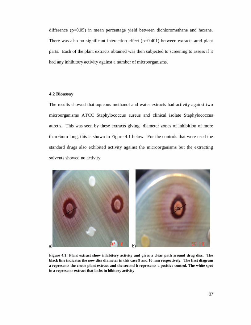
4.2 Bioassay
The results showed that aqueous methanol and water extracts had activity against two
microorganisms ATCC Staphylococcus aureus and clinical isolate Staphylococcus
aureus. This was seen by these extracts giving diameter zones of inhibition of more
than 6mm long, this is shown in Figure 4.1 below. For the controls that were used the
standard drugs also exhibited activity against the microorganisms but the extracting
solvents showed no activity.
a)
b)
Figure 4.1: Plant extract show inhibitory activity and gives a clear path around drug disc. The black line indicates the new dics diameter in this case 9 and 10 mm respectively. The first diagram a represents the crude plant extract and the second b represents a positive control. The white spot in a represents extract that lacks in hibitory activity
9 10mm

38
The activity of the plant extracts is summarised in the following tables (Table 4.3 and
4.4). the results for Kitui and Ngong crude extract activity against selected
microorganisms was similar except in Ngong where the Stem water extract exhibited
activity against clinical isolate of Salmonella typhi. The standard drugs gentamycine
and kanamycine had activity against the bacterial strains while the chloroamphenical
had activity against the fungal strains. The discs that contained the extractants only, i.e
(hexane, dichloromethane, aqueous methanol and water) did not show any activity
against the microorganisms.
Table 4.3 Antimicrobial activity of plant extracts from Kitui against selected microorganisms
ANTIMICROBIAL ACTIVITY IN EXTRACTS
ROOTS STEM STEM BARK EXTRACTANTS DRUGS
Microbe A B C D A B C D A B C D A B C D Gen Kan Chlo
1 - - - - - - - - - - - - - - - - - - +
2 - - - - - - - - - - - - - - - - + + -
3 - - - - - - - - - - - - - - - - + + -
4 - - + + - - + + - - + + - - - - + + -
5 - - - - - - - - - - - - - - - - - - +
6 - - - - - - - - - - - - - - - - - - +
7 - - - - - - - - - - - - - - - - + + -
8 - - + + - - + + - - + + - - - - + + -
(+) Inhibiton active inhibition zone diameter > 6mm; (-)No Inhibition inhibition zone diameter=6mm
Extracting solvent 20µl A: Hexane, B: Dichloromethane, C: Aqueous methanol, D: Water
Standard drugs Gen: Gentamycine, Kan: Kanamycine, Chlo: Chloroamphenical
Microbes 1: ATCC Candida albicans, 2: ATCC Escherichia coli, 3: ATCC Pseudomonas aeroginosa, 4: ATCC Staphylococus aureus, 5: clin Cryptococcus neofumonus, 6: clin. Microsporum gypseum, 7: clin. Salmonella typhi, 8: clin. Staphylococus aureus

39
Table 4.4 Antimicrobial activity of plant extracts from Ngong against selected microorganisms
ANTIMICROBIAL ACTIVITY IN EXTRACTS
ROOTS STEM STEM BARK EXTRACTANTS DRUGS
Microbe A B C D A B C D A B C D A B C D Gen Kan Chlo
1 - - - - - - - - - - - - - - - - - - +
2 - - - - - - - - - - - - - - - - + + -
3 - - - - - - - - - - - - - - - - + + -
4 - - + + - - + + - - + + - - - - + + -
5 - - - - - - - - - - - - - - - - - - +
6 - - - - - - - - - - - - - - - - - - +
7 - - - - - - - + - - - - - - - - + + -
8 - - + + - - + + - - + + - - - - + + -
(+) Inhibiton active inhibition zone diameter > 6mm; (-)No Inhibition inhibition zone diameter=6mm
Extracting solvent 20µl A: Hexane, B: Dichloromethane, C: Aqueous methanol, D: Water
Standard drugs Gen: Gentamycine, Kan: Kanamycine, Chlo: Chloroamphenical
Microbes 1: ATCC Candida albicans, 2: ATCC Escherichia coli, 3: ATCC Pseudomonas aeroginosa, 4: ATCC Staphylococus aureus, 5: clin Cryptococcus neofumonus, 6: clin. Microsporum gypseum, 7: clin. Salmonella typhi, 8: clin. Staphylococus aureus
Further analysis of the crude extracts to assess variation in disc diameter showed that
the crude extracts that had an activity had minimal variation on inhibition of the
microorganisms (Table 4.5). This was not significantly different (p>0.05). Those that
did not show activity had a disc diameter of 6 mm thus no variation in inhibitory
activity. The table 4.5 gives the variation on inhibitory activity on Staphylococcus
aureus since this is the microorganism that showed susceptiblity to the drug. The other
microbes show no effect to the crude plant extracts.

40
Table 4.5: Variation in inhibitory activity
INHIBITION ZONE DIAMETERS (mm) Site Plant
part Extract ATCC
Staphylococuss aureus
Clin. Staphylococus aureus
Kitui Roots Hexane 6 6 Dichlorometane 6 6 Aq. Methanol 10.3 11 Water 10 10 Stem Hexane 6 6 Dichlorometane 6 6 Aq. Methanol 8 8.3 Water 7.3 7.3 Stem
bark Hexane 6 6
Dichlorometane 6 6 Aq. Methanol 9.3 9.3 Water 8.7 9.3 Ngong Roots Hexane 6 6 Dichlorometane 6 6 Aq. Methanol 9.3 9.3 Water 9 9 Stem Hexane 6 6 Dichlorometane 6 6 Aq. Methanol 8.3 8.3 Water 8.3 8.3 Stem
bark Hexane 6 6
Dichlorometane 6 6 Aq. Methanol 7.3 7.3 Water 7.3 7.3
4.3 Chromatographic analysis – HPLC
For this analysis all the extracts from Kitui and Ngong were analysed. The stem, bark
and root extracts gave almost similar peak areas and thus for this analysis those of the
stem were recorded. Under the current chromatographic conditions the organic and
aqueous parts of the extracts were satisfactorily separated. The chromatograms show
peak areas at varied retention times. The total run time for the chromatograms was 35
minutes and peak areas between 0 – 20 mins represent aqueous components of the
extracts while those between 21 – 35 mins represent organic components of the crude

41
plant extracts. There was significant difference (p<0.01) in the results of the
chromatograms due to the extracts. However, there was no significant difference
(p>0.05) in the chromatographic results due to site or plant part. This results confirms
those that were obtained from the extraction and the bioassay and we see that extract
quantity and activity is not due to site of collection of extract or plant part it is obtained
from but more on the crude extract itself.
In Figure 4.2 there is an appearance of three peak areas the first one is between 0 – 4
min, this represents some aqueous components in the hexane extract. The peak height
in both the Kitui and Ngong sample is observed at less than 214 nm. A second peak of
aqueous compounds that is appearing in this chromatogram at between 12 – 20 min.
The last one which is the largest peak area is appearing between 24 – 30 min. This peak
areas is observered at 214nm absorbance. The Ngong chromatogram seems to exhibit
more clearly the chromatogram expected of a hexane extract than the kitui one due to
the presence of two peaks between 0 – 20 min. These peaks may be due to
contamination by impurities and the peak is a result of materials that are able to dissolve
in the impurities.

42
Figure 4.2: Chromatograms of stem hexane extracts top; Kitui sample, bottom; Ngong
sample
Figure 4.3 shows the dichloromethane extracts between 0 – 4 min there is a small peak
that is present in the Kitui sample but not the Ngong sample. Between 10 – 20 min
there are two peak areas in the Kitui sample and one peak area in the Ngong sample.
This could be a component that is specific to dichloromethane that is a compound that
easily dissolves in dichloromethane and allows for its extraction from the plant

43
material. At 24 – 30 min is a large peak area that represents the organic compounds
extracted from the plant material.
Figure 4.3: Chromatograms of stem Dichloromethane extracts top;Ngong sample, bottom;
Kitui sample
Prominent increase in the proportion of medium polarity components is seen in Figure
4.4 as compared to the dichloromethane extracts. This is an expected observation. The
aqueous components, which are observed in the peak area between 0 – 4 min have also
increased in area and they are observed at almost 214 nm. The peak area between 24 –

44
30 min is small in both the Kitui and Ngong aqueous methanol extract and this shows
less amounts of the organic components in the extracts.
Fig 4.4: Chromatogram of stem aq. methanol extracts top;Kitui sample,
bottom;Ngong sample
Figure 4.5 shows large peak areas for aqueous components in comparison to the
medium and organic ones. The Ngong polar components are less than those of Kitui
but more than the aqueous components of Ngong. These extracts show both the

45
aqueous and organic components and this could suggest that both polar are able to
dissolve in water thus are extracted by water. In the Ngong extracts the large aqueous
peak area could contain some impurities, which are soluble in water, as this large peak
area is not expected.

46
Figure 4.5: chromatograms of stem water extracts top;Kitui sample, bottom; Ngong
sample

47
CHAPTER 5
DISCUSSION
5.1 Yield
Osyris lanceolata contains the active ingredient in all the three plant parts used in this
experiment. It is seen that crude extracts of aqueous methanol and water are found in
all three plant parts with minimal variation in their quantities. The hexane and
dichloromethane extracts are also found in all the plant parts but these do not seem to
contain the active ingredient. However the fact that all the crude extracts are found in
all the three plant samples used is an indication of equal distribution of the secondary
metabolites within the samples collected. The average percentages by which the crude
extracts are obtained range from 0.1 to 3.9, these are an indicator of what the extracting
solvents are able to absorb out of the plant samples and previous studies (Anesin and
Prez 1993) show that this is about the expected quantity. Extracting solvents dissolve
some specific components out of plant materials and when the solvent is evaporated the
weight of the crude extract is not so much. However this small quantities have proven
to be of much medicinal value (Elof, 1998).
The difference caused by geographical location on average yield is also observed to be
minimal. Both localities were rocky and hilly and the samples were found on the forest
margins. There was a slight difference in rainfall between the two areas. Some studies
seem to suggest that geographical location has little impact on the quality and quantity
of ingredients that plants produce which are used against microorganisms (Anesin and
Prez, 1993). However this study cannot conclusively support such contibutions as Kitui
and Ngong are almost similar in their geography and more areas need to be covered to
be able to make such a conclusion. It is however worth noting that the two areas under

48
study do not seem to affect much the quantity of crude extract or the activity of the
active crude extracts. This work supports traditional use of Osyris lanceolata in
treatment of ailments as observed in Kitui and Ngong.
The role of extraction solvents cannot be belittled, the average percentage yield
analysis, it is the extraction solvent that is seen to have the greatest impact on the
amount of crude extract obatined. Plants contain physiologically active compounds and
various extracting solvent and solvent mixtures are able to dissolve them out from plant
samples (Somken et al., 1999). Extracting solvents are specific on what they can
dissolve. A mixture of extracting solvents may be able to disslove more substances and
this could explain why aqueous methanol was able to obtain the highest percentage of
crude extract. Studies show use of various solvents and solvent mixtures in extraction
from plant materials (Shapoval et al., 1994) and the varied amount of crude extracts
obtined. There is no standard solvent system that is known to extract the highest
amount of crude extract from any given plant material but it is valuable to know what
amounts each solvent or solvent mixture is able to extract form a given weight of plant
material. This is vital information in case of industrial application of such studies.
5.2 Bioassay
The study was designed to obtain preliminary information on the anti microbial activity
of Osyris lanceolata on certain microorganisms. Osyris lanceolata is used in traditional
medicine and the reports that it can treat skin disorders (Mohamed and Musya, 2005)
especially those caused by Staphylococcus aureus were supported by the bioassay
results of this study. The results showed that Osyris lanceolata water and aqueous
methanol crude extracts had activity against both the standard strain and clinical isolates
of Staphylococcus aureus. Stomach upsets caused by Salmonella typhi, which is a

49
pathogen that causes stomach upsets (Agunn et al., 2005), may not be treated by Osyris
lanceolata crude extracts.
Only the aqueous methanol and water extracts showed activity against the
microorganisms while the hexane and dichloromethane crude extracts did not show
inhibitory activity. The broad anti microbial action of the aqueous extracts could be
ascribed to the anionic components, which are naturally occurring in most plant
materials (Darout et al., 2000). The other extracts showed no action as anti microbial
agents. This may be due to little diffusion properties in the agar medium or effect of the
active principal by the steps of extraction methods (El Astal et al., 2005). This is a
good indicator for traditional healers as in most cases they use aqueous extracts (Eloff,
1998).
Escherichia coli showed no response to each of the extracts tested against it. Earlier
studies show that Escherichia coli have moderate sensitivity to other plants namely
Helichrysum italicum and Phytilacca dodecanda (Cowan, 1999). The observed
resistance probably could be due to cell membrane permeability or due to other genetic
factors (El Astal et al., 2005).
Pseudomonas aeroginosa, which is also resistant to different antibiotics, was also
resistant to the plant extracts. This bacteria’s control is very difficult by therapeutic
means (Gislene et al., 2000).
Staphylococcus aureus was susceptible to the aqueous methanol and water crude
extracts in this study. Other studies have also shown susceptiblity of the microbe to
different plant extacts (Okemo et al., 2001; Madamombe and Afolayan, 2003). This
could be due to the fact that the cell wall of gram-positive bacteria is less complex than
that of gram-negative bacteria and lack the natural sieve effect against large molecules
due to the small pores in their cell envelope (Gould and Booker, 2000).

50
Candida albicans has become resistant to the already limited toxic and expensive anti
Candida agents available in the market. This factor necessitates the search for new anti
fungal agents (Deborah et al., 2006). Yet the plant extract did not show activity against
Candida albicans. It may be speculated here that the extracts would not be useful in the
treatment of diarrhea caused by gastrointestinal Candida infection and skin lesions due
to the fungal infection.
Microsporum gypsum did not show any response to any of the extracts of Osyris
lanceolata, this could suggest to some extent lack of lapachol. This component when
available in plant extracts is capable of inhibiting activity of Micosporum gypsum
(Rasadah et al., 1998).
5.3 Chromatographic analysis
The chromatographic conditions allowed for the separation of the components of the
extracts. For this study we were looking for clusters of either aqueous or organic
componets and this was determined by the retention time. The extracts were crude and
not pure and thus determinig the specific component of the extract shown by the peaks
would not give true information on the extracts. The chromatograms were run for 35
min and typical retention times for aqueous components and organic components is
between 0 – 20 min and 21 – 35 min respectively (Chang et al., 2008).
The hexane and dichloromethane extracts showed large peak ares of organic
components and small peak ares for the aqueous components. This suggests presence
of more organic components than the aqueous components. The aqueous methanol and
water extracts showed almost similar amounts of aqueous and organic components.
Relating this to the inhibitory activity, the aqueous methanol and water extracts were
able to inhibit some of the microorganisms while the hexane and dichloromethane

51
extracts did not show any inhibitory activity. Seemingly presence of both aqueous and
organic components in almost equal amounts has something to do with the inhibitory
activity of the aqueous methanol and water crude extracts. Studies have been able to
show that single components of extracts may not show inhibitory activity (Jembere and
Hassanali, 2001) but that more than one component may exhibit synergistic effect thus
causing inhibitory activity. In this case the non-polar components may not have
inhibitory activity. However they may be able to activate the aqueous components
causing them to exhibit inhibitory activity. Studies on synergistic effects of pure
extracts have shown some interesting results and more studies need to be carried out on
Osyris lanceolata extracts.
The bioassay results showed that aqueous methanol and water extracts contained an
active ingredient against Staphylococcus aureus. From the chromatograms we could
suggest that this active component is polar. This is because the organic component is
found in all the extracts but the aqueous one is found in only the aqueous methanol and
water extracts in large quantities. The polar component may on the other hand not
contain the active ingredient, but may be largely responsible for the inhibitory activity
against Staphylococcus aureus.
The chromatographic analysis was also able to give us a general view of the number of
pure components that could be gotten from the crude extracts if separation and
purification was done. This could be seen in the peak areas as smaller peaks were
present within the lager peak areas. This provides a good basis for purification studies
and HPLC results have normally been used to prepare and carry out purification
experiments (Jembere and Hassanali, 2001). The chromatoographic analysis supports
the bioassay results and give a basis for more experimental work on the plant Osyris
lanceolata.

52
In general, the mechanism by which microorganisms survive the action of antimicrobial
agents is poorly understood and remains debatable (Okemo et al., 2001). On the other
hand, the chemical constituents of these extracts may have a causal role in protecting
plants from microbial attack in vivo. Nevertheless, at least in part, if not all they should
be valuable in the multi-chemical defense against bacterial attack. The compounds
responsible for bioactivity are unknown at this point and isolation, purification and
identification of bioactive compounds of Osyris lanceolata is crucial to a fuller
understanding of the observed activity.

53
CHAPTER 6
CONCLUSION AND RECOMMENDATIONS
6.1 Conclusion
The study was designed to assess the anti microbial effect if any of Osyris lanceolata
crude extracts on selected microorganisms. Using five bacteria and three fungi the
study was carried out using the sequential extraction and disc diffusion techniques. The
disc diffusion technique provides a quick and clear way of analysis of the actual ability
of the plant extract to inhibit or not inhibit any microorganism. Further studies were
done on the extracts by separation of the crude extracts using the high performance
liquid chromatographic technique. The chromatograms were analyzed and the peak
areas were categorised as either of polar or non-polar components based on the
retention times that they exhibited and absorption was at 214nm with total run time of
35 min. The technique enabes a visual display of presence or absence of the
components. This was used as an indicator of the likely nature of the active ingredient
as either polar or non-polar(Chang et al., 2008).
The results clearly showed that the aqueous methanol and water crude extracts had
inhibitory activity against Staphylococcus aureus. Hexane, dichloromethane, aqueous
methanol and water extracts did not have inhibitory activity on any of the other
microorganisms used in the study. The extracts that showed activity were from all three
sample plant parts used. This gave an indication of equal distribution of the active
ingredient among the sample parts used in the study. Further the variation of activity
between Kitui and Ngong samples was minimal and this showed that in this case the
geographical location did not affect the activity of the extracts. This is important as
from our study plants grown in Ngong or Kitui will not vary in activity and will give an

54
assurance of high quality medicinal value. The results of the chromatographic runs
showed us that all the plant extracts contained non-polar components but the active
extracts contained the non-polar and polar components. This showed us that the polar
components could be responsible for the inhibitory activity. The non-polar components
were not responsible for activity but may have some role in enhancing the action of the
polar components.
This study shows that Osyris lanceolata aqueous methanol and water crude extracts
have inhibitory activity against Staphylococcus aureus both clinical and standard
strains.
The study provides some basic information on the activity of Osyris lanceolata against
microorganisms and also background information for further studies on the plant. No
doubt, the plant has a great potential as a cash crop and a source of medicine in
marginal arid and semi-arid areas of Kenya.
6.2 Recommendations
The present study has unraveled some information on activity of Osyris lanceolata on
microorganisms but at the same time new questions have arisen. For instance, the
actual active ingredient and its structure is not known and the mode of action of the
plant extracts on the microorganism was beyond the scope of the current study. Thus,
the results of this study have opened new areas of research namely:
1. Bioactive compounds of Osyris lanceolata should be isolated, purified and fully
characterized.
2. Investigation on the mechanism of action of the isolated compound on the
microorganism should be revealed whether it has any competitive advantage over the
antimicrobial agents currently in use.

55
3. Pharmacokinetics studies on how fast the isolated compound is metabolized in the liver
to harmless product should be carried out.
4. Futher studies on the inhibitory action of the organic extracts using surfactants that
assist the extracts to diffuse in aqueous medium.

56
REFERENCE
Agunn A., Yusuf S., Onyilogi G. A., Zezi A. U. and Abdurham E. M. (2005).
Evaluation of five medicinal plants used in diarrhea treatment in Nigeria. Journal of
Ethanopharmacology. 101:27-30.
Almagboul A. Z., Bashir A. K., Farouk A., Salih A. K. M. (1985). Anti-microbial
activity of certain Sudanese plants used in folkloric medicine: Screening for anti-
microbial activity. Fitoterapia. 56:331-337.
Anesin C., Prez C. (1993). Screening for plants used in Argentine folk medicine for
antimicrobial activity. Journal of Ethnopharmacology. 49:147-156.
Artizzu N., Bonsignoe L., Coltiglia F., Loy G. (1995). Studies of the diuretic and
anti-microbial activity of Cydon Dactylon essential oil. Fitoterapia. 66:174-175.
Beentje H. (1994). Description and ecology of some 1850 woody species indigenous to
Kenya or that have been naturalized. Kenya Trees, Shrubs and Lianas. National
Museums of Kenya.
Brian K. R. and Turner T.D. (1975). Extraction procedures. The practical evaluation
of phytopharmaceuticals. Wright Scentechemical, Bristol UK. Page 129-139.

57
Carralho v., Melo V. M., Aguian A., Matos F. S. (1988). Toxcicity evaluation of
medicinal plant extracts by the brine shrimp bioassay. Cienciae cultura. 40:1109-1111.
Chhabra S. C. and Uisco, F. C. (1991). Antibacterial activity of some Tanzanian
plants used in traditional medicine. Fitoterapia, LXII 6: 499-500.
Clark A. M., El-Feraley F. S. and Li W. S. (1981). Anti microbial activity of phenolic
constituents of Magnolia grandiflora L. Journal of Pharmacological Science. 70:951.
Coates P. K. (1977). Ecology of Woody species. Trees of Southern Africa. Page 12 –
15.
Cowan M. M. (1999). Plant products as antimicrobial agents. Clinical Microbiology
Reviews. 12: 564 - 582
Cruz F. G., Roque N. F., Giesbrecht A. M., Davino S. C. (1996). Anti-biotic activity
of diterperes from Mikania triangularis. Fitoterapia. 67:189-190.

58
Darout I., Cristy A., Skaug N., Egeberg P. (2000). Identification and quatification of
some potential anionic components in Miswak extracts. Indian Journal of
Pharmacology. 32:11-14.
Dawson E. (1944). Some Ethnobotanical Notes on the Seri Indians. Desert Plant Life.
9:133-138.
Deborah K. B., Mercky I. N., Olipa D. N., Cosan C. J., Zakana H. W. (2006).
Screening of Tanzanian Medicinal plants for anti Candida activity. BMC
Complementary and Alternative Medicine. 6:11
Dery B. B., Otsyina R., Ng'atigwa C. (1999). Indigenous Knowledge of Medicinal
Trees and Setting Priorities for their Domestication in Shinyanga Region, Tanzania.
International Centre for Research in Agroforestry, Nairobi. 87.
Dhar A. K. and Bose S. K. (1968). Studies of Versicolin, a new anti fungal antibiotic
from Aspergillus versicolor: isolation and purification. Applied microbiology. 16; 749.
El Astal Z. Y, Ashour A, Kerrit A. (2005). Antimicrobial activity of some medicinal
plant extracts in Palestine. Pakistan Journal of Medicinal Sciences. 21(2):187-193.
Elof J. N. (1998). Which extract should be used for the screening and isolation of anti-
microbial components from plants? Journal of Ethnopharmacology. 60:1-6.

59
Eloff J. N. (2000). On expressing the antibacterial activity of plant extracts—a small
first step in applying scientific knowledge to rural primary health care, South African
Journal of Science 96: 16–118.
Elof J. N. (2004). Quantifying the bioactivity of plant extracts during screening and
bioguided fractionation. Phytomedicine. 11:370-371.
Faleiro M. L., Miguel M. G., Ladeiro F., Vebabcio F.,Tavares R., Brito J. C.,
Figueirdo A. C., Barrso J. G., Pedro L. G. (2003). Antimicrobial activity of essential
oils isolated from Portuguese endemic species of Thymus. Letters in Applied
Microbiology. 36: 35–40
Gislene G. F, Juliana L, Paulo C, Giuliana L. (2000). Antibacterial activity of plant
extracts and phytochemicals on antibioticresistant bacteria. Brazilian Journal of
Microbiology. 31:247-256.
Gosh S. K. (1994). PhD. Thesis. Studies on antimicrobial properties of some
photochemicals from higher plants. University of Calculta, India: 22, 23, 73.

60
Gould D, Booker C. (2000). Applied microbiology for nurses. Aardvark Editorial,
Mcndham, Suffolk. 75-94.
Govaerts R. (2001). How many species of seed plants are there? Taxon. 50:1085-1090.
Griffin P. M. And Tauxe R. V. (1991). The epidemiology of infections caused by
Escherichia coli O157:H7, other enterohemorrhagic E. Coli and the associated
hemolytic uremic syndrome. Epidemiology Review. 13:60 – 98.
Hamza K. F. S. (1997). Introduction to non-wood forest products. In a compendium.
Sokoine University of Agriculture, Tanzania. 82.
Ikran M., Inamul H. (1984). Screening of medicinal plants for anti-microbial
activities. Fitoterapia. 55:62-64.
Izzo A. A., DiCarlo G., Biscardi D., Fusco R., Mascolo N., Borreli F., Fasulo M. P.,
Atwe G. (1995). Biological screening of Italian medicinal plants for anti-microbial
activity. Phytotheraphy Research. 9:281-286.

61
Ivan B. S., Mateus L. B. P., Antonio D. V. and Riad N. Y. (2006). Anti bacterial
activity of Brazillian Amazon plant extracts. Brazillian Journal of Infectious diseases.
10:400 – 402.
Jansen A. M., Cheffer J. J. C., Svendsen A. B. (1987). Anti-microbial activity of
essential oils a 1976-1986 literature review. Aspects of test methods. Planta Med.
40:395-398.
Karmali M. A. (1992). The association of Verocytotoxin and the classification of
hemolytic-uremic syndrome in Kaplan, B. S. Hemolytic Uremic Syndrome and
Thrombocytopenic Purpura, Marcel Dekker, New York, pp. 199 – 212.
Kelmanson J. E., Jagen A. K., Van staden J. (2000). Zulu medicinal plants with
anti-bacterial activity. Journal of Ethnopharmacology. 69:241-246.
Kubo L., Muroi H., Hirejina M. (1993). Structure-antibacterial activity relationships
of anacardic acids. Journal of Agricultural food chemistry. 41:136-142.
Lemos T. L. G., Monte F. J. Q., Mantos F. J. A., Alencar J. W., Craverio A. A.,
Barbosa R. C. S. B., Lima E. D. (1992). Chemical composition and anti-microbial
activity of essential oils from Brazilian plants. Fitoterapia. 63:266-268.

62
Madamombe I, Afolayan A. (2003). Evaluation of antimicrobial activity of extracts
from South African Usnea barbata. Pharmaceutical Biology. 41:199-202.
Martinez M. J., Bentacourt J., Alonso-Gonzalez N., Jauregui A. (1996). Screening
of come Cuban medicinal plants for anti-microbial activity. Journal of
Ethnopharmacology. 52:171-176.
Martinez M. J., Vasquez S. M., Espinasa-Perez C., Dias M., Herrera-Sanchez M.
(1994). Anti-microbial properties of argentative A isolated from Parthnium
argentatum. Fitoterapia. 65:371-372.
McCallum S. J., Corkill J. and Gallagher M. (2001). Superinfection with
transmissible strain of Pseudomonas aeruginosa in adults with cystic fibrosis
chronically colonized by P. aeruginosa. The Lancet. 358:558 – 560.
McChesney J. D., Clark A. M., Silveira R. E. (1991). Antimicrobial diterperes of
Croton sonderianus. Pharmaceutical Research. 8:1243-1247.
Mohamed A. M. and Musya D. K. (2005). The status of Osyris lanceolata Hochst. &
Stendel “the money tree”: An overexploited medicinal plant in southern drylands,

63
Kenya. A preliminary report presented to National project coordinator (NPC) – Natural
Forests, KEFRI.
Mullika T. C., Suvimol S., Veena S. N. and Wandee G. (2005). Anti microbial effects
of Thai medicinal plants against acne-inducing bacteria. Journal of
Ethnopharmacology. 101:330 – 333.
Munekazu I., Hideki T., Toshiyuki T., Fujio A., Yasuko K., Ryoyu S. and Kenichi
M. (1996). Anti bacterial activity of xanthones from guttiferaeous plants against
methicillin-resistant Staphylococcus aureus. Journal of Pharmacy and Pharmacology.
48:861 – 865.
Murphy C. R. (1993) Gas chromatography-Mass spectrometry. Mass Spectrometry of
Lipids. Plenum Press. New York and London. Page 58.
Nascimento S. C., Chiappeta A., Lima R. M. O. (1990). Anti-microbial and
cytotoxic activities in plants from Pemambuco, Brazil. Fitoterapia. 61:353-355.
Navon-Venezia S., Ben-Ami R. and Carmeli Y. (2005). Update on Pseudomonas
aeruginosa and Acinetobacter baumannii infections in the helthcare setting. Current
Opinion in Infectious Diseases. 18:306 – 313.

64
Obritsch M. D., Fish D. N. And MacLaren R. (2005). Nosocomial infections due to
multidrug-resistant Pseudomonas aeruginosa: epdemiology and treatment options.
Pharmacotherapy. 10:1353 – 1364.
Okemo P., Mwatha W., Chhabra S., Fabry W. (2001). The kill kinetics of
Azadirachta indica A. juss.(Meliaceae) extracts on Staphylococcus aureus, E. coli,
Psedomonas aeruginosa and Candida albicans. African Journal of Science and
Technology. 2:113-118.
Pai C. H., Ahmed N., Lior H., Hohnson W. M., Sims H. V. and Woods D. E. (1988).
Epidermiology of sporadic diarrhea due to verocytotoxin-producing Escherichia coli: a
2-year prospective study. Journal of Infectious Disease. 157:1054 – 1057.
Palmer E. and Pitman N. (1972). Covering all known indigenous species in RSA,
South west Africa, Botswana, Lesotho and Swaziland. Trees of Southern Africa.
Balkema. Cape Town. Page 20 – 25.
Pamplona G. D. and Roger M. D. (2000). Plants for the stomach. Encyclopedia of
medicinal plants, Vol 2.
Prance G. T. (1991). What is ethnobotany today? Journal of Ethnopharmacology.
32:209-216.

65
Pooley E. (1993). The complete field guide to trees of Natal, Zululand and Transkei.
Natural flora publications Trust, Durban. Page 34 – 39.
Rasadah M. A, Peter J. H, Hoo T. S. (1998). Antifungal Activity of Some
Bignoniaceae Found in Malaysia. Phytotherapy Research. 12: 331–334.
Riley L. W. (1987). The epidermiologic, clinical and microbiologic features of
hemorrhagic colitis. Annual Review of Microbiology. 41: 383 – 407.
Robert C. M. (1993). Sample introduction. Mass spectrometry of lipids. Plenum
press, New York and London. Page 58.
Santos Filho D., Sarti S. J., Bastos J. K., Leitao Filho H. F., Machado J. O., Araujo
M. L. C., Lopes W. D., Abreu J. E. (1990). Effect of plant extracts on bacteria. Rev.
Cien. Farm. 12:39-46.
Santos P. R. V., Oliveria A. C. X., Tomassini T. C. B. (1995). Control of microbial
products. Fitoterapicos Rev. Farm. Bioquim. 31:35-38.
Sapu C. 2000. Folk medicinal plants of the Nagas in India. Asia Folklore studies.
58:205

66
Saxena G., McCutcheon A. R., Farner S., Toners G. H. N., Hancock R. E. W.
(1994). Anti-microbial constituents of Rhus glabra. Journal of Ethnopharmacology.
42:95-199.
Schippmann U., Leaman D. J., Cunningham A. B. (2002). Impact of Cultivation and
Gathering of Medicinal Plants on Biodiversity: Global Trends and Issues. In (FAO).
Biodiveristy and the ecosystem approach in agriculture, forestry and fishries. Satellite
event on the occasion of the Ninth regular session of the commission on genetic
resources for food and agriculture. Inter departmental working group on biological
diversity for food and agriculture, Rome.
Schmidt E., Lotter M. and McCleland W. (2002). Trees and Shrubs of Mpumalunga
and Kruge National Park. Jacana, Johannesberg. Page 44.
Scotland S. M., Rowe B., Smith H.R., Willshaw G. A. and Gross R. J. (1998).
Verocytotoxin-producing strains of Escherichia coli from children with HUS and their
detection by specific DNA probes. Journal of Medical Microbiology. 25:237 – 243.
Sekiguchi J., Asagi T. and Miyoshi-Akiyama T. (2005). Multidrug-resistant
Pseudomonas aeruginosa strain that caused an outbreak in a neurosurgery ward and its

67
aac(6’)-lae gene cassette encoding a novel aminoglycoside acetyltransferase.
Antimicrobial agents Chemotherapy. 49:3734 – 3742.
Shapoval E. E. S., Silvenia S. M., Miranda M. L., Alice C. B., Henriqes A. T.
(1994). Evaluation of some pharmacological activities of Eugenia uniflora. Journal of
Ethnopharmacology. 44:136-142.
Srinivansal D., Nathan S., Shuresh T., Perumassamy O. (2001). Anti-microbial
activity of certain Indian medicinal plants used in folkloric medicine. Journal of
Ethnopharmacology. 74:271-220.
Sokmen A., Jones B. M., Erturk M. (1999). The in vitro antibacterial activity of
Turkish medicinal plants. Journal of Ethnopharmacology. 67:79-86.
Supayang V, Amornrat L, Wanpen J, Trechada S, Souwalak P and Thanomjit S.
(2004). Effective medicinal plants against enterohaemorrhagic Escherichia coli
O157:H7. Journal of Ethnopharmacology 94:49-54
Thomas V. and Grant R. (2002). Sappi Tree Spotting. Highveld and Drakensberg.
Jacana, Johannesberg. Page 21.

68
Van wyke B. and Van wyke P. (1997). Field guide to trees of Southern Africa. Struik
publishers Cape town. Page 536.