evaluation of 14 nonlinear deformation algorithms applied ... · evaluation of 14 nonlinear...
TRANSCRIPT

Evaluation of 14 nonlinear deformation
algorithms applied to human brain MRI
registration
Arno Klein a,∗, Jesper Andersson b, Babak A. Ardekani c,d,
John Ashburner e, Brian Avants f , Ming-Chang Chiang g,
Gary E. Christensen h, D. Louis Collins i, James Gee f ,
Pierre Hellier j,k, Joo Hyun Song h, Mark Jenkinson b,
Claude Lepage i, Daniel Rueckert m, Paul Thompson g,
Tom Vercauteren n,ℓ, Roger P. Woods o, J. John Mann a,
Ramin V. Parsey a
aNew York State Psychiatric Institute, Columbia University, NY, NY 10032, USA
bFMRIB Centre, University of Oxford, Department of Clinical Neurology, John
Radcliffe Hospital, Oxford OX3 9DU, UK
cNathan Kline Institute, Orangeburg, NY, 10962, USA
dNew York University School of Medicine, NY, NY 10016, USA
eFunctional Imaging Laboratory, Wellcome Trust Centre for Neuroimaging, London
WC1N 3BG, UK
f Penn Image Computing and Science Laboratory, Department of Radiology,
University of Pennsylvania, Philadelphia, PA 19104-2644, USA
gLaboratory of Neuro Imaging, UCLA School of Medicine, Los Angeles, CA
90095-7332, USA
hDept. of Electrical and Computer Engineering, University of Iowa, Iowa City, IA
52242, USA
iMcConnell Brain Imaging Center, Montreal Neurological Institute, Montreal, QC
H3A 2B4, Canada
jINRIA Rennes, Bretagne Atlantique Research Centre, Campus universitaire de
Beaulieu, 35042 Rennes Cedex, France
kINSERM, Visages U746, IRISA, Campus de Beaulieu, Rennes, France
ℓINRIA Sophia Antipolis - Méditerranée, 06902 Sophia Antipolis, France
mVisual Information Processing, Department of Computing, Imperial College,
London SW7 2BZ, UK
nMauna Kea Technologies, 75010 Paris, France
oDepartment of Neurology, David Geffen School of Medicine at UCLA, Los
Angeles, CA 90095, USA

Abstract
All fields of neuroscience that employ brain imaging need to communicatetheir results with reference to anatomical regions. In particular, comparativemorphometry and group analysis of functional and physiological data requirecoregistration of brains to establish correspondences across brain structures. It is wellestablished that linear registration of one brain to another is inadequate for aligningbrain structures, so numerous algorithms have emerged to nonlinearly registerbrains to one another. This study is the largest evaluation of nonlinear deformationalgorithms applied to brain image registration ever conducted. Fourteen algorithmsfrom laboratories around the world are evaluated using 8 different error measures.More than 45,000 registrations between 80 manually labeled brains were performedby algorithms including: AIR, ANIMAL, ART, Diffeomorphic Demons, FNIRT,IRTK, JRD-fluid, ROMEO, SICLE, SyN, and four different SPM5 algorithms(“SPM2-type” and regular Normalization, Unified Segmentation, and the DARTELToolbox). All of these registrations were preceded by linear registration betweenthe same image pairs using FLIRT. One of the most significant findings of thisstudy is that the relative performances of the registration methods under comparisonappear to be little affected by the choice of subject population, labeling protocol,and type of overlap measure. This is important because it suggests that the findingsare generalizable to new subject populations that are labeled or evaluated usingdifferent labeling protocols. Furthermore, we ranked the 14 methods according tothree completely independent analyses (permutation tests, one-way ANOVA tests,and indifference-zone ranking) and derived three almost identical top rankings ofthe methods. ART, SyN, IRTK, and SPM’s DARTEL Toolbox gave the best resultsaccording to overlap and distance measures, with ART and SyN delivering the mostconsistently high accuracy across subjects and label sets. Updates will be publishedon the http://www.mindboggle.info/papers/ website.
∗ Corresponding authorEmail address: [email protected] (Arno Klein).URL: http://www.binarybottle.com (Arno Klein).
2

1 Introduction
Brain mapping – mapping the structures, physiology, functions, andconnectivity of brains in individuals and in different populations – is possibledue to a diverse but often disconnected array of brain imaging technologies andanalysis methods. To make the best use of brain image data, researchers haveattempted for over 40 years to establish a common reference frame such as athree-dimensional coordinate or labeling system to consistently and accuratelycommunicate the spatial relationships within the data (Talairach and Szikla,1967; Talairach and Tournoux, 1988; Drury et al., 1996; Fischl et al., 1999;Clouchoux et al., 2005). A common reference frame helps us to:1. communicate and compare data
(across subjects, time, conditions, and image types),2. classify data
(by meaningful spatial positions or extent), and3. find patterns in data
(to infer structural or functional relationships).These three benefits are contingent on one serious premise: positions and sizesin one brain must correspond to positions and sizes in another brain to makecomparisons.
This premise almost universally does not hold when brain image data arecompared across individuals. The noise that this introduces is often acceptedby researchers who generally assume that if they have found correspondingfeatures across two brains, the intervening points between those featurescorrespond to one another as well. Brains are so variable in shape that theresimply may not exist a point-to-point correspondence across any two brains,or even in the same brain over time.
Explicit manual labeling of brain regions is the preferred approach forestablishing anatomical correspondence, but it is too prohibitive in termsof time and resources, particularly in cases where neuroanatomists are notavailable, in intraoperative or other time-sensitive scenarios, and in high-throughput environments that need to process dozens to thousands of brainimages. 1 .
1 To indicate the level of investment required to manually label brain anatomy, theCenter for Morphometric Analysis (CMA) at the Massachusetts General Hospital(MGH) expects at least one month of training to train new technicians to the pointof acceptable inter-rater reliability using their Cardviews (Caviness et al., 1996)labeling protocol and software; once trained, it takes hours to weeks to manuallylabel a single brain. For 12 of the brains used in this study, a trained assistant tooktwo weeks to label each brain. At this rate, performing a modest imaging study with20 subjects and 20 controls would require 20 months devoted strictly to labeling.Manual labeling also suffers from inconsistencies within and across human labelers
3

Automatically determining anatomical correspondence is almost universallydone by registering brains to one another or to a template. There hasbeen a proliferation of different approaches to perform image registrationthat demands a comparison to guide choices regarding algorithms, softwareimplementation, setup and parameters, and data preprocessing options. Tobetter enable individuals to make these choices, the Valmet software tool(http://www.ia.unc.edu/public/valmet/) (Gerig et al., 2001) and theNon-rigid Image Registration Evaluation Project (NIREP) (http://www.nirep.org) were developed. The Windows-based Valmet was in 2001 thefirst publicly available software tool for measuring (as well as visualizing)the differences between corresponding image segmentations, but has receivedonly one minor update since 2001 (in 2004). It uses several algorithms tocompare segmentations: overlap ratio, Hausdorff distance, surface distance,and probabilistic overlap. The NIREP project “has been started to develop,establish, maintain, and endorse a standardized set of relevant benchmarks andmetrics for performance evaluation of nonrigid image registration algorithms.”The initial phase of the project will include 16 manually labeled brain images(32 labeled regions in 8 men and 8 women) and four evaluation metrics: 1.relative overlap (equivalent to the “union overlap” defined in the Materialsand methods section), 2. variance of the registered intensity images foran image population, 3. inverse consistency error between a forward andreverse transformation between two images, and 4. transitivity (how wellall the pairwise registrations of the image population satisfy the transitivityproperty).
In this study we set out to evaluate what we believe are the most importantnonlinear deformation algorithms that have been implemented in fullyautomated software programs and applied to human brain image registration.We measure accuracy at the scale of gross morphologi cal structures (gyri,sulci, and subcortical regions) acquired by magnetic resonance imaging (MRI).There have been two significant prior studies that compared more than threenonlinear deformation algorithms for evaluating whole-brain registration.
The first was communicated in a series of publications by Hellier et al.(Hellier et al., 2001a, 2002, 2003); they compared five different fully automatednonlinear brain image registration software programs using the same set ofquantitative measures. These included global measures comparing 17 deformedMRI source images and one target image: average brain volume, gray matteroverlap, white matter overlap, and correlation of a measure of curvature, andlocal measures of distance and shape between corresponding principal sulci.Our study includes a version of each of the five methods and is differentprimarily because (1) all tests were conducted by a single individual (the firstauthor) who had not authored any of the software packages, but received
(Caviness et al., 1996; Fiez et al., 2000; Towle et al., 2003)
4

guidance from the principal architects of the respective algorithms, (2) itsfocus is on manually labeled anatomical regions, and (3) each and every brainwas used as a source and as a target for registration rather than selecting asingle target.
The second is a recent paper (Yassa and Stark, 2009) that compares nonlinearregistration methods applied to regions in the medial temporal lobe; six of themethods are fully automated and two are semi-automated (requiring manualidentification of landmarks). They apply these methods either to manuallylabeled brain regions, to weighted masks for these regions, or to the originalunlabeled brains, as in our study. The four methods that they applied tounlabeled brains (and evaluated on regions in the medial temporal lobe) arethe Talairach piecewise linear approach and three SPM programs (includedin our study). Registering labeled regions obviously requires that the regionsbe labeled; their ROI-AL approach ‘labels to register’ rather than ‘registersto label’ or ‘registers without labels.’ They used two evaluation measures onpairs of images (20 MRI volumes total): an overlap measure (equivalent to the“target overlap” defined in the Materials and methods section) and a measureof blur in a group average of coregistered images. What sets our study apartfrom both of these prior studies is the unparalleled scale and thoroughness ofthe endeavor:
What sets our study apart from both of these prior studies is the unparalleledscale and thoroughness of the endeavor:
• over 14 nonlinear algorithms• each algorithm applied at least 2,168 times (over 45,000 registrations total)• 80 manually labeled brain images• 4 different whole-brain labeling protocols (56 to 128 labeled regions)• 8 different evaluation measures• 3 independent analysis methods
This study evaluates 15 registration algorithms, one linear (FLIRT) and 14nonlinear: AIR, ANIMAL, ART, Diffeomorphic Demons, FNIRT, IRTK, JRD-fluid, ROMEO, SICLE, SyN, and four different SPM5 algorithms (“SPM2-type” and regular Normalization, Unified Segmentation, and the DARTELToolbox; DARTEL was also run in a pairwise manner and all four SPMalgorithms were run with and without removal of skulls from the images). Thelinear algorithm was included as an initialization step to establish a baselineprior to applying the nonlinear algorithms. Comparisons among the algorithmsand their requirements are presented in Table 1 and in the Appendix, softwarecommands are in Supplementary section 7, and brief descriptions are inSupplementary section 8. Many of them are in common use for registeringstructural MRIs to each other or to templates for neuromorphometric researchor as an intermediary to compare functional or physiological data (Gholipour
5

et al., 2007), but some of them exist only as pre-release code made availableby their respective authors for this study. See the “Algorithms excluded fromthe study” section in the Discussion for algorithms excluded from the study.Additional materials and updated information will be made publicly availablevia the website http://www.mindboggle.info/papers/.
2 Materials and methods
In this section, we first briefly describe the acquisition and preparation ofthe brain image and label data. Then we outline the preprocessing (brainextraction and formatting), linear registration, and nonlinear registrationstages applied to the data, our evaluation measures, and our analysis methods.The first author performed these latter steps on an OSX system (Mac Pro2-Quad-Core (8-processor) Intel Xeon, 3 GHz, 6 GB RAM) with a 10.4operating system, except where noted (see Supplementary section 7).CustomPython (http://www.python.org) and Matlab (http://www.mathworks.com) software programs performed the preprocessing steps, called the differentprograms to process thousands of pairs of images, computed the results forevaluation, and produced the visualizations in the Results section.
2.1 Data preparation: images, labels, brain extraction, and formatting
6

Algorithm Deformation ≃dof Similarity Regularization
FLIRT Linear, rigid-body 9, 6 norm. CR
AIR 5th-order polynomial warps 168 MSD (opt.intensityscaling)
Incremental increase of polynomialorder; MRes: sparse-to-fine voxelsampling
ANIMAL Local translations 69K CC MRes, local Gaussian smoothing;stiffness parameter weights meandeformation vector at each node
ART Non-parametric,homeomorphic
7M norm. CC MRes median and low-pass Gaussianfiltering
DiffeomorphicDemons
Non-parametric,diffeomorphic displacementfield
21M SSD MRes: Gaussian smoothing
FNIRT Cubic B-splines 30K SSD Membrane energy*
MRes: down- to up-sampling; numberof basis components
IRTK Cubic B-splines 1.4M norm. MI None used in the study; MRes: controlmesh and image
JRD-fluid Viscous fluid: variationalcalculus (diffeomorphic)
2M Jensen-Rényidivergence
Compressible viscous fluid governed bythe Navier-Stokes equation forconservation of momentum; MRes
ROMEO Local affine (12 dof) 2M Displacedframedifference
First-order explicit regularizationmethod, brightness constancyconstraint
MRes: adaptive multigrid (octreesubdivision), Gaussian smoothing
SICLE 3-D Fourier series(diffeomorphic)
8K SSD Small-deformation linear elasticity,inverse consistency
MRes: number of basis components
SyN Bi-directionaldiffeomorphism
28M CC MRes Gaussian smoothing of thevelocity field, transformationsymmetry
SPM5:
“SPM2-type”Normalization
Discrete cosine transforms 1K MSD Bending energy, basis cutoff
Normalization Discrete cosine transforms 1K MSD Bending energy, basis cutoff
UnifiedSegmentation
Discrete cosine transforms 1K Generativesegmentationmodel
Bending energy, basis cutoff
DARTEL Toolbox Finite difference model of avelocity field (constant overtime, diffeomorphic)
6.4M Multinomialmodel(“congealing”)
Linear-elasticity; MRes: full-multigrid(recursive)
Table 1Deformation model, approximate number of degrees of freedom (dof), similarity measure, and regularizationmethod for each of the algorithms evaluated in this study. The dof is estimated based on the parameters anddata used in the study; approximate equations, where available, are given in each algorithm’s descriptionin the Supplementary section 8. Software requirements, input, and run time for the algorithms are inthe Appendix. *Since this study was conducted, FNIRT uses bending energy as its default regularizationmethod. MRes=multiresolution; norm=normalized; MSD=mean squared difference; SSD=sum of squareddifferences; CC=cross-correlation; CR=correlation ratio; MI=mutual information
7

Fig. 1. Brain image data. The study used four different image datasets with a totalof 80 brains. The datasets contain different numbers of subjects (n) and differentnumbers of labeled anatomical regions (r) derived from different labeling protocols:LPBA40 (LONI Probabilistic Brain Atlas: n=40, r=56), IBSR18 (Internet BrainSegmentation Repository: n=18, r=84), CUMC12 (Columbia University MedicalCenter: n=12, r=128), and MGH10 (Massachusetts General Hospital: n=10, r=74).A sample brain from each dataset is shown. For each brain, there are three columns(left to right): original T1-weighted MRI, extracted brain registered to nonlinearMNI152 space, and manual labels registered to nonlinear MNI152 space (used toextract the brain). Within each column the three rows (top to bottom) correspondto sagittal (front facing right), horizontal (front facing top, right on right side), andcoronal (right on right side) views. The LPBA40 brains had already been extractedand registered to MNI (MNI305 vs. MNI152) space (Shattuck et al., 2008). Thescale, position, and contrast of the MR images have been altered for the figure. Thecolors for the manual labels do not correspond across datasets.
8

2.1.1 Image acquisition and manual labels
Brain image data (T1-weighted MRIs and corresponding manual labels) for80 normal subjects were acquired from four different sources (see Fig. 1 andTable 2, and Caveats section in the Discussion regarding label reliability):
LPBA40: 40 brain images and their labels used to construct the LONIProbabilistic Brain Atlas (LPBA40) at the Laboratory of Neuro Imaging(LONI) at UCLA (Shattuck et al., 2008) are available online (http://www.loni.ucla.edu/Atlases/LPBA40). They were preprocessed accordingto existing LONI protocols to produce skull-stripped brain volumes. Thesevolumes were aligned to the MNI305 atlas (Evans et al., 1993) using rigid-body transformation to correct for head tilt and reduce bias in the manuallabeling process. This produced a transform from native space to labelingspace and an associated inverse transform. In each of the 40 subjects, 56structures were manually labeled according to custom protocols (http://www.loni.ucla.edu/Protocols/LPBA40) using BrainSuite software (http://brainsuite.usc.edu/). Brain masks were constructed from the manuallabels and projected back to the native (labeling) space to produce brain-onlyMRI volumes. These volumes were then corrected for non-uniformity usingBrainSuite’s Bias Field Corrector. Sulci were used as boundaries; white mattervoxels that occurred between the boundaries of sulci and their surroundinggray matter were included in the structure. This is the only dataset wherewhite matter is included with gray-matter regions.
After all of the registrations were conducted, we found errors in two of theLPBA40 subjects, particularly with the right putamen. We brought this toLONI’s notice and it is being corrected for future downloads. The impact ofthese errors on the present study appears to be negligible, as may be seen inFigs. 7 and 13, where there appears to be little difference between the averagevalues for the left and right putamen.
IBSR18: 18 brain images acquired at different laboratories are availablethrough the Internet Brain Segmentation Repository (http://www.cma.mgh.harvard.edu/ibsr/) as IBSR v2.0. The T1-weighted images have beenrotated to be in Talairach alignment (Talairach and Tournoux, 1988) andhave been processed by the CMA (Center for Morphometric Analysis,Massachusetts General Hospital (MGH) in Boston) ‘autoseg’ bias fieldcorrection routines. They were manually labeled with NVM software (http://neuromorphometrics.org:8080/nvm/), resulting in 84 labeled regions.
CUMC12: 12 subjects were scanned at the Columbia University MedicalCenter on a 1.5 T GE scanner. Images were resliced coronally to a slicethickness of 3 mm, rotated into cardinal orientation, then segmented andmanually labeled by one technician trained according to the Cardviews
9

Dataset Subjects Agesµ=mean
Volume (mm) Voxel (mm) TR(ms)
TE(ms)
flip∠
LPBA40 40 (20 , 20 ) 19–40µ=29.20
256×256×124 38=0.86×0.86×1.52=0.78×0.78×1.5
10-12.5
4.2-4.5
20
IBSR18 18 (14 , 4 ) 7–71µ=38.4+4 “juve-niles”
256×256×128 8=0.94×0.94×1.56=0.84×0.84×1.54=1×1×1.5
CUMC12 12 (6 , 6 )right-handed
26–41µ=32.7
256×256×124 0.86×0.86×1.5 34 5 45
MGH10 10 (4 , 6 ) 22–29µ=25.3
256×256×128 1×1×1.33 6.6 2.9 8
Table 2MRI acquisition parameters. Dataset, number and ages of subjects, volume and voxel dimensions in nativespace, TR, TE, and flip angle. The images were registered to either the nonlinear MNI152 or MNI305 atlas(see text) in a 181×217×181 volume of 1mm3 voxels.
labeling scheme (Caviness et al., 1996) created at the CMA, andimplemented in Cardviews software (http://www.cma.mgh.harvard.edu/manuals/parcellation/). The images have 128 labeled regions.
MGH10: 10 subjects were scanned at the MGH/MIT/HMS Athinoula A.Martinos Center for Biomedical Imaging using a 3 T Siemens scanner andstandard head coil. The data were inhomogeneity-corrected, affine-registeredto the MNI152 template (Evans et al., 1992), and segmented using SPM2software (Friston et al., 1995). The images were manually labeled by Tourvilleof Boston University using Ghosh’s ASAP software (Nieto-Castanon et al.,2003); the labeling protocol (Tourville and Guenther, 2003) is similar toCardviews, and in the version used for this study produces 74 labeled regions.
2.1.2 Brain extraction
To register the brains with each other, we extracted each brain from its whole-head image by constructing a mask from the corresponding manually labeledimage (see Fig. 1). However, since white matter and cerebrospinal fluid werenot fully labeled in all of the images, they had to be filled to create solidmasks. For this, the non-background image in each sagittal slice was dilatedby one pixel, any holes were filled, and then the image was eroded by onepixel. This procedure was repeated sequentially on the resulting volume for thecoronal, horizontal, and again for the sagittal slices, and resulted in a volumecontaining the filled brain mask. This manual label-based skull-strippingprocedure was performed on each MRI volume in the IBSR18, CUMC12, andMGH10 sets, but not for those in the LPBA40 set; the LPBA40 images hadalready been similarly prepared, but dilated and eroded with a larger andspherical structural element (neighborhood) (Shattuck et al., 2008). All fourSPM algorithms were also run on whole-head images.
10

2.1.3 File preparation
All image and label volumes were in right-handed orientation and wereconverted to Analyze 7.5 (.img, .hdr) format (except for MINC format usedby ANIMAL) because it was the most common image format accepted bythe different software programs, and the only format presently compatiblewith AIR, ART, JRD-fluid, and SICLE (see Appendix B). This itself was acause of difficulties, because the different software packages deal with Analyzeheader information differently, in particular with respect to leftÐright flippingand origin location. Because of this and because of discrepancies betweenbrain and atlas origins for some of the data sets, origin and orientationinformation was removed from each of the image and label volumes using FSL’s“fslorient -deleteorient” and “fslchfiletype” commands. The NiFTI data format,accepted by most of the f/MRI software packages, obviates these concernsand is recommended over the Analyze format (http://nifti.nimh.nih.gov/). Exceptions to the above steps were made for SPM5’s template-basedalgorithms (Normalization, Unified Segmentation, and DARTEL Toolbox, butnot “SPM2-type” Normalization): Analyze images were flipped right-to-left toleft-handed orientation, and header orientation discrepancies were correctedusing spm_get_space.m (other algorithms were unaffected after the fslorientcommand above).
Some extra preparation had to be done to accommodate the recommendationsfor running the individual software packages (see Appendix B), whichincluded writing parameter files, intensity correction, padding, smoothing,and reorientation (in the case of SPM). For example, parameter files wererequired for ROMEO, IRTK, and for each registration pair when using SICLE,and command-line parameters had to be reset to make some of the programsrun in less than an hour or so per registration. SICLE required considerablepreparation: we wrote a Python script to generate the input parameter filesand create output directories, normalized intensities in Matlab, and paddedversions of all of the image volumes so that their dimensions were divisible by16 (e.g., 181×217×181 files were padded to 224×224×192).
2.2 Linear registration as initialization
We linearly registered 40 of the brain images to a template using FMRIBSoftware Library’s (FSL) FLIRT (with the following settings: 9-parameter,correlation ratio, trilinear interpolation; see Fig. 1). The template was the“nonlinear MNI152,” the nonlinear average template in MNI space used byFSL (MNI152_T1_1mm_brain: 181×217×181 voxels, 1×1×1 mm/voxel).The remaining 40 images were from the LPBA40 set and had already beenregistered to the MNI305 atlas.
11

We then rigidly registered each of the 80 brains in MNI space, Is, to each ofthe other brains in its group, It, again using FLIRT (6-parameter, correlationratio, trilinear interpolation). This resulted in 2,168 linear transforms Xs→t
and transformed images in MNI space Is→t (a straight arrow denotes linearregistration), with 2,088 of them representing non-identical source-target pairs(402+182+122+102−80). These linearly transformed source images, or “linearsource images,” serve as the input to each of the algorithms under comparison.
We applied the above linear and rigid-body transforms (with nearest-neighborinterpolation) to the corresponding manually labeled volumes Ls, resulting inthe “linear source labels” Ls→t below (and in Figs. 2 and 3).
2.3 Nonlinear registration
Each of the nonlinear registration algorithms in the study then registeredeach of the 2,168 linear source images Is→t to its corresponding targetimage It. We applied the resulting nonlinear transformation X[s→t];t (withnearest-neighbor interpolation) to the corresponding linear source labels Ls→t,producing warped source labels L[s→t];t (a curved arrow denotes nonlinearregistration). These labels are compared against the manual labels of thetarget, Lt, for evaluating registration performance. See Figs. 2 and 3 for thecontext and Supplementary section 7 for the software commands used for eachalgorithm. Note that some structures were removed during preprocessing priorto computing the transforms, such as the cerebellum in the LPBA40 set, butwere included when applying the transforms to the source labels.
2.4 Evaluation measures
We used volume and surface overlap, volume similarity, and distance measuresto evaluate how well individual anatomical regions as well as total brainvolumes register to one another. For this section and for Fig. 4, source S refersto a registered image to be compared with its registration target T (in our case,the warped source labels L[s→t];t and the target labels Lt). These evaluationmeasures assume the manual label sets are correct, or “silver standards.”
2.4.1 Volume overlap
We used three overlap agreement measures and two overlap error measures,each quantifying some fraction of source S and target T volumes where theirlabels agree or disagree. For information on overlap measures, including casesfor multiple and fractional labels, see (Crum et al., 2005). The first overlap
12

Fig. 2. Registration equations. The three stages of the study were to compute,apply, and evaluate registration transforms. To compute the transforms, we linearlyregistered each source image Is to a target image It (both already in MNI space),resulting in a “linear source image” Is→t as well as a linear transform Xs→t (a straightarrow denotes linear registration). Each nonlinear algorithm Ai then registered(warped) the linear source image to the same target image, generating a second,nonlinear transform X[s→t];t (a curved arrow denotes nonlinear registration). Weapplied the linear transform to the source labels Ls to give the corresponding “linearsource labels” Ls→t, and applied the nonlinear transform to Ls→t to produce thefinal warped source labels L[s→t];t. Finally, we compared these labels to the manuallabels for the target, Lt, using a set of evaluation measures Eq.
agreement measure is the “target overlap,” TO, the intersection between twosimilarly labeled regions r in S and T divided by the volume of the region inT , where || indicates volume computed as the number of voxels:
TOr =|Sr ∩ Tr|
|Tr|(1)
Target overlap is a measure of sensitivity. When summed over a set of multiplelabeled regions, we have the total overlap agreement measure for a givenregistration:
TO =
∑r |Sr ∩ Tr|∑
r |Tr|(2)
Our second overlap agreement measure is the “mean overlap,” MO, a specialcase of the Kappa coefficient (Zijdenbos et al., 1994) sometimes called theDice coefficient; it is the intersection divided by the mean volume of the tworegions, which may again be summed over multiple regions:
MO = 2
∑r |Sr ∩ Tr|∑
r (|Sr|+ |Tr|)(3)
13

Fig. 3. Overview. This diagram provides an overview of the study for a singlenonlinear registration algorithm, placing example preprocessed data from Figure 1into the equations of Figure 2. The three stages include linear registration, nonlinearregistration, and evaluation (left to right). The four different datasets (LBPA40,IBSR18, CUMC12, and MGH10) are aligned along the left in four different versions:images, surfaces derived from the images, labels, and borders derived from the labels.A source and target are drawn from each version (image volumes are shown ascoronal slices for clarity). A source image Is is linearly then nonlinearly registeredto a target image It. The linear and nonlinear transforms (Xs→t and X[s→t];t) areapplied to the corresponding source labels Ls. The resulting nonlinearly transformedlabels L[s→t];t are compared against the target labels Lt. This comparison is used tocalculate volume overlap and volume similarity per region. The target surface St isintersected with the target labels Lt and warped source labels L[s→t];t to calculatesurface overlap. Borders between each labeled region and all adjacent labeled regionsare constructed from Lt and L[s→t];t, and average distances between the resultingborders Bt and B[s→t];t are calculated per region.
14

Fig. 4. Overlap. This study uses volume and surface overlap, volume similarity,and distance measures to evaluate the accuracy of registrations. The equations forthe three overlap measures: target overlap, mean overlap, and union overlap use theterms in this schematic Venn diagram of two partially overlapping objects, a sourceS and a target T . Their intersection is denoted by S ∩ T and their union by S ∪ T .S\T indicates the set (theoretic complement) of elements in S but not in T .
Our third overlap agreement measure is the “union overlap,” UO, or Jaccardcoefficient (Gee et al., 1993; Jaccard, 1912), the intersection over the union:
UO =
∑r |Sr ∩ Tr|∑r |Sr ∪ Tr|
(4)
UO can be converted to MO by the following (Heckemann et al., 2006):
MO =2× UO
1 + UO(5)
To complement the above agreement measures, we also computed falsenegative (FN) and false positive (FP ) errors. For these errors we characterizethe source as a tentative set of labels for the target, and again assume thatthe target’s manual labels are correct. These error measures can range fromzero to one; a value of zero is achieved for perfect overlap.
A false negative error for a given region is the measure of how much of thatregion is incorrectly labeled. It is computed as the volume of a target regionoutside the corresponding source region divided by the volume of the targetregion. As before, it is computed in voxels and summed over a set of multiplelabeled regions each with index r:
FN =
∑r |Tr\Sr|∑
r |Tr|(6)
where Tr\Sr indicates the set (theoretic complement) of elements in Tr butnot in Sr.
A false positive error for a given region is the measure of how much of thevolume outside that region is incorrectly assigned that region’s label. It is
15

computed as the volume of a source region outside the corresponding targetregion divided by the volume of the source region:
FP =
∑r |Sr\Tr|∑
r |Sr|(7)
2.4.2 Surface overlap
We anticipated that imaging artifacts affecting cortical thickness could biasour overlap measures, because (for the same cortical area) thicker regions willhave relatively higher volume overlap agreements than thinner regions due tolower surface-to-volume ratios. We tried to reduce this bias by computingoverlap agreement only on the target surfaces of the brain images, notthroughout the entire target volumes. Computing overlap agreement on thesurfaces should also decrease the impact of segmentation biases, when manuallabels extend into white matter, especially for the LPBA40 set, where whitematter between sulcal structures were also assigned the structures’ labels.
We used Freesurfer software (http://surfer.nmr.mgh.harvard.edu/, ver-sion 1.41) to construct cerebral cortical surfaces (Dale et al., 1999) for eachof the original 80 full-head images, and converted the Freesurfer-generatedsurfaces to each brain’s native space with Freesurfer’s “mri_surf2vol”command. We then linearly registered each surface to MNI space using theinitial affine transform from the original brain image to the MNI template(“Linear registration as initialization” section). Each resulting target surfacewas intersected with its corresponding target label volume Lt and warpedsource label volume L[s→t];t. We compared these target surface labels withthe warped source surface labels using the same overlap agreement and errormeasures used for the volumes.
2.4.3 Volume similarity
The volume similarity coefficient, V S, is a measure of the similarity betweensource and target volumes. Although this measure does not reflect registrationaccuracy (source and target regions can be disjoint and still have equalvolumes), it is a conventional measure included for retrospective evaluationof prior studies. It is equal to the differences between two volumes divided bytheir mean volume, here again summed over multiple regions:
V S = 2
∑r(|Sr| − |Tr|)∑r (|Sr|+ |Tr|)
(8)
16
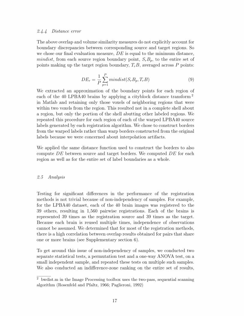
2.4.4 Distance error
The above overlap and volume similarity measures do not explicitly account forboundary discrepancies between corresponding source and target regions. Sowe chose our final evaluation measure, DE is equal to the minimum distance,mindist, from each source region boundary point, SrBp, to the entire set ofpoints making up the target region boundary, TrB, averaged across P points:
DEr =1
P
P∑p=1
mindist(SrBp, TrB) (9)
We extracted an approximation of the boundary points for each region ofeach of the 40 LPBA40 brains by applying a cityblock distance transform 2
in Matlab and retaining only those voxels of neighboring regions that werewithin two voxels from the region. This resulted not in a complete shell abouta region, but only the portion of the shell abutting other labeled regions. Werepeated this procedure for each region of each of the warped LPBA40 sourcelabels generated by each registration algorithm. We chose to construct bordersfrom the warped labels rather than warp borders constructed from the originallabels because we were concerned about interpolation artifacts.
We applied the same distance function used to construct the borders to alsocompute DE between source and target borders. We computed DE for eachregion as well as for the entire set of label boundaries as a whole.
2.5 Analysis
Testing for significant differences in the performance of the registrationmethods is not trivial because of non-independency of samples. For example,for the LPBA40 dataset, each of the 40 brain images was registered to the39 others, resulting in 1,560 pairwise registrations. Each of the brains isrepresented 39 times as the registration source and 39 times as the target.Because each brain is reused multiple times, independence of observationscannot be assumed. We determined that for most of the registration methods,there is a high correlation between overlap results obtained for pairs that shareone or more brains (see Supplementary section 6).
To get around this issue of non-independency of samples, we conducted twoseparate statistical tests, a permutation test and a one-way ANOVA test, on asmall independent sample, and repeated these tests on multiple such samples.We also conducted an indifference-zone ranking on the entire set of results,
2 bwdist.m in the Image Processing toolbox uses the two-pass, sequential scanningalgorithm (Rosenfeld and Pfaltz, 1966; Paglieroni, 1992)
17

testing for practical rather than statistical significance (see below). For eachtest, the underlying measure is target overlap averaged across all regions.
2.5.1 Permutation tests
We performed permutation tests to determine if the means of a small set ofindependent overlap values obtained by each of the registration methods arethe same, after (Menke and Martinez, 2004) and according to the followingpermutation algorithm:
1. Select a subset of P independent brain pairs2. Select a pair of methods (two vectors of P total overlap values)3. Subtract the two vectors and compute the mean difference D
4. Select a subset of the elements from one of the vectors5. Swap this subset across the two vectors6. Subtract the resulting vectors; compute the mean difference Dp
7. Repeat steps #4-6 N times8. Count the number of times n where 3 abs(Dp)≥abs(D)9. Compute the exact p-value: p= n
N
10. Repeat steps #1-9; compute the fraction of times where p≤0.05
The subset of brain pairs was selected so that each brain was used only once,corresponding to the “no dependence” condition in Supplementary section6. There were 20, 9, 6, and 5 independent brain pairs for the LPBA40,IBSR18, CUMC12, and MGH10 datasets, respectively, as well as 20, 9, 6,and 5 corresponding average target overlap values obtained by each method.
The number of permutations N for each subset of brain pairs was eitherthe exhaustive set of all possible permutations (212=4,096 for CUMC12 and210=1,024 for MGH10) or 1,000 permutations (LPBA40 and IBSR18) to keepthe duration of the tests under 24 hours. The number of p-values calculatedwas either 100,000 (CUMC12 and MGH10) or 10,000 (LPBA40 and IBSR18).
2.5.2 One-way ANOVA
We also performed a standard one-way ANOVA to test if the means of similarsubsets of independent average target overlap values obtained by each ofthe registration methods are the same. We then subjected these results to amultiple comparison test using Bonferroni correction to determine which pairsof means are significantly different (disjoint 95% confidence intervals aboutthe means, based on critical values from the t distribution). We repeated these
3 abs()=absolute value
18

ANOVA and multiple comparison tests 20 times, each time randomly selectingindependent samples from each of the datasets. These tests are not expectedto be as accurate as the permutation tests because some of the overlap valueshave skew distributions and because the p-values are not exact.
2.5.3 Indifference-zone ranking
Our third evaluation between methods tested practical significance ratherthan statistical significance. For example, if a region is registered to anotherregion of equal volume and results in an offset of a single voxel, this isnot considered a significant misregistration, but offsets greater than this areconsidered significant. An evaluation measure of registration accuracy for agiven region within a given brain pair is calculated for two different registrationmethods. If these two values are within delta of one another (referred to asan “indifference zone” when ranking (Bechhofer, 1954)), they are consideredequal. The delta must correspond to a practical difference in registration. Ifwe model a region as a cube, then a single-voxel offset along the normal to oneof its faces would mean the voxels on that face of the cube reside outside ofits target Ñ this is equal to one-sixth of its surface. We therefore set delta toone-sixth of a target region’s surface. For the IBSR18, CUMC12, and MGH10datasets, we assumed the surface to be that of a cube (6×edge2−12×edge,where edge = the edge length of a cube with the volume of the target region,in voxels). For the LPBA40 dataset, we set the surface to the number of voxelsbordering adjacent regions, extracted as in the “Distance error” section.
Our implementation of indifference-zone ranking compared the 15 differentregistration methods to each other in the following manner. For each regionin a given label set and for each pair of registered brains we constructed a15×15 matrix, where each row and each column corresponded to a registrationmethod. Each element of the matrix was assigned the value −1, 0, or 1, for thecases when the evaluation measure for the method corresponding to its rowwas at least delta less than, within delta of, or at least delta greater than thatof the method corresponding to its column. Then we calculated the mean ofthese −1, 0, 1 values across all registration pairs for each region to constructFigs. 7, 8, 9, and 10 (the latter three in Supplementary section 3).
3 Results
Results for the initial run are in Supplementary section 1, for the trivial case,where each brain was registered to itself, are in Supplementary section 2,volume similarity results are in Supplementary section 4, and distance errorresults are in Supplementary section 5.
19

3.1 Overlap results
3.1.1 Whole-brain averages
After the initial run and changes described in Supplementary section 1, out of2,168 registrations per algorithm, the only cases where target overlap valueswere less than 25% were SPM’s DARTEL (79 cases; the majority were fromone brain) Normalize (15 cases), ANIMAL (2 cases), and ROMEO (1 case)for the LPBA40 set and Diffeomorphic Demons (1 case) for the IBSR18 set.
The target, union, and mean overlap values for volumes as well as surfaces (andthe inverse of their false positive and false negative values), averaged over allregions, gave almost identical results when corrected for baseline discrepancies.Distributions of target overlap values are shown in Fig. 5. What is remarkableis that the relative performances of these methods appear to be robust notjust to type of overlap measure, but also to subject population and labelingprotocol, as evidenced by the similar pattern of performances of the methodsacross the label sets. This is particularly the case across IBSR18, CUMC12,and MGH10 sets. The pattern is more subtle in LPBA40 because that labelset has fewer labeled regions that are larger and extend into white matter,and therefore results in higher and more similar absolute overlap values.
We ran all 2,168 registrations again on whole-head images (before skull-stripping) using SPM’s Normalize, Unified Segmentation, and DARTEL, andthe results were comparable or better with the skull-stripped images. Therelative overlap performance of the SPM programs agrees with Yassa and Stark(Yassa and Stark, 2009): DARTEL performs better than Unified Segmentationwhich performs better than Normalize. Because the SPM DARTEL resultswere very similar for its original and pairwise implementations, we haveincluded only the pairwise results; this is a fair comparison because the othermethods do not include optimal average template construction.
3.1.2 Region-based results
The pattern of region-based overlap values is almost indistinguishable acrossthe methods, discounting baseline differences (data not shown). In Fig. 6 wepresent volume and surface target overlap data for individual regions in theiranatomical context (LPBA40 set). For the most part this figure suggeststhat the overlap values are approximately the same for volume and surfacemeasures, corroborating whole-brain averages, but also exposes discrepanciesat the level of regions (FLIRT and SICLE) 4 .
4 The worse surface overlaps of the cerebellum (for all the methods except ROMEO)are probably due to the fact that the cerebellum was removed from the LPBA40 set
20

Fig. 5. Overlap by registration method. These box and whisker plots show the targetoverlap measures between deformed source and target label volumes averaged firstacross all of the regions in each label set (LPBA40, IBSR18, CUMC12, and MGH10)then across brain pairs. Each box represents values obtained by a registrationmethod and has lines at the lower quartile, median, and upper quartile values;whiskers extend from each end of the box to the most extreme values within 1.5times the interquartile range from the box. Outliers (+) have values beyond theends of the whiskers. Target, union and mean overlap measures for volumes andsurfaces (and the inverse of their false positive and false negative values) all producedresults that are almost identical if corrected for baseline discrepancies. Similaritiesbetween relative performances of the different registration methods can even be seenhere across the label sets. (SPM_N*=“SPM2-type” normalization, SPM_N=SPM’sNormalize, SPM_US=Unified Segmentation, SPM_D=DARTEL pairwise)
Most of the regions in the brain volume plots are hidden from view, so for acomplete picture at the scale of individual regions, Figs. 7, 8, 9, and 10 presentrelative performances of the different methods for each region as color-codedtables for each of the four label sets (their construction is described in the“Indifference-zone ranking” section under “Materials and methods”; Figs. 8, 9,and 10 are in Supplementary section 3). If all of the methods had performed
prior to computing the registration transforms, but the transforms were applied tothe full label set (including the cerebellum).
21

equally well, the color tables would be a uniform color. However, some of themethods performed better than average, particularly against simple linearregistration (FLIRT). By visual inspection, we can see that ART, IRTK,SyN, and SPM’s DARTEL have consistently high accuracy for the IBSR18,CUMC12, and MGH10 label sets relative to the other methods, and that inaddition to ART, IRTK, and SyN, FNIRT and JRD-fluid also appear to havehigh relative accuracy for the LPBA40 set. As expected, we observed for all ofthe methods higher overlap values for larger sized regions, because of smallersurface-to-volume ratios (not shown).
3.2 Rankings
We ranked the registration methods in three independent ways: permutationtests, confidence intervals obtained from one-way ANOVA tests withBonferroni correction, and indifference-zone ranking.
3.2.1 Permutation, ANOVA, and indifference-zone rankings
Table 3 presents the top three ranks of registration methods according to thepercentage of permutation tests whose p-values were less than or equal to0.05, and Table 4 according to relative target overlap scores. For both tables,members within ranks 1, 2, and 3 have means lying within one, two, and threestandard deviations of the highest mean, respectively. Only ART and SyN arein the top rank for all four label sets and for all tests.
For the one-way ANOVA tests, rank 1 methods have means lying withinthe 95% confidence interval of the best method and rank 2 methods haveconfidence intervals that overlap the confidence interval of the best method.These rankings were in almost complete agreement among the target, union,and mean overlap values (and distance errors for the LPBA40 set). Becausethese results were very similar to the permutation test ranks, and becausethese tests are expected to be less accurate than the permutation tests, theyare not included.
22

Fig. 6. Volume and surface overlap by registration method: LPBA40 regions.These brain images show the mean target overlap calculated across all 1,560brain pairs for the (A) volume and (B) surface of each LPBA40 region, anddepicts that mean as a color (blue indicates higher accuracy). The valuesfor each registration method are projected on one of the LPBA40 brains,seen from the left, looking down from 30, with the frontal pole facingleft. (SPM_N*=“SPM2-type” Normalize, SPM_N=Normalize, SPM_US=UnifiedSegmentation, SPM_D=DARTEL pairwise)
23

Fig. 7. Indifference-zone ranking of the registration methods: LPBA40 overlaps.This matrix uses a color scale that reflects the relative performance of theregistration methods (with blue indicating higher accuracy). Each colored rectanglerepresents the average score for a given method for a given region, averagedover 1,560 LPBA40 registrations. The scores are −1, 0, 1 values indicatingthe pairwise performance of the method relative to each of the other methods(see text), according to target volume overlap (union and mean overlap resultsare almost identical). The colors (and color range) are not comparable tothose of the other label sets (Figures 8, 9, and 10 in Supplementary section3). (SPM_N*=“SPM2-type” Normalize, SPM_N=Normalize, SPM_US=UnifiedSegmentation, SPM_D=DARTEL pairwise)
24

LPBA40 µ (SD) IBSR18 µ (SD) CUMC12 µ (SD) MGH10 µ (SD)
rank
1ART .82 (.35) SPM_D .83 (.27) SPM_D .76 (.24) SyN .77 (.37)
SyN .60 (.38) SyN .72 (.51) SyN .74 (.51) ART .72 (.45)
FNIRT .49 (.66) IRTK .67 (.53) IRTK .74 (.50) IRTK .61 (.51)
JRD-fluid .49 (.66) ART .60 (.70) ART .60 (.70)
2
IRTK .43 (.63) JRD-fluid .30 (.82) SPM_D .27 (.23)
D.Demons .13 (.82) D.Demons .27 (.69)
SPM_US .11 (.83) JRD-fluid .24 (.66)
ROMEO .06 (.63)
3
ROMEO .08 (.73) FNIRT .16 (.82) D.Demons .20 (.84)
SPM_D .07 (.29) D.Demons .05 (.84) FNIRT .18 (.81)
JRD-fluid .17 (.81)
Table 3Permutation test ranking of the registration methods by label set. This table lists the methods that attainedthe top three ranks after conducting permutation tests between mean target overlaps (averaged acrossregions) for each pair of methods, then calculating the percentage of p-values less than or equal to 0.05 (of100,000 tests for CUMC12 and MGH10 or of 10,000 tests for LPBA40 and IBSR18; µ=mean, SD=standarddeviation). Methods within ranks 1, 2, and 3 have positive mean percentages lying within one, two, andthree standard deviations of the highest mean, respectively. Values are not comparable across label sets(columns). (SPM_D=DARTEL pairwise)
LPBA40 µ (SD) IBSR18 µ (SD) CUMC12 µ (SD) MGH10 µ (SD)
rank
1
ART .35 (.07) SPM_D .50 (.19) SPM_D .47 (.17) SyN .39 (.06)
SyN .34 (.24) SyN .40 (.12) IRTK .42 (.07) ART .36 (.07)
IRTK .35 (.15) SyN .41 (.06)
ART .33 (.08) ART .35 (.05)
2 JRD-fluid .18 (.13)
3
JRD-fluid .20 (.08) FNIRT .06 (.11) JRD-fluid .07 (.07) IRTK .26 (.07)
IRTK .18 (.15) D.Demons .01 (.08) FNIRT .07 (.09) SPM_D .25 (.28)
FNIRT .17 (.08) ROMEO .01 (.28) D.Demons .05 (.05)
SPM_D .14 (.31)
Table 4Indifference-zone ranking of the registration methods by label set. This table lists the methods that attainedthe top three ranks after averaging scores across all brain regions then across all registration pairs (µ=mean,SD=standard deviation). The scores reflect a pairwise comparison between methods, according to targetoverlap (see text). Methods within ranks 1, 2, and 3 have positive means lying within one, two, and threestandard deviations of the highest mean, respectively. Values are not comparable across label sets (columns).(SPM_D=DARTEL pairwise)
25

4 Discussion
This study evaluates 15 registration algorithms (one linear, 14 nonlinear)based primarily on overlap measures of manually labeled anatomical regions.The scale and thoroughness are unprecedented (over 45,000 registrations, 80manually labeled brain images representing 4 different labeling protocols,8 different evaluation measures, and 3 independent analysis methods). Wehope that the method of evaluation as well as the results will be useful tothe neuroscience community. As they become available, additional materialsand updated information will be made publicly available via the websitehttp://www.mindboggle.info/papers/.
One of the most significant findings of this study is that the relativeperformances of the registration methods under comparison appear to belittle affected by the choice of subject population, labeling protocol, andtype of overlap measure. This is important because it suggests that thefindings are generalizable to new healthy subject populations that are labeledor evaluated using different labeling protocols. Furthermore, we ranked themethods according to three completely independent analyses and derived threealmost identical top rankings. However, in order to make recommendations, itis important to place these results in the context of the wider range of softwarepackages available and the caveats inherent in registration in general and withrespect to this study in particular, as we do below.
Although we were not able to see a pattern in the results that would allow usto rank algorithms by deformation model, similarity measure, or regularizationmethod, there is a modest correlation between the number of degrees offreedom of the deformation and registration accuracy (0.29, or 0.45 if oneexcludes Diffeomorphic Demons), and between the number of degrees offreedom and year (0.55) (see Table 5). This finding corroborates Hellier’sevaluation: “The global measures used show that the quality of the registrationis directly related to the transformation’s degrees of freedom” (Hellier et al.,2003). The four algorithms whose mean rank is less than two (SyN, ART,IRTK, and SPM’s DARTEL Toolbox) all have millions of degrees of freedomand all took at least 15 min per registration, and all but one (IRTK) werecreated in the last three years. Of the remaining 10 algorithms, seven havefewer than a million degrees of freedom, seven took less than 15 min, and sixwere created over three years ago.
26

Algorithm mean rank dof run time: minutes year
SyN 1.00 28M 77 (15.1) 2008
ART 1.00 7M 20.1 (1.6) [Linux] 2005
IRTK 1.63 1.4M 120.8 (29.3) 1999
SPM5 DARTEL Toolbox 1.88 6.4M 71.8 (6.3) 2007
JRD-fluid 2.50 2M 17.1 (1.0) [Solaris] 2007
Diffeomorphic Demons 3.00 21M 8.7 (1.2) 2007
FNIRT 3.00 30K 29.1 (6.0) 2008
ROMEO 3.50 2M 7.5 (0.5) 2001
ANIMAL 69K 11.2 (0.4) 1994
SICLE 8K 33.5 (6.6) 1999
SPM5 Unified Segmentation 1K ≃1 2005
“SPM2-type” Normalize 1K ≃1 1999
SPM5 Normalize 1K ≃1 1999
AIR 168 6.7 (1.5) 1998
Table 5Mean rank, degrees of freedom (dof), average run time, and year of publication for each algorithm. The 14nonlinear deformation algorithms are ordered by mean rank (best at top), which was computed for eachalgorithm by averaging the target overlap ranks in Tables 3 and 4 (assigned by the permutation tests andindifference-zone rankings). The six algorithms at the bottom are of equal rank (4) since they were notin the top three ranks. For details on architecture and run time, see Appendix. Except for FNIRT andDiffeomorphic Demons, the dof and mean rank sequences roughly match.
4.1 Algorithms excluded from the study
We excluded semi-automated approaches that require even minimal manualintervention to reduce bias. A significant example is the forerunner of modernnonlinear registration methods, the original Talairach coordinate referencingsystem (Talairach and Szikla, 1967; Talairach and Tournoux, 1988), a piece-wise linear registration method that requires the identification of landmarksin a brain image. Although the Talairach system is well suited to labelingregions proximal to these landmarks (Grachev et al., 1998), it does not dealadequately with nonlinear morphological differences, especially when appliedto the highly variable cortex (Grachev et al., 1999; Mandl et al., 2000; Rolandet al., 1997; Xiong et al., 2000). Other examples that require landmarks includemodern nonlinear algorithms such as Large Deformation Diffeomorphic MetricMapping (personal communication with Michael Miller)(Beg et al., 2005) andCaret (http://brainmap.wustl.edu/, personal communication with DavidVan Essen and Donna Dierker)(Essen et al., 2001).
We also excluded some of the primary software programs for auto-matically labeling cortical anatomy: Freesurfer (http://surfer.nmr.mgh.
27

harvard.edu/)(Fischl et al., 2002, 2004), BrainVisa (http://brainvisa.info)(Cointepas et al., 2001), HAMMER (https://www.rad.upenn.edu/sbia/software/index.html)(Shen and Davatzikos, 2002), and Mindboggle(http://www.mindboggle.info)(Klein and Hirsch, 2005; Klein et al., 2005),because their cortical labeling algorithms are tied to their own labeled brainatlas(es). We considered this problematic for three reasons: (1) we wantedto evaluate brain registration algorithms, not brain labeling algorithms orparticular atlas-based approaches, (2) their atlas labels are inconsistent withthe protocols used to label the brains in this study which would makeevaluation difficult, and (3) creating new atlases for each of these requiresconsiderable knowledge of the software. Freesurfer and BrainVisa differ fromall of the other methods mentioned in this paper because they register surfacesrather than image volumes. Mindboggle differs from the others because itis based on combinatoric feature-matching and uses multiple independentatlases. And of the four, HAMMER is the only one that can transform anarbitrary set of labels when registering a source brain to a target brain.However, because we were not able to obtain reasonable results, we did notinclude it in the study. We also tested the PASHA algorithm (Cachier et al.,2003) with and without intensity normalization but because we obtained veryinconsistent results across the datasets we decided not to include it in thestudy either. We also excluded other programs that do not allow one to applytransforms to separate image volumes.
4.2 Caveats
4.2.1 General caveats
There are numerous caveats that must be taken into account when evaluatingregistration data. The very question of correspondence between brainsthat we raised at the beginning of this paper is revisited at every stage:at the level of anatomy, image acquisition, image processing, registration(including similarity measure, transformation model, regularization method,etc.), evaluation measures, and analysis based on these measures. We will focushere on the most fundamental level of correspondence, at the primary level ofanatomy, and on the effects of registration on anatomical correspondence.
If we consider the scale of gross anatomy or patterns of functional activityor physiological data, then we may seek correspondences at the level oftopographical, functional, or physiological boundaries without assuming one-to-one mapping of the points of the boundaries or the points withinthese regions of interest. In other words, another way of approaching this“correspondence problem,” and by extension the elusive common referenceframe, is as a partial mapping between brains, independent of naming or
28

spatial conventions. The common reference frame is used simply as a referenceof comparison or evaluation, not as a rigid framework for comprehensivelyannotating brain image data, as is often done.
If we cannot expect every brain to have a one-to-one mapping with everyother brain, then if possible we need to compare similar brains. This caneasily lead to the confound where image correspondence is mistaken foranatomic correspondence (Crum et al., 2003; Rogelj et al., 2002). Choosing arepresentative brain with which to establish correspondences with a givenbrain results in a Catch-22 where determining similarities itself entailsdetermining correspondences between the brains. A few approaches aroundthis dilemma include the use of an established average template or probabilisticatlas as an intermediary registration target (as is standardly done with SPM),construction of such a template from the subject group that includes the brainin question, and decision fusion strategies for combining multiple, tentativebrain registrations or labels for a given target brain (Kittler et al., 1998;Rohlfing et al., 2004; Warfield et al., 2004; Klein et al., 2005). With all ofthese approaches, however, there still remains the distinct possibility that agiven brain is not adequately represented by the majority of the set of brainsto which it is being compared. Indeed, it is possible that substructures withina brain are most similar to a minority (or even a single, or no instance) of theset of brains, and would be overridden by the majority.
The evaluation measures and analysis methods used in this paper arepredicated on the assumption that, at the macroscopic scale of topographicanatomical regions, there are correspondences across a majority of brainsthat can effectively guide registrations. It is very important to stress thatwe cannot make inferences about the accuracy of registrations within thesemacroscopic regions. Therefore our overlap evaluation measures not onlyignore misregistration within a labeled region but are insensitive to foldingin the deformations, which would impact studies such as deformation-basedmorphometry. More generally, our evaluation measures rely on informationwhich is not directly included in the images, which is good for evaluating theregistrations, but they do not inform us about the intrinsic properties of thespatial transformations. Example measures of the intrinsic properties of spatialtransformations include inverse consistency error, transitivity error, and “meanharmonic energy” (where the Jacobian determinant of the transformation isaveraged over the volume).
Another general caveat comes from recent evidence that nonlinear registrationto average templates affects different brain regions in different ways that leadto relative distortions in volume that are difficult to predict (Allen et al.,2008). The evidence was based on varying the target template and registrationmethod (AIR and piecewise linear). Although our study was not concernedwith absolute volumetry, and nonlinear registrations were conducted from one
29

brain to another without the use of a template, we share the caution raisedby their study.
4.2.2 Specific caveats
Caveats that are specfic to our study mirror the general caveats raisedabove: anatomical and labeling variability of the subject brains, quality oftheir images, the preprocessing steps the images were subjected to, theimplementation of the registration algorithms, and our evaluation and analysismethods. With regard to the first three caveats, we made the assumption thateach label set consists of a subject group of normal individuals whose brainimages were acquired, preprocessed, and labeled in a consistent manner. Someof the co-authors have commented that the quality of the images in this studyis worse than the quality of the images that they are used to applying theiralgorithms to. Some of the reasons for this are that the images for these labelsets were acquired years ago, are incomplete (for example, only the CUMC12set includes the cerebellum in registered images and labels), many are of lowcontrast, and all of them were linearly transformed to a template space thatinvolved two trilinear interpolation steps (see below). All of the algorithmsperformed worst on the IBSR18 set, whose images were acquired from varioussources and are of varying quality, flouting our assumption above regardingconsistency.
Each brain image was labeled only once. Because there are no intra- or inter-labeler data for these images, we cannot know how accurately and consistentlythey were labeled, let alone have an idea of the degree of confidence for anyof the label boundaries. We can only estimate based on labeling tests for twoof the labeling protocols (Caviness et al., 1996; Shattuck et al., 2008). Wetherefore had to treat these label sets as “silver standards” whose hard labelboundaries are considered correct.
Regarding pre-processing, the brain images of each label set were consistentlypreprocessed, and each registration method that performed preprocessingsteps did so in a consistent manner across all images. However, thesepreprocessing steps may be suboptimal for particular registration methods.For example, aside from SPM’s algorithms, we did not test registrationaccuracy for whole-head images. Although most of the co-authors indicatedthat they believe their registration methods would perform better on properlyskull-stripped images than on whole-head images 5 , we are not aware of anypublished study that has made this comparison. Likewise, we are aware
5 FNIRT is an exception: In the beta version used in this study, zero values areinterpreted as missing data; FNIRT will not use the information for the edge of thecortex in the registration with this setting, which may result in misregistration ofthe surface of the brain.
30

of no comparisons between the registration of interpolated versus non-interpolated (bias-field corrected and uncorrected, intensity normalized andnon-normalized, etc.) images. All of the images in this study were linearlyinterpolated twice, once to linearly register each brain to a template, and asecond time to linearly register each source brain to a target brain in thetemplate space, prior to nonlinear registration. We did this to be consistent,because all of the registration methods we compared do not accept anaffine transform to initialize registration. The first author has observed muchmore accurate nonlinear registrations with ART (on a separate set of brainimages) when using nearest-neighbor (or no) interpolation on a preliminarylinear registration step, most noticeably in occipital-parietal boundaries. Thissuggests that, at the very least, ART would perform much better than thisstudy suggests. More work will need to be conducted to see how consistent theimprovements are and which algorithms are affected most by interpolation.
Regarding the registration methods themselves, each one has a similaritymeasure, transformation model, regularization method, and optimizationstrategy. Unfortunately, we could only evaluate each algorithm in its entirety.A superior transformation model coupled with an unsuitable similaritymeasure, for example, would most likely lead to suboptimal results. Byextension, a poor selection of parameter settings will lead to poor registrations.We could only evaluate each algorithm using the software parameters thatwere recommended by their authors. Perhaps the most crucial assumption ofour study is that these parameter settings for each method were appropriatefor all of our brain images. We fully expect that each registration algorithmcould perform better given the opportunity to experiment with these settings.This is one aspect of our study that sets it apart from comparison studiessuch as Hellier’s (Hellier et al., 2001a, 2002, 2003), where the authors of thesoftware packages were allowed to tweak and run their own programs on thefull test set of brain images. The commands that were run for this studywere recommended by the authors of their respective software programs afterhaving seen only one or two of the 80 images from one of the four datasets(FNIRT, IRTK, SICLE, SyN, and SPM’s DARTEL Toolbox 6 ), or no imagesat all.
When reslicing the source label volumes, we used nearest-neighborinterpolation to preserve label values. An alternative approach is recommendedwhere the source label volume is first split into N binary volumes, with onelabel per volume (Collins, personal communication). Each volume is then
6 Updated versions of these software packages were used after the authors of thepackages saw an image or two, or their recommended commands or parameter fileswere altered to set the number of iterations or control point spacing to reducecomputation time, or the authors needed to determine if intensity correction waswarranted (see Supplementary section 1).
31

resampled using the nonlinear transformation with a tricubic or truncated synckernel instead of nearest-neighbor interpolation. The resulting N temporaryvolumes are finally combined into a single volume, where each voxel label isset to the label of the structure that has the highest value. This presumablygives more consistent behavior at structure edges, especially in areas wherethe deformation changes local volumes or where more than three structuresmeet. Others have implemented variants of this approach (Crum et al., 2004;Shattuck et al., 2008). We were unable to follow this recommendation dueto computational and storage constraints, and were advised that the resultswould be only marginally different.
4.3 Recommendations
Bearing in mind the caveats mentioned above, particularly those regardingparameter settings, the first author makes the following recommendationsbased on the results of this study. All of the software packages undercomparison are freely available via the Internet or from the authors themselves(except for JRD-fluid, run on LONI’s servers) and all but one (SICLE) areeasy to install. They vary in the extent of their documentation, primarilybecause the pre-release software packages are new and very much under activedevelopment.
The highest-ranking registration methods were SyN, ART, IRTK, and SPM’sDARTEL Toolbox (see Tables 3, 4, and 5). SyN and ART gave consistentlyhigh-ranking results and were the only methods that attained top rank for alltests and for all label sets. IRTK and SPM’s DARTEL were competitive withthese two methods.
All four of these methods are available on Unix-type systems, and all but ARTare available for the Windows operating system. Of the four, only SPM requiresa commercial software package (Matlab) and has a graphical user interface(which was not used in the study). If flexibility is desired, SyN provides themost options and the closest documentation to a manual for command-lineparameters. If resources are an issue, note that SyN requires at least 1 GBRAM and 87 MB storage per x, y, z set of transform files (followed by ARTat 67 MB for our data). If time is a constraint, ART is the fastest of the four.If consistency is the top priority, ART had the fewest outliers and among thetightest distributions of the four methods. If interested in particular regions,please refer to Figs. 7, 8, 9, and 10 (the latter three are in Supplementarysection 3) to determine which of the 15 methods had the highest relativeaccuracy for those regions across the label sets.
For time-sensitive scenarios, such as intraoperative imaging, and in high-
32

throughput environments that need to process dozens to thousands of brainimages, Diffeomorphic Demons and ROMEO are reasonable candidates.
With regard to the evaluation protocol, based on the experience of conductingthis study the first author recommends caution when choosing an imageformat and preprocessing steps, particularly when comparing across methods,recommends avoiding interpolation prior to running nonlinear registration,and recommends the model of Pierre Jannin et al. for defining and reportingreference-based validation protocols (Jannin et al., 2006).
With regard to designing and distributing registration algorithms, thefirst author recommends where possible creating separable components forthe similarity measure, transformation model, regularization method, andoptimization strategy. This would aid users and evaluators who would wantto alter or improve upon these individual components.
5 Acknowledgments
The first author would like to extend his sincere gratitude to the participantsin this study for their guidance and support in the use of their software,which in some cases took the form of new pre-release software and reslicingalgorithms. He is grateful to his colleagues in the Division of MolecularImaging and Neuropathology, and thanks Steve Ellis, Todd Ogden, SatrajitGhosh, and Jack Grinband for their helpful discussions. And of course hethanks his two closest colleagues Deepanjana and Ellora. This work waspartially funded by the National Institutes of Health through NIH grant P50-MH062185. The LPBA40 MR and label data were provided by the Laboratoryof Neuro Imaging at UCLA and are available at http://www.loni.ucla.
edu/Atlases/LPBA40. The IBSR18 MR and label data were provided by theCenter for Morphometric Analysis at Massachusetts General Hospital and areavailable at http://www.cma.mgh.harvard.edu/ibsr/. The CUMC12 datawere provided by Brett Mensh, and the MGH10 data were provided by SatrajitGhosh and Jason Tourville. The contributions to this paper by Babak A.Ardekani were supported by Grant Number R03EB008201 from the NationalInstitute of Biomedical Imaging And Bioengineering (NIBIB) and the NationalInstitute of Neurological Disorders and Stroke (NINDS). The contributions tothis paper by Gary E. Christensen and Joo Hyun Song were supported by NIHgrant EB004126. Mark Jenkinson would like to thank the UK BBSRC (DavidPhillips Fellowship). John Ashburner is funded by the Wellcome Trust.
33

A Supplementary data
Supplementary data associated with this article can be found below, in theonline version, at doi:10.1016/j.neuroimage.2008.12.037, and on http:
//www.mindboggle.info/papers/.
B Algorithm requirements
34

Algorithm Code Computer Input Setup Run time: minutes
FLIRT (FSL 4.0) C++ OSX, Linux, Win,... Analyze, NiFTI
AIR 5.25 C OSX, Unix, Win,...ANSI C compiler
Analyze 8-/16-bit Remove nonbrainstructures
6.7 (1.5)
ANIMAL(AutoReg 0.98k)
C, Perl OSX, Linux, Unix MINC Intensity correction(option)
11.2 (0.4)
ART C++ OSX, Linux Analyze 20.1 (1.6) [Linux]
DiffeomorphicDemons
C++ Most (ITKcompilable)
Analyze, NifTI,DICOM,... (ITK)
8.7 (1.2)
FNIRT beta C++ OSX, Linux, Unix Analyze, NiFTI(writes to Analyze)
29.1 (6.0)
IRTK C++ OSX, Linux, Win Analyze, NiFTI,VTK, GIPL
Parameter file 120.8 (29.3)
JRD-fluid C++ Sun Analyze 17.1 (1.0) [Solaris]
ROMEO C++ OSX, Linux, Win Analyze, NiFTI,DICOM,... (ITK)
Parameter file 7.5 (0.5)
900+MB RAM Intensity correction(Hellier, 2003)
SICLE C++ OSX, Linux, Solaris,Alpha, Win
Analyze (7.5) 8-bit Dimensions divisibleby 16
33.5 (6.6)
g77/gfortran Intensity correction
lapack, f2c Isotropic
1+GB RAM Individual parameterfiles
SyN beta C++ Most (ITKcompilable)
Analyze, NiFTI,DICOM,... (ITK)
77 (15.1)
1+GB RAM
SPM5: Matlab Most (Matlab)
“SPM2-type”Normalization
Matlab 6.5 onwards Analyze, NiFTI Smooth targets(Gaussian 8mmFWHM)
<1
Normalization Matlab 6.5 onwards Analyze, NiFTI Left-handedorientation
<1
UnifiedSegmentation
Matlab 6.5 onwards Analyze, NiFTI Left-handedorientation
≃1
DARTELToolbox (pairs)
Matlab 7.0 onwards Analyze, NiFTI Left-handedorientation
71.8 (6.3)*
Origin near anteriorcommissure
Table B.1Algorithm requirements, input, and run time. The run time average (and standard deviation) is estimatedfrom a sample of registrations and includes the time to compute the source-to-target transform but not toapply it to resample the source labels. *SPM’s DARTEL Toolbox requires time to construct a template persubject group. The time listed is for the pairwise implementation; for the normal toolbox implementation,it took 17 minutes per brain, or 17.5 hours to run all 80 brains (LPBA40: 480 min., IBSR18: 220 min.,CUMC12: 195 min., MGH10: 158 min.). All programs were run on an OSX system (Mac Pro Quad-CoreIntel Xeon, 3GHz, 6GB RAM) with a 10.4 operating system, except for ROMEO (10.5 operating system),ART (the OSX version was made available after the study; Dell PowerEdge 6600 Enterprise server withfour 2.8GHz Intel Xeon processors and 28GB of RAM running Redhat linux, approximately 1.25-1.5 timesslower than the OSX machine), and JRD-fluid (run on LONI’s servers: SUN Microsystem workstations witha dual 64-bit AMD Opteron 2.4 GHz processor running Solaris).
35

Supplementary section 1: Initial run
A few algorithms resulted in consistently low accuracies or occasional failuresin their registrations. We did our best to rectify these problems. For example,ROMEO’s initial run produced inconsistent results, so Hellier providedintensity correction code which we applied to all of the data prior to registeringwith ROMEO. Of the 2,168 registrations, there were 19 failures for AIR,accompanied by the message: “Registration terminated due to a Hessian matrixthat was not positive definite” (even with the “-q” option). We were ableto correct all of these cases by skipping the “alignlinear” step (and relyingon the preliminary linear alignment with FLIRT). SyN had seven failures,which we corrected by first running Avant’s TranslateRegistration program(this program had no effect on other registrations).
SPM’s DARTEL Toolbox resulted in highly variable results for the LPBA40set (and low results obtained with one of the MGH10 images), most likelybecause of the inconsistent way that an older version of the code dealtwith zeros in the images. DARTEL estimates its spatial transformation byregistering gray and white matter maps produced by the segmentation step.Regions in these images, which had been set to zero in the skull-stripped data,sometimes contained information from the tissue probability maps used by thesegmentation. We downloaded an updated version of DARTEL that correctsfor this and ran it again (in both a pairwise as well as average templatemanner).
In addition to addressing variable results, we had to set reasonable timeconstraints. SyN’s default parameters were found to be too computationallyintensive, so Avants recommended a different number of iterations to keepcomputation under an hour per registration. Likewise, Rueckert provideda parameter file with reduced control point spacing to reduce IRTK’scomputation time. IRTK, SICLE, and SPM’s Unified Segmentation andDARTEL Toolbox were run by their authors on one registration pair to ensurethat the first author’s implementation and execution of their software wascorrect. 7
7 Likewise for PASHA and HAMMER, but these were excluded from the study (seeDiscussion).
36

Supplementary section 2: The trivial case: self-registration results
For the trivial case, where each brain was registered to itself, all of themethods performed nearly perfectly as measured by volume similarity,with the exception of SPM’s DARTEL Toolbox for the LPBA40 set.According to target volume overlap, the methods that gave less than perfectresults were: ANIMAL and ROMEO (for all four label sets), “SPM2-type”Normalization (IBSR18, CUMC12, and MGH10), and ROMEO, SICLE, andSPM’s DARTEL (LPBA40). The few cases of low values were obtained withSPM’s DARTEL, and only for the LPBA40 set according to overlap anddistance measures.
The results were almost identical for the target, union, and mean volumeand surface overlap measures. Not surprisingly, the deviations from perfectregistration in the trivial case are revisited in the non-trivial, inter-brainregistrations results.
37

Supplementary section 3: More indifference-zone rankings
See Figures 8, 9, and 10 for indifference-zone ranking of the registrationmethods for the IBSR18, CUMC12, and MGH10 data, respectively. Theserankings are based on target overlap results, and correspond to theindifference-zone ranking for the LPBA40 data in Figure 7.
Fig.8 Indifference-zone ranking of the registration methods: IBSR18 overlaps.This matrix was constructed as in Figure 7 for target overlap rankings averagedacross 306 registration pairs using the 84 regions of the IBSR18 dataset(union and mean overlap results are almost identical). Blue indicates higheraccuracy; the colors (and color range) are not comparable to those of theother label sets (Figures 7, 9, and 10). (SPM_N*=“SPM2-type” Normalize,SPM_N=Normalize, SPM_US=Unified Segmentation, SPM_D=DARTELpairwise)
Fig.9 Indifference-zone ranking of the registration methods: CUMC12overlaps. This matrix was constructed as in Figure 7 for target overlap rankingsaveraged across 132 registration pairs using the 128 regions of the CUMC12dataset (union and mean overlap results are almost identical). Blue indicateshigher accuracy; the colors (and color range) are not comparable to those of theother label sets (Figures 7, 8, and 10). (SPM_N*=“SPM2-type” Normalize,SPM_N=Normalize, SPM_US=Unified Segmentation, SPM_D=DARTELpairwise)
Fig.10 Indifference-zone ranking of the registration methods: MGH10overlaps. This matrix was constructed as in Figure 7 for target overlap rankingsaveraged across 90 registration pairs using the 74 regions of the MGH10dataset (union and mean overlap results are almost identical). Blue indicateshigher accuracy; the colors (and color range) are not comparable to those ofthe other label sets (Figures 7, 8, and 9). (SPM_N*=“SPM2-type” Normalize,SPM_N=Normalize, SPM_US=Unified Segmentation, SPM_D=DARTELpairwise)
38

39

40

41

Supplementary section 4: Volume similarity results
The volume similarity measures, averaged across all of the regions in each labelset, are shown in Figure 11. Volume similarity is only an indirect measure ofregistration accuracy, however it can be used to expose gross discrepancies involume, if not shape. For each label set, all of the methods resulted in verysimilar median values. The LPBA40 set and SPM DARTEL exhibited thelargest number of outliers.
Fig.11 Volume similarity by registration method. These box and whisker plots(constructed as in Figure 5) show the volume similarity measures betweendeformed source and target labels, averaged first across all of the regions ineach label set (LPBA40, IBSR18, CUMC12, and MGH10) then across brainpairs, with highest similarity at the top. (SPM_N*=“SPM2-type” Normalize,SPM_N=Normalize, SPM_US=Unified Segmentation, SPM_D=DARTELpairwise)
42

Supplementary section 5: Distance results
The box and whisker plot in Figure 12 shows the distance errors for theLPBA40 label set. They exhibit a roughly similar pattern across the methodsas the overlap values in Figure 5, although regional differences may be seenacross Figures 13 and 7.
Fig.12 Distance error by registration method. The box and whisker plotwas constructed as in Figure 5 except that the measure is distance errorbetween deformed source and target label boundaries, averaged first acrossall of the regions in the LPBA40 label set then across brain pairs, withlowest errors toward the top. The brain images (constructed as in Figure6) show the mean distance error per region as a color (blue indicateshigher accuracy). (SPM_N*=“SPM2-type” normalization, SPM_N=SPM’sNormalize, SPM_US=Unified Segmentation, SPM_D=DARTEL pairwise)
Fig.13 Indifference-zone ranking of the registration methods: LPBA40distance errors. This matrix was constructed as in Figure 7, except for distanceerror rankings rather than overlap rankings. Blue indicates lower error andhigher accuracy. (SPM_N*=“SPM2-type” normalization, SPM_N=SPM’sNormalize, SPM_US=Unified Segmentation, SPM_D=DARTEL pairwise)
43

44

45

Supplementary section 6: Dependence test
To determine the degree of correlation across source-target registration pairs,we selected and organized pairs from the LPBA40 brains (e.g., a→b) into twocolumns (a→b, c→d), where rows were independent of one another (no brainappeared in multiple rows), but the two columns had one of four dependencyrelationships to one another, as follows:
(1) No dependence; each brain was used only once (10 rows):a→b, c→d
(2) source and target dependence; both brains were used twice (20 rows):a→b, b→a
(3) source dependence; the source is used twice (13 rows):a→b, a→c
(4) target dependence; the target is used twice (13 rows):a→b, c→b
We then replaced each registration pair with the target overlap calculated forthat pair for each registration method, and computed the correlation betweenthe left and right columns for each dependency condition. We repeated thisprocedure 1,000 times, using a new set of pairs satisfying each condition, andaveraged the results. The values for the no dependence condition are close tozero as we would expect for independent pairs. All of the other conditions,however, result in high correlations for most of the methods. One interestingdetail is that there is an asymmetry between conditions 3 and 4. For example,for some methods, even when pairs that share a source brain are correlated,pairs that share a target brain are not necessarily correlated.
46

Supplementary section 7: Software setup and commands
Most of the programs offer a range of flexible options for their similaritymetric, regularization method, etc. Therefore the following commands eachrepresent but a single implementation of its underlying algorithm. Thenonlinear deformation programs are listed in alphabetical order after the linearregistration program FLIRT.
Python and Matlab programs were used to call all of the commands below, andwere run on an OSX system (Mac Pro 2-Quad-Core (8-processor) Intel Xeon,3GHz, 6GB RAM) with a 10.4 operating system, except for ROMEO (10.5operating system, same hardware), ART (Dell PowerEdge 6600 Enterpriseserver with four 2.8GHz Intel Xeon processors and 28GB of RAM runningRedhat linux), and JRD-fluid (run on LONI’s servers: SUN Microsystemworkstations with a dual 64-bit AMD Opteron 2.4 GHz processor runningSolaris). IRTK was run on some of the LPBA40 brains on servers at ImperialCollege, and the results were confirmed to be identical to those obtained bythe first author.
FLIRT linear and rigid alignment commands were run before any of the othercommands, and the resulting linearly transformed brain images were reslicedusing trilinear interpolation. For all algorithms, manual labels were reslicedusing nearest-neighbor interpolation.
FLIRT (FSL v4.0)Linearly align to MNI space (9-parameter):flirt -in 〈source〉 -ref 〈MNI152〉 -omat 〈output transform〉.mat -out 〈outputbrain〉 -bins 256 -cost corratio -searchrx -90 90 -searchry -90 90 -searchrz -9090 -dof 9 -interp trilinearwhere 〈MNI152〉 is the nonlinear MNI152 template used by FSL:MNI152_T1_1mm_brain
Rigidly align in MNI space (6-parameter):flirt -in 〈above output brain〉 -ref 〈target〉 -omat 〈output transform〉.mat -out〈starting point for all other algorithms〉 -bins 256 -cost corratio -searchrx -3030 -searchry -30 30 -searchrz -30 30 -dof 6 -interp trilinear
Reslice using 6-parameter output transform:flirt -in 〈labeled source〉 -ref 〈labeled target〉 -applyxfm -init 〈transform〉.mat-out 〈output labels〉 -paddingsize 0.0 -interp nearestneighbour
AIR v5.25Align (12-parameter affine):alignlinear 〈target〉.img 〈source〉.img 〈output affine transform〉.air -m 12 -t1 1-t2 1 -q
47

Warp (2nd-order, 3rd-order, 4th-order and then 5th-order 168-parameter):align_warp 〈target〉.img 〈source〉.img 〈output transform〉.warp -m 2 5 -f〈above affine transform〉 -t1 1 -t2 1 -q
Reslice:reslice_warp 〈align_warp transform〉.warp 〈output labels〉.img -a 〈labeledsource〉.img -n 0 -o
ANIMAL (MNI AutoReg v0.98k)A Perl script performs non-linear fitting. The original ANIMAL parameterswere optimized Robbins (Robbins et al., 2004). The resulting Perl script wasmodified again by Janke and Lepage. The script calls minctracc and theparameters and hierarchical steps are as follows:
‘-nonlinear’, ‘corrcoeff’, ‘-weight’,1, ‘-stiffness’,1, ‘-similarity’,0.3,‘-sub_lattice’,6
’step’: 32 16 12 8 6 4
’blur_fwhm’: 16 8 6 4 3 2
’iterations’: 20 20 20 20 20 10
Warp: 〈Perl script〉.pl -clobber -normalize 〈source〉.mnc 〈target〉.mnc 〈outputtransform〉.xfm
Reslice: mincresample -transform 〈transform〉.xfm -like 〈labeled target〉.mnc〈source〉.mnc 〈output labels〉.mnc -nearest_neighbour -keep_real_range -short
Convert to Analyze format: mnc2nii -short 〈above output labels〉.mnc 〈outputlabels〉.nii
ART
Warp: 3dwarper -R -trg 〈target〉.img -obj 〈source〉 -u 〈output transform〉.wrp-o 〈output brain〉.img -A -sd 8.0
Reslice: applywarp3d -nn -w 〈transform〉.wrp 〈labeled source〉
Diffeomorphic Demons:
Warp: DemonsRegistration -f 〈target〉.hdr -m 〈source〉.hdr -0 〈outputtransform〉.hdr -e -s2 -i30x20x10
Reslice: ResampleImage -i 〈labeled source〉.hdr -f 〈transform〉.hdr -o 〈outputlabels〉.img
48

FNIRT
Warp: fnirt - -config=schedule_01.cnf - -ref=〈target〉 - -in=〈source〉 - -cout〈output transform〉
Reslice: applywarp -i 〈labeled source〉 -r 〈target〉 -w 〈transform〉 -o 〈outputlabels〉 - -interp=nn
IRTK
In the current study no regularization was used.The parameter file called for a 2.5 mm minimum control point spacing, andthe non-rigid registration parameters were:Lambda1, 2, and 3 = 0Control point spacing in X, Y, and Z = 20
Rigidly align: rreg 〈target〉.hdr 〈source〉.hdr -dofout 〈outputtransform〉_rreg.dof
Affinely align: areg 〈target〉.hdr 〈source〉.hdr -dofin 〈rreg transform〉_rreg.dof-dofout 〈output transform〉_areg.dof
Warp: nreg 〈target〉.hdr 〈source〉.hdr -dofin 〈areg transform〉_areg.dof -dofout〈output transform〉_nreg.dof -parin ITK_parameters_2.5mm.txt
Reslice: transformation 〈labeled source〉.hdr 〈output labels〉.hdr -dofin 〈nregtransform〉_nreg.dof -target 〈target〉.hdr
JRD-Fluid:Warp: mix_fluidmap_nohassle_column.out 〈source〉.img〈dimensions〉 〈target〉.img 〈dimensions〉 〈output directory〉 source_le 0.95 0〈parameter〉,
where parameter is set to 4 for LONI LPBA40 images (181x217x181 voxels)and set to 20 for all other images registered to the nonlinear MNI152 template.
Reslice: resample_8bit_image_NN.out 〈labeled source〉.img 〈above outputdirectory〉/trn_DXP_ftle 〈above output directory〉/trn_DYP_ftle 〈aboveoutput directory〉/trn_DZP_ftle 〈dimensions〉 〈output labels〉
ROMEO
Warp and reslice: Romeo.py -t 〈target〉 -s 〈source〉 -l 〈labeled source〉 -o〈output brain〉 -d 〈output labels〉
SICLE
Preparation: lereg2.exe -gf global.param -p input_files/〈source-target pairparameter file〉.in | tee 〈source-target pair output file〉.out
Warp: rpp2.exe 〈source-target pair parameter file〉.in -def -jac -gf global.param
49

Reslice: deform3d.exe -coeff 〈source-target pair output stem〉_res10000_iter00020.coeffs -t 〈labeled source〉.hdr-o 〈output labels〉.hdr -interp N
SPM5’s “SPM2-type” Normalization
Warp (in Matlab): spm_normalise(“〈target〉.img”, “〈source〉.img”, “〈outputtransform〉.mat”);
Reslice (in Matlab): spm_write_sn(“〈labeled source〉.img”, “〈outputlabels〉.mat”, struct(“interp”,0));
SPM5’s Normalize, Unified Segmentation, and DARTEL Toolbox
Matlab scripts were used to compose the transforms for these methods, andmay be obtained from http://www.mindboggle.info/papers/.
SyN
Linearly align (optional): TranslationRegistration 〈target〉.hdr 〈source〉.hdr〈output linear transform〉
Warp: perl arnoMVSN.pl 〈target〉.hdr 〈source〉.hdr 〈above linear transform〉 “-c 5 -n3 -i30x99x11 -l 0.5 -s 2 -a 0.05 ” 〈SyN directory〉 0 0 0 〈user name〉 1
Reslice: WarpImageBackward 〈labeled source〉.hdr 〈transform filestem〉warp〈output labels〉.hdr donearestneighbor
We corroborated results obtained with the above commands for the newer,publicly released ANTS software (http://www.picsl.upenn.edu/ANTS/)using the following commands:
Warp: ANTS 3 -m PR[〈target〉.nii, 〈source〉.nii, 1, 2] -o 〈output transform〉.nii-r Gauss[2,0] -t SyN[0.5] -i 30x99x11 –use-Histogram-Matching
Reslice: WarpImageMultiTransform 3 〈labeled source〉.nii 〈output labels〉.nii-R 〈target〉.nii 〈transform〉Warp.nii 〈transform〉Affine.txt –use-NN
50

Supplementary section 8: Algorithm descriptions
8.1 FLIRT: FMRIB’s Linear Image Registration Tool
FLIRT was developed by Jenkinson (Jenkinson and Smith, 2001) at theFMRIB Centre at the University of Oxford, UK. FLIRT can be downloadedwith the FMRIB Software Library (FSL) at http://www.fmrib.ox.ac.uk/
fsl/.
FLIRT is an automated linear (affine) registration tool based around a multi-start, multi-resolution global optimisation method. It can be used for inter-and intra-modal registration with 2-D or 3-D images. In addition, it can be runwith a number of different transformation models (degrees of freedom) and itimplements a general cost function weighting scheme for all cost functions.
8.2 AIR: Automated Image Registration
AIR was developed by Woods (Woods et al., 1998) at the David Geffen Schoolof Medicine at UCLA, CA. The C source code may be downloaded at http:
//air.bmap.ucla.edu.
AIR aligns a pair of images by minimizing the mean squared difference betweenthe image designated for resampling and the image to which it is beingregistered. First and second derivatives of the cost function are computed andused to iteratively adjust the model parameters. In addition to an optionalintensity scaling parameter, the algorithm adjusts the elements of polynomialsof the desired order. Registration begins with a first order polynomial (i.e.,an affine transformation) and the order is incremented sequentially until theorder specified by the user is reached. Polynomial orders up to twelfth order areimplemented. At each order, registration begins with sparse sampling of thedata and proceeds to denser sampling, per user specifications, with subsequentiterations. Criteria for advancing to denser sampling or to higher orderpolynomials include the magnitude of the predicted improvement in the costfunction, the number of iterations without improvement in the cost function,and the total number of iterations. In some instances, full Newton-basedminimization cannot proceed because the Hessian matrix of second derivativesis not positive definite. The likelihood of a non-positive definite Hessianmatrix can optionally be reduced by omitting certain contributions to thesecond derivatives using a strategy similar to that employed by the Levenberg-Marquardt algorithm. The optimized polynomial transformation is stored, andseparate programs allow either of the two original images to be resampled intothe space defined by the other image using a variety of interpolation algorithms(nearest neighbor interpolation, trilinear interpolation, sinc interpolation, andhybrids of these methods). Diagnostics are available to verify that no regionswith non-positive Jacobians are present. Polynomial transformations can bemathematically combined with any number of affine linear transformations,
51

allowing data to be resampled directly from or to any space that has an affinerelationship to the original images.
(degrees of freedom (dof) = 168)
8.3 ANIMAL: Automatic Nonlinear Image Matching and Anatomical Labeling
ANIMAL was developed by Collins et al. (Collins et al., 1994, 1995; Collinsand Evans, 1997; Robbins et al., 2004) of the Montreal Neurological Institute,Canada. The original ANIMAL is available for download at http://www.bic.mni.mcgill.ca/users/louis/MNI_ANIMAL_home/readme/. Lepage provideda Perl script (see below) for implementing the multi-resolution strategy.
ANIMAL is based on multi-scale, 3-D cross-correlation. Spatial registrationis completed automatically as a two step process. The first accounts forthe linear part of the transformation by using correlation between Gaussian-blurred features extracted from both volumes. In the second step, ANIMALestimates the 3-D deformation field required to account for this variability. Thedeformation field is built by sequentially stepping through the target volumein a 3-D grid pattern. At each grid-node i, the deformation vector required toachieve local registration between the two volumes is found by optimizationof three translational parameters (txi, tyi, tzi) that maximize the objectivefunction evaluated only in the neighborhood region surrounding the node.The algorithm is applied iteratively in a multi-scale hierarchy, so that imageblurring and grid size are reduced after each iteration, thus refining the fit.The multi-scale approach also makes the procedure very robust and ensuresthat the algorithm converges to the global minimum.
A Perl script (nlfit) implements the multi-resolution fitting strategy to mapbrains into stereotaxic space at the Montreal Neurological Institute. At theheart of this procedure is minctracc, the program that automatically findsthe best non-linear transformation to map one volumetric data set (storedin MINC format) to another. The program uses optimization over a userselectable number of parameters to identify the best transformation mappingvoxel values of the first data set into the second.
(dof≤ 3× ≃23,000 nodes (1.5M brain voxels / 4mm each direction) = 69,000)
8.4 ART: Automated Registration Tool
ART was developed by Ardekani et al. (Ardekani et al., 2005) at the NathanKline Institute, NY. The executables may can be downloaded at http://
www.nitrc.org/projects/art/. Ardekani revised the registration programto allow one to specify an output file and revised the resampling code toenable nearest-neighbor interpolation.
52

ART uses local normalized cross-correlation between the source and targetimages as its similarity measure. It determines a displacement vector fielddefined for each grid point on the target image using a non-parametric free-form multi-resolution approach. The displacement vector field obtained at eachresolution level is regularized by median and low-pass filtering.
Prior to non-linear registration, there are options for the program to determinea linear rigid-body (6-parameter) registration between the target and subjectimages followed by a linear affine (12-parameter) registration (Ardekani et al.,1995).
(dof ≃ 3 ×#brain voxels ≃ 7million)
8.5 Demons: Diffeomorphic Demons
Diffeomorphic Demons was developed by Vercauteren et al. (Vercauteren et al.,2007) at Mauna Kea Technologies and at INRIA Sophia Antipolis, France; it isimplemented as part of the finite difference solver framework within the InsightToolkit (ITK) and can be downloaded at http://hdl.handle.net/1926/510.A graphical user interface is provided as part of MedINRIA (Toussaint et al.,2007). Vercauteren provided a version of the resampling code with an optionfor nearest-neighbor interpolation that doesn’t require origin information.
This non-parametric algorithm generalizes Thirion’s Demons algorithm(Thirion, 1998) to produce a diffeomorphic spatial transformation (Ver-cauteren et al., 2007). This method alternates between the computation ofwarping forces inspired from optical flow theory and the regularization ofthese forces by a simple Gaussian smoothing.
The Demons algorithm may be characterized as the optimization of a globalenergy function (Cachier et al., 2003) where correspondences act as a hiddenvariable in the registration process. The regularization criterion is thenconsidered as a prior on the smoothness of the spatial transformation s, andpoint correspondences between image pixels (a vector field c) are allowed tohave some error.
Given a fixed image F (.) and a moving image M(.), the following global energyis optimized:
E(c, s) =1
σ2i
Sim (F, M c) +1
σ2x
dist (s, c)2 +1
σ2T
Reg (s) , (B.1)
where σi accounts for the noise on the image intensity, σx accounts forspatial uncertainty on the correspondences and σT controls the amount ofregularization. Classically, Sim (F, M s) = 1
2‖F −M s‖2, dist (s, c) =
‖c− s‖ and Reg (s) = ‖∇s‖2 but the regularization can also be modified
53

to handle fluid-like constraints.
Within this framework, the Demons registration can be explained as analternate optimization over s and c. The optimization is performed withinthe complete space of dense non-rigid transformations by taking a series ofadditive steps, s ← s + u. In constrast, the diffeomorphic demons algorithmoptimizes E(c, s) over a space of diffeomorphisms. This is done in (Vercauterenet al., 2007) by using an intrinsic update step, s ← s exp(u), on the groupof diffeomorphisms.
(dof ≃ 3 ×#voxels = 21million)
8.6 FNIRT: FMRIB’s Nonlinear Image Registration Tool
FNIRT was developed by Andersson et al. (Andersson et al., 2008) of theFMRIB Centre at the University of Oxford, UK. Andersson provided pre-release software for use in this study.
FNIRT is the FMRIB tool for small-displacement non-linear registration. Thedisplacement fields are modelled as linear combinations of a basis set of splinesof order two or higher, with a default of three (cubic splines). Regularisationof the field is based on bending energy (default) or membrane energy (theprior default used in this study), with optimization by multi-scale Levenberg-Marquardt minimization. The registration is initialized and run to convergencewith sub-sampled images, a field of low resolution and a high regularizationweight. The images and the fields from the first step are then up-sampled, theregularization modified and it is again run to convergence. This is repeateduntil the required warp resolution and level of regularization is achieved. Aftereach resolution step the field is projected back onto the space of fields withJacobians within a predefined range (Karacali and Davatzikos, 2004).
The important and unique aspects of FNIRT are with respect to itscost function. Normally when using a sum-of-squares (SOS) cost functionone minimizes the difference between a warped source image and sometarget. FNIRT minimizes the SOS between a warped source image and theexpectation of an image in target space. This expectation is based on an actualimage in target space and an intensity model. The intensity model will havesome set of parameters that is determined along with the warp parametersas part of the optimization. This model can be very simple, for example justa linear scaling of the target image, in which case the parameters will bea single scale factor. The model can also be quite complex, if for exampleone wants to model a spatially varying flip-angle (which is a real problemwith high-field scanners) it will consist of some set (e.g. 5) fields. Each ofthese fields is modeled as a linear combination of some basis set (also splines)and will consist of a few thousand parameters. For each voxel the expectedintensity would then be a 5th-order polynomial of the intensity in the target
54

image, where the coefficients are given by the values of the five fields at thatvoxel. This way it is possible to accurately model an image where (due toinhomogeneities) one area is strongly T1-weighted and another area has astrong T2-component. There is a set of different intensity models in FNIRTand the decision of which to use is made based on the properties of the twoimages one attempts to match.
(dof ≃ 30,000)
8.7 IRTK: Image Registration Toolkit
IRTK was developed by Rueckert et al. (Rueckert et al., 1999; Studholmeet al., 1999; Rueckert et al., 2006) of Imperial College, UK. The executablesare available for download at http://www.doc.ic.ac.uk/~dr/software/.
IRTK uses a combined transformation T which consists of a globaltransformation and a local transformation:
T(x) = Tglobal(x) + Tlocal(x) (B.2)
The global transformation describes the overall differences between thetwo subjects and is represented by an affine transformation. The localtransformation describes any local deformation required to match theanatomies of the subjects. IRTK uses a free-form deformation (FFD) modelbased on B-splines. The basic idea of FFDs is to deform an object bymanipulating an underlying mesh of control points. The resulting deformationcontrols the shape of the 3-D object and can be written as the 3-D tensorproduct of the familiar 1-D cubic B-splines,
Tlocal(x) =3∑
l=0
3∑m=0
3∑n=0
Bl(u)Bm(v)Bn(w)ci+l,j+m,k+n (B.3)
where c denotes a mx ×my ×mz lattice of control points which parameterisethe free-form deformation, i, j, k denote the indices of the control points andu, v, w correspond to the relative positions of x in lattice coordinates. Thelattice of control points is defined as a grid with uniform spacing whichis placed on the underlying reference image. The optimal transformation isfound using a gradient descent minimisation of a cost function associatedwith the global transformation parameters as well as the local transformationparameters. The cost function comprises two competing goals: The first termrepresents the cost associated with the voxel-based similarity measure, in thiscase normalised mutual information (Studholme et al., 1999), while the secondterm corresponds to a regularization term which constrains the transformationto be smooth.
55

In the current study no regularization was used. This will most likely not affectresults evaluated with overlap measures, but may affect studies interestedin folding in the deformation fields. A penalty term for folding in thetransformation is described in (Rueckert et al., 2006).
(dof ≃ 3 ×#control points = 1,422,843)
8.8 JRD-fluid: Jensen-Rényi Divergence fluid
JRD-fluid was developed by Chiang et al. (Chiang et al., 2007) at LONI,UCLA, CA. Chiang provided the command options for the executablesand revised the resampling code to include an option for nearest-neighborinterpolation. All registrations were run on LONI’s servers.
JRD-fluid is based on an information-theoretic measure, the Jensen-Rényidivergence. JRD is derived from the joint histogram of two images. Usingvariational calculus methods, the driving forces are defined throughout thedeforming image to maximize the JRD between it and the target image. Aviscous fluid regularizer was applied to guarantee diffeomorphic (i.e., smooth,one-to-one) deformation mappings. The resulting partial differential equationwas solved iteratively by convolving the applied force field with the Green’sfunction of the linear differential operator.
(dof ≃ 1283 = 2,097,152)
8.9 ROMEO: Robust multigrid elastic registration based on optical flow
ROMEO was developed by Hellier et al. (Hellier et al., 2001b) at INRIARennes, France. Hellier provided the executables for this study.
The ROMEO registration method expresses the registration process as theminimization of a cost function depending on two terms: an optical flow-basedsimilarity measure and a regularization term. The optical flow hypothesis,introduced by Horn and Schunck (Horn and Schunck, 1981), assumes that theluminance of a physical point does not change when the point moves with theflow:
f(s + ws, t1)− f(s, t2) = 0 (B.4)
where s is a voxel of the volume, t1 and t2 are the indexes of the volumes(temporal indexes for a dynamic acquisition, indexes in a database for multi-subject registration), f is the luminance function and w the expected 3-Ddisplacement field.
Generally, a linear expansion of this equation is preferred: ∇f(s, t) · ws +ft(s, t) = 0, where ∇f(s, t) stands for the spatial gradient of luminance andft(s, t) is the voxelwise difference between the two volumes. The resulting setof undetermined equations has to be complemented with some prior on the
56

deformation field. This prior is defined according to the quadratic differenceof the deformation field computed between neighbours. Using an energy-basedframework the regularization problem may be formulated as the minimizationof the following cost function:
U(w; f) =∑s∈S
[∇f(s, t) ·ws + ft(s, t)]2 + α
∑<s,r>∈C
||ws −wr||2 (B.5)
where S is the voxel lattice, C is the set of neighboring pairs w.r.t. a givenneighborhood system V on S (< s, r >∈ C ⇔ s ∈ V(r)), and α controls thebalance between the two energy terms. The first term is the linear expansionof the luminance conservation equation and represents the interaction betweenthe field and the data. The second term is the smoothness constraint. In orderto cope with large displacements, an incremental multi-resolution procedureis used to construct a pyramid of volumes by successive Gaussian blurring andsubsampling.
(dof ≃ 2 million for 300,000 rigid and 50,000 affine estimates)
8.10 SICLE: Small-deformation, Inverse-Consistent, Linear-Elastic imageregistration
SICLE was developed by Christensen et al. (Christensen, 1999; Christensenand Johnson, 2001; Johnson and Christensen, 2002) at the University of Iowa.Song provided the executables and helped to install the software.
SICLE is based on the principle of jointly estimating the forward h andreverse g transformations between two images while minimizing the inverseconsistency error ||h(x) − g−1(x)|| + ||g(x) − h−1(x)||. Ideally, the forwardtransformation h from image T to S and the reverse transformation g fromS to T should be uniquely determined and should be inverses of one another.However, estimating h and g independently as with most uni-directionalimage registration algorithms rarely results in an inverse consistent set oftransformations due to a large number of local minima. Jointly estimatingthe forward and reverse transformations provides additional correspondenceinformation helping to minimize correspondence errors.
The SICLE image registration algorithm iteratively minimizes the followingcost function
C =σ
∫Ω|Ti(hi,j(x))− Tj(x)|2 + |Tj(hj,i(x))− Ti(x)|2dx
] + ρ
∫Ω||Lui,j(x)||2 + ||Luj,i(x)||2dx
+ χ
∫Ω||ui,j(x)− uj,i(x)||2 + ||uj,i(x)− ui,j(x)||dx. (B.6)
57

where the parameters σ, ρ, and χ are weighting constants used to vary theinfluence of each term of the cost function and Ω is the image domain.The intensity of the images are normalized between 0 and 1. The firstintegral of the cost function defines the correspondence (squared intensitydifference) between the deformed template and target images and betweenthe deformed target and template images, respectively. The second integralis used to regularize the forward and reverse displacement fields ui,j and uj,i
respectively, and is minimized when the forward and reverse displacementfields satisfy the properties of the linear elastic model. In the linear elasticmodel, the linear differential operator Lu(x) = α∇2u(x)+β∇(∇·u(x))+γu(x)penalizes large second and cross derivatives in the x, y, and z directions in thedisplacement fields. The constant weighting parameters α, β, and γ are usedto vary the influence of the terms of Lu(x). The third integral is called theinverse consistency constraint and is minimized when the forward and reversetransformations hi,j and hj,i, respectively, are inverses of each other.
The cost function in Equation B.6 is minimized using the gradient descentmethod described in (Christensen and Johnson, 2001) where each componentof the displacement field is parameterized in terms of a 3-D Fourier series. Amulti-resolution approach in the frequency and spatial domains is taken toestimate the Fourier Series coefficients by first estimating the low frequencycomponents then increasing the number of harmonics as the estimationprogresses.
(dof = 7,986 for 10 harmonics)
8.11 SPM5: Statistical Parametric Mapping
SPM5 registration algorithms were developed by Ashburner et al.(Ashburnerand Friston, 1999, 2005; Ashburner, 2007) at the Functional ImagingLaboratory, UK. Normalize, Unified Segmentation, and the DARTEL Toolboxare part of the SPM5 package and may be downloaded from http://
www.fil.ion.ucl.ac.uk/spm/software/spm5/. Ashburner provided Matlabscripts for composing the Normalization, Unified Segmentation and DARTELToolbox transforms.
Five methods were evaluated from the SPM5 package: regular and “SPM2-type” Normalization, Unified Segmentation, and regular and pairwiseimplementations of the DARTEL Toolbox. It is expected that the SPM userwill register original brain images (non-skull-stripped and in their native space)to idealized templates. The template images supplied with SPM5 conform tothe space defined by the ICBM, NIH P-20 project, and approximate thatof the space described in the atlas of Talairach and Tournoux (Talairachand Tournoux, 1988). In this study, the “SPM2-type” Normalization and thepairwise DARTEL were the only SPM methods that were applied in the same
58

manner as all of the other non-SPM algorithms, that is, by directly registeringone skull-stripped brain image in MNI space to another (rather than to oneanother via a coregistration template).
SPM5 regular and “SPM2-type” Normalization (Ashburner and Friston, 1999)
The algorithms work by minimizing the sum of squares difference betweenthe image which is to be normalized and a linear combination of one or moretemplate images (in the case of “SPM2-type” Normalization, a single targetimage). The first step of the normalization is to determine the optimum 12-parameter affine transformation. A Bayesian framework is used, such thatthe registration searches for the solution that maximizes the a posterioriprobability of it being correct. That is, it maximizes the product of thelikelihood function (derived from the residual squared difference) and theprior function (which is based on the probability of obtaining a particularset of zooms and shears).
The affine registration is followed by estimating nonlinear deformations,whereby the deformations are defined by a linear combination of 3-D discretecosine transform (DCT) basis functions. The parameters represent coefficientsof the deformations in three orthogonal directions. The matching involvessimultaneously minimizing the bending energies of the deformation fields andthe residual squared difference.
(dof ≃ 1,000)
SPM5 Unified Segmentation (Ashburner and Friston, 2005)
This approach uses a probabilistic generative model that combines imageregistration, tissue classification, and bias correction. The log-likelihoodobjective function is based on a mixture of Gaussians, and is extended toincorporate a smooth intensity variation and nonlinear registration with tissueprobability maps. A small-deformation registration model is used, which isparameterised by a linear combination of around 1,000 cosine transform basisfunctions.
In addition to simple inter-subject registration, the Unified Segmentationapproach also performs classification of brain tissues into gray and whitematter, as well as bias correction and rudimentary skull-stripping.
(dof ≃ 1,000)
SPM5 DARTEL Toolbox: Diffeomorphic Anatomical Registration usingExponentiated Lie algebra (Ashburner, 2007)
59

This approach is an extension of the approach described by Ashburner(Ashburner, 2007), and is intended to register tissue class images from multiplesubjects with a common template. The tissue class images are typicallygray and white matter, which have been extracted using the SPM5 UnifiedSegmentation approach. The generative model assumes that the templateencodes the mean of a multinomial distribution. Pre-computed templates canbe used, but the toolbox also allows them to be iteratively generated from apopulation of subjects using a “congealing” approach. Nonlinear registration isconsidered as a local optimization problem, which is done using a Levenberg-Marquardt strategy. The necessary matrix solutions are obtained in reasonabletime using a multi-grid method. A constant Eulerian velocity framework isused, which allows a rapid scaling and squaring method to be used in thecomputations.
(dof = 121×145×121×3 = 6,368,835)
8.12 SyN: Symmetric Normalization
SyN was developed by Brian Avants et al. (Avants et al., 2008) at theUniversity of Pennsylvania. Brian Avants provided the executables for a betaversion for this study. Several implementation improvements have been madesince the algorithm was evaluated in this work. SyN is available in the ANTStoolkit; a new release with tutorial is available at http://www.picsl.upenn.edu/ANTS/.
The symmetric normalization (SyN) methodology uses a symmetricparameterization of the shortest path of diffeomorphisms connecting twoneuroanatomical configurations. The SyN formulation uses a bidirectionalgradient descent optimization which gives results that are unbiased withrespect to the input images. SyN also provides forward and inverse continuummappings that are consistent within the discrete domain and enablesboth large and subtle deformations to be captured. Specific performancecharacteristics depend upon the range of similarity metrics chosen for thestudy and the velocity field regularization. The current study uses Gaussiansmoothing of the velocity field and a gradient-based optimization of anapproximate cross-correlation (CC) similarity metric with CC evaluated ina window of size 5x5x5 voxels. A variety of other similarity measures areavailable, including robust optical flow, mutual information and additionalcorrelation measures.
(dof ≃ 4 ×#voxels = 28million)
60

References
Allen, J. S., Bruss, J., Mehta, S., Grabowski, T., Brown, C. K., Damasio, H.,2008. Effects of spatial transformation on regional brain volume estimates.NeuroImage 42, 535–547.
Andersson, J., Smith, S., Jenkinson, M., 2008. FNIRT - FMRIB’s Non-linearImage Registration Tool. Human Brain Mapping 2008, Poster #496.
Ardekani, B., Braun, M., Hutton, B. F., Kanno, I., Iida, H., Aug. 1995. A fullyautomatic multimodality image registration algorithm. Journal of ComputerAssisted Tomography 19, 615–623.
Ardekani, B. A., Guckemus, S., Bachman, A., Hoptman, M. J., Wojtaszek,M., Nierenberg, J., Mar. 2005. Quantitative comparison of algorithms forinter-subject registration of 3D volumetric brain MRI scans. Journal ofNeuroscience Methods 142, 67–76.
Ashburner, J., Oct. 2007. A fast diffeomorphic image registration algorithm.NeuroImage 38, 95–113.
Ashburner, J., Friston, K. J., 1999. Nonlinear spatial normalization using basisfunctions. Human Brain Mapping 7, 254–266.
Ashburner, J., Friston, K. J., Jul. 2005. Unified segmentation. NeuroImage 26,839–851.
Avants, B. B., Epstein, C. L., Grossman, M., Gee, J. C., 2008. Symmetric dif-feomorphic image registration with cross-correlation: Evaluating automatedlabeling of elderly and neurodegenerative brain. Medical Image Analysis 12,26–41.
Bechhofer, R. E., Mar. 1954. A single-sample multiple decision procedure forranking means of normal populations with known variances. The Annals ofMathematical Statistics 25, 16–39.
Beg, M. F., Miller, M. I., Trouvé, A., Younes, L., Feb. 2005. Computinglarge deformation metric mappings via geodesic flows of diffeomorphisms.International Journal of Computer Vision 61, 139–157.
Cachier, P., Bardinet, E., Dormont, D., Pennec, X., Ayache, N., 2003. Iconicfeature based nonrigid registration: the PASHA algorithm. Computer Visionand Image Understanding 89, 272–298.
Caviness, V., Meyer, J., Makris, N., Kennedy, D., 1996. MRI-basedtopographic parcellation of human neocortex: an anatomically specifiedmethod with estimate of reliability. Journal of Cognitive Neuroscience 8,566–587.
Chiang, M.-C., Dutton, R. A., Hayashi, K. M., Lopez, O. L., Aizenstein,H. J., Toga, A. W., Becker, J. T., Thompson, P. M., 2007. 3D patternof brain atrophy in HIV/AIDS visualized using tensor-based morphometry.NeuroImage 34, 44–60.
Christensen, G., Johnson, H., 2001. Consistent image registration. IEEETransactions on Medical Imaging 20, 568–582.
Christensen, G. E., 1999. Consistent linear-elastic transformations for imagematching. Proceedings of Information Processing in Medical Imaging: IPMI
61

99 1613, 224–237.Clouchoux, C., Coulon, O., Rivière, D., Cachia, A., Mangin, J.-F.,
Régis, J., 2005. Anatomically constrained surface parameterization forcortical localization. Medical Image Computing and Computer-AssistedIntervention: MICCAI 2005 3750, 344–351.
Cointepas, Y., Mangin, J.-F., Garnero, L., Poline, J.-B., Benali, H., Jun.2001. BrainVISA: Software platform for visualization and analysis of multi-modality brain data. NeuroImage 13, 98.
Collins, D. L., Evans, A. C., 1997. ANIMAL: validation and applications ofnon-linear registration-based segmentation. International Journal of PatternRecognition and Artificial Intelligence 11, 1271–1294.
Collins, D. L., Holmes, C. J., Peters, T. M., Evans, A. C., 1995. Automatic3-d model-based neuroanatomical segmentation. Human Brain Mapping 3,190–208.
Collins, D. L., Neelin, P., Peters, T. M., Evans, A. C., Apr. 1994. Automatic3D intersubject registration of mr volumetric data in standardized talairachspace. Journal of Computer Assisted Tomography 18, 192–205.
Crum, W. R., Camara, O., Rueckert, D., Bhatia, K. K., Jenkinson, M., Hill,D. L. G., 2005. Generalised overlap measures for assessment of pairwise andgroupwise image registration and segmentation. Medical Image Computingand Computer-Assisted Intervention: MICCAI 2005 3749, 99–106.
Crum, W. R., Griffin, L. D., Hill, D. L. G., Hawkes, D. J., 2003. Zen and theart of medical image registration: correspondence, homology, and quality.NeuroImage 20, 1425–1437.
Crum, W. R., Rueckert, D., Jenkinson, M., Kennedy, D., Smith, S. M., 2004.A framework for detailed objective comparison of non-rigid registrationalgorithms in neuroimaging. Medical Image Computing and Computer-Assisted Intervention: MICCAI 2004 3216, 679–686.
Dale, A. M., Fischl, B., Sereno, M. I., Feb. 1999. Cortical surface-based analysisI: Segmentation and surface reconstruction. NeuroImage 9, 179–194.
Drury, H. A., Essen, D. C. V., Anderson, C. H., Lee, C. W., Coogan, T. A.,Lewis, J. W., 1996. Computerized mappings of the cerebral cortex: Amultiresolution flattening method and a surface-based coordinate system.Journal of Cognitive Neuroscience 8, 1–28.
Essen, D. C. V., Drury, H. A., Dickson, J., Harwell, J., Hanlon, D., Anderson,C. H., 2001. An integrated software suite for surface-based analyses ofcerebral cortex. Journal of the American Medical Informatics Association :JAMIA 8, 443–59.
Evans, A. C., Collins, D. L., Mills, S. R., Brown, E. D., Kelly, R. L., Peters,T. M., Oct. 1993. 3D statistical neuroanatomical models from 305 MRIvolumes. Nuclear Science Symposium and Medical Imaging Conference 3,1813–1817.
Evans, A. C., Collins, D. L., Milner, B., 1992. An MRI-based stereotactic brainatlas from 300 young normal subjects. Proc. of the 22nd Symposium of theSociety for Neuroscience, Anaheim 408.
62

Fiez, J. A., Damasio, H., Grabowski, T. J., 2000. Lesion segmentation andmanual warping to a reference brain: intra- and interobserver reliability.Human Brain Mapping 9, 192–211.
Fischl, B., Salat, D. H., Busa, E., Albert, M., Dieterich, M., Haselgrove,C., van der Kouwe, A., Killiany, R., Kennedy, D., Klaveness, S., Montillo,A., Makris, N., Rosen, B., Dale, A. M., 2002. Whole brain segmentation:Automated labeling of neuroanatomical structures in the human brain.Neuron 33, 341–355.
Fischl, B., Sereno, M. I., Tootell, R. B. H., Dale, A. M., 1999. High-resolutionintersubject averaging and a coordinate system for the cortical surface.Human Brain Mapping 8, 272–284.
Fischl, B., van der Kouwe, A., Destrieux, C., Halgren, E., Ségonne, F., Salat,D. H., Busa, E., Seidman, L. J., Goldstein, J., Kennedy, D., Caviness, V.,Makris, N., Rosen, B., Dale, A. M., 2004. Automatically parcellating thehuman cerebral cortex. Cerebral Cortex 14, 11–22.
Friston, K. J., Ashburner, J., Poline, J. B., Frith, C. D., Heather, J. D.,Frackowiak, R. S. J., 1995. Spatial registration and normalization of images.Human Brain Mapping 2, 165–189.
Gee, J. C., Reivich, M., Bajcsy, R., 1993. Elastically deforming 3D atlas tomatch anatomical brain images. Journal of Computer Assisted Tomography17, 225–236.
Gerig, G., Jomier, M., Chakos, M., 2001. Valmet: A new validation toolfor assessing and improving 3D object segmentation. Lecture Notes inComputer Science 2208, 516–524.
Gholipour, A., Kehtarnavaz, N., Briggs, R., Devous, M., Gopinath, K., 2007.Brain functional localization: A survey of image registration techniques.IEEE Transactions on Medical Imaging 26, 427–451.
Grachev, I. D., Berdichevsky, D., Rauch, S. L., Heckers, S., Alpert, N. M., 1998.Anatomic landmark-based method for assessment of intersubject imageregistration techniques: Woods vs Talairach [abstract]. 8th Annual Meetingof the Organization for Human Brain Mapping, Brighton, England.
Grachev, I. D., Berdichevsky, D., Rauch, S. L., Heckers, S., Kennedy, D. N.,Caviness, V. S., Alpert, N. M., 1999. A method for assessing the accuracyof intersubject registration of the human brain using anatomic landmarks.NeuroImage 9, 250–268.
Heckemann, R. A., Hajnal, J. V., Aljabar, P., Rueckert, D., Hammers, A.,Oct. 2006. Automatic anatomical brain MRI segmentation combining labelpropagation and decision fusion. NeuroImage 33, 115–126.
Hellier, P., 2003. Consistent intensity correction of MR images. InternationalConference on Image Processing: ICIP 2003 1, 1109–1112.
Hellier, P., Ashburner, J., Corouge, I., Barillot, C., Friston, K., 2002. Intersubject registration of functional and anatomical data using SPM. MedicalImage Computing and Computer-Assisted Intervention: MICCAI 2002 2489,590–597.
Hellier, P., Barillot, C., Corouge, I., Gibaud, B., Goualher, G. L., Collins,
63

D. L., Evans, A., Malandain, G., Ayache, N., Christensen, G. E., Johnson,H. J., Sep. 2003. Retrospective evaluation of intersubject brain registration.IEEE transactions on medical imaging 22, 1120–30.
Hellier, P., Barillot, C., Corouge, I., Gibaud, B., Goualher, G. L., Collins, L.,Evans, A., Malandain, G., Ayache, N., 2001a. Retrospective evaluation ofinter-subject brain registration. Lecture Notes in Computer Science 2208,258–266.
Hellier, P., Barillot, C., Mémin, E., Pérez, P., 2001b. Hierarchical estimationof a dense deformation field for 3D robust registration. IEEE Transactionson Medical Imaging 20, 388–402.
Horn, K., Schunck, B., 1981. Determining optical flow. Artificial Intelligence17, 185–203.
Jaccard, P., 1912. The distribution of flora in the alpine zone. The NewPhytologist 11, 37–50.
Jannin, Grova, Maurer, 2006. Model for defining and reporting reference-basedvalidation protocols in medical image processing. International Journal ofComputer Assisted Radiology and Surgery 1, 63–73.
Jenkinson, M., Smith, S., Jun. 2001. A global optimisation method for robustaffine registration of brain images. Medical Image Analysis 5, 143–156.
Johnson, H., Christensen, G., 2002. Consistent landmark and intensity-basedimage registration. IEEE Transactions on Medical Imaging 21, 450–461.
Karacali, B., Davatzikos, C., Jul. 2004. Estimating topology preserving andsmooth displacement fields. IEEE Transactions on Medical Imaging 23, 868–880.
Kittler, J., Hatef, M., Duin, R. P. W., Matas, J., 1998. On combining classifiers.IEEE Transactions on Pattern Analysis and Machine Intelligence 20, 226–239.
Klein, A., Hirsch, J., 2005. Mindboggle: a scatterbrained approach to automatebrain labeling. NeuroImage 24(2), 261–280.
Klein, A., Mensh, B., Ghosh, S., Tourville, J., Hirsch, J., 2005. Mindboggle:Automated brain labeling with multiple atlases. BMC Medical Imaging 5:7.
Mandl, R., Jansma, J., Slagter, H., Collins, D., Ramsey, N., 2000.On the validity of associating stereotactic coordinates with anatomicalnomenclature. Human Brain Mapping: HBM 2000: S539.
Menke, J., Martinez, T., 2004. Using permutations instead of student’st distribution for p-values in paired-difference algorithm comparisons.In: Neural Networks, 2004. Proceedings. 2004 IEEE International JointConference on. Vol. 2. pp. 1331–1335 vol.2.
Nieto-Castanon, A., Ghosh, S. S., Tourville, J. A., Guenther, F. H., 2003.Region of interest based analysis of functional imaging data. NeuroImage19, 1303–1316.
Paglieroni, D. W., 1992. Distance transforms: properties and machine visionapplications. CVGIP: Graph. Models Image Process. 54, 56–74.
Robbins, S., Evans, A. C., Collins, D. L., Whitesides, S., Sep. 2004. Tuningand comparing spatial normalization methods. Medical Image Analysis 8,
64

311–323.Rogelj, P., Kovacic, S., Gee, J., 2002. Validation of a nonrigid registration
algorithm for multimodal data. Proc. SPIE 4684, 299–307.Rohlfing, T., Russakoff, D. B., Maurer, C. R., 2004. Performance-based
classifier combination in atlas-based image segmentation using expectation-maximization parameter estimation. IEEE Transactions on Medical Imaging23, 983–994.
Roland, P., Geyer, S., Amunts, K., Schormann, T., Schleicher, A., Malikovic,A., Zilles, K., 1997. Cytoarchitectural maps of the human brain in standardanatomical space. Human Brain Mapping 5, 222–227.
Rosenfeld, A., Pfaltz, J. L., 1966. Sequential operations in digital pictureprocessing. J. ACM 13, 471–494.
Rueckert, D., Aljabar, P., Heckemann, R. A., Hajnal, J. V., Hammers, A.,2006. Diffeomorphic registration using B-splines. 9th International Confer-ence on Medical Image Computing and Computer-Assisted Intervention:MICCAI 2006 4191, 702–709.
Rueckert, D., Sonoda, L., Hayes, C., Hill, D., Leach, M., Hawkes, D., 1999.Nonrigid registration using free-form deformations: application to breastMR images. IEEE Transactions on Medical Imaging 18, 712–721.
Shattuck, D. W., Mirza, M., Adisetiyo, V., Hojatkashani, C., Salamon, G.,Narr, K. L., Poldrack, R. A., Bilder, R. M., Toga, A. W., Feb. 2008.Construction of a 3D probabilistic atlas of human cortical structures.NeuroImage 39, 1064–1080.
Shen, D., Davatzikos, C., Nov. 2002. Hammer: hierarchical attribute matchingmechanism for elastic registration. IEEE transactions on medical imaging21, 1421–39.
Studholme, C., Hill, D. L. G., Hawkes, D. J., 1999. An overlap invariantentropy measure of 3D medical image alignment. Pattern Recognition 32,71–86.
Talairach, J., Szikla, G., 1967. Atlas d’anatomie stereotaxique du telencephale:etudes anatomo-radiologiques. Masson & Cie, Paris.
Talairach, J., Tournoux, P., 1988. Co-planar stereotaxic atlas of the humanbrain. Thieme Medical Publishers, New York.
Thirion, J. P., Sep. 1998. Image matching as a diffusion process: an analogywith Maxwell’s demons. Medical Image Analysis 2, 243–260.
Tourville, J., Guenther, F., 2003. A cortical and cerebellar parcellation systemfor speech studies. Boston University Technical Reports CAS/CNS-03-022.
Toussaint, N., Souplet, J.-C., Fillard, P., 2007. MedINRIA: Medical imagenavigation and research tool by INRIA. Proceedings of MICCAI’07Workshop on Interaction in Medical Image Analysis and Visualization 4791,1–8.
Towle, V. L., Khorasani, L., Uftring, S., Pelizzari, C., Erickson, R. K., Spire, J.-P., Hoffmann, K., Chu, D., Scherg, M., Jul. 2003. Noninvasive identificationof human central sulcus: a comparison of gyral morphology, functional mri,dipole localization, and direct cortical mapping. NeuroImage 19, 684–97.
65

Vercauteren, T., Pennec, X., Perchant, A., Ayache, N., 2007. Non-parametricdiffeomorphic image registration with the demons algorithm. Medical ImageComputing and Computer-Assisted Intervention: MICCAI 2007 4792, 319–326.
Warfield, S., Zou, K., Wells, W., 2004. Simultaneous truth and performancelevel estimation (STAPLE): an algorithm for the validation of imagesegmentation. IEEE Transactions on Medical Imaging 23, 903–921.
Woods, R. P., Grafton, S. T., Holmes, C. J., Cherry, S. R., Mazziotta,J. C., Feb. 1998. Automated image registration: I. general methodsand intrasubject, intramodality validation. Journal of Computer AssistedTomography 22, 139–152.
Xiong, J., Rao, S., Jerabek, P., Zamarripa, F., Woldorff, M., Lancaster, J.,Fox, P. T., Sep. 2000. Intersubject variability in cortical activations duringa complex language task. NeuroImage 12, 326–339.
Yassa, M. A., Stark, C. E. L., 2009. A quantitative evaluation of cross-participant registration techniques for MRI studies of the medial temporallobe. NeuroImage 44, 319–327.
Zijdenbos, A., Dawant, B., Margolin, R., Palmer, A., 1994. Morphometricanalysis of white matter lesions in mr images: method and validation. IEEETransactions on Medical Imaging 13, 716–724.
66