evaluating the efficacy of trichoderma spp and bacillus substilis as
TRANSCRIPT

1324
AJCS 8(9):1324-1335 (2014) ISSN:1835-2707
Evaluating the efficacy of Trichoderma spp and Bacillus substilis as biocontrol agents against
Magnaporthe grisea in rice
Hamdia Ali
1 and Kalaivani Nadarajah
2*
1School of Biosciences and Biotechnology, Faculty of Science and Technology, Universiti Kebangsaan Malaysia,
43600 UKM Bangi, Selangor, Malaysia 2 School of Environmental and Natural Resources Sciences, Faculty of Science and Technology, Universiti
Kebangsaan Malaysia, 43600 UKM Bangi, Selangor, Malaysia.
*Corresponding author: [email protected]
Abstract
Rice blast causes yield losses to rice farmers worldwide. Although this problem is currently being addressed through the use of
resistant rice varieties, fungicide and rotation farming, these methods alone do not form a durable, long lasting solution in mitigating
disease. Here we obtained Trichoderma isolates from soil and tested their efficacy as biocontrol agents against Magnaporthe grisea.
Twenty-two Trichoderma isolates were identified and used in dual culture assays to determine antagonistic ability before isolates
showing promise were administered to rice plants grown in greenhouse conditions where the efficacy of these isolates was tested
individually or as dual inoculums with Bacillus substilis UKM1. The results showed that the dual inoculation of biocontrol agents
caused significant (p ≤0.05) inhibition of M. grisea as compared to a single agent. In addition, we observed the inhibitory effect of
Trichoderma T2 under SEM where the hypha of the biocontrol agent coiled and possibly enzymatically degraded the pathogen.
Trichoderma T2 in combination with B. substilis UKM1 gave the highest reduction in pre-post damping off, disease infection and
disease severity caused by M. grisea in greenhouse conditions. From the greenhouse experimentations, the biocontrol agents were
more efficient in inhibiting rice blast when applied prior to transplantation as compared to when applications were made at seed
setting stage.
Keywords: Magnaporthe grisea, biocontrol, dual plate culture, hyphal coiling, Trichoderma spp, Bacillus substilis UKM1.
Abbreviations: PDA_potatoe dextrose agar, SEM_scanning electron microscope, IRRI_International Rice Research Institute.
Introduction
Magnaporthe grisea (anamorf Pyricularia grisea Sacc.
synonym Pyricularia oryzae Cav.) causes rice blast disease
in rice cultivation areas worldwide (BPS, 2010; Chin, 1975;
Kato, 2001). The disease causes yield losses from between 1-
100% in Japan (Kato, 2001), 70% in China, 21-37% in Bali
Indonesia (Suprapta and Khalimi, 2012), and 30-50% in
South America and Southeast Asia (Baker et al., 1997;
Scardaci et al., 1997). Disease severity has increased recently
due to the use of intensive agronomic practices that favor
disease development. Blast disease severity is triggered by
excess of N fertilization (Faria et al., 1982; Correa-Victoria et
al., 2004) as well as rainfall and high humidity. Cultural
practice, cultivating resistant varieties and the use of
synthethic fungicides are the three strategies used to control
rice blast (BPTP, 2009; Ghazanfar et al., 2009; IRRI, 2010).
Although the use of resistant cultivars is known to be the
most effective control strategy, Pyricularia oryzae develops
new races rapidly which results in the breakdown of rice
resistance. Thus the use of resistant cultivar is limited to a
certain place and time (BPTP, 2009). Based on this reason,
the use of resistant cultivar should be combined with other
control strategies that are environmentaly friendly and
effective. The control of this disease through fungicide
application however has adverse effects on the environment
and negatively affects the soil microbiota. These chemicals
either induce the generation of mutant varieties or reduce or
suppress other microbial populations. Studies have shown
that various types of microorganisms are a potential
substitute for inorganic chemical compounds (fertilizer and
pesticides) that can be applied in the field in a wide scale. A
number of microbes have been reported to be effective as
biological control agents of plant diseases i.e. Bacillus,
Bdellovibrio, Dactylella, Gliocladium, Penicillium,
Pseudomonas, and Trichoderma (Fravel, 1988). Soil
microorganisms associated with the rhizospheres of plants
have been known to contribute in many processes in the soil
which in turn may influence the plants growth and
progression (Tilak et al., 2005; Shimoi et al., 2010).
Unfortunately, most microorganisms used as biocontrol
agents have relatively narrow spectrum of activity compared
to synthetic pesticides, and often exhibit inconsistent
performance under particular agro-ecosystem. The
inconsistent performance of the biocontrol agents is largely
due to the utilization of single bio-control agents to suppress
single plant-pathogen under any environmental condition.
Single biocontrol agents are not likely to be active in all soil
environments in which they are applied or against all
pathogens that attack the host plant (Altomare et al., 1999;
Hafedh et al., 2005; Hafedh et al., 2006; Harman and
Kubicek 1998; Kumar 1995; Lewis and Papavizas 1987;
Tamimi and Hadwan 1985; Ziedan 1998; Ziedan and Elewa

1325
2000). Trichoderma has a wide spectrum of biotypes that
ranges from effective soil colonizers to non-plant symbionts
(Cardona and Rodriguez 2006; Meraj-ul et al., 2012) that live
in the rhizosphere and are able to successfully colonize the
plant epidermis. They have been used as biocontrol agents
against plant pathogens (Abeysingne 2007; Andrei et al.,
2012; Harman et al., 2004; Sundaramoorthy et al., 2012) and
are believed to antagonize plant pathogens through
competition for substrate, antibiosis, and parasitism (Belen
Suarez et al., 2005).
Several investigators have pointed out that Trichoderma and
B. subtilis species are appealing candidates for control of
blast disease in rice. A lot of studies have described using B.
subtilis individually or in combination with other
microorganisms like Trichoderma spp. or chemical fungicide
to control sheath blight and rice blast in rice plants
(Abeysingne 2007; Andrei et al., 2012; Lixuan et al., 2008;
Yang et al., 2009). Trichoderma and Bacillus strains are able
to grow in a wide range of pH and are available in all soil
types. They are capable of secreting hydrolytic enzymes and
causing mycoparasitism of fungal pathogens of plants. In
order to identify successful biocontrol agents, continuous
screening of new isolates is needed for effective formulation
of biocontrol agents against specific pathogens. However
obtaining the best concoction that consists of different
antagonistic organisms that work under different field
condition is required before any bioagent can be developed
commercially (Baha 2002; Harman 2000; Ou 1985). Here we
present the isolation and characterization of the antagonistic
properties of Trichoderma isolates derived from soil. These
isolates were then scored for pre-post damping off, disease
incidence and severity individually or in dual inoculation
with B. substilis UKM1 in greenhouse experiments using rice
and our local Malaysian field isolate of M. grisea. The effect
of these isolates in controlled and field like conditions was
compared and data derived was statistically analyzed. In
addition, the reaction between Trichoderma T2 and M. grisea
in dual culture assays was observed under SEM to shed some
light on the mode of antagonism between these cultures.
Results
In vitro study of biocontrol agents and their effect on M.
grisea
The twenty-two isolates had differences in efficiency of
suppressing radial growth of M. grisea four days post-
inoculation. Table 1 shows the antagonistic activity of all
twenty-two isolates. The best antagonistic results were
obtained with Trichoderma isolates T2, T3, T4, T6, T7, T9,
T11, T18 and T22. These isolates scored highest (+++) in
suppression of the pathogen. This shows that the above
Trichoderma isolates prevented the growth of M. grisea by
causing 100% coverage/over growth of the 9 cm plates as
seen in Fig. 1. Trichoderma isolates that scored (+++)
covered the whole plate within 4 days. Some of these isolates
(T1, T5, T8, T13, T14, T16, T20 and T21) caused moderate
inhibition (++) while others (T10, T12 and T19) showed low
levels of inhibition (+). Isolate T15 showed no influence
towards the suppression of radial growth of the pathogen.
Fig. 2A represents the interaction between M. grisea and B.
subtilis UKM1, a bacterial biocontrol agent that was isolated
from a previous studies. The bacterial culture within the wells
inhibited the progression of the fungus. Fig. 2B shows the
entire 9 cm petri dish is covered completely by M.grisea 7
days post subculturing. The inhibition zone observed in Fig.
2A was calculated roughly to be 16 % in comparison with
control plates. Inhibition zones were calculated 7 days post
incubation according to the formula by Mojica-Marin et al.
(2008). No growth of fungus was observed beyond the
inoculated wells 14 days post experiment.
Biological control agents and their effect on M. grisea in
greenhouse study
Although isolates with the highest antagonistic activity have
been identified via the dual culture method, all 22
Trichoderma isolates were used in the greenhouse
experiment as it has been reported previously that the dual
culture data may not be replicable in greenhouse or field
experiments. The experiments were designed to test: (1) The
effect of Trichoderma isolates on the growth rate of rice
plants (2) The inhibitory effect of Trichoderma isolates and
or Bacillus subtilis UKM1 on pathogen when used
individually or in dual inoculations, and (3) The ability of the
best isolates to function in field like conditions.
The first set of experiments tested the efficiency of the
Trichoderma isolates in enhancing growth and seedling
germination in rice. The experiment conducted under
greenhouse conditions found that Trichoderma isolates
showed different levels of growth enhancement and increased
Pre and Post seedlings emergence in the MR219 rice variety
(p ≤ 0.05) (Fig. 3 A-D). The data presented indicates that the
isolates induced seedling progression (Fig. 3A). There have
been previous reports that showed Trichoderma isolates were
able to induce growth of plants in various environments and
conditions (Alfredo and Aleli, 2011; Bell et al., 1982;
Harman, 2000; Schuster and Schmoll, 2010). Certain isolates
(T2, T5, T7, T11 and T21) showed enhanced growth 60 days
post transplanting by approximately 4.3 to 8.7 % (Fig. 3A)
when. treated with Trichoderma spp. only. Therefore
Trichoderma isolates do have a positive impact in improving
growth. This observation will require further validation
through growth and yield related experiments in both
greenhouse and field conditions. In the presence of M. grisea,
these isolates were efficient in reducing progression of
disease (Fig. 3B and D).
The greenhouse experiment showed that Trichoderma
isolates T2, T3, T7, T8, T11 and T21 were effective in
inducing significant pre emergence damping off in seedlings
in the presence of M. grisea (p≤0.05) (28.6 to 50.3 %) (Fig.
3B). The post emergence damping off showed that
Trichoderma isolates T5 and T21, resulted in significant
(12.3 %; Fig. 3D). Although isolates T8 and T21 exhibited
moderate degree of antagonistic activity in dual culture
experiments (Table 1), these isolates provided better
antagonistic values against M. grisea, which indicates a
possibility of effective mycoparasitism in soil against
pathogen (Fig. 4B and D) (Alfredo and Aleli, 2011; Bell et
al., 1982; Harman, 2000; Schuster and Schmoll, 2010).
Certain isolates (T2, T7, T9, T11 and T21) showed identical
results in the dual culture technique and the greenhouse
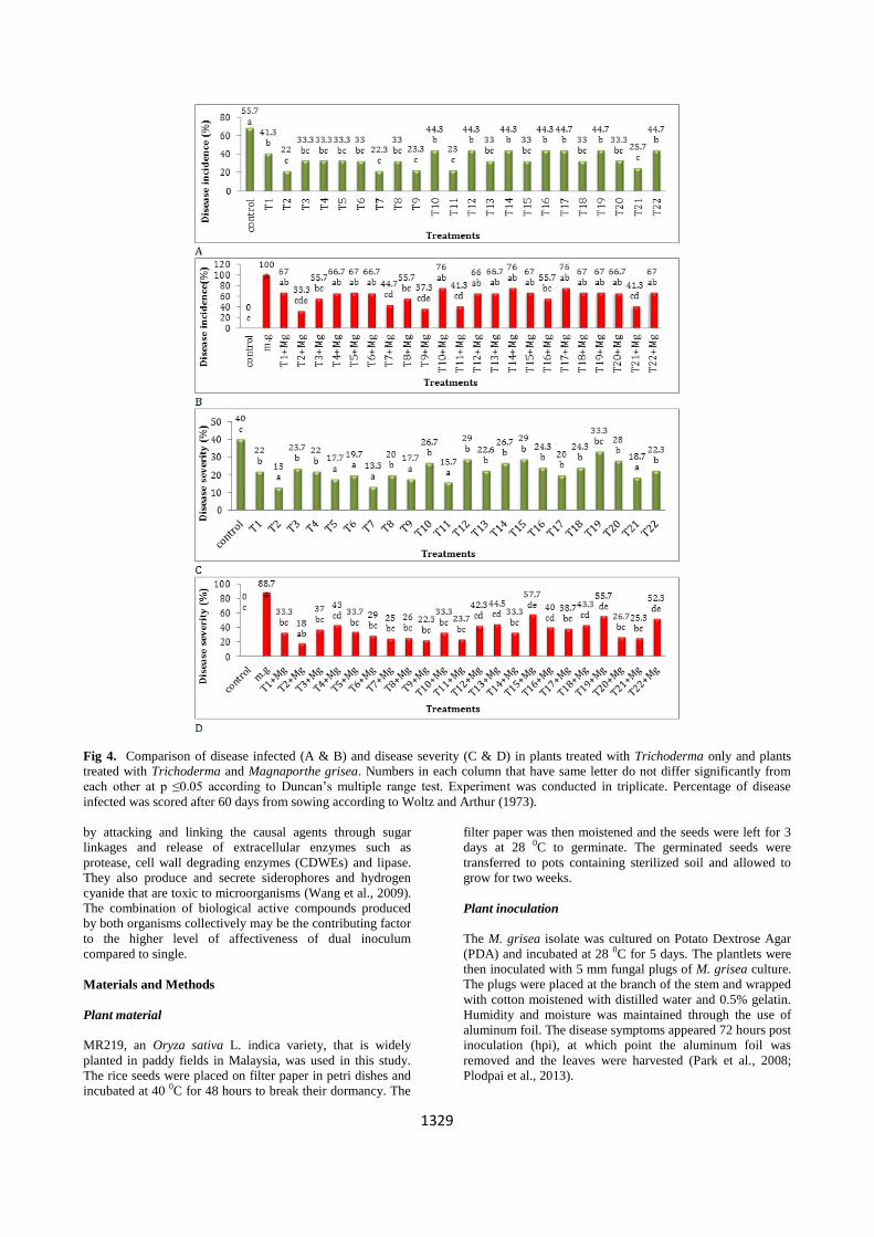
experimets. Trichoderma isolate T2 significantly reduced the
disease incidence parameter (p≤0.05) (33.3 %) compared to
treatment with pathogen only (100 %) (Fig. 4B). Majority of
the isolates showed significant reduction (18% to 57.7%) in
disease severity in comparison to control (88.7 %) (Fig. 4D).
Inoculation of Trichoderma isolates and B. substilis UKM1
showed that isolates T2, T3, T4, T5, T7, T8, T9 and T11
reduced pre emergence damping off (4.3 % for all of them)
while control untreated with Trichoderma isolates only

1326
scored 29.3 % (Fig. 5A). Post emergence of disease in seedlings was recorded as 4.3 to 12 % for the above isolates
Table 1. Antagonistic activity between Trichoderma spp. and Magnaporthe grisea under laboratory condition.
Treatments **Degree of antagonism after 4 days
T2+ Magnaporthe grisea +++
T3+ Magnaporthe grisea +++
T4+ Magnaporthe grisea +++
T6+ Magnaporthe grisea +++
T7+ Magnaporthe grisea +++
T9+ Magnaporthe grisea +++
T11+ Magnaporthe grisea +++
T18+ Magnaporthe grisea +++
T22+ Magnaporthe grisea +++
T8+ Magnaporthe grisea ++
T20+ Magnaporthe grisea ++
T14+ Magnaporthe grisea ++
T17+ Magnaporthe grisea ++
T5+ Magnaporthe grisea ++
T13+ Magnaporthe grisea ++
T21+ Magnaporthe grisea ++
T16+ Magnaporthe grisea ++
T1+ Magnaporthe grisea ++
T12+ Magnaporthe grisea +
T19+ Magnaporthe grisea +
T10+ Magnaporthe grisea +
T15+ Magnaporthe grisea -
*Mean of three plates (9 cm diameter) were used as replicates for each treatment
**According to scale by Alfredo and Aleli (2011) that involve four degrees:
(+++ ) The antagonistic fungus was able to grow over the pathogen and pathogen growth completely inhibited. (++) The pathogen growth completely inhibited, but
antagonist was not able to grow over the pathogen. (+) Mutual inhibition initially, but antagonist was overgrown by pathogen. Pathogen growth not inhibited, antagonist
was overgrown by pathogen.
Fig 1. Effect of different treatments on growth of pathogen.
as compared to control untreated with Trichoderma only
(Fig. 5C and D). In observing the disease infected plants,
isolates T2 and T7 showed the most significant reduction in
disease incidence i.e. 12 % for both in control (Fig. 6A) and
33 and 33.3 % respectively for both when M.grisea is present
(Fig. 6B). Fig. 6 shows that the percentage of disease severity
was also significantly reduced (p≤0.05) for isolates T2 and
T7 where the values for both were 4.3 % in control (Fig. 6C)
and 8.7 % (Fig. 6D) in pathogen infested soil.
From the experiments conducted and presented above it was
observed that the inhibition of M.grisea was higher in dual
inoculation. Therefore we selected the top six Trichoderma
spp (T2, T7, T8, T9, T11 and T21) that worked effectively
with B. substilis UKM1 to inhibit M. grisea in control
conditions for use in (non-autoclaved field soil). Figure 7
shows that the antagonistic effects of the six Trichoderma
isolates were higher when non autoclaved soil was employed
where isolates T2 and T11 had the most significant
differences (p≤0.05) in pre-post emergence parameters (4.3
% for all readings) (Fig. 7 A and B).
The disease incidence and severity data showed high
reduction in M. grisea infections (Fig. 7C and D).
Trichoderma T2 was the most effective antagonist in
reducing disease incidence and severity parameters (22 and
4.5 % respectively) (Fig. 7 C and D) compared to infections
in natural treatment (data not shown). Figure 8 shows MR219

1327
Fig 2. Antagonistic interaction between Bacillus subtilis UKM1 and M. grisea. (A) M. grisea only as control, the mycelium covers
the 9 cm plate over a 4-day period. (B) B. subtilis UKM1 and M. grisea, the arrows refer to the inhibition zones by B. subtilis UKM1.
The inhibition zone was scored 7 days from culture.
Fig 3. Comparison between pre (A & B) and post emergence (C & D) damping off in rice seedlings treated with 22 Trichoderma
isolates, and Trichoderma spp. in combination with Magnaporthe grisea under greenhouse condition. Numbers in each column that
have same alphabet do not differ significantly from each other at p ≤0.05 according to Duncan’s multiple range tests. Pre and post-
emergence test were conducted in triplicate, according to Ziedan (1998).

1328
rice plants treated with the top six Trichoderma spp in
combination with B. substilis UKM1. The plants exhibit
good growth with rich green foliage (Fig. 8A and B). Figure
8C shows the MR219 plantlet artificially inoculated with M.
grisea in the presence of the dual inoculum of biocontrol
agents. The plants survived to seed setting stage with
minimal disease symptoms. This can be seen clearly by
comparing with the untreated plants which showed infected
sheaths, stunted growth and no or lack of seed setting at the
same age (Fig. 8D and E).
Observation of reaction between Trichoderma T2 and M.
grisea under SEM
A previous study conducted between T. harzianum and R.
solani reported that mycoparasitic mechanism in early stages
could not be observed because the interaction starts soon
after penetration of hyphal cell walls by Trichoderma spp.
Therefore time factor is crucial in observing the process
under a microscope. The mechanism of mycoparasitism is
not very clear between Trichoderma T2 and hyphae of M.
grisea (Elad et al., 1983). Figure 9B shows the interaction
between the mycelium of Trichoderma isolate T2 and M.
grisea using the scanning electron microscope (SEM). The
electron-micrographs show the hyphal coiling of M. grisea
by Trichoderma T2. Hyphal coiling has been reported by
several researchers as the mode of action of Trichoderma on
pathogenic fungi (Van Eck, 1978; Mondal and Hyakumachi,
1998). Chitinolytic assays conducted on isolate T2 showed
high endochitinase activity and this enzyme is reported as a
CWDE that can break down cell walls of organisms
(Kalaivani et al., 2014). We believe the pathogen may have
been degraded by this enzyme as we were not able to
reisolate M. grisea from the dual culture plates (Fig. 9).
Discussion
In recent years the use of bioformulations in crop protection
has gained great interest as a safe and effective solution to the
chemical alternative (Suryadi et al., 2013). This study has
looked into the efficiency of single and dual bioinoculums in
controlling rice blast disease at the greenhouse level.
Antagonistic effect of Trichoderma isolate T1 to T22 in
dual culture plates
Through the dual culture technique we observed that all
twenty two isolates had varying levels of inhibition on
pathogen growth. The rate of inhibition was calculated as
percentage of overgrowth of Trichoderma in petri dish.
Trichoderma isolates T2, T7, T8, T9, T11 and T21 showed
the highest degree of inhibition (+++). As observed in the
dual culture assays, the antagonist out grew the pathogen in
the petri dish within four days (Fig. 1). Fuji et al. (1978) and
Vinale et al. (2008) reported that Trichoderma spp. produced
secondary metabolites such as antibiotics (6-pentyl-alpha-
pyrone (6pp), isocyanide derivatives), acids (heptelidic and
koningic acid), peptaibols and cell wall degrading enzymes
(CDWE) that are implicated in inhibits of radial growth of
many phytopathogenic fungi (Verma et al., 2007).
Antagonistic effect of Bacillus substilis UKM1 against M.
grisea
Bacillus is a genus of Gram positive bacteria that produces
endospores and is a potential biological control agent due to
its resistance to heat and drought conditions (Wayne et al.,
2000). B. substilis UKM1 while presenting some inhibitory
effect (16%) on the growth of the phytopathogen (Fig. 2), it
was not as efficient as the Trichoderma isolates (T2, T7,T8,
T9, T11 and T21 – produced 80-100% inhibition) in reducing
radial growth on its own. Therefore B. substilis UKM1 is not
an effective biocontrol agent against M. grisea. These values
are low compared to other B. substilis biocontrol isolates
such as B. subtilis NSRS 89-24 which resulted in
approximately 60 % inhibition of P. grisea in dual culture
test (Harman, 2000; Leelasuphakul et al., 2006). It was also
reported that B. subtilis produced > 50% inhibition of radial
growth of pathogens where this inhibition was
experimentally attributed to the release of several antifungal
metabolites such as subtilin, bacitracin, bacillin and
bacillomycin (Moubarak and Abdel-Monaim 2011;
Soleimani et al., 2005; Zaghloul et al., 2007).
Hyphal interaction between Trichoderma T2 and M. grisea
Trichoderma T2 was grown on a dual culture plates and
harvested for SEM analysis when both cultures were in
contact. Macroscopic observation of the fungal growth in
dual cultures revealed that the pathogens growth was
inhibited soon after contact with the antagonist. As observed
by the SEM analysis, Trichoderma was found growing and
coiling around the hyphal structure of M. grisea. Based on
microscopic, dual culture assays and biochemical analysis
(chitinolytic activity) (Kalaivani et al., 2014) we believe that
the mechanism by which Trichoderma antagonizes the
pathogen is by mycoparasitism which involves the
production of enzymes and secondary metabolites. Specific
compounds such as chitinolytic enzymes are able to degrade
the pathogens cell walls and inhibit the production and
release of active compounds by the pathogens. This was
further substanciated by the inabiliy to reisolate the pathogen
from the dual culture plates 4-days post incubation.
Greenhouse experiments for single and dual biocontrol
applications
From the greenhouse analysis conducted using the 22
Trichoderma isolates, six isolates showed improved pre and
post emergengence in rice when used alone and in
combination with the pathogen (Fig 3). This therefore
indicates that Trichoderma is a good candidate at increasing
growth though this would require further study growth, yield
and developmental studies. Similarly when scored for disease
incidence and disease severity the same isolates showed the
most promise in reducing loss of plants due to disease (Fig.
4). Figures 5 and 6 showed the effect of dual inoculation on
pre-post emergence, disease incidence and disease severity.
The reduction in all four parameters scored in dual
incoculation indicates that it is a more efficent way to control
M. grisea. The antagonistic effect of the 22 Trichoderma
isolates against M. grisea, individually or in combination
with B. subtilis UKM1 were more evident in uncontrolled
soil conditions. The reason for this could be that Trichoderma
is a saprophytic fungus, and the non-autoclaved soil
contained a rich source of several microorganisms from
which it may obtain food and other chemical exudates to
increase its growth, sustainance and effectiveness. Therefore
the results show that the six Trichoderma isolates can be used
under field condition successfully when a suitable
formulation has been obtained for field use against
pathogens. Trichoderma and Bacillus effect plant pathogens
1329
Fig 4. Comparison of disease infected (A & B) and disease severity (C & D) in plants treated with Trichoderma only and plants
treated with Trichoderma and Magnaporthe grisea. Numbers in each column that have same letter do not differ significantly from
each other at p ≤0.05 according to Duncan’s multiple range test. Experiment was conducted in triplicate. Percentage of disease
infected was scored after 60 days from sowing according to Woltz and Arthur (1973).
by attacking and linking the causal agents through sugar
linkages and release of extracellular enzymes such as
protease, cell wall degrading enzymes (CDWEs) and lipase.
They also produce and secrete siderophores and hydrogen
cyanide that are toxic to microorganisms (Wang et al., 2009).
The combination of biological active compounds produced
by both organisms collectively may be the contributing factor
to the higher level of affectiveness of dual inoculum
compared to single.
Materials and Methods
Plant material
MR219, an Oryza sativa L. indica variety, that is widely
planted in paddy fields in Malaysia, was used in this study.
The rice seeds were placed on filter paper in petri dishes and
incubated at 40 0C for 48 hours to break their dormancy. The
filter paper was then moistened and the seeds were left for 3
days at 28 0C to germinate. The germinated seeds were
transferred to pots containing sterilized soil and allowed to
grow for two weeks.
Plant inoculation
The M. grisea isolate was cultured on Potato Dextrose Agar
(PDA) and incubated at 28 0C for 5 days. The plantlets were
then inoculated with 5 mm fungal plugs of M. grisea culture.
The plugs were placed at the branch of the stem and wrapped
with cotton moistened with distilled water and 0.5% gelatin.
Humidity and moisture was maintained through the use of
aluminum foil. The disease symptoms appeared 72 hours post
inoculation (hpi), at which point the aluminum foil was
removed and the leaves were harvested (Park et al., 2008;
Plodpai et al., 2013).

1330
Fig 5. Comparison between rice plants treated with 22 Trichoderma isolates, B. subtilis UKM1 with or without Magnaporthe grisea
under greenhouse condition. Pre (A & B) and post emergence (C & D) damping off in seedlings. Numbers in each column that have
same letters do not differ significantly from each other at p ≤0.05 according to Duncan’s multiple range tests. Pre and post-
emergence experiments were conducted in triplicate for each isolate according to Ziedan (1998).
Isolation of Trichoderma spp.
A serial dilution was conducted on soil sample obtained from
the National Forest Reserve, Malaysia (Hamdia and
Kalaivani, 2013). Fungal cultures obtained were maintained
on PDA. Pure cultures of fungal isolates was examined
macro and microscopically to determine isolates that were
Trichoderma spp. Macro and microscopic examination of
cultures showed that there were 22 different isolates of
Trichoderma obtained from the soil sample and these were
designated isolate Trichoderma T1 to T22.
Antagonistic activity between Trichoderma isolates and M.
grisea
Antagonistics studies were conducted using dual culture
technique. Each PDA plate was divided equally into two
portions where in one portion a 5 mm M. grisea fungal plug
was placed while the second half was inoculated with 5mm
fungal plugs of any one of the T1 to T22 isolates (Hamdia
and Kalaivani, 2013). The plates were incubated at 280C for 4
days and the antagonistic activity was scored according to the
scale developed by Alfredo and Aleli, (2011).

1331
Fig 6. Comparison between rice plants treated with 22 Trichoderma isolates and B. subtilis UKM1 in combination with or without
Magnaporthe grisea under greenhouse condition. (A & B) Disease infected and(C & D) disease severity. Numbers in each column
that have same letters do not differ significantly from each other at p ≤0.05 according to Duncan’s multiple range tests. Three
replicates for each isolate. Percentage of disease infected was scored after 60 days from sowing according to Woltz and Arthur
(1973).
Preparation of Trichoderma isolate for observation under
scanning electron microscope (SEM)
Fresh cultures from interaction area between Trichoderma T2
and M. grisea was used for observation under the scanning
electron microscope. Small specimens were cut by hand with
a sharp razor and sliced to 1cm2 sections. The samples were
put into separate vials and fixed with 4% glutaraldehyde for
12-24 hours at 4 0C. The samples were washed three times
with phosphate buffer (PBS) and then dehydrated in an
alcohol series of 50%, 70%, 80%, 85%, 90%, 95%, and three
changes of 100% alcohol followed by three changes of 100%
acetone for 30 min each. The specimen was than stuck to a
stub coated with gold or colloidal silver via a sputter coater.
The coated specimens were viewed under the scanning
electron microscope (SEM - XL 30, Philips) operated at 10 to
15 Kv at various magnifications to obtain the best images.

1332
Fig 7. Comparison between rice plants treated with six Trichoderma isolates with or without Magnaporthe grisea under greenhouse
condition (A) pre and (B) post emergence damping off, (C) disease infected and (D) disease severity. Numbers in each column that
have same letter do not differ significantly from each other at p ≤0.05 according to Duncan’s multiple range test. pre and post
emergence damping off was determined according to Ziedan (1998). Percentage of disease infected and severity were scored after 60
days from sowing according to Woltz and Arthur (1973).
Magnifications of 500X to 10,000X were used for spores and
mycelium morphology studies.
Greenhouse experiments
As for the greenhouse experiment, the following
experimental design was establihed. Experiments was
conducted as follows: Control without microbes, M. grisea
only, B. subtilis UKM1 only, 22 Trichoderma isolates only
(tested individually), B. subtilis UKM1 + 22 Trichoderma
isolates (tested individually), M.grisea + 22 Trichoderma
isolates (tested individually), M. grisea + B. subtilis UKM1
+ 22 Trichoderma isolates (tested individually). The
concentration of Trichoderma isolates and M. grisea was
determined via Haemocytometer (Duncan, 1995; Goswani
and Kistler, 2004). The M. grisea inoculum was set at 1x10⁷
spore/mL while Trichoderma spp inoculum was set at 1x10⁸
spore /mL. B. subtilis UKM1 inoculum size was set at 2x10⁸
cell /mL (Harman and Kubicek, 1998). The soil was
moistened and mixed thoroughly every other day for 1 week.
Seedlings were then sown into the infested pots (8
seedlings/pot). Seedlings were visually examined for any

1333
Fig 8. Effect of dual inoculation against M. grisea. (a) Plants inoculated individually with six Trichoderma spp. + B. subtilis UKM1
+ M. grisea. (1) M. grisea only, and showing initial symptoms (2) Control natural infection (3) Bacillus subtilis UKM1 only (4) B.
substilis UKM1 and Trichoderma T2 (5) Isolate T2 (6) Isolate T7 (7) Isolate T8 (8) Isolate T9 (9) Isolate T11, and (10) Isolate T21.
(A) 6 Trichoderma isolates + M. grisea. (B) 6 Trichoderma isolates + B. substilis UKM1 inoculation pre M. grisea inoculation (C) 6
Trichoderma isolates + B. subtilis UKM1 + M. grisea at seed setting stage in the end of growth (D) Control, M. grisea only. (E)
Control, untreated natural infection (field soil). The arrows in (D & E) refer to diseased tissue.
Fig 9. (A) Trichoderma isolate T2 on PDA plate (A1) Branches, hyphae, chlamydospores and conidia in more than one point
(polyplastic). (A2) The arrow refers to the ornamented walls (verricose) of spores that accumulate in balls, and sporophore that carry
many flask-shaped monophialidic. (B) Dual culture plate showing interaction between Trichoderma T2 and M. grisea. T2 covers the
growth of M. grisea > 90 %, right side of the plate has Trichoderma, and left for M. grisea. (B1 & B2) SEM of tip region of
Trichoderma isolate T2 and M. grisea, branches of T2 completely associated and coiling around hyphae of M. grisea.
1334
signs of infection seven days post transplantation. The Pre-
and Post-emergence damping off data was collected two
weeks post transplantation. The data for pre-emergence and
post-emergence damping off was calculated using the
following formula:
Disease infected and severity were determined 60 days from
planting according to the following formula:
}
Disease severity was assessed 60 days from planting in
greenhouse via the 0 – 5 scale developed by (Woltz and
Arthur 1973). Where a score of 0 = healthy plants, 1 =
yellowing characteristic, 2 = wilting of one third leaves,
3=wilting of two third leaves, 4=whole plant wilted, 5= plant
is dead. The percentage data obtained from observations and
calculated using the formulae above were statistically
evaluated using a randomized complete block design via the
Analysis of Variance (Three Way ANOVA) .Three
replications were used for each parameter calculated. The
ANOVA analysis was conducted via the Statistical Analysis
System (SAS) versi 8.0.
Conclusion
This study shows that the Trichoderma isolates obtained from
our screening were effective in inhibiting M. grisea. Isolate
T2 was the most effective and produced higher levels of
inhibition of disease incidence and severity when used as
dual inoculum with B. substilis UKM1. Overall the best
results were with T2 and B. substilis UKM1 in uncontrolled
environment.
Acknowledgements
We would like to thank the Ministry of Agriculture, Malaysia
for the eScienceFund awarded (UKM 05-01-02 SF1013) to
conduct this research. In addition the Ministry of Education
Malaysia has provided some assistance to this research
through the awarded grant LRGS/TD/2011/UPM-
UKM/KM/01.
References
Abeysingne S (2007) Biological control of Fusarium solani f. sp.
phaseoli the causal agent of root rot of bean using Bacillus
subtilis CA32 and Trichoderma harzianum RU01. Ruhuna J.
Sci., 2: 82-88.
Alfredo MS, and Aleli Cornelia RP (2011) Biological control of
sheath blight of upland rice with Trichoderma species. J Trop.
Plant Pathol. 69: 1-9.
Altomare C, Norvell WA, Bjbrkman T, and Harman GE (1999)
Solubilization of phosphates and micronutrients by the
plantgrowth promoting and biocontrol fungus Trichoderma
harzianum Rifai 1295-22. Appl Env Microbiol. 65 : 2926-
2933.
Andrei SS, Roberto DNS, Alexandre SGC, Tatsuya N, Eliane
FN, and Cirano JU (2012) Trichoderma harzianum expressed
sequence tags for identification of genes with putative roles in
mycoparasitism against Fusarium solani. Biol Control. 61:
134-140.
Baha AA (2002) Enzyme activities Trichoderma harzianum in
soil and Tomato yield growth. Ph.D Thesis, Faculty
Agriculture, Baghdad University.
Baker B, Zambryski P, Staskawicz B and Dinesh-Kumar SP
(1997) Signaling in plant- microbe interactions. Science. 276:
726-733.
Suarez MB, Sanz L, Luis S, Isabel Chamorro M, Rey M,
Gonzalez FJ, Llobell A, Monte E (2005) Proteomic analysis of
secreted proteins from Trichoderma harzianum: Identification
of a fungal cell wall-induced aspartic protease. Fungal Gen.
Biol. 42: 924-934.
Bell DK, Wells HD and Markham CR (1982) In vitro
antagonism of Trichoderma species against six fungal plant
pathogens. Phytopathol. 72 : 379-382.
BPS (2010) Informasi data luas panen, produksi tanaman padi
seluruh provinsi. Jakarta : Badan Pusat Statistik.
BPTP (2009) Penyakit blas. Balai Besar Penelitian Tanaman
Padi, Sukamadi, Subang Jawa Barat.
Cardona R and Rodriguez H (2006) Effect of Trichoderma
harzianum fungus on the incidence of the charcoal rot disease
on sesame. Rev Facultad Agron. LUZ, 23: 42-47.
Chin KM (1975) Fungicidal control of the rice blast disease.
Mardi Research Bulletin. 2(2): 82-84. Correa-Victoria FJ, Tharreau D, Martinez C, Vales M, Escobar
F, Prado G, et al. (2004) Studies on the rice blast pathogen,
resistance genes, and implications for breeding for durable
blast resistance in Colombia. In: Kawasaki S, editor. Rice
blast: interaction with rice and control. Proceedings of the third
international rice blast conference. Dordrecht, the Netherlands.
Kluwer Academic Publishers. pp. 214-27.
Elad Y, Chet I, Boyle P, and Henis Y (1983) Parasitism of
Trichoderma spp. on Rhizoctonia solani and S clerotium rolfsii
- Scanning Electron Microscopy and Fluorescence
Microscopy. Phytopathol. 73: 85-88.
Faria JCD, Prabhu AS, and Zimmermann FJP (1982) Effect of
Nitrogen fertilization and fungicidal sprays on blast and yield
of upland rice. Pesq. Agropec. Bras. 17(6): 847-852.
Fravel DR, (1988) Role of Antibiosis in the Biocontrol of Plant
Diseases. Ann Rev Phytopathol. 26: 75-91.
Fuji K, Fujita E, Takaishi Y, Fujita T, Arita I, Komatsu M and
Hiratsuka N (1978) New antibiotics, trichopolyns A and B:
Isolation and biological activity. Experientia. 34: 237-239.
Ghazanfar MU, Wakil W, Sahi ST and Saleem-il-Yasin (2009)
Influence of various fungicides on the management of rice
blast disease. Mycopathol. 7(1): 29-34.
Hafedh HZA, Aboud HM, Fattah FA and Khlaywi SA (2005)
Evaluation the antagonistic efficiency of thirty-four isolates of
Trichoderma spp. against Macrophomina phaseolina under
laboratory and greenhouse conditions. Arab J Plant Protect.
23(1): 44-50.
Hafedh HZA, Aboud HM, Musa NK, Gasam FH and Abid SHM
(2006) The effect of pH on growth and sporulation of
Trichoderma spp. Proceedings of the 9th Arab Congress of
Plant Protection, November 19-23, Damascus. Syria. pp. 218-
218.
Hamdia ZA, Kalaivani N (2013) Evaluating the efficacy of
Trichoderma and Bacillus isolates as biological control agents
against Rhizoctonia solani. Res J Appl Sci. 8(1):72-81.
1335
Harman GE, Hayes CK, Lorito M, Broadway RM, Di pietro A,
Peterbaur C and Tronsmo A (1993) Chitinolytic enzymes of
Trichoderma harzianum: purification of chitobiosidase and
endochitinase. Phytopathol. 83: 313-318.
Harman GE and Kubicek CP (1998) Trichoderma and
Gliocladium enzymes, biological control and commercial
applications. Taylor and Francis. London Vol. 2. p. 393.
Harman GE (2000) Myths and dogmas of biocontrol. Plant Dis.
84(4): 377-393.
Harman GE, Howell CR, Vitarbo A, Chet I, Lorito M (2004)
Trichoderma species eopportunistic, avirulent plant symbionts.
Nat Rev Microbiol. 2: 43-56.
IRRI (2010) Rice blast. International Rice Research Institute.
www.knowledgebank.irri.org/factsheetsPDFs/.RiceFactSheets.
Mar 2010.
Kalaivani N, Hamdia ZA and Nurfarahana SO (2014) The
isolation and characterization of an endochitinase gene from a
Malaysian isolate of Trichoderma sp. Aust J Crop Sci. 8(5):
711-721.
Kato H (2001) Rice blast disease. Pesticide Outlook February
2001. pp. 23-25.
Kim SY and Kim DK (2009) Evidence of a potential adaptation
of Magnaporthe oryzae for increased phosphorothiolate-
fungicide resistance on rice. Crop Prot. 28: 940-946.
Kumar KK, Maruthasalam S, Loganthan M, Sudhakar D and
Balasubramaniam P (2005) An improved Agrobacterium-
mediated transformation protocol for recalcitrant elite indica
rice cultivars. Plant Mol Biol Rep. 23: 6-73.
Leelasuphakul W, Pranom S and Souwalak PH (2006)
Purification, characterization and synergistic activity of 1,3-
glucanase and antibiotic extract from an antagonistic Bacillus
subtilis NSRS 89-24 against rice blast and sheath blight.
Enzyme Microbial Technol. 38: 990-997.
Lewis JA and Papavizas GC (1987) Reduction of inoculum of
Rhizoctonia solani in soil by germlings of Trichoderma
hamatum. Plant Pathol. 36: 438-446.
Lixuan R, Shiming S, Xingming Y, Yangchun X, and Qiwe QS
(2008) Intercropping with aerobic rice suppressed Fusarium
wilt in watermelon. Soil Biol Biochem. 40: 834-844.
Meraj-ul H and Nandkar PB (2012) Antagonistic effect of
rhizospheric Trichoderma isolates against tomato damping-off
pathogen, Fusarium oxysporum f.sp. lycopersici. Intl J Res
BioSci. 1(2): 27-3.
Mojica-Marin V, Luna-Olvera HA, Sandoval-Corondo CF,
Pereyra-Alferez P, Morales-Ramos LH, Hernandez-Luna CE
and Alvarado-Gomez, OG (2008) Antagonistic activity of
selected strains of Bacillus thuringiensis against Rhizoctonia
solani of chili pepper. Afr J Biotech. 7(9): 127-1276.
Mondal SN and Hyakumachi MP (1998) Carbon Loss and
Germinability, Viability, and Virulence of Chlamydospores of
Fusarium solani f. sp. phaseoli After Exposure to Soil at
Different pH Levels, Temperatures, and Matric Potentials.
Ecol Popul Biol. 1217-02R.
Moubarak MY and Abdel-Monaim MF (2011) Effect of bio-
control agents on yield, yield components and root rot control
in two wheat cultivars at New Valley region. Egypt J Cereals
Oilseeds. 2(6): 77-87.
Ou S (1985) Rice diseases. 2nd ed. Kew. Surrey: Commonwealth
Mycological Institute.
Park DS, Sayler RJ, Hong YG, Nam MH and Yang Y (2008) A
method for inoculation and evaluation of rice sheath blight
disease. Plant Dis. 92: 25-29.
Plodpai P, Chuenchitt S, Petcharat V, Chakthong S, Piyawan S
and Voravuthikunchai SS (2013) Anti-Rhizoctonia solani
activity by Desmos chinensis extracts and its mechanism of
action. Crop Prot. 43: 65-71.
Scardaci SC (2003) A new diseases in California.University of
California. Davis: Agronomy Fact Sheet Series 1997-2.
Retrieved 2010-10-20.
Schuster A and Schmoll M (2010) Biology and biotechnology of
Trichoderma. Appl Microbiol Biotech. 87: 787-799.
Shimoi S, Inoue K, Kitagawa H, Yamasaki M, Tsushima S, Park
P and Iked K (2010) Biological control for rice blast disease
by employing detachment action with gelatinolytic bacteria.
Biol Control. 55: 85-91.
Soleimani MJ, Shamsbakhsh M, Taghavi M and Kazemi SH
(2005) Biological control of stem and root rot of wheat caused
by Bipolaris spp. by using antagonistic bacteria, fluorescent
Pseudomonas and Bacillus sp. J Biol Sci. 5(3): 347-353.
Sundaramoorthy S, Raguchander T, Ragupathi N and
Samiyappan R (2012) Combinatorial effect of endophytic and
plant growth promoting rhizobacteria against wilt disease of
Capsicum annum L. caused by Fusarium solani. J Biol
Control. 60: 59-67.
Suprapta DN and Khalimi K (2012) Pengembangan agen hayati
untuk mengendalikan penyakit blas, memacu pertumbuhan dan
meningkatkan hasil tanaman padi. Laporan Penelitian Riset
Invensi Udayana. Universitas Udayana, Denpasar. pp. 42
Suryadi Y, Susilowati DN, Riana E and Mubarik NR (2013)
Management of rice blast disease (Pyricularia oryzae) using
formulated bacterial consortium. Emir J Food Agric. 25(5):
349-357.
Tamimi KM and Hadwan HA (1985) Biological effect of
Neurospora sitophila and Trichoderma harzianum on the
growth of a range of sesamum wilt causing fungi in vitro. Ind
Phytopathol. 38(2): 292-196.
Tilak KVBR, Ranganayaki N, Pal KK, De R, Saxena AK,
Nautiyal CS, Mittal S, Tripathi AK and Tripathi and Johri BN
(2005) Diversity of plant growth and soil health supporting
bacteria. Curr Sci. 89(1): 136-150.
Van Eck WH 1978 Autolysis of chlamydospores of Fusarium
solani f. sp. cucurbitaein chitin and laminarin amended soils.
Soil Biol Biochem. 10(2): 89-93.
Verma M, Satinder KB, Tyagi RD, Surampalli RY and Valero
JR (2007) Antagonistic fungi, Trichoderma spp. Biochem
Engin J. 37: 1-20.
Vinale F, Sivasithamparam K, Ghisalberti E, Marra R, Woo S
and Lorito M (2008) Trichoderma-plant-pathogen interactions.
Soil Biol Biochem. 40(1): 1-10.
Wang H, Wen K, Zhao X, Wang X, Li A and Hong H (2009)
The inhibitory activity of endophytic Bacillus sp. strain CHM1
against plant pathogenic fungi and its plant growth-promoting
effect. Crop Prot. 28: 634-639.
Woltz SS and Arthur WE (1973) Fusarium wilt of
chrysanthemum, effect of nitrogen source and lime on disease
development. Phytopathol. 63(1): 155-157.
Yang D, Wang B, Wang J, Chen Y and Mingguo Z (2009)
Activity and efficacy Bacillus subtilis strain NJ-18 against
rice sheath blight and sclerotinia stem rot of rape. J Biol
Control. 51: 61-65.
Zaghloul RA, Hanafy EA, Neweigy NA and Khalifa NA (2007)
Application of biofertilization and biological control for
tomato production. 12th Conference of Microbiology; Cairo,
Egypt. pp. 198-212.
Ziedan EHE (1998) Integrated control of wilt and root rot
disease. Ph.D. Thesis, Faculty of Agriculture, Ain Shams
University pp.169.
Ziedan EHE and Elewa IS (2000) Treatment of sesame
transplants with Trichoderma spp. and chitosan as control
measures against wilt disease Fusarium oxysporum f. sp.
sesami. The Ninth Congress of Phytopathology, Giza, Egypt 8-
10 May.