eukaryotic cells and their cell bodies: cell - annals of botany
TRANSCRIPT

doi:10.1093/aob/mch109, available online at www.aob.oupjournals.org
INVITED REVIEW
Eukaryotic Cells and their Cell Bodies: Cell Theory Revised
FRANTISÏ EK BALUSÏ KA1 , 2* , DIETER VOLKMANN1 and PETER W. BARLOW 3 , ²
1Institute of Cellular and Molecular Botany, University of Bonn, Kirschallee 1, 53175 Bonn, Germany; 2Institute of
Botany, Slovak Academy of Sciences, DuÂbravska cesta 14, 842 23 Bratislava, Slovakia; 3School of Biological Sciences,
University of Bristol, Woodland Road, Bristol BS8 1UG, UK
Received: 9 January 2004 Returned for revision: 20 February 2004 Accepted: 2 March 2004 Published electronically: 20 May 2004
d Background Cell Theory, also known as cell doctrine, states that all eukaryotic organisms are composed ofcells, and that cells are the smallest independent units of life. This Cell Theory has been in¯uential in shapingthe biological sciences ever since, in 1838/1839, the botanist Matthias Schleiden and the zoologist TheodoreSchwann stated the principle that cells represent the elements from which all plant and animal tissues areconstructed. Some 20 years later, in a famous aphorism Omnis cellula e cellula, Rudolf Virchow annunciatedthat all cells arise only from pre-existing cells. General acceptance of Cell Theory was ®nally possible onlywhen the cellular nature of brain tissues was con®rmed at the end of the 20th century. Cell Theory then rapidlyturned into a more dogmatic cell doctrine, and in this form survives up to the present day. In its current version,however, the generalized Cell Theory developed for both animals and plants is unable to accommodate thesupracellular nature of higher plants, which is founded upon a super-symplasm of interconnected cells intowhich is woven apoplasm, symplasm and super-apoplasm. Furthermore, there are numerous examples ofmultinucleate coenocytes and syncytia found throughout the eukaryote superkingdom posing serious problemsfor the current version of Cell Theory.d Scope To cope with these problems, we here review data which conform to the original proposal of DanielMazia that the eukaryotic cell is composed of an elemental Cell Body whose structure is smaller than the celland which is endowed with all the basic attributes of a living entity. A complement to the Cell Body is the CellPeriphery Apparatus, which consists of the plasma membrane associated with other periphery structures.Importantly, boundary stuctures of the Cell Periphery Apparatus, although capable of some self-assembly, arelargely produced and maintained by Cell Body activities and can be produced from it de novo. These boundarystructures serve not only as mechanical support for the Cell Bodies but they also protect them from the hostileexternal environment and from inappropriate interactions with adjacent Cell Bodies within the organism.d Conclusions From the evolutionary perspective, Cell Bodies of eukaryotes are proposed to represent vestiges ofhypothetical, tubulin-based `guest' proto-cells. After penetrating the equally hypothetical actin-based `host'proto-cells, tubulin-based `guests' became specialized for transcribing, storing and partitioning DNA moleculesvia the organization of microtubules. The Cell Periphery Apparatus, on the other hand, represents vestiges ofthe actin-based `host' proto-cells which have become specialized for Cell Body protection, shape control, moti-lity and for actin-mediated signalling across the plasma membrane. ã 2004 Annals of Botany Company
Key words: Actin, Cell Body, Cell Periphery Apparatus, Cell Theory, coenocytes, cytoskeleton, nucleus, plasmamembrane, plasmodesmata, polarity, syncytia, tubulin.
MULTICELLULARITY VERSUSSUPRACELLULARITY
Supracellular plants do not ®t with the classical Cell Theory
`. . . something truly fundamental is missing in our image ofthe cell . . .' Daniel Mazia (1987)
The cell doctrine is ®rmly embedded in all biologicaldisciplines and acts as a general paradigm of organismal andtissue construction and function (Wolpert, 1995;Mazzarello, 1999; Nurse, 2000). Mainstream biologiststake this concept for granted and use it to underpinsophisticated reductionistic approaches by which to under-stand the molecular basis of cellular development (Pollard,
2003). However, those who are aware of the most recentadvances in plant cell biology (see also Rustom et al., 2004)are convinced that Cell Theory, as it now stands, isabsolutely incompatible with a cell-based organization ofhigher plants (Fig. 1) and requires an update (Box 1).Indeed, formulation of organismal theory of plant develop-ment, in which it is stated that it is not the cell but the wholemulticellular organism that is the primary unit of plant life(Kaplan, 1992; Sitte, 1992; Barlow, 1994; Korn, 1999;Niklas, 2000; Wojtaszek, 2001; Tsukaya, 2002), hasprecipitated a crisis for Cell Theory as applied to plants.Organismal theory is an idea whose formulation andreformulation occurs with each successive generation ofbiologists (e.g. Sinnott, 1960; and before him all the wayback to de Bary, 1864; see also Barlow, 1982). Furthermore,after a hundred years of discussion, the endosymbioticconcept of cell organization and evolution is now ®nallywidely accepted (Margulis, 1993; McFadden, 1999; Martin
* For correspondence. E-mail [email protected]² PWB dedicates his contribution to this paper to his friend and mentor,
Professor Paul E. Polani FRS, on the occasion of his 90th birthday,1 January 2004.
Annals of Botany 94/1, ã Annals of Botany Company 2004; all rights reserved
Annals of Botany 94: 9±32, 2004
Dow
nloaded from https://academ
ic.oup.com/aob/article/94/1/9/222826 by guest on 02 D
ecember 2021

et al., 2001; Gray et al., 2001; Cavalier-Smith, 2002a). Theimplication of this concept is that present-day eukaryoticcells represent assemblages of `cells within a cell'. Othereven more obvious examples of `cells within a cell' are thesperm cells of higher plants (Mogensen, 1992; Palevitz andTiezzi, 1992; Southworth, 1992), endosperm of higherplants (Olsen, 2001; Brown et al., 2004) and spores withinyeast mother cells (Knop and Strasser, 2000; Nickas et al.,2003; Shimoda, 2004). Interestingly in this respect, andrelevant to our further argumentation, is that sperm cells ofhigher plants do not contain any F-actin but do haveprominent microtubules (Palevitz and Tiezzi, 1992), sug-gesting that the actin cytoskeleton is neither essential foreukaryotic cellular life nor for cell divisions (Palevitz andTiezzi, 1992; for a similar conclusion on somatic plant cellssee BalusÏka et al., 2001c; Vantard and Blanchoin, 2002).Concerning the last-mentioned point, genetic and pharma-cological evidence convincingly document that it is themicrotubular cytoskeleton which is essential for celldivision and the formation of multicellular organisms (forplant cells see Mayer et al., 1999; Mayer and JuÈrgens,2002).
All these problems with Cell Theory were forecast byThomas Henry Huxley in 1853, who was convinced thatcells were not anatomically independent but that they wereinterconnected into supracellular assemblages (Richmond,2001). Therefore, for Huxley, cells could not be theelementary units of life. In fact, current advances in plantcell biology reveal that this view is correct for all higherplants (Fig. 1). Strictly speaking, higher plants aresupracellular organisms because almost all the cells of agiven plant organism are interconnected via cell-to-cellchannels known as plasmodesmata (Lucas et al., 1993;Zambryski and Crawford, 2000) that form primarily across
the division wall at cytokinesis, and secondarily acrossselected, already established walls (Ehlers and Kollmann,2001). Their mode of development attests to the necessity ofdirect cell±cell communication during plant development.These complex, communicative and contractile channels(Blackman et al., 1999; Zambryski and Crawford, 2000;BalusÏka et al., 2001b) are not only lined with the plasmamembrane but are also traversed by endoplasmic reticulum.This latter feature, together with the well-known continuitybetween endoplasmic reticulum elements and nuclearenvelopes, means that all nuclei of a given plant arepotentially in direct contact and are part of a structurallyintegrated supracellular network of nuclei interconnectedvia endoplasmic reticulum elements (Lucas et al., 1993). Itis not possible to interpret this phenomenon correctly usingcell doctrine as it stands now because this is based on thebelief that cells are physically separated and structurallyindependent. In fact, recent advances in animal cell biologyalso reveal that cells are also not isolated from each other insome situations (Rustom et al., 2004). We are, however, stillfar away from understanding how individual nuclei of asupracellular network of plant nuclei might communicatewith each other via the intervening cytoplasmic channels.
A consequence of the fact that the cytoplasms of plantcells are interconnected via plasmodesmata is that theindividuality of the cell is given up in favour of an integratedand corporate cytoplasm that bene®ts the whole organism.This supracellular, or organismal, approach towards multi-cellularity seems to have allowed sessile plants to adapt tolife on land and to evolve even within hostile environments.The continuity of cellular units allows potentially unre-stricted exchange of information throughout the plant body,the informational signals being used to rapidly coordinategenome transcription that can either neutralize or takeadvantage of environmental challenges (BalusÏka et al.,2004). Thus, whereas animals and humans are perhaps trulymulticellular organisms, higher plants are composed ofcommunicative cytoplasms.
The current crisis of the Cell Theory in plants (Kaplanand Hagemann, 1991; Kaplan, 1992; Korn, 1999;Wojtaszek, 2001) is quite paradoxical if we consider thatRobert Hooke in 1665 and Nehemiah Grew in 1682discovered cells from observations on higher plant tissues(Wolpert, 1995; Harris, 1999; Nurse, 2000). It took morethan 250 years until the Cell Theory was de®nitely acceptedfor animals and humans, neurons being the last type of cellto be de®nitely de®ned as such (Mazzarello, 1999). Plantsalso served as useful objects for the discovery of thenucleus, the plasma membrane, cell cycle and cytokinesis(Harris, 1999; see also Boxes 2±4). Thus, plants seemalways to have been at the forefront of Cell Theory, evennow when it needs updating in order to accommodate thesupracellular nature of higher plants. Numerous examples ofmultinucleate cells (Fig. 2) in almost all eukaryoticorganisms, direct cytoplasmic continuity in some animalcells (Rustom et al., 2004), as well as the ability to form theplasma membrane de novo (Shimoda, 2004)Ðall thesesuggest that the Cell Theory is in crisis elsewhere too, andthat it is not solely a plant-speci®c problem.
F I G . 1. The supracellular nature of higher plants is incompatible with thecurrent version of Cell Theory. Plant cells are not physically separated.Cytoplasms of `cells' are interconnected via plasmodesmata andendoplasmic reticulum into supracellular assemblies bounded by aplasma membrane. Enclosed within discrete cytoplasmic domains areunitary complexes of nucleus and perinuclear microtubules. Eachcomplex we term a Cell Body in accordance with Daniel Mazia'sconception of this structure. Cortical microtubules are not shown in this
highly simpli®ed scheme.
10 BalusÏka Ð Cell Theory Revised
Dow
nloaded from https://academ
ic.oup.com/aob/article/94/1/9/222826 by guest on 02 D
ecember 2021

Unique organization of microtubules and Golgi apparatus inmultinuclear syncytia±coenocytes of animals and lowerplants resembles situations in supracellular plants
There are several well-known examples where not onlyplant cells but also several animal cell types do not conformto the traditional view of cells as the smallest unit of life.Mention can be made of the many examples of multi-nucleate coenocytes and syncytia throughout the eukaryotickingdom (Fig. 2). Coenocytes are formed as a result of theuncoupling of mitosis from cytokinesis. Whereas mitosis isa conservative and persistent living process, cytokinesisappears to be less conservative, more sporadic, and can evenbe absent; this results in situations where numerous nucleicome to be present within the con®nes of a `mother' cell.Besides the already mentioned yeast spores (Shimoda,2004), good examples of coenocytic plants are the multi-nucleate algae (Woodcock, 1971; Goff and Coleman, 1987;McNaughton and Goff, 1990) and also the male and femalegametophyte tissues of higher plants (Brown and Lemmon,1992, 2001; McCormick, 1993; Reiser and Fischer, 1993;Russell, 1993; Brown et al., 1994a, b, 1996; Huang andSheridan, 1994, 1996; Smirnova and Bajer, 1998; Oteguiand Staehelin, 2000, 2003; Ranganath, 2003). In animals,well-studied examples of the coenocytic state are found inoogenesis and in the early embryogeny of Drosophila(St Johnson and NuÈsslein-Volhard, 1992; Foe et al., 2000;Mazumdar and Mazumdar, 2002). The simplest coenocytewould be a cell with two or four nuclei, as occurs in plants inthe anther tapetum and in the liver of many rodents(D'Amato, 1977). There are also several examples ofcoenocytes elicited by mutations that prevent cytokinesis(Sipiczki et al., 1993; Adam et al., 2000).
A syncytium, another multinucleate form, derives fromuninucleate cells that have fused together. Examples ofhomotypic cell fusion and hence of homokaryotic multi-nucleate syncytium formation in animal systems aremyotubes, which are essential for muscle differentiation,multinucleate osteoclasts, which are active in bone resorp-tion and homeostasis, and the syncytiotrophoblast, which ischaracteristic of the mammalian placenta (Cross et al.,
1994; Solari et al., 1995; Shemer and Podbilewicz, 2000,2003; Taylor, 2002). There are also examples of fusionsbetween different animal cell types: neurons and bonemarrow-derived stem cells can both form stable hetero-karyons (Kozorovitskiy and Gould, 2003; Weimann et al.,2003). Moreover, huge multinucleate syncytia can beinduced by viruses such as HIV and measles (Sylwesteret al., 1993; Cathomen et al., 1998). Intriguingly, animalsyncytia behave like single cells, mimicking their polarintegrity and showing pseudopod extensions and actin-based motility (Lewis and Albrecht-Buehler, 1987;Sylwester et al., 1993). In plants, syncytia are formed bymeans of the enlargement of plasmodesmata, dissolution ofthe original cell walls and consequent merging of neigh-bouring cytoplasmic domains (Fink, 1999). In some cases,syncytium formation is the normal mode of plant cellulardevelopment, like articulated laticifers (Mahlberg andSabharwal, 1966); in other cases, it is a response to achallenge from organisms that burrow into plant tissue andconvert it into the nutritive syncytial nurse cells of insectand nematode galls (Jones and Northcote, 1972).
A major hallmark of plant cells is that they organize theirmicrotubules from sites upon a nuclear surface (Lambert,1993; Mizuno, 1993; BalusÏka et al., 1996, 1997a; Schmit,2003). Often they also organize microtubules at the cellcortex from the secondary microtubule organizing centres(MTOCs) which have been derived from primary MTOCsthat lie on the nuclear surface (BalusÏka et al., 1997a). In thecase of those animal cells which embark upon coenocytic orsyncytial developmental pathways, the typical centrosome-based organization of their microtubules is abandoned andthe whole nuclear surface starts to organize microtubules, asis known from plant cells (Tassin et al., 1985a; Sylwesteret al., 1993; Lu et al., 2001; Mulari et al., 2003). In this way,the animal coenocyte or syncytium is similar to theindividual plant `cell', suggesting that this type of animal`cell', too, may be a supracellular continuum of many nucleiand cytoplasms.
The above suggestion can be followed using another lineof evidence involving the Golgi apparatus. For animal cells,it is well known that localization of the Golgi complex isdependent on microtubules while, at the same time, theGolgi complex acts as a microtubule-organizing organelle(Tassin et al., 1985b; Kronenbusch and Singer, 1987; Hoet al., 1989; Cole et al., 1996; Bloom and Goldstein, 1998;Burkhardt, 1998; Chabin-Brion et al., 2001). But in the caseof the animal cell syncytium, the Golgi apparatus undergoesa dramatic reorganization and acquires features that corres-pond to what is found in supracellular higher plants wherenumerous small Golgi stacks are closely associated withendoplasmic reticulum export sites (Boevink et al., 1998;Brandizzi et al., 2002). For instance, during myogenesis inanimals, similarly to cells devoid of microtubules (Coleet al., 1996), perinuclear Golgi apparatus re-arranges intonumerous small Golgi stacks that are closely associated withthe endoplasmic reticulum exit sites (Ralston, 1993; Luet al., 2001; Ralston et al., 2001). Golgi mini-stacks andmicrotubules organized around nuclei were also reported formaturing mouse oocytes (Moreno et al., 2002). Thus, theplant microtubular and Golgi apparatus organizations are
F I G . 2. Cell Bodies are obvious in multinucleate coenocytes andsyncytia, structures which have been reported in almost all majortaxonomic groups of eukaryotes. Importantly, perinuclear radiating arraysof Cell Body microtubules are critical for the regular spacing of nuclei
and Cell Bodies in the multinucleate cytoplasmic community.
BalusÏka Ð Cell Theory Revised 11
Dow
nloaded from https://academ
ic.oup.com/aob/article/94/1/9/222826 by guest on 02 D
ecember 2021

directly related to their supracellular nature in both plantsand animals.
Coenocytic and syncytial nuclei organize cytoplasmicdomains via radiating microtubules and they obey thecytonuclear rule
One characteristic feature of the majority of syncytia andcoenocytes is that their nuclei are regularly spaced withinthe cytoplasm (Goff and Coleman, 1987; McNaughton andGoff, 1990; Bresgen et al., 1994; Bruusgaard et al., 2003)and this is apparently due to the assembly of perinuclearradiating microtubules (Woodcock, 1971; Brown andLemmon, 1992, 2001; Brown et al., 1994a, b, 2004;Huang and Sheridan, 1994, 1996; Otegui and Staehelin,2000, 2003). Each individual nucleus of both syncytia andcoenocytes controls a cytoplasmic domain (Fig. 2), the sizeof which depends on the DNA content and volume of thatnucleus. These nucleo-cytoplasmic domains, despite lack-ing any obvious physical borders, behave like independentstructural entities (Goff and Coleman, 1987; McNaughtonand Goff, 1990; Brown and Lemmon, 1992, 2001; Brownet al., 1994a, b, 1996; Reinsch and GoÈnczy, 1998; Pickett-Heaps et al., 1999). Distinct nucleo-cytoplasmic domainsare organized also in animal syncytial myotubes (Hall andRalston, 1989; Bruusgaard et al., 2003), where the individ-ual nuclei even maintain their own transcription andtranslation domains (Rotundo and Gomez, 1990; Ralstonand Hall, 1992). Individual nuclei of multinucleate muscle®bres exert control also over distinct cell surface domains(Rossi and Rotundo, 1992). Thus, characteristic cytogeneticpatterns could theoretically be set up within a coenocyticstructure without the need for any de®ning cell membranesor wall boundaries, the cytoplasmic domains being patrolledby the microtubules radiating from the nuclear surface.
In plants, there are numerous studies showing thatradiating perinuclear microtubules are essential for theregular spacing of nuclei (Goff and Coleman, 1987;McNaughton and Goff, 1990; Brown and Lemmon, 1992,2001; Brown et al., 1994a, b, 1996, 2004; BalusÏka et al.,1996, 1997a, b, 1998; Pickett-Heaps et al., 1999). Animportant feature is that the whole nuclear surface is activein the initiation and maintenance of minus-ends ofmicrotubules, while dynamic plus-ends exert pushing/pulling forces when contacting the cell boundary, or whenapproaching plus-ends of microtubules radiating from otheradjacent nuclei. This phenomenon allows each nucleus toactively conquer and maintain its own unique cytoplasmicspace which does not encroach upon the spaces controlledby neighbouring nuclei (Strasburger, 1893; Hertwig, 1903;Trombetta, 1939; Pickett-Heaps et al., 1999; Gregory,2001a, b).
The nuclear spacing is often in the form of regularhexagonal arrays, this feature being indicative of theisomorphic space-claiming force of individual nuclei-MTcomplexes. Interestingly, correct patterning and polarity areexpressed throughout animal syncytia and plant coenocytes(St Johnston and NuÈsslein-Volhard, 1992; Boisnard-Loriget al., 2001; Sùrensen et al., 2002; Brown et al., 2004). Thisis perhaps an expression of precisely regulated `cell-like'
domains of varying strength, each maintained by preciselyregulated activities of perinuclear radiating microtubules(Goff and Coleman, 1987; McNaughton and Goff, 1990;Brown and Lemmon, 1992, 2001; Bresgen et al., 1994;Brown et al., 1994a, b, 1996; BalusÏka et al., 1996; Pickett-Heaps et al., 1999; Bruusgaard et al., 2003).
THE CELL BODY CONCEPT
Cell Body represents the smallest autonomous andself-reproducing unit of eukaryotic life
`The Cell Body pervades the whole interphase cell andcondenses into a mitotic apparatus during mitosis' DanielMazia (1993)
The supracellular nature of higher plants, as well as ofcoenocytes and syncytia found in almost all eukaryotes,implies that it is not the cell but some subcellular structurewhich represents the elementary unit of eukaryotic life. Infact, such ideas have often been expressed in the past. Thecytoskeleton was unknown in these early times, and so theseideas were doomed to be forgotten (Harris, 1999). Butalready the very early studies on plant microtubulesrevealed that these structures controlled the spatial distri-bution of chromosomes during mitosis (Ledbetter andPorter, 1963) and of whole nuclei during interphase(Kiermayer, 1968; Woodcock, 1971). These features werealso con®rmed for animal cells (Slautterback, 1963;Aronson, 1971). However, the close connections betweenDNA and tubulin molecules throughout the cell cycle aswell as in postmitotic eukaryotic cells became obvious onlylater (see Box 4), providing a completely new perspectiveupon what came to be known as the cytoskeleton.
Daniel Mazia was the ®rst to realise that a closeconnection between DNA and tubulin molecules wouldhave an immediate impact upon Cell Theory. He was alsothe ®rst to suggest that the nucleus with its associatedmicrotubules formed a composite structure which he calledCell Body (Mazia, 1993; Epel and Schatten, 1998).Although this concept was left almost unnoticed, werevealed that it is obviously also valid for plant cells(BalusÏka et al., 1997a, 1998). Importantly, Cell Bodyrepresents the smallest unit of life which is capable of self-organization, self-reproduction and of responsiveness todiverse external stimuli (Mazia, 1993; BalusÏka et al., 1997a,1998, 2000b, 2001a; Epel and Schatten, 1998).
This new perspective improves our understanding ofseveral, at ®rst sight unrelated, phenomena like the C-valueenigma and the related nucleotypic effect of DNAmolecules, irrespective of their encoded informationalcontent (Bennett, 1972; Gregory, 2001a, b). Cell Bodyconcept also provides insight into cancer which results fromimpaired genome±centrosome stability (Lingle et al., 1998;Anderson et al., 2001; Brinkley, 2001; Maser and DePinho,2002; Nigg, 2002). The association between DNA andtubulin allows an unprecedent expansion of genome size(Gregory, 2001a, b) because it enables a high ®delity ofsegregation, motility and propagation of large DNA-basedstructures like mitotic chromosomes and even whole nuclei(Mazia, 1984, 1987; Inoue and Salmon, 1995; Reinsch and
12 BalusÏka Ð Cell Theory Revised
Dow
nloaded from https://academ
ic.oup.com/aob/article/94/1/9/222826 by guest on 02 D
ecember 2021

GoÈnczy, 1998; Adames and Cooper, 2000; Compton, 2000;Tran et al., 2001; McIntosh et al., 2002; Kusch et al., 2003).This unique molecular coupling between DNA and tubulinallows DNA-based structures, including individual chromo-somes and whole nuclei, to express motility and exploratorybehaviour.
Nucleus as the most ancient endosymbiont of eukaryotic cell
The Cell Body concept permits an understanding ofcellular organization of eukaryotes from an evolutionaryperspective. As happens in science, after a long time inoblivion, the endosymbiotic theory of ConstantinMereshkowsky has ®nally, after almost 100 years ofdiscussion, become widely accepted for both of theseorganelles (Mereshkowsky, 1905, 1910; Margulis, 1993;Rizzotti, 2000; Martin et al., 2001; Cavalier-Smith, 2002a).Current advances in molecular and cellular biology haveprovided conclusive evidence that eukaryotic cells arecomposite structures that incorporate ancient and originallyfree-living cells (Gray et al., 2001; Martin et al., 2001;Timmis et al., 2004). This feature is especially obvious inplant cells containing both mitochondria and plastids(McFadden, 1999). Even peroxisomes seem to haveendosymbiotic origins (de Duve, 1996; Katz, 1999).
In contrast, the evolutionary origin of nuclei remainsobscure and serves as a matter of hot debate (Margulis,1993; Lake and Rivera, 1994; Margulis et al., 2000; Martinet al., 2001; Cavalier-Smith, 2002a; Dolan et al., 2002). Inhis original theory, Mereshkowsky proposed that nucleiwere also of endosymbiotic origin (Mereshkowsky, 1905,1910; Martin et al., 2001). Now, in the last 10 years, the ®rststrong data have been published in line with this idea thatthe nucleus could be the vestige of an originally free-livingproto-cell (Gupta et al., 1994; Gupta and Golding, 1996;Horiike et al., 2001; Dolan et al., 2002; Hartman andFedorov, 2002). Several authors consider as almost acceptedthat the nucleus is of endosymbiotic origin, the onlydisputed point being the identity of the `guest' and `host'proto-cells (Margulis et al., 2000; Horiike et al., 2001;Dolan et al., 2002; Hartman and Fedorov, 2002). Such anorigin of the nucleus would also explain the unexpected®nding of RNA-to-protein translation within the nucleus(Hentze, 2001). Intriguingly, this nuclear translation seemsto be dependent upon ongoing DNA-to-RNA transcription,a situation resembling that which occurs in prokaryotes(Iborra et al., 2001; Pederson, 2001).
If the nucleus is the most ancient example of a `cell withincell', then the Cell Body concept is in the right position toexplain why there is a subcellular unit of eukaryotic life,composed of nucleus and perinuclear microtubules, capableof autonomous existence reproducing itself once per cellcycle. The Cell Body concept can also cope with the well-known fact that the nucleus±microtubule complex oftendivides independently of the cell in which it resides, thusresulting in the coenocytic condition found in all eukar-yotes. Looking at this problem from the opposite end, thesupracellular nature of higher plants, as well as the existenceof coenocytes and syncytia throughout the eukaryoticsuperkingdom, can be understood much better if nuclei
are considered as vestiges of originally free-living pro-eukaryotic cells. A legacy of these ancient symbioticinteractions is that eukaryotic cells continue to show tightlinks between nuclei, centrosomes and microtubules in theform of Cell Bodies. This legacy may also be re¯ected in theepixenosomes, unique bacterial ectosymbionts located atthe cell periphery of hypotrich ciliates (Petroni et al., 2000).These organelles consist of tubulin-based tubules and DNA/basic proteins complexes resembling eukaryotic chromatin(Jenkins et al., 2002) and possessing some of the charac-teristics of the predecessors of eukaryotic Cell Bodies.
It is well-known that coenocytic and syncytial organisms,such as, for example, slime-molds and Acetabularia,propagate from uninucleate spores. This feature mightalso be relevant for the surprising observation that nakednucleo-cytoplasmic aggregates released from cut siphonousalgae can regenerate de novo the lost plasma membrane(O'Neil and La Claire II, 1984; Pak et al., 1991; Kim et al.,2001; Kim and Klotchkova, 2001; Ram and Babbar, 2002).This ability can be used for propagation, in this case via theformation of nucleated but envelope-less protoplasts which,after their release, form a plasma membrane de novo (Kimand Klotchkova, 2001). In yeast cells, too, the plasmamembrane is formed de novo during spore formation(Shimoda, 2004). Similarly, the nuclei of syncytial osteo-clasts can form uninucleate cells by means of a buddingprocess during which individual nuclei (in reality, CellBodies) are enclosed within a regenerating plasma mem-brane (Solari et al., 1995). It is important to mention in thisrespect that cytokinetic plant cells also form a plasmamembrane de novo. This involves the active participation ofdaughter Cell Bodies following their division at mitosis. Useis made of the Cell Body-based radiating microtubules(BalusÏka et al., 1996) to position new plasma membrane(Pickett-Heaps et al., 1999; Brown and Lemmon, 2001)arising from homotypic fusions of endosomes containinginternalized cell wall pectins (F. BalusÏka, unpubl. data).This process resembles a large-scale repair of a damagedcell periphery, which is also based on homotypic fusions ofendosomes and lysosomes (McNeil and Terasaki, 2001;Reddy et al., 2001; McNeil et al., 2003). In a similarfashion, the ®nal stage of animal cytokinesis is based onde novo fomation of the plasma membrane (Bowerman andSeverson, 1999) via the interdigitating microtubules knownas the midbody. Closure of the midbody requires thepresence of a mother centriole to close the intercellularbridge (Doxsey, 2001; Khodjakov and Rieder, 2001; Piehlet al., 2001). Interestingly, centrosomes and their micro-tubules drive cytokinesis in brown algae (Nagasato andMotomura, 2002).
Several features of centrosomes suggest that thesestructures might be considered as highly reduced vestigesof a putative endosymbiont which, having reduced itscontent and structure, retains only the centrosomes andmicrotubules (Margulis, 1993). This idea receives supportfrom recent data on nucleomorphs (Cavalier-Smith andBeaton, 1999; Keeling et al., 1999; Gilson, 2001) where theextreme reduction of endosymbiotic cells has led to theevolution of certain almost vanishingly small organisms.Other data document that, in some situations, centrosomes
BalusÏka Ð Cell Theory Revised 13
Dow
nloaded from https://academ
ic.oup.com/aob/article/94/1/9/222826 by guest on 02 D
ecember 2021
can behave independently of nuclei and chromosomes(Balczon et al., 1995; Fukasawa et al., 1996; Piehl et al.,2001; Rieder et al., 2001; Burakov et al., 2003; Maloneet al., 2003). In fact, centrosomes emerge as a real commandcentres for cellular control (Doxsey, 2001), an idea forecastby Theodore Boveri in 1888 (Boveri, 1888; Mazia, 1987).
Cell Body: cell within a cell
If the case is strong for the endosymbiotic origin of theeukaryotic nucleus, then the question is this: how couldprimitive proto-cells have accomplished such a fusion?Unfortunately, these fusion events took place in suchancient times that they are nearly beyond scienti®c imagin-ation based on any human experience. Consequently,proposed scenarios, models and answers can only bespeculations and visions (Forterre and Philippe 1999;Woese, 2002; Brooke and Holland, 2003). Nevertheless,analysis of extant cells can give some clues.
Phagocytosis is often considered as the only possibility ofacquiring endosymbionts (Cavalier-Smith, 2002a).However, it is not necessary to rely on this quite complexprocess for the earliest merging of two ancient pro-eukaryotic cells. In any case, phagocytosis is not helpfulin solving this mystery as these primitive proto-cells wouldhave lacked the complex and signalling-competent actin-based cytoskeleton which is necessary for the phagocytosis-like uptake of a `guest' cell by a `host' cell. Importantly,phylogenetic analysis of small GTPases suggests thatphagocytosis developed relatively late in eukaryotic evolu-tion, after the nucleus and secretory pathway were alreadywell-established (JeÂkely, 2003).
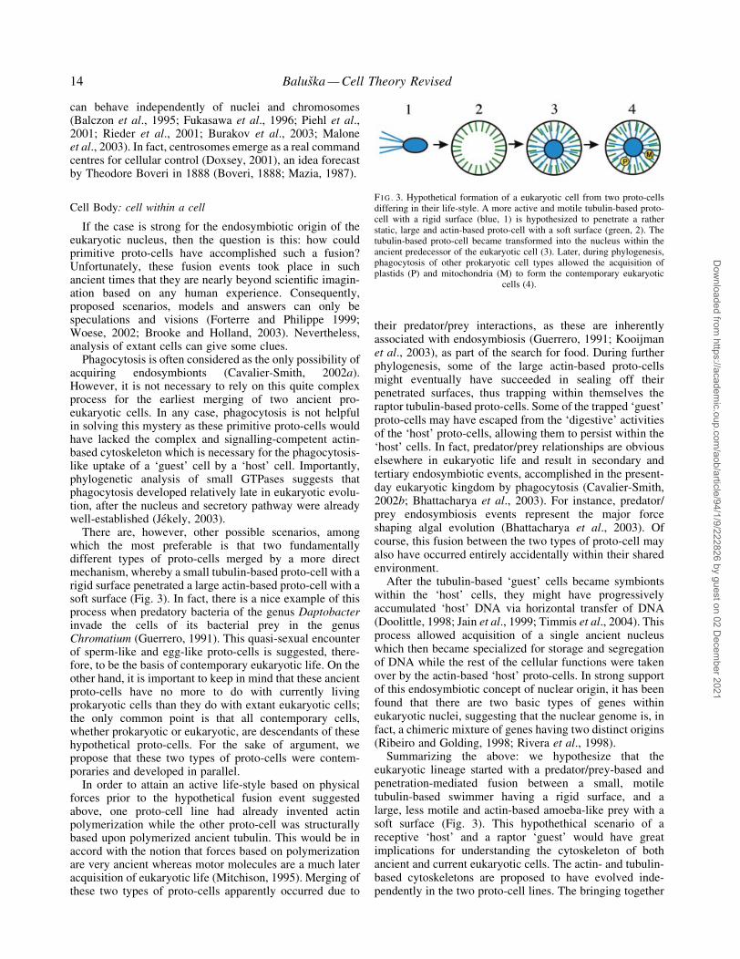
There are, however, other possible scenarios, amongwhich the most preferable is that two fundamentallydifferent types of proto-cells merged by a more directmechanism, whereby a small tubulin-based proto-cell with arigid surface penetrated a large actin-based proto-cell with asoft surface (Fig. 3). In fact, there is a nice example of thisprocess when predatory bacteria of the genus Daptobacterinvade the cells of its bacterial prey in the genusChromatium (Guerrero, 1991). This quasi-sexual encounterof sperm-like and egg-like proto-cells is suggested, there-fore, to be the basis of contemporary eukaryotic life. On theother hand, it is important to keep in mind that these ancientproto-cells have no more to do with currently livingprokaryotic cells than they do with extant eukaryotic cells;the only common point is that all contemporary cells,whether prokaryotic or eukaryotic, are descendants of thesehypothetical proto-cells. For the sake of argument, wepropose that these two types of proto-cells were contem-poraries and developed in parallel.
In order to attain an active life-style based on physicalforces prior to the hypothetical fusion event suggestedabove, one proto-cell line had already invented actinpolymerization while the other proto-cell was structurallybased upon polymerized ancient tubulin. This would be inaccord with the notion that forces based on polymerizationare very ancient whereas motor molecules are a much lateracquisition of eukaryotic life (Mitchison, 1995). Merging ofthese two types of proto-cells apparently occurred due to
their predator/prey interactions, as these are inherentlyassociated with endosymbiosis (Guerrero, 1991; Kooijmanet al., 2003), as part of the search for food. During furtherphylogenesis, some of the large actin-based proto-cellsmight eventually have succeeded in sealing off theirpenetrated surfaces, thus trapping within themselves theraptor tubulin-based proto-cells. Some of the trapped `guest'proto-cells may have escaped from the `digestive' activitiesof the `host' proto-cells, allowing them to persist within the`host' cells. In fact, predator/prey relationships are obviouselsewhere in eukaryotic life and result in secondary andtertiary endosymbiotic events, accomplished in the present-day eukaryotic kingdom by phagocytosis (Cavalier-Smith,2002b; Bhattacharya et al., 2003). For instance, predator/prey endosymbiosis events represent the major forceshaping algal evolution (Bhattacharya et al., 2003). Ofcourse, this fusion between the two types of proto-cell mayalso have occurred entirely accidentally within their sharedenvironment.
After the tubulin-based `guest' cells became symbiontswithin the `host' cells, they might have progressivelyaccumulated `host' DNA via horizontal transfer of DNA(Doolittle, 1998; Jain et al., 1999; Timmis et al., 2004). Thisprocess allowed acquisition of a single ancient nucleuswhich then became specialized for storage and segregationof DNA while the rest of the cellular functions were takenover by the actin-based `host' proto-cells. In strong supportof this endosymbiotic concept of nuclear origin, it has beenfound that there are two basic types of genes withineukaryotic nuclei, suggesting that the nuclear genome is, infact, a chimeric mixture of genes having two distinct origins(Ribeiro and Golding, 1998; Rivera et al., 1998).
Summarizing the above: we hypothesize that theeukaryotic lineage started with a predator/prey-based andpenetration-mediated fusion between a small, motiletubulin-based swimmer having a rigid surface, and alarge, less motile and actin-based amoeba-like prey with asoft surface (Fig. 3). This hypothethical scenario of areceptive `host' and a raptor `guest' would have greatimplications for understanding the cytoskeleton of bothancient and current eukaryotic cells. The actin- and tubulin-based cytoskeletons are proposed to have evolved inde-pendently in the two proto-cell lines. The bringing together
F I G . 3. Hypothetical formation of a eukaryotic cell from two proto-cellsdiffering in their life-style. A more active and motile tubulin-based proto-cell with a rigid surface (blue, 1) is hypothesized to penetrate a ratherstatic, large and actin-based proto-cell with a soft surface (green, 2). Thetubulin-based proto-cell became transformed into the nucleus within theancient predecessor of the eukaryotic cell (3). Later, during phylogenesis,phagocytosis of other prokaryotic cell types allowed the acquisition ofplastids (P) and mitochondria (M) to form the contemporary eukaryotic
cells (4).
14 BalusÏka Ð Cell Theory Revised
Dow
nloaded from https://academ
ic.oup.com/aob/article/94/1/9/222826 by guest on 02 D
ecember 2021

of actin and tubulin within the same cell resulted in anew quality due to the fact that these at ®rst uniquepro-eukaryotic cells were equipped with a more complexcytoskeleton. This feature endowed these ancient pro-eukaryotes with tremendous advantages, resulting in anexplosive evolution of early eukaryotic life. It might alsohave allowed these new cells to survive the most criticalphases of evolution in which extremely harsh conditionscould cause bottlenecks for the predecessor proto-cellpopulations yet allow the pro-eukaryotes to ¯ourish. Thisscenario also gives a possibility of understanding thecytoskeleton of eukaryotic cells from a completely newprespective.
Tubulin-based ¯agellate sperm cells, lacking F-actin,penetrate into large actin-based egg cells to generate plantCell Bodies
A hypothetical penetration or fusion event between twoancient proto-cells can explain not only the origin of theeukaryotic nucleus but can also serve as a useful paradigmfor understanding sexual reproduction of present-daymulticellular organisms where, invariably, two haploidcells fuse together to form a diploid zygote (Fig. 4). Theproto-cell fusion event is also reminiscent of the ancientChinese Yin/Yang concept. The tubulin-based sperm cell issmall and motile (Yang), whereas the large, actin-based eggcell (Yin) is non-motile and lacks a centrosome. Thesestructural features, as well as the mode of sexual cell fusion,might resemble the ancient fusion event which may havegiven rise to the pro-eukaryotic cell.
Higher plants seem not to ®t completely into this schemeas they do not have obvious motile sperm cells equippedwith ¯agellae (Fig. 4A). However, plant sperms lost their¯agellae only secondarily (Poort et al., 1996) as a result oftheir adaptation to life on land. In this situation, actin-driventip growth of pollen tubes (AÈ stroÈm et al., 1995; Raudaskoskiet al., 2001; Laitiainen et al., 2002) provides the actualvehicle for the tubulin-based sperm cells' transport (Fig. 5)towards the egg within the female gametophyte (Sil¯ow andLefebvre, 2001). Tip growth in plants is represented notonly by pollen tubes but also by root hairs, where it is drivenby actin polymerization and is tubulin-independent(Bibikova et al., 1999; Gibbon et al., 1999; BalusÏka et al.,2000a; Raudaskoski et al., 2001; Vidali et al., 2001;Foissner et al., 2002; Laitiainen et al., 2002; SÏamaj et al.,2002).
Sperm cells of higher plants have not only lost their¯agellae, but they are also devoid of F-actin (Pierson et al.,1986; Heslop-Harrison et al., 1988; Palevitz and Tiezzi,1992). In fact, higher plant sperm is the only knownexample of a plant cell that lacks F-actin. On the other hand,sperm cells are equipped with a prominent tubulin-basedcytoskeleton in the form of bundled microtubules (Piersonet al., 1986; Palevitz and Liu, 1992; Palevitz and Tiezzi,1992) whose assembly is directed by g-tubulin (Palevitzet al., 1994). From the cytoskeletal point of view, the spermcell resembles a mitotic spindle (mitotic Cell Body) whichrepresents the most basic form of Cell Body (Mazia, 1993;BalusÏka et al., 1998).
Nevertheless, lower plants do still possess ¯agellatesperm cells (Li et al., 1989; Vaughn et al., 1993; Renzagliaand Garbary, 2001; Sil¯ow and Lefebvre, 2001; Sakaushiet al., 2003), and these cells closely resemble the motilesperm cells of other eukaryotic organisms, not only withrespect to their tubulin-based ¯agellae but also on accountof the importance of centrin for their MTOCs (Vaughn et al.,1993; Hart and Wolniak, 1998). For instance, the mostancient gymnosperm species, cycads and Ginkgo biloba,release from their pollen tubes multi¯agellated sperm cellswhich actively swim towards the egg cells using tubulin-based ¯agellae (Li et al., 1989; Renzaglia and Garbary,2001; Sil¯ow and Lefebvre, 2001).
CELL BODY VERSUS CELL PERIPHERYAPPARATUS
Tubulin-based mitosis versus actin-based cytokinesis fromthe Cell Body perspective: divisions of `guest' and `host'cells?
It is undisputable that mitosis and cytokinesis, althoughtightly coupled in most cells, can often be uncoupled,suggesting that these two processes are actually independ-
F I G . 4. Sexual reproduction of current eukaryotic organisms is based onsimilar sequences of events with the tubulin-based sperm cellspenetrating the actin-based oocytes. Sperms of most higher plants (A) arenon-¯agellated, and thus lack active tubulin-based motility, as is the casein most other eukaryotic organisms (B). However, this is a secondarytrait associated with the adaptation of plants to life on land. This hasenforced a `dry' mode of pollination in contrast to the motile `wet' modeof gamete penetration still found in lower plants and some primitive
gymnosperms (cycads, Gingko).
F I G . 5. Pollen (A) and pollen tubes (B) of higher plants constitute agood example of `cells within a cell', a mode of organization which isnot compatible with the current version of Cell Theory. The sperm cell isimmobile and lacks F-actin, but it contains abundant microtubules (blue).In contrast, the vegetative nucleus forms an active Cell Body withradiating perinuclear microtubules and assembles a dense F-actin cap
(not shown) which drives tip-growth of the pollen tube.
BalusÏka Ð Cell Theory Revised 15
Dow
nloaded from https://academ
ic.oup.com/aob/article/94/1/9/222826 by guest on 02 D
ecember 2021

ent, even though they usually cooperate to bring about celldivision. The very nature of these processes implies thatthey are based on different principles. It is obvious thatmitosis represents the division of the tubulin-based `guest'cell (now in the form of Cell Body), whereas cytokinesiscorresponds to the division of the actin-based `host' cell.
It is well known that nuclear division (Cell Body divisionor mitosis) is an extremely conservative process drivensolely by the microtubular cytoskeleton (Pickett-Heaps,1969; Hyman and Karsenti, 1996). In contrast, cytokinesis,which divides the cytoplasm as well as the cell boundarycomplex, is less conservative (Ueda and Nagasaki, 2004),and is driven mainly by the actin cytoskeleton, although italso requires the cooperation of microtubules (Hyman andKarsenti, 1996; Glotzer, 1997; Hales et al., 1999; Karsentiand Vernos, 2001; Guertin et al., 2002). Moreover, mitosisnot only precedes cytokinesis temporally but also instructscytokinesis spatially (Glotzer, 2004). This more conservednature of mitosis and less conserved nature of cytokinesis,combined with the many examples of mitosis not followedby cytokinesis, suggests that mitosis is much more import-ant for eukaryotic life. Importantly, the plasma membranecan form de novo during cytokinesis, and this process is theninstructed and regulated by Cell Bodies (for sporulation inyeast see Knop and Strasser, 2000; Nickas et al., 2003;Shimoda, 2004).
The coenocyte-like nature of higher plants deviates fromthis scheme slightly because here cytokinesis is based moreon microtubules than on actin ®laments (Staehelin andHepler, 1996; Assaad, 2001; BalusÏka et al., 2001c;Bednarek and Falbel, 2003). Owing to the evolutionaryloss of the compact centrosomes and the acquisition ofabundant cortical micortubules (Mazia, 1987; BalusÏka et al.,1997a), plant cytokinesis has undergone dramatic changesduring the evolution of supracellular higher plants. Forexample, cytokinesis in lower plants is either partially orfully actin-dependent (McIntosh et al., 1995; Sawitzky andGrolig, 1995; HoÈftberger and LuÈtz-Meindl, 1999;Karyophyllis et al., 2000), whereas in higher plants it isdirected preferentially by the microtubular Cell Body.Under stress situations, however, plant cells sometimesrevert to a cleavage-like cytokinesis resembling animalcytokinesis (Herth and Meyer, 1978; Sonobe, 1990; Cleary,2001). It is as though the basic and ancient cytokineticprocess is still embedded in contemporary plant cells andcan reassert itself as a default upon severe challenge whenall other division systems are prone to failure.
On the other hand, animal cells experimentally madedevoid of centrosomes also fail to complete a truecytokinesis, leaving the daughter cells coupled by cyto-plasmic bridges (Doxsey, 2001; Khodjakov and Rieder,2001; Piehl et al., 2001) resembling plasmodesmata.Interestingly in this respect, in higher plants, centriole andcentrosome-based centrin localize to both plasmodesmata(Blackman et al., 1999) and cytokinetic cell plates (DelVecchio et al., 1997; Harper et al., 2000). Moreover, plantcells lack myosin II (Reichelt and Kendrick-Jones, 2000).The signi®cance of this is that, in animal as well as yeastmutant cells devoid of class II myosins, there are aberrationsin the ®nal phases of their cytokinesis, with a failure to
separate the daughter cells (Bi et al., 1998; Tolliday et al.,2003). This, in turn, suggests that the coenocyte-like higherplants perhaps evolved their apparent multicellularity byprocesses that resulted from the loss (or the non-acquistionby evolution) of myosin II and compact centrosomes.Moreover, remains of MTOCs might have become trappedwithin cell-to-cell channels which failed to constrict due tothe absence of myosin II. Intriguingly, centrin and plant-speci®c myosin VIII are found at contractile cell-to-cellplasmodesmatal channels in plants (Blackman et al., 1999;BalusÏka et al., 2001b). This ®nding is potentially veryrelevant because centrioles are known to be essential for the®nal stage of animal cytokinesis (Khodjakov and Rieder,2001; Piehl et al., 2001).
Actin-based Cell Periphery Complex versus tubulin-basedCell Body: Yin and Yang principles imply sexual nature ofthe cytoskeleton
Vasiliev (1987) was the ®rst to propose that eukaryoticcells are based on a symbiosis-like coexistence of two co-operating, yet competing domains: an actin-based cellperiphery termed actinoplast, and a tubulin-based tubulo-plast (see also Figs 3, 4), an idea that clearly foreshadowsMazia's Cell Body concept. These two cellular domainssegregate completely during mitosis when the tubulin-basedmitotic spindle, or naked Cell Body, is divested of actin andthe cells revert to the primitive nature that is characteristicof the early eukaryotic cells (Fig. 6). As discussed above,this feature is also a characteristic of sperm cells of higherplants. In contrast, plant cells entering into interphasedeploy their microtubules at the cell periphery (BalusÏkaet al., 1997a) while actin and diverse actin-binding proteinsaccumulate within their nuclei and participate in theorganization of nuclear structure and chromatin activities(like DNA transcription) as well as in the maturation andtransport of RNA molecules (Olave et al., 2002; Pedersonand Aebi, 2002; Kandasamy et al., 2003; Kraus et al., 2003;Shumaker et al., 2003).
Obviously, both actin and tubulin are important for theorganization of eukaryotic cells and therefore it is notsurprising that both these proteins are among the mostconserved of eukaryotic proteins. Strikingly, tight parallelsexist between this symbiotic-like organization of the actin-based Cell Periphery Apparatus and the tubulin-based CellBody, both assemblies being the vestiges of an ancienthypothetical actin-based `host' cell and a tubulin-based`guest' cell (Fig. 3). As mentioned above, this sequence ofevents is recapitulated during the sexual reproduction ofeukaryotic organisms when, invariably, fusion between atubulin-based sperm cell and an actin-based oocyte givesrise to a new multicellular organism (Fig. 4). After fusion ofthe tubulin-based sperm cell with the actin-based oocyte,followed by the fusion of their haploid nuclei (Cell Bodies),the centrosome-less oocyte acquires the sperm centrosomewhich then takes control of the spatial arrangement ofmicrotubules in the fertilized zygote.
This sexual background to the current cytoskeleton, andthe joining of the two ancient and Yin-Yang-like cyto-skeletal systems into one cell, may explain the extreme
16 BalusÏka Ð Cell Theory Revised
Dow
nloaded from https://academ
ic.oup.com/aob/article/94/1/9/222826 by guest on 02 D
ecember 2021

rapidity of the prokaryotic±eukaryotic switch and theconsequent lack of fossil records of `transition' organisms(Dacks and Doolittle, 2001). The actin cytoskeletonremained associated preferentialy with the ¯exible cellboundary which thereby drives an actin-based motility(Pantaloni et al., 2001), whereas the microtubular cyto-skeleton evolved, together with DNA and associatedproteins, into the Cell Body. Both basic types of cyto-skeleton exert mechanical forces via polymerizationand depolymerization of their respective polymers, resem-bling the force generation of present-day prokaryotic life,which is also based on actin-like and tubulin-like proteins(van den Ent et al., 2001a, b; Ben-Yehuda and Losick,2002; Carballido-LoÂpez and Errington, 2003; Daniel andErrington, 2003). On the other hand, more advanced force-generating systems, such as molecular motors which useactin- and tubulin-based polymers as tracks, are trueeukaryotic inventions accomplished only as a consequenceof the increased complexity of eukaryotic cells (Mitchison,1995; Vale, 2003). Interestingly, not only present-daycellular parasites but also endosomes and phagosomes(Merri®eld et al., 1999; Taunton et al., 2000; Zhang et al.,2002; Fehrenbacher et al., 2003; Southwick et al., 2003) useactin polymerization as a driving force for their motilities(Machesky, 1999; Maly and Borisy, 2001; Pantaloni et al.,2001; Pollard and Borisy, 2003).
Actin- and tubulin-based cytoskeletal systems can sup-port cellular and subcellular movements independently ofeach other. Cellular fragments containing portions of cellperiphery and an actin polymerization machinery, butlacking nuclei and microtubules, are still capable ofautonomous directional motility (Albrecht-Buehler, 1980;Euteneuer and Schliwa, 1984; Malawista and Chevance deBois¯eury, 1984; Verkhovsky et al., 1998; Maly and Borisy,2001). On the other hand, tubulin-based Cell Bodies are alsoinherently motile. The characteristic motility of Cell Bodieswithin eukaryotic cells (BalusÏka et al., 2001a) stronglyimplicates the independent nature of this part of theeukaryotic cell. As mentioned above, perinuclear micro-tubules, capable of both pushing and pulling forces, act aseffective instruments to allow Cell Bodies to claim a certain
amount of the cytoplasmic space. If one of them is lesseffective in this activity, then unequal daughter cells of adivision are the result; the weaker Cell Body has a smallerin¯uence and gains a correspondingly smaller cytoplasmicspace (Pickett-Heaps et al., 1999; Brown and Lemmon,2001).
A nice example of this situation is the ®rst mitoticdivision of a pollen nucleus to produce a large vegetativecell, which supports pollen tube growth, and a smallgenerative cell designed to form sperm cells devoid of F-actin. Such rudimentary Cell Bodies of the sperm cells areinactive and are fully dependent upon the metabolicactivities of the vegetative nucleus and pollen tube.Another example of such a `tug-of-war' between CellBodies having different strengths is the ®rst division of thefertilized zygote, which is often asymmetric and therebyde®nes the anterior±posterior body axis of most multi-cellular organisms (Wallenfang and Seydoux, 2000; Lyczaket al., 2002; Wodarz, 2002). Smaller cells typically give riseto the posterior/shoot poles of multicellular organisms, andthen ultimately they become specialized for the develop-ment of sexual organs and organs of movement. The largercells produce, again via asymmetric division, anterior/rootpoles specialized for the uptake of nutritive substances andfor neuronal-like activities (for plants see JuÈrgens, 2000,2003; BalusÏka et al., 2004).
Centering of tubulin-based Cell Body and its modulation viaactin-based Cell Periphery Apparatus
Recently, we reviewed data reporting that the actin-basedcell periphery participates in the positioning of the CellBody by means of interactions between the dynamic plus-ends of microtubules, which emanate from the Cell Body,and the actin-rich Cell Periphery Apparatus (BalusÏka et al.,2000b, 2001a). In the most typical situation, the Cell Bodysettles at the geometrical centre of the cell as a result of acentripetal pushing force directed from the cell periphery.Dynamic microtubules lacking association with centro-somes and nuclei, but equipped with microtubular motors,are also capable of this centering phenomenon if the minus-ends of microtubules focus upon cellular inclusions, such asmelanophores, while their plus-ends radiate towards the cellperiphery (Rodionov and Borisy, 1997). Centrosomesreleased from their inherent nuclear association use thesame mechanism for positioning and centring (Rieder et al.,2001; Euteneuer and Schliwa, 1992; Burakov et al., 2003).
Cell Bodies make use of interactions with the cellperiphery-enriched actin cytoskeleton (Pruyne andBretscher, 2000) to maintain their positions (Burakovet al., 2003). Dynamic microtubules explore the surround-ing perinuclear cytoplasmic space (Holy et al., 1997;Faivre-Moskalenko and Dogterom, 2002). The property ofmicrotubule instability, which is affected by reaching thecell boundary, is crucial for this explorative behaviour(Komarova et al., 2002). It allows mitotic spindles andinterphase nuclei to perform rotations in the cytoplasm,these movements also being navigated by the actincytoskeleton which accumulates under the plasma mem-brane (Reinsch and GoÈnczy, 1998; Adames and Cooper,
F I G . 6. During mitosis, all microtubules (blue) retract from the actin-rich(green) cell periphery and participate in the assembly of the mitoticspindle, which represents the most basic form (or transformation) of theCell Body. In this state, the Cell Body is specialized for the segregationof large amounts of chromosomal DNA (red). Mitosis is one of the most
conserved process found within the eukaryotic superkingdom.
BalusÏka Ð Cell Theory Revised 17
Dow
nloaded from https://academ
ic.oup.com/aob/article/94/1/9/222826 by guest on 02 D
ecember 2021

2000; Tran et al., 2001; Burakov et al., 2003; Kusch et al.,2003). The identity of critical molecules that link the plus-ends of microtubules with the actin cytoskeleton at the cellcortex has recently been illuminated in yeast and animalcells (Goode et al., 2000; Pruyne and Bretscher, 2000;Glynn et al., 2001; Ishizaki et al., 2001; Gundersen, 2002;Kodama et al., 2003). Interestingly, plant cells express ahomologue of Kar9p (Gardiner and Marc, 2003) which isresponsible for linking Cell Body microtubules to the actin-rich cell cortex (Segal et al., 2002).
Accumulations of actin at distinct cell periphery domainsattract and stabilize nearby microtubules, and these ultim-ately polarize the Cell Body (BalusÏka et al., 2000b, 2001a).The centring and polarizing properties of Cell Bodies areessential not only for division of unicellular yeast cells(Pruyne and Bretscher, 2000) but also for cell-to-cellcommunication, as evidenced by actin-based synapticcontacts both in animal and plant cells (Dustin andColman, 2002; BalusÏka et al., 2003a, b, c; Barlow et al.,2004). In plants, polar transport of auxin is inherently linkedto the overall polarity of the Cell Bodies (BalusÏka et al.,2003a, b, c; Barlow et al., 2004). This in turn leads to apreferred orientation of mitotic division. Cell Bodies ofanimal cells are also polarized via immunological synapses(Sancho et al., 2002). In fact, in what seems to be part of acellular `arms race', active Cell Bodies organizing lyso-some-based secretion of lytic substances can be consideredto behave as some sort of `killer machines' (Bossi et al.,2002; Clark et al., 2003).
From the Yin/Yang perspective, mitosis might be viewedas a phase in which the two types of cytoskeleton areseparated from each other, and revert back to the ancientcon®guration of the cytoarchitecture (Fig. 6). Mitoticsegregation of DNA-based mitotic chromosomes is organ-ized and driven solely via microtubules, which retract fromall cellular areas and are then free to build up the spindleapparatus. Conversely, the actin cytoskeleton retracts fromthe cell's interior and associates preferentially with the CellPeriphery Apparatus (Fig. 5). When mitotis and cytokinesisare both concluded, tubulin and actin-based cytoskeletonsinterpenetrate again and form the integrated cytoskeletalnetwork of eukaryotic cells (Goode et al., 2000; Kodamaet al., 2003).
Cell Body-based exocytosis versus Cell Periphery-basedendocytosis
From a phylogenetical perspective, the Cell Body conceptgives us some clues to speculate on how it came about thateukaryotic cells developed two quite contrasting pathwaysfor vesicular membrane traf®cking. The secretory pathwayis organized by the Cell Body: it starts at the nuclearenvelope (VorõÂsÏek, 2000; Matynia et al., 2002), continuesvia endoplasmic reticulum and Golgi apparatus, andculminates with secretory vesicles fusing with the plasmamembrane (Fig. 7). Secretion is tightly coupled with nuclearorganization (Nanduri and Tartakoff, 2001) and is under thespatial control of the Cell Body microtubules (Bloom andGoldstein, 1998; MuÈsch, 2004).
Importantly, the outwardly directed exocytic pathway isphylogenetically older than the inwardly directed endo-cytotic pathway (JeÂkely, 2003), which is organized by theCell Periphery Apparatus (Fig. 7). The endocytic pathwaystarts at the plasma membrane (Conner and Schmid, 2003)with actin-dependent internalization steps (Engquist-Goldstein and Drubin, 2003), and proceeds deeper into thecytoplasm via different types of endosomes (Fig. 7) pro-pelled by comet-like actin tails (Merri®eld et al., 1999;Taunton et al., 2000; Zhang et al., 2002; Fehrenbacher et al.,2003; Southwick et al., 2003). This pathway, which isevolutionarily speaking a more recent one, is a vestige of theactivities of the ancient actin-based `host' proto-cell whichrepresents a transformation of its actin-based plasmamembrane. These internalization pathways, includingprimitive versions of phagocytic and endocytic pathways,allowed the symbiotic acquisition of further organelles ofeukaryotic cells; these acquired organelles were the fore-runners of the present-day mitochondria and plastids(McFadden, 1999; Gray et al., 2001). Nowadays theseendocytotic pathways are hijacked by viruses and bacteria,allowing them to intrude into eukaryotic cells (Brock et al.,2003; Stamm et al., 2003; Wang et al., 2003) and then, afterentering the cell, to exploit the actin cytoskeleton for theirintracellular, as well as cell-to-cell, migration (Goldberg,2001; Fehrenbacher et al., 2003; Stamm et al., 2003).
Using the actin cytoskeleton, the most primitive eukar-yotic cells exploited this second, endocytotic pathway ofvesicular traf®cking not only for cellular nutrition (Connerand Schmid, 2003) but also for complex cell-to-cell
F I G . 7. The Cell Body organizes the exocytic secretory pathway (blue),which is composed of endoplasmic reticulum (ER), Golgi apparatus (GA)and secretory vesicles (SV). In contrast, the Cell Periphery Apparatusorganizes the endocytic secretory pathway (green), which is composed ofendocytic vesicles (EV), recycling vesicles (RV), early endosomes (EE)
and late endosomes (LE).
18 BalusÏka Ð Cell Theory Revised
Dow
nloaded from https://academ
ic.oup.com/aob/article/94/1/9/222826 by guest on 02 D
ecember 2021

signalling pathways which have now become a prevalentfeature of multicellular organisms (Gundel®nger et al.,2003; Stevens, 2003). The best examples here are adhesiondomains specialized for vesicular cell-to-cell communica-tion in neuronal, immunological and plant synapses (Dustinand Colman, 2002; Barlow et al., 2004). Moreover, besidesthe endosymbiotic acquisition of the power-houses ofeukaryotic cellsÐthe mitochondria and chloroplasts(McFadden, 1999; Gray et al., 2001) ± there were secondaryendosymbiotic events in which one primitive eukaryoteenclosed a second eukaryote (Cavalier-Smith and Beaton,1999; Douglas et al., 2001; Gilson, 2001; Cavalier-Smith,2002b). This reveals that there is an inherent tendency forendosymbiosis which has operated throughout the evolutionof biological systems.
Small GTP-binding proteins from the Cell Body perspective:the unique status of Ran family
Besides the nucleus, cytoskeleton and vesicle traf®ckingmachinery, all eukaryotic cells are characterized by the Rassuperfamily of small GTPases that are key regulators ofboth cytoskeletal dynamics and vesicular traf®ckings. Thephylogenetic analysis of small GTPases reveals that themost ancient eukaryotic cells were equipped with asecretory machinery but, as mentioned above, lacked themolecules which would support endocytosis and phagocy-tosis (JeÂkely, 2003). The nuclear envelope is part of theexocytic pathway (VorõÂsÏek, 2000; Matynia et al., 2002;Shimoda, 2004) that is organized along Cell Body micro-tubules radiating from the nuclear envelope towards the cellperiphery. It is probable that the symbiotic origin of thenucleus (Gupta et al., 1994; Gupta and Golding, 1996;Horiike et al., 2001; Hartman and Fedorov, 2002) isinherently linked with the acquisition of this pathway.
Most small GTPases localize to the membranes ofeukaryotic cells where they act as biological switches,activating or terminating biological processes. Particularsubcellular localizations of these membranous targets arespeci®ed by post-translational modi®cations with farnesyl,palmitoyl, myristolyl and geranylgeranyl lipid groups(Takai et al., 2001; Vernoud et al., 2003). Members of theRas family are predominantly localized to the plasmamembrane where they activate stimulus±responsive serine/threonine kinases (Takai et al., 2001; Vernoud et al., 2003),while members of the Rho family organize a cytoskeleton inassociation with phagocytic and endocytic membranes(Ridley, 2001; Etienne-Manneville and Hall, 2002).Members of the Rab family organize endocytic pathways(Zerial and McBride, 2001) while Arf members localizepreferentially to endoplasmic reticulum and Golgi apparatus(Pasqualato et al., 2002; Spang, 2002). Interestingly, cellfusion is regulated by the plasma membrane-associatedGTPase ARF6 (Chen et al., 2003; Taylor, 2003).
Of all the known families of small GTPases, only the Ranfamily members lack lipid attachment modules. They arethus not localized to membranes but are, instead, abundantwithin the nucleus. Ran GTPases shuttle to the cytoplasmand organize diverse nuclear features and processes, such asnuclear architecture (Clarke and Zhang, 2001), the assembly
of nuclear pores (Ryan et al., 2003), the sorting out of thenuclear envelope as a specialized domain of endoplasmicreticulum (Hetzer et al., 2002; Mattaj, 2004), nucleocyto-plasmic transport (GoÈrlich and Kutay, 1999), targetting ofnuclear proteins (Narayanan et al., 2003), as well ascentrosome activity (Di Fiore et al., 2003; Keryer et al.,2003), kinetochore function (Arnaoutov and Dasso, 2003),nuclear chromatin- and chromosome-driven polymerizationof microtubules (Carazo-Salas et al., 1999; Ohba et al.,1999; Wilde et al., 2001; Kalab et al., 2002) and spindlecheckpoints (Li et al., 2003). Because Ran GTPases alsoregulate DNA synthesis (Moore, 2001; Yamaguchi andNewport, 2003) and cell-cycle progression (Moore, 2001),this class of small GTPases emerges as a central organizingcomponent of the Cell Body, linking together DNA- andtubulin-based structures.
INHERENT DNA±TUBULIN INTERACTIONS
`Happy marriage' or `master±slave' relationships?
The evolutionary transition from prokaryotes to eukaryotes,which is still one of the greatest puzzles for contemporarybiology, was marked by the unprecedented molecularcoupling of tubulin with DNA. In prokaryotes, DNA isassociated with membranes which thereby allow its repli-cation and partitioning, whereas eukaryotes use exclusivelymicrotubules to partition huge amounts of DNA with high®delity. Recently, DNA segregation in bacteria was shownto rely on polymerization of actin-like ParM protein(Mùller-Jensen et al., 2003) This suggests that DNA hasan inherent tendency to enslave cytoskeletal molecules,irrespective of its association with either prokaryotic oreukaryotic cellular organization.
The association of DNA with nuclear proteins, especiallyhistones (Malik and Henikoff, 2003), as well as theassociation of chromatin with tubulin-based microtubules,are the most characteristic features of eukaryotic cells(BalusÏka et al., 1997a). Double-stranded (but not the single-stranded) DNA binds to the microtubule-associated proteintau, which somehow protects the DNA double helix (Huaand He, 2003; Hua et al., 2003). This latter feature togetherwith those processes that drive mitosis indicate that DNA isperhaps the dominant partner in this molecular relationship.This would imply some kind of molecular slavery relation-ship between tubulin and DNA, the latter playing the role ofmaster.
Importantly, this `master±slave' relationship allows theCell Bodies to exhibit exploratory properties in space andtime (Kirschner and Gerhart, 1998; West-Eberhard, 1998;BalusÏka et al., 2001a). These properties are essential fordriving cellular polarities (Pruyne and Bretscher, 2000;BalusÏka et al., 2001a) as well as for pattern formation,morphogenesis and development of complex multicellularorganisms (BalusÏka et al., 2003b). Due to the abandonmentof the inherent association between DNA and membranes,which is the hallmark of prokaryotes, eukaryotic DNAbecame free to engage in extensive proliferation. Usingspecialized nuclear proteins that direct tubulin polymeriza-tion (Oegema et al., 1997; Wittmann et al., 2000; Du et al.,
BalusÏka Ð Cell Theory Revised 19
Dow
nloaded from https://academ
ic.oup.com/aob/article/94/1/9/222826 by guest on 02 D
ecember 2021
2001; Keryer et al., 2003; Rabitsch et al., 2003; Raemaekerset al., 2003; Schatz et al., 2003; for a review see BalusÏkaet al., 1997a), DNA enslaved the microtubular cytoskeleton(see also Box 2) and exploited it as the vehicle to move largeDNA assemblages, such as mitotic chromosomes or evenwhole nuclei. Cell Bodies clearly manifest this master±slaverelationship.
The inherent relationship between eukaryotic DNA andmicrotubules is so strong that even the extremely reducednucleomorphs, which have undergone up to 1000-foldreduction of their genomic DNA mass (Cavalier-Smith andBeaton, 1999; Gilson, 2001), still retain genes for a-, b- andg-tubulins, although they lack most other proteins typical ofeukaryotic cells (Keeling et al., 1999). As mentioned, a verystrong argument for an inherent association between DNAmolecules and tubulins can be found in the unique nature ofepixenosomes, which are ectosymbionts located on thesurface of marine ciliates (Petroni et al., 2000). They mightbe considered to be highly reduced symbiotic Cell Bodiesconsisting only of DNA and tubulins (Jenkins et al., 2002).
MAZIA'S VISION OF FLEXIBLE LINEARCENTROSOMES IN PLANT CELLS: GAMMA-TUBULIN, EB1 AND SPC98P HOLD THE KEY
One outstanding mystery of higher plant cells, which hasperplexed scientists for many years, is the apparent absenceof corpuscular centrosomes. This failure to identify anyde®nite centrosome in the cells of higher plants stimulatedMazia to propose, in an entirely speculative manner, theconcept of a `¯exible' linear centrosome which should beable to modify its three-dimensional arrangement (Mazia,1987). He was proposing a `¯exible string' composed ofdiscrete units which are, in a way similar to DNA, capableof folding and hence attaining secondary and tertiary ordersof structural organization.
At the time of Mazia's suggestion, g-tubulin was notknown (Oakley and Oakley, 1989), and no MTOCcomponent had been identi®ed in plant cells, despite thefact that the concept of a MTOC had already been proposedfor them (Pickett-Heaps, 1969). Now, however, numerousdata are accumulating that indicate that plant g-tubulins,together with other proteins, correspond to the putativediscrete units which represent the ¯exible linear centrosomeof higher plant cells (Liu et al., 1993; Joshi and Palevitz,1996; Canaday et al., 2000; Panteris et al., 2000;Dibbayawan et al., 2001; Drykova et al., 2003; Horio andOakley, 2003; Kumagai et al., 2003; Schmit, 2003;Shimamura et al., 2004). In addition to g-tubulin, veryrecent advances have identi®ed the tubulin plus-end bindingprotein EB1 (Rehberg et al., 2002; Rogers et al., 2002)which, in plant cells, also marks the mobile minus-ends(Chan et al., 2003). These ®ndings strengthen Mazia's viewof a ¯exible and dispersed centrosome in plant cells (Chanet al., 2003; Lloyd and Chan, 2004). This interpretation issupported by the situation known from Dictyosteliumdiscoideum where EB1 is an integral part of the centrosome,is independent of microtubules (Rehberg et al., 2002), andemerges from centrosomes on tips of growing microtubules(Piehl et al., 2004). SPC98p and SPC97p are other well-
known components of MTOCs, and they are part of aubiquitous microtubule nucleator complex with a molecularmass of about 280 kDa (Moritz and Agard, 2001). SPC98phas also been identi®ed in plant cells (Erhardt et al., 2002)where it localizes to three distinct sites: intranuclear dots,the nuclear surface, and at sites near the plasma membrane(Seltzer et al., 2003). All this conforms very well with theCell Body concept as elaborated in this review.
OUTLOOK
Genome evolution is associated with a huge variation innuclear DNA amounts. Eukaryotic organisms at either thesame or different levels of complexity differ considerably intheir DNA amounts. The genome of Amoeba, for example,is about 200 times larger than the human genome, repre-senting one of the most astonishing examples of the C-valueenigma (Gregory, 2001a). In the plant genus Luzula, whosespecies are dif®cult to distinguish morphologically from oneanother, diploid DNA values vary 15-fold and chromosomenumbers 10-fold (species with low chromosome numbershaving higher DNA amounts) (Barlow and Nevin, 1976).Not many molecular biologists are aware of the fact thatcoding DNA comprises only <10 % of the whole genome,the rest of the DNA being of unknown function (Cavalier-Smith and Beaton, 1999). Importantly, the non-coding DNAhas an effect on cell size via its so-called nucleotypicin¯uence (Bennett, 1972; Gregory, 2001a, b), althoughthere is no plausible explanation for this phenomenon(Gregory, 2001a).
Fortunately, the Cell Body concept is poised to explainthese enigmas. DNA-binding proteins stored within nucleioften regulate tubulin polymerization (BalusÏka et al.,1997a), while the dynamism of cytoplasmic microtubulesregulates the access to the nucleus of proteins sequesteredwithin the cytoplasm (for transcription factors see Oegemaet al., 1997; Wittmann et al., 2000; Du et al., 2001; Keryeret al., 2003; Rabitsch et al., 2003; Raemaekers et al., 2003;Schatz et al., 2003; for a review see BalusÏka et al., 1997a).This, in turn, regulates the binding of these proteins to theDNA, irrespective of its coding capacity, and in¯uences theassembly of the nuclear matrix (BalusÏka and Barlow, 1993;BalusÏka et al., 1995a, b, 1997b).
Dynamic microtubules also stabilize the nucleocytoplas-mic ratio (Trombetta, 1939), this being a measure of theef®ciency with which the microtubules of the Cell Bodypatrol the cytoplasmic domain surrounding the nucleus.DNA can independently increase or diminish in amount,and a given nucleocytoplasmic ratio can be maintained solong as the microtubules radiating from the nucleusdominate the surrounding cytoplasmic domain in proportionto nuclear volume. As mentioned above, nucleomorphs arenot relict nuclei, as generally assumed (Cavalier-Smith andBeaton, 1999; Gilson, 2001), but are relict Cell Bodies. Thisis evidenced by their expression of tubulin genes, althoughthis does not lead to the formation of microtubules (Keelinget al., 1999). Similarly, epixenosomes represent anotherexample of highly reduced Cell Bodies composed only oftubulins and DNA (Jenkins et al., 2002). Importantly, theminiaturized genomes of nucleomorphs do not scale with
20 BalusÏka Ð Cell Theory Revised
Dow
nloaded from https://academ
ic.oup.com/aob/article/94/1/9/222826 by guest on 02 D
ecember 2021

the cell size of their hosts (Gilson, 2001). Becausenucleomorphs lack non-coding DNA, which in mosteukaryotic genomes is much more abundant than codingDNA (Gregory, 2001a, b), one could propose that the non-coding DNA is relevant for the Cell Body on account of itsinteraction with tubulin molecules via diverse tubulin/DNA-associated proteins, of which NuMa is the most instructiveexample (Levesque et al., 2003; Tulu et al., 2003).Recently, a putative plant homologue of NuMa was reportedfor Arabidopsis (Gardiner and Marc, 2003).
In the framework of the Cell Body concept, the non-coding DNA could control nuclear structure via its ability tocontrol both internal nuclear architecture and the availabil-ity of nuclear proteins that have tubulin-polymerizingactivity (BalusÏka et al., 1997a). This would be in a fullagreement with the proposition that non-coding DNA actsas nucleoskeletal DNA (Cavalier-Smith and Beaton, 1999).On the other hand, the dynamic properties of cytoplasmicmicrotubules can make a direct impact on nuclear architec-ture. They can either exert pushing forces on the nuclearsurface or sequester, within the cytoplasm, proteins criticalfor structuring the nuclear chromatin and for regulatinggenome expression. In support of these notions, we havereported elsewhere upon the close relationships betweennuclear size, chromatin structure and dynamicity ofcytoplasmic microtubules (BalusÏka and Barlow, 1993;BalusÏka et al., 1995a, b, 1997b).
Thus, the Cell Body concept not only predicts that DNAregulates tubulin assembly within the cytoplasm but alsothat the assembled microtubules control the availablity ofnuclear proteins, sequestered within the cytoplasm, for thedecondensation of chromatin which controls DNA replica-tion and transcription (Oegema et al., 1997; Du et al., 2001;Wittmann et al., 2000; Keryer et al., 2003; Rabitsch et al.,2003; Raemaekers et al., 2003; Schatz et al., 2003; for areview see BalusÏka et al., 1997a). From the point of view ofthe Cell Body, there is no difference between coding DNAand non-coding DNA; both are predicted to interact, directlyor indirectly, with the sequestered nuclear proteins.However, uncovering those proteins which interact directlywith the non-coding skeletal DNA (Cavalier-Smith andBeaton, 1999) will require concentrated activity frommolecular biologists. Unfortunately, this will take sometime because current scienti®c efforts are focusing solely onthe genetic coding properties of DNA.
The importance of non-coding DNA for interactions withthe tubulin-based cytoskeketon is well known fromcentromeres (repetitive non-coding DNA sequences),which organize, via associations of numerous DNA bindingproteins, kinetochores that are specialized for the attach-ment of mitotic chromomes to microtubules of the mitoticspindle (De Wulf et al., 2003). Recent advances in studieson centromeres and kinetochores reveal a lack of DNAsequence speci®city for the establishment and maintenanceof centromere DNA identity, as well as kinetochoreassembly (Sullivan et al., 2001; Amor and Choo, 2002).
Clearly, the centromere±kinetochore complex is assem-bled and maintained via self-propagating epigenetic mech-anisms based on chromatin structures, but independent ofDNA sequences (Amor and Choo, 2002). These ®ndings are
exciting in that they reveal the need for the Cell Bodyconcept, especially because of the unique power of thisconcept to explain the role of non-coding DNA as a centralplayer responsible for the linkage between the DNA-basednuclear chromatin with the tubulin-based cytoskeleton. Thisfeature allows the Cell Body to couple genomic information(encoded within DNA sequences and handed over to RNAmolecules) with epigenetic information (embodied withinthe inherent physical properties of DNA structures, whichcan store and propagate this information via complex DNA±protein and protein±protein templating processes) (Gregoryand Herbert, 1999; Zuckerkandl, 2002). The crucial ques-tion to answer is what molecules accomplish the inherentDNA/tubulin-based cytoskeleton interactions. The ®rstclues are emerging in this respect. First, linker histone H1was reported to exert a dual function, acting as some kind ofmicrotubule-associated protein which stabilizes preformedmicrotubules (Multigner et al., 1992; Saoudi et al., 1995;Kaczanowski and Jerzmanowski, 2001). Second, micro-tubule-associated protein tau binds to double-stranded, butnot single-stranded, DNA and does so reversibly in thepresence of histones (Hua et al., 2003). It also apparentlyprotects the double helix structure from damaging freeradicals (Hua and He, 2003).
Cell Bodies can sense electric and magnetic ®elds, anduse them as cues for the orientation of mitotic divisions(Denegre et al., 1998; Zhao et al., 1999; Song et al., 2002;Valles, 2002). Relevant in this respect are the microtubuleswhich, in this sensory context, have even been proposed toact like the `nerves' of cells (Albrecht-Buehler, 1998) due totheir ability to perceive and transmit light (Albrecht-Buehler, 1992, 1994, 1998). The orientated self-organ-ization of microtubules is, in some circumstances, alsodependent upon gravitational ®elds (Tabony and Job, 1992;Papaseit et al., 1999, 2000; Tabony et al., 2001). Moreover,dynamic microtubules can sense another critical physicalparameter of environment: temperature. This may be viaCa2+ liberated from cell walls (Plieth et al., 1999) and fromthe endocytotic components of the Cell PeripheryApparatus. Sensing the physical environment is inherentlycombined with the ability of microtubules to organize intoradial arrays of the Cell Body that scan the plasmamembrane boundaries of the cytoplasm, exerting either apushing or a pulling force on any object or boundary towhich they are attached. Thus, the combination of all theseproperties allows the microtubular mitotic spindle (specia-lized form of the Cell Body optimized for its multiplication)not only to act as an ideal tool for separating large amountsof DNA molecules with high ®delity but also to endow theotherwise passive DNA-storing nuclei with sensory andexploratory properties (Kirschner and Mitchison, 1986;Kirschner and Gerhardt, 1998). Clearly, microtubules arecentral for these abilities of Cell Bodies to, ®rst, sensephysical properties of their environment and, second, to usethis information directly in morphogenesis (Kirschner andMitchison, 1986; Hyman and Karsenti, 1996; Papaseit et al.,1999). These two phenomena are especially critical forsessile higher plants (BalusÏka et al., 1997a, 1998;Wasteneys, 2002).
BalusÏka Ð Cell Theory Revised 21
Dow
nloaded from https://academ
ic.oup.com/aob/article/94/1/9/222826 by guest on 02 D
ecember 2021

CONCLUSIONS
In conclusion, the Cell Body concept proposed here is usefulfor understanding eukaryotic cells in their whole complex-ity. This concept not only explains how eukaryotic cellscame to be formed from their proto-cellular predecessors,and in particular how the eukaryotic nucleus was formedand why it is so intimately linked with centrosomes andmicrotubules, but it also explains why mitosis andcytokinesis can be accomplished independently of eachother. Moreover, this concept reveals the fundamentallysexual nature of the eukaryotic cytoskeleton, and explainsdifferences between exocytosis and endocytosis from anevolutionary point of view. Last, but not least, the Cell Bodyconcept allows, for the ®rst time, an explanation of the C-value enigma from the perspective of nucleotypic DNA±tubulin interactions.
A FINAL NOTE
This review is dedicated to the memory of Daniel Mazia(1912±1996) who was well aware of the unique nature ofDNA±tubulin interactions. The apparent absence of acorpuscular centrosome in cells of higher plants was apuzzle to be solved, and Mazia approached this enigmaticproblem by proposing the existence of a thread-like ¯exiblecentrosome which would be in a position to attain higher-order structures. Daniel Mazia discovered and isolated themitotic apparatus of sea urchins (Mazia and Dan, 1952)even before microtubules were known. Following this, andwith the legacy of Theodore Boveri in mind (Mazia, 1987),he devoted almost his whole life to understanding howcentrosomes, microtubules and nuclei (or mitotic chromo-somes) interact to build the structural and functional unitwhich he termed the Cell Body (Mazia, 1994).Unfortunately, his death prevented him from formulatingthis concept to the full. Here, we make an attempt to do this,employing a holistic approach that embraces both theevolutionary as well as the structural and functional aspectsof eukaryotic life. We ®nd that these approaches can besatisfactorily integrated into the miracle of the Cell Body.
LITERATURE CITED
Adam JC, Pringle JR, Pfeifer M. 2000. Evidence for functionaldifferentiation among Drosophila septins in cytokinesis andcellularization. Molecular Biology of the Cell 11: 3123±3135.
Adames NR, Cooper JA. 2000. Microtubule interactions with the cellcortex causing nuclear movements in Saccharomyces cerevisiae.Journal of Cell Biology 149: 863±874.
Albrecht-Buehler, G. 1980. Autonomous movement of cytoplasmicfragments. Proceedings of the National Academy of Sciences of theUSA 77: 6639±6643.
Albrecht-Buehler, G. 1992. A rudimentary form of cellular `vision'.Proceedings of the National Academy of Sciences of the USA 89:8288±8292.
Albrecht-Buehler G. 1994. The cellular infrared detector appears to becontained in the centrosome. Cell Motility and the Cytoskeleton 27:262±271.
Albrecht-Buehler G. 1998. Altered drug resistance of microtubules incells exposed to infrared light pulses: are microtubules the `nerves'of cells? Cell Motility and the Cytoskeleton 40: 183±192.
Anderson GR, Stoler DL, Brenner BM. 2001. Cancer: the evolvedconsequence of a destabilized genome. BioEssays 23: 1037±1046.
Amor DJ, Choo KHA. 2002. Neocentromeres: role in human disease,evolution, and centromere study. American Journal of HumanGenetics 71: 695±714.
Arnaoutov A, Dasso M. 2003. The Ran GTPase regulates kinetochorefunction. Developmental Cell 5: 99±111.
Aronson JF. 1971. Demonstration of a colcemid-sensitive attractive forceacting between the nucleus and a cell center. Journal of Cell Biology51: 579±583.
Assaad F. 2001. Plant cytokinesis. Exploring the links. Plant Physiology126: 509±516.
AÈ stroÈm H, Sorri O, Raudaskoski M. 1995. Role of microtubules in themovement of the vegetative nucleus and generative cell in tobaccopollen tubes. Sexual Plant Reproduction 8: 61±69.
Balczon R, Bao L, Zimmer WE, Brown K, Zinkowski RP, BrinkleyBR. 1995. Dissociation of centrosome replication events from cyclesof DNA synthesis and mitotic division in hydroxyurea-arrestedChinese hamster ovary cells. Journal of Cellular Biology 130: 105±115.
BalusÏka F, Barlow PW. 1993. The role of the microtubular cytoskeletonin determining nuclear chromatin structure and passage of maize rootcells through the cell cycle. European Journal of Cell Biology 61:160±167.
BalusÏka F, Barlow PW, Hauskrecht M, Kubica SÏ , Parker JS,Volkmann D. 1995a. Microtubule arrays in maize root cells.Interplay between the cytoskeleton, nuclear organization and post-mitotic cellular growth patterns. New Phytologist 130: 177±192.
BalusÏka F, BacigaÂlova K, Oud JL, Hauskrecht M, Kubica SÏ . 1995b.Rapid reorganization of microtubular cytoskeleton accompaniesearly changes in nuclear ploidy and chromatin structure inpostmitotic cells of barley leaves infected with powdery mildew.Protoplasma 185: 140±151.
BalusÏka F, Barlow PW, Parker JS, Volkmann D. 1996. Symmetricreorganizations of radiating microtubules around pre-mitotic andpost-mitotic nuclei of dividing cells organized within intact rootmeristems. Journal of Plant Physiology 149: 119±128.
BalusÏka F, Volkmann D, Barlow PW. 1997a. Nuclear components withmicrotubule-organizing properties in multicellular eukaryotes:functional and evolutionary considerations. International Review ofCytology 175: 91±135.
BalusÏka F, SÏamaj J, Volkmann D, Barlow PW. 1997b. Impact of taxol-mediated stabilization of microtubules on nuclear morphology,ploidy levels and cell growth in maize roots. Biology of Cell 89:221±231.
BalusÏka F, Lichtscheidl IK, Volkmann D, Barlow PW. 1998. The plantcell body: a cytoskeletal tool for cellular development andmorphogenesis. Protoplasma 202: 1±10.
BalusÏka F, Salaj J, Mathur J, Braun M, Jasper F, SÏamaj J et al. 2000a.Root hair formation: F-actin-dependent tip growth is initiated bylocal assembly of pro®lin-supported F-actin meshworks accumulatedwithin expansin-enriched bulges. Developmental Biology 227: 618±632.
BalusÏka F, Volkmann D, Barlow PW. 2000b. Actin-based domains ofthe `cell periphery complex' and their associations with polarized`cell bodies' in higher plants. Plant Biology 2: 253±267.
BalusÏka F, Volkmann D, Barlow PW. 2001a. Motile plant cell body: a`bug' within a `cage'. Trends in Plant Sciences 6: 104±111.
BalusÏka F, Cvrckova F, Kendrick-Jones J, Volkmann D. 2001b. Sinkplasmodesmata as gateways for phloem unloading. Myosin VIII andcalreticulin as molecular determinants of sink strength? PlantPhysiology 126: 39±46.
BalusÏka F, JaÂsik J, Edelmann HG, Salajova T, Volkmann D. 2001c.Latrunculin B induced plant dwar®sm: plant cell elongation is F-actin dependent. Developmental Biology 231: 113±124.
BalusÏka F, SÏamaj J, Menzel D. 2003a. Polar transport of auxin: carrier-mediated ¯ux across the plasma membrane or neurotransmitter-likesecretion? Trends Cell Biology 13: 282±285.
BalusÏka F, Wojtaszek P, Volkmann D, Barlow PW. 2003b. Thearchitecture of polarized cell growth: the unique status of elongatingplant cells. BioEssays 25: 569±576.
BalusÏka F, SÏamaj J, Wojtaszek P, Volkmann D, Menzel D. 2003c.Cytoskeleton ± plasma membrane ± cell wall continuum: emerginglinks revisited. Plant Physiology 136: 482±491.
BalusÏka F, Mancuso S, Volkmann D, Barlow PW. 2004. Root apices as
22 BalusÏka Ð Cell Theory Revised
Dow
nloaded from https://academ
ic.oup.com/aob/article/94/1/9/222826 by guest on 02 D
ecember 2021

plant command centres: the unique `brain-like' status of the root apextransition zone. BioloÂgia (Bratislava) 59 (Supplement 13): In press
Barlow PW. 1982. `The plant forms cells, not cells the plant': the origin ofde Bary's aphorism. Annals of Botany 49: 269±271.
Barlow PW. 1994. Cell divisions in meristems and their contribution toorganogenesis and plant form. In: Ingram DS, Hudson A, eds. Shapeand form in plants and fungi. London: Academic Press, 169±193.
Barlow PW, Nevin D. 1976. Quantitative karyology of some species ofLuzula. Plant Systematics and Evolution 125: 77±86.
Barlow PW, Volkmann D, BalusÏka F. 2004. Polarity in roots. In:Lindsey K, ed. Polarity in plants. Oxford: Blackwell Publishers,192±241.
de Bary A. 1864. Die Mycetozoen (Schleimpilzen), Leipzig: Engelmann.Bednarek SY, Falbel TG. 2003. Membrane traf®cking during plant
cytokinesis. Traf®c 3: 621±629.Bennett MD. 1972. Nuclear DNA content and minimum generation time
in herbaceous plants. Proceedings of the Royal Society of London,Series B 181: 109±135.
Ben-Yehuda S, Losick R. 2002. Asymmetric cell division in B. subtilisinvolves a spiral-like intermediate of the cytokinetic protein FtsZ.Cell 109: 257±266.
Bhattacharya D, Yoon HS, Hackett JD. 2003. Photosynthetic eukaryotesunite: endosymbiosis connects the dots. BioEssays 26: 50±60.
Bi E, Maddox P, Lew DJ, Salmon ED, McMillan JN, Yeh E, PringleJR. 1998. Involvement of an actomyosin contractile ring inSaccharomyces cerevisiae cytokinesis. Journal of Cell Biology142: 1301±1312.
Bibikova TN, Blanca¯or E, Gilroy S. 1999. Microtubules regulate tipgrowth and orientation in root hairs of Arabidopsis thaliana. PlantJournal 17: 657±665.
Blackman LM, Harper JDI, Overall RL. 1999. Localization of acentrin-like protein to higher plant plasmodesmata. EuropeanJournal of Cell Biology 78: 297±304.
Bloom GS, Goldstein LSB. 1998. Cruising along microtubule highways:how membranes move through the secretory pathway. Journal ofCell Biology 140: 1277±1280.
Boevink P, Oparka K, Santa-Cruz S, Martin B, Betteridge A, HawesC. 1998. Stacks on tracks: the plant golgi apparatus traf®cs on anactin/ER network. Plant Journal 15: 441±447.
Boisnard-Lorig C, Colon-Carmona A, Bauch M, Hodge S, Doerner P,Bancharel E, Dumas C, Haseloff J, Berger F. 2001. Dynamicanalyses of the expression of the HISTONE::YFP fusion protein inArabidopsis show that syncytial endosperm is divided in mitoticdomains. Plant Cell 13: 495±509.
Bossi G, Trambas C, Booth S, Clark R, Stinchcombe J, Grif®ths GM.2002. The secretory synapse: the secrets of a serial killer.Immunological Reviews 189: 152±160.
Boveri T. 1888. Zellen-studien II. Die Befruchtung und Teilung des Eiesvon Ascaris megalocephala. Jena: Fisher.
Boveri T. 1902a. Zellen-studien IV. UÈ ber die Natur der Centrosomen.Jena: Fisher.
Boveri T. 1902b. UÈ ber mehrpolige Mitosen als Mittel zur Analyse desZellkerns. Verhandlungen der physikalisch-medizinischenGesselschaft zu WuÈrzburg 35: 67±90.
Bowerman B, Severson AF. 1999. Cell division: plant-like properties ofanimal cell cytokinesis. Current Biology 9: R658±R660.
Brandizzi F, Snapp EL, Roberts AG, Lippincot-Schwartz J, Hawes C.2002. Membrane protein transport between the endoplasmicreticulum and the Golgi in tobacco leaves is energy dependent butcytoskeleton independent: evidence from selective photobleaching.Plant Cell 14: 1293±1309.
Bresgen N, Czihak G, Linhart J. 1994. Computer modeling of blastodermformation in Drosophila. Naturwissenschaften 81: 417±418.
Brinkley BR. 2001. Managing the centrosome numbers game: from chaosto stability in cancer cell division. Trends in Cell Biology 11: 18±21.
Brock SC, Goldenring JR, Crowe Jr. JE. 2003. Apical recyclingsystems regulate directional budding of respiratory syncytial virusfrom polarized epithelial cells. Proceedings of the National Academyof Sciences of the USA 100: 15143±15148.
Brooke NM, Holland PWH. 2003. The evolution of multicellularity andearly animal genomes. Current Opinion in Genetics & Development13: 599±603.
Brown RC, Lemmon BE. 1992. Cytoplasmic domain: a model for spatial
control of cytokinesis in reproductive cells of plants. EMSA Bulletin22: 48±53.
Brown RC, Lemmon BE. 2001. The cytoskeleton and spatial control ofcytokinesis in the plant cell life cycle. Protoplasma 215: 35±49.
Brown RC, Lemmon BE, Olsen O-A. 1994a. Endosperm development inbarley: microtubule involvement in the morphogenetic pathway.Plant Cell 6: 1241±1252.
Brown RC, Lemmon BE, Olsen O-A. 1994b. Development of endospermin Arabidopsis thaliana. Sexual Plant Reproduction 12: 32±42.
Brown RC, Lemmon BE, Olsen O-A. 1996. Development of theendosperm in rice (Oryza sativa L.): cellularization. Journal of PlantResearch 109: 301±313.
Brown RC, Lemmon BE, Nguyen H. 2004. Events during the ®rst fourrounds of mitosis establish three developmental domains in thesyncytial endosperm of Arabidopsis thaliana. Protoplasma 222:167±174.
Bruusgaard JC, Liestùl K, Ekmark M, Kollstand K, Gundersen K.2003. Number and spatial distribution of nuclei in the muscle ®bresof normal mice studied in vivo. Journal of Physiology 551.2: 467±478.
Burakov A, Nadezhdina E, Slepchenko B, Rodionov V. 2003.Centrosome positioning in interphase cells. Journal of Cell Biology162: 963±969.
Burkhardt J. 1998. The role of microtubule-based motor proteins inmaintaining the structure and function of the Golgi complex.Biochimica et Biophysica Acta 1404: 113±116.
Canaday J, Stoppin-Mellet V, Mutterer J, Lambert A-M. 2000. Higherplant cells: gamma-tubulin and microtubule nucleation in the absenceof centrosomes. Microscopy Research and Techniques 49: 487±495.
Carazo-Salas RE, Guarguaglini G, Gruss OJ, Segref A, Karsenti E,Mattaj IW. 1999. Generation of GTP-bound Ran by RCC1 isrequired for chromatin-induced mitotic spindle formation. Nature400: 178±181.
Carballido-LoÂpez R, Errington J. 2003. The bacterial cytoskeleton:in vivo dynamics of the actin-like protein Mbl of Bacillus subtilis.Developmental Cell 4: 19±28.
Cathomen T, Mrkic B, Spehner D, Drillien R, Naef R, Pavlovic J,Aguzzi A, Billeter MA, Cattaneo R. 1998. A matrix-less measlesvirus is infectious and elicits extensive cell fusion: consequences forpropagation in the brain. EMBO Journal 17: 3899±3908.
Cavalier-Smith T. 2002a. The phagotrophic origin of eukaryotes andphylogenetic classi®cation of Protozoa. International Journal ofSystematic and Evolutionary Microbiology 52: 297±354.
Cavalier-Smith T. 2002b. Chloroplast evolution: secondarysymbiogenesis and multiple losses. Current Biology 12: R62±R64.
Cavalier-Smith T, Beaton MJ. 1999. The skeletal function of non-codingDNA: new evidence from ancient cell chimeras. Genetics 106: 3±13.
Chabin-Brion K, Marceiller J, Perez F, Settegrana C, Drechou A,Durand G, PouÈs C. 2001. The Golgi complex is a microtubule-organizing organelle. Molecular Biology of the Cell 12: 2047±2060.
Chan J, Calder GM, Doonan JH, Lloyd CW. 2003. EB1 reveals mobilemicrotubule nucleation sites in Arabidopsis. Nature Cell Biology 5:967±971.
Chen EH, Pryce BA, Tzeng JA, Gonzalez GA, Olson EN. 2003. Controlof myoblast fusion by a guanine nucleotide exchange factor, loner,and its effector ARF6. Cell 114: 751±762.
Clark RH, Stinchcombe JC, day A, Blott E, Booth S, Bossi G, HamblinT, Davies EG, Grif®ths GM. 2003. Adaptor protein 3-dependentmicrotubule-mediated movement of lytic granules to theimmunological synapse. Nature Immunology 4: 1111±1120.
Clarke PR, Zhang C. 2001. Ran GTPase: a master regulator of nuclearstructure and function during the eukaryotic division cycle? Trendsin Cell Biology 11: 366±371.
Cleary AL. 2001. Plasma membrane ± cell wall connections: roles inmitosis and cytokinesis revealed by plasmolysis of Tradescantiavirginiana leaf epidermal cells. Protoplasma 215: 21±34.
Cole NB, Sciaky N, Marotta A, Song J, Lippincott-Schwartz J. 1996.Golgi dispersal during microtubule disruption: regeration of Golgistacks at peripheral endoplasmic reticulum exit sites. MolecularBiology of the Cell 7: 631±650.
Compton DA. 2000. Spindle assembly in animal cells. Annual Reviews ofBiochemistry 69: 95±114.
BalusÏka Ð Cell Theory Revised 23
Dow
nloaded from https://academ
ic.oup.com/aob/article/94/1/9/222826 by guest on 02 D
ecember 2021

Conner SD, Schmid SL. 2003. Regulated portals of entry into the cell.Nature 422: 37±44.
Cross JC,Werb Z, Fisher SJ. 1994. Implantation and the placenta: keypieces of the development puzzle. Science 266: 1508±1518.
Dacks JB, Doolittle WF. 2001. Reconstructing/deconstructing the earliesteukaryotes: how cooperative genomics can help. Cell 107: 419±425.
D'Amato, F. 1977. Nuclear cytology in relation to development.Cambridge: Cambridge University Press.
Daniel RA, Errington J. 2003. Control of cell morphogenesis in bacteria:two distinct ways to make a rod-shaped cell. Cell 113: 767±776.
de Duve C. 1996. The birth of complex cells. Scienti®c American 274: 38±45.
Del Vecchio AJ, Harper JDI, Vaughn KC, Baron AT, Salisbury JL,Overall RL. 1997. Centrin homologues in higher plants areprominently associated with the developing cell plate. Protoplasma196: 224±234.
Denegre JM, Valles Jr JM, Lin K, Jordan WB, Mowry KL. 1998.Cleavage planes in frog eggs are altered by strong magnetic ®elds.Proceedings of the National Academy of Sciences of the USA 95:14729±14732.
De Wulf P, McAinsh AD, Sorger PK. 2003. Hierarchical assembly of thebudding yeast kinetochore from multiple subcomplexes. Genes andDevelopment 17: 2902±2921.
Dibbayawan TP, Harper JDI, Marc J. 2001. A g-tubulin antibodyagainst a plant peptide sequence localizes to cell division speci®cmicrotubule arrays and organelles in plants. Micron 32: 671±678.
Di Fiore B, Ciciarello M, Mangiacasale R, Palena A, Tassin A-M,Cundari E, Lavia P. 2003. Mammalian RanBP1 regulatescentrosome cohesion during mitosis. Journal of Cell Science 116:3399±3411.
Dolan MF, Melnitsky H, Margulis L, Kolnicki R. 2002. Motilityproteins and the origin of the nucleus. The Anatomical Record 268:290±301.
Doolittle WF. 1998. You are what you eat: a gene transfer ratchet couldaccount for bacterial genes in eukaryotic nuclear genomes. Trends inGenetics 14: 307±311.
Douglas S, Zauner S, Fraunholz M, Beaton M, Penny S, Deng L-T,et al. 2001. The highly enslaved genome of an enslaved algalnucleus. Nature 410: 1091±1096.
Doxsey SJ. 2001. Centrosome as command centres for cellular control.Nature Cell Biology 3: E105±E107.
Drykova D, Cenklova V, Sulimenko V, Voic J, DraÂber P, Binarova P.2003. Plant g-tubulin interacts with ab-tubulin dimers and formsmembrane-associated complexes. Plant Cell 15: 465±480.
Du Q, Stukenberg PT, Macara IG. 2001. A mammalian Partner ofinscuteable binds NuMA and regulates mitotic spindle organization.Nature Cell Biology 3: 1069±1075.
Dustin ML, Colman DR. 2002. Neural and immunological synapticrelations. Science 298: 785±789.
Ehlers K, Kollmann R. 2001. Primary and secondary plasmodesmata:structure, origin, and functioning. Protoplasma 216: 1±30.
Epel D, Schatten G. 1998. Daniel Mazia: a passion for understanding howcells reproduce. Trends in Cell Biology 8: 416±418.
Engquist-Goldstein AÊ E, Drubin DG. 2003. Actin assembly andendocytosis: from yeast to mammals. Annual Reviews of Cell andDevelopmental Biology 19: 287±332.
Erhardt M, Stoppin-Mellet V, Campagne S, Canaday J, Mutterer J,Fabian T, et al. 2002. Higher plant SPC98p orthologues and g-tubulin localize at microtubule nucleation sites and are involved inmicrotubule nucleation. Journal of Cell Science 115: 2423±2431.
Etienne-Manneville S, Hall A. 2002. Rho GTPases in cell biology.Nature 420: 629±635.
Euteneuer U, Schliwa M. 1984. Persistent directional motility of cells andcytoplasmic fragments in the absence of microtubules. Nature 310:58±61.
Euteneuer U, Schliwa M. 1992. Mechanism of centrosome repositioningduring the wound response in BSC-1 cells. Journal of Cell Biology116: 1157±1166.
Faivre-Moskalenko C, Dogterom M. 2002. Dynamics of microtubuleasters in microfabricated chambers: the role of catastrophes.Proceedings of the National Academy of Sciences of the USA 99:16788±16793.
Fehrenbacher K, Huckaba T, Yang H-C, Boldogh I, Pon L. 2003. Actin
comet tails, endosomes and endosymbionts. Journal of ExperimentalBiology 206: 1977±1984.
Fink S. 1999. Pathological and regenerative plant anatomy. Encyclopediaof Plant Anatomy, vol. 14, part 6. Berlin: Gebruder Borntraeger.
Flemming W. 1882. Zellsubstanz, Kern und Zelltheilung. Leipzig: Vogel.Foe VE, Field CM, Odell GM. 2000. Microtubules and mitotic cycle
phase modulate spatio-temporal distributions of F-actin and myosinII in Drosophila syncytial blastoderm embryos. Development 127:1767±1787.
Foissner I, Grolig F, Obermeyer G. 2002. Reversible proteinphosphorylation regulates the dynamic organization of the pollentube cytoskeleton: effects of calyculin A and okadaic acid.Protoplasma 220: 1±15.
Forterre P, Philippe H. 1999. Where is the root of the universal tree oflife? BioEssays 21: 871±879.
Fukasawa K, Choi T, Kuriyama R, Rulong S, Vande Woude GF. 1996.Abnormal centrosome ampli®cation in the absence of p53. Science271: 1744±1747.
Gardiner J, Marc J. 2003. Putative microtubule-associated proteins fromthe Arabidopsis genome. Protoplasma 222: 61±74.
Gibbon BC, Kovar DR, Staiger CJ. 1999. Latrunculin B has differenteffects on pollen germination and tube growth. Plant Cell 11: 2349±2364.
Gilson PR. 2001. Nucleomorph genomes: much ado about practicallynothing. Genome Biology 2: 1022.1±1022.5.
Glotzer M. 1997. The mechanism and control of cytokinesis. CurrentOpinion in Cell Biology 9: 815±823.
Glotzer M. 2004. Cleavage furrow positioning. Journal of Cell Biology164: 347±351.
Glynn JM, Lustig RJ, Berlin A, Chang F. 2001. Role of bud6p and tea1pin the interaction between actin and microtubules for theestablishment of cell polarity in ®ssion yeast. Current Biology 11:836±845.
Goff LJ, Coleman AW. 1987. The solution of the cytological paradox ofisomorphy. Journal of Cell Biology 104: 739±748.
Goldberg MB. 2001. Actin-based motility of intracellular microbialpathogens. Microbiology and Molecular Biology Reviews 65: 595±626.
Goode BL, Drubin DG, Barnens G. 2000. Functional cooperationbetween the microtubule and actin cytoskeletons. Current Opinion ofCell Biology 12: 63±71.
GoÈrlich D, Kutay U. 1999. Transport between the cell nucleus and thecytoplasm. Annual Reviews of Cell and Developmental Biology 15:607±660.
Gray MW, Burger G, Lang BF. 2001. The origin and early evolution ofmitochondria. Genome Biology 2: 1018.1±1018.5.
Gregory TR. 2001a. The bigger the C-value, the larger the cell: genomesize and red blood cell size in vertebrates. Blood Cells, Molecules,and Disease 27: 830±843.
Gregory TR. 2001b. Coincidence, coevolution, or causation? DNAcontent, cell size, and the C-value enigma. Biological Reviews 76:65±101.
Gregory TR, Hebert PDN. 1999. The modulation of DNA content:proximate causes and ultimate consequences. Genome Research 9:317±324.
Guertin DA, Trautmann S, McCollum D. 2002. Cytokinesis ineukaryotes. Microbiology and Molecular Biology Reviews 66: 155±178.
Guerrero R. 1991. Predation as prerequisite to organelle origin:Daptobacter as example. In: Margulis L, Fester R, eds. Symbiosisas a source of evolutionary innovation. Cambridge, MA: MIT Press,106±117.
Gundel®nger ED, Kessels MM, Qualmann B. 2003. Temporal andspatial coordination of exocytosis and endocytosis. Nature ReviewsMolecular Cell Biology 4: 127±139.
Gundersen GG. 2002. Evolutionary conservation of microtubule-capturemechanisms. Nature Reviews Molecular Cell Biology 3: 296±304.
Gupta RS, Aitken K, Falah K, Singh B. 1994. Cloning of Giaria lambliaheat shock protein HSP70 homologs: implications regarding originsof eukaryotic cells and of endoplasmic reticulum. Proceedings of theNational Academy of Sciences of the USA 91: 2895±2899.
Gupta RS, Golding GB. 1996. The origin of the eukaryotic cell. Trends inBiochemical Sciences 21: 166±171.
24 BalusÏka Ð Cell Theory Revised
Dow
nloaded from https://academ
ic.oup.com/aob/article/94/1/9/222826 by guest on 02 D
ecember 2021

Hall ZW, Ralston E. 1989. Nuclear domains in muscle cells. Cell 59:771±772.
Hales KG, Bi E, Wu J-Q, Adam JC, Yu I-C, Pringle JR. 1999.Cytokinesis: an emerging uni®ed theory for eukaryotes? CurrentOpinion in Cell Biology 11: 717±725.
Harper JDI, Fowke LC, Gilmer S, Overall RL, Marc J. 2000. A centrinhomologue is localised across the developing cell plate ingymnosperms and angiosperms. Protoplasma 211: 207±216.
Harris H. 1999. The birth of the cell. New Haven and London: YaleUniversity Press.
Hart PE, Wolniak SM. 1998. Spermiogenesis in Marsilea vestita: atemporal correlation between centrin expression and blepharoplastdifferentiation. Cell Motility and Cytoskeleton 41: 39±48.
Hartman H, Fedorov A. 2002. The origin of the eukaryotic cell: agenomic investigaion. Proceedings of the National Academy ofSciences of the USA 99: 1420±1425.
Hentze MW. 2001. Believe it or not ± translation in the nucleus. Science293: 1058±1059.
Herth W, Meyer Y. 1978. Cytology of budding and cleavage in tobaccomesophyll protoplasts cultivated in saline medium. Planta 142: 11±21.
Hertwig R. 1903. UÈ ber Korrelation von Zell- und KerngroÈsse und ihreBedeutung fuÈr die Geschletliche Differenzierung und die Teilung derZelle. Biologisches Zentralblatt 23: 49±62.
Heslop-Harrison J, Heslop-Harrison Y, Cresti M, Tiezzi A, MoscatelliA. 1988. Cytoskeletal elements, cell shaping and movement in theangioperm pollen tube. Journal of Cell Science 91: 49±60.
Hetzer M, Gruss OJ, Mattaj IW. 2002. The Ran GTPase as a marker ofchromosome position in spindle formation and nuclear envelopeassembly. Nature Cell Biology 4: E177-E184.
Ho WC, Allan VJ, van Meer G, Berger EG, Kreis TE. 1989.Reclustering of scattered Golgi elements occurs along microtubules.European Journal of Cell Biology 48: 250±263.
HoÈftberger M, LuÈtz-Meindl U. 1999. Septum formation in the desmidXanthidium (Chlorophyta): effects of cytochalasin D and latrunculinB suggest the involvement of actin micro®laments. Journal ofPhycology 35: 768±777.
Holy TE, Dogterom M, Yurke B, Leibler S. 1997. Assembly andpositioning of microtubule asters in microfabricated chambers.Proceedings of the National Academy of Sciences of the USA 94:485±488.
Horiike T, Hamada K, Kanaya S, Shinozawa T. 2001. Origin ofeukaryotic cell nuclei by symbiosis of Archaea and Bacteria isrevelaed by homology-hit analysis. Nature Cell Biology 3: 210±214.
Horio T, Oakley BR. 2003. Expression of Arabidopsis g-tubulin in ®ssionyeast reveals conserved and novel functions of g-tubulin. PlantPhysiology 133: 1926±1934.
Howard A, Pelc S. 1953. Synthesis of deoxyribonucleic acid in normaland irradiated cells and its relation to chromosome breakage.Heredity 6 (Supplement): 261±273.
Hua Q, He R-Q. 2003. Tau could protect DNA double helix structure.Bichimica et Biophysica Acta ± Proteins and Proteomics 1645: 205±211.
Hua Q, He R-Q, Haque N, Qu M-H, del Carmen Alonso A, Grundke-Iqbal I, Iqbal K. 2003. Microtubule associated protein tau binds todouble-stranded but not single-stranded DNA. Cellular andMolecular Life Sciences 60: 413±421.
Huang B-Q, Sheridan WF. 1994. Female gametophyte development inmaize: microtubular organization and embryo sac polarity. Plant Cell6: 845±861.
Huang B-Q, Sheridan WF. 1996. Embryo sac development in the maizeindeterminate gametophyte1 mutant: abnormal nuclear behavior anddefective microtubule organization. Plant Cell 8: 1391±1407.
Hyman AA, Karsenti E. 1996. Morphogenetic properties of microtubulesand mitotic spindle assembly. Cell 84: 401±410.
Iborra FJ, Jackson DA, Cook PR. 2001. Coupled transcription andtranslation within nuclei of mammalian cells. Science 293: 1139±1141.
Inoue S, Salmon ED. 1995. Force generation by microtubule assembly/disassembly in mitosis and related movements. Molecular Biology ofthe Cell 6: 1619±1640.
Ishizaki T, Morishima Y, Okamoto M, Furuyashiki T, Kato T,
Narumiya S. 2001. Coordination of microtubules and the actincytoskeleton by the Rho effector mDia. Nature Cell Biology 3: 8±14.
Jain R, Rivera MC, Lake JA. 1999. Horizontal gene transfer amonggenomes: the complexity hypothesis. Proceedings of the NationalAcademy of Sciences of the USA 96: 3801±3806.
Jenkins C, Samudrala R, Anderson I, Hedlund BP, Petroni G,Michailova N, Pinel N, Overbeek R, Rosati G, Staley JT. 2002.Genes for the cytoskeletal protein tubulin in the bacterial genusProsthecobacter. Proceedings of the National Academy of SciencesUSA 99: 17049±17054.
JeÂkely G. 2003. Small GTPases and the evolution of the eukaryotic cell.BioEssays 25: 1129±1138.
Jones MGK, Northcote DH. 1972. Nematode-induced syncytium: amultinucleate transfer cell. Journal of Cell Science 10: 789±809.
Joshi HC, Palevitz BA. 1996. g-Tubulin and microtubule organization inplants. Trends in Cell Biology 6: 41±44.
JuÈrgens G. 2000. Apical-basal pattern formation in Arabidopsisembryogenesis. EMBO Journal 20: 3609±3616.
JuÈrgens G. 2003. Growing up green: cellular basis of plant development.Mechanisms of Development 120: 1395±1406.
Kalab P, Weis K, Heald R. 2002. Visualization of a Ran-GTP gradient ininterphase and mitotic Xenopus egg extracts. Science 295: 2452±2456.
Kandasamy MK, McKinney EC, Meagher RB. 2003. Cell cycle-dependent association of Arabidopsis actin-related proteins AtARP4and AtARP7 with the nucleus. Plant Journal 33: 939±948.
Kaplan DR. 1992. The relationship of cells to organisms in plants:problem and implications of an organismal perspective. InternationalJournal of Plant Sciences 153: S28-S37.
Kaplan DR, Hagemann W. 1991. The relationship of cell and organismin vascular plants. BioScience 41: 693±703.
Karsenti E, Vernos I. 2001. The mitotic spindle: a self-made machine.Science 294: 543±547.
Karyophyllis D, Katsaros C, Dimitriadis I, Galatis B. 2000. F-actinorganization during the cell cycle of Sphacelaria rigidula(Phaeophyceae). European Journal of Phycology 35: 25±33.
Katz LA. 1999. The tangled web: gene genealogies and the origin ofeukaryotes. American Naturalist 154: S137±S145.
Kaczanowski S, Jerzmanowski A. 2001. Evolutionary correlationbetween linker histones and microtubular structures. Journal ofMolecular Evolution 53: 19±30.
Keeling PJ, Deane JA, Hink-Schauer C, Douglas SE, Maier U-G,McFadden GI. 1999. The secondary endosymbiont of thecryptomonad Guillardia theta contains alpha-, beta-, and gamma-tubulin genes. Molecular Biology of Evolution 16: 1308±1313.
Keryer G, Di Fiore B, Celati C, Lechtreck KF, Mogensen M, DelouveÂeA, Lavia P, Bornens M, Tassin A-M. 2003. Part of Ran isassociated with AKAP450 at the centrosome: involvement inmicrotubule-organizing activity. Molecular Biology of the Cell 14:4260±4271.
Khodjakov A, Rieder CL. 2001. Centrosomes enhance the ®delity ofcytokinesis in vertebrates and are required for cell cycle progression.Journal of Cell Biology 153: 237±242.
Kiermayer O. 1968. The distribution of microtubules in differentiatingcells of Micrasterias denticulata BreÂb. Planta 83: 223±236.
Kim GH, Klotchkova TA, Kang Y-M. 2001. Life without a cellmembrane: regeneration of protoplasts from disintegrated cells of themarine green alga Bryopsis plumosa. Journal of Cell Science 114:2009±2014.
Kim GH, Klotchkova TA. 2001. From protoplasm to swarmer:regeneration of protoplasts from disintegrated cells of themulticellular marine green alga Microdictyon umbilicatum(Chlorophyta). Journal of Phycology 38: 174±183.
Kirschner M, Gerhart J. 1998. Evolvability. Proceedings of the NationalAcademy of Sciences of the USA 95: 8420±8427.
Kirschner M, Mitchison TJ. 1986. Beyond self assembly: frommicrotubules to morphogenesis. Cell 45: 329±342.
Knop M, Strasser K. 2000. Role of the spindle pole body of yeast inmediating assembly of the prespore membrane during meiosis.EMBO Journal 19: 3657±3667.
Kodama A, Karakesisoglou I, Wong E, Vaezi A, Fuchs E. 2003. ACF7:an essential integrator of microtubule dynamics. Cell 115: 343±354.
Komarova YA, Vorobjev IA, Borisy GG. 2002. Life cycle of MTs:
BalusÏka Ð Cell Theory Revised 25
Dow
nloaded from https://academ
ic.oup.com/aob/article/94/1/9/222826 by guest on 02 D
ecember 2021

persistent growth in the cell interior, asymmetric transitionfrequencies and effects of the cell boundary. Journal of CellScience 115: 3527±3539.
Kooijman SALM, Auger P, Poggiale JC, Kooi BW. 2003. Quantitativesteps in symbiogenesis and the evolution of homeostasis. BiologicalReviews 78: 435±463.
Korn RW. 1999. Biological organization ± a new look at an old problem.BioScience 49: 51±57.
Kozorovitskiy Y, Gould E. 2003. Stem cell fusion in the brain. NatureCell Biology 5: 952±954.
Kraus SW, Chen C, Penman S, Heald R. 2003. Nuclear actin and protein4.1: essential interactions during nuclear assembly in vitro.Proceedings of the National Academy of Sciences of the USA 100:10752±10757.
Kronenbusch PJ, Singer SJ. 1987. The microtubule-organizing complexand the Golgi apparatus are co-localized around the entire nuclearenvelope of interphase cardiac myocytes. Journal of Cell Science 88:25±34.
Kumagai F, Nagata T, Yahara N, Moriyama Y, Horio T, Naoi K,Hashimoto T, Murata T, Hasezawa S. 2003. g-Tubulin distributionduring cortical microtubule reorganization at the M/G1 interface intobacco BY-2 cells. European Journal of Cell Biology 82: 43±51.
Kusch J, Liakopoulos D, Barral Y. 2003. Spindle asymmetry: a compassfor the cell. Trends in Cell Biology 13: 562±569.
Laitiainen E, Nieminen KM, Vihinen H, Raudaskoski M. 2002.Movement of generative cell and vegetative nucleus in tobaccopollen is dependent on microtubule cytoskeleton but independent ofthe synthesis of callose plugs. Sexual Plant Reproduction 15: 195±204.
Lake JA, Rivera MC. 1994. Was the nucleus the ®rst endosymbiont?Proceedings of the National Academy of Sciences of the USA 91:2880±2881.
Lambert AM. 1993. Microtubule-organizing centers in higher plants.Current Opinion in Cell Biology 5: 116±122.
Ledbetter MC, Porter KR. 1963. A "microtubule" in plant ®ne structure.Journal of Cell Biology 19: 239±250.
Levesque AA, Howard L, Gordon MB, Compton DA. 2003. Afunctional relationship between NuMA and kid is involved in bothspindle organization and chromosome alignment in vertebrate cells.Molecular Biology of Cell 14: 3541±3552.
Lewis L, Albrecht-Buehler G. 1987. Distribution of multiplecentrospheres determines migration of BHK syncitia. Cell Motilityand Cytoskeleton 7: 282±290.
Li H-Y, Cao K, Zheng Y. 2003. Ran in the spindle checkpoint: a newfunction for a versatile GTPase. Trends in Cell Biology 13: 553±557.
Li Y, Wang FH, Knox RB. 1989. Ultrastructural analysis of the ¯agellarapparatus in sperm cells of Ginkgo biloba. Protoplasma 149: 57±63.
Lingle WL, Lutz WH, Ingle JN, Maihle NJ, Salisbury JL. 1998.Centrosome hypertrophy in human breast tumors: implications forgenomic stability and cell polarity. Proceedings of the NationalAcademy of Sciences of the USA 95: 2950±2955.
Liu B, Marc J, Joshi HC, Palevitz BA. 1993. A g-tubulin-related proteinassociated with the microtubule arrays of higher plants in a cellcycle-dependent manner. Journal of Cell Science 104: 1217±1228.
Lloyd CW, Chan J. 2004. Microtubules and the shape of plants to come.Nature Reviews Molecular Cell Biology 5: 13±23.
Lu Z, Joseph D, Bugnard E, Zaal KJM, Ralston E. 2001. Golgicomplex reorganization during muscle differentiation: visualizationin living cells and mechanisms. Molecular Biology of the Cell 12:795±808.
Lucas WJ, Ding B, van der Schoot C. 1993. Plasmodesmata and thesupracellular nature of plants. New Phytologist 125: 435±476.
Lyczak R, Gomes J-E, Bowerman B. 2002. Heads or tails: cell polarityand axis formation in the early Coenorhabditis elegans embryo.Developmental Cell 3: 157±166.
McCormick S. 1993. Male gametophyte development. Plant Cell 5:1265±1275.
McIntosh K, Pickett-Heaps JD, Gunning BES. 1995. Cytokinesis inSpirogyra: integration of cleavage and cell-plate formation.International Journal of Plant Sciences 156: 1±8.
McIntosh JR, Grishchuk EL, West RR. 2002. Chromosome-microtubuleinteractions during mitosis. Annual Reviews of Cell andDevelopmental Biology 18: 193±219.
McFadden GI. 1999. Endosymbiosis and evolution of the plant cell.Current Opinion of Plant Biology 2: 513±519.
McNaughton EE, Goff LJ. 1990. The role of microtubules in establishingnuclear spatial patterns in multinucleate green algae. Protoplasma157: 19±37.
McNeil PL, Terasaki M. 2001. Coping with the inevitable: how cellsrepair a torn surface membrane. Nature Cell Biology 3: E124±E129.
McNeil PL, Miyake K, Vogel SS. 2003. The endomembrane requirementfor cell surface repair. Proceedings of the National Academy ofSciences of the USA 100: 4592±4597.
Machesky L. 1999. Rocket-based motility: a universal mechanism?Nature Cell Biology 1: E29±E31.
Mahlberg P, Sabharwal P. 1966. Mitotic waves in laticifers of Euphorbiamarginata. Science 152: 518±519.
Malawista SE, Chevance de Bois¯eury A. 1984. The cytokineplast:puri®ed, stable, and functional motile machinery from human bloodpolymorphonuclear leukocytes. Journal of Cell Biology 95: 960±973.
Malik HS, Henikoff S. 2003. Phylogenomics of the nucleosome. NatureStructural Biology 10: 882±891.
Malone CJ, Misner L, Le Bot N, Tsai M-C, Campbell JM, Ahringer J,White JG. 2003. The C. elegans Hook protein, ZYG-12, mediatesthe essential attachment between the centrosome and nucleus. Cell115: 825±836.
Maly IV, Borisy GG. 2001. Self-organization of a propulsive actinnetwork as an evolutionary process. Proceedings of the NationalAcademy of Sciences of the USA 98: 11324±11329.
Margulis L. 1993. Symbiosis in cell evolution. San Francisco: W.H.Freeman & Co.
Margulis L, Dolan MF, Guerrero R. 2000. The chimeric eukaryote:origin of the nucleus from the karyomastignot in amitochondriateprotists. Proceedings of the National Academy of Sciences of the USA97: 6954±6959.
Martin W, Hoffmeister M, Rotte C, Henze K. 2001. An overview ofendosymbiotic models for the origins of eukaryotes, their ATP-producing organelles (mitochondria and hydrogenosomes), and theirheterotrophic lifestyle. Biological Chemistry 382: 1521±1539.
Maser RS, DePinho RA. 2002. Connecting chromosomes, crisis, andcancer. Science 297: 565±569.
Mattaj IW. 2004. Sorting out the nuclear envelope from the endoplasmicreticulum. Nature Reviews Molecular Cell Biology 5: 65±69.
Matynia A, Salus SS, Sazer S. 2002. Three proteins required for earlysteps in the protein secretory pathway also affect nuclear envelopestructure and cell cycle progression in ®ssion yeast. Journal of CellScience 115: 421±431.
Mayer U, Herzog U, Berger F, Inze D, JuÈrgens G. 1999. Mutation in thePILZ group genes disrupt the microtubule cytoskeleton and uncouplecell cycle progression from cell division in Arabidopsis embryo andendosperm. European Journal of Cell Biology 78: 100±108.
Mayer U, JuÈrgens G. 2002. Microtubule cytoskeleton: a track record.Current Opinion in Plant Biology 5: 494±501.
Mazia D. 1984. Centrosomes and mitotic poles. Experimental CellResearch 153: 1±15.
Mazia D. 1987. The chromosome cycle and the centrosome cycle in themitotic cycle. International Review of Cytology 100: 49±92.
Mazia D. 1993. The cell cycle at the cellular level. European Journal ofCell Biology 61(Suppl. 38): 14.
Mazia D, Dan K. 1952. The isolation and biochemical characterization ofthe mitotic apparatus of dividing cells. Proceedings of the NationalAcademy of Sciences of the USA 38: 826±838.
Mazumdar A, Mazumdar M. 2002. How one becomes many: blastodermcellularization in Drosophila melanogaster. BioEssays 24: 1012±1022.
Mazzarello P. 1999. A unifying concept: the history of Cell Theory.Nature Cell Biology 1: E13±E15.
Mereshkowsky C. 1905. UÈ ber Natur und Ursprung der Chromatophorenim P¯anzenreiche. Biologisches Zentralblatt 25: 593±604.
Mereshkowsky C. 1910. Theorie der zwei Plasmaarten als Grundlage derSymbiogenesis, einer neuen Lehre von der Entstehung derOrganismen. Biologisches Zentralblatt 30: 278±303, 321±347,353±367.
Merri®eld CJ, Moss SE, Ballestrem C, Imhof BA, Giese G,Wundedrlich I, Almers W. 1999. Endocytic vesicles move at thetips of actin tails in cultured mast cells. Nature Cell Biology 1: 72±74.
26 BalusÏka Ð Cell Theory Revised
Dow
nloaded from https://academ
ic.oup.com/aob/article/94/1/9/222826 by guest on 02 D
ecember 2021
Miescher F. 1871. UÈ ber die chemische Zusammensetzung der Eiterzellen.Hoppe-Seyler's medicinisch-chemische Untersuchungen 4: 441±460.
Mitchison TJ. 1995. Evolution of a dynamic cytoskeleton. PhilosophicalTransactions of Royal Society of London Series B 349: 299±304.
Mitchison T, Kirschner M. 1984. Dynamic instability of microtubulegrowth. Nature 312: 232±237.
Mizuno K. 1993. Microtubule-nucleation sites on nuclei of higher plantcells. Protoplasma 173: 77±85.
Mogensen HL. 1992. The male germ unit: concept, composition, andsigni®cance. International Review of Cytology 140: 129±147.
Mùller-Jensen J, Borch J, Dam M, Jensen RB, Roepstorff P, GerdesK. 2003. Bacterial mitosis: ParM of plasmid R1 moves plasmid DNAby an actin-like insertional polymerization mechanism. MolecularCell 12: 1477±1487.
Moore JD. 2001. The Ran-GTPase and cell-cycle control. BioEssays 23:77±85.
Moreno RD, Schatten G, Ramalho-Santos J. 2002. Golgi apparatusdynamics during mouse oocyte in vitro maturation: effect of themembrane traf®cking inhibitor brefeldin A. Biology of Reproduction66: 1259±1266.
Moritz M, Agard DA. 2001. Gamma-tubulin complexes and microtubulenucleation. Current Opinion in Cell Biology 11: 174±181.
MuÈsch A. 2004. Microtubule organization and function in epithelial cells.Traf®c 5: 1±9.
Mulari MTK, Patrikainen L, Kaisto T, MetsikkoÈ K, Salo JJ,VaÈaÈnaÈnen HK. 2003. The architecture of microtubular networkand Golgi orientation in osteoclasts ± major differences betweenavian and mammalian species. Experimental Cell Research 285:221±235.
Multigner L, Gagnon J, Van Dorsselaer A, Job D. 1992. Stabilisation ofsea urchin ¯agellar microtubules by histone H1. Nature 360: 33±39.
Nagasato C, Motomura T. 2002. In¯uence of the centrosome incytokinesis of brown algae: polyspermic zygotes of Scytosiphonlomentaria (Scytosiphonales, Phaeophyceae). Journal of CellScience 115: 2541±2548.
Nanduri J, Tartakoff AM. 2001. The arrest of secretion response inyeast: signaling from the secretory path to the nucleus via Wscproteins and Pkc1p. Molecular Cell 8: 281±289.
Narayanan A, Eifert J, Marfatia KA, Macara IG, Corbett AH, TernsRM, Terns MP. 2003. Nuclear RanGTP is not required for targetingsmall nucleolar RNAs to the nucleolus. Journal of Cell Science 116:177±186.
Nickas ME, Schwartz C, Neiman AM. 2003. Ady4p and Spo74p arecomponents of the meiotic spindle pole body that promote growth ofthe prespore membrane in Saccharomyces cerevisiae. EukaryoticCell 2: 431±445.
Nigg EA. 2002. Centrosome aberrations: cause or consequence of cancerprogression? Nature Reviews Cell Biology 2: 1±11.
Niklas KJ. 2000. The evolution of plant body plans ± a biomechanicalperspective. Annals of Botany 85: 411±438.
Nurse P. 2000. The incredible life and times of biological cells. Science289: 1711±1716.
Oakley CE, Oakley BR. 1989. Identi®cation of a g-tubulin, a newmember of the tubulin superfamily encoded by mipA gene ofAspergillus nidulans. Nature 338: 662±664.
Oegema K, Marshall WF, Sedat JW, Alberts BM. 1997. Two proteinsthat cycle asynchronously between centrosomes and nuclearstructures: Drosophila CP60 and CP190. Journal of Cell Science110: 1573±1583.
Ohba T, Nakamura M, Nishitani H, Nishimoto T. 1999. Self-organization of microtubule asters induced by GTP-bound Ran.Science 284: 1356±1358.
Olave IA, Reck-Peterson SL, Crabtree GR. 2002. Nuclear actin andactin-related proteins in chromatin remodelling. Annual Reviews ofBiochemistry 71: 755±781.
Olsen O-A. 2001. Endosperm development: cellularization and cell fatespeci®cation. Annual Reviews of Plant Physiology and PlantMolecular Biology 52: 233±267.
O'Neil RM, La Claire II JW. 1984. Mechanical wounding induces theformation of extensive coated membranes in giant algal cells.Science 225: 331±333.
Otegui M, Staehelin LA. 2000. Syncytial-type cell plates: a novel kind of
cell plate involved in endosperm celllularization of Arabidopsis.Plant Cell 12: 933±947.
Otegui M, Staehelin LA. 2003. Electron tomographic analysis of post-meiotic cytokinesis during pollen development in Arabidopsisthaliana. Planta 218: 501±515.
Pak JY, Solorzano C, Arai M, Nitta T. 1991. Two distinct steps forspontaneous generation of subprotoplasts from disintegratedBryopsis cell. Plant Physiology 96: 819±825.
Palevitz BA, Liu B. 1992. Micro®lament (F-actin) in generative cells andsperm: an evaluation. Sexual Plant Reproduction 5: 89±100.
Palevitz BA, Tiezzi A. 1992. Organization, composition, and function ofthe generative cell and sperm cytoskeleton. International Review ofCytology 140: 149±185.
Palevitz BA, Liu B, Joshi C. 1994. g-tubulin in tobacco pollen tubes:association with generative cell and vegetative microtubules. SexualPlant Reproduction 7: 209±214.
Pantaloni D, Le Clainche C, Carlier M-F. 2001. Mechanisms of actin-based motility. Science 292: 1502±1506.
Panteris E, Apostolakos P, Graf R, Galatis B. 2000. g-Tubulin colocalizeswith microtubule arrays and tubulin paracrystals in dividingvegetative cells of higher plants. Protoplasma 210: 179±187.
Papaseit C, Vuillard L, Tabony J. 1999. Reaction-diffusion microtubuleconcentration patterns occur during biological morphogenesis.Biophysical Chemistry 79: 33±39.
Papaseit C, Vuillard L, Tabony J. 2000. Microtubule self-organization isgravity-dependent. Proceedings of the National Academy of Sciencesof the USA 97: 8364±8368.
Pasqualato S, Renault L, Cher®ls J. 2002. Arf, Arl, Arp and Sar proteins:a family of GTP-binding proteins with a structural device for `front-back' communication. EMBO Reports 3: 1035±1041.
Pederson T. 2001. Is the nucleus in need of translation? Trends in CellBiology 11: 395±397.
Pederson T, Aebi U. 2002. Actin in the nucleus: what form and what for?Journal of Structural Biology 140: 3±9.
Petroni G, Spring S, Schleifer K-H, Verni F, Rosati G. 2000. Defensiveextrusive ectosymbionts of Euplotidium (Ciliophora) that containmicrotubule-like structures are bacteria related to Verrucomicrobia.Proceedings of the National Academy of Sciences of the USA 97:1813±1817.
Pickett-Heaps JD. 1969. The evolution of the mitotic apparatus: anattempt at comparative ultrastructural cytology in dividing plantcells. Cytobios 3: 257±280
Pickett-Heaps JD, Gunning BES, Brown RC, Lemmon BE, Cleary AL.1999. The cytoplast concept in dividing plant cells: cytoplasmicdomains and the evolution of spatially organized cell division.American Journal of Botany 86: 153±172.
Piehl M, Nordberg J, Euteneuer U, Bornens M. 2001. Centrosome-dependent exit of cytokinesis in animal cells. Science 291: 1550±1553.
Piehl M, Tulu US, Wadsworth P, Cassimeris L. 2004. Centrosomematuration: measurement of microtubule nucleation throughout thecell cycle by using GFP-tagged EB1. Proceedings of the NationalAcademy of Sciences of the USA 101: 1584±1588.
Pierson ES, Derksen J, Traas JA. 1986. Organization of micro®lamentsand microtubules in pollen tubes grown in vitro or in vivo in variousangiosperms. European Journal of Cell Biology 41: 14±18.
Plieth, C. Hansen, U.-P. Knight H. Knight MR. 1999. Temperaturesensing in plants: the primary mechanisms of signal perception andcalcium response. Plant Journal 18: 491±497.
Pollard TD. 2003. The cytoskeleton, cellular motility, and the reductionistagenda. Nature 422: 741±745.
Pollard TD, Borisy GG. 2003. Cellular motility driven by assembly anddisassembly of actin ®laments. Cell 112: 453±465.
Poort RJ, Visscher H, Dilcher DL. 1996. Zoidogamy in fossilgymnosperms: the centenary of a concept, with special reference toprepollen of late Paleozoic conifers. Proceedings of the NationalAcademy of Sciences of the USA 93: 11713±11717.
Pruyne D, Bretscher A. 2000. Polarization of cell growth in yeast. II. Therole of the cortical actin cytoskeleton. Journal of Cell Science 113:571±585.
Rabitsch KP, Pentronczki M, Javerzat J-P, Genier S, Chwalla B,Schleifer A, Tanaka TU, Nasmyth K. 2003. Kinetochore
BalusÏka Ð Cell Theory Revised 27
Dow
nloaded from https://academ
ic.oup.com/aob/article/94/1/9/222826 by guest on 02 D
ecember 2021

recruitment of two nucleolar proteins is required for homologsegregation in meiosis I. Developmental Cell 4: 535±548.
Raemaekers T, Ribbeck K, Beaudouin J, Annaert W, Van Camp M,Stockmans I, et al. 2003. NuSAP, a novel microtubule-associatedprotein involved in mitotic spindle organization. Journal of CellBiology 162: 1017±1029.
Ralston E. 1993. Changes in architecture of the Golgi complex and othersubcellular organelles during myogenesis. Journal of Cell Biology120: 399±409.
Ralston E, Hall ZW. 1992. Restricted distribution of mRNA producedfrom a single nucleus in hybrid myotubes. Journal of Cell Biology119: 1063±1068.
Ralston E. Ploug T, Kalhovde J, Lùmo T. 2001. Golgi complex,endoplasmic reticulum exit sites, and microtubules in skeletal muscle®bers are organized by ptterned activity. Journal of Neuroscience 21:875±883.
Ram M, Babbar SB. 2002. Transient existence of life without a cellmembrane: a novel strategy of siphonous seaweed for survival andpropagation. BioEssays 24: 588±590.
Ranganath RM. 2003. Female gametophyte development in higher plants± meiosis and mitosis break the cellular barrier. Plant Biology 5: 42±49.
Raudaskoski M, AÊ stroÈm H, Laitiainen E. 2001. Pollen tubecytoskeleton: structure and function. Journal of Plant GrowthRegulation 20: 113±130.
Reddy A, Caler EV, Andrews NW. 2001. Plasma membrane repair ismediated by Ca2+-regulated exocytosis of lysosomes. Cell 106: 157±169.
Rehberg M, Graf R. 2002. Dictyostelium EB1 is a genuine centrosomalcomponent required for proper spindle formation. Molecular Biologyof Cell 13: 2301±2310.
Reichelt S, Kendrick-Jones J. 2000. Myosins. In: Staiger CJ, BalusÏka F,Volkmann D, Barlow PW, eds. Actin: a dynamic framework formultiple plant cell functions. Dordrecht: Kluwer AcademicPublishers, 29±44.
Reinsch S, GoÈnczy P. 1998. Mechanisms of nuclear positioning. Journalof Cell Science 111: 2283±2295.
Reiser L, Fischer RL. 1993. The ovule and the embryo sac. Plant Cell 5:1291±1301.
Renzaglia KS, Garbary DJ. 2001. Motile gametes of land plants:diversity, development, and evolution. Critical Reviews of PlantSciences 20: 107±213.
Ribeiro S, Golding GB. 1998. The mosaic nature of the eukaryoticnucleus. Molecular Biology and Evolution 15: 779±788.
Richmond ML. 2001. Thomas Henry Huxley's developmental view of thecell. Nature Reviews Molecular Cell Biology 3: 61±65.
Ridley AJ. 2001. Rho proteins: linking signaling with membranetraf®cking. Traf®c 2: 303±310.
Rieder CL, Faruki S, Khodjakov A. 2001. The centrosome invertebrates: more than a microtubule-organizing center. Trends inCell Biology 11: 413±419.
Rivera MC, Jain R, Moore JE, Lake JA. 1998. Genomic evidence fortwo functionally distinct gene classes. Proceedings of the NationalAcademy of Sciences of the USA 95: 6239±6244.
Rizzotti M. 2000. Early evolution. Basel: BirkhaÈuser.Rodionov VI, Borisy GG. 1997. Self-centring activity of cytoplasm.
Nature 386: 170±173.Rogers SL, Rogers GC, Sharp DJ, Vale RD. 2002. Drosophila EB1 is
important for proper assembly, dynamics, and positioning of themitotic spindle. Journal of Cell Biology 158: 873±884.
Rossi SG, Rotundo RL. 1992. Cell surface acetylcholinesterase moleculeson multinucleated myotubes are clustered over the nucleus of origin.Journal of Cell Biology 119: 1657±1667.
Rotundo RL, Gomez AM. 1990. Nucleus-speci®c translation andassembly of acetylcholinesterase in multinucleated muscle cells.Journal of Cell Biology 110: 715±719.
Russell SD. 1993. The egg cell: development and role in fertilization andearly embryogenesis. Plant Cell 5: 1349±1359.
Rustom A, Saffrich R, Markovic I, Walther P, Gerdes H-H. 2004.Nanotubular highways for intercellular organelle transport. Science303: 1007±1010.
Ryan KJ, McCaffery JM, Wente SR. 2003. The Ran GTPase cycle is
required for yeast nuclear pore complex assembly. Journal of CellBiology 160: 1041±1053.
Sachs J. 1892. Physiologische Notizen. II. BeitraÈge zur Zelltheorie. Flora75: 57±67.
Sakaushi S, Okoshi M, Miyamura S, Hori T. 2003. Swimming behaviorand ultrastructure of sperm of Lygodium japonicum (Pteridophyta).Sexual Plant Reproduction 16: 113±122.
SÏamaj J, Ovecka M, Hlavacka A, Lecourieux F, Meskiene I,Lichtscheidl I, et al. 2002. Involvement of the mitogen-activatedprotein kinase SIMK in regulation of root hair tip-growth EMBOJournal 21: 3296±3306.
Sancho D, Vicente-Manzanares M, Mittelbrunn M, Montoya MC,GordoÂn-Alonso M, Serrador JM, SaÂnchez-Madrid F. 2002.Regulation of microtubule-organizing center reorientation andactomyosin cytoskeleton rearrangement during immune inter-actions. Immunology Reviews 189: 84±97.
Saoudi Y, Paintrand I, Multigner L, Job D. 1995. Stabilization andbundling of subtisilin-treated microtubules induced by microtubuleassociated proteins. Journal of Cell Science 108: 357±367.
Sawitzky H, Grolig F. 1995. Phragmoplast of the green alga Spirogyra isfunctionally distinct from the higher plant phragmoplast. Journal ofCell Biology 130: 1359±1371.
Schatz CA, Santarella R, Hoenger A, Karsenti E, Mattaj IW, GrussOJ, Carazo-Salas RE. 2003. Importin a-regulated nucleation ofmicrotubules by TPX2. EMBO Journal 22: 2060±2070.
Schmit A-C. 2003. Acentrosomal microtubule nucleation in higher plants.International Review of Cytology 220: 257±289.
Segal M, Bloom K, Reed SI. 2002. Kar9p-independent microtubulecapture at Bud6p cortical sites primes spindle polarity before budemergence in Saccharomyces cerevisiae. Molecular Biology of Cell13: 4141±4155.
Seltzer V, Pawlowski T, Campagne S, Canaday J, Erhardt M, EvrardJ-L, Herzog E, Schmit A-C. 2003. Multiple microtubule nucleationsites in higher plants. Cell Biology International 27: 267±269.
Shemer G, Podbilewicz B. 2000. Fusomorphogenesis: cell fusion in organformation. Developmental Dynamics 218: 30±51.
Shemer G, Podbilewicz B. 2003. The story of cell fusion: big lessonsfrom little worms. BioEssays 25: 672±682.
Shimamura M, Brown RC, Lemmon BE, Akashi T, Mizuno K,Nishihara N et al. 2004. g-Tubulin in basal land plants:characterization, localization, and implication in the evolution ofacentriolar microtubule organizing centers. Plant Cell 16: 45±59.
Shimoda C. 2004. Forespore membrane assembly in yeast: coordinatingSPBs and membrane traf®cking. Journal of Cell Science 117: 389±396.
Shumaker DK, Kuczmarski ER, Goldman RD. 2003. Thenucleoskeleton: lamins and actin are major players in essentialnuclear functions. Current Opinion in Cell Biology 15: 358±366.
Sil¯ow CD, Lefebvre PA. 2001. Assembly and motility of eukaryotic ciliaand ¯agella. Lessons from Chlamydomonas reinhardtii. PlantPhysiology 127: 1500±1507.
Sinnott EW. 1960. Plant morphogenesis. New York: McGraw-Hill.Sipiczki M, Grallert B, Miklos I. 1993. Mycelial and syncytial growth of
Shizosaccharomyces pombe induced by novel septation mutations.Journal of Cell Science 104: 485±493.
Sitte P. 1992. A modern concept of the `Cell Theory': a perspective oncompeting hypothesis of structure. International Journal of PlantSciences 153: S1±S6.
Slautterback DB. 1963. Cytoplasmic microtubules. Journal of CellBiology 18: 367±388.
Smirnova EA, Bajer AS. 1998. Early stages of spindle formation andindependence of chromosome and microtubule cycles in Haemanthusendosperm. Cell Motility and Cytoskeleton 40: 22±37.
Solari F, Domenget C, Gire V, Woods C, Lazarides E, Rousset B,Jurdic P. 1995. Multinucleated cells can continuously generatemononucleated cells in the absence of mitosis: a study of cells of theavian osteoclast lineage. Journal of Cell Science 108: 3233±3241.
Song B, Zhao M, Forrester JV, McCaig CD. 2002. Electrical cuesregulate the orientation and frequency of cell division and the rate ofwound healing in vivo. Proceedings of the National Academy ofSciences of the USA 99: 13577±13582.
Sonobe S. 1990. Cytochalasin B enhances cytokinetic cleavage in
28 BalusÏka Ð Cell Theory Revised
Dow
nloaded from https://academ
ic.oup.com/aob/article/94/1/9/222826 by guest on 02 D
ecember 2021

miniprotoplasts isolated from cultured tobacco cells. Protoplasma155: 239±242.
Sùrensen MB, Mayer U, Lukowitz W, Robert H, Chambrier P,JuÈrgens G, Somerville C, Lepiniec L, Berger F. 2002.Cellularisation in the endosperm of Arabidopsis thaliana iscoupled to mitosis and shares multiple components withcytokinesis. Development 129: 5567±5576.
Southwick FS, Li W, Zhang F, Zeile WL, Purich DL. 2003. Actin-basedendosome and phagosome rocketing in macrophages: activation bythe secratagogue antagonists lanthanum and zinc. Cell Motility andCytoskeleton 54: 41±55.
Southworth D. 1992. Freeze fracture of male reproductive cells.International Review of Cytology 140: 187±204.
Spang A. 2002. ARF1 regulatory factors and COPI vesicle formation.Current Opinion in Cell Biology 14: 423±427.
Staehelin LA, Hepler PK. 1996. Cytokinesis in higher plants. Cell 84:821±824.
Stamm LM, Morisaki JH, Gao L-Y, Jeng RL, McDonald KL, Roth R,Takeshita S, Heuser J, Welch MD, Brown EJ. 2003.Mycobacterium marinum escapes from phagosomes and ispropelled by actin-based motility. Journal of ExperimentalMedicine 198: 1361±1368.
Stevens CF. 2003. Neurotransmitter release at central synapses. Neuron40: 381±388.
St Johnson D, NuÈsslein-Volhard C. 1992. The origin of pattern andpolarity in the Drosophila embryo. Cell 68: 201±219.
Strasburger E. 1875. Zellbildung und Zelltheilung. Jena: Herman Dabis.Strasburger E. 1893. UÈ ber die WirkungssphaÈre der Kerne und die
ZellgroÈsse. Histologische BeitraÈge 5: 97±124.Strasburger E. 1901. UÈ ber Plasmaverbindungen p¯anzlicher Zellen.
Jahrbuch fuÈr Wissenschaftliche Botanik 36: 493±610.Sullivan BA, Blower MD, Karpen GH. 2001. Determining centromere
identity: cyclical stories and forking paths. Nature Reviews Genetics2: 584±596.
Swift H. 1950. The constancy of deoxyribose nucleic acid in plant nuclei.Proceedings of the National Academy of Sciences of the USA 36:643±654.
Sylwester A, Wessels D, Anderson SA, Warren RQ, Shutt DC,Kennedy RC, Soll DR. 1993. HIV-induced syncytia of a T cell lineform a single giant pseudopods and are motile. Journal of CellScience 106: 941±953.
Tabony J, Job D. 1992. Gravitational symmetry breaking in microtubulardissipative structures. Proceedings of the National Academy ofSciences of the USA 89: 6848±6952.
Tabony J, Pochon N, Papaseit C. 2001. Microtubule self-organizationdepends upon gravity. Advances in Space Research 28: 529±535.
Takai Y, Sasaki T, Matozaki T. 2001. Small GTP-binding proteins.Physiological Reviews 81: 153±208.
Tangl E. 1879. UÈ ber offene Communicationen zwischen den Zellen desEndosperms einiger Samen. Jahrbuch fuÈr Wissenschaftliche Botanik12: 170±190.
Tassin AM, Maro B, Bornens M. 1985a. Fate of microtubule-organizingcenters during myogenesis in vitro. Journal of Cell Biology 100: 35±46.
Tassin AM, Paintrand M, Berger EG, Bornens M. 1985b. The Golgiapparatus remains associated with microtubule organizing centersduring myogenesis. Journal of Cell Biology 101: 630±638.
Taunton J, Rowning BA, Coughlin ML, Wu M, Moon RT, MitchisonTJ, Larabell CA. 2000. Actin-dependent propulsion of endosomesand lysosomes by recruitment of N-WASP. Journal of Cell Biology148: 519±530.
Taylor MV. 2002. Muscle differentiation: how two cells become one.Current Biology 12: R224±R228.
Taylor MV. 2003. Muscle differentiation: signalling cell fusion. CurrentBiology 13: R964-R966.
Timmis JN, Ayliffe MA, Huang CY, Martin W. 2004. Endosymbioticgene transfer: organelle genomes forge eukaryotic chromosomes.Nature Reviews Genetics 5: 123±135.
Tolliday N, Pitcher M, Li R. 2003. Direct evidence for a critical role ofmyosin II in budding yeast cytokinesis and the evolvability of newcytokinetic mechanisms in the absence of myosin II. MolecularBiology of the Cell 14: 798±809.
Tran PT, Marsh L, Doye V, Inoue S, Chang F. 2001. A mechanism of
nuclear positioning in ®ssion yeast based on microtubule pushing.Journal of Cell Biology 153: 379±411.
Trombetta VV. 1939. The cytonuclear ratio in developing plant cells.American Journal of Botany 26: 519±529.
Tsukaya H. 2002. Intepretation of mutants in leaf morphology: geneticevidence for a compensatory system in leaf morphogenesis thatprovides a new link between cell and organismal theories.International Review of Cytology 217: 1±39.
Tulu US, Rusan NM, Wadsworth P. 2003. Peripheral, non-centrosome-associated microtubules contribute to spindle formation incentrosome-containing cells. Current Biology 13: 1894±1899.
Ueda TQP, Nagasaki A. 2004. Variations on a theme: the many modes ofcytokinesis. Current Opinion in Cell Biology 16: 55±60.
Vale RD. 2003. The molecular motor toolbox for intracellular transport.Cell 112: 467±480.
Valles JM Jr. 2002. Model of magnetic ®eld-induced mitotic apparatusreorientation in frog eggs. Biophysical Journal 82: 1260±1265.
van den Ent F, Amos L, Lowe J. 2001a. Bacterial ancestry of actin andtubulin. Current Opinion of Microbiology 4: 634±638.
van den Ent F, Amos L, Lowe J. 2001b. Prokaryotic origin of the actincytoskeleton. Nature 413: 39±44.
Vantard M, Blanchoin L. 2002. Actin polymerization processes in plantcells. Current Opinion in Plant Biology 5: 502±506.
Vasiliev JM. 1987. Actin cortex and microtubular system inmorphogenesis: cooperation and competition. Journal of CellScience, Supplement 8: 1±18.
Vaughn KC, Sherman TD, Renzaglia KS. 1993. A centrin homologue isa component of the multilayered structure in bryophytes andpteridophytes. Protoplasma 175: 58±66.
Verkhovsky AB, Svitkina TM, Borisy GG. 1998. Self-polarization anddirectional motility of cytoplasm. Current Biology 9: 11±20.
Vernoud V, Horton AC, Yang Z, Nielsen E. 2003. Analysis of the smalGTPase gene superfamily of Arabidopsis. Plant Physiology 131:1191±1208.
Vidali L, McKenna ST, Hepler PK. 2001. Actin polymerization isessential for pollen tube growth. Molecular Biology of the Cell 12:2534±2545.
VorõÂsÏek J. 2000. Functional morphology of the secretory pathwayorganelles in yeast. Microscopy Research and Techniques 51: 530±546.
Wallenfang MR, Seydoux G. 2000. Polarization of the anterior-posterioraxis of C. elegans is a microtubule-directed process. Nature 408: 89±92.
Wang MQ, Kim W, Gao G, Torrey TA, Morse III HC, De Camilli P,Goff SP. 2003. Endophilins interact with Moloney murine leukemiavirus Gag and modulate virion production. Journal of Biology 3: 4.1±4.17.
Wasteneys GO. 2002. Microtubule organization in the green kingdom:chaos or self-order? Journal of Cell Science 115: 1345±1354.
Watson JD, Crick FH. 1953. Molecular structure of nucleic acids: astructure for deoxyribonucleic acid. Nature 171: 737±738.
Weimann JM, Jahansson CB, Trejo A, Blau HM. 2003. Stablereprogrammed heterokaryons form spontaneously in Pukinjeneurons after bone marrow transplant. Nature Cell Biology 5: 959±966.
Weisenberg RC. 1972. Microtubule formation in vitro in solutionscontaining low calcium concentrations. Science 177: 1104±1105.
Weisenberg RC, Borisy GG, Taylor EW. 1968. The colchicine-bindingprotein of mammalian brain and its relation to microtubules.Biochemistry 7: 4466±4479.
West-Eberhard MJ. 1998. Evolution in the light of developmental andcell biology, and vice versa. Proceedings of the National Academy ofSciences of the USA 95: 8417±8419.
Wilde A, Lizarraga SB, Zhang L, Wiese C, Gliksman NR, WalczakCE, Zheng Y. 2001. Ran stimulates spindle assembly by alteringmicrotubule dynamics and the balance of motor activities. NatureCell Biology 3: 221±227.
Wittmann T, Wilm M, Karsenti E, Vernos I. 2000. TPX2, a novelXenopus MAP involved in spindle pole organization. Journal of CellBiology 149: 1405±1418.
Wodarz A. 2002. Establishing cell polarity in development. Nature CellBiology 4: E39±E44.
BalusÏka Ð Cell Theory Revised 29
Dow
nloaded from https://academ
ic.oup.com/aob/article/94/1/9/222826 by guest on 02 D
ecember 2021

Woese CR. 2002. On the evolution of cells. Proceedings of the NationalAcademy of Sciences of the USA 99: 8742±8747.
Wojtaszek P. 2001. Organismal view of plant and a plant cell. ActaBiochemica Polonica 48: 443±451.
Wolpert L. 1995. The evolution of the Cell Theory. PhilosophicalTransactions of Royal Society of London, Series B 349: 227±233.
Woodcock CLF. 1971. The anchoring of nuclei by cytoplasmicmicrotubules in Acetabularia. Journal of Cell Science 8: 611±621.
Yamaguchi R, Newport J. 2003. A role for Ran-GTP and Crm1 inblocking re-replication. Cell 113: 115±125.
Zambryski P, Crawford K. 2000. Plasmodesmata: gatekeepers for cell-
to-cell transport of developmental signals in plant cells. AnnualReviews of Cell and Developmental Biology 16: 393±421.
Zerial M, McBride H. 2001. Rab proteins as membrane organizers.Nature Reviews Molecular Cell Biology 2: 107±118.
Zhang F, Southwick FS, Purich DL. 2002. Actin-based phagosomemotility. Cell Motility and Cytoskeleton 53: 81±88.
Zhao M, Forrester JV, McCaig CD. 1999. A small, physiologicalelectric ®eld orients cell division. Proceedings of the NationalAcademy of Sciences of the USA 96: 4942±4946.
Zuckerkandl E. 2002. Why so many non-coding nucleotides? Theeukaryote genome as an epigenetic machine. Genetica 115: 105±129.
BOX 1
Updated cellular doctrine
The Cell Body is a primary element of organismal structure.The plasma membrane, being a component of the complexCell Periphery Apparatus, encloses the Cell Body to form acomplete Cell. The Cell Periphery Apparatus is a second-ary, largely self-assembled structure that is guided andmaintained in its position by the Cell Body, providing it withboth a protective layer and a mechanical support.Evolutionarily, the actin-based portion of the outer bound-ary represents the vestige of an actin-based `host' proto-cellthat was penetrated by a tubulin-based `guest' proto-cell.The latter proto-cell, in tight coordination with the `host'DNA, was subsequently transformed into a compositeDNA/tubulin-based Cell Body on account of the greataf®nity of DNA for tubulin molecules. Importantly, theactive Cell Body of contemporary eukaryotic cells canparticipate in elaborating the plasma membrane de novo.This can happen either occasionally during cell wounding orregularly during cytokinesis and meiosis. On the other hand,the Cell Body cannot be formed de novo and can be formedonly from a pre-existing Cell Body. Therefore, the Cell Bodyrepresents the smallest autonomous and self-reproducingunit of eukaryotic life.
BOX 2
Cell Body manifesto: four basic principles
I. `Templating' PrincipleTwo basic types of templates store and propagate informa-tion (with Positive Feedback Loops operating betweenthem).1. Molecular templates based on complementarity of mol-
ecules (e.g. DNA and RNA);2. Structural templates based on topological order of
vectorial structures (arrangement of microtubule organ-izing centres and the microtubules which arise fromthem, non-coding DNA interacting with speci®cproteins).
II. `Molecular Slavery' PrincipleDNA is proposed to act as a `master' molecule withcytoskeletal molecules acting as its `slaves'. The assemblyand spatial distribution of cytoskeletal polymers is directly(with tubulin as a primary `slave') and indirectly (with actinas a secondary `slave') controlled by DNA molecules and
their associated proteins which together assemble intochromatin.1. DNA enslaves tubulin, the primary slave, in order:d to gain motility (microtubules move large DNA-based
structures such as nuclei and mitotic chromosomes);d to bring about the evolution of its structure and base
sequence, and to gather environmental information(microtubules provide DNA with a sensory apparatusoptimized to gather and process information via theproperties of dynamically unstable microtubules).
2. DNA enslaves actin, the secondary `slave', via nuclear±cytoplasmic shuttling of G-actin, pro®lin and actin-depolymerizing factor, all of which are intrinsicallylinked to the plasma membrane and derived membrane-enclosed compartments (endosomes). The actin-associ-ated elements together form part of the Cell PeripheryApparatus.
3. For survival, the Cell Body participates in the organ-ization of the Cell Periphery Apparatus (cell boundarycomposed of plasma membrane and extracellular matrix/cell wall). These boundary structures enable the CellBody:
d to be protected from the hostile outside world;d to accumulate information about the outside world;d to interact with the outside world directly via endocytosis,
allowing the Cell Body to `taste' its surroundings.
III. Cytoskeleton as a Force GeneratorThe cytoskeleton is specialized for the conversion ofchemical energy into mechanical energy. It generates twotypes of force:1. A primitive force based on the polymerization of tubulin
and actin: i.e. microtubules push or pull DNA-basedstructures and membranes, actin ®laments push mem-branes.
2. A more advanced type of force based on molecularmotors (myosins, kinesins, dyneins) which drag mem-branous cargoes along tracks based on polymerizedmicrotubules and actin ®laments.
IV. Principle of Membrane Boundary and Compart-mentalizationThe Cell Periphery Apparatus that encloses The Cell is aboundary that represents a vestige of the `host' cell. It isspecialized for the protection of the Cell Body. A majorportion of cellular DNA is stored within nuclei separatedfrom the rest of the cell by a nuclear envelope which, similar
30 BalusÏka Ð Cell Theory Revised
Dow
nloaded from https://academ
ic.oup.com/aob/article/94/1/9/222826 by guest on 02 D
ecember 2021

to other endosymbiotic organelles, is composed of twomembranes (that are probably descended from the ancientboundary membranes of `host' and `guest' cells).
BOX 3
The Cell Body and its history
The history of the Cell Body starts in 1892 with Julius Sachswho, when faced with the curious internal structure ofseveral species of siphonous coenocytic algae, concludedthat the nucleus organizes a distinct cytoplasmic domain,even in the absence of any obvious cytoplasmic boundary.Sachs coined the term `energid' to describe the nucleus withan associated portion of cytoplasm. He then postulated thatthe algal siphons, which he was examining at that time, werepolyenergids as opposed to the more usual monoenergidiccells of higher plants with only one nucleus (Sachs, 1892;see also Sitte, 1992). In fact, similar views had beenproposed some 30 years before those of Sachs, by MaxSchultze studying multinucleate muscle cells of animals(Schultze, 1861; cited in Harris, 1999), and by Anton deBary working on multinucleate plasmodia of slime moulds(de Bary, 1864; cited in Harris, 1999). In¯uenced by theseand other multinucleate situations, Eduard Strasburgerproposed, in 1893, the concept of the karyoplasmic ratio
(now known as the nucleocytoplasmic ratio), stating thatthere is a positive interrelationship between nuclear andcellular sizes.
More than 100 years after Julius Sachs and EduardStrasburger, Daniel Mazia tackled many of these sameproblems from the perspective of centrosomes, microtu-bules and their roles in partitioning the mitotic chromo-somes within which the whole genome resides (Mazia,1993; Epel and Schatten, 1998). Mazia proposed that theeukaryotic cell is a confederation of two independent units:a tubulin-based Cell Body composed of nucleus and acomplement of perinuclear microtubules, and an actin-based Cell Periphery Apparatus organized at the plasmamembrane (Mazia, 1993). Although this proposal wasformulated for the unitary cells that compose animalorganisms, we have shown that the Cell Body concept isalso valid for the supracellular `confederation' of intercon-nected cells that comprise plant organisms (BalusÏka et al.,1998, 2000b, 2001a). Obviously, this concept is of generalapplicability throughout the eukaryotic superkingdom. Weconclude that the DNA-based nucleus and its associatedtubulin-based microtubules form the Cell Body, and that thisitem represents the smallest autonomous and self-reproduc-ing unit of the eukaryotic life.
BalusÏka Ð Cell Theory Revised 31
Dow
nloaded from https://academ
ic.oup.com/aob/article/94/1/9/222826 by guest on 02 D
ecember 2021
BOX 4 Milestones on the path towards the Cell Body concept. A story of two dominant molecules in eukaryotic life, DNAand tubulin, interacting together and using a large battery of supporting proteins to build up the Cell BodyÐthe smallestself-replicating and autonomous unit of eukaryotic life. Discoveries and concepts made using plants are highlighted in bold.
1665: R. Hooke discovers cells in plants (Harris, 1999).1830: J. Purkinje and G. Valentin discover cells in animals (Harris, 1999).1831: R. Brown discovers the nucleus in plant cells (Harris, 1999).1838/1839: M. Schleiden and Th. Schwann annunciate a general Cell Theory (Harris, 1999).1871: F. Miescher discovers nuclein, which was later identi®ed as DNA (Harris, 1999).1875±1882: W. Flemming and E. Strasburger discover chromosomes, mitosis and cytokinesis.1879: E. Tangl describes `open communications' for direct cell-to-cell transport in plant cells, which E. Strasburger (1901)
named plasmodesmata. Recent discovery reveals also that animal cells can be linked together via direct cytoplasmic channels,which even transport organelles (Rustom et al., 2004).
1888: Th. Boveri discovers centrosomes as well as the individuality and continuity of chromosomes.1892: J. Sachs, studying coenocytic algae, postulates the energid as a nuclear unit equipped with a portion of cytoplasm
(the earliest version of the Cell Body concept applied to coenocytes).1893: E. Strasburger postulates the karyoplasmic ratio (now known as the nucleo-cytoplasmic ratio) in which a nucleus claims
a certain amount of surrounding cytoplasmic space by means of an unspeci®ed in¯uence. This `sphere of in¯uence' isunder the control of the nucleus (the earliest version of the Cell Body concept applied generally to multicellular organisms).
1901: Th. Boveri discovers the cyclical nature of centrosomes and their link to chromosomes as units of heredity (chromosomal theoryof heredity).
1905/1910: C. Mereshkowsky proposes that the nucleus was the ®rst endosymbiont of eukaryotic cells.1950: H. Swift de®nes DNA amounts of haploid (1C-value) and diploid (2C-value) nuclei.1952: D. Mazia and K. Dan isolate intact mitotic spindle apparatus.1953: A. Howard and S. Pelc report that DNA synthesis is restricted to the discrete period of interphase, later known as S-phase,
and thus lay the basis for the cell cycle concept.1953: J. Watson and F. Crick discover the double-helix structure of DNA molecules, which allows DNA to self-replicate and to serve as
a template which instructs the formation of RNA molecules.1963: M. C. Ledbetter and K. Porter, as well as D. B. Slautterback, discover microtubules in plants using electron microscopy and
propose that they form mitotic spindles.1968: R. Weisenberg, G. Borisy, and E. Taylor discover tubulin, a protein from which microtubules are formed.1968: O. Kiermayer reports that perinuclear microtubules position nuclei in differentiating cells of the desmid, Micrasterias
denticulata.1969: J. Pickett-Heaps proposes the concept of Microtubule Organizing Centre (MTOC), according to which microtubules and
their orientation are seeded by a structural template.1971: C. L. F. Woodcock shows that coenocytic nuclei of Acetabularia caps are positioned by perinuclear microtubules.1972: R. Weisenberg succeeds in polymerizing microtubules from tubulin in vitro.1972: M. Bennett postulates the Nucleotype Concept, according to which DNA, irrespective of its informational content and role
in heredity, determines the size of the cell.1984: T. Mitchison and M. Kirschner discover the dynamic instability of microtubules.1987: D. Mazia proposes that chromosome- and centrosome-based cycles of mitotic cells are closely associated and mutually interdependent.1987: J. M. Vasiliev proposes that the eukaryotic cell consists of two co-operating and competing systems: an actin-based cell periphery
called actinoplast, and a tubulin-based tubuloplast. The cell is viewed as some kind of symbiotic association between these two typesof cytoplasmic organization.
1992: R. M. Brown and B. E. Lemmon study the development of female gametophytes of higher plants and postulate the CytoplasmicDomain as a cytoplasmic space controlled by a given nucleus via radiating arrays of microtubules from which new plasmamembranes may be formed at the line of interaction between neighbouring arrays of microtubules which are also radiatingfrom the surfaces of adjacent nuclei. This domain corresponds to the `sphere of in¯uence' postulated by Strasburger in 1893(see above).
1993: A.-M. Lambert and K. Mizuno postulate and show that the whole nuclear surface acts as a MTOC in plant cells. K. Mizunoidenti®es proteinaceous nuclear factors essential for the MTOC nature of nuclear surface. Also, radiating arrays ofmicrotubules are formed around small puri®ed nuclear particles.
1994: D. Mazia postulates the Cell Body concept for animal cells.1997: F. BalusÏka, D. Volkmann and P. W. Barlow review the many nuclear, DNA- and chromatin-associated nuclear proteins which
stimulate polymerization of microtubules when released into the cytoplasm. Their controlled release into the cytoplasm duringinterphase, and their bulk release during mitosis, is proposed to regulate the distribution of microtubules. Cell Body ispostulated to underlie the nucleo-cytoplasmic ratio.
2003: J. Chan, G. Calder, J. H. Doonan and C. W. Lloyd show that EB1 marks the elusive MTOCs of plant cells as mobile sites,supporting the Daniel Mazia's idea of a ¯exible plant centrosome.
2004: F. BalusÏka, D. Volkmann and P. W. Barlow postulate that the Cell Body represents the smallest autonomous unit of eukaryoticlife, and that it can be structurally uncoupled from the Cell Periphery Apparatus (cell boundary) and functionally uncoupledfrom cytokinesis.
32 BalusÏka Ð Cell Theory Revised
Dow
nloaded from https://academ
ic.oup.com/aob/article/94/1/9/222826 by guest on 02 D
ecember 2021