Étienne létourneau, fabiola vallejo castaneda, nancy el bared, danny duplan, martin hinse...
TRANSCRIPT

Étienne Létourneau, Fabiola Vallejo Castaneda, Nancy El Bared, Danny Duplan, Martin HinsePresented by: Étienne Létourneau2015 Joint Congress, Montreal, Quebec – May 28
CALCULATING DOSAGE FOR CBCT

Overview
Objectives Methods and Calibration Measurements and Results Acquisition adjustments Future work
Objectives
To measure the Cone Beam Computed Tomography (CBCT) dose-to-organs using Optically Stimulated Luminescence (OSL) dosimeters : We want to know what we are giving to the patient!
To reduce the dose by adjusting the CBCT acquisition parameters.

Purpose
In radiation therapy (RT), the CBCT is used for patient positioning prior to treatment.
CBCT dose seems negligible compared to the treatment dose. Why bother?– A.L.A.R.A– CBCT gives dose to healthy tissues– A patient can receive as many CBCT scans as
treatment fractions (sometimes more) – In some cases, the treatment dose to a given organ is
already reaching the recommended limits– The goal of CBCT is matching (most of the time using
bones), and matching doesn’t require high quality
Methods and Calibration
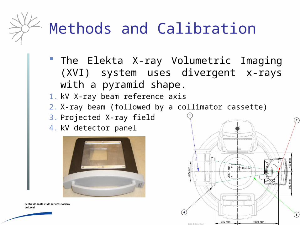
The Elekta X-ray Volumetric Imaging (XVI) system uses divergent x-rays with a pyramid shape.
1. kV X-ray beam reference axis
2. X-ray beam (followed by a collimator cassette)
3. Projected X-ray field
4. kV detector panel
Methods and Calibration
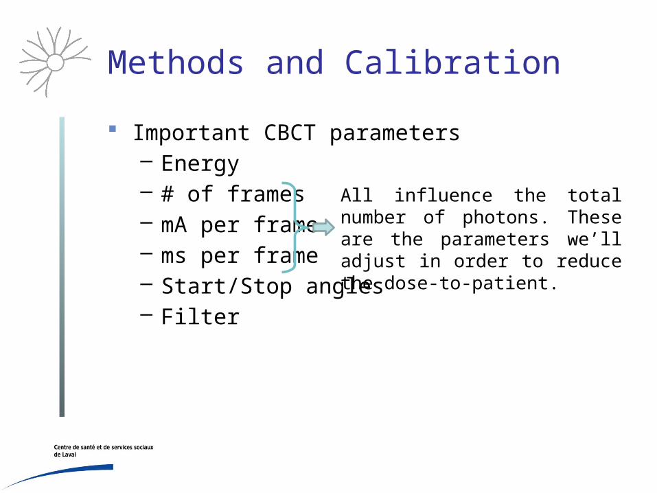
Important CBCT parameters– Energy– # of frames– mA per frame– ms per frame– Start/Stop angles– Filter
All influence the total number of photons. These are the parameters we’ll adjust in order to reduce the dose-to-patient.

Methods and Calibration
Optically Stimulated Luminescence Dosimeters (OSLD) work in a similar way than Thermoluminescent Dosimeters (TLD) using the electrons trapped in a crystalline structure.
Electrons are released using light instead of heat. Landauer Nanodots have a useful energy range
from 5 keV to 20 MeV.

Methods and Calibration
How did we calibrate OSLD?Half Value Layer (HVL) measurements using
aluminum sheets
Air-kerma calibration factor obtained from standard lab
in the kV range for a Farmer ionization chamber
Absolute dose measurement with the
Farmer chamber (AAPM TG 61 in-air method)
Establish a link between the dose read by the chamber
and the number of counts in OSLD
Methods and Calibration
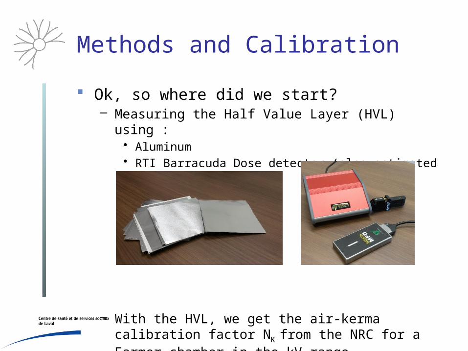
Ok, so where did we start?– Measuring the Half Value Layer (HVL) using :
• Aluminum • RTI Barracuda Dose detector (also estimated kVp and
mAs)
– With the HVL, we get the air-kerma calibration factor NK
from the NRC for a Farmer chamber in the kV range

Methods and Calibration
Using AAPM Task Group 61 in-air method:
• dose at water phantom surface• : corrected free-in-air chamber reading• : air-kerma calibration factor • : mean mass energy-absorption water-to-air ratio• : chamber stem correction factor (1.000 in our case)• : backscatter factor
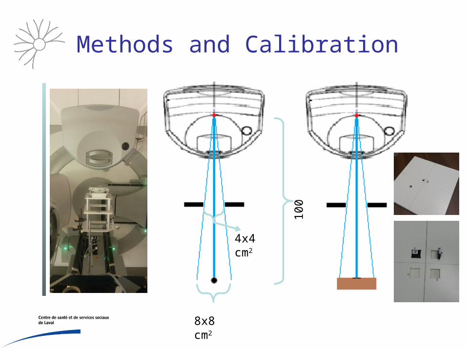
Methods and Calibration
8x8 cm2
4x4 cm2
100
cm

Measurements and Results
We filled an heterogeneous anthropomorphic adult phantom with OSLs.
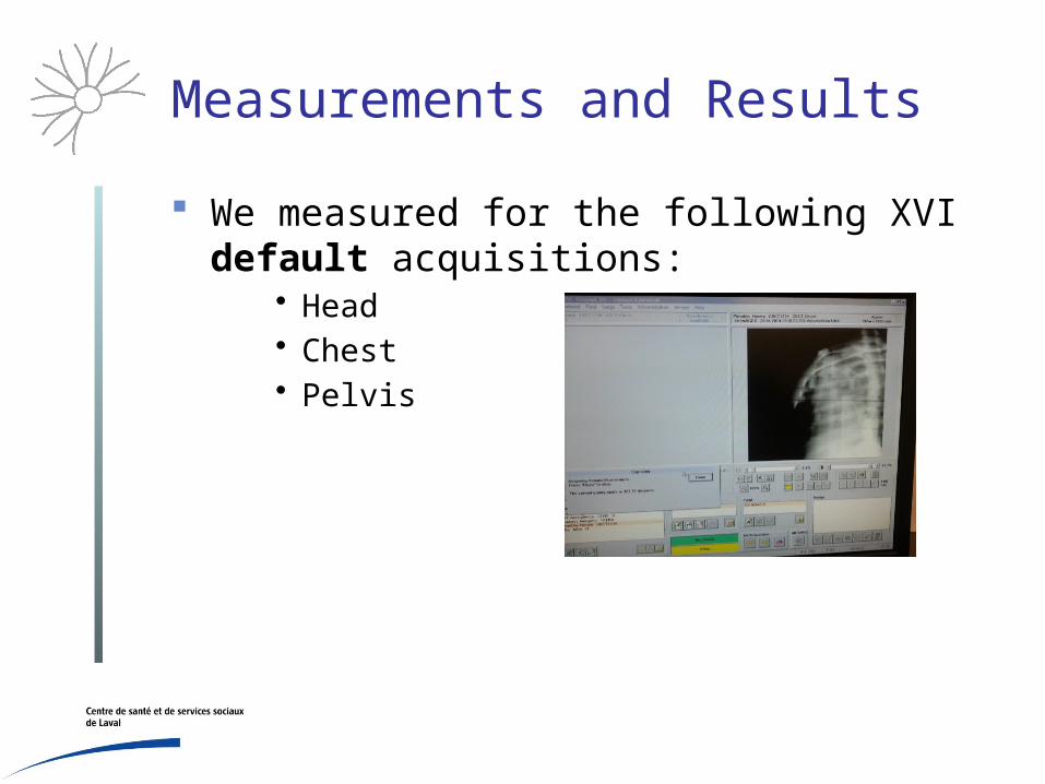
Measurements and Results
We measured for the following XVI default acquisitions:
• Head• Chest• Pelvis
Measurements and Results
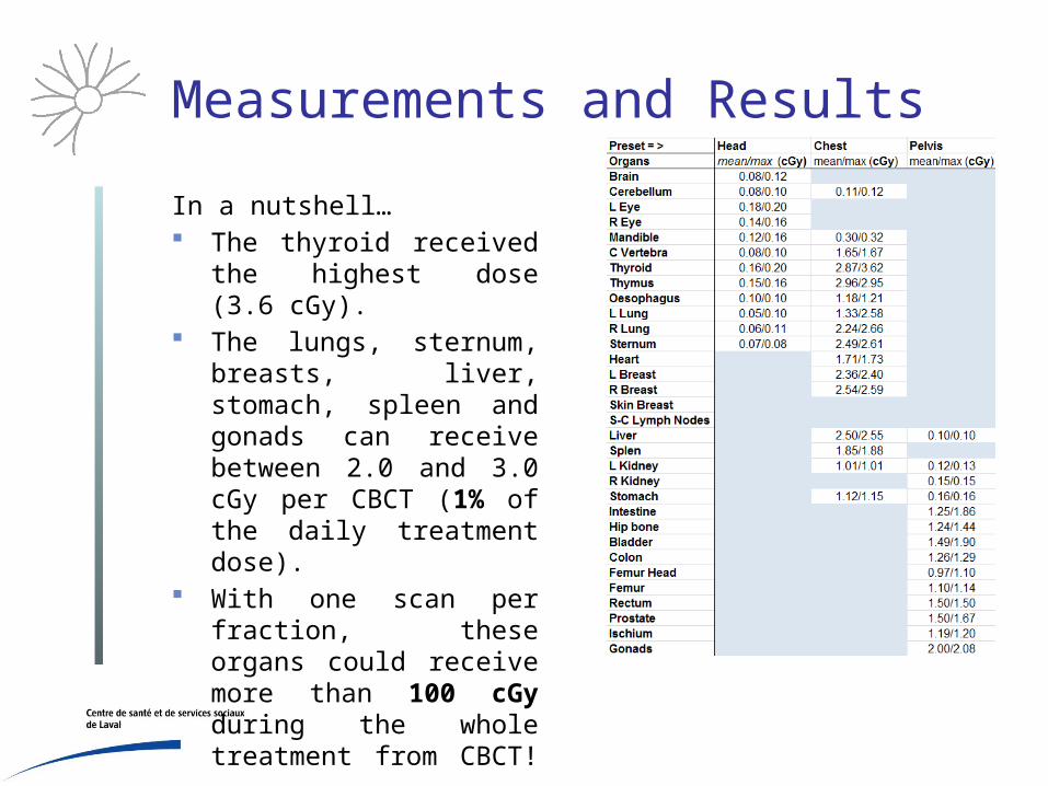
In a nutshell… The thyroid received the
highest dose (3.6 cGy). The lungs, sternum,
breasts, liver, stomach, spleen and gonads can receive between 2.0 and 3.0 cGy per CBCT (1% of the daily treatment dose).
With one scan per fraction, these organs could receive more than 100 cGy during the whole treatment from CBCT!

Measurements and Results
These results match similar studies:– Song et al. Med. Phys. 35: 480-486 (2008)
• XVI dose from 0.1 to 3.5 cGy/scan • Using ionization chamber in phantom
– Hyer et al. Appl. Clin. Med. Phys. 11 181-97 (2010)• XVI dose from 0.1 to 3.0 cGy/scan • Fiber-Optic-Coupled devices
– Alei, Ding and Guan. Med Phys. 37(1) (2007)• XVI dose from 0.1 to 2.8 cGy/scan • Using TLD and Monte-Carlo simulation

Measurements and Results
These results need to be interpret carefully :– Depending on the patient size: Since the number of
photons are always the same, a larger patient might receive less dose and vice versa.
– OSLD response depends on the radiation angle.– The absorbed dose in a given material depends on the
electronic density and the energy of the interacting photon.
– However, these results still a good approximation

Acquisition adjustments
We’ve adjusted the CBCT acquisition parameters (mA per frame, ms per frame and total number of frames).
AcquisitionMaximum
dose (cGy)% of daily
treatment doseRatio with
default dose
Head 0.1 0.1% 1/2
Pelvis 1.0 0.7% 1/4
Chest (low dose) 0.5 0.4% 1/8
Chest (ultra low dose)
0.2 0.2% 1/22
Acquisition adjustments
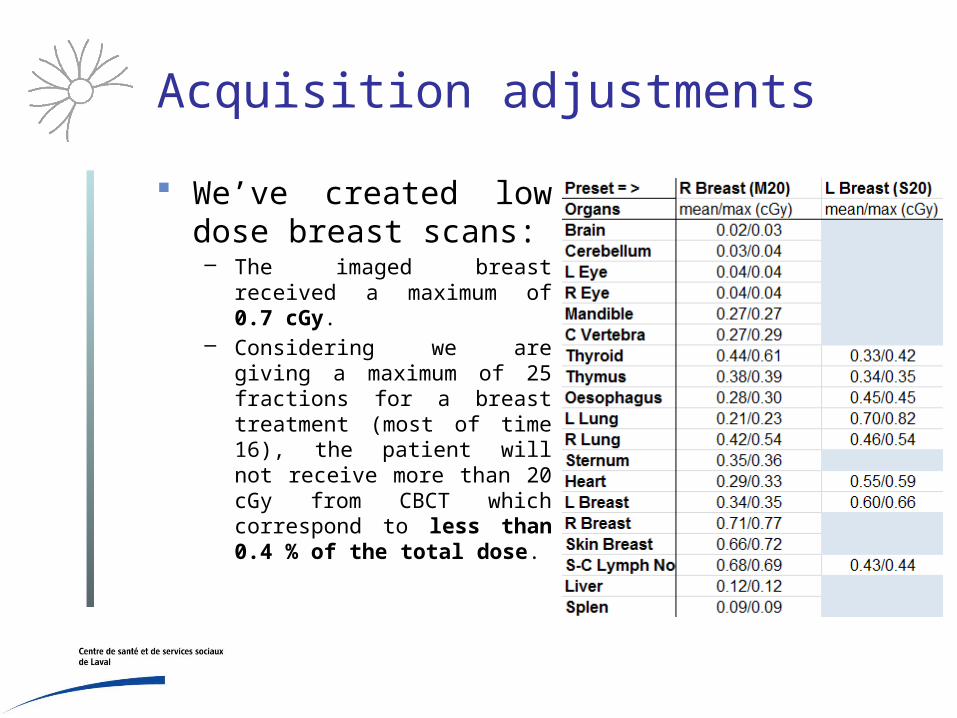
We’ve created low dose breast scans:– The imaged breast received a
maximum of 0.7 cGy.– Considering we are giving a
maximum of 25 fractions for a breast treatment (most of time 16), the patient will not receive more than 20 cGy from CBCT which correspond to less than 0.4 % of the total dose.
Acquisition adjustments
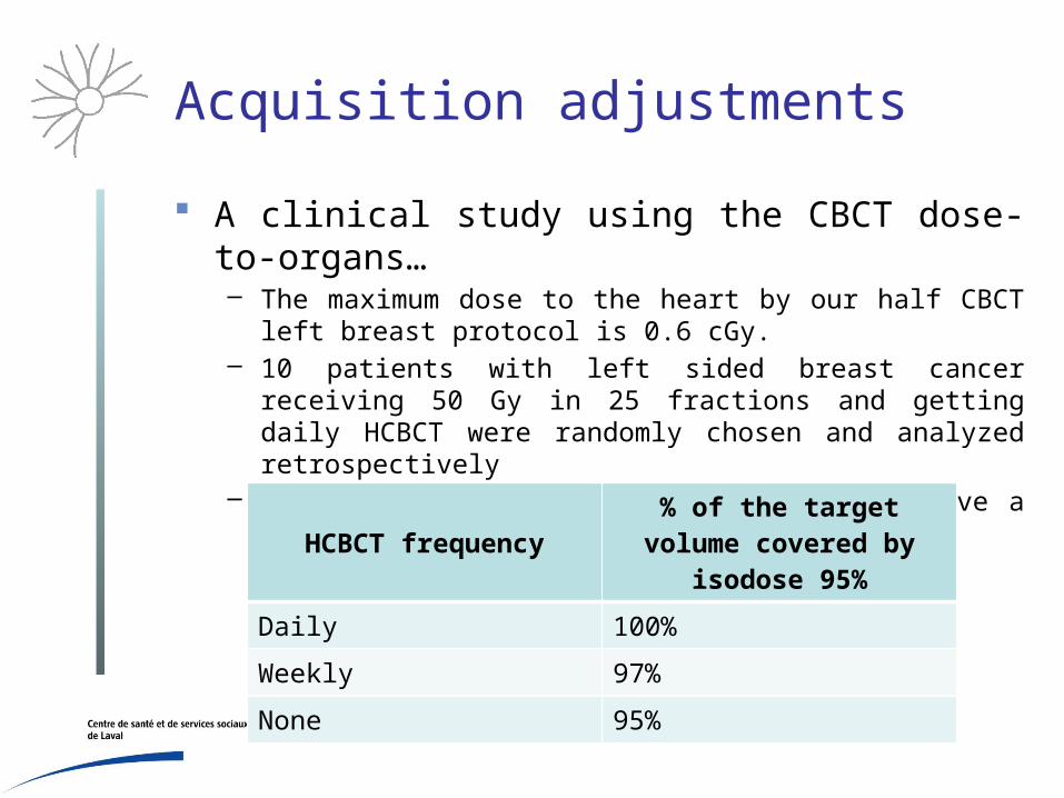
A clinical study using the CBCT dose-to-organs…– The maximum dose to the heart by our half CBCT left breast
protocol is 0.6 cGy.– 10 patients with left sided breast cancer receiving 50 Gy in 25
fractions and getting daily HCBCT were randomly chosen and analyzed retrospectively
– Assuming daily HCBCT, the heart will receive a maximum extra dose of 15 cGy.
HCBCT frequency% of the target volume
covered by isodose 95%
Daily 100%
Weekly 97%
None 95%

Future work
Do a similar experiment on CT scan Compared with different CBCT scans such as the
Varian On-Board Imaging (OBI) Encourage other centers to adjust their
acquisition parameters even if they can’t measure the dose-to-organs

Acknowledgement
Martin Hinse, Nancy El Bared, Danny Duplan, Fabiola Vallejo and all the CICL staff. This research is a beautiful example of interdisciplinary team work.
Merci à vous!
Be there for my presentation on dose to cardiac implantable electronic device, Thursday June 11 AM, Dosimetry and Quality Control session
References
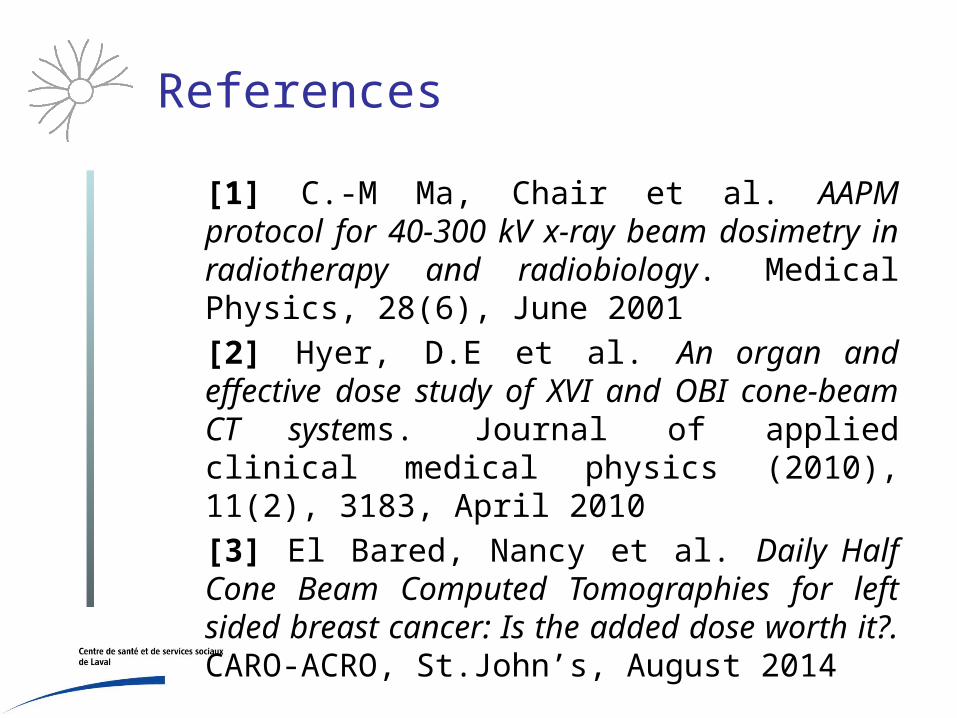
[1] C.-M Ma, Chair et al. AAPM protocol for 40-300 kV x-ray beam dosimetry in radiotherapy and radiobiology. Medical Physics, 28(6), June 2001
[2] Hyer, D.E et al. An organ and effective dose study of XVI and OBI cone-beam CT systems. Journal of applied clinical medical physics (2010), 11(2), 3183, April 2010
[3] El Bared, Nancy et al. Daily Half Cone Beam Computed Tomographies for left sided breast cancer: Is the added dose worth it?. CARO-ACRO, St.John’s, August 2014

THANK YOU!
QUESTIONS?