establishment of anterior–posterior axis in the mouse embryo
TRANSCRIPT
13H. Kondoh and A. Kuroiwa (eds.), New Principles in Developmental Processes, DOI 10.1007/978-4-431-54634-4_2, © Springer Japan 2014
Abstract The anterior–posterior (A–P) axis is the fi rst established and morphologically discernible axis of the body during mouse development. From embryonic day (E) 4.5 to E6.5 of mouse embryos, the formation of the distal vis-ceral endoderm (DVE) followed by that of the anterior visceral endoderm (AVE) breaks the A–P symmetry of the embryo. The DVE progenitor cells arise in primi-tive endoderm (PrE) cells of the late blastocyst with an asymmetrical distribution. This asymmetry may contribute to the determination of the A–P axis in later embryos. At E5.5, DVE cells mature, and migrate from the distal tip to the future anterior side. The DVE migration guides the migration of newly formed AVE and trigger the extensive movement of visceral endoderm (VE) cells in a wide area. Our observations revise the earlier model about AVE development, namely, that the AVE is directly derived from the DVE.
Keywords Anterior–posterior axis • AVE • Blastocyst • DVE • Implantation
2.1 Introduction
Vertebrates have three principal body axes: the anterior–posterior (A–P), dorsal–ventral (D–V), and left–right (L–R) axes. In many animal species, the A–P axis is the fi rst morphologically discernible body axis. How and at which stage of development is the A–P axis determined? The answer depends on the animal species. Establishment of the A–P axis is based on early molecular asymmetry. In Drosophila melanogaster , the A–P axis is defi ned during oogenesis by the asymmetrical deposition of maternal mRNAs along two poles of the oocyte (Huynh and St. Johnston 2004 ).
Chapter 2 Establishment of Anterior–Posterior Axis in the Mouse Embryo
Katsuyoshi Takaoka
K. Takaoka (*) Developmental Genetics Group, Graduate School of Frontier Biosciences, Osaka University , 1-3 Yamada-oka , Suita , Osaka 565-0871 , Japan e-mail: [email protected]

14
In the mouse, the initial process to develop A–P asymmetry has been poorly understood, although several recent reports showed that early A–P molecular asym-metry can be traced back at least to the peri-implantation embryos.
The fertilized mouse egg undergoes cell divisions, increasing the number of blas-tomeres, and reaches the early blastocyst stage at E3.5 (Fig. 2.1 ). The early blasto-cyst is composed of two types of cells: inner cell mass (ICM) and trophectoderm (TE). The ICM contributes to the future embryonic tissues whereas TE contributes
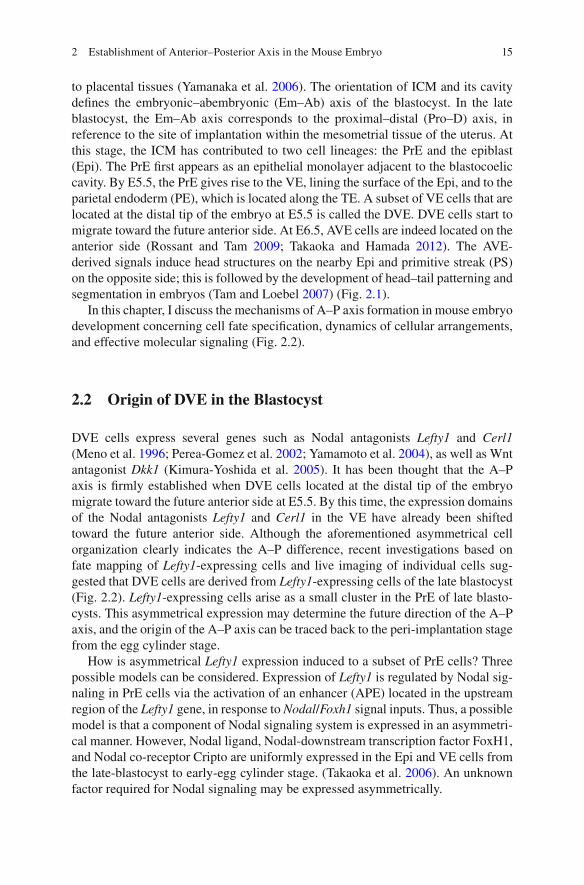
Fig. 2.1 Axis formation of early mouse embryo development. In the early blastocyst, the Em–Ab (embryo–abembryo) axis is defi ned by the position of the inner cell mass ( ICM ). After the implan-tation, the Em–Ab axis and the Pro–D axis of the late blastocyst are aligned in the same direction. The proximal side corresponds to the mesometrial side in the uterus. From E5.5 to E6.5, the ante-rior–posterior ( A–P ) axis of an embryo is established by DVE migration, which is followed by AVE formation. The cell types in the embryos are color coded. AVE anterior visceral endoderm, DVE distal visceral endoderm, E embryonic day, Epi epiblast, Exe extra-embryonic ectoderm, PrE primitive ectoderm, PE parietal endoderm , PS primitive streak, VE visceral endoderm, Em – Ab embryonic–abembryonic axis, Pro–D proximal–distal axis, A–P anterior–posterior axis
K. Takaoka
15
to placental tissues (Yamanaka et al. 2006 ). The orientation of ICM and its cavity defi nes the embryonic–abembryonic (Em–Ab) axis of the blastocyst. In the late blastocyst, the Em–Ab axis corresponds to the proximal–distal (Pro–D) axis, in reference to the site of implantation within the mesometrial tissue of the uterus. At this stage, the ICM has contributed to two cell lineages: the PrE and the epiblast (Epi). The PrE fi rst appears as an epithelial monolayer adjacent to the blastocoelic cavity. By E5.5, the PrE gives rise to the VE, lining the surface of the Epi, and to the parietal endoderm (PE), which is located along the TE. A subset of VE cells that are located at the distal tip of the embryo at E5.5 is called the DVE. DVE cells start to migrate toward the future anterior side. At E6.5, AVE cells are indeed located on the anterior side (Rossant and Tam 2009 ; Takaoka and Hamada 2012 ). The AVE- derived signals induce head structures on the nearby Epi and primitive streak (PS) on the opposite side; this is followed by the development of head–tail patterning and segmentation in embryos (Tam and Loebel 2007 ) (Fig. 2.1 ).
In this chapter, I discuss the mechanisms of A–P axis formation in mouse embryo development concerning cell fate specifi cation, dynamics of cellular arrangements, and effective molecular signaling (Fig. 2.2 ).
2.2 Origin of DVE in the Blastocyst
DVE cells express several genes such as Nodal antagonists Lefty1 and Cerl1 (Meno et al. 1996 ; Perea- Gomez et al. 2002 ; Yamamoto et al. 2004 ), as well as Wnt antagonist Dkk1 (Kimura- Yoshida et al. 2005 ). It has been thought that the A–P axis is fi rmly established when DVE cells located at the distal tip of the embryo migrate toward the future anterior side at E5.5. By this time, the expression domains of the Nodal antagonists Lefty1 and Cerl1 in the VE have already been shifted toward the future anterior side. Although the aforementioned asymmetrical cell organization clearly indicates the A–P difference, recent investigations based on fate mapping of Lefty1 -expressing cells and live imaging of individual cells sug-gested that DVE cells are derived from Lefty1 -expressing cells of the late blastocyst (Fig. 2.2 ). Lefty1 -expressing cells arise as a small cluster in the PrE of late blasto-cysts. This asymmetrical expression may determine the future direction of the A–P axis, and the origin of the A–P axis can be traced back to the peri-implantation stage from the egg cylinder stage.
How is asymmetrical Lefty1 expression induced to a subset of PrE cells? Three possible models can be considered. Expression of Lefty1 is regulated by Nodal sig-naling in PrE cells via the activation of an enhancer (APE) located in the upstream region of the Lefty1 gene, in response to Nodal / Foxh1 signal inputs. Thus, a possible model is that a component of Nodal signaling system is expressed in an asymmetri-cal manner. However, Nodal ligand, Nodal-downstream transcription factor FoxH1, and Nodal co-receptor Cripto are uniformly expressed in the Epi and VE cells from the late-blastocyst to early-egg cylinder stage. (Takaoka et al. 2006 ). An unknown factor required for Nodal signaling may be expressed asymmetrically.
2 Establishment of Anterior–Posterior Axis in the Mouse Embryo

16 K. Takaoka

17
Fig
. 2.2
A
–P a
xis
form
atio
n du
ring
mou
se e
mbr
yo d
evel
opm
ent:
orig
ins
and
mig
ratio
ns o
f th
e D
VE
and
the
AV
E in
em
bryo
pat
tern
ing
alon
g th
e A
–P a
xis.
T
he D
VE
line
age
is s
how
n in
gre
en a
nd th
e A
VE
line
age
is s
how
n in
pur
ple
cell
s ; s
olid
col
orin
g in
dica
tes
cells
that
exp
ress
Lef
ty1 ,
whe
reas
col
orin
g on
ly in
ou
tline
ind
icat
es c
ells
with
out
Lef
ty1
expr
essi
on. (
a) A
t E
4.2,
Lef
ty1
is e
xpre
ssed
in
the
DV
E p
roge
nito
rs (
gree
n ). (
b) I
mm
atur
e D
VE
cel
ls, w
hich
mai
ntai
n Le
fty1
exp
ress
ion
but a
re n
egat
ive
for
othe
r D
VE
mar
kers
suc
h as
Cer
l1 a
nd H
ex , a
re f
ound
at t
he d
ista
l tip
of
the
embr
yo a
t E5.
2. (
c) A
t E5.
5, m
atur
ed D
VE
ce
lls e
xpre
ss L
efty
1 an
d ot
her D
VE
mar
kers
. Sha
pes
of D
VE
cel
ls c
hang
e to
col
umna
r. D
VE
cel
ls m
igra
te to
the
futu
re a
nter
ior s
ide
of th
e em
bryo
. (d)
At E
5.7,
D
VE
cel
ls m
igra
te a
way
fro
m th
e di
stal
tip,
and
VE
cel
ls n
egat
ive
for
Left
y1 e
xpre
ssio
n ( o
pen
mag
enta
sha
pes )
mov
e to
the
dist
al ti
p an
d be
com
e ne
w A
VE
ce
lls. (
e) A
t E6.
0, D
VE
cel
ls th
at h
ave
reac
hed
the
embr
yoni
c/ex
tra-
embr
yoni
c ju
nctio
n m
igra
te la
tera
lly a
nd lo
se e
xpre
ssio
n of
DV
E m
arke
rs, i
nclu
ding
Lef
ty1 .
A
VE
cel
ls r
emai
n ne
wly
gen
erat
ed a
t the
dis
tal t
ip a
nd a
lso
mig
rate
tow
ard
the
prox
imal
sid
e. (
f) A
t E6.
5, th
e A
VE
occ
upie
s th
e an
teri
or s
ide
of th
e em
bryo
, an
d cr
eate
s A
–P p
atte
rnin
g in
the
Epi
. AV
E a
nter
ior
visc
eral
end
oder
m, D
VE
dis
tal
visc
eral
end
oder
m, E
em
bryo
nic
day,
Epi
epi
blas
t, E
xe e
xtra
-em
bryo
nic
ecto
derm
, PrE
pri
miti
ve e
ctod
erm
, PS
prim
itive
str
eak,
VE
vis
cera
l end
oder
m, P
ro–D
pro
xim
al–d
ista
l axi
s, A
–P a
nter
ior–
post
erio
r ax
is
2 Establishment of Anterior–Posterior Axis in the Mouse Embryo

18
The second model assumes crosstalk of these signaling systems with other sig-naling pathways. Wnt is a candidate signaling system that crosstalks with other systems. The nuclear localization of β-catenin, which is indicative of active Wnt signaling, was observed in late blastocysts detected in a single Epi cell that faces the PrE (Chazaud and Rossant 2006 ). In addition, investigation on microarray profi ling of β-catenin mutant embryos identifi ed Cripto, a Nodal co-receptor, as a β-catenin target gene (Morkel et al. 2003 ). These results suggest that Nodal signaling is read-ily activated with Wnt signal input.
The third model, the restriction of Lefty1 expression to a subset of ICM cells, involves a self-enhancement lateral inhibition (SELI) mechanism, which amplifi es a small difference of Nodal signaling activities into a robust asymmetry, as has been demonstrated for the left–right embryo patterning (Nakamura et al. 2006 ; Nakamura and Hamada 2012 ) (Fig. 2.3 ). Lefty1 is induced by Nodal signaling, and Nodal is
Fig. 2.3 Model for the self-enhancement lateral inhibition ( SELI ) mechanism in DVE formation. (a) Observation: Lefty1 -positive cells ( green ), putative DVE progenitors, appear as a very small population of PrE cells ( blue ). (b) Scheme of cross-regulation between Nodal and Lefty 1. Lefty1 ( green ) is induced by Nodal signaling ( magenta ), and Nodal signaling is inhibited by a negative feedback. (c) Model: PrE cells have a threshold for the Nodal signaling activity that allows Lefty1 activation (step 1). By a stochastic mechanism, the activity of Nodal signaling exceeds the thresh-old in a fraction of PrE cells ( yellow arrows , step 2). Then, Lefty1 expression is activated in these cells, and the activated Lefty1 inhibits Nodal signaling in other PrE cells (step 3). The DVE is formed (step 4)
K. Takaoka

19
regulated by the Activin / Nodal signaling-dependent enhancer of Nodal (ASE) in a subset of ICM cells (Granier et al. 2011 ). These coordinated activities of Nodal and its feedback inhibitor Lefty meet the requirements for the SELI system.
2.3 Maturation of DVE and DVE Migration
From E4.5 to E5.5, expression of Lefty1 is cell autonomously maintained in the DVE progenitor to mature DVE cells. At E5.5, a greater variety of genes such as Cerl1 , Hex , and Dkk1 are expressed in the DVE, suggesting that signaling systems change as the DVE cells become mature. In this signaling system, bone morphoge-netic protein (BMP) signaling plays an important role. BMP4 , which is expressed in ExE (extra-embryonic ectoderm) cells, generates a gradient of BMP signaling in the embryonic Epi and VE tissues. The DVE progenitors are confi ned to the distal tip of embryos corresponding to the BMP signaling-negative portion from E5.2 to E5.5. The BMP signaling thus restricts the DVE region to the distal tip of embryos. Indeed, ExE-removed embryos show an expansion of the DVE region (Rodriguez et al. 2005 ; Yamamoto et al. 2009 ) (Fig. 2.4 ).
Fig. 2.4 Changes in the network of signaling systems during the developmental stages for DVE migration and AVE formation. Major changes in the spatial organization of the signaling system are caused by the migrations of DVE ( green ) and AVE ( magenta ) cells. After E5.5, the DVE migration transforms the P–D differences in the signaling systems into the A–P differences. At E6.0 and E6.5, signals from the AVE specify the nearby Epi by inhibiting Nodal signaling and Wnt signaling. On the opposite side far from the AVE, Nodal and Wnt signaling remains active, which results in the formation of the primitive streak ( light blue ). Activities of bone morphogenetic pro-tein ( BMP ), Nodal, and Wnt are indicated by purple and blue triangles , respectively
2 Establishment of Anterior–Posterior Axis in the Mouse Embryo

20
Alongside their maturation, DVE cells migrate to the future anterior side of the early egg cylinder embryos. Immediately before their migration, three morphologi-cal changes take place in the DVE cells. The fi rst is “visceral endoderm thickening” (VET), which makes the entire VE a monolayer of tall columnar cells (Rivera-Perez et al. 2003 ; Srinivas 2006 ; Takaoka et al. 2011 ) (Fig. 2.2 ).
Second, lamellipodia- and fi lopodia-like structures are formed in migrating DVE cells (Rakeman and Anderson 2006 ; Migeotte et al. 2010 ). In the VE-specifi c con-ditional mutant embryo lacking the Rho family GTPase Rac1, a component of WAVE complex (a regulator of the actin cytoskeleton), the lamellipodia- and fi lopodia- like structures fail to form. As a result, DVE cells fail to migrate away from the distal tip. In Nap1 mutant embryos also, lacking a component of WAVE complex, DVE cells fail to migrate (Rakeman and Anderson 2006 ). These observa-tions indicate that dynamic cytoskeletal reorganization regulated by the WAVE complex is required for the correct DVE migration.
Third, at a more macroscopic level, DVE cells form multicellular rosettes. Rosettes of fi ve or more cells, sharing a central vertex, are formed in the sim-ple epithelium of VE during the migration of DVE. Formation of the cellular rosettes depends on PCP signaling, and is supposed to buffer the structural dis-turbance in cell packing in an epithelium caused by DVE migration (Trichas et al. 2012 ).
2.4 The Direction of the A–P Axis
At E5.5, DVE cells migrate to the future anterior side. It is a critical issue whether the direction of A–P is determined before the DVE migration. First, several studies suggest that embryos have A–P asymmetry before the DVE migration. One sug-gestion is that the expression domains of the Nodal antagonists Lefty1 and Cerl1 are already shifted toward the future anterior side before E5.5. Forced expression of Nodal antagonist Lefty1 at E5.2 causes asymmetry in Nodal signaling and initi-ates the migration of DVE (Yamamoto et al. 2004 ). Similarly, Wnt antagonist and Dkk1 expression domains change dynamically between before and after DVE migration, and the ectopic activation of Dkk1 can alter the direction of DVE cell migration (Kimura-Yoshida et al. 2005 ). These reports suggested that embryos may have A–P pre-patterning before E5.5. However, DVE still migrates unilater-ally toward the proximal side in mutant mice that lack both Lefty1 and Cerl1 (Perea-Gomez et al. 2002 ; Yamamoto et al. 2004 ) or those lacking Dkk1 (del Barco Barrantes et al. 2003 ). The lack of migration defects in these mutant embryos may be caused by functional redundancy between Nodal-dependent and Wnt-dependent mechanisms. The process that determines the direction of the A–P axis may involve parallel mechanisms.
K. Takaoka

21
2.5 Role of DVE and AVE
Recent investigations have indicated that the descendant of the DVE contributed mainly to the most proximal portion of AVE and VE in the lateral region, but not to the entire AVE (Fig. 2.2 ). Time-lapse observation of DVE migration and AVE for-mation showed that the extensive movement of VE cells is initiated by DVE migra-tion at E5.5 (Takaoka et al. 2011 ). While DVE cells migrate, the majority of AVE cells are newly formed at the distal end, migrate toward the proximal side following DVE migration, and eventually occupy the entire region of the AVE (Takaoka et al. 2011 ) (Fig. 2.2 ).
What is the role of the DVE in AVE formation? In DVE-ablated embryos caused by genetic manipulation, AVE cells are newly formed at the distal end but fail to migrate. Under this condition, extensive movement of VE cells does not occur, and the migration of the AVE cells is stalled (Takaoka et al. 2011 ). For instance, in mutant embryos lacking Cripto, a coreceptor of Nodal, the AVE is formed in the absence of DVE in the absence of DVE but does not migrate at all (Chu and Shen 2010 ). These observations suggest that the DVE guides the AVE to the anterior side by initiating extensive movement of visceral endoderm cells.
AVE cells function as head organizer by expressing a few signal protein antago-nists, that is, Nodal antagonists Lefty1 and Cerl1, and Wnt antagonist Dkk1. Theses antagonists protect nearby Epi cells from posterior-inducing Wnt and Nodal signal-ing (Fig. 2.4 ), specifying AVE-underlain Epi to head tissue. In fact, explant culture assays have shown that Epi cells without VE will adopt the posterior identity, whereas implanted AVE cause Epi to assume an anterior identity (Kimura et al. 2000 ).
2.6 Reinterpretation of Embryo Phenotypes Defective in A–P Axis Formation
It had been believed that the mature DVE is the direct precursor for the entire AVE and its migration toward the future anterior side away from the distal tip is the mechanism of AVE formation (Beddington and Robertson 1999 ) (Fig. 2.5 ). However, the recent observation that DVE does not contribute to the major part of AVE renders it necessary to reinterpret the phenotypes of previously reported mutant mouse embryos based on the following four criteria: (1) DVE formation; (2) DVE migration; (3) AVE formation; and (4) AVE migration (Table 2.1 ).
In Nodal -null (Waldrip et al. 1998 ; Mesnard et al. 2006 ) and Smad2 -null (Waldrip et al. 1998 ) mutants, the Epi is specifi ed to neural tissues precociously, and neither DVE nor AVE forms (Fig. 2.6 ). In Cripto -null (Ding et al. 1998 ; Chu and Shen 2010 ) and Eomes VE-conditional null embryos, Cerl1 , a DVE marker, is expressed at very reduced levels or not at all at E5.5. Cerl1 -positive AVE cells are newly formed close to the distal tip of the mutant embryo from E6.0 to E6.5, but they fail to migrate proximally and remain at the distal tip of the embryo, as in the
2 Establishment of Anterior–Posterior Axis in the Mouse Embryo

22
DVE- ablated embryo (Takaoka et al. 2011 ; Morris et al. 2012 ) (Fig. 2.6 ). In other mutants also, such as Otx2 -null (Kimura-Yoshida et al. 2005 ), Rac1 -null (Sugihara et al. 1998 ; Migeotte et al. 2010 ), and Pten -null (Bloomekatz et al. 2012 ) mutants, the DVE is formed at the distal dip at E5.5, but fails to migrate. Thus, cells that express DVE/AVE markers remain close to the distal tip of the embryo at E6.0 and E6.5 (Fig. 2.6 ). These observations indicate that the major role of DVE is to guide the AVE migration.
2.7 Conclusion
The A–P axis of mouse embryos is fully established by the directed movement of DVE and AVE cells, the latter known as s head organizer. DVE progenitor cells are distributed on one side of the primitive endoderm cells at E4.2. The direction of the A–P axis of later embryos may depend on this asymmetry. At E5.5, DVE cells that express Nodal antagonists Lefty1 and Cer1, and Wnt antagonist Dkk1,
Fig. 2.5 Comparison of previously proposed model and current model concerning the roles of DVE in AVE formation. Previous model ( upper ): The DVE is specifi ed at the distal tip of the embryo at E5.5, then migrates toward the anterior and proximal side to form the AVE. It has been thought that all AVE cells are derived from DVE. New model ( lower ): Origin of the DVE can be traced back to the Lefty1 -positve primitive endoderm cells. The migration of DVE cells triggers global VE movements, and AVE cells are newly produced at the distal tip. In the proximally located migrating cells, DVE markers are lost. At E6.5, newly formed AVE cells occupy the ante-rior side of VE cells
K. Takaoka

23
become mature and migrate to the future anterior side. Immediately before their migration, morphological changes occur in the VE cells, namely, visceral endo-derm thickening, formation of lamellipodia- and fi lopodia-like processes, and organization of multicellular rosettes. DVE migration toward the future anterior side has the role of guiding the AVE cells to the anterior side of the egg cylinder embryo, and triggers extensive rearrangement of visceral endoderm cells in a wide area.
Important questions that remain to be answered are these: how are DVE progeni-tors selected among PrE cells in the blastocyst, and at which stage of development is the direction of DVE migration determined?
Table 2.1 Reinterpretation of mutant mouse phenotypes that include defective anterior visceral endoderm (AVE) formation
Gene Genetic modifi cation
DVE formation at E5.5
DVE migration
AVE formation at E6.5
AVE migration References
Nodal Null No – No – Brennan et al. ( 2001 ), Camus et al. ( 2006 ) and Mesnard et al. ( 2006 )
Smad2 Null No – No – Nomura and Li ( 1998 ) and Brennan et al. ( 2001 )
Cripto Null No – Yes No Ding et al. ( 1998 ), Kimura et al. ( 2001 ) and Chu and Shen ( 2010 )
Eomes VE-specifi c null
Low No Yes No Nowotschin et al. ( 2013 )
Otx2 Null Yes No Yes No Kimura et al. ( 2000 , 2001 ) and Kimura- Yoshida et al. ( 2005 )
Rac1 VE-specifi c null
Yes No Yes No Migeotte et al. ( 2010 )
Lefty1 Null Expansion Yes Yes Yes Yamamoto et al. ( 2004 )
Lefty1 Cerl1
Double null N.D. N.D. Yes Delay Perea-Gomez et al. ( 2002 ) and Yamamoto et al. ( 2004 )
2 Establishment of Anterior–Posterior Axis in the Mouse Embryo

24
Acknowledgments We thank members of the Hamada laboratory for discussion.
References
Beddington RS, Robertson EJ (1999) Axis development and early asymmetry in mammals. Cell 96(2):195–209. doi: 10.1016/S0092-8674(00)80560-7
Bloomekatz J, Grego-Bessa J, Migeotte I, Anderson KV (2012) Pten regulates collective cell migration during specifi cation of the anterior-posterior axis of the mouse embryo. Dev Biol 364(2):192–201. doi: 10.1016/j.ydbio.2012.02.005
Brennan J, Lu CC, Norris DP, Rodriguez TA, Beddington RS, Robertson EJ (2001) Nodal signalling in the epiblast patterns the early mouse embryo. Nature (Lond) 411(6840):965–969. doi: 10.1038/35082103
Camus A, Perea-Gomez A, Moreau A, Collignon J (2006) Absence of Nodal signaling promotes precocious neural differentiation in the mouse embryo. Dev Biol 295(2):743–755. doi: 10.1016/j.ydbio.2006.03.047
Chazaud C, Rossant J (2006) Disruption of early proximodistal patterning and AVE formation in Apc mutants. Development(Camb) 133(17):3379–3387. doi: 10.1242/dev.02523
Fig. 2.6 Grouping of mouse mutant phenotypes defective in the A–P axis development. (a) Wild-type embryos. (b) Precocious neural differentiation occurs in the Epi; thus, the DVE and the AVE fail to form throughout E5.5 and E6.5 stages. (c) The DVE does not form at E5.5. The AVE is newly formed at the distal tip at E6.0 but fails to migrate. As a result, Epi cells with posterior identity develop at the proximal side of the embryo. (d) The DVE forms at the distal tip of the embryo at E5.5 but fails to migrate. Cells positive for DVE/AVE markers remain at the distal tip at E6.0 and E6.5. As a result, Epi cells with posterior identity develop at the proximal side of the embryo, similar to (c)
K. Takaoka

25
Chu J, Shen MM (2010) Functional redundancy of EGF-CFC genes in epiblast and extraembry-onic patterning during early mouse embryogenesis. Dev Biol 342(1):63–73. doi: 10.1016/j.ydbio.2010.03.009
del Barco Barrantes I, Davidson G, Grone HJ, Westphal H, Niehrs C (2003) Dkk1 and noggin cooperate in mammalian head induction. Genes Dev 17(18):2239–2244. doi: 10.1101/gad.269103
Ding J, Yang L, Yan YT, Chen A, Desai N, Wynshaw-Boris A, Shen MM (1998) Cripto is required for correct orientation of the anterior-posterior axis in the mouse embryo. Nature (Lond) 395(6703):702–707. doi: 10.1038/27215
Granier C, Gurchenkov V, Perea-Gomez A, Camus A, Ott S, Papanayotou C, Iranzo J, Moreau A, Reid J, Koentges G, Saberan-Djoneidi D, Collignon J (2011) Nodal cis-regulatory elements reveal epiblast and primitive endoderm heterogeneity in the peri-implantation mouse embryo. Dev Biol 349(2):350–362. doi: 10.1016/j.ydbio.2010.10.036
Huynh JR, St. Johnston D (2004) The origin of asymmetry: early polarisation of the Drosophila germline cyst and oocyte. Curr Biol 14(11):R438–449. doi: 10.1016/j.cub.2004.05.040
Kimura C, Yoshinaga K, Tian E, Suzuki M, Aizawa S, Matsuo I (2000) Visceral endoderm medi-ates forebrain development by suppressing posteriorizing signals. Dev Biol 225(2):304–321. doi: 10.1006/dbio.2000.9835
Kimura C, Shen MM, Takeda N, Aizawa S, Matsuo I (2001) Complementary functions of Otx2 and Cripto in initial patterning of mouse epiblast. Dev Biol 235(1):12–32. doi: 10.1006/dbio.2001.0289
Kimura-Yoshida C, Nakano H, Okamura D, Nakao K, Yonemura S, Belo JA, Aizawa S, Matsui Y, Matsuo I (2005) Canonical Wnt signaling and its antagonist regulate anterior-posterior axis polarization by guiding cell migration in mouse visceral endoderm. Dev Cell 9(5):639–650. doi: 10.1016/j.devcel.2005.09.011
Meno C, Saijoh Y, Fujii H, Ikeda M, Yokoyama T, Yokoyama M, Toyoda Y, Hamada H (1996) Left-right asymmetric expression of the TGF beta-family member lefty in mouse embryos. Nature (Lond) 381(6578):151–155. doi: 10.1038/381151a0
Mesnard D, Guzman-Ayala M, Constam DB (2006) Nodal specifi es embryonic visceral endoderm and sustains pluripotent cells in the epiblast before overt axial patterning. Development (Camb) 133(13):2497–2505. doi: 10.1242/dev.02413
Migeotte I, Omelchenko T, Hall A, Anderson KV (2010) Rac1-dependent collective cell migration is required for specifi cation of the anterior-posterior body axis of the mouse. PLoS Biol 8(8):e1000442. doi: 10.1371/journal.pbio.1000442
Morkel M, Huelsken J, Wakamiya M, Ding J, van de Wetering M, Clevers H, Taketo MM, Behringer RR, Shen MM, Birchmeier W (2003) Beta-catenin regulates Cripto- and Wnt3- dependent gene expression programs in mouse axis and mesoderm formation. Development (Camb) 130(25):6283–6294. doi: 10.1242/dev.00859
Morris SA, Grewal S, Barrios F, Patankar SN, Strauss B, Buttery L, Alexander M, Shakesheff KM, Zernicka-Goetz M (2012) Dynamics of anterior-posterior axis formation in the developing mouse embryo. Nat Commun 3:673. doi: 10.1038/ncomms1671
Nakamura T, Hamada H (2012) Left-right patterning: conserved and divergent mechanisms. Development (Camb) 139(18):3257–3262. doi: 10.1242/dev.061606
Nakamura T, Mine N, Nakaguchi E, Mochizuki A, Yamamoto M, Yashiro K, Meno C, Hamada H (2006) Generation of robust left-right asymmetry in the mouse embryo requires a self- enhancement and lateral-inhibition system. Dev Cell 11(4):495–504. doi: 10.1016/j.devcel.2006.08.002
Nomura M, Li E (1998) Smad2 role in mesoderm formation, left-right patterning and craniofacial development. Nature (Lond) 393(6687):786–790. doi: 10.1038/31693
Nowotschin S, Costello I, Pillisezek A, Kwon GS, Mao CA, Klein WH, Robertson EJ, Hadjantonakis AK (2013) The T-box transcription factor Eomesodermin is essential for AVE induction in the mouse embryos. Genens Dev 27(9):997–1002. doi: 10.1101/gad.215152.113
Perea-Gomez A, Vella FD, Shawlot W, Oulad-Abdelghani M, Chazaud C, Meno C, Pfi ster V, Chen L, Robertson E, Hamada H, Behringer RR, Ang SL (2002) Nodal antagonists in the anterior visceral endoderm prevent the formation of multiple primitive streaks. Dev Cell 3(5):745–756
2 Establishment of Anterior–Posterior Axis in the Mouse Embryo

26
Rakeman AS, Anderson KV (2006) Axis specifi cation and morphogenesis in the mouse embryo require Nap1, a regulator of WAVE-mediated actin branching. Development (Camb) 133(16):3075–3083. doi: 10.1242/dev.02473
Rivera-Perez JA, Mager J, Magnuson T (2003) Dynamic morphogenetic events characterize the mouse visceral endoderm. Dev Biol 261(2):470–487. doi: 10.1016/S0012-1606(03)00302-6
Rodriguez TA, Srinivas S, Clements MP, Smith JC, Beddington RS (2005) Induction and migration of the anterior visceral endoderm is regulated by the extra-embryonic ectoderm. Development (Camb) 132(11):2513–2520. doi: 10.1242/dev.01847
Rossant J, Tam PP (2009) Blastocyst lineage formation, early embryonic asymmetries and axis patterning in the mouse. Development (Camb) 136(5):701–713. doi: 10.1242/dev.017178
Srinivas S (2006) The anterior visceral endoderm-turning heads. Genesis 44(11):565–572. doi: 10.1002/dvg.20249
Sugihara K, Nakatsuji N, Nakamura K, Nakao K, Hashimoto R, Otani H, Sakagami H, Kondo H, Nozawa S, Aiba A, Katsuki M (1998) Rac1 is required for the formation of three germ layers during gastrulation. Oncogene 17(26):3427–3433. doi: 10.1038/sj.onc.1202595
Takaoka K, Hamada H (2012) Cell fate decisions and axis determination in the early mouse embryo. Development (Camb) 139(1):3–14. doi: 10.1242/dev.060095
Takaoka K, Yamamoto M, Shiratori H, Meno C, Rossant J, Saijoh Y, Hamada H (2006) The mouse embryo autonomously acquires anterior-posterior polarity at implantation. Dev Cell 10(4):451–459. doi: 10.1016/j.devcel.2006.02.017
Takaoka K, Yamamoto M, Hamada H (2011) Origin and role of distal visceral endoderm, a group of cells that determines anterior-posterior polarity of the mouse embryo. Nat Cell Biol 13(7):743–752. doi: 10.1038/ncb2251
Tam PP, Loebel DA (2007) Gene function in mouse embryogenesis: get set for gastrulation. Nat Rev Genet 8(5):368–381. doi: 10.1038/nrg2084
Trichas G, Smith AM, White N, Wilkins V, Watanabe T, Moore A, Joyce B, Sugnaseelan J, Rodriguez TA, Kay D, Baker RE, Maini PK, Srinivas S (2012) Multi-cellular rosettes in the mouse visceral endoderm facilitate the ordered migration of anterior visceral endoderm cells. PLoS Biol 10(2):e1001256. doi: 10.1371/journal.pbio.1001256
Waldrip WR, Bikoff EK, Hoodless PA, Wrana JL, Robertson EJ (1998) Smad2 signaling in extra-embryonic tissues determines anterior-posterior polarity of the early mouse embryo. Cell 92(6):797–808
Yamamoto M, Saijoh Y, Perea-Gomez A, Shawlot W, Behringer RR, Ang SL, Hamada H, Meno C (2004) Nodal antagonists regulate formation of the anteroposterior axis of the mouse embryo. Nature (Lond) 428(6981):387–392. doi: 10.1038/nature02418
Yamamoto M, Beppu H, Takaoka K, Meno C, Li E, Miyazono K, Hamada H (2009) Antagonism between Smad1 and Smad2 signaling determines the site of distal visceral endoderm formation in the mouse embryo. J Cell Biol 184(2):323–334. doi: 10.1083/jcb.200808044
Yamanaka Y, Ralston A, Stephenson RO, Rossant J (2006) Cell and molecular regulation of the mouse blastocyst. Dev Dyn 235(9):2301–2314. doi: 10.1002/dvdy.20844
K. Takaoka

http://www.springer.com/978-4-431-54633-7