establishing a high titer transient gene expression ... · joão nuno dos santos pereira licenciado...
TRANSCRIPT

João Nuno dos Santos Pereira
Licenciado em Bioquímica
Establishing a High Titer Transient Gene Expression Process in
Conditioned Media for CHO-DG44 Cells
Dissertação para obtenção do Grau de Mestre em
Biotecnologia
Orientador: Florian M. Wurm, Prof., EPFL
Júri: Presidente: Prof. Doutor Rui Manuel Freitas Oliveira
Arguente: Doutora Paula Maria Marques Leal Sanches Alves
Outubro, 2011

i
Copyright
Establishing a high titer transient gene expression process in conditioned media for
CHO-DG44 cells ©
João Nuno dos Santos Pereira
FCT/UNL
UNL
A Faculdade de Ciências e Tecnologia e a Universidade Nova de Lisboa têm o direito,
perpétuo e sem limites geográficos de arquivar e publicar esta dissertação através de
exemplares impressos reproduzidos em papel ou de forma digital, ou por qualquer outro
meio conhecido ou que venha a ser inventado, e de a divulgar atraves de repositórios
cientificos e de admitir a sua côpia e distribuiçao com objectivos educacionais ou de
investigacao, não comerciais, desde que seja dado crédito ao autor e editor

ii

iii
“Muitos anos depois, diante do pelotão de fuzilamento, o Coronel Aureliano Buendía havia de recordar aquela tarde remota em que seu pai o levou para conhecer o gelo.
(...)
Desconcertado, sabendo que os meninos esperavam uma expllicação imediata, José Arcadio Buendía atreveu-se a murmurar:
- É o maior diamante do mundo.
- Não – corrigiu o cigano – É gelo!”
Gabriel García Marquez “Cem anos de Solidão”

iv
v
“Many years later, as he faced the firing squad, Colonel Aureliano Buendía was to remember that distant afternoon when his father took him to discover ice.
(...)
Disconcerted, knowing the children were waiting for an immediate explanation, José Arcadio Buendía ventured a murmur:
- It’s the largest diamond in the world.
- No – the gipsy countered – It’s ice!”
Gabriel García Marquez “One Hundred Years of Solitude”

vi

vii
Abstract
Transient gene expression (TGE) allows for fast protein production in mammalian cells
and has become a very important technology in the product development pipeline of
biopharmaceuticals. Polyethylenimine (PEI) mediated, high-density transfections have
allowed for transient processes exceeding ~300mg/L in CHO-DG44 cells. As such, the
bottleneck of TGE is no more in the titers, but in the scale-up to volumes higher than 1L,
because of the need for a medium exchange before transfection. It is known that if the
transfection is done in a running culture, without a medium exchange (i.e in conditioned
medium), the yields obtained are very low (~5 mg/L). In CHO-DG44 cells, this problem
was explored from the point of view of transfection efficiency, gene delivery and
transcription. A new insight is presented in this work: The low productivities are not due
to a deficient gene delivery, but instead, to lower mRNA levels that we hypothesize to be
related to a lower gene accessibility of the transfected plasmid. Further, the yields were
improved from ~5mg/L to ~90mg/L (18-fold) by optimizing the conditions for transfecting
in conditioned medium and utilizing sodium butyrate as a transcription enhancer. These
results are expected to open paths for the successful scale-up of TGE.
Keywords: Transient gene expression; Recombinant protein production; CHO cells:
Conditioned medium; Scale-up

viii

ix
Resumo
A expressão transiente de genes (ETG) permite a produção de proteínas recombinantes
em células de mamífero num curto espaço de tempo e tornou-se uma tecnologia muito
importante no pipeline de desenvolvimento de biofármacos. Transfecções de grande
densidade celular mediadas por polietileno-amina (PEI) têm permitido processos
transientes excendendo os 300mg/L em células CHO-DG44. Desta forma, a limitação da
ETG actualmente não está nos títulos, mas sim no scale-up para volumes maiores que
1L, devido à necessidade de efectuar uma mudança de meio de cultura antes da
transfecção. É sabido que se a transfecção é feita numa cultura corrente, sem uma
mudança de meio de cultura (em meio condicionado), a productividade é muito baixa
(~5mg/L). Em células CHO-DG44, este problema foi explorado do ponto de vista da
efficiência da transfecção, da transferência genética e da transcripção. Neste revelou-se
que as baixas productividades não se devem a uma transferência genética deficiente,
mas sim a uma limitação na transcripção. Discute-se também a hipótese de que esta
deficiência está relacionada com uma baixa acessibilidade do plasmídeo aos
mecanismos de transcripção. A productividade também foi melhorada de 5mg/L para
90mg/L (18 vezes) através da optimização das condições de transfecção e utilizando
butirato de sódio como um agente promotor da transcripção. Espera-se que estes
resultados abram caminhos para o sucesso do scale-up da ETG.
Palavras Chave: Expressão transiente de genes; Produção de proteínas recombinantes;
Células CHO; Meio condicionado; Scale-up

x

xi
Table of Contents
1. Introduction .................................................................................................................. 1
1.1. Therapeutic proteins produced from mammalian cells ....................................... 1 1.2. Transient gene expression .................................................................................. 2
1.2.1. Transient gene expression for the production of recombinant proteins ...... 2 1.2.2. Expression vectors for transient gene expression ...................................... 3 1.2.3. Non-viral gene delivery vehicles ................................................................. 3 1.2.4. Cell lines for transient gene expression ...................................................... 4 1.2.5. Transient gene expression in CHO cells ..................................................... 4 1.2.6. Scale-up of transient gene expression ........................................................ 5 1.2.7. Transient gene expression in conditioned medium .................................... 6
1.3. The CHO-DG44 cell line ..................................................................................... 6 1.4. TubeSpin® bioreactor 50 and orbitally shaken bioreactors ................................ 7
2. Goal of this thesis ........................................................................................................ 9 3. Materials and Methods .............................................................................................. 11
3.1. Cell culture ........................................................................................................ 11 3.2. Transfections ..................................................................................................... 11
3.2.1. Plasmid DNA preparation.......................................................................... 11 3.2.2. Plasmids used ........................................................................................... 11 3.2.3. Preparation of the stock solution of 25kDa linear polyethylenimine ......... 12 3.2.4. Transfection with a medium exchange (fresh medium) ............................ 12 3.2.5. Transfection without a medium exchange (conditioned medium) ............ 12 3.2.6. Transfection for the media screening experiment ..................................... 12
3.3. Reporter protein assays .................................................................................... 13
3.3.1. Transfection efficiency: %GFP positive cells ............................................ 13 3.3.2. ELISA: Recombinant antibody concentration ........................................... 13
3.4. Plasmid DNA extraction and quantification ....................................................... 13 3.5. mRNA extraction and quantification.................................................................. 13 3.6. Detection of DNA in the medium with PicoGreen ® ......................................... 14 3.7. Nuclear extraction and nuclear pDNA quantification ........................................ 14
4. Results and Discussion ............................................................................................. 15
4.1. Transfection without a medium exchange results in lower recombinant protein
yields .................................................................................................................... 15
4.1.1. Introduction ............................................................................................... 15 4.1.2. Comparison between a TGE process in fresh media and in conditioned
media ......................................................................................................... 15 4.1.3. The transfection efficiency decreases with the time of conditioning of the
media ......................................................................................................... 16 4.1.4. Transfection efficiency in conditioned media vs fresh media.................... 17 4.1.5. Conclusion ................................................................................................ 18
xii
4.2. Plasmid DNA delivery in conditioned media ..................................................... 18
4.2.1. Introduction ................................................................................................ 18 4.2.2. The transfected plasmid DNA is not detectable in the cell culture medium
after transfection in conditioned and in fresh medium .............................. 18 4.2.3. Plasmid DNA is being delivered to the cells in conditioned media, but
mRNA levels are lower .............................................................................. 20 4.2.4. Quantification of pDNA in the cell nuclei ................................................... 22 4.2.5. Conclusion ................................................................................................. 23
4.3. Improving a TGE process without a medium exchange by process
development at small scale ........................................................................................... 24
4.3.1. Media screening ........................................................................................ 24 4.3.2. Increasing the PEI and DNA concentrations improves transfection
efficiency and protein production .............................................................. 25 4.3.3. Strategies to improve mRNA levels and protein production in TGE
processes in conditioned medium ............................................................. 27 4.3.4. Conclusion ................................................................................................. 28
5. Future perspectives and conclusion .......................................................................... 31 6. Bibliography ............................................................................................................... 33

xiii
Table of Figures
Figure 1.1: The drug development pipeline. Source: (http://www.phrma.org/research-
development) ............................................................................................................... 1
Figure 1.2: Comparison of the process time-line for stable gene expression (SGE) and
transient gene expression (TGE). ................................................................................ 2
Figure 1.3 – Left to Right: Disposable TubeSpin® bioreactors 50 and 600 (TPP,
Switzerland; www.tpp.ch) ............................................................................................ 7
Figure 4.1 – Comparison between a TGE process in fresh medium (■) and in conditioned
medium (■). n=2 ........................................................................................................ 15
Figure 4.2 – Transfection efficiency (% of GFP positive cells 24-post transfection) in
conditioned medium and in fresh medium. n=2 ......................................................... 16
Figure 4.3 – The effect of the time of conditioning of the medium on the transfection
efficiency (% of GFP positive cells 24-post transfection. n=2 ................................... 16
Figure 4.4 – Transfection efficiency (% of GFP positive cells 24-post transfection) of cells
transfected in fresh media (■) and in conditioned media (■). A medium exchange to
fresh medium or conditioned medium was performed 4h post-transfection. n=2 ..... 17
Figure 4.5 – Agarose gel electrophoresis (1%) of medium supernatant after transfection
in conditioned medium and in fresh medium. The bands show the plasmid used for
transfection, in supercoiled and relaxed form. Both sets (fresh medium and
conditioned medium) were run on the same gel, with the same volume of
supernatant loaded in every well. .............................................................................. 19
Figure 4.6 – Detection of DNA in the culture medium after transfection using the
PicoGreen® dye. n=2 ................................................................................................ 19
Figure 4.7 – Plasmid DNA copy number per cell (x1000) at different time-points after
transfection in fresh medium and in conditioned medium. n=2 ................................. 20
Figure 4.8 – Top: LC-mRNA fold increase relative to β-actin after transfection in fresh
medium and conditioned medium. Bottom: Protein titers correlating to the mRNA
levels when transfections are performed in fresh medium and in conditioned
medium. n=2 .............................................................................................................. 21
Figure 4.9 – Plasmid DNA copy number (x1000) per nuclei 24h post-transfection when
transfections are performed in fresh medium and in conditioned medium . n=2 ...... 22
Figure 4.10 – Screening of 38 cell culture media formulations for growth (left) and
productivity on a 7 day batch TGE process in fresh medium (■) and in conditioned
medium (■). The cell growth calculation has an error of <5%. The 38 media samples
were provided by Excellgene SA (Monthey, CH) ...................................................... 24
Figure 4.11 – Transfection efficiency (% of GFP positive cells 24-post transfection) in
conditioned medium at different concentrations of DNA and PEI. Control (0.6μg
DNA/million cells; 3μg PEI/million cells). n=2 ............................................................ 25

xiv
Figure 4.12 – Fold increase in productivity in conditioned medium at different
concentrations of DNA and PEI. Control (0.6μg DNA/million cells; 3μg PEI/million
cells) ........................................................................................................................... 26
Figure 4.13 – Viable cell density (straight lines) and cell viability (dashed lines) when
transfections are performed in conditioned medium with a DNA concentration of
1μg/million cells and two different concentrations of PEI. ......................................... 26
Figure 4.14 – The use of Dimethyl sulfoxide (DMSO) and Sodium Butyrate (NaBut) for
the enhancement of protein production when the transfections are performed in
conditioned medium. Protein concentration measured at day 7 post transfection.
Control (transfection performed with 1μg of DNA and 4μg of PEI per million cells) .. 27
Figure 4.15 - Yield comparison between the initial process (optimized for fresh medium),
the process with the improved transfection conditions for conditioned medium and
the improved process with the addition of Sodium Butyrate (NaBut). ....................... 28

xv
Table of Tables
Table 1.1: Top 10 biopharmaceutical sales 2010 (adapted from (Walsh 2010)) ................ 1
Table 3.1 – Primers used for the quantification of DNA by qPCR .................................... 13
Table 4.1 – Summary of the improvements of the TGE process in CHO-DG44 cells in
conditioned medium ................................................................................................... 29

xvi

xvii
List of Abreviations
CaPi - Calcium phosphate precipitation
cDNA - Complementary DNA
CHO - Chinese Hamster Ovary cells
DHFR - Dihydrofolate reductase
DMSO - Dimethyl sulfoxide
EBNA1 - Epstein-Barr nuclear antigen 1
EBV - Epstein - Barr virus
EF1-α - Elongation Factor 1 alfa
ELISA - Enzyme Linked Immunosorbent assay
GFP - Green Fluorescent Protein
HCMV - Human Cytomegalovirus
HEK293 - Human Embryo Kidney 293
IgG - Imunnoglobulin G
mRNA - Messenger RNA
NaBut - Sodium butyrate
PBS - Phosphate Saline Buffer
PCV - Packed Cell volume
pDNA - Plasmid DNA
PEI - Polyethylenimine
PTFE - Polytetrafluoroethylene
SGE - Stable Gene Expression
STR - Stirred Tank Reactor
SV40 - Simian Vacuolating virus 40
TGE - Transient Gene Epxression
WPRE - Woodchuck hepatitis virus Posttranscriptional Regulatory
Element
μ - Growth rate (h-1
)

xviii
1
1. Introduction
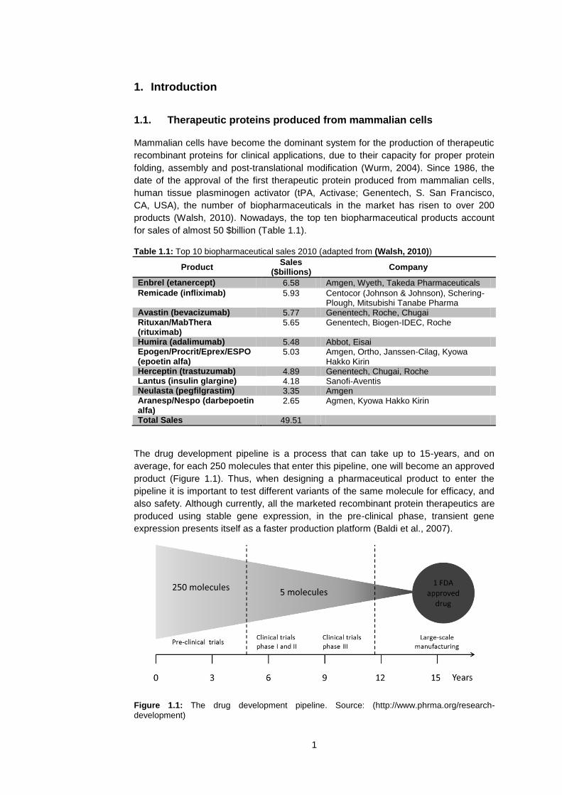
1.1. Therapeutic proteins produced from mammalian cells
Mammalian cells have become the dominant system for the production of therapeutic
recombinant proteins for clinical applications, due to their capacity for proper protein
folding, assembly and post-translational modification (Wurm, 2004). Since 1986, the
date of the approval of the first therapeutic protein produced from mammalian cells,
human tissue plasminogen activator (tPA, Activase; Genentech, S. San Francisco,
CA, USA), the number of biopharmaceuticals in the market has risen to over 200
products (Walsh, 2010). Nowadays, the top ten biopharmaceutical products account
for sales of almost 50 $billion (Table 1.1).
Table 1.1: Top 10 biopharmaceutical sales 2010 (adapted from (Walsh, 2010))
Product Sales
($billions) Company
Enbrel (etanercept) 6.58 Amgen, Wyeth, Takeda Pharmaceuticals
Remicade (infliximab) 5.93 Centocor (Johnson & Johnson), Schering-Plough, Mitsubishi Tanabe Pharma
Avastin (bevacizumab) 5.77 Genentech, Roche, Chugai Rituxan/MabThera (rituximab)
5.65 Genentech, Biogen-IDEC, Roche
Humira (adalimumab) 5.48 Abbot, Eisai Epogen/Procrit/Eprex/ESPO (epoetin alfa)
5.03 Amgen, Ortho, Janssen-Cilag, Kyowa Hakko Kirin
Herceptin (trastuzumab) 4.89 Genentech, Chugai, Roche Lantus (insulin glargine) 4.18 Sanofi-Aventis Neulasta (pegfilgrastim) 3.35 Amgen Aranesp/Nespo (darbepoetin alfa)
2.65 Agmen, Kyowa Hakko Kirin
Total Sales 49.51
The drug development pipeline is a process that can take up to 15-years, and on
average, for each 250 molecules that enter this pipeline, one will become an approved
product (Figure 1.1). Thus, when designing a pharmaceutical product to enter the
pipeline it is important to test different variants of the same molecule for efficacy, and
also safety. Although currently, all the marketed recombinant protein therapeutics are
produced using stable gene expression, in the pre-clinical phase, transient gene
expression presents itself as a faster production platform (Baldi et al., 2007).
Figure 1.1: The drug development pipeline. Source: (http://www.phrma.org/research-
development)
2
1.2. Transient gene expression
Transient gene expression (TGE) is defined as the introduction of foreign genetic
material in a cell host, without selection for stable integration of the transfected
plasmid in the host genome. This approach has been used for a long time (Graham e
van der Eb, 1973) and offers many advantages to research: it is rapid, simple and can
be applied to a large number of cell lines (Wurm e Bernard, 1999)(Geisse, 2009).
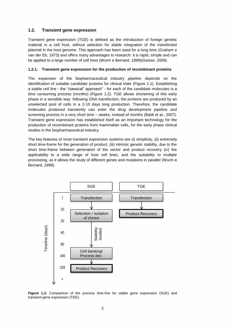
1.2.1. Transient gene expression for the production of recombinant proteins
The expansion of the biopharmaceutical industry pipeline depends on the
identification of suitable candidate proteins for clinical trials (Figure 1.1). Establishing
a stable cell line - the “classical” approach” - for each of the candidate molecules is a
time consuming process (months) (Figure 1.2). TGE allows shortening of this early
phase in a sensible way: following DNA transfection, the proteins are produced by an
unselected pool of cells in a 2-10 days long production. Therefore, the candidate
molecules produced transiently can enter the drug development pipeline and
screening process in a very short time – weeks, instead of months (Baldi et al., 2007).
Transient gene expression has established itself as an important technology for the
production of recombinant proteins from mammalian cells, for the early phase clinical
studies in the biopharmaceutical industry.
The key features of most transient expression systems are (i) simplicity, (ii) extremely
short time-frame for the generation of product, (iii) intrinsic genetic stability, due to the
short time-frame between generation of the vector and product recovery (iv) the
applicability to a wide range of host cell lines, and the suitability to multiple
processing, as it allows the study of different genes and mutations in parallel (Wurm e
Bernard, 1999).
Figure 1.2: Comparison of the process time-line for stable gene expression (SGE) and
transient gene expression (TGE).
Tim
elin
e (
da
ys)
Transfection
Product Recovery
Sta
bili
ty
stu
die
s
Selection / Isolation of clones
Transfection
Cell banking/ Process dev.
Product Recovery
SGE TGE

3
1.2.2. Expression vectors for transient gene expression
For transient gene expression, the expression vector can be non-viral and viral and
has to be designed to transfer the foreign gene into mammalian cells. The choice of
the vector depends on the application, the host cell, the time limitation and the safety
(Baldi et al., 2007).
There are three essential elements for a non-viral TGE vector: A constitutive
promoter, a transcription terminator and a prokaryotic cassette with a replication origin
and a selection marker for vector production in bacteria. The most commonly used
promoters are derived from viral genomes and include the human cytomegalovirus
immediate early promoter (HCVM), the simian virus (SV40) early promoter and the
Rous sarcoma virus long-terminal repeat promoter. Constitutive cellular promoters like
the human elongation factor-1-alpha (EF-1α) have also been shown to be efficient
(Baldi et al., 2007). Other elements can be used to enhance transient protein
expression. The insertion of an intron between the promoter and the 5’-end of the
cDNA, or the inclusion of the Woodchuck hepatitis virus post-transcriptional regulatory
element (WPRE) in the 3’UTR region of the transgene mRNA (Baldi et al.,
2007)(Wulhfard et al., 2008). Mammalian cells expressing the Epstein-Barr virus
(EBV) nuclear antigen 1 (EBNA1), like the HEK293-EBNA1 cell line, are capable of
episomal amplification of plasmid DNA containing the EBV or the SV40 origin of
replication. This strategy has also been used for improving the recombinant protein
production by increasing the plasmid copy number in transiently transfected cells (Van
Craenenbroeck et al., 2000).
Viral expression vectors are also used for TGE. The Semliki Forest Virus (SFV) is the
most widely used viral expression vector for TGE. The baculovirus system has also
been used for transient gene expression in both mammalian and insect cell lines (Kost
e Condreay, 2002). For mammalian cells, the baculovirus system has several
advantages, including: The absence of viral replication, the lack of cytotoxicity and the
simplicity of vector production. Vaccinia virus vectors have also been used for
transient gene expression, although they raise more regulatory concerns, as they
require containment in bio-safety level 2 laboratories (Baldi et al., 2007)(Sutter e
Moss, 1992).
1.2.3. Non-viral gene delivery vehicles
Several DNA carriers for gene delivery into mammalian cells have been used.
However, only a few are applicable to large-scale TGE, due to their cost-efficiency.
The non-viral DNA vehicles can be categorized in (i) cationic lips, (ii) inorganic
compounds and (ii) cationic polymers. Calcium phosphate (CaPi) precipitation was the
first transfection method, described by Graham in 1973 (Graham e van der Eb, 1973),
and still the most widely used method employing inorganic compounds (Jordan et al.,
1996)(Baldi et al., 2007).
Cationic lipids are very effective transfection agents, however, they are too expensive
for large-scale transfections, and are not considered for transient gene expression for
manufacturing purposes (Baldi et al., 2007).
CaPi is one of the most efficient DNA delivery methods, allowing transfection
efficiencies of 80-90% in HEK293 cells (Meissner et al., 2001). The requirement for
serum during the transfection, however, is a critical constraint. Furthermore, CHO
cells require an osmotic shock to be transfected with this method (Baldi et al., 2007).
Currently, the most widely used DNA delivery agent for TGE is 25kDa linear

4
polyethylenimine, which was demonstrated as an efficient gene carrier in 1995
(Boussif et al., 1995).Transfections with 25kDa PEI have been the most productive to
date with reported antibody yields up to 1g/L in HEK293 cells and 300mg/L in CHO
cells (Rajendra et al., 2011).
1.2.4. Cell lines for transient gene expression
HEK293 cells are the most commonly used cell lines in TGE, due to the ease of
maintenance and the high transfection efficiencies observed (Baldi et al.,
2007)(Geisse, 2009). Several genetically modified or clonally selected HEK293
variants have been developed. HEK293 cell lines grow well in adherent mode in
medium supplemented with serum or in suspension in serum free media (Geisse,
2009).
However, when TGE is used as a screening tool for drug development, it is preferable
to obtain CHO-derived material for pre-clinical trials (Baldi et al., 2007). The
pharmaceutical industry prefers to use CHO cell lines for production because of the
established track record of this cell line in manufacturing (Wurm, 2004)(Derouazi et
al., 2006a).
Only recently major attempts to use CHO cells as hosts for TGE have emerged. The
first large scale transient transfection of CHO cells in a bioreactor was reported in
2004 (Derouazi et al., 2004). The interest in CHO cells for transient gene expression
is high. However, the overall productivity of CHO cells for the manufacturing of
recombinant proteins is lower than HEK239 cells (Geisse, 2009).
Other cell lines like COS cells and Vero cells (both derivative of CV-1 cells of Simian
origin), various carcinoma cells, and recently human embryonic stem cells have also
been used for transient transfections, although with no proven success for protein
manufacturing (Geisse, 2009).
1.2.5. Transient gene expression in CHO cells
Until recently, the yields of transiently produced proteins from CHO cells have been
far behind the yields obtained in HEK293 cells, which have achieved 1 g/L (Backliwal
et al., 2008). Before 2008, the yields of TGE processes in CHO cells were in the 1-
25mg/L range. Since then, several improvements in the process were made, and
yields of 80-100mg/L were reported (Wulhfard et al., 2008)(Ye et al., 2009)(Wulhfard
et al., 2010). Very recently, a TGE process in CHO cells achieving antibody yields of
300mg/L was reported (Rajendra et al., 2011).
Several factors are responsible for these improvements: Mild hypothermia culture
conditions, vector design, high density transfections and the optimization of PEI and
DNA concentrations for transient gene expression (Wulhfard et al., 2008)(Rajendra et
al., 2011)
The highest yields reported so far in CHO cells have been obtained by transfecting
the cells with linear 25kDa PEI following incubation of the culture at 30-30ºC
(Wulhfard et al., 2008)(Rajendra et al., 2011). The enhancement of TGE in mild
hypothermia conditions (31ºC) correlates with an accumulation of cells in the G1
phase of the cell cycle, reduced cellular metabolism, greater cell viability, increased
steady state levels of transgene mRNAs, and increased cell size as compared to cells
kept at 37ºC(Wulhfard et al., 2008). The vector design, as for example, the inclusion
of the Woodchuck hepatitis virus post-transcriptional element (Wulhfard et al., 2008)

5
or the inclusion of an intron between the promoter and the 5’-end of the transgene
cDNA have shown to improve recombinant protein production(Baldi et al., 2007).
1.2.6. Scale-up of transient gene expression
Some private companies have reported high yields in transient gene expression at
large-scale. However, they do not publish the methods used for these breakthrough
achievements, as they are of great commercial value.
The first review on large scale TGE was published in 1999 (Wurm e Bernard, 1999).
Regarding the DNA delivery method, at that time polyethylenimine was already seen
as a major technology for large-scale operations, as it is cost-efficient and it can be
applied in big quantities. However, the state of the art delivery method for large scale
TGE was still Calcium phosphate. The cell lines dominating the field were HEK-293,
COS cells (Simian origin) and baby hamster kidney (BHK) cells. Only later CHO cells
started to give satisfying levels of protein expression when transfected transiently
(Derouazi et al., 2004).
For large-scale processes, mostly standard stirred-tank (STR) bioreactors have been
used, but in recent years, the trend to use disposable bioreactors has increased. For
large-scale TGE processes, orbital shaking bioreactors have been shown to be
efficient (Zhang et al., 2010a), but also the Wave™ bioreactor has proven to be well
suited for TGE processes (Geisse e Henke, 2005).
For transient gene expression in CHO cell lines, the largest scales reported were of 5-
100 L in orbital shaking bioreactors, with obtained antibody titers in the range of 20-
60mg/L (Muller et al., 2007)(Stettler et al., 2007) and in Wave™ bioreactors with
yields of 9 mg/L(Geisse, 2009). More recently, one transient 14-day fed-batch process
with CHO cells yielding 80mg/L at the 20 L scale was reported, being the highest titer
every reported for large scale TGE process in CHO cells (Ye et al., 2009). These
processes, although being at large-scale, include (i) a centrifugation step prior to
transfection to eliminate the spent medium and (ii) a low cell density at the time of
transfection.
Transfections at high cell density for TGE processes have proved to be the most high-
yielding, in both CHO and HEK-293 cell lines (Backliwal et al., 2008)(Rajendra et al.,
2011). The high density transfection procedure described for CHO cells is performed
at a cell density of 4-5 million cells/mL (Rajendra et al., 2011), whereas for HEK-293
cells, it is performed at a cell density of 20 million cells/mL (Backliwal et al., 2008).
The high-density HEK-293 transfections, while very efficient, are only feasible at small
scales, as these cells are not known to achieve such densities (20 million cells/mL) in
batch cultures. Thus, this high cell densities at the time of transfection can only be
achieve by concentrating the cells before a medium exchange, i. e with a
centrifugation step. From this point of view, the high density process in CHO cells is
more likely to be successfully scaled-up, as CHO cells, grow easily to densities of 4-5
million cells/mL in serum-free media. However, for high-density transfections in CHO
cells, there is still a need for a medium exchange by centrifugation to remove the
spent medium (Rajendra et al., 2011).
While a centrifugation step for a medium exchange is very easy to perform at smaller
scales of operation, it is not a viable alternative for large scale processes (over 2 L)
and it is a limiting step on scaling-up high-yielding transient gene expression
processes. Therefore, the alternative is to transfect a large scale culture without a
6
medium exchange (i.e in conditioned medium). However, this results in lower
transfection efficiencies and loss of productivity (Schlaeger e Christensen, 1999).
1.2.7. Transient gene expression in conditioned medium
For the purpose of transient gene expression, conditioned medium is defined has the
spent medium where cells have been cultivated for more than 2 days. This medium is
lower in nutrients due to cell growth, it contains cell derived compounds (proteins,
glycans, etc.) and other molecules that are the product of cell metabolism.
Transfecting cells in conditioned medium results in lower transfection efficiencies. This
has been shown for cationic lipid mediated gene transfer (Belting e Petersson, 1999)
and for PEI-mediated gene transfer (Schlaeger e Christensen, 1999). The contrary
has also been claimed (but not shown) at least in one publication on PEI-mediated
TGE in HEK293 cells (Raymond et al., 2011). However, the titers of these transient
processes in HEK293-cells are still 40-fold lower than those obtained when a medium
exchange is performed: 4000 mg/L vs 100mg/L (Divor Kiseljak, unpublished data)
(Raymond et al., 2011)).
Processes where the transfection is done into a running culture have been reported
for HEK293 cells (Raymond et al., 2011). For CHO cells, a transient gene expression
procedure where the transfection is performed without a medium exchange has not
been published yet.
It has been shown that the negative effect of the conditioned medium on the
transfection efficiency is mainly due to the presence of cell-secreted high molecular
weight compounds. It is thought that these compounds interact with the PEI-DNA
complex, inhibiting the transfection procedure, although the mechanisms and the
molecular entity of these compounds are not known.
Cell secreted high molecular weight compounds have been shown to interfere with
cation-mediated gene delivery, and although they are not studied in this work, they
are worth to mention. Positively and negatively charged molecules interact with DNA
and the cationic delivery vehicle, and interfere with the transfection process. These
include proteoglycans and glycosaminoglycans, like heparin or dextran sulphate.
(Mislick e Baldeschwieler, 1996)(Ruponen et al., 2001)(Ruponen et al., 2004).
1.3. The CHO-DG44 cell line
Most of the therapeutic proteins in the market are produced in Chinese Hamster
Ovary (CHO) cells, but also other cell lines such as NS0 (mouse myeloma), BHK
(Baby Hamster Kidney), HEK-293 (Human embryo kidney) and PerC 6 (Human retinal
cells). All these cell lines, among others, have gained regulatory approval for the
production of recombinant proteins (Wurm, 2004).
For manufacturing purposes, cell culture is done in adherent or suspension culture,
being suspension, by large, the most common. Suspension adapted CHO cell lines
are the dominant host for the mass production of therapeutic products (Wurm, 2004).
The CHO cells used in protein manufacturing originated in 1957 by immortalization of
a cell from a primary culture of ovarian cells of a Chinese Hamster (Hacker et al.,
2007).

7
The CHO-K1, a glycine dependent strain derived from the original CHO cell line, was
mutagenized to generate CHO-DXB11, a cell line lacking DHFR activity (Urlaub et al.,
1983). Subsequently, a CHO-pro3 strain was mutagenized to generate the CHO-
DG44, which also lacks DHFR activity. Because of that, these two DHFR-minus
strains require glycine, hypoxanthine and thymidine (GTH) for cell growth (Kaufman et
al., 1985)
The DHFR-minus CHO cells were not originally intended for manufacturing of
recombinant proteins, but several pioneering experiments were done on a stable
transfection with an exogenous dhfr gene via selection in a GTH-minus medium
(Hacker et al., 2007). The CHO-DG44 cell line has thus become a dominant, and well
characterized, mammalian cell host for recombinant protein manufacturing.
1.4. TubeSpin® bioreactor 50 and orbitally shaken bioreactors
The TubeSpin® Bioreactor 50 (TPP, Switzerland) is, an orbitally shaken scale-down
system, successfully used in the screening and optimization processes of suspension
cells(De Jesus et al., 2004). It has a working volume from 1 to 35mL and 5 openings
of different sizes above the gas permeable sterile PTFE filter of the screw cap which
allow for custom optimization of the gas transfer. The sterile gas exchange is done
through the 0.22 µm filter membrane. High cell densities can be achieve with only
passive aeration through the opening cap (Muller et al., 2005). With passive aeration,
the mass transfer coefficient of oxygen for the TubeSpin® Bioreactors 50 (kLa) is
above 15h-1
and from the 50mL scale up to the 2000L scale the kLa is in the range of
4-30h-1
(Zhang et al., 2009)(Tissot et al., 2011). This disposable bioreactor system
allows bioprocesses to be run without probes or controllers (Tissot et al., 2011). For
these reasons, the TubeSpin® bioreactor 50 is very advantageous for the high-
throughput study of culture conditions or optimization of transfection processes.
Furthermore, the orbitally shaken culture system has been scaled-up to volumes
higher than 1000L in a very straightforward approach(Zhang et al., 2010b) which
leaves the door open to further volumetric scale-up. Transient gene expression has
been performed in these bioreactors at the 100 L scale (Zhang et al., 2010b).
Figure 1.3 – Left to Right: Disposable TubeSpin® bioreactors 50 and 600 (TPP, Switzerland;
www.tpp.ch)

8

9
2. Goal of this thesis
Currently, one of the key problems for TGE in CHO cells is its volumetric scale-up with
high yields. This can be approached by developing a high-density transfection
procedure without a medium exchange (i.e in conditioned medium). However, it is
known that if the transfection is done in a running culture, without a medium
exchange, the yields obtained are very low (Schlaeger e Christensen, 1999).
This work examines this problem from two different perspectives:
1. Analysis of gene delivery in fresh medium vs conditioned medium.
2. A process development approach for the establishment of a high-titer TGE
process for CHO-DG44 cells in conditioned medium.
By comparing the gene delivery in the TGE process in fresh medium (high yielding)
and in conditioned medium (low yielding) it was expected to gain better insights about
what is happening to the transfected DNA. This was done by quantitative analysis of
the transfected plasmid DNA and comparison with the respective mRNA levels and
protein production. Studies were done in order to unravel the factors that are limiting
the protein production when the transfections are done in conditioned medium. These
insights were applied in the design of strategies for the improvement of yields.
With a step-by-step approach of process development, using the TubeSpin®
bioreactor 50 system, several parameters known to have an influence of transient
gene expression process productivities were studied: (i) The study of different cell
culture media formulations (ii) The optimization of the PEI-DNA ratios for high density
transfections in conditioned medium and (iii) the study of chemicals known to have a
positive effect on the mRNA levels and protein productivities in cell culture processes.
This work was aimed at the identification of important parameters for conditioned
medium transfections and strategies that can be used to improve TGE processes in
CHO cells for larger scales. A process development approach was also followed in
order to develop a fully scalable TGE process for CHO cells that does not involve a
medium exchange prior to transfection.

10

11
3. Materials and Methods
3.1. Cell culture
Suspension-adapted CHO DG44 cells (Urlaub chasin) were routinely grown in 250-mL
squared shaped glass bottles (Schott Glass, Mainz, Germany) as previously
described (Whulfard 2008) at 37ºC in 85% humidity and 5% CO2. The cells were
cultivated in ProCHO5 medium (Lonza, Verviers, Belgium) (Henceforth refered to as
“ProCHO5”) supplemented with 14µg/L hypoxanthine, 4µg/L thymidine, and 4mM
glutamine (SAFC Biosciences, St. Louis, MO). The cells were passaged 2 times per
week and maintained in exponential growth between 0.3 – 6 million cells/mL.
The suspension cell culture done was done at a working volume of at most 40% of the
nominal volume of the vessel. The bottles were fixed to a horizontal model ES-W
orbital shaker with a rotational diameter of 2.5 cm (Kühner AG, Birsfelden,
Switzerland) using double sided adhesive transfer tape (3M Corp, Minneapolis, MN)
and agitated at 110 rpm.
Before transfection and at the time of transfection the cell density was always
determined by the packed cell volume method (PCV) (Stettler 2006) with the VoluPAC
system (Sartorius AG , Göttingen, Germany). For CHO-DG44 cells, a PCV of 0.18-0.2
correlates to a cell density of 1 million cells/mL. Manual cell counting after transfection
was performed, when necessary, using the Trypan blue exclusion method.
3.2. Transfections
3.2.1. Plasmid DNA preparation
Plasmid DNA was produced by transformation of E.Coli DH5α and growth of the
bacteria in LB broth with ampicillin. The plasmid DNA purification was performed with
a Nucleobond AX anion exchange column (Macherey-Nagel, Düren, Germany)
according to the manufacturer’s protocol. Plasmid DNA concentration was measured
with a NanoDrop 2000 spectrophotometer (Thermo Scientific, Wilmington, DE). The
quality ratios 260/280 and 260/230 were measured and the accepted values were of
at least 1.8 and 2.0, respectively.
3.2.2. Plasmids used
3.2.2.1. Plasmid pXLGCHO
-A3 (pA3)
The dual expression vector pXLGCHO
-A3 with the human anti-Rhesus D IgG1 heavy
and light chain cDNAs cloned in separate expression cassettes in a head-to-head
orientation was kindly provided by Excellgene SA. It was constructed by removing the
IgG light chain expression cassette from pXLGHEK
-RhLC and cloning it into the unique
AflII site of pXLGHEK-
RhHC. Each expression cassette included the human CMV
immediate early promoter/enhancer, an artificial intron in the 5′-untranslated region,
the cDNA for IgG light or heavy chain with the translational start codon present within
the Kozak consensus sequence, the woodchuck hepatitis virus post-transciptional
regulatory element (WPRE) in the 3′-untranslated region, and the bovine growth
hormone polyadenylation signal (Rajendra et al., 2011).
12
3.2.2.2. Plasmid pMYKEF1-EGFP (pEGFP)
The plasmid pMYKEF1-EGFP has been previously described (Derouazi et al., 2006b).
It was generated by the enhanced GFP gene from pEGFP-N1 (Clontech, Palo Alto,
CA) cloned as an EcoRI/NotI fragment into EcoRI/NotI-digested pMYKEF-1 (Kim et al.
2002) to produce pMYKEF1-EGFP.
3.2.3. Preparation of the stock solution of 25kDa linear polyethylenimine
For the preparation of the 1mg/mL stock solution of 25kDa linear polyethylenimine
(PEI), 1 g of PEI is dissolved in 800mL dH2O by adding 1N HCl (PEI will not dissolve
until the pH is lower than 7). The pH is adjusted to 7.0 with 1N NaOH and dH2O is
added until the volume is 1L. If the pH is lower than 7, 1N NaOH is added. The
solution is sterile filtered trought a 0.22µm filter and stored at -80ºC in 40 mL aliquots
in 50mL tubes.
3.2.4. Transfection with a medium exchange (fresh medium)
All transfections were done in duplicate at a final volume of 5 mL in TubeSpin®
Bioreactors 50, unless specified otherwise. Cells from the seed culture were spin
down and re-suspended in 5 mL of ProCHO5 medium at a density of 5 million
cells/mL (PCV = 1 - 1.1). The cells were transfected with 15ug (at a concentration of
~1mg/mL) of DNA (95% pXLGCHO
-A3 and 5% pMYKEF1-EGFP) and 75 ug of of
25kDa linear polyethylenimine (PEI) (at a concentration of ~1mg/mL). The DNA was
added directly to the cells followed by addition of PEI, and the culture was incubated
at 31ºC with 5% CO2 and 85% humidity at a shaking speed of 180 rpm.
3.2.5. Transfection without a medium exchange (conditioned medium)
For transfecting without a medium exchange, cells from the seed train, in exponential
growth phase, were sub-cultivated when at a density of ~5million cells/mL into fresh
medium at a density of 0.4-0.5 million cells/mL. The cell culture was incubated at 37ºC
as previously described, and on day 3 (72h) the transfection was performed. On the
day of transfection cell density was controlled with the packed cell volume (PCV)
method. When the PCV of the culture reached 1.05-1.1, cells were transfected with
different concentrations of DNA and PEI (as described in the results and discussion
section) by direct addition of DNA and PEI, and incubated at 31ºC with 5% CO2 and
85% humidity at a shaking speed of 180 rpm. Sodium butyrate and Dimethyl sulfoxide
were added to the culture at the time of transfection, when specified.
3.2.6. Transfection for the media screening experiment
Thirty-eight different commercially available cell growth media were inoculated with
0.5 million cells/mL, and cells were allowed to grow in this media in the previously
described cell culture conditions. After 3 days, the medium supernatant (i.e the
conditioned medium) was harvested by centrifugation. Cells from the seed culture
were then transfected as described in 3.2.4, using the harvested conditioned medium
and also fresh medium out of the bottle.
13
3.3. Reporter protein assays
3.3.1. Transfection efficiency: %GFP positive cells
Cells transfected with the pEGFP plasmid were diluted 20x in PBS to a density of ~0.3
million cells/mL in a 96-Well plate and the percentage of GPF positive cells was
measured with a GUAVA Easy-CyteTM
multi-well system (GUAVA Technologies,
Hayward, CA).
3.3.2. ELISA: Recombinant antibody concentration
After transfection with the pA3 plasmid, the IgG1 (human anti-Rhesus-D IgG)
concentration in the medium was determined by sandwhich ELISA using an anti-
human kappa light chain antibody (coating IgG) (Biosource International, Camarillo,
CA) for the IgG capture and alkaline phosphatase conjugated anti-human IgG for
detection (detection IgG) (Invitrogen AG). Microtest™ 96-well ELISA plates (BD
Biosciences, Bedford, MA) were incubated for 3-4 hours at 37ºC with 100 µL of 1000x
diluted (in PBS with 0.1% of tween-20) coating IgG. For the assay, the samples were
diluted in a blocking buffer and added to each well. A standard of known concentration
was also added to the plate. The incubation time was 45 min at 37ºC. After this
incubation time, the plate was washed 3 times and incubated again at 37ºC with 100
uL of 1000x diluted (in blocking buffer) detection IgG. After 45 min of incubation, the
plate was washed and the substrate solution was added. Absorbance was measured
at 405 nm against 490 nm using a microplate reader (SPECTRAmax™ 340,
Molecular devices, Palo Alto, CA).
3.4. Plasmid DNA extraction and quantification
At different times after transfection, 1 million cells were sampled and centrifuged,
followed by two consecutive washes with PBS buffer. Total DNA was isolated using
DNeasy blood and tissue kit ( Qiagen, Hombrechtikon, Switzerland ) according to the
manufacturer’s instructions. Quantitative real-time PCR (qPCR) was performed used
Absolute qPCR SYBR green ROX mix (ABgene UK, Epsom, UK) in Ligthcycler® 480
optical 96-well reaction plates ( Roche Diagnostics, Mannheim, Germany). DNA
isolated from cells was diluted 100 x with high pure water before use for qPCR. A
standard curve was made by sequential dilution of the plasmid of interest in water.
The primers used for the quantification of the pA3 plasmid amplify a region of the
gene of the IgG light-chain and were purchase from SAFC Biosciences (St. Louis,
MO, USA (Table 3.1). All the experiments were performed on a Lightcycler® 480
(Roche Diagnostics, Mannheim, Germany). The following thermal cycling program
was used: preheating at 95ºC for 5 min, 35 cycles of heating at 95ºC for 15s, 59ºC for
30s and 72ºC for 45s. For each sample, 4 measurements were performed.
Table 3.1 – Primers used for the quantification of DNA by qPCR
Gene of Interest Forward primer (5’ – 3’) Reverse primer (5’ – 3’)
IgG light chain TGTCTTCATCTTCCCGCC GCGTTATCCACCTTCCACTGT Β-Actin GCTCTTTTCCAGCCTTCCTT GAGCCAGAGCAGTGATCTC
3.5. mRNA extraction and quantification
At different times after transfection, 1 million cells were collected and centrifuged,
followed by two consecutive washes with PBS buffer. Total RNA was extracted using
the GenElute mRNA kit (Sigma-Aldrich GbmH, Buchs, Switzerland) according to the
14
manufacturer’s protocol. Samples were treated with 1 U of DNAse I (Invitrogen AG,
Basel, Switzerland) for 15’ at room temperature, the enzyme was then inhibited by
addition of EDTA and by heating.
First-strand cDNA synthesis was performed from 1µg of RNA with the M-MLV reverse
transcriptase (Invitrogen AG) using oligo dT (New England Biolabs, Ipswich, MA) as
the primer. Each reaction was diluted 10-fold in RNAse-free water for quantitative
PCR. The oligolucleotide primers for the amplification (Table 3.1) were purchased
from SAFC Biosciences (St. Louis, MO, USA).
The samples from the first-strand cDNA synthesis were mixed with the appropriate
primer pair and the reaction mix from the Absolute qPCR SYBR Green ROX mix
(ABgene UK, Epsom, UK), and amplified according to the manufacturer’s protocol. All
experiments were performed on a Lightcycler® 480 (Roche Diagnostics, Mannheim,
Germany). The comparative Ct method was used to calculate the relative quantity of
IgG light chain mRNAs relative to the quantity of β-actin mRNA (Housekeeping gene).
3.6. Detection of DNA in the medium with PicoGreen ®
The PicoGreen® dye from the Quant-iT™ PicoGreen® dsDNA Assay Kit (Invitrogen
AG) was diluted in TE Buffer at a 200-fold dilution according to the manufacturer’s
instructions to make a working solution. Then, at different times post-transfection,
cells were centrifuged and medium supernatant removed. Afterwards, 100µL of the
medium samples were sampled into a black 96-well plate, followed by the addition of
50µL of the PicoGreen® working solution. The plate was incubated in the dark for 5
minutes and Fluorescence (excitation 480 nm, emission 520 nm) was measured in a
TECAN Safire2 ™ Spectrofluorometer (Tecan Trading, Männedorf, Switzerland).
3.7. Nuclear extraction and nuclear pDNA quantification
1 million viable cells were collected, centrifuged (4ºC, 400g, 5min), and washed twice
with PBS buffer. The cell pellet was resuspended in 1mL nuclear extraction buffer (10
mM Tris-HCl, pH 7.4, 3 mM MgCl2, 10 mM NaCl, and 0.5% Nonidet P-40 (NP40)), and
a 10 min incubation in ice followed. Nuclei were then collected by centrifugation (4ºC,
400g, 5min) and re-suspended in PBS buffer. The nuclei extraction method used was
based on a publish protocol (Wang et al., 2007) and validated in house by
microscopy, to be sure that only intact nuclei were isolated, and not cells. Nuclear
DNA extraction and quantification was executed as previously described for the total
DNA extraction (3.4).

15
4. Results and Discussion
4.1. Transfection without a medium exchange results in lower
recombinant protein yields
4.1.1. Introduction
Transiente gene expression (TGE) processes are known to be less efficient if at the
time of transfection a medium exchange to remove the spent medium is not
performed (Schlaeger e Christensen, 1999). In this section, a transient gene
expression process that was developed involving a medium exchange by
centrifugation is characterized. Two production runs, one with and one without a
medium exchange (i.e. in conditioned medium) are compared side by side in terms of
protein titers.
Further, the negative influence of the conditioned medium on the transfection
efficiency is analyzed in a time-dependent way and the influence of the conditioned
medium on transfection efficiency and protein production is studied.
4.1.2. Comparison between a TGE process in fresh media and in conditioned
media
In order to study this TGE process in conditioned medium and in fresh medium, two
transient production runs were done in parallel. In one process, a medium exchange
was performed before transfection, whereas in the other process, the transfection was
done without a medium exchange (i.e in conditioned medium) (Figure 4.1).
Figure 4.1 – Comparison between a TGE process in fresh medium (■) and in conditioned medium (■). n=2
0
2
4
6
8
10
0 2 4 6 8 10 12
Via
ble
ce
ll d
en
sity
(M
illio
n c
ells
/mL
)
Time post-transfection (days)
0%
20%
40%
60%
80%
100%
0 2 4 6 8 10 12
Cell
via
bili
ty (
%)
Time post-transfection (days)
0
2
4
6
8
10
0
100
200
300
400
500
0 2 4 6 8 10 12
IgG
1 A
nti-R
he
su
s D
(m
g/L
) T
ran
sf.
Con
dtiio
ne
d m
ed
ia
IgG
1 A
nti-R
he
su
s D
(m
g/L
) T
ran
sf.
Fre
sh
me
dia
Time post-transfection (days)
16
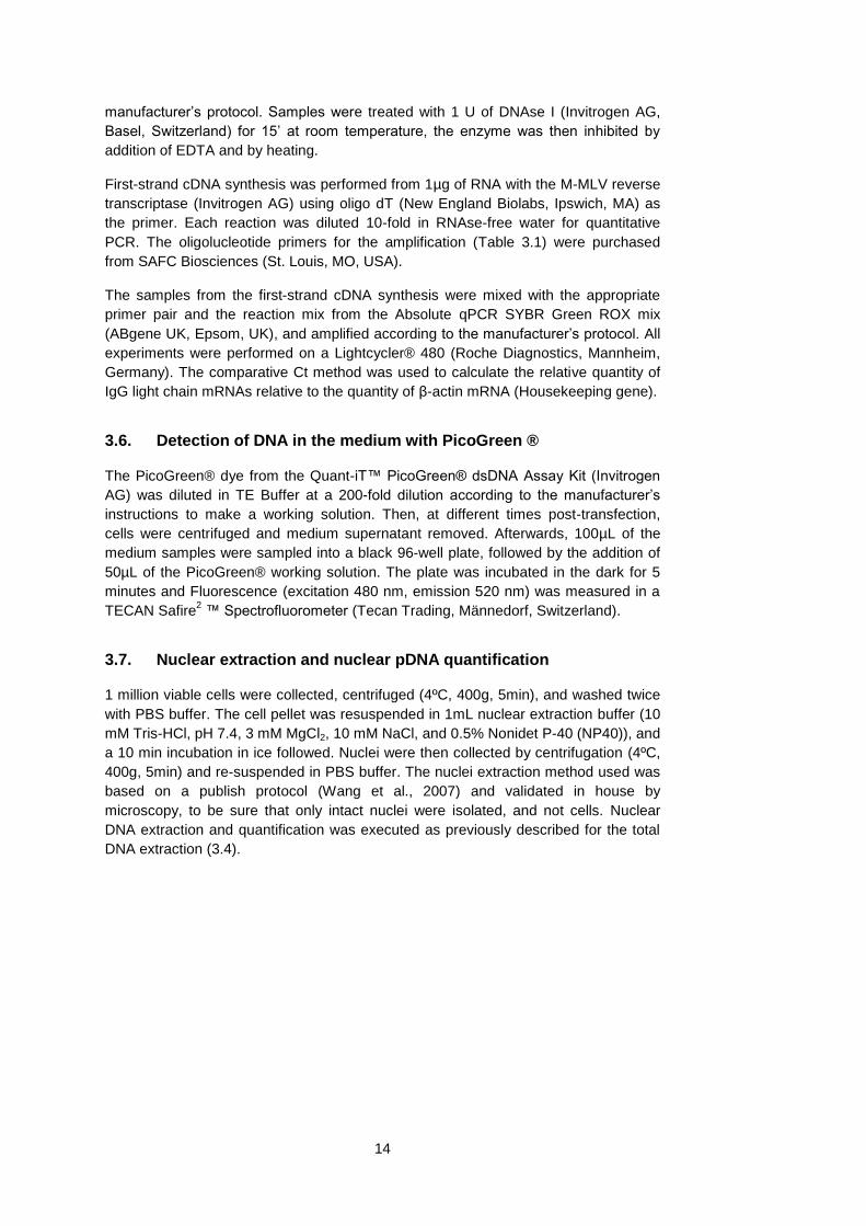
The titers obtained on a 10 day batch process are very low when the transfection is
performed without a medium exchange, compared to when a medium exchange is
done to eliminate the conditioned medium: ~450 mg/L vs ~6 mg/L. Although the titer
obtained is low, the cell viability and cell density are high throughout the culture and it
is worth noticing that in the process in conditioned medium, the viable cell density and
the cell viability are higher (this was observed throughout all the experiments
performed). If the transfection is performed in spent medium, the cell viability, and
consequently the cell density are always higher than when the same transfection
procedure is done in fresh medium. This makes sense, as the spent medium contains
many factors secreted by cells, which might act with a serum-like effect by protecting
the cells, but making the transfection less efficient. The low transfection efficiency can
be seen by analyzing the %GFP positive cells 24h post transfection by flow cytometry
(Figure 4.2). This data correlates with the low productivity of the process – the
negative effect of the conditioned medium on transfection is already observed on the
percentage of cells that express GFP at 24 hours post-transfection.
Figure 4.2 – Transfection efficiency (% of GFP positive cells 24-post transfection) in conditioned medium and in fresh medium. n=2
4.1.3. The transfection efficiency decreases with the time of conditioning of
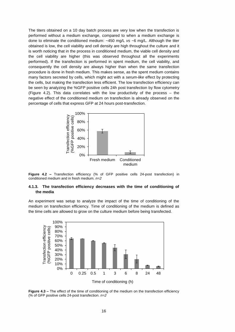
the media
An experiment was setup to analyze the impact of the time of conditioning of the
medium on transfection efficiency. Time of conditioning of the medium is defined as
the time cells are allowed to grow on the culture medium before being transfected.
Figure 4.3 – The effect of the time of conditioning of the medium on the transfection efficiency (% of GFP positive cells 24-post transfection. n=2
0%
20%
40%
60%
80%
100%
Fresh medium Conditionedmedium
Tra
nsfe
ction e
ffic
iency
(%G
FP
positiv
e c
ells
)
0%
10%
20%
30%
40%
50%
60%
70%
80%
90%
100%
0 0.25 0.5 1 3 6 8 24 48
Tra
nsfe
ction e
ffic
iency
(%G
FP
positiv
e c
ells
)
Time of conditioning (h)
17
It is possible to observe that the transfection efficiency decreases with the time of
conditioning of the medium (Figure 4.3), i.e. if the cells are transfected in the same
medium where they were grown, the transfection efficiency decreases. This effect can
already be seen at 3-6h of conditioning time and limits the possibility of growing the
cells and transfecting without a medium exchange prior to transfection. It had been
previously suggested that this is due to cell derived components that interfere with the
transfection process (Ralph Duhr, EPFL) – The longer the time of culture, more
secreted compounds will be in the medium, the more conditioned the medium will be
resulting in a lower transfection efficiency.
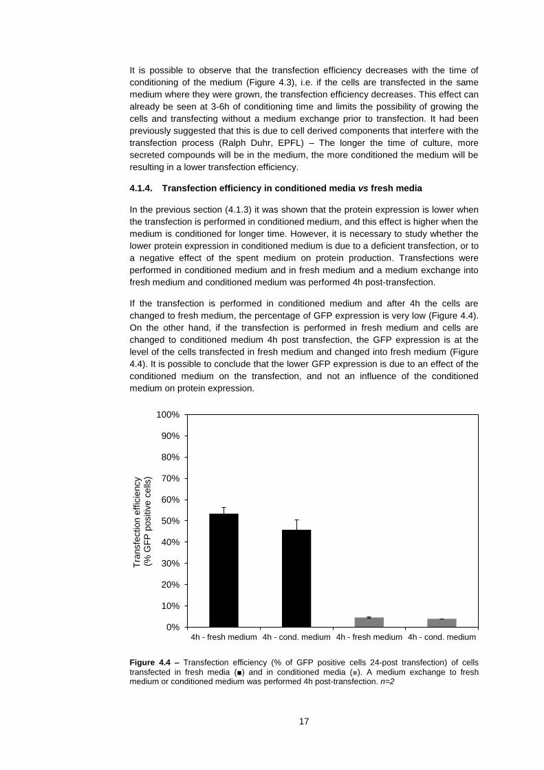
4.1.4. Transfection efficiency in conditioned media vs fresh media
In the previous section (4.1.3) it was shown that the protein expression is lower when
the transfection is performed in conditioned medium, and this effect is higher when the
medium is conditioned for longer time. However, it is necessary to study whether the
lower protein expression in conditioned medium is due to a deficient transfection, or to
a negative effect of the spent medium on protein production. Transfections were
performed in conditioned medium and in fresh medium and a medium exchange into
fresh medium and conditioned medium was performed 4h post-transfection.
If the transfection is performed in conditioned medium and after 4h the cells are
changed to fresh medium, the percentage of GFP expression is very low (Figure 4.4).
On the other hand, if the transfection is performed in fresh medium and cells are
changed to conditioned medium 4h post transfection, the GFP expression is at the
level of the cells transfected in fresh medium and changed into fresh medium (Figure
4.4). It is possible to conclude that the lower GFP expression is due to an effect of the
conditioned medium on the transfection, and not an influence of the conditioned
medium on protein expression.
Figure 4.4 – Transfection efficiency (% of GFP positive cells 24-post transfection) of cells
transfected in fresh media (■) and in conditioned media (■). A medium exchange to fresh medium or conditioned medium was performed 4h post-transfection. n=2
0%
10%
20%
30%
40%
50%
60%
70%
80%
90%
100%
4h - fresh medium 4h - cond. medium 4h - fresh medium 4h - cond. medium
Tra
nsfe
ction e
ffic
iency
(%
GF
P p
ositiv
e c
ells
)
18
4.1.5. Conclusion
Transient gene expression processes, with and without a medium exchange, were
studied and compared. Furthermore, the reasons for the low protein production
observed in TGE processes in conditioned medium were assessed: (i) The low protein
production is not due to a low cell viability, as the cellular viability post-transfection is
even higher when cells are transfected in conditioned medium, in comparison to fresh
medium (ii) A higher conditioning time of the medium leads to a lower transfection
efficiency, suggesting that compounds that accumulate in the medium could interfere
with the transfection procedure (iii) The lower protein expression is due to a negative
effect of the conditioned medium on the transfection.
One might think that the lower transfection efficiency (the percentage of GFP positive
cells 24-h post transfection) is due to a reduced DNA delivery into the cell due to cell
derived components in the conditioned medium that interfere with the PEI-DNA
complex (Mislick e Baldeschwieler, 1996)(Ruponen et al., 2001). This hypothesis is
explored in the following section.
4.2. Plasmid DNA delivery in conditioned media
4.2.1. Introduction
The results presented in the previous section lead to the conclusion that the
transfection in conditioned medium is less efficient that in fresh medium and that this
is the reason for the lower protein production. However, the observed transfection
efficiency is representative of the percentage of GFP expressing cells. The expression
of the reporter protein GFP, although being an indicator, does not give any information
about whether or not the transfected DNA is internalized by the cell.
In this chapter, DNA delivery into the cells is studied. This is done first from the point
of view of DNA disappearing from the culture medium after transfection, and
afterwards by quantifying the copy number of transfected plasmid DNA inside the
cells. Further, exogenous mRNA levels are studied and the isolation of transfected
plasmid DNA from the cell nuclei is also done.
4.2.2. The transfected plasmid DNA is not detectable in the cell culture
medium after transfection in conditioned and in fresh medium
A first approach to study the disappearance of DNA from the culture medium was to
collect medium supernatant samples at different time points after transfection, and
analyzing them on a 1% agarose gel (Figure 4.5).
When the transfection is performed in conditioned medium the GFP expression is
lower. It was expected that DNA would not be internalized by the cells and would stay
in the cell culture medium after transfection. Surprisingly, it was observed that 1h after
transfection, no DNA was detected in the medium, both when the transfection is
performed in conditioned medium, or in fresh medium.

19
Figure 4.5 – Agarose gel electrophoresis (1%) of medium supernatant after transfection in
conditioned medium and in fresh medium. The bands show the plasmid used for transfection, in supercoiled and relaxed form. Both sets (fresh medium and conditioned medium) were run on the same gel, with the same volume of supernatant loaded in every well.
In order to confirm the results of the medium analysis on the agarose gel, a different
methodology was used. As before, supernatant samples were collected at different
time-points after transfection, and analyzed for their fluorescence when treated with
the DNA intercalating dye PicoGreen® (Figure 4.6). The decrease in fluorescence of
the medium samples collected after transfection shows the same trend as the agarose
gel analysis - One hour after transfection, DNA is not detectable in the cell culture
medium. A control experiment (not show) was done, where DNA was incubated with
medium to assess whether or not its disappearance from the medium could be due to
degradation, and this hypothesis was excluded.
Figure 4.6 – Detection of DNA in the culture medium after transfection using the PicoGreen® dye. n=2
If the DNA is not detectable in the culture medium after transfection, there is a strong
indication that it is being internalized by the cell. The evidence of internalization of
DNA by the cells when the transfection is done in conditioned medium is surprising,
because the protein expression is reduced.
0
100
200
300
400
500
600
0 10 20 30 40 50 60 70
RF
U
(Rela
tive F
luore
scence U
nits)
Time post-transfection ( min )
Fresh medium
Cond medium

20
4.2.3. Plasmid DNA is being delivered to the cells in conditioned media, but
mRNA levels are lower
The information obtained by analyzing the medium supernatant is strongly indicative
that the plasmid DNA is being taken up by the cell, but is not conclusive. In order to
further study the DNA delivery into the cell, total intracellular DNA was extracted after
transfection, and quantitative real-time PCR was used to assess the pDNA copy
number per cell. Transfections were performed in fresh medium and in conditioned
medium, and at different time-points post-transfection, samples were taken and the
plasmid copy number per cell was quantified (Figure 4.7).
Figure 4.7 – Plasmid DNA copy number per cell (x1000) at different time-points after transfection in fresh medium and in conditioned medium. n=2
It was expected to detect pDNA inside the cells, even if the transfection is done in
conditioned medium, as the plasmid DNA was disappearing from the cell culture
medium. However, it was thought that when the transfection was performed in
conditioned medium, a decrease in the plasmid copy number per cell would be seen
over time and this could explain the lower protein production. The hypothesis was that
in conditioned medium, cell derived compounds in the medium would interfere with
the PEI-DNA complex, and a weaker complex would enter the cell (Ruponen et al.,
2004). A weaker PEI-DNA complex makes DNA less protected inside the cell and
more subject to degradation by nucleases. A big decrease in the pDNA copy number
per cell after transfection could explain the low protein expression observed and
would confirm this hypothesis, however, this was not observed.
What is shown here is that the pDNA copy number per cell when the transfection is
performed in conditioned medium is even slightly higher than when the transfection is
performed in fresh medium (this phenomenon was always observed in different qPCR
experiments). More importantly, the pDNA copy number is stable up to 3 days after
transfection. Longer duration studies cannot be made because DNA extraction and
quantification is not reliable when cells have a viability lower than 70%. The slight
decrease observed in the pDNA copy number is due to cell division, as cells coming
from cell division will not contain the transiently transfected plasmid.
The copy number per cell and the stability of the plasmid DNA copy number is
contradictory to the low protein productivity when the transfection is performed in
conditioned medium. However, lower mRNA levels can explain the low protein
production when the transfection is performed in conditioned medium (Figure 4.8).
0
5
10
15
20
25
30
35
40
0 20 40 60 80
pD
NA
copy n
um
ber
per
cell
x1000
Time post-transfection (h)
Fresh medium
Cond medium
21
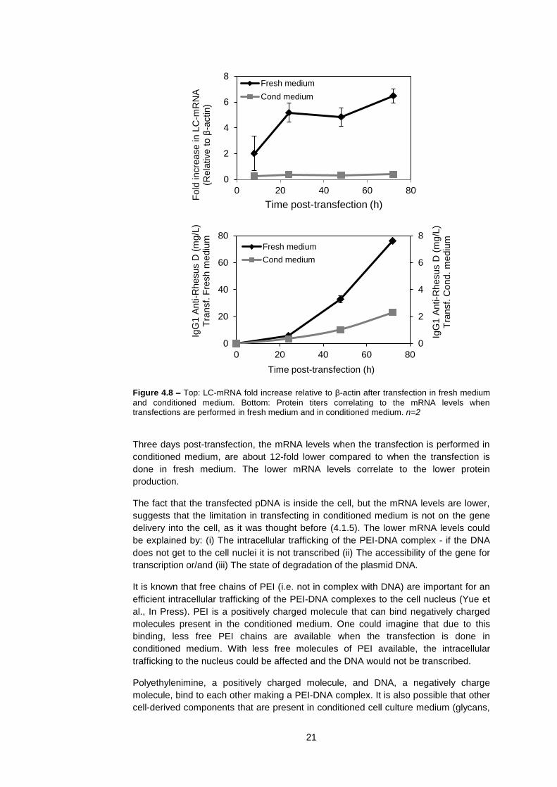
Figure 4.8 – Top: LC-mRNA fold increase relative to β-actin after transfection in fresh medium
and conditioned medium. Bottom: Protein titers correlating to the mRNA levels when transfections are performed in fresh medium and in conditioned medium. n=2
Three days post-transfection, the mRNA levels when the transfection is performed in
conditioned medium, are about 12-fold lower compared to when the transfection is
done in fresh medium. The lower mRNA levels correlate to the lower protein
production.
The fact that the transfected pDNA is inside the cell, but the mRNA levels are lower,
suggests that the limitation in transfecting in conditioned medium is not on the gene
delivery into the cell, as it was thought before (4.1.5). The lower mRNA levels could
be explained by: (i) The intracellular trafficking of the PEI-DNA complex - if the DNA
does not get to the cell nuclei it is not transcribed (ii) The accessibility of the gene for
transcription or/and (iii) The state of degradation of the plasmid DNA.
It is known that free chains of PEI (i.e. not in complex with DNA) are important for an
efficient intracellular trafficking of the PEI-DNA complexes to the cell nucleus (Yue et
al., In Press). PEI is a positively charged molecule that can bind negatively charged
molecules present in the conditioned medium. One could imagine that due to this
binding, less free PEI chains are available when the transfection is done in
conditioned medium. With less free molecules of PEI available, the intracellular
trafficking to the nucleus could be affected and the DNA would not be transcribed.
Polyethylenimine, a positively charged molecule, and DNA, a negatively charge
molecule, bind to each other making a PEI-DNA complex. It is also possible that other
cell-derived components that are present in conditioned cell culture medium (glycans,
0
2
4
6
8
0 20 40 60 80F
old
incre
ase in L
C-m
RN
A
(Rela
tive t
o β
-actin)
Time post-transfection (h)
Fresh medium
Cond medium
0
2
4
6
8
0
20
40
60
80
0 20 40 60 80
IgG
1 A
nti-R
hesus D
(m
g/L
) T
ransf.
Cond.
mediu
m
IgG
1 A
nti-R
hesus D
(m
g/L
) T
ransf.
Fre
sh m
ediu
m
Time post-transfection (h)
Fresh medium
Cond medium

22
proteins, etc.) can bind to this complex making it tighter. A tighter PEI-DNA complex
might not be as accessible to transcription as a normal PEI-DNA complex. For
example, if histone proteins would be present in the cell culture medium, they could
bind to DNA making it less accessible for transcription. A tighter PEI-DNA complex
would also make the DNA less susceptible to degradation by nucleases and would
explain the higher pDNA copy numbers observed when the transfection is performed
in conditioned medium (Figure 4.7).
It is not likely that the DNA is degraded, as extracellular degradation does not occur
(4.2.2) and intracellular degradation by exo-nucleases would destroy the whole
plasmid making it undetectable by qPCR. However, the hypothesis that the DNA is
degraded can only be fully excluded by a southern hybridization, which was not done
in this work.
The intracellular localization of the transfected pDNA is studied on the next section.
The gene accessibility for transcription was not studied by molecular biology means,
but this hypothesis was taken into consideration on the studies on process
development for the improvement of productivity and it will be discussed further in this
work.
4.2.4. Quantification of pDNA in the cell nuclei
Quantification of intracellular pDNA copy numbers has shown that the DNA is being
delivered to the cell when cells are transfected in either fresh medium or conditioned
medium. However, the mRNA levels and the protein production are lower in
conditioned medium, which means that the delivered DNA is not being utilized by the
cell.
For a plasmid to be transcribed it has to reach the cellular nucleus. Cell nuclei from
cells transfected in fresh and conditioned media were isolated 24 hours post-
transfection and the pDNA copy number was quantified. It was observed that when
cells are transfected in conditioned medium, and in fresh medium, an equivalent
plasmid copy number is detected inside the cell nucleus. (Figure 4.9).
Figure 4.9 – Plasmid DNA copy number (x1000) per nuclei 24h post-transfection when transfections are performed in fresh medium and in conditioned medium . n=2
0
5
10
15
20
25
30
Fresh medium Cond medium
Nucle
ar
pD
NA
copy n
um
ber
(per
cell)
24h p
ost-
transfe
ction (
x1000)

23
The fact that the same pDNA copy number is found in the nucleus 24h post-
transfection, whether cells are transfected in fresh medium of in conditioned medium
is a new insight on the understanding of what happens in the transfection when cells
are transfected in conditioned medium and in fresh medium.
4.2.5. Conclusion
The systematic study of the fate of the transfected pDNA when transfections are
performed conditioned medium and in fresh medium revealed that there are no
apparent differences in the number of pDNA copies that are delivered into the cells
between these two conditions (Figure 4.7). Further, it revealed that, although the
transfected pDNA is present in the cell nuclei, it is not being transcribed into mRNA,
which results in a lower protein expression (Figure 4.8; Figure 4.9).
If the transfected DNA is present in the cell nuclei, the possibility of an incorrect
cellular trafficking of the PEI-DNA complex when transfections are performed in
conditioned medium can be put aside. However, questions are raised on why the
pDNA is not being transcribed into mRNA. As previously discussed, there are two
main hypotheses that could explain the lower mRNA levels: (i) The transfected DNA is
not in a condition which allows it to be correctly transcribed (i.e degraded), (ii) The
DNA is not accessible to the RNA polymerase.
In mammalian cells, the enzymes responsible for DNA degradation, the exo-
nucleases, are not sequence specific. It is then reasonable to assume that if there
would be intracellular DNA degradation due to an inefficient transfection, this would
affect all the plasmid, and a lower pDNA copy number would be detected, which is not
the case (Figure 4.7).
The reason behind the lower mRNA levels could be a lower accessibility of the
plasmid to transcription by the RNA polymerase. As discussed before (4.2.3), it is
possible that charged proteins (for example, histones) or other components of the
conditioned medium bind to the PEI-DNA complex making it tighter and less
accessible to the RNA polymerase transcription complex.

24
4.3. Improving a TGE process without a medium exchange by process
development at small scale
Several strategies were studied in order to improve the TGE process without a
medium exchange for CHO-DG44 cells transfected with polyethylenimine (PEI). In
order to improve the process, the impact of several parameters was studied: (i) The
cell culture medium, (ii) The increase of the concentrations of PEI and DNA (iii) The
study of Sodium butyrate (NaBut) and Dimethyl sulfoxide (DMSO) as enhancers of
mRNA levels and protein production.
4.3.1. Media screening
Thirty-eight different commercially available media formulations were tested for TGE
in conditioned medium. The aim was to find out if the problem of transfecting in
conditioned medium can be solved by changing the cell culture media. The 38 media
formulations were tested for cell growth and for transfections in fresh medium and in
conditioned medium. Before the transfection, cell growth was accessed with the PCV
method, and 7 days post-transfection the protein titer was measured by ELISA (Figure
4.10).
Figure 4.10 – Screening of 38 cell culture media formulations for growth (left) and productivity
on a 7 day batch TGE process in fresh medium (■) and in conditioned medium (■). The cell growth calculation has an error of <5%. The 38 media samples were provided by Excellgene SA (Monthey, CH)
1920212223242526272829303132333435363738
1 10 100 1000
123456789
101112131415161718
IgG1 Anti-Rhesus D log(mg/L)
0 0.01 0.02 0.03 0.04
123456789
101112131415161718
Growth rate μ (h-1)
Chem
ically
defined m
edia
1920212223242526272829303132333435363738
Non c
hem
ically
defined m
ediu
m

25
Through this screening approach, 15 media formulations that allow for a fast cellular
growth were identified (μ>0.03 h-1
, doubling time < 23h). Taking into account the
control transfections in fresh medium, only in 13 media formulations, transient
antibody production was observed (>10 mg/L. More importantly, only in 5 media
formulations high titers were achieved (>100mg/L).
In all the media formulations studied, a significant reduction in productivity is observed
when the transfections are performed in conditioned medium (productivity is
presented in a logarithmic scale). The highest productivity obtained in conditioned
medium (24 mg/L, number 11) is still far from the maximum productivity obtained in
fresh medium (450mg/L, number 19).
Changing the cell culture medium requires adaptation of the cells to the new medium
and establishment of a cell bank in the new medium after adaptation. Taking these
factors into account, it was considered that the slight improvement obtained with the
medium formulation 11 for transfections in conditioned medium was not significant
enough to consider a change from one medium to another. Other alternatives for the
improvement of titers in conditioned medium were explored in the following sections.
4.3.2. Increasing the PEI and DNA concentrations improves transfection
efficiency and protein production
The hypothesis of increasing the PEI and DNA concentrations in order to improve the
TGE process in conditioned medium was studied. The rationale behind this approach
is that some of the negative effects of the conditioned medium on the transfection
could be surpassed by “overloading” the cells with DNA. Accordingly, by utilizing a
higher DNA concentration, the PEI concentration also needs to be increased.
The process that had been previously established for transfecting in fresh media
utilizes 3μg/million cells of PEI and 0.6 μg/million cells of DNA. Cells were transfected
after being cultivated for 3 days, when the cell density reached 5-5.5 million cells/mL.
At the time of transfection the concentrations of PEI and DNA were varied in a range
of 3-8 μg PEI/million cells and 0.6-1.2 μg DNA/million cells. The transfection efficiency
was measured 24-h post transfection (Figure 4.11) and antibody yields were
measured after 7 days and are represented as a fold difference compared to the
control (Figure 4.12).
Figure 4.11 – Transfection efficiency (% of GFP positive cells 24-post transfection) in
conditioned medium at different concentrations of DNA and PEI. Control (0.6μg DNA/million cells; 3μg PEI/million cells). n=2
0%
3%
6%
9%
12%
15%
0.6 0.8 1 1.2
Tra
nsfe
ction e
ffie
icney
(% G
FP
positic
e c
ells
DNA (μg/million cells)
3 ug PEI/million cells 4 ug PEI/million cells 5 ug PEI/million cells
6 ug PEI/million cells 7 ug PEI/million cells 8 ug PEI/million cells

26
Figure 4.12 – Fold increase in productivity in conditioned medium at different concentrations of
DNA and PEI. Control (0.6μg DNA/million cells; 3μg PEI/million cells)
As observed in both Figure 4.11 and Figure 4.12, increasing only the PEI
concentration from 3μg/million cells to 4μg/million cells already improves the process
(keeping DNA at 0.6 μg/million cells). One can imagine that the extra PEI can bind to
some of the charged compounds in the conditioned medium that interfere with the
transfection, and enhance this process. However, the best results are obtained when
the DNA concentration is also increased, and the optimal DNA concentration for this
process is 1μg/million cells. The DNA concentration of 1.2μg/million cells was not
considered for further studies, as the cell viability drops very fast with this
concentration of DNA (data not shown).
Considering the concentration of DNA of 1μg/million cells the optimal, further
investigations were done to decide on whether to use 4 or 5 μg/million cells of PEI, as
this are the two lowest concentrations of PEI that result in an improved process
(Figure 4.12).
Figure 4.13 – Viable cell density (straight lines) and cell viability (dashed lines) when
transfections are performed in conditioned medium with a DNA concentration of 1μg/million cells and two different concentrations of PEI.
0
1
2
3
4
0.6 0.8 1 1.2
Fold
in
cre
ase in p
roductivity
IgG
1 A
nti-R
hesus D
DNA (μg/million cells)
3 ug PEI/million cells 4 ug PEI/million cells 5 ug PEI/million cells
6 ug PEI/million cells 7 ug PEI/million cells 8 ug PEI/million cells
0%
20%
40%
60%
80%
100%
0
1
2
3
4
5
6
7
0 2 4 6 8 10
Cell
via
bili
ty
Via
ble
cell
density
(m
illio
n c
ells
/mL)
Time post-transfection (days)
4 ug PEI/million cells
5 ug PEI/million cells
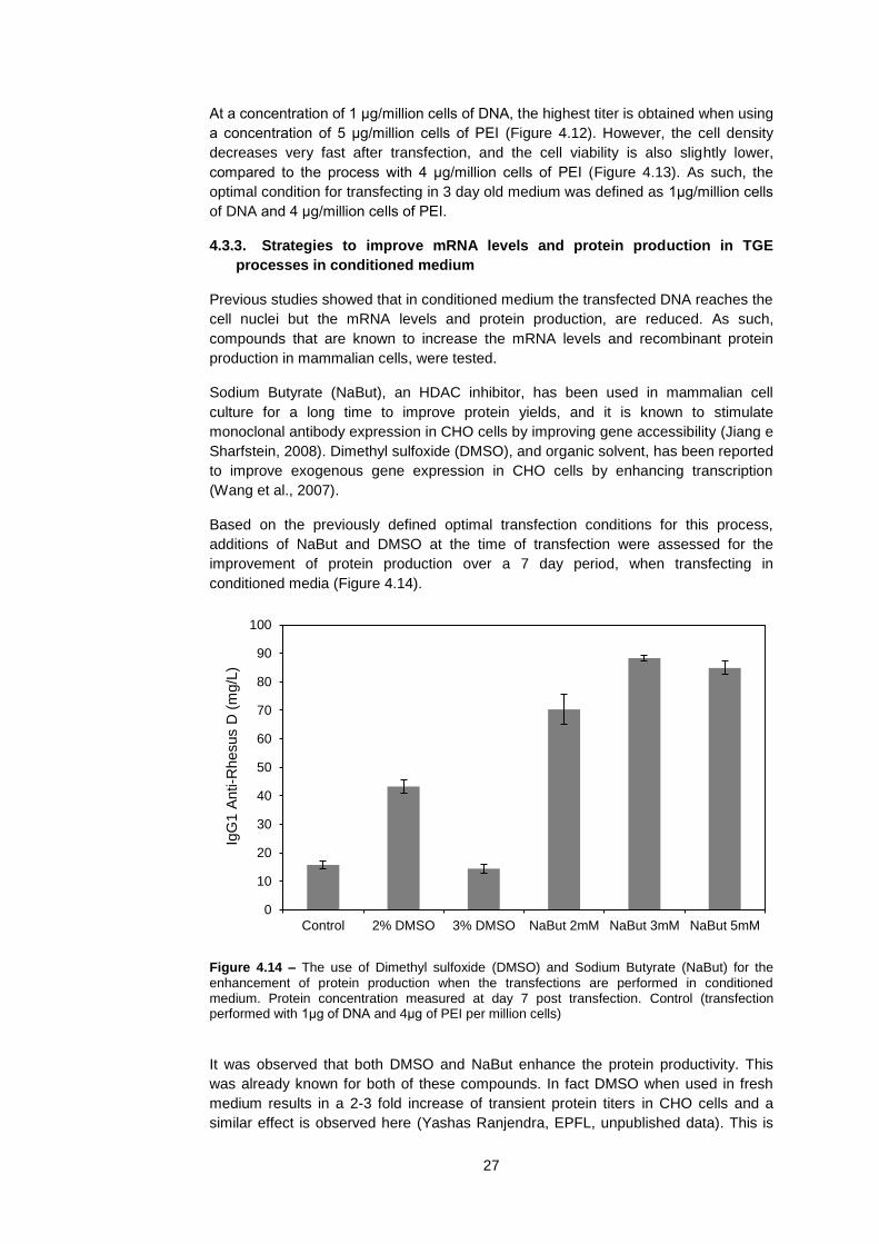
27
At a concentration of 1 μg/million cells of DNA, the highest titer is obtained when using
a concentration of 5 μg/million cells of PEI (Figure 4.12). However, the cell density
decreases very fast after transfection, and the cell viability is also slightly lower,
compared to the process with 4 μg/million cells of PEI (Figure 4.13). As such, the
optimal condition for transfecting in 3 day old medium was defined as 1μg/million cells
of DNA and 4 μg/million cells of PEI.
4.3.3. Strategies to improve mRNA levels and protein production in TGE
processes in conditioned medium
Previous studies showed that in conditioned medium the transfected DNA reaches the
cell nuclei but the mRNA levels and protein production, are reduced. As such,
compounds that are known to increase the mRNA levels and recombinant protein
production in mammalian cells, were tested.
Sodium Butyrate (NaBut), an HDAC inhibitor, has been used in mammalian cell
culture for a long time to improve protein yields, and it is known to stimulate
monoclonal antibody expression in CHO cells by improving gene accessibility (Jiang e
Sharfstein, 2008). Dimethyl sulfoxide (DMSO), and organic solvent, has been reported
to improve exogenous gene expression in CHO cells by enhancing transcription
(Wang et al., 2007).
Based on the previously defined optimal transfection conditions for this process,
additions of NaBut and DMSO at the time of transfection were assessed for the
improvement of protein production over a 7 day period, when transfecting in
conditioned media (Figure 4.14).
Figure 4.14 – The use of Dimethyl sulfoxide (DMSO) and Sodium Butyrate (NaBut) for the
enhancement of protein production when the transfections are performed in conditioned medium. Protein concentration measured at day 7 post transfection. Control (transfection performed with 1μg of DNA and 4μg of PEI per million cells)
It was observed that both DMSO and NaBut enhance the protein productivity. This
was already known for both of these compounds. In fact DMSO when used in fresh
medium results in a 2-3 fold increase of transient protein titers in CHO cells and a
similar effect is observed here (Yashas Ranjendra, EPFL, unpublished data). This is
0
10
20
30
40
50
60
70
80
90
100
Control 2% DMSO 3% DMSO NaBut 2mM NaBut 3mM NaBut 5mM
IgG
1 A
nti-R
hesus D
(m
g/L
)

28
consistent with the hypothesis that DMSO enhances transcription, and thus, enhances
mRNA levels and protein titers by having a positive effect on the activity of the RNA
polymerase (Chen e Zhang, 2005).
For transfections in conditioned medium it is seen that NaBut increases the titers
more than 5-fold. However, if the transfection is performed fresh medium the addition
of NaBut does not have such a big impact (data not shown).
Based on the 5-fold improvement seen when adding NaBut for transfecting in
conditioned mediumThis is in accordance with the hypothesis proposed before, that
components of the conditioned medium bind to the PEI-DNA complex, are internalized
during the transfection, and decrease the gene accessibility, and thus, a lower protein
production is observed (4.2.3;4.2.5). It could even be that cell derived histone proteins
present in the conditions medium are binding to the DNA, altought this is not proved.
However, the addition of NaBut helps to overcome the negative effects of the
conditioned medium in TGE processes.
4.3.4. Conclusion
The advantage of process development at small scale is that it allows the study of
different conditions at the same time. Here, the improvement of the transfection
conditions for transfecting in conditioned medium yielded a 3-fold improvement on the
protein titers. Combined with the addition of NaBut, the titers of the already improved
process are increased more than 5-fold (Figure 4.15).
Figure 4.15 - Yield comparison between the initial process (optimized for fresh medium), the
process with the improved transfection conditions for conditioned medium and the improved process with the addition of Sodium Butyrate (NaBut).
An 18-fold improvement of the process was achieved by combining the improvement
of the transfection conditions with the addition of NaBut. Table 4.1 shows a summary
0
10
20
30
40
50
60
70
80
90
100
Initial PEI:DNA ratio Improved PEI:DNA ratio Improved PEI:DNA ratio +NaBut
IgG
1 A
nti-R
hesus D
(m
g/L
)

29
of what was done to improve this TGE process in CHO-DG44 cells in conditioned
medium.
Table 4.1 – Summary of the improvements of the TGE process in CHO-DG44 cells in
conditioned medium
Initial process Improved process
25kDa Polyethylenimine (PEI) (μg/million cells)
3 4 (33% more)
DNA (μg/million cells)
0.6 1 (66% more)
Chemical addition None 3mM of Sodium
Butyrate
Yield ~5 mg/L ~90mg/L (18-fold)

30
31
5. Future perspectives and conclusion
In this work, transient gene expression processes in conditioned medium in CHO-
DG44 cells were studied. The investigations on DNA delivery provided new insights
on the understanding of why these processes are less efficient. By quantifying
intracellular plasmid DNA concentrations, it was unraveled that the DNA delivery into
the cell is not a limitation when transfecting in conditioned medium and it is not the
factor responsible for the lower mRNA levels and protein production. The hypothesis
of incorrect intracellular trafficking of the plasmid was put aside by extracting the cell
nuclei after transfection and quantifying the intracellular pDNA. It was observed that
the transfected DNA was inside the nuclei 24h post transfection when the cells are
transfected in either fresh or conditioned medium. It was hypothesized that there
might be a problem of gene accessibility and that this could be the reason for the
lower mRNA levels when the transfection is performed in conditioned medium.
As argued before, it is not likely that the plasmid DNA is degraded, as its detection is
very stable by qPCR. However, a definitive proof can only be obtained by a southern
hybridization of the DNA isolated from the cells after the transfection. This should be
included in further work done on this topic.
The trafficking of the DNA to the nucleus was studied with a simple method of nuclear
extraction that is based on a detergent. However, it would also be interesting to label
DNA and PEI with fluorescent probes and with microscopy, study the differences of
the transfection process with DNA and PEI in fresh and in conditioned medium.
The hypothesis of reduced gene accessibility due to compounds of the conditioned
medium binding to PEI-DNA complexes, and making them less accessible, is not easy
to prove. First, it is necessary to quantify cell-derived compounds in the conditioned
medium, by measuring DNA concentrations, protein concentrations and glycan
concentrations in the cell culture medium. This however, is both challenging and time
consuming, as cell culture media is a highly rich mixture of components, that include
proteins and glycans, and it would be difficult to analytically distinguish cell derived
components in this mixture. More specifically, it would be interesting to detect the
presence of DNA binding proteins in the conditioned medium. Further research should
also look into in vitro studies to detect the possibility of a PEI-DNA complex being
formed in conjugation with other charged molecules like proteins (positively charged
and negatively charged) or glycans.
In the process development part of this work, the concentrations of of DNA and PEI
that had been used for TGE processes involving a medium exchange before
transfection, were optimized to define a TGE process without a medium exchange
(i.e. in conditioned medium). An increase of the DNA concentration and PEI
concentration lead to a 3-fold improvement of the yields.
Further, the possibility of enhancing mRNA levels and protein production with Sodium
Butyrate (NaBut) and Dymethyl Sulfoxide (DMSO) was investigated. Although both
chemicals improved the recombinant protein yields, NaBut proved the most efficient.
A yield improvement of more than 5-fold was obtained by using Sodium Butyrate and
this might be due to enhanced gene accessibility. This is a good example of how
systematic research combined with process development tools can be used to solve
otherwise complex biotechnological problems. All together, the TGE process in
conditioned medium in CHO-DG44 cells was improved 18-fold, from ~5mg/L to
~90mg/L. Regarding the available literature, this was the first TGE process with
significant yields in CHO cells that was performed without a medium exchange.

32
A remark to small scale cell culture systems should be made, as it would not have
been possible to conduct this kind of process development work at larger scales. The
TubeSpin® bioreactor 50 proved to be very useful, as it allows high density
suspension cell culture work in volumes that can go to as low as 5 mL. This allowed,
for example, 48 different bioprocesses (i.e. 48 small-scale bioreactors) to be run at the
same time when optimizing the PEI and DNA concentrations.
In this approach, the use of feeding was not looked into. As the transfections are
performed in conditioned media, the concentrations of medium compounds are
changed, and the medium has already accumulated metabolites as lactate and
ammonia and is depleted of glutamine. A feeding approach would enhance the cell
metabolism and extend the process for a longer period of time, resulting in higher
yields. Also, only 3 concentrations of NaBut were tested and a more refined screening
of different concentrations of NaBut should be done. The timing of addition of NaBut
could also be important as in this study NaBut was only added at the time of
transfection. It could also be that adding NaBut before the transfection, or some hours
after has a better effect on the productivity.
The development of TGE processes without a medium exchange is one of the
principles that allow the scale-up of transient gene expression and these processes
should continue to be developed and scaled-up. High yields obtained by transient
transfections at large culture volumes would allow the manufacturing of Kg amounts of
protein for the market in time-spans of as short as 1-2 weeks. This would be a very
strong competitive advantage for biotechnology companies as they could enter the
market faster, without having to spent months developing a stable cell line and a
process for it. If high-yielding TGE is mastered at large-scale, the door is opened for a
paradigm shift in the protein manufacturing industry, but that remains to be seen.

33
6. Bibliography
Backliwal G, Hildinger M, Chenuet S, Wulhfard S, De Jesus M, Wurm FM. 2008. Rational vector design and multi-pathway modulation of HEK 293E cells yield recombinant antibody titers exceeding 1 g/l by transient transfection under serum-free conditions. Nucleic Acids Research 36:e96.
Baldi L, Hacker D, Adam M, Wurm F. 2007. Recombinant protein production by large-scale transient gene expression in mammalian cells: state of the art and future perspectives. Biotechnology Letters 29:677-684.
Belting M, Petersson P. 1999. Intracellular Accumulation of Secreted Proteoglycans Inhibits Cationic Lipid-mediated Gene Transfer. Journal of Biological Chemistry 274:19375 -19382.
Boussif O, Lezoualc’h F, Zanta MA, Mergny MD, Scherman D, Demeneix B, Behr JP. 1995. A versatile vector for gene and oligonucleotide transfer into cells in culture and in vivo: polyethylenimine. Proceedings of the National Academy of Sciences 92:7297 -7301.
Chen Z, Zhang Y. 2005. Dimethyl sulfoxide targets phage RNA polymerases to promote transcription. Biochemical and Biophysical Research Communications 333:664-670.
Van Craenenbroeck K, Vanhoenacker P, Haegeman G. 2000. Episomal vectors for gene expression in mammalian cells. European Journal of Biochemistry 267:5665-5678.
Derouazi M, Martinet D, Besuchet Schmutz N, Flaction R, Wicht M, Bertschinger M, Hacker DL, Beckmann JS, Wurm FM. 2006a. Genetic characterization of CHO production host DG44 and derivative recombinant cell lines. Biochemical and Biophysical Research Communications 340:1069-1077.
Derouazi M, Flaction R, Girard P, de Jesus M, Jordan M, Wurm F. 2006b. Generation of Recombinant Chinese Hamster Ovary Cell Lines by Microinjection. Biotechnology Letters 28:373-382-382.
Derouazi M, Girard P, Van Tilborgh F, Iglesias K, Muller N, Bertschinger M, Wurm FM. 2004. Serum-free large-scale transient transfection of CHO cells. Biotechnol. Bioeng. 87:537-545.
Geisse S. 2009. Reflections on more than 10 years of TGE approaches. Protein Expression and Purification 64:99-107.
Geisse S, Henke M. 2005. Large-scale Transient Transfection of Mammalian Cells: A Newly Emerging Attractive Option for Recombinant Protein Production. Journal of Structural and Functional Genomics 6:165-170.
Graham FL, van der Eb AJ. 1973. A new technique for the assay of infectivity of human adenovirus 5 DNA. Virology 52:456-467.
Hacker DL, De Jesus M, Wurm FM. 2007. 25 years of recombinant proteins from reactor-grown cells -- Where do we go from here? Biotechnology Advances 27:1023-1027.
De Jesus MJ, Girard P, Bourgeois M, Baumgartner G, Jacko B, Amstutz H, Wurm FM. 2004. TubeSpin satellites: a fast track approach for process development with animal cells using shaking technology. Biochemical Engineering Journal 17:217-223.
Jiang Z, Sharfstein ST. 2008. Sodium butyrate stimulates monoclonal antibody over-expression in CHO cells by improving gene accessibility. Biotechnol. Bioeng. 100:189-194.
Jordan M, Schallhorn A, Wurm FM. 1996. Transfecting Mammalian Cells: Optimization of Critical Parameters Affecting Calcium-Phosphate Precipitate Formation. Nucleic Acids Research 24:596 -601.
Kaufman RJ, Wasley LC, Spiliotes AJ, Gossels SD, Latt SA, Larsen GR, Kay RM. 1985. Coamplification and coexpression of human tissue-type plasminogen activator and murine dihydrofolate reductase sequences in Chinese hamster ovary cells. Mol. Cell. Biol. 5:1750-1759.
Kost TA, Condreay JP. 2002. Recombinant baculoviruses as mammalian cell gene-delivery vectors. Trends in Biotechnology 20:173-180.

34
Meissner P, Pick H, Kulangara A, Chatellard P, Friedrich K, Wurm FM. 2001. Transient gene expression: Recombinant protein production with suspension-adapted HEK293-EBNA cells. Biotechnol. Bioeng. 75:197-203.
Mislick KA, Baldeschwieler JD. 1996. Evidence for the role of proteoglycans in cation-mediated gene transfer. Proceedings of the National Academy of Sciences 93:12349 -12354.
Muller N, Derouazi M, Van Tilborgh F, Wulhfard S, Hacker D, Jordan M, Wurm F. 2007. Scalable transient gene expression in Chinese hamster ovary cells in instrumented and non-instrumented cultivation systems. Biotechnology Letters 29:703-711.
Muller N, Girard P, Hacker DL, Jordan M, Wurm FM. 2005. Orbital shaker technology for the cultivation of mammalian cells in suspension. Biotechnol. Bioeng. 89:400-406.
Rajendra Y, Kiseljak D, Baldi L, Hacker DL, Wurm FM. 2011. A simple high-yielding process for transient gene expression in CHO cells. Journal of Biotechnology 153:22-26.
Raymond C, Tom R, Perret S, Moussouami P, L’Abb D, St-Laurent G, Durocher Y. 2011. A simplified polyethylenimine-mediated transfection process for large-scale and high-throughput applications. Methods In Press, Corrected Proof. http://www.sciencedirect.com/science/article/pii/S1046202311000818.
Ruponen M, Honkakoski P, Tammi M, Urtti A. 2004. Cell-surface glycosaminoglycans inhibit cation-mediated gene transfer. J. Gene Med. 6:405-414.
Ruponen M, Rönkkö S, Honkakoski P, Pelkonen J, Tammi M, Urtti A. 2001. Extracellular Glycosaminoglycans Modify Cellular Trafficking of Lipoplexes and Polyplexes. Journal of Biological Chemistry 276:33875 -33880.
Schlaeger E-J, Christensen K. 1999. Transient gene expression in mammalian cells grown in serum-free suspension culture. Cytotechnology 30:71-83.
Stettler M, Zhang X, Hacker DL, de Jesus M, Wurm FM. 2007. Novel Orbital Shake Bioreactors for Transient Production of CHO Derived IgGs. Biotechnol Progress 23:1340-1346.
Sutter G, Moss B. 1992. Nonreplicating vaccinia vector efficiently expresses recombinant genes. Proceedings of the National Academy of Sciences 89:10847 -10851.
Tissot S, Oberbek A, Reclari M, Dreyer M, Hacker DL, Baldi L, Farhat M, Wurm FM. 2011. Efficient and reproducible mammalian cell bioprocesses without probes and controllers? New Biotechnology In Press, Corrected Proof. http://www.sciencedirect.com/science/article/pii/S1871678411000409.
Urlaub G, Kós E, Carothers AM, Chasin LA. 1983. Deletion of the diploid dihydrofolate reductase locus from cultured mammalian cells. Cell 33:405-412.
Walsh G. 2010. Biopharmaceutical benchmarks 2010. Nat Biotech 28:917-924. Wang W, Yi X, Zhang Y. 2007. Gene transcription acceleration: main cause of
hepatitis B surface antigen production improvement by dimethyl sulfoxide in the culture of Chinese hamster ovary cells. Biotechnol. Bioeng. 97:526-535.
Wulhfard S, Baldi L, Hacker DL, Wurm F. 2010. Valproic acid enhances recombinant mRNA and protein levels in transiently transfected Chinese hamster ovary cells. Journal of Biotechnology 148:128-132.
Wulhfard S, Tissot S, Bouchet S, Cevey J, de Jesus M, Hacker DL, Wurm FM. 2008. Mild Hypothermia Improves Transient Gene Expression Yields Several Fold in Chinese Hamster Ovary Cells. Biotechnol Progress 24:458-465.
Wurm FM. 2004. Production of recombinant protein therapeutics in cultivated mammalian cells. Nat Biotech 22:1393-1398.
Wurm F, Bernard A. 1999. Large-scale transient expression in mammalian cells for recombinant protein production. Current Opinion in Biotechnology 10:156-159.
Ye J, Kober V, Tellers M, Naji Z, Salmon P, Markusen JF. 2009. High-level protein expression in scalable CHO transient transfection. Biotechnol. Bioeng. 103:542-551.
35
Yue Y, Jin F, Deng R, Cai J, Chen Y, Lin MCM, Kung H-F, Wu C. In Press. Revisit complexation between DNA and polyethylenimine -- Effect of uncomplexed chains free in the solution mixture on gene transfection. Journal of Controlled Release In Press, Corrected Proof. http://www.sciencedirect.com/science/article/pii/S0168365910008424.
Zhang X, Burki C-A, Stettler M, De Sanctis D, Perrone M, Discacciati M, Parolini N, DeJesus M, Hacker DL, Quarteroni A, Wurm FM. 2009. Efficient oxygen transfer by surface aeration in shaken cylindrical containers for mammalian cell cultivation at volumetric scales up to 1000L. Biochemical Engineering Journal 45:41-47.
Zhang X, Garcia I, Baldi L, Hacker D, Wurm F. 2010a. Hyperosmolarity enhances transient recombinant protein yield in Chinese hamster ovary cells. Biotechnology Letters 32:1587-1592.
Zhang X, Stettler M, De Sanctis D, Perrone M, Parolini N, Discacciati M, De Jesus M, Hacker D, Quarteroni A, Wurm F. 2010b. Use of Orbital Shaken Disposable Bioreactors for Mammalian Cell Cultures from the Milliliter-Scale to the 1,000-Liter Scale. Em: . Disposable Bioreactors. Springer Berlin / Heidelberg. Advances in Biochemical Engineering/Biotechnology, Vol. 115, pp 33-53. http://dx.doi.org/10.1007/10_2008_18.