essentials of dental radiography for dental assistants and hygienists chapter copyright ©2012 by...
TRANSCRIPT
Essentials of Dental Radiographyfor Dental Assistants and Hygienists
CHAPTER
Copyright ©2012 by Pearson Education, Inc.All rights reserved.
Essentials of Dental Radiography for Dental Assistants and Hygienists, Ninth EditionEvelyn M. Thomson • Orlen N. Johnson
NINTH EDITION
Producing Quality Radiographs
4
Copyright ©2012 by Pearson Education, Inc.All rights reserved.
Essentials of Dental Radiography for Dental Assistants and Hygienists, Ninth EditionEvelyn M. Thomson • Orlen N. Johnson
Objectives
• Define the key words.• Evaluate a radiographic image identifying
the basic requirements of acceptability.• Differentiate between radiolucent and
radiopaque areas on a dental radiograph.• Define radiographic density and contrast.• Differentiate between subject contrast and
film contrast.
Copyright ©2012 by Pearson Education, Inc.All rights reserved.
Essentials of Dental Radiography for Dental Assistants and Hygienists, Ninth EditionEvelyn M. Thomson • Orlen N. Johnson
Objectives
• List the factors that influence magnification and distortion.
• List the geometric factors that affect image sharpness.
• Summarize the factors affecting the radiographic image.
• Describe how mA, kVp, and exposure time affect image density.
Copyright ©2012 by Pearson Education, Inc.All rights reserved.
Essentials of Dental Radiography for Dental Assistants and Hygienists, Ninth EditionEvelyn M. Thomson • Orlen N. Johnson
Objectives
• Discuss how kVp affects image contrast.• Explain target-surface, object-image
receptor, and target-image receptor distances.
• Demonstrate the practical use of the inverse square law.
Copyright ©2012 by Pearson Education, Inc.All rights reserved.
Essentials of Dental Radiography for Dental Assistants and Hygienists, Ninth EditionEvelyn M. Thomson • Orlen N. Johnson
Key Words
• Contrast• Crystal• Definition• Density• Distortion• Exposure chart• Exposure factors• Exposure time
Copyright ©2012 by Pearson Education, Inc.All rights reserved.
Essentials of Dental Radiography for Dental Assistants and Hygienists, Ninth EditionEvelyn M. Thomson • Orlen N. Johnson
Key Words
• Extraoral radiography• Film contrast• Focal spot• Geometric factors• Grid• Intensifying screen• Intraoral radiography• Inverse square law
Copyright ©2012 by Pearson Education, Inc.All rights reserved.
Essentials of Dental Radiography for Dental Assistants and Hygienists, Ninth EditionEvelyn M. Thomson • Orlen N. Johnson
Key Words
• Kilovoltage peak (kVp)• Long-scale contrast• Magnification• Milliampere (mA)• Motion• Object-image receptor distance• Penumbra
Copyright ©2012 by Pearson Education, Inc.All rights reserved.
Essentials of Dental Radiography for Dental Assistants and Hygienists, Ninth EditionEvelyn M. Thomson • Orlen N. Johnson
Key Words
• Position indicating device (PID)• Radiographic contrast• Radiolucent• Radiopaque• Sharpness• Short-scale contrast
Copyright ©2012 by Pearson Education, Inc.All rights reserved.
Essentials of Dental Radiography for Dental Assistants and Hygienists, Ninth EditionEvelyn M. Thomson • Orlen N. Johnson
Key Words
• Subject contrast• Target-image receptor distance• Target-object distance• Target-surface distance
Copyright ©2012 by Pearson Education, Inc.All rights reserved.
Essentials of Dental Radiography for Dental Assistants and Hygienists, Ninth EditionEvelyn M. Thomson • Orlen N. Johnson
Introduction
• Each patient presents with a unique set of characteristics for which a customized approach to exposure settings is needed.
• The dental radiographer has an ethical responsibility to produce the highest diagnostic quality radiographs for patients who agree to be exposed to ionizing radiation.
Copyright ©2012 by Pearson Education, Inc.All rights reserved.
Essentials of Dental Radiography for Dental Assistants and Hygienists, Ninth EditionEvelyn M. Thomson • Orlen N. Johnson
Introduction
• To consistently produce diagnostic quality radiographs at the lowest possible radiation dose, the dental radiographer needs to understand the inter-relationships of the components of the dental x-ray machine.
Copyright ©2012 by Pearson Education, Inc.All rights reserved.
Essentials of Dental Radiography for Dental Assistants and Hygienists, Ninth EditionEvelyn M. Thomson • Orlen N. Johnson
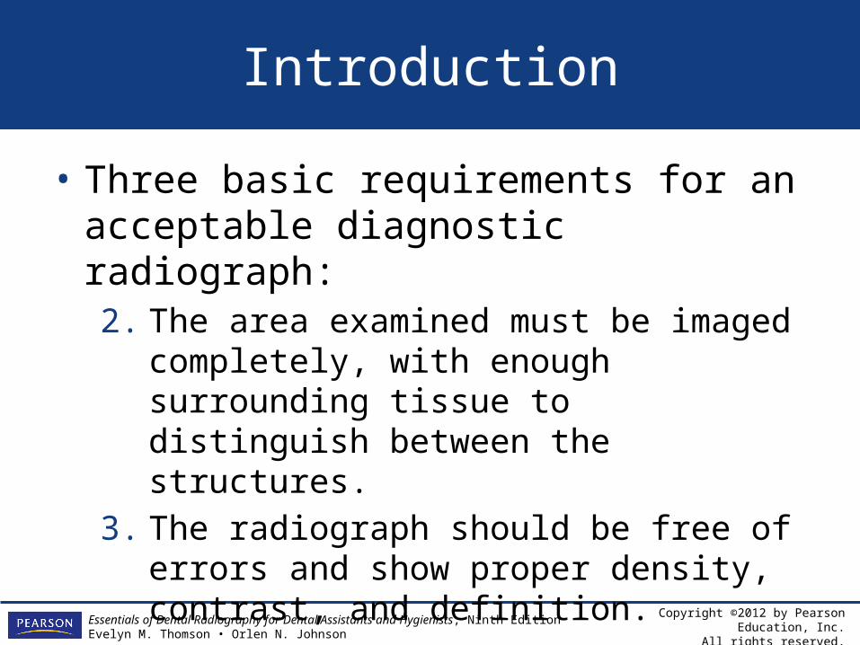
Introduction
• Three basic requirements for an acceptable diagnostic radiograph: 1. All parts of the structures recorded must be
imaged as close to their natural shapes and sizes as the patient’s oral anatomy will permit. Distortion and superimposition of structures should be at a minimum.
Copyright ©2012 by Pearson Education, Inc.All rights reserved.
Essentials of Dental Radiography for Dental Assistants and Hygienists, Ninth EditionEvelyn M. Thomson • Orlen N. Johnson
Introduction
• Three basic requirements for an acceptable diagnostic radiograph: 2. The area examined must be imaged
completely, with enough surrounding tissue to distinguish between the structures.
3. The radiograph should be free of errors and show proper density, contrast, and definition.
Copyright ©2012 by Pearson Education, Inc.All rights reserved.
Essentials of Dental Radiography for Dental Assistants and Hygienists, Ninth EditionEvelyn M. Thomson • Orlen N. Johnson
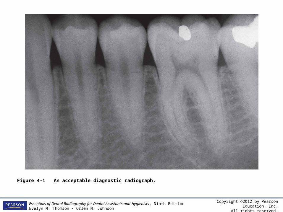
Figure 4-1 An acceptable diagnostic radiograph.
Copyright ©2012 by Pearson Education, Inc.All rights reserved.
Essentials of Dental Radiography for Dental Assistants and Hygienists, Ninth EditionEvelyn M. Thomson • Orlen N. Johnson
Terminology
• Radiolucent• Radiopaque• Density• Contrast
– Short-scale contrast – Long-scale contrast
• Sharpness
Copyright ©2012 by Pearson Education, Inc.All rights reserved.
Essentials of Dental Radiography for Dental Assistants and Hygienists, Ninth EditionEvelyn M. Thomson • Orlen N. Johnson
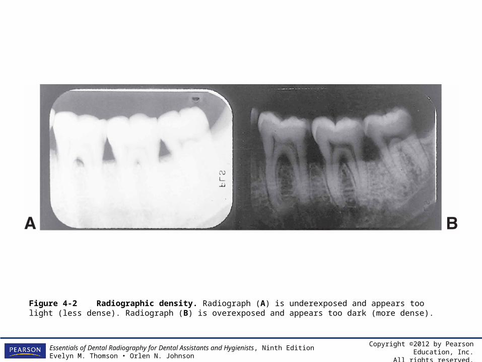
Figure 4-2 Radiographic density. Radiograph (A) is underexposed and appears too light (less dense). Radiograph (B) is overexposed and appears too dark (more dense).

Copyright ©2012 by Pearson Education, Inc.All rights reserved.
Essentials of Dental Radiography for Dental Assistants and Hygienists, Ninth EditionEvelyn M. Thomson • Orlen N. Johnson
Figure 4-3 Penetrometer tests demonstrate radiographically that a longer contrast scale results from the use of 100 kilovolt exposures. Dental radiographs exposed at 100 kVp have long-scale contrast. Radiographs exposed at 60 kVp have short-scale contrast.
Copyright ©2012 by Pearson Education, Inc.All rights reserved.
Essentials of Dental Radiography for Dental Assistants and Hygienists, Ninth EditionEvelyn M. Thomson • Orlen N. Johnson
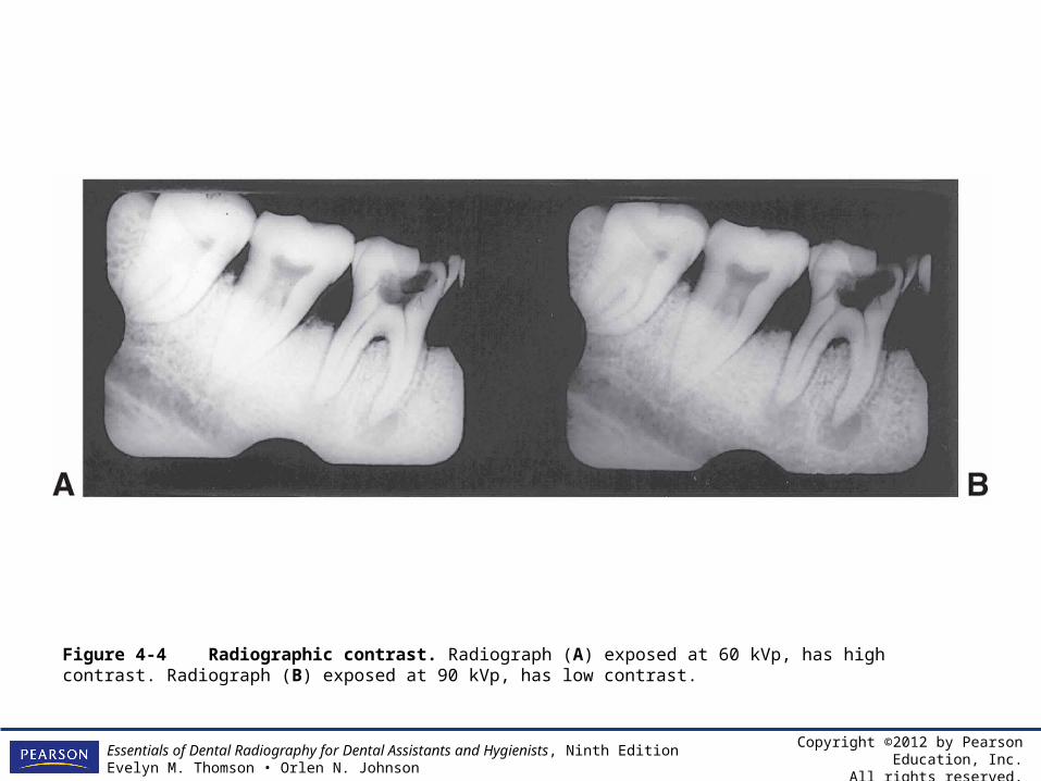
Figure 4-4 Radiographic contrast. Radiograph (A) exposed at 60 kVp, has high contrast. Radiograph (B) exposed at 90 kVp, has low contrast.
Copyright ©2012 by Pearson Education, Inc.All rights reserved.
Essentials of Dental Radiography for Dental Assistants and Hygienists, Ninth EditionEvelyn M. Thomson • Orlen N. Johnson
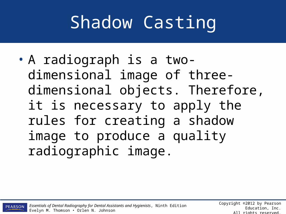
Shadow Casting
• A radiograph is a two-dimensional image of three-dimensional objects. Therefore, it is necessary to apply the rules for creating a shadow image to produce a quality radiographic image.
Copyright ©2012 by Pearson Education, Inc.All rights reserved.
Essentials of Dental Radiography for Dental Assistants and Hygienists, Ninth EditionEvelyn M. Thomson • Orlen N. Johnson
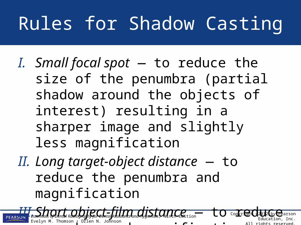
Rules for Shadow Casting
I. Small focal spot — to reduce the size of the penumbra (partial shadow around the objects of interest) resulting in a sharper image and slightly less magnification
II. Long target-object distance — to reduce the penumbra and magnification
III. Short object-film distance — to reduce penumbra and magnification
Copyright ©2012 by Pearson Education, Inc.All rights reserved.
Essentials of Dental Radiography for Dental Assistants and Hygienists, Ninth EditionEvelyn M. Thomson • Orlen N. Johnson
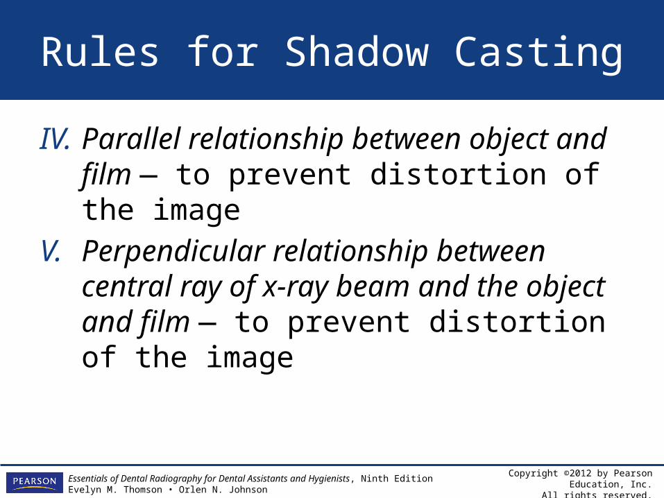
Rules for Shadow Casting
IV. Parallel relationship between object and film — to prevent distortion of the image
V. Perpendicular relationship between central ray of x-ray beam and the object and film — to prevent distortion of the image
Copyright ©2012 by Pearson Education, Inc.All rights reserved.
Essentials of Dental Radiography for Dental Assistants and Hygienists, Ninth EditionEvelyn M. Thomson • Orlen N. Johnson
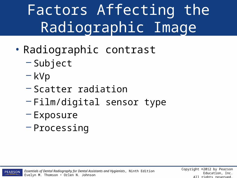
Factors Affecting the Radiographic Image
• Radiographic contrast– Subject– kVp– Scatter radiation– Film/digital sensor type– Exposure – Processing
Copyright ©2012 by Pearson Education, Inc.All rights reserved.
Essentials of Dental Radiography for Dental Assistants and Hygienists, Ninth EditionEvelyn M. Thomson • Orlen N. Johnson
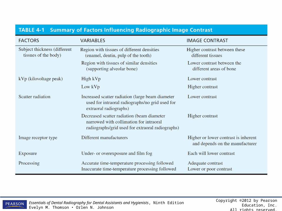
Table 4-1 Summary of Factors Influencing Radiographic Image Contrast
Copyright ©2012 by Pearson Education, Inc.All rights reserved.
Essentials of Dental Radiography for Dental Assistants and Hygienists, Ninth EditionEvelyn M. Thomson • Orlen N. Johnson
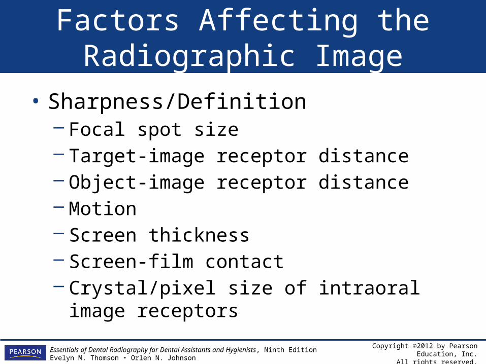
Factors Affecting the Radiographic Image
• Sharpness/Definition– Focal spot size– Target-image receptor distance– Object-image receptor distance– Motion– Screen thickness– Screen-film contact– Crystal/pixel size of intraoral image receptors
Copyright ©2012 by Pearson Education, Inc.All rights reserved.
Essentials of Dental Radiography for Dental Assistants and Hygienists, Ninth EditionEvelyn M. Thomson • Orlen N. Johnson
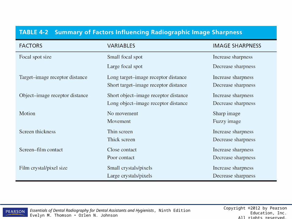
Table 4-2 Summary of Factors Influencing Radiographic Image Sharpness
Copyright ©2012 by Pearson Education, Inc.All rights reserved.
Essentials of Dental Radiography for Dental Assistants and Hygienists, Ninth EditionEvelyn M. Thomson • Orlen N. Johnson
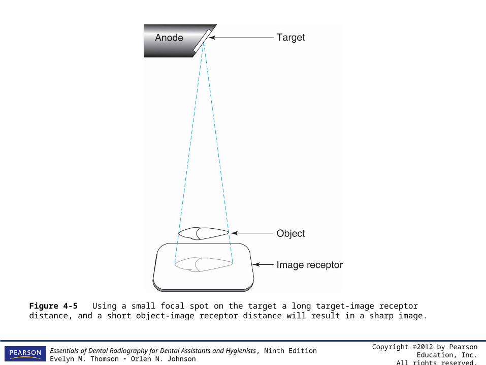
Figure 4-5 Using a small focal spot on the target a long target-image receptor distance, and a short object-image receptor distance will result in a sharp image.
Copyright ©2012 by Pearson Education, Inc.All rights reserved.
Essentials of Dental Radiography for Dental Assistants and Hygienists, Ninth EditionEvelyn M. Thomson • Orlen N. Johnson
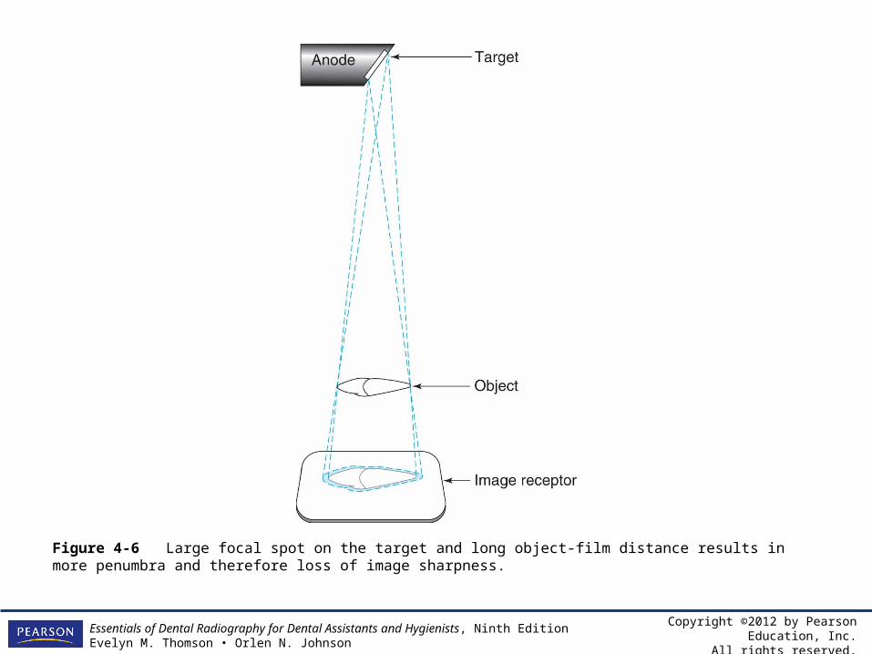
Figure 4-6 Large focal spot on the target and long object-film distance results in more penumbra and therefore loss of image sharpness.
Copyright ©2012 by Pearson Education, Inc.All rights reserved.
Essentials of Dental Radiography for Dental Assistants and Hygienists, Ninth EditionEvelyn M. Thomson • Orlen N. Johnson
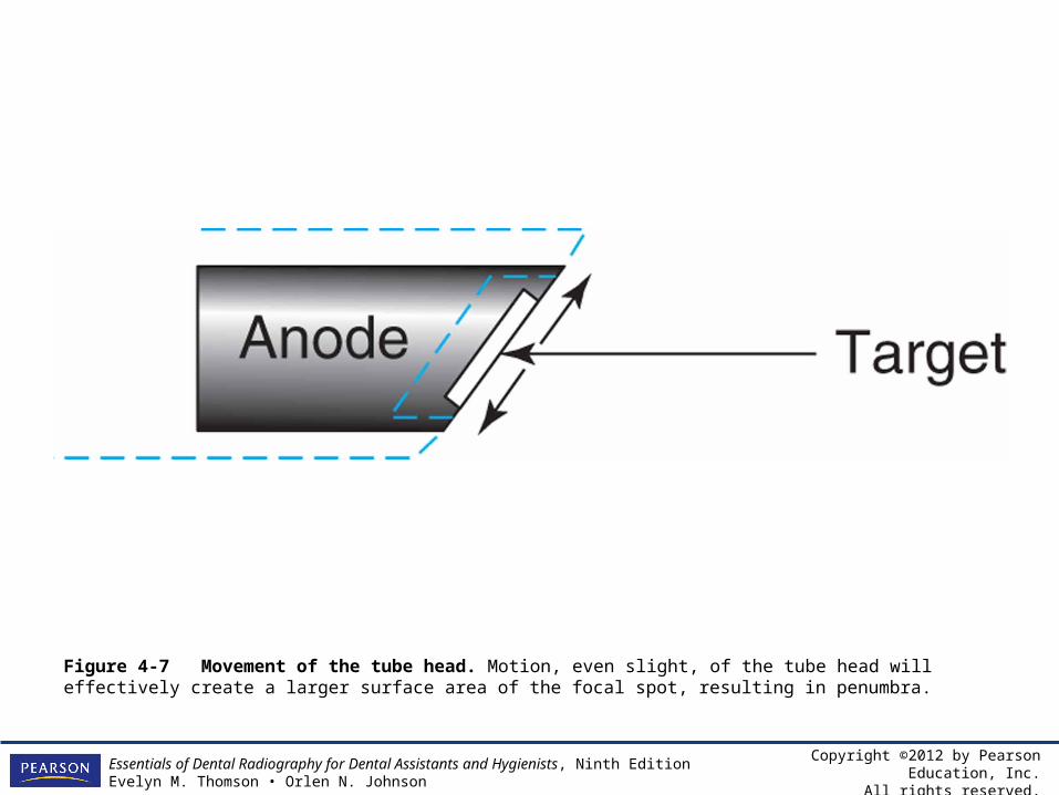
Figure 4-7 Movement of the tube head. Motion, even slight, of the tube head will effectively create a larger surface area of the focal spot, resulting in penumbra.
Copyright ©2012 by Pearson Education, Inc.All rights reserved.
Essentials of Dental Radiography for Dental Assistants and Hygienists, Ninth EditionEvelyn M. Thomson • Orlen N. Johnson
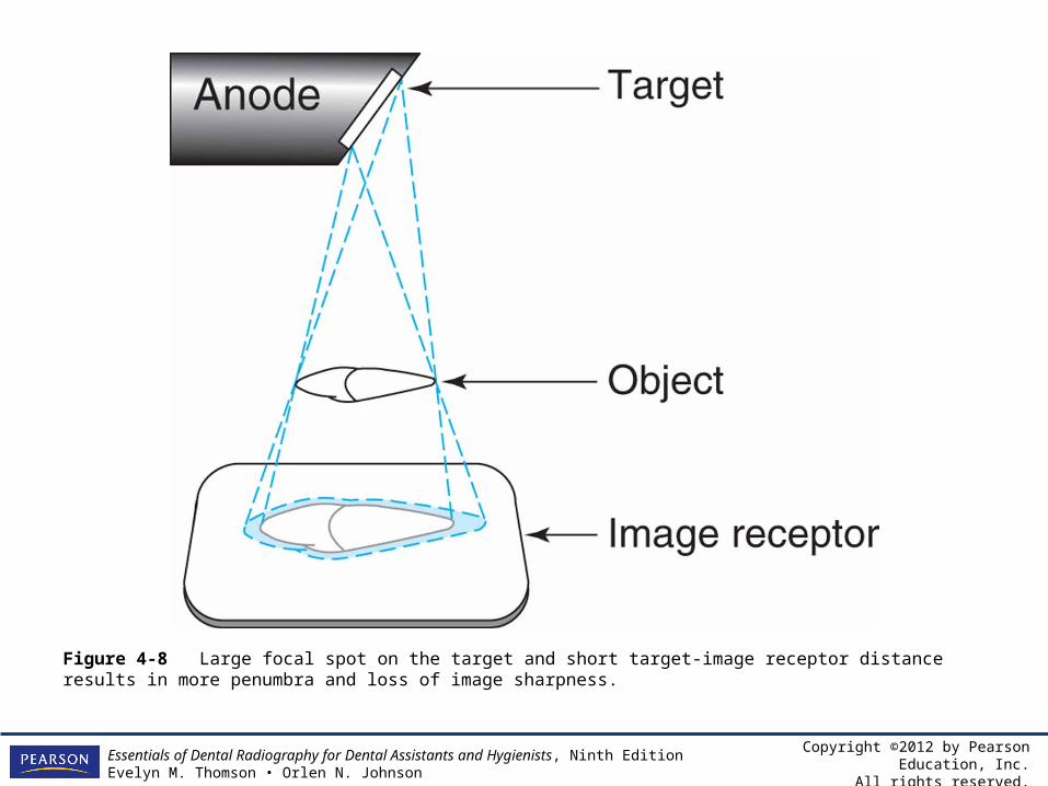
Figure 4-8 Large focal spot on the target and short target-image receptor distance results in more penumbra and loss of image sharpness.
Copyright ©2012 by Pearson Education, Inc.All rights reserved.
Essentials of Dental Radiography for Dental Assistants and Hygienists, Ninth EditionEvelyn M. Thomson • Orlen N. Johnson
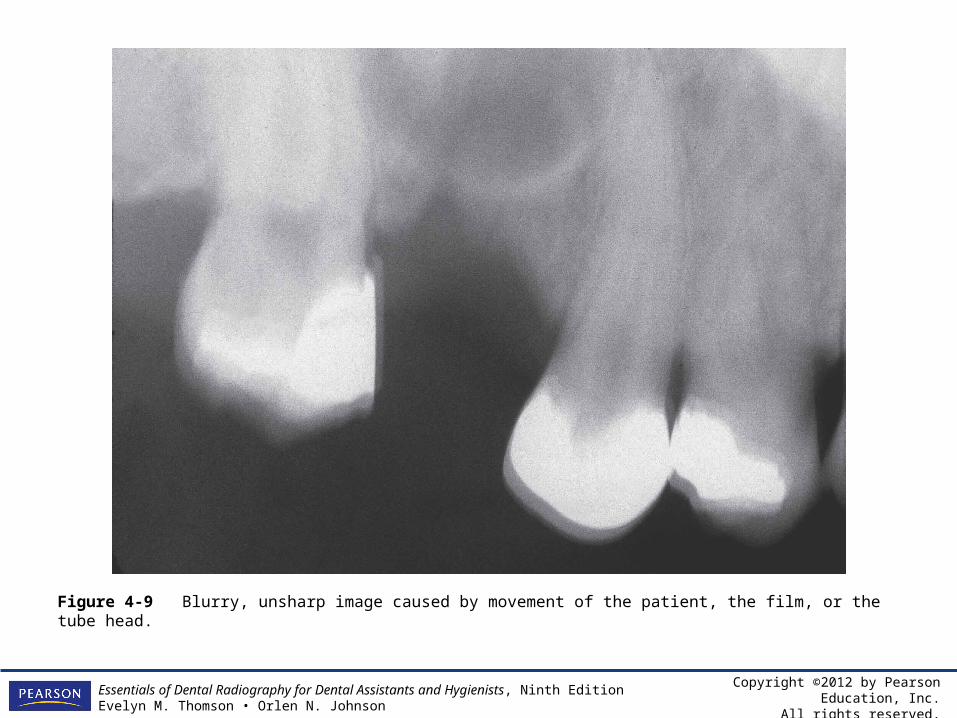
Figure 4-9 Blurry, unsharp image caused by movement of the patient, the film, or the tube head.
Copyright ©2012 by Pearson Education, Inc.All rights reserved.
Essentials of Dental Radiography for Dental Assistants and Hygienists, Ninth EditionEvelyn M. Thomson • Orlen N. Johnson
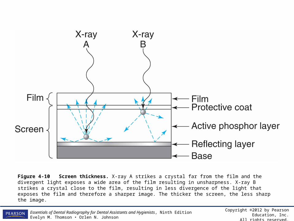
Figure 4-10 Screen thickness. X-ray A strikes a crystal far from the film and the divergent light exposes a wide area of the film resulting in unsharpness. X-ray B strikes a crystal close to the film, resulting in less divergence of the light that exposes the film and therefore a sharper image. The thicker the screen, the less sharp the image.
Copyright ©2012 by Pearson Education, Inc.All rights reserved.
Essentials of Dental Radiography for Dental Assistants and Hygienists, Ninth EditionEvelyn M. Thomson • Orlen N. Johnson
Factors Affecting the Radiographic Image
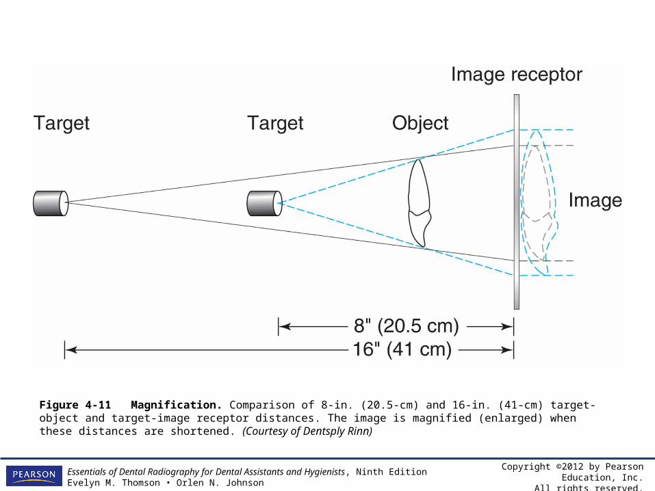
• Magnification/enlargement is mostly influenced by the target-object distance and the object-image receptor distance. The target-object distance is determined by the length of the PID.
Copyright ©2012 by Pearson Education, Inc.All rights reserved.
Essentials of Dental Radiography for Dental Assistants and Hygienists, Ninth EditionEvelyn M. Thomson • Orlen N. Johnson
Factors Affecting the Radiographic Image
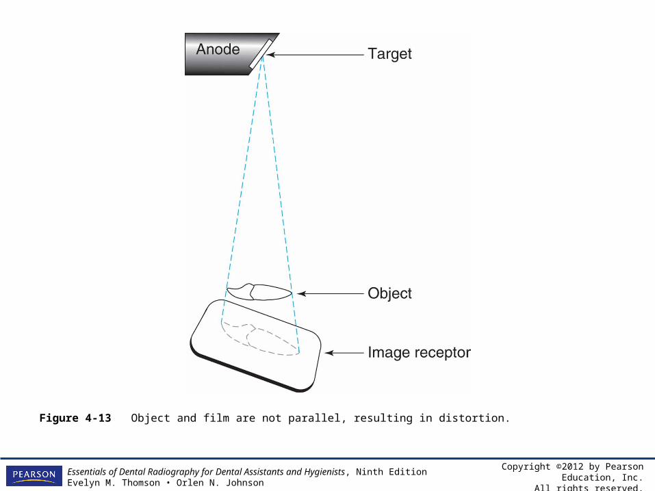
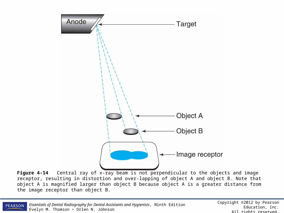
• Distortion is the result of unequal magnification of different parts of the same object. Distortion results when the image receptor is not parallel to the object (Figure 4-13) and/or when the central ray of the x-ray beam is not perpendicular to the object and the plane of the image receptor (Figure 4-14)
Copyright ©2012 by Pearson Education, Inc.All rights reserved.
Essentials of Dental Radiography for Dental Assistants and Hygienists, Ninth EditionEvelyn M. Thomson • Orlen N. Johnson
Figure 4-11 Magnification. Comparison of 8-in. (20.5-cm) and 16-in. (41-cm) target-object and target-image receptor distances. The image is magnified (enlarged) when these distances are shortened. (Courtesy of Dentsply Rinn)
Copyright ©2012 by Pearson Education, Inc.All rights reserved.
Essentials of Dental Radiography for Dental Assistants and Hygienists, Ninth EditionEvelyn M. Thomson • Orlen N. Johnson
Figure 4-13 Object and film are not parallel, resulting in distortion.
Copyright ©2012 by Pearson Education, Inc.All rights reserved.
Essentials of Dental Radiography for Dental Assistants and Hygienists, Ninth EditionEvelyn M. Thomson • Orlen N. Johnson
Figure 4-14 Central ray of x-ray beam is not perpendicular to the objects and image receptor, resulting in distortion and over-lapping of object A and object B. Note that object A is magnified larger than object B because object A is a greater distance from the image receptor than object B.
Copyright ©2012 by Pearson Education, Inc.All rights reserved.
Essentials of Dental Radiography for Dental Assistants and Hygienists, Ninth EditionEvelyn M. Thomson • Orlen N. Johnson
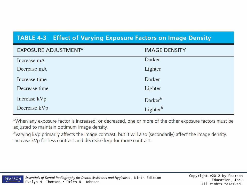
Table 4-3 Effect of Varying Exposure Factors on Image Density
Copyright ©2012 by Pearson Education, Inc.All rights reserved.
Essentials of Dental Radiography for Dental Assistants and Hygienists, Ninth EditionEvelyn M. Thomson • Orlen N. Johnson
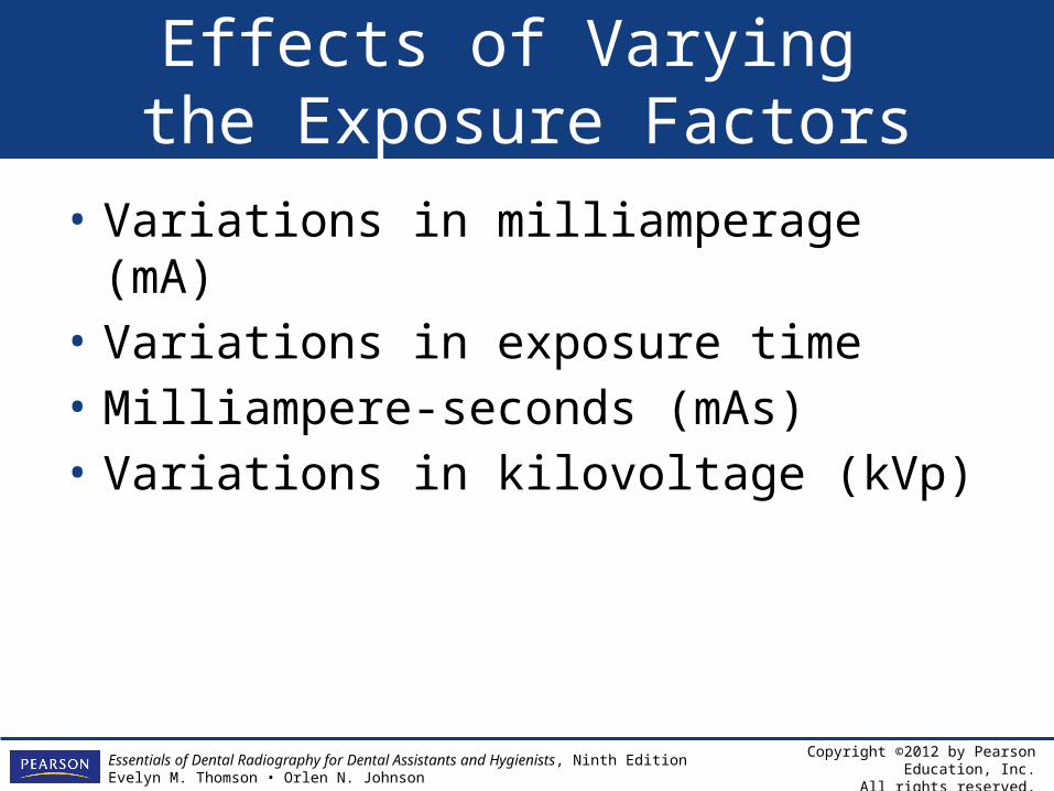
Effects of Varying the Exposure Factors
• Variations in milliamperage (mA)• Variations in exposure time• Milliampere-seconds (mAs)• Variations in kilovoltage (kVp)
Copyright ©2012 by Pearson Education, Inc.All rights reserved.
Essentials of Dental Radiography for Dental Assistants and Hygienists, Ninth EditionEvelyn M. Thomson • Orlen N. Johnson
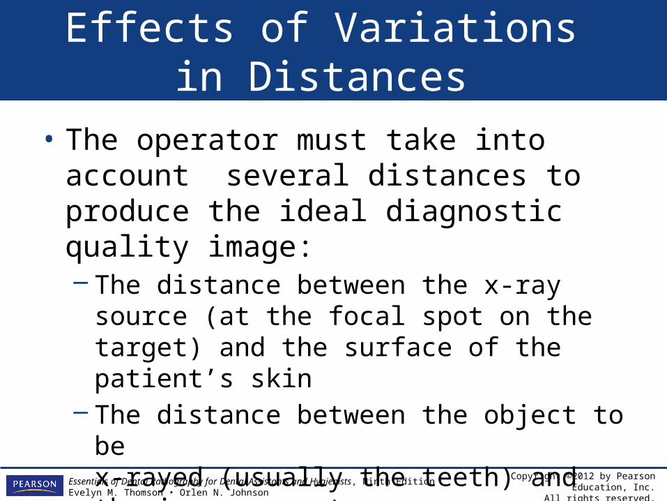
Effects of Variations in Distances
• The operator must take into account several distances to produce the ideal diagnostic quality image:– The distance between the x-ray source (at the
focal spot on the target) and the surface of the patient’s skin
– The distance between the object to be x-rayed (usually the teeth) and the image receptor
Copyright ©2012 by Pearson Education, Inc.All rights reserved.
Essentials of Dental Radiography for Dental Assistants and Hygienists, Ninth EditionEvelyn M. Thomson • Orlen N. Johnson
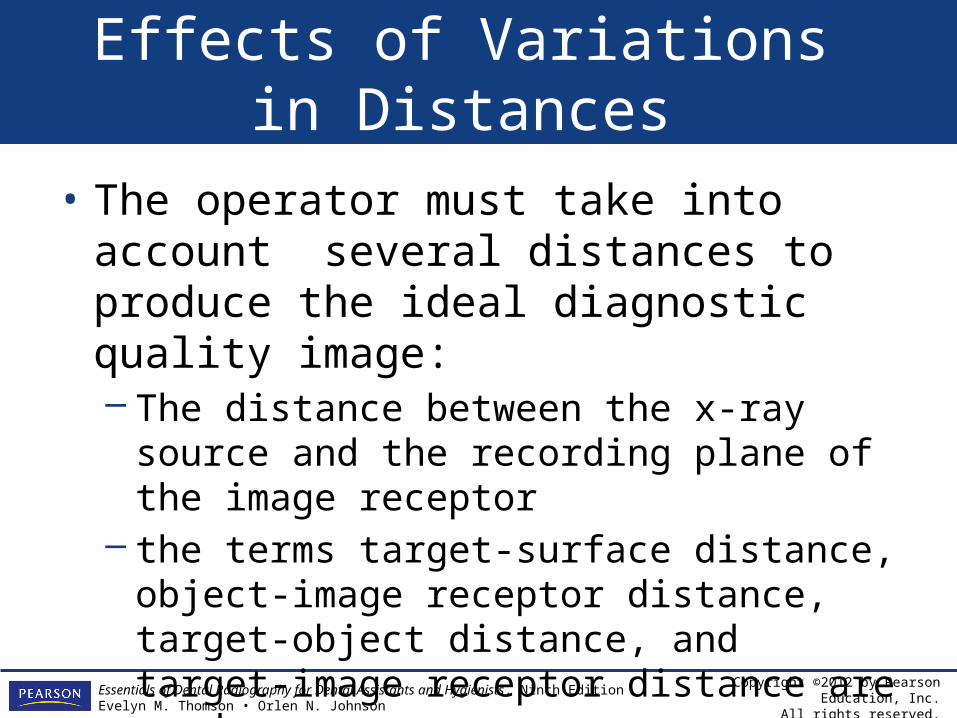
Effects of Variations in Distances
• The operator must take into account several distances to produce the ideal diagnostic quality image:– The distance between the x-ray source and
the recording plane of the image receptor– the terms target-surface distance, object-
image receptor distance, target-object distance, and target-image receptor distance are used.
Copyright ©2012 by Pearson Education, Inc.All rights reserved.
Essentials of Dental Radiography for Dental Assistants and Hygienists, Ninth EditionEvelyn M. Thomson • Orlen N. Johnson
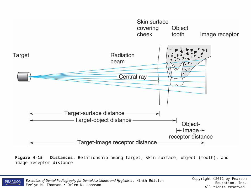
Figure 4-15 Distances. Relationship among target, skin surface, object (tooth), and image receptor distance
Copyright ©2012 by Pearson Education, Inc.All rights reserved.
Essentials of Dental Radiography for Dental Assistants and Hygienists, Ninth EditionEvelyn M. Thomson • Orlen N. Johnson
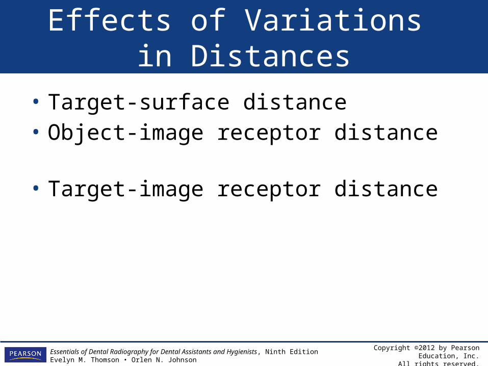
Effects of Variations in Distances
• Target-surface distance• Object-image receptor distance• Target-image receptor distance
Copyright ©2012 by Pearson Education, Inc.All rights reserved.
Essentials of Dental Radiography for Dental Assistants and Hygienists, Ninth EditionEvelyn M. Thomson • Orlen N. Johnson
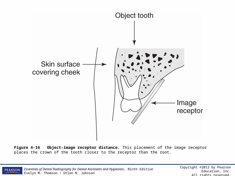
Figure 4-16 Object-image receptor distance. This placement of the image receptor places the crown of the tooth closer to the receptor than the root.
Copyright ©2012 by Pearson Education, Inc.All rights reserved.
Essentials of Dental Radiography for Dental Assistants and Hygienists, Ninth EditionEvelyn M. Thomson • Orlen N. Johnson
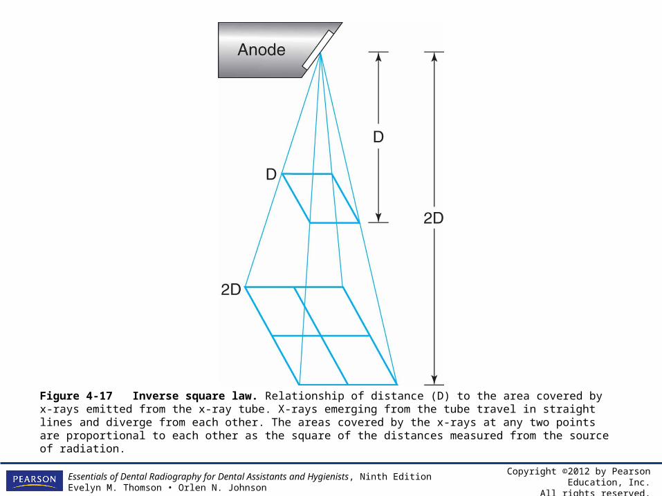
Figure 4-17 Inverse square law. Relationship of distance (D) to the area covered by x-rays emitted from the x-ray tube. X-rays emerging from the tube travel in straight lines and diverge from each other. The areas covered by the x-rays at any two points are proportional to each other as the square of the distances measured from the source of radiation.
Copyright ©2012 by Pearson Education, Inc.All rights reserved.
Essentials of Dental Radiography for Dental Assistants and Hygienists, Ninth EditionEvelyn M. Thomson • Orlen N. Johnson
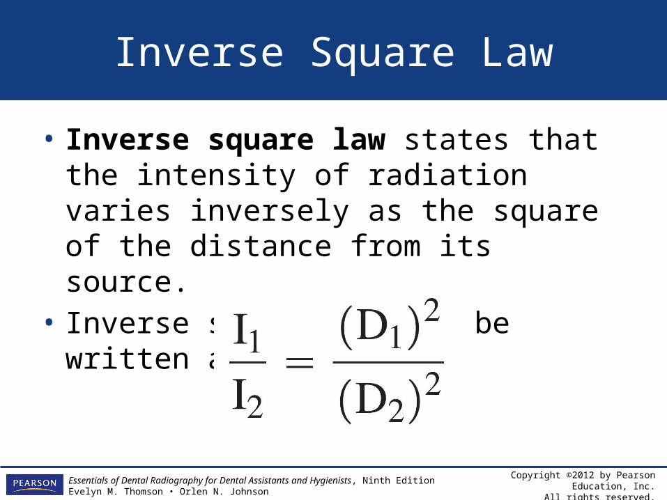
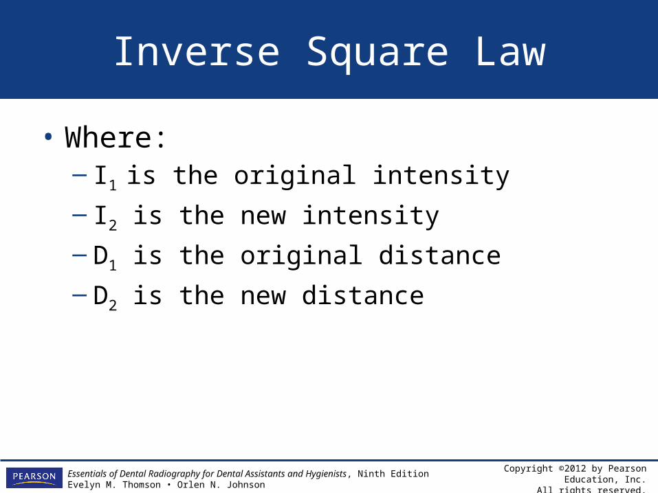
Inverse Square Law
• Inverse square law states that the intensity of radiation varies inversely as the square of the distance from its source.
• Inverse square law may be written as:
Copyright ©2012 by Pearson Education, Inc.All rights reserved.
Essentials of Dental Radiography for Dental Assistants and Hygienists, Ninth EditionEvelyn M. Thomson • Orlen N. Johnson
Inverse Square Law
• Where:– I1 is the original intensity
– I2 is the new intensity
– D1 is the original distance
– D2 is the new distance
Copyright ©2012 by Pearson Education, Inc.All rights reserved.
Essentials of Dental Radiography for Dental Assistants and Hygienists, Ninth EditionEvelyn M. Thomson • Orlen N. Johnson
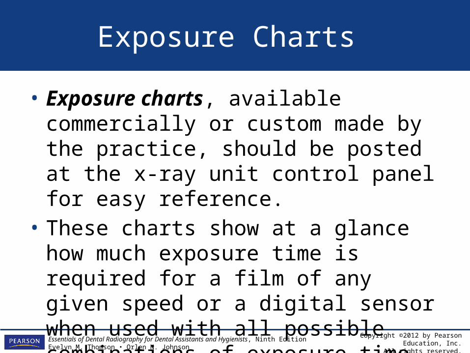
Exposure Charts
• Exposure charts, available commercially or custom made by the practice, should be posted at the x-ray unit control panel for easy reference.
• These charts show at a glance how much exposure time is required for a film of any given speed or a digital sensor when used with all possible combinations of exposure time, milliamperage, and peak kilovoltage.
Copyright ©2012 by Pearson Education, Inc.All rights reserved.
Essentials of Dental Radiography for Dental Assistants and Hygienists, Ninth EditionEvelyn M. Thomson • Orlen N. Johnson
Review: Chapter Summary
• An acceptable diagnostic radiograph must show the areas of interest completely and with minimum distortion and maximum sharpness.
• When evaluating a radiographic image the oral health care professional should utilize appropriate scientific terminology.
Copyright ©2012 by Pearson Education, Inc.All rights reserved.
Essentials of Dental Radiography for Dental Assistants and Hygienists, Ninth EditionEvelyn M. Thomson • Orlen N. Johnson
Review: Chapter Summary
• The dental radiographer must have a working knowledge of the factors that affect the radiographic image.
Copyright ©2012 by Pearson Education, Inc.All rights reserved.
Essentials of Dental Radiography for Dental Assistants and Hygienists, Ninth EditionEvelyn M. Thomson • Orlen N. Johnson
Recall: Study Questions
• General• Chapter Review

Copyright ©2012 by Pearson Education, Inc.All rights reserved.
Essentials of Dental Radiography for Dental Assistants and Hygienists, Ninth EditionEvelyn M. Thomson • Orlen N. Johnson
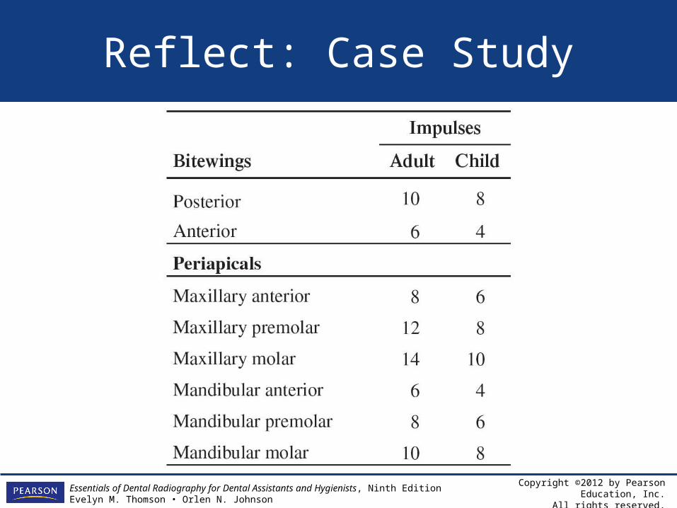
Reflect: Case Study
• You have just been hired to work in a new oral healthcare facility. Prior to providing patient services, you are asked to help develop exposure settings and equipment recommendations for the practice. The equipment and image receptor manufacturers’ suggestions are as follows:
F Speed Film; 8-in. (20.5 cm) PID; 85 kVp
Copyright ©2012 by Pearson Education, Inc.All rights reserved.
Essentials of Dental Radiography for Dental Assistants and Hygienists, Ninth EditionEvelyn M. Thomson • Orlen N. Johnson
Reflect: Case Study
Copyright ©2012 by Pearson Education, Inc.All rights reserved.
Essentials of Dental Radiography for Dental Assistants and Hygienists, Ninth EditionEvelyn M. Thomson • Orlen N. Johnson
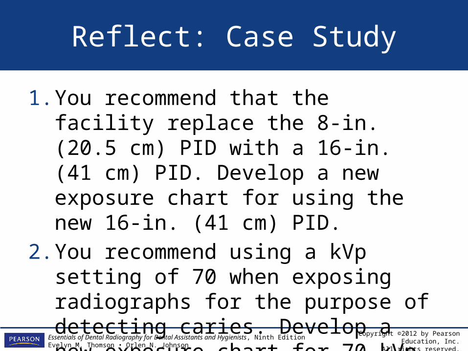
Reflect: Case Study
1. You recommend that the facility replace the 8-in. (20.5 cm) PID with a 16-in. (41 cm) PID. Develop a new exposure chart for using the new 16-in. (41 cm) PID.
2. You recommend using a kVp setting of 70 when exposing radiographs for the purpose of detecting caries. Develop a new exposure chart for 70 kVp.
Copyright ©2012 by Pearson Education, Inc.All rights reserved.
Essentials of Dental Radiography for Dental Assistants and Hygienists, Ninth EditionEvelyn M. Thomson • Orlen N. Johnson
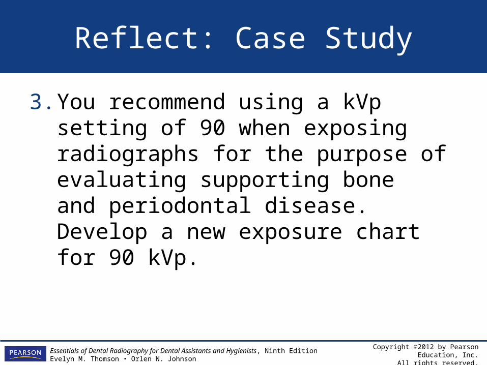
Reflect: Case Study
3. You recommend using a kVp setting of 90 when exposing radiographs for the purpose of evaluating supporting bone and periodontal disease. Develop a new exposure chart for 90 kVp.

Copyright ©2012 by Pearson Education, Inc.All rights reserved.
Essentials of Dental Radiography for Dental Assistants and Hygienists, Ninth EditionEvelyn M. Thomson • Orlen N. Johnson
Relate: Laboratory Application
• Proceed to Chapter 4, Laboratory Application, to complete this activity.