escherichia coli and quantification of... · lb luria-b ert ani lba luria-bertani agar lps lipopo...
TRANSCRIPT

Distribution and Quantification of Escherichia coli and Escherichia coli 0157: H7 in Organic Vegetables
at Farm Level \
Lim Poh Yiin
QR 101 E81 L731 Bachelor of Science with Honours 1013 (Resource Biotechnology)
2013

Pusat Khidmat Maklumat Akademi UNlVERSm MALAYSIA SARAWAK
Distribution and Quantification of Escherichia coli and Escherichia coli 0157: H7 in Organic Vegetables at Farm Level
P.KHIDMAT MAKLUMAT AKADEMIK
11111 1IIIIfiiifli 11111111 1000246758
Lim Poh Yiin (26729)
A thesis submitted in partial fulfillment of the requirements for the degree of
Bachelor of Science with Honours
(Resource Biotechnology)
Supervisor: Dr. Lesley Maurice Bilung
Co-supervisor: Dr. Micky Vincent
Resource Biotechnology
Department of Molecular Biology
Faculty of Resource Science and Technology
Universiti Malaysia Sarawak
2013
ACKNOWLEDGEMENT
First of all, I would like to convey my deepest gratitude to my supervisor, Dr. Lesley
Maurice Bilung and co-supervisor, Dr. Micky Vincent, for their keen guidance and advices
throughout my bench work and write-up process. I would also like to thank Dr. Lesley for her
upmost encouragement when I faced with difficulty in conducting this research.
Second, I would like to forward this acknowledgement to the post-graduate students
in Microbiology Lab, UNIMAS, namely Ms. Velnetti Linang, Ms. Christy Chan Siew Wei
and Mr. Yong Sy Fuh for their help and kindness in guiding me the appropriate practices to
conduct my project.
Third, I would also like to thank my fellow friends namely Chai Sze Fan, Chai Siaw
Yew, Lillian Sea Shun Yi, Chin Zhao Ning, Hu Li Tze, Nurulhuda Najihah bt Zainal Abidin
and Nur Bainun binti Mohd Zin for their kind act when I needed support and assist in the
laboratory.
Fourth, I would like to thank Ms. Chua Ann Ann from N & N Farm Sdn. Bhd. for her
kind involvement in providing free samples for my project. Further, Ms. Chua was also very
kind to share with me her experience in organic farming.
Lastly, my greatest gratitude is forwarded to my beloved family members, especially
my parents who had supported me from every aspect such as fmancial, emotion, faith and
confidence.
DECLARATION
I hereby declare that the thesis entitled "Distribution and Quantification of Escherichia coli
and Escherichia coli 0157: H7 in Organic Vegetables at Farm Level" submitted herewith for
Degree of Bachelor of Science (Honours) at Universiti Malaysia Sarawak is my own work
(except for the cited references) and has not been previously submitted by me at any other
University for any purposes.
Name
Signature
Date
II
....

Pusat Khidmat Maklumat Akademik UNIVERSm MALAYSIA SARAWAK
TABLE OF CONTENTS
ACKNOWLEDGEMENT I
DECLARA TION II
TABLE OF CONTENTS III
LIST OF ABBREVIATIONS v
LIST OF TABLES VII
LIST OF FIGURES VIII
ABSTRACT 1
CHAPTER 1 INTRODUCTION 2
CHAPTER 2 LITERATURE REVIEW 5
2.1 Genus, Morphology and Features ofEscherichia coli 5
2.2 History ofE. coli and E. coli 0157: H7 5
2.3 Enterohermorrhagic E. coli (EHEC) or Shigatoxigenic 6
E. coli (STEC)
2.4 Foodbome Outbreaks related to E. coli 0157: H7 7
2.4.1 Transmission ofBacteria into Food 8
2.5 Organic Farming 9
CHAPTER 3 MATERIALS AND METHODS 11
3.1 Sample Collection 11
3.2 Sample Processing 12
3.2.1 Sample Enrichnment 12
3.2.2 Serial Dilutions 12
III ..'
3.2.3 Most Probable Number (MPN) method 12
3.2.4 Colony-Forming Unit (CFU) 12
3.3 DNA Extraction PCR Amplification 13
3.4 PCR Amplification 13
3.5 Agarose Gel Electrophoresis (AGE) 14
3.6 Antibiotic Susceptibility Test (AST) 14
3.7 Multiple Antibiotic Resistance Indexing (MARl) 16
CHAPTER 4 RESULTS AND DISCUSSIONS 17
CHAPTER 5 CONCLUSIONS 25
REFERENCES 26
IV
C
LIST OF ABBREVIATIONS
AGE
AMP
AST
bp
CDC
CFU
CLSI
CN
DAEC
DNA
DO
E. coli
EAEC
EHEC
EIEC
EMB
EPEC
EtBr
ETEC
F
HUS
KF
Agarose Gel Electrophoresis
Ampicillin
Antimicrobial Susceptibility Test
Base pairs
Chloramphenicol
Centers for Disease Control and Prevention
Colony-forming Unit
Clinical and Laboratory Standards Institute
Gentamicin
Diffusely Adherent E. coli
Deoxyribonucleotides
Doxycycline
Escherichia coli
Enteroaggregative E. coli
Enterohemorrhagic E. coli
Enteroinvasive E. coli
Eosin-methylene Blue
Enteropathogenic E. coli
Ethidium Bromide
Enterotoxigenic E. coli
Nitrofurantoine
Hemolytic uremic syndrome
Cephalothin
v

00
LB Luria-B ert ani
LBA Luria-Bertani Agar
LPS Lipopo lysaccharides
MARl Multiple Antibiotic Resistance Index
MHA Mueller-Hinton Agar
ml Milliliter
MPN Most Probably Number
NaCI Sodium chloride
Optical Density
PCR Polymerase Chain Reaction
S Streptomycin • SOM Skim Organik Malaysia (Malaysian Organic Scheme)
STEC Shiga-toxin-producing E. coli
TBE Tris-Borate EDTA
JJ.g Microgram
JJ.L Micro litre
VI
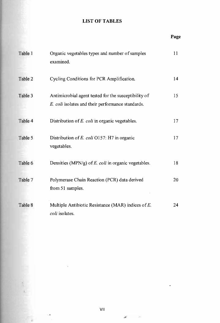
LIST OF TABLES
Table 1
Table 2
Table 3
Table 4
Table 5
Table 6
Table 7
Table 8
Page
Organic vegetables types and number ofsamples 11
examined.
Cycling Conditions for PCR Amplification. 14
Antimicrobial agent tested for the susceptibility of 15
E. coli isolates and their performance standards.
Distribution ofE. coli in organic vegetables. 17
Distribution ofE. coli 0157: H7 in organic 17
vegetables.
Densities (MPN/g) ofE. coli in organic vegetables. 18
Polymerase Chain Reaction (PCR) data derived 20
from 51 samples.
Multiple Antibiotic Resistance (MAR) indices ofE. 24
coli isolates.
VII ..
~.
,...... I
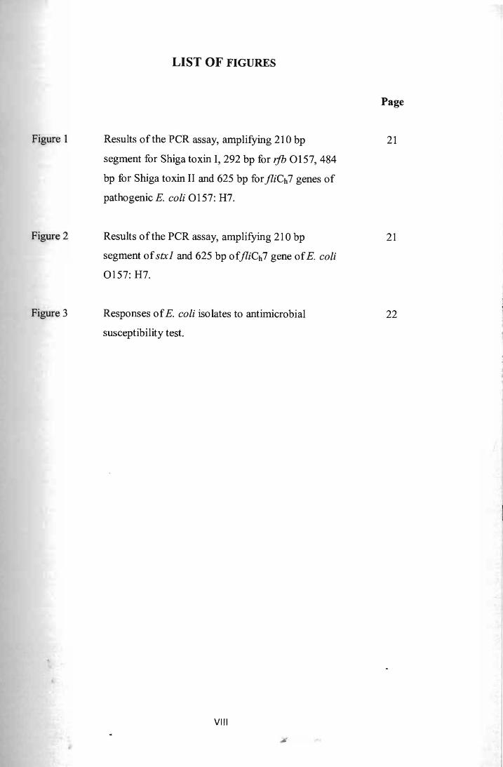
LIST OF FIGURES
Page
Figure 1 Results of the PCR assay, amplifying 210 bp
segment for Shiga toxin 1,292 bp for rjb 0157,484
bp for Shiga toxin II and 625 bp for jliCh7 genes of
pathogenic E. coli 0157: H7.
21
Figure 2 Results of the PCR assay, amplifying 210 bp
segment ofstxl and 625 bp ofjliCh7 gene ofE. coli
0157: H7.
21
Figure 3 Responses ofE. coli isolates to antimicrobial
susceptibility test.
22
VIII
..:

Distribution and Quantification of Escherichia coli and Escherichia coli 0157: H7 in Organic Vegetables at Farm Level
Lim Pob Yiin
Resource Biotechnology Faculty of Resource Science and Technology
Universiti Malaysia Sarawak
ABSTRACT
Organic vegetables are grown without the use of synthetic fertilizers and pesticides, thus they attract many consumers whom are concerned with their health. However, several researches had reported the presence of foodbome pathogens such as Escherichia coli (E. coli) and Escherichia coli 0157: H7 in organic vegetables. The aim of this study is to detect and characterize E. coli as well as E. coli 0157: H7 in raw organic vegetables from four organic farms located at Kuching and Siburan areas. Fifty one samples were collected and analyzed using Most Probable Number (MPN) supplemented with Polymerase Chain Reaction (PCR) method. E. coli was successfully isolated and enumerated from the organic vegetables samples. Characterization of nine of the E. coli isolates through antibiotics susceptibility test (AST) showed that five of them were multi resistant and possessed high risk of contamination by having Multiple Antibiotic Resistance (MAR) Index of 0.571 and 0.286. Fortunately, none of the samples tested harbored for pathogenic E. coli 0157: H7.
Key words: Escherichia coli, Escherichia coli 0157: H7, organic vegetables, antibiotics susceptibility tests, MPN-PCR
ABSTRAK
Sayur-sayuran organik ditumbuh tanpa penggunaan baja-baja sintetik dan racun perosak. Ini telah menarik perhatian daripada ramai pelanggan yang lebih prihatin terhadap kesihatan mereka. Bagaimanapun, patogen daripada makanan seperti Escherichia coli (E. coli) dan E. coli 0157: H7 telah dilaporkan dalam pelbagai kajian ten tang sayur-sayuran organik. Objektif kajian ini adalah untuk mengesan dan memperbahagikan ciri-ciri E. coli dan E. coli 0157: H7 dalam sayur-sayuran organik mentah pada peringkat kebun. 51 sampel telah diperolehi dari 4 kebun organik di kawasan Kuching dan Siburan. Mereka telah dianalisis dengan kaedah MPN serta PCR. E. coli telah berjaya dikesan dan diasingkan daripada sampel-sampel tersebut. Pembahagian ciri-ciri E. coli juga telah dilakukan melalui ujian kecenderungan antibiotik (AST) dan ujian tersebut telah menunjukkan sebahagian daripada mereka adalah cenderung terhadap pelbagai antibiotik. Mereka juga didapati membawa risiko yang tinggi dalam pencemaran makanan, dengan mempunyai Indeks Ringtangan Pelbagai Antibiotik (MARl) berni/ai 0.571 dan 0.286. Namun, antara semua sampel sayur-sayuran organik yang dikaji, E. coli 0157: H7 yang patogenik gagal dikesan.
Kata kunci: Escherichia coli, Escherichia coli 0157: H7, sayur-sayuran organik, ujian kecenderungan antibiotik, MPN-PCR
1
CHAPTER 1
INTRODUCTION
Organic produces are vegetables and fruits grown and harvested without using any
synthetic fertilizers and pesticides. Besides that, synthetic methods in producing organic
products such as applying irradiation and artificial ripening of fruits are prevented in organic
agriculture (F AO, 2013). Instead, organic farming adapts certain systems whereby soil is
manipulated to be remained fertile for longer period and pests control can be done via natural
means (FAO, 2013). Recently, the demand and interest for organic produces have been
increasing as they are believed to be safer and healthier to consumers. Furthermore,
researchers had also recommended that people should consume at least five servings of
vegetables and fruits per day to maintain their health (James, 2006). However, organic
produces may still contain natural contaminants from the environment such as soil, water and
livestocks' manure (James, 2006). Since most of the organic fertilizers are made up of animal
manure, this provides risk that pathogenic bacteria can be introduced to organic plants
through spreading of manure in the environment (Mukherjee et. aI., 2004). According to a
study performed by Beuchat and Ryu (1997), bacteria can be introduced to the fresh produces
in the farm or during any stages of handling the produces (James, 2006). For example,
enterobacteriaceae such as Escherichia coli (E. coli) which can be found naturally in the soil,
water, animals and plants. Hence, Sivapalasingam et al. (2004) stated that they were not
surprised for the occurrence of produce-related foodborne diseases that has been increasing
during the past three decades (Anno us et aI., 2009).
E. coli is common enteric bacteria that are found in the gastrointestinal tracts of
humans. and animals (CDC, 2012a). Most of them are harmless towards the hosts whereas
some of them can cause diseases. Pathogenic E. coli normally causes diarrhea and thus they
2
are also known as diarrheagenic E. coli. According to Buchanan and Doyle (1997), there are
six classes of diarrheagenic E. coli recognized. These includes Shiga-toxin-producing E. coli
(STEC) or enterohemorrhagic E. coli (EHEC), enterotoxigenic E. coli (ETEC),
enteroaggregative E. coli (EAEC), enteroinvasive E. coli (EIEC), enteropathogenic E. coli
(EPEC), and diffusely adherent (DAEC). In this study, only EHEC known as E. coli 0157:
H7 was investigated.
E. coli 0157: H7 is the most commonly isolated pathogenic strain of E. coli (CDC,
2012a). It causes wide outbreak of infections in the world every year. They are responsible
fo r many types of foodbome diseases such as bloody diarrhea and hemolytic uremic
syndrome (HUS) (CDC, 2012a). However, even though there were a lot of infection
outbreaks caused by E. coli over the years, it has not been confirmed that fresh produces
contain E. coli 0157: H7 as most of the experiments conducted failed in isolating the target
strain. According to investigation done by Mukherjee et at. (2004), they also, could not
isolate any E. coli 0157: H7 from 40 farms in Minnesota.
So far, there is no reported outbreak of E. coli in Malaysia. However, the government
and health department of the country had issued warnings to citizens that had visited Europe
countries in year 2011 to be investigated at nearby health centers. This is due to an outbreak
of E. coli believed to be originated from cucumbers from Germany that year. The outbreak
had caused 15 deaths in Germany and hundreds of illness (The Star, 2011). It was reported
that the cucumbers were contaminated by EHEC bacteria, thus able to cause diseases such as
I{US and bloody diarrhea.
There are over 100, 000 cases per year reported to be caused by EHEC (DuPont,
2007). Among these, E. coli 0157:H7 is responsible for 73, 000 cases with more than 2,000
hospitalizations and 60 deaths per year in United States (CDC, 2005). Normally outbreaks of
3

E. coli 0157: H7 are caused by contamination of the food products such as fresh produces
and undercooked ground beef by uncleaned irrigation water, animals' manures, uncleaned
harvesting tools and the hygienic level of workers processing the food. Several studies have
reported the presence of E. coli 0157: H7 in some organic foods in Malaysia (Chang et. aI.,
20l3). Therefore, the purpose of this research was to detect the presence ofE. coli and E. coli
0157: H7 in raw organic vegetables in Kuching and Siburan areas. Another aim of this
research was to determine the antibiotic susceptibility profile ofthe E. coli and E. coli 0157:
H7 isolates.
4 ...
Pusat Khidmat Maklumat Akademik UNTVERSITI MALAYSIA SARAWA](
CHAPTER 2
LITERATURE REVIEW
2.1 Genus, Morphology and Features of Escherichia coli
Escherichia coli (E. coli) belongs to the family Enterobacteriaceae and genus
Escherichia. It is a Gram-negative, non endospore-forming, motile and facultative anaerobic
bacterium. Under the microscope, it is seen as straight rods with the size of 2.0 x 0.5 p,m.
When being plated on Eosin Methylene Blue (EMB) agar, the bacterium will form colonies
of dark blue-black color with metallic green sheen (American Society for Microbiology,
2012).
E. coli can be found naturally in the gastrointestinal tract of human being and warm
blooded animals such as cattle and domestic livestock (ECDC, 2013). In general, most of the
strains of this bacterium are beneficial towards the host. It helps to maintain the environment
of the intestines by suppressing the growth of foreign bacteria (FDA, 2012). However, there
are also some serotypes ofE. coli which are harmful towards us such as E. coli 0157: H7, E.
coli 0145 and E. coli 026 (CDC, 2012a).
2.2 History ofE. coli and E. coli 0157: H7
In year 1885, Theodor Escherich, a German pediatrician, discovered E. coli in normal
individual's faeces and at that time, he named the bacterium as Bacterium coli commune. The
name was given because the bacterium was found in the colon part of the people. However,
in the later years, genus Bacterium had been eliminated and subsequently, Bacterium coli
commune was renamed Escherichia coli, named after its pioneer discoverer.
5

One of the serotype called E. coli 0157: H7 was fIrst identifIed and recognized as
pathogenic E. coli in year 1982 when an outbreak was caused by it (Riley et aI., 1983). This
serotype was found to cause hemorrhagic colitis and leads to Hemolytic uremic syndrome
(HUS). After 11 years, another outbreak was caused by E. coli 0157: H7 and it infected
mUltiple states in the United States. Researchers by then discovered the connection between
the bacterium with undercooked ground beef patties from a fast-food restaurant (Bell et. aI.,
1994). A year later, the bacterium became widely known and people were warned against it.
2.3 Enterohermorrhagic E. coli (EHEC) or Shigatoxigenic E. coli (STEC)
E. coli 0157: H7 is the predominant serotype under EHEC or STEC. It was named
according to the 0 and H antigen displayed on the outer membrane. The outer membrane of
the bacterium consists of lipopolysaccharides (LPS) molecule including the 0 antigen. The
antigen is encoded by rjb gene cluster (O'Brien et al., 2005). Meanwhile, H antigen can be
found on the flagella of the bacterium itself It is encoded by jliCh7 genes (O'Brien et aI.,
2005). The presence of virulence genes such as stXj and Slx2 indicates that this bacterium is
capable to produce Shiga toxin (Franz et. al., 2007). This toxic can cause severe damages to
the lining of gastrointestinal tracts (FDA, 2012). This serotype of the bacterium is thought to
be the most common cause of bacterial diarrhea following Salmonella spp. Over the past
years, it has been associated with unpasteurized dairy products, contaminated water and
juices, fresh produces and also raw or undercooked ground beef meat (Boyce, 2012).
E. coli 0157: H7 is pathogenic and can cause an acute disease known as hemorrhagic
colitis (FDA, 2012). The symptoms of the disease include the occurrence of bloody diarrhea
after feW days of watery diarrhea, abdominal cramp and in some cases; fever ana vomiting
may also happen (Cohen and Giannella, 1992). Usually, the sickness will last for an average
6
,j.:

of eight days (FDA, 2012). For some patients of hemorrhagic colitis, if they are not treated
accordingly, there is a chance they may develop HUS. Some of the consequences that might
occur with HUS are anemia, thrombocytopenia and even kidney failure (Boyce, 2012).
Furthennore, it may lead to severe illness such as nerve or brain damage, or even death in
older patients (Boyce, 2012). Other symptoms of disease caused by infection ofE. coli 0157:
H7 are pneumonia and respiratory diseases (CDC, 2012a). E. coli 0157: H7 is more
significant if compared to other bacteria that cause gastrointestinal diseases because it can
cause infection even in low doses and affecting people with any age group, with young kids
and old people at greater risk. Besides, it is also found out to be more acid tolerant than other
bacteria (Buchanan and Doyle, 1997).
2.4 Foodborne Outbreaks related to E. coli 0157: H7
EHEC or STEC causes approximately 100, 000 cases per year according to DuPont
(2007). According to the Centers for Disease Control and Prevention (CDC) (2005), E. coli
0157: H7 causes 73,000 diseases with at least 2,000 hospitalizations and 60 deaths per year
in the United States. Produce-related outbreaks were first reported in 1991 and it has been
emerging as an infection vehicle until nowadays (Rangel et aI., 2005). According to the
epidemiology study conducted by Rangel et al. (2005), it was reported that most produce
associated outbreaks are linked to vegetables such as lettuce, salad, coleslaw and sprouts. In
Japan, an E. coli 0157 outbreak linked with raw radish sprouts was reported and it had
infected 12, 000 people with 12 deaths (Michino et aI., 1999).
Recently in September 2006, CDC reported a multistate outbreak associated with
fresh spinach caused by this bacterium. The outbreak spread throughout 26 states of the
7

United States, Canada and even New Mexico. The consumption of freshly packaged baby
spinach was the cause and 205 cases of illness and 3 deaths were reported (CDC, 2006).
Besides that, in December 2012, another report was lodged by CDC on another
outbreak affecting 5 states of US caused by the same bacterium serotype and associated with
organic spinach and spring mix blend. It infected 33 persons in which 13 of them were
hospitalized and 2 of them diagnosed with HUS. Fortunately, no death was reported during
the outbreak (CDC, 2012b).
2.4.1 Transmission of Bacteria into Food
For STEC, there are three routes of transmission which are through food, person-to
person and from the environment such as contaminated water and animals (DuPont, 2007).
Feca1-oral route mostly occur at nurseries, and some in schools, housing communities and
public facilities (Rangel et ai., 2005). Fresh produces such as vegetables and fruits are
susceptible to contamination of the bacterium Likewise, organic farms are also at risks of
contamination with the bacterium as various studies have reported organic produces are more
likely to be contaminated with the bacterium than conventional farms. However, it has yet to
be proven whether the statement is true (Mukherjee et al., 2004). So far, there is only one
confirmed report regarding the consumption of organic vegetable which led to foodbome
outbreak (Tschape et al., 1995). All produces have equal chances to be contaminated in fields
by animal's manure, irrigation water or during processing, harvesting, transporting and
storing (Rangel et ai., 2005). Cattle, bovine and domestic livestock manure had been used as
fertilizer in growing produces. However, it is known that animal manure may contain
gastroenteritis such as E. coli, Salmonella, Mycobacterium paratuberculosis and Listeria spp.
(PeU, 1997). An experiment done by Soloman et al. (2002) has also demonstrated that E. coli
8
0157: H7 can enter through the root system of lettuce plant and finally be found in the edible
parts ofthe lettuce (Solomon et al., 2002)
To minimize the occurrence of such bacteria in the manure, a method called
composting is introduced to the farmers. It is believed that composting the manure before
using the manure directly as fertilizer can help to remove large number of bacteria in it as
heat is generated over time (Hussong et al., 1985).
2.5 Organic Farming
Organic produces are produce that are grown without the use of any synthetic
fertilizer, herbicides and pesticides. In an organic farm, it is common to fmd animal manures
composted to be used as fertilizers. Any synthetic methods such as irradiation and
introducing artificial ripening of fruits are not allowed in an organic farm. Apart from that,
organic farming also adapts certain systems whereby soil is being manipulated to be fertile
for longer period and pests control can be done in natural ways (FAO, 2013).
There are quite a number of reports on the occurrence of foodborne pathogens in
organic produces. Tshape et al. (1995) reported that green butter made with organic parsley
contaminated with verotoxinogenic Citrobacter freundii was the cause of a severe
gastroenteritis followed by HUS outbreak in a nursery school and kindergarten in Germany.
In year 2001, McMahon and Wilson had conducted a study on the occurrence of foodborne
pathogens in 86 commercially available organic vegetables in Northern Ireland. Thirty-four
percent of the tested samples were found to be contaminated with Aeromonas spp.
(McMahon and Wilson, 2001).
9

In Malaysia, Chang et al. (2013) had reported the low contamination ofE. coli 0157:
H7 in organic four- winged beans and white radish sold at supermarkets and retail groceries
respectively. The highest prevalence ofE. coli 0157: H7 in their study was found in organic
chickens where 40% ofthe tested samples (n= 20) harboured the bacteria.
10 ..

CHAPTER 3
MATERIALS AND METHODS
3.1 Sample collection
A total of 51 samples made up of seven types of organic vegetables (Table 1) were
collected randomly from four organic fanns in Kuching and Siburan, Malaysia. Among all
four organic farms, only one is certified by the Malaysian Organic Scheme (SOM) and the
others claimed to be practicing organic farming. Samples were picked randomly in three
different sites at the fann and kept in sterile ziplock bags labeled. The samples were
transported in an ice box to the laboratory. Analysis of the samples was conducted within 24
hours upon collection of samples.
Table 1. Organic vegetables types and number of samples examined
English name Scientific name Total number of samples
Romaine lettuce Lactuca sativa 9
Chinese broccoli Brassica oleracea 9
Tomato L ycopersicon esculentum 6
Salad (Sorrel) Rumex acetosa 6
Chinese cabbage Brassica rapa 12
Other leafy greens 9
(Spinach and Mustard)
TOTAL 51
11
.."

3.2 Sample Processing
3.2.1 Sample Enrichment
Twenty five grams of the organic vegetable samples were enriched in 225 ml of
Luria-Bertani (LB) Broth and homogenized in a sterile stomacher bag for 2 minutes. Then,
the mixture was incubated at 37°e for 24 hours.
3.2.2 Serial Dilutions
The incubated sample mixture was subjected to ten-fold serial dilutions. One rnl of the
sample mixture was diluted with 9 rnl of sterile saline solution (0.85% NaCl). Then, 1 rnl of
the aliquot from first dilution was transferred to second test tube with dilution factor of 10-2.
The same procedures were carried out until dilution factor of 10-7.
3.2.3 Most Probable Number (MPN)
One rnl of aliquot from dilution 10-1 until 10-3 were transferred into triplicate MPN
tubes containing 9 rnl of LB. The tubes were then incubated at 37°e for 24 hours. A tube
containing only LB was prepared and used as a control. On the next day, test tubes that
showed turbidity were then subjected to DNA extraction and peR for the detection of
ifb0157,fliCh7, Shiga toxin I and II genes.
3.2.4 Colony-Forming Unit (CFU)
Meanwhile, 100 JLL of the aliquot from dilution 10-3 until 10-7 were plated on Eosin
Methylene Blue (EMB) agar. The plates were incubated at 37°e for 24 hours. After
incubation, green metallic sheen colonies forming on the EMB agar were CQunted. The
isolates were then stored on LBA.
12
3.3 DNA Extraction
DNA extraction was conducted using the boil cell method as described by Apun et al.
(2011) with minor modifications. One ml of aliquot from each overnight MPN tube was
centrifuged at 13,000 rpm for 5 minutes. Then, the supernatant was discarded and the pellet
was resuspended with 200 ilL of sterile distilled water. The mixture was vortexed slightly to
make sure that remaining pellet was dissolved. Following that, the mixture was boiled for 20
minutes and immediately cooled for 20 minutes at -20°C. After cooling, the mixture was
centrifuged again at 13,000 rpm for 3 minutes. Final supernatant containing DNA was
transferred to a new micro centrifuge tube and store at -20°C until needed for PCR assay.
3.4 peR Amplification
Four pairs of primers (SIt!, SIt!L ifbE, jliCh 7) targeting Shiga toxin I and II genes,
0157 antigen gene and H7 antigen gene were used in the PCR amplification for detection of
pathogenic E. coli 0157: H7. PCR amplification was performed in 25 ilL reaction mixture
consisting 2.5 ilL of lOX PCR buffer, 1.25 ilL of 10 mM deoxynucleoside triphosphate
(dNTP) mix, 2.5 ilL of25 mM MgCh, 0.50 ilL of 5 UIIlL Taq Polymerase, 10 ilL of template
DNA and 0.5 ilL of each primer pairs (Slti-FISltI-R, SltII-F,SltII-R, ifb-Flifb-R, jliCh 7r FI
fliCh7-R). E. coli 0157: H7 reference strains EDL 933 was also included in the PCR assay as
positive control. The thermal cycling conditions were as stated in Table 2.
13

Table 2. Cycling Conditions for PCR Amplification
Process Temperature Duration Cycle(s)
(OC) (s)
Initial Denaturation 94 300 1
Denaturation
Annealing
Extension
94
59
72
30
60
60 } 35
Final Extension 72 420 1
3.5 Agarose Gel Electrophoresis (AGE)
Amplified PCR products were subjected to AGE. Two percent agarose gel was
prepared using IX Tris-Borate EDTA (TBE) buffer. Four micro litre of PCR product was
mixed with 1 p.L of6X loading dye and then loaded into the wens prepared. After loading the
peR products in the well, 3 p.L of 100 bp marker was loaded into the well as an indicator of
the band size. After that, the PCR products were electrophoresed at 85 V for 1 hOUT. Then,
the gel was stained in Ethidium Bromide (EtBr) for 20 minutes before it was viewed under an
ultraviolet transilluminator and photographed usmg gel documentation system
(AlphaDigiDoc RT). The expected sizes of amplicons for rjb0157,jliCh7 and Shiga toxin-
producing genes were 292 bp, 625 bp, 210 bp and 484 bp respectively.
3.6 Antimicrobial Susceptibility Test (AST)
AST was performed usmg disc diffusion method according to the Clinical and
Laboratory Standards Institute (CLSI). First, the bacterial isolate from LBA was revived by
incubating overnight in LB at 37°C. The viability was tested by obtaining OD600 value
ranging from 0.8 to 1.0. After that, sterile cotton bud was used to transfer and spread the
bacteria on Mueller-Hinton Agar (MBA) plate. Antibiotics tested include ampicillin
14