erythropoietin (epo) regulates hypoxic ventilation …©institute of veterinary physiology chart 1...
TRANSCRIPT

© Institute of Veterinary Physiology Chart 1
Erythropoietin (Epo) regulates hypoxic ventilation by interacting with brainstem and carotid bodies
University of ZürichUniversité Claude Bernard Lyon1
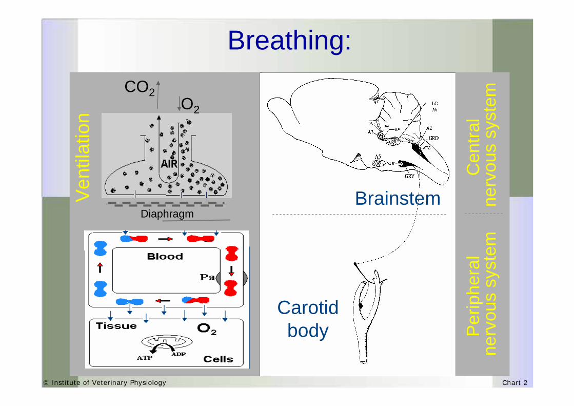
© Institute of Veterinary Physiology Chart 2
O2
CO2
Diaphragm
Ven
tilat
ion
Breathing:
Carotid body
Brainstem
Cen
tral
nerv
ous
syst
emP
erip
hera
lne
rvou
s sy
stem

© Institute of Veterinary Physiology Chart 3
Ventilation underHYPOXIA (diminished PpO2)
PaO2

© Institute of Veterinary Physiology Chart 4
Molecules controlling the ventilation:
Epotarget genes
Hypoxia-activatedMolecules
(HIF-1)
NeurotransmittersAmines
Steroids
(Pascual, 2004)
(Kline, 2002)

© Institute of Veterinary Physiology Chart 5
Epo Epo expression occurs in:expression occurs in:
brain
testis
lung
kidney
liver
but also in other organs including:
© Institute of Veterinary Physiology Chart 6
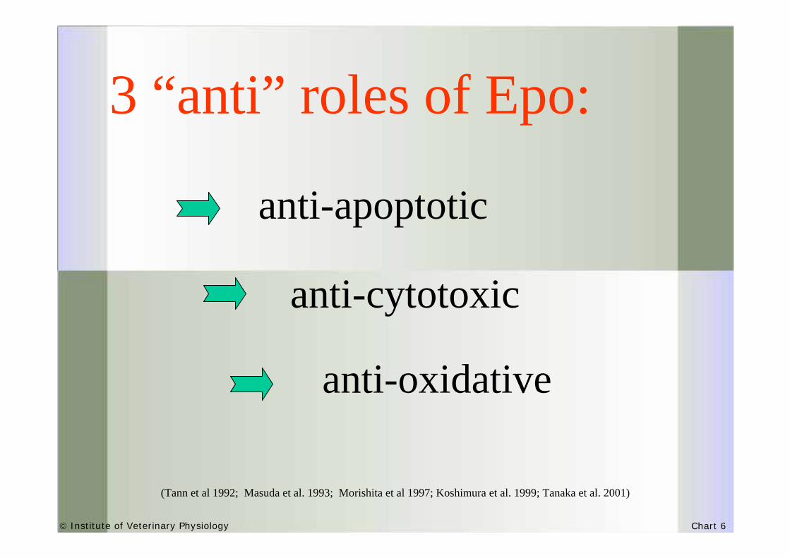
3 “anti” roles of Epo:
anti-apoptotic
anti-cytotoxic
anti-oxidative
(Tann et al 1992; Masuda et al. 1993; Morishita et al 1997; Koshimura et al. 1999; Tanaka et al. 2001)

© Institute of Veterinary Physiology Chart 7
Epo and Epo receptor (Epo and Epo receptor (EpoREpoR) in brain) in brain
Epo &EpoR
are expressedin the
mammalian andhuman brain
are expressedin the
mammalian andhuman brain
cerebral Epo is induced by hypoxia
cerebral Epo is induced by hypoxia
are expressed by neurons
and astrocytes
are expressed by neurons
and astrocytes

© Institute of Veterinary Physiology Chart 8
Does brain-derived Epo
play a role in the control of ventilation under
hypoxia?
© Institute of Veterinary Physiology Chart 9
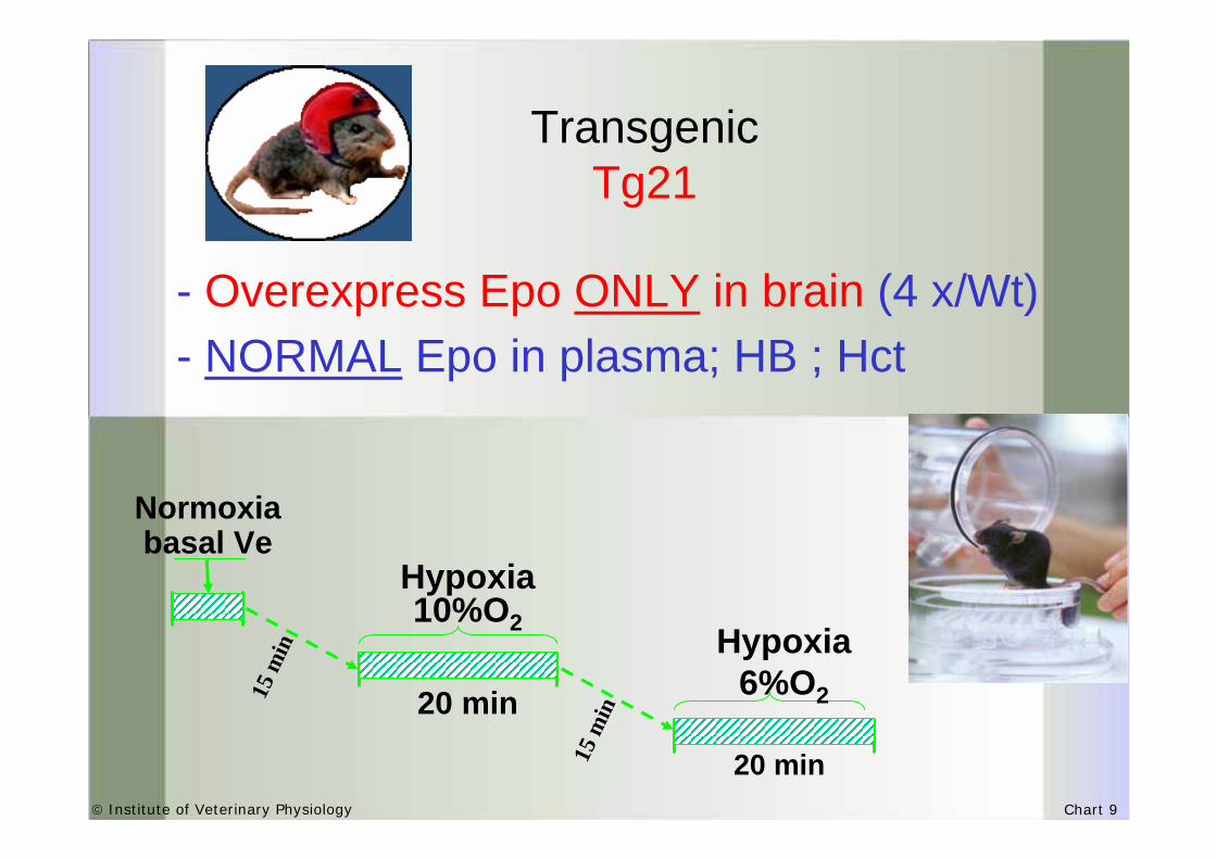
- Overexpress Epo ONLY in brain (4 x/Wt)- NORMAL Epo in plasma; HB ; Hct
Transgenic Tg21
Normoxiabasal Ve
20 min
Hypoxia 10%O2
20 min
Hypoxia 6%O215
min
15 m
in

© Institute of Veterinary Physiology Chart 10
Tg21 maintain high ventilation under at 6% hypoxia
∗ ∗ ∗ ∗ ∗
80
105
130
155
180
205
230
VE
(ml/m
in/1
00g)
exposure time (min)
Tg21WT
21% O2 10% O2 6% O2

© Institute of Veterinary Physiology Chart 11
Tg21 enhance ventilation after chronic hypoxia
• hypoxic chamber• 3 days at 10% O2
basal Ve
20 min
Hypoxia 10%O2
20 min
Hypoxia 6%O2

© Institute of Veterinary Physiology Chart 12
EpoR is expressed in the resp.areas of the brainstem
Legends:
4V, 4th ventricle; AP, area postrema; NTS, nucleus tractus solitarius; 7n, facial nerve; PBC, Pre-Bötzinguercomplex; NA, nucleus ambiguus; PY, pyramidal decussation; LC, locus ceruleus. Catecholaminergic areas in the medulla oblongata: A1C1 & A2C2 and in the pons: A5 & A6

© Institute of Veterinary Physiology Chart 13
Chemodenervation is life-threatening in Wtbut not in Tg21
Chemodenervation

© Institute of Veterinary Physiology Chart 14
Is the Epo impact on ventilation a
neuro-protectivephenomenon?

© Institute of Veterinary Physiology Chart 15
0
50
100
150
200
1 2
Activated STAT-5 35 kDa-
Perc
ent o
f con
trol
wt tg21
020406080
100120140
1 2
Activated ERK-1 44 kDa-
Perc
ent o
f con
trol
wt tg210
20406080
100120
1 2
Activated ERK-2
42 kDa-
Perc
ent o
f con
trol
wt tg21
0
15
30
45
60
1 2
JAK-2 45 kDa-
wt tg21
Perc
ent o
f con
trol
STAT5
JNK
PKBMAPK
AktERk1 ERk2
020406080
100120
1
46 kDa-
*P
erce
nt o
f con
trol
wt tg21
020406080
100120
1 2
*
62 kDa-
Perc
ent o
f con
trol
wt tg21

© Institute of Veterinary Physiology Chart 16
Are Catecholamines
involved in the impactof Epo
in ventilation?

© Institute of Veterinary Physiology Chart 17
Catecholaminergic group cells in the brainstem

© Institute of Veterinary Physiology Chart 18
WT Tg21
A1C
1A
2C2
A5
A6

© Institute of Veterinary Physiology Chart 19
pmol
esN
A/20
min
Nor
adre
nalin
e
0
10
20
30 *
Wt Tg
pmol
esD
OPA
/20m
in
pmol
esN
E
TH
act
ivity
NE
con
tent
A2C2 cell group
4
6
8
10
WT Tg21
*
10
15
20
25
WT Tg21
*

© Institute of Veterinary Physiology Chart 20
does plasma-derived Epo
impact hypoxic ventilation via CB?

© Institute of Veterinary Physiology Chart 21
TH
ECAICA
CCA
TH
EpoR is expressed in Carotid bodies glomic cells
Epo-REpo-R
ECAICA
CCA
Legends:
TH, tyrosine hydroxilase; Epo-R, erythropoietin receptor; ICA, internal carotid artery; ECA, external carotid artery; CCA, commun carotid artery

© Institute of Veterinary Physiology Chart 22
Epo injected (i.v.) in Wt mice modulates the hypoxic ventilatory pattern
Injection of Epo (iv)(2000 U/Kg: 50 U/mouse) Wt C57/Bl6
Epo does notcross the BBB
VE = RR x VT
© Institute of Veterinary Physiology Chart 23
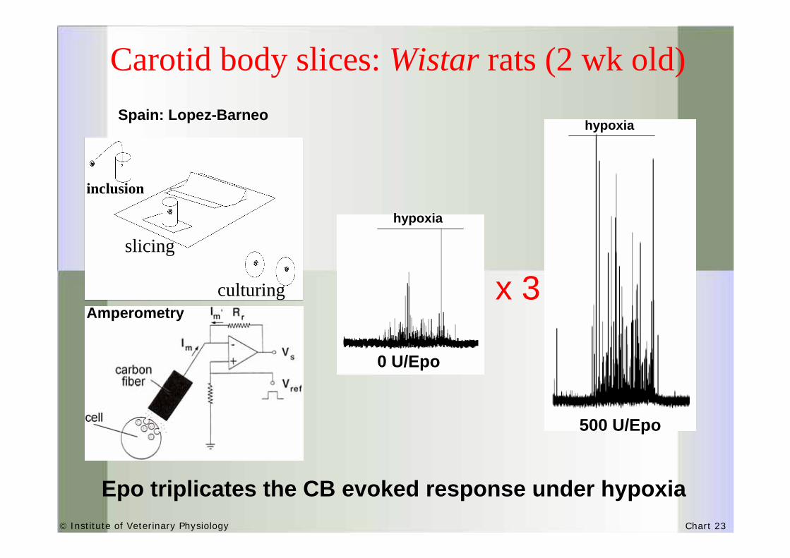
Carotid body slices: Wistar rats (2 wk old)
Epo triplicates the CB evoked response under hypoxia
500 U/Epo
x 3
0 U/Epo
hypoxia
hypoxia
inclusion
slicing
culturingAmperometry
Spain: Lopez-Barneo

© Institute of Veterinary Physiology Chart 24
Conclusion
Brain-derived Epo (via EpoR in brainstem) and systemic Epo (via EpoR in carotid bodies) modulate ventilation under hypoxia
These results suggest that Epo has crucial role in the fine-tuning of oxygen homeostasis
High altitude dwellers?
Acute and Chronic mountain sickness?
Lance Amstrong…Epo doping?

© Institute of Veterinary Physiology Chart 25
C Soulage C BecskeiL Ogunshola J VogelT GorrS KellerD Furrer
Acknowledgements
Prof. M GassmannProf. V JosephProf. JM Pequignot

© Institute of Veterinary Physiology Chart 26
Is the Epo effect on ventilation a selective Is the Epo effect on ventilation a selective action?action?
Epo mediates general integrity to the tissue
retinal protection
brain injury(stroke)spinalcord
injury
heartinfarct
development?
gonads?

© Institute of Veterinary Physiology Chart 27
Hello, hello,…. how much oxygen do you have over there?