erj express. published on june 1, 2010 as doi: 10.1183...
TRANSCRIPT

Prognostic factors in pathological stage IB non-small cell lung cancer greater than 3 cm
Jung-Jyh Hung1,2,3, Wen-Juei Jeng4, Wen-Hu Hsu3, Shiou-Fu Lin5, Chih-Cheng Hsieh1,3,
Biing-Shiun Huang3, Min-Hsiung Huang3, Jung-Sen Liu2, Teh-Ying Chou1,5 and Yu-Chung
Wu3
1Institute of Clinical Medicine, National Yang-Ming University, Taipei, Taiwan
2Department of Surgery, Cathay General Hospital and School of Medicine, Fu Jen Catholic
University, Taipei, Taiwan
3Division of Thoracic Surgery, Department of Surgery, Taipei Veterans General Hospital and
School of Medicine, National Yang-Ming University, Taipei, Taiwan
4Department of Internal Medicine, Chang Gung Memorial Hospital and School of Medicine,
Chang Gung University, Taipei, Taiwan
5Department of Pathology and Laboratory Medicine, Taipei Veterans General Hospital, Taipei,
Taiwan
Address correspondence to:
Dr. Yu-Chung Wu
Division of Thoracic Surgery, Department of Surgery, Taipei Veterans General Hospital, No.
201, Section 2, Shih-Pai Road, Taipei 112, Taiwan
. Published on June 1, 2010 as doi: 10.1183/09031936.00014109ERJ Express
Copyright 2010 by the European Respiratory Society.
Phone: 886-(2)-2875-7546; Fax: 886-(2)-2873-1488
E-mail: [email protected]
Drs Teh-Ying Chou and Yu-Chung Wu contributed equally to this article.
Running title: Stage IB Lung Cancer Greater than 3 cm

ABSTRACT
Significant heterogenity of stage IB (sixth edition of the TNM staging system) non-small
cell lung cancer (NSCLC) has been identified, and further subclassification according to tumor
size has been proposed. The aim of this study is to evaluate the prognostic factors in patients
with resected stage IB NSCLC greater than 3 cm.
From January 1980 to December 2000, 525 patients underwent surgical resection for
stage IB NSCLC greater than 3 cm at Taipei Veterans General Hospital. The clinicopathologic
characteristics of these patients were retrospectively reviewed.
The 5- and 10-year overall survival rates were 44.9% and 27.3%, respectively. Age (P <
0.001), tumor size (P = 0.002), extent of pulmonary resection (P = 0.002), histological type (P
= 0.005) and number of mediastinal lymph nodes dissected/sampled (P = 0.004) were
significant predictors for overall survival in multivariate analysis. Patients with tumor size > 7
cm, or > 5 to ≤ 7 cm, had a worse survival than those with tumor size > 3 to ≤ 5 cm. However,
visceral pleural invasion did not influence overall survival.
Stage IB NSCLC with a diameter greater than 3 cm may be subclassified according to
tumor size with regardless of visceral pleural invasion.
Key words: Non-small cell lung cancer, stage IB, survival, tumor size, visceral pleural
invasion

Introduction
Lung cancer is the leading cause of cancer-related death worldwide. Histological
identification and tumor staging play a critical role for the optimal management of lung cancer.
The Union Internationale Contre le Cancer was the first organization to classify lung cancer by
tumor, node, metastasis (TNM) staging system in 1968. The TNM staging system for lung
cancer was first applied by the American Joint Committee on Cancer in 1974 [1]. The fifth
edition of the TNM staging system for lung cancer was published in 1997, and stage I
non-small cell lung cancer (NSCLC) was subdivided into IA (T1N0M0, tumor size ≤ 3 cm) and
IB (T2N0M0, tumor size > 3 cm) [2]. In addition to tumor size greater than 3 cm, the current T2
descriptor also includes tumors that invade the visceral pleura regardless of size, tumors that
involve the main bronchus � 2 cm distal to the carina, and tumors that result in associated
atelectasis and obstructive pneumonitis that extends to the hilar region but does not involve the
entire lung radiographically [2]. The current (sixth) edition of the TNM staging system for lung
cancer was published in 2002 [3], without changes to the previous edition [2]. Significant
heterogenity of stage IB patients has been identified in several studies, and further
subclassification according to tumor size has been proposed [4-7]. The seventh edition of the
TNM classification of lung cancer has been published in 2009. The changes to the sixth edition
of the TNM staging system for lung cancer were based upon the proposals from the
International Association for the Study of Lung Cancer (IASLC). The IASLC lung cancer

staging project committee has recommended that T2 tumors be classified into T2a (> 3 to ≤ 5
cm), T2b (> 5 to ≤ 7 cm), and T3 (> 7 cm) [8-10].
Among the three non�size-based T2 descriptors, visceral pleural invasion (VPI) is the
main criteria [4, 11, 12]. Hammar suggested a classification of pleural invasion as follows: Px
and P0, lack of pleural invasion beyond the elastic layer; P1, invasion beyond the elastic layer;
P2, invasion to the surface of the visceral pleura; and P3, invasion of the parietal pleura and/or
chest wall [13, 14]. According to his proposal, P0 is not a T descriptor and the T category in
such cases should be assigned by other criteria. P1 or P2 correspond to T2, and P3 corresponds
to T3. The International Staging Committee of the IASLC has proposed the definition of VPI
as invasion beyond the elastic layer (PL1) including invasion to the visceral pleural surface
(PL2) [15]. They also recommend that elastic stains be used in cases when the distinction
between PL0 and PL1 is not clear based on evaluation of hematoxylin and eosin sections [15].
Although VPI has generally been reported as a poor prognostic factor [16-21], some studies
have demonstrated that VPI was not a prognostic factor for survival [4, 6, 7, 11, 22, 23]. The
prognostic value of VPI in patients of early stage NSCLC with larger tumor size has remained
to be demonstrated.
In our previous study [24], we have demonstrated that VPI did not influence overall
survival in resected stage I NSCLC with a diameter of 3 cm or less. Therefore, we recommend
that patients with tumor size ≤ 3 cm but are staged as stage IB (T2N0M0) due to VPI to be
treated as stage IA (T1N0M0). According to our proposal, the category of stage IB NSCLC
only consists of patients with stage I NSCLC with a diameter greater than 3 cm. In this regard,
we analyzed the prognostic factors of survival in stage IB NSCLC with a diameter greater than
3 cm and evaluate the validity of the new staging system of T descriptors proposed by the
IASLC. Furthermore, we investigated the prognostic value of VPI and its relationship with
tumor size in these patients.
Materials and methods
From January 1980 to December 2000, a total of 597 patients underwent surgical
resection for pathologic stage IB (T2N0M0) NSCLC at Taipei Veterans General Hospital. Of
these, 525 (87.9%) patients who had tumors with a diameter greater than 3 cm were identified
and included in this retrospective study. The preoperative staging workup, including chest and
upper abdomen computed tomographic scans, bronchoscopic examination and nuclear
medicine survey (bone and brain), was done as previous described [24, 25]. Mediastinoscopy
was not a routine preoperative staging procedure, and was performed only when enlarged
mediastinal lymph nodes (diameter over 1.0 cm) were shown by computed tomographic scan.
Among the 194 patients with available data on whether pre-operative mediastinoscopy was
performed for staging, 12 (6.2%) underwent mediastinoscopy before operation. In the study
period, positron emission tomography scan was not available as a staging modality. Patients
with suspected distant metastasis were excluded from consideration of operation. Complete
resection of lung cancer with mediastinal lymph node dissection/sampling was performed in all
patients as previously described [24, 25]. No patient received adjuvant chemotherapy after
surgical resection. Histological typing was determined according to the World Health
Organization classification [26]. Determination of disease stages was based on the TNM
classification of the International Union Against Cancer [3].
VPI was examined in tumor sections with hematoxylin and eosin stain. VPI was classified
according to Hammar�s suggestion [13, 14]: Px and P0, lack of pleural invasion beyond the
elastic layer; P1, invasion beyond the elastic layer; P2, invasion to the surface of the visceral
pleura; and P3, invasion of the parietal pleura and/or chest wall. Presence of VPI was defined
as tumors with P1 and P2, whereas absence of VPI was defined as tumors with Px and P0.
Elastic stains were performed in tumor sections when the status of VPI was indeterminate by
hematoxylin and eosin stains.
The hospital charts of all patients, including pathologic and surgical reports, were
reviewed to collect data of clinicopathologic characteristics and survival. Patient demographics,
pack years, tumor location, histological type of the tumor, histologic grade, tumor size (> 3 to ≤
5 vs. > 5 to ≤ 7 vs. > 7 cm), extent of pulmonary resection, presence of VPI and number of
mediastinal lymph nodes dissected/sampled were documented. The number of mediastinal
lymph nodes dissected/sampled, including N1 and N2 nodes, was recorded from pathologic
reports. All patients were followed up at our outpatient department quarterly in the first two

years after resection and semi-annually thereafter. The length of survival was defined as the
interval in months between the date of surgical resection and the date of either death or the last
follow-up.
The overall survival rate was calculated by the Kaplan-Meier method [27]. The χ2 test, the
independent-sample t test, or the one-way analysis of variance test was used to compare
between groups with respect to categorical and continuous variables as appropriate. Univariate
and multivariate analyses were performed by means of the Cox proportional hazards model
using SPSS software (version 16.0; SPSS, Chicago, Illinois, USA). Variables with P value less
than 0.05 after the univariate analysis were entered into multivariate analysis. Statistical
analysis was considered to be significant when the probability value was < 0.05.
Results
The median follow-up time for these 525 patients with surgically resected stage IB
NSCLC with a diameter greater than 3 cm was 50.3 months (95% confidence interval [CI],
43.8 to 58.0 months). The characteristics of these patients are listed in Table 1. There were 16
patients lost to follow-up. At the last follow-up session, 119 patients were alive (including 5
patients alive with recurrent cancers), 210 patients died of other causes without evidence of
tumor recurrence, and 180 patients (16.2%) died of cancer. Twenty-three postoperative deaths
(4.4%) occurred, ten patients with pneumonectomy, four with bilobectomy, five with

lobectomy and four with wedge resection. The 5- and 10-year overall survival rates were
44.9% and 27.3%, respectively (Figure 1).
The relationship between clinicopathologic characteristics and tumor size is listed in
Table 2. The tumor size of > 3 to ≤ 5 cm group consisted with more female patients than > 5 to
≤ 7 cm (P = 0.001) and > 7 cm (P = 0.023) groups. The group of > 5 to ≤ 7 cm consisted of
higher number of pack years than that of > 3 to ≤ 5 cm (P = 0.001). The group with tumor size
of > 3 to ≤ 5 cm consisted with less squamous cell carcinoma than > 5 to ≤ 7 cm (P = 0.001) and
> 7 cm (P = 0.001) groups. Patients with tumor size > 7 cm had a higher frequency undergoing
pneumonectomy or bilobectomy than those of > 5 to ≤ 7 cm (P = 0.009) and those of > 3 to ≤ 5
cm (P < 0.001). No association between VPI (P = 0.232) or other clinicopathologic
characteristics and tumor size was detected.
Univariate analysis indicated that age (hazard ratio [HR] = 1.020, 95% CI, 1.008 to 1.033;
P = 0.001), gender (HR of male = 1.408; 95% CI, 1.057 to 1.880; P = 0.019), pack years (HR =
1.004; 95% CI, 1.000 to 1.007; P = 0.037), tumor size (P < 0.001), and extent of pulmonary
resection (HR for bilobectomy and pneumonectomy = 1.374; 95% CI; 1.109 to 1.703; P =
0.004) and number of mediastinal lymph node dissected (HR = 0.988, 95% CI, 0.978 to 0.999;
P = 0.035) had a significant influence on overall survival (Table 3). The median survivals for
patients with tumor size > 3 to ≤ 5 cm, > 5 to ≤ 7 cm, and > 7 cm were 59.6 months (95% CI,
48.762 to 70.505 months), 43.9 months (95% CI, 31.318 to 56.415 months), and 23.7 months

(95% CI, 11.837 to 35.629 months), respectively (Figure 2). The median survivals for patients
with and without VPI were 54.4 months (95% CI, 41.705 to 67.162 months) and 49.0 months
(95% CI, 41.705 to 67.162 months), respectively (Figure 3). VPI was not associated with an
increased hazard of death in this population of patients (P = 0.424).
Variables with P value less than 0.05 after the univariate analysis were entered into
multivariate analysis. Histological type was also entered for mutual adjustment. Only age
(HR = 1.028, 95% CI, 1.014 to 1.043; P < 0.001), tumor size (P = 0.002), extent of pulmonary
resection (HR for bilobectomy and pneumonectomy = 1.456; 95% CI; 1.149 to 1.846; P =
0.002), histological type (P = 0.005), and number of mediastinal lymph nodes
dissected/sampled (HR = 0.983, 95% CI, 0.971 to 0.994; P = 0.004) were still significant
prognostic indicators in multivariate analysis (Table 4). Patients with tumor size > 7 cm (HR =
1.724; 95% CI, 1.231 to 2.415) and > 5 to ≤ 7 cm (HR = 1.377; 95% CI, 1.059 to 1.790) had a
worse survival than those with tumor size > 3 to ≤ 5 cm (P = 0.002). Patients with
adenocarcinoma (HR = 1.377; 95% CI, 1.059 to 1.790) had a worse survival than those with
squamous cell carcinoma (P = 0.005).
Discussion
This study investigated the prognostic role of conventional clinicopathologic factors in
patients with resected stage I NSCLC of diameter greater than 3cm. The 5- and 10-year overall
survival rates were 44.9% and 27.3%, respectively. Age, tumor size, extent of pulmonary
resection, histological type and number of mediastinal lymph nodes dissected/sampled were
significant predictors for overall survival in multivariate analysis. VPI did not influence overall
survival.
The number of mediastinal lymph nodes dissected/sampled alternatively represents the
quality of lymphadenectomy and affects the survival rate for patients with resected stage I
NSCLC [24, 25]. In our previous study [24], we have demonstrated that number of mediastinal
lymph nodes dissected/sampled was a prognostic factor for overall survival in resected stage I
NSCLC with a diameter of 3 cm or less. For resected stage I NSCLC with a diameter of 3 cm or
less, patients with 15 or less mediastinal lymph nodes dissected/sampled had worse survival
outcome than those with that more than 15 [24]. In the current study, number of mediastinal
lymph nodes dissected/sampled was entered into univariate and multivariate analyses as a
continuous variable. Patients with more mediastinal lymph nodes dissected/sampled had better
survival. Number of mediastinal lymph nodes dissected/sampled could be used as a marker of
adequate mediastinal lymph node dissection/sampling and a prognostic predictor in early stage
NSCLC with larger tumor size.
The prognostic factors subcommittee of the International Staging Committee of the IASLC
has published a paper regarding the impact of additional prognostic factors in NSCLC [28].
Histologic cell type was a significant prognostic factor in stage IIIA NSCLC, with squamous
cell carcinoma having a better prognosis in comparison to other cell type [28]. For early-stage

(stage I-II) NSCLC in their study, histologic cell type was not a prognostic factor for survival
in their study [28]. In our report, squamous cell carcinoma is a better prognostic factor for stage
IB NSCLC greater than 3 cm.
Tumor size is a significant prognostic factor for stage I NSCLC [11, 16, 22]. The use of 3
cm as a cut-off value has been applied to divide patients with T1N0M0 as stage IA from those
with T2N0M0 as stage IB since 1997 [2]. Further categorization of tumor size in stage I
NSCLC has been proposed in many studies [5-7, 22, 29-31]. Padilla and colleagues [5]
reported that tumor size was the only predictor for worse survival in stage IB patients. Jones
and associates [4] reported that increased tumor size and histologic grade were significant
independent predictors of a worse overall survival in stage IB NSCLC. Carbone and coworkers
[6] demonstrated that a tumor size of greater than 5 cm was a poor prognostic factor in T2
NSCLC. For the revision of the seventh edition of TNM staging system, the IASLC lung
cancer staging project committee has recommended that T2 tumors be classified into T2a (> 3
to ≤ 5 cm), T2b (> 5 to ≤ 7 cm), and T3 (> 7 cm) [8-10]. In our study, tumor size was a
significant predictor for overall survival in resected stage IB NSCLC with a diameter greater
than 3 cm. Patients with tumor size > 7 cm and > 5 to ≤ 7 cm survived shorter than those with
tumor size > 3 to ≤ 5 cm. Our results support the IASLC proposals for the revision of the T2
tumors in the seventh edition of the TNM Classification for lung cancer. We further showed
that patients with tumor size > 5 cm consisted with more squamous cell carcinoma than those

with tumor size > 3 to ≤ 5 cm. Patients with larger tumor size also had a higher frequency
undergoing more extensive pulmonary resection (pneumonectomy or bilobectomy).
The reported frequency of VPI in stage I NSCLC patients ranged between 18 to 21% [17,
21]. Jones and coworkers [4] reported that the frequency of VPI in patients with stage IB
NSCLC was 36.4%, while Kang and associates [18] reported only 23%. In our study, the
frequency of VPI in stage IB NSCLC with a diameter greater than 3 cm was 25.6% (121 of 472
patients). The relationship between frequency of VPI and tumor size has not been well
demonstrated. In Manac�h and colleagues� report [17], the frequency of VPI significantly
increased as tumor size increased (10% in tumor size 3 cm or less, 19.6% in > 3 to ≤ 5 cm and
33% in > 5 cm). Shimizu and coworkers [21] also demonstrated that tumors with a diameter
greater than 3 cm had higher frequency of VPI. However, no correlation between tumor size
and VPI was found in Kang and associates� report [18]. Our results showed that there was no
association between VPI and tumor size in stage IB NSCLC with a diameter greater than 3 cm.
Although VPI is the most common criteria of the three non�size-based T2 descriptors [4,
11, 12], its prognostic value for survival has remained controversial [4, 6, 7, 11, 16, 21-23].
VPI was shown to correlate with a higher frequency of mediastinal lymph node involvement,
and thus a poor survival [17, 18, 21]. However, the impact of VPI in stage I NSCLC is less clear.
The effect of tumor size on the impact of VPI is remained unclear. Only a few reports regarding
this issue, and the results were inconsistent [20, 22]. Some studies have demonstrated VPI as a
poor prognostic factor in stage I or stage IB NSCLC [16-18, 21]. Ou and colleagues [12]
reported that presence of VPI, hilar atelectasis or obstructive pneumonitis in T2 tumors > 3 cm
is an independent poor prognostic factor for survival. However, VPI could not be separated
from hilar atelectasis and obstructive pneumonitis in their database [12, 32]. Therefore, the
specific effect of VPI in stage IB NSCLC could not be analyzed in their study [12].
Lopez-Encuentra and coworkers [11] reported that VPI was not a prognostic factor in stage I
NSCLC. Jones and colleagues [4] demonstrated that VPI did not influence overall survival in
stage IB NSCLC. In Martini and coworkers� study [22], they showed that VPI, although a
contributing adverse factor in patients with larger tumors, did not influence overall survival in
stage I NSCLC with a diameter greater than 3 cm (P = 0.18). The International Staging
Committee of the IASLC has published proposals for the definition of VPI as invasion beyond
the elastic layer (PL1) including invasion to the visceral pleural surface (PL2) [15]. They also
recommend the use of elastic stains in cases when the status of VPI is indeterminate [15]. In the
current study, we used elastic stains in documenting VPI in sections where the status of
invasion is indeterminate by hematoxylin and eosin stains. VPI was not a prognostic factor of
overall survival in resected stage IB NSCLC with a diameter greater than 3 cm. Our previous
study has demonstrated that VPI was not a prognostic factor for overall survival in stage I
NSCLC with a diameter of 3 cm or less [24]. Compared to the patients of stage I NSCLC with
a diameter greater than 3 cm, the overall survival was significantly better in patients of stage I
NSCLC 3 cm or less in diameter with VPI (P = 0.012). In the current study, we further showed
that tumor size is the most determined factor of survival in stage IB NSCLC with a diameter
greater than 3 cm. Therefore, small tumors (≤ 3 cm) of stage IB NSCLC with VPI should be
treated as T1 disease (stage IA) but not T2 disease. VPI did not influence overall survival in
larger tumor (> 3 cm) of stage IB NSCLC.
There are some limitations of this study that should be mentioned. This is a retrospective
study with long study period. Data are lacking in some patients for some variables. The
information of whether pre-operative mediastinoscopy was done was only available in 37% of
patients in the study. Not all patients had received radical mediastinal lymph nodes dissection
in our cohort. However, we provided the number of mediastinal lymph nodes
dissected/sampled in nearly all patients to alternatively represent the quality of
lymphadenectomy. Furthermore, the lack of data on the frequency of other T2 descriptors
(tumors that involve the main bronchus � 2 cm distal to the carina and tumors that result in
associated atelectasis and obstructive pneumonitis that extends to the hilar region but does not
involve the entire lung radiographically) in the study population was another weakness of our
study. However, the main criterion of non�size-based T2 descriptors is VPI [4, 11, 12]. NSCLC
is rarely staged as stage IB only according to hilar atelectasis and obstructive pneumonitis [4,
11, 12, 32].

In conclusion, age, tumor size, extent of pulmonary resection, histological type and
number of mediastinal lymph nodes dissected/sampled were prognostic factors for overall
survival in resected stage IB NSCLC with a diameter greater than 3 cm. We suggest
subclassification of stage IB NSCLC with a diameter greater than 3 cm according to tumor size,
with regardless of VPI.

Acknowledgments
The authors are grateful to Dr Liang-Shun Wang of Shuang Ho Hospital for contribution to
this article. We also thank Mr. Jung-Hsing Lin for his assistance regarding in data collection.

References
1. Mountain CF, Carr DT, Anderson WA. A system for the clinical staging of lung cancer. Am
J Roentgenol Roentgenol Radium Ther Nucl Med 1974; 120: 130-138.
2. Mountain CF. Revisions in the international system for staging lung cancer. Chest 1997;
111: 1710-1717.
3. Sobin L, Wittekind Ch, eds. TNM Classification of Malignant Tumors, Sixth Edition. New
York: Wiley-Liss, 2002: 99-103.
4. Jones DR, Daniel TM, Denlinger CE, et al. Stage IB nonsmall cell lung cancers: are they all
the same? Ann Thorac Surg 2006; 81: 1958-1962.
5. Padilla J, Calvo V, Peñalver JC, et al. Survival and risk model for stage IB non-small cell
lung cancer. Lung Cancer 2002; 36: 43-48.
6. Carbone E, Asamura H, Takei H, et al. T2 tumors larger than five centimeters in diameter
can be upgraded to T3 in non-small cell lung cancer. J Thorac Cardiovasc Surg 2001; 122:
907-912.
7. Takeda S, Fukai S, Komatsu H, et al. Impact of large tumor size on survival after resection
of pathologically node negative (pN0) non-small cell lung cancer. Ann Thorac Surg 2005;
79: 1142-1146.
8. Rami-Porta R, Ball D, Crowley J, et al. The IASLC Lung Cancer Staging Project:
proposals for the revision of the T descriptors in the forthcoming (seventh) edition of the
TNM classification for lung cancer. J Thorac Oncol 2007; 2: 593-602.
9. Groome PA, Bolejack V, Crowley JJ, et al. The IASLC Lung Cancer Staging Project:
validation of the proposals for revision of the T, N, and M descriptors and consequent stage
groupings in the forthcoming (seventh) edition of the TNM classification of malignant
tumours. J Thorac Oncol 2007; 2: 694-705.
10. Goldstraw P, Crowley J, Chansky K, et al. The IASLC Lung Cancer Staging Project:
proposals for the revision of the TNM stage groupings in the forthcoming (seventh) edition
of the TNM Classification of malignant tumours. J Thorac Oncol 2007; 2: 706-714.
11. López-Encuentra A, Gómez de la Cámara A, Rami-Porta R, et al. Previous tumour as a
prognostic factor in stage I non-small cell lung cancer. Thorax 2007; 62: 386-390.
12. Ou SH, Zell JA, Ziogas A, et al. Prognostic significance of the non-size-based AJCC T2
descriptors: visceral pleura invasion, hilar atelectasis, or obstructive pneumonitis in stage
IB non-small cell lung cancer is dependent on tumor size. Chest 2008; 133: 662-669.
13. Hammar SP. Common Tumors. In Dail DH, Hammar SP, (Eds.), Pulmonary Pathology,
2nd Ed. New York: Springer-Verlag, 1994. Pp. 1138.
14. Hammar SP. Common Tumors. In Dail DH, Hammar SP, (Eds.), Pulmonary Pathology, 1st
Ed. New York: Springer-Verlag, 1988. Pp. 727�845.

15. Travis WD, Brambilla E, Rami-Porta R, et al. Visceral pleural invasion: pathologic criteria
and use of elastic stains: proposal for the 7th edition of the TNM classification for lung
cancer. J Thorac Oncol 2008; 3: 1384-1390.
16. Harpole DH Jr, Herndon JE II, Young WG Jr, et al. Stage I non-small cell lung cancer.
Cancer 1995; 76: 787-796.
17. Manac�h D, Riquet M, Medioni J, et al. Visceral pleura invasion by non-small cell lung
cancer: an underrated bad prognostic factor. Ann Thorac Surg 2001; 71: 1088-1093.
18. Kang JH, Kim KD, Chung KY. Prognostic value of visceral pleura invasion in non-small
cell lung cancer. Eur J Cardiothorac Surg 2003; 23: 865-869.
19. Osaki T, Nagashima A, Yoshimatsu T, et al. Visceral pleural involvement in non-small cell
lung cancer: prognostic significance. Ann Thorac Surg 2004; 77: 1769-1773.
20. Shimizu K, Yoshida J, Nagai K, et al. Visceral pleural invasion classification in non-small
cell lung cancer: a proposal on the basis of outcome assessment. J Thorac Cardiovasc Surg
2004; 127: 1574-1578.
21. Shimizu K, Yoshida J, Nagai K, et al. Visceral pleural invasion is an invasive and
aggressive indicator of non-small cell lung cancer. J Thorac Cardiovasc Surg 2005; 130:
160-165.
22. Martini N, Bains MS, Burt ME, et al. Incidence of local recurrence and second primary
tumors in resected stage I lung cancer. J Thorac Cardiovasc Surg 1995; 109: 120-129.
23. Padilla J, Calvo V, Penalver JC, et al. Surgical results and prognostic factors in early
non-small cell lung cancer. Ann ThoracSurg 1997; 63: 324-326.
24. Hung JJ, Wang CY, Huang MH, et al. Prognostic factors in resected stage I non-small cell
lung cancer with a diameter of 3 cm or less: visceral pleural invasion did not influence
overall and disease-free survival. J Thorac Cardiovasc Surg 2007; 134: 638-643.
25. Wu YC, Lin CF, Hsu WH, et al. Longterm results of pathological stage I non-small cell
lung cancer: validation of using the number of totally removed lymph nodes as a staging
control. Eur J Cardiothorac Surg 2003; 24: 994-1001.
26. World Health Organization: Histological typing of lung tumors. 2nd ed. Geneva: World
Health Organization, 1981.
27. Kaplan EL, Meier P. Nonparametric estimation for incomplete observations. J Am Stat
Assoc 1958; 53: 457-481.
28. Sculier JP, Chansky K, Crowley JJ, et al. The impact of additional prognostic factors on
survival and their relationship with the anatomical extent of disease expressed by the 6th
Edition of the TNM Classification of Malignant Tumors and the proposals for the 7th
Edition. J Thorac Oncol 2008; 3: 457-466.
29. Birim O, Kappetein AP, Takkenberg JJ, et al. Survival after pathological stage IA non-
small cell lung cancer: Tumor size matters. Ann Thorac Surg 2005; 79: 1137-1141.
30. Port JL, Kent MS, Korst RJ, et al. Tumor size predicts survival within stage IA non-small
cell lung cancer. Chest 2003; 124: 1828-1833.
31. López-Encuentra A, Duque-Medina JL, Rami-Porta R, et al. Staging in lung cancer: is 3
cm a prognostic threshold in pathologic stage I non-small cell lung cancer? A multicenter
study of 1,020 patients. Chest 2002; 121: 1515-1520.
32. Hung JJ, Liu JS, Wu YC, et al. The effect of tumor size on non-size-based descriptors in
staging of stage I non-small cell lung cancer. Chest 2009; 135: 1695.

Table 1. Characteristics of 525 patients of resected stage IB non-small cell lung cancer with a
diameter greater than 3 cm
Variables No. of patients (%)
Age, years (mean ± SD) 65.7 ± 8.8
Sex
Male 437 (83.2)
Female 88 (16.8)
Pack years (mean ± SD) 27.2 ± 25.2
Tumor location
Right lung 385 (73.3)
Left lung 140 (26.7)
Tumor size, cm (mean ± SD) 5.2 ± 2.0
> 3 to ≤ 5 362 (69.0)
> 5 to ≤ 7 111 (21.1)
> 7 52 (9.9)
Histological type
Squamous cell carcinoma 272 (51.8)
Adenocarcinoma 188 (35.8)
Bronchioalveolar carcinoma 30 (5.7)
Large cell carcinoma 29 (5.5)
Adenosquamous carcinoma 6 (1.1)
Histologic grade
Well differentiated 46 (8.8)
Moderately differentiated 174 (33.1)

Poorly differentiated 63 (12.0)
Unknown 242 (46.1)
Extent of pulmonary resection
Lobectomy or wedge resection 381 (72.6)
Pneumonectomy or bilobectomy 144 (27.4)
Visceral pleural invasion
Absent 351 (66.9)
Present 121 (23.0)
Unknown 53 (10.1)
Number of LNs dissected/sampled (mean ± SD) 15.4 ± 10.1
LN ≤ 15 298 (56.7)
LN > 15 222 (42.3)
Unknown 5 (1.0)
SD, Standard deviation; LN, lymph node.

Table 2. Relationship between tumor size and clinicopathologic variables in patients of resected stage IB non-small
cell lung cancer with a diameter greater than 3 cm
> 3 to ≤ 5 cm
(n=362)
> 5 to ≤ 7 cm
(n=111)
> 7 cm
(n=52)
Variables
No. (%) No. (%)
No. (%)
P value
Age, years (mean ± SD) 65.7 ± 9.3 65.4 ± 7.6 65.8 ± 8.0 0.947
Sex
Male 286 (79.0) 103 (92.8) 48 (92.3) 0.001
Female 76 (21.0) 8 (7.2) 4 (7.7)
Pack years (mean ± SD) 24.2 ± 25.2 34.6 ± 23.9 31.4 ± 24.0 < 0.001
Tumor location
Right lung 274 (75.7) 77 (69.4) 34 (65.4) 0.165
Left lung 88 (24.3) 34 (30.6) 18 (34.6)
Histological type
Squamous cell carcinoma 165 (45.6) 71 (64.0) 36 (69.2) < 0.001
Others 197 (54.4) 40 (36.0) 16 (30.8)
Histologic grade*
Well to moderately differentiated 154 (79.4) 44 (71.0) 22 (81.5) 0.339
Poorly differentiated 40 (20.6) 18 (29.0) 5 (18.5)
Extent of pulmonary resection
Lobectomy or wedge resection 279 (77.1) 77 (69.4) 25 (48.1) < 0.001
Pneumonectomy or bilobectomy 83 (22.9) 34 (30.6) 27 (51.9)
Visceral pleural invasion*

Absent 238 (72.1) 76 (79.2) 37 (80.4) 0.232
Present 92 (27.9) 20 (20.8) 9 (19.6)
Number of LNs dissected/sampled*
≤ 15 200 (55.7) 68 (61.8) 30 (58.8) 0.512
> 15 159 (44.3) 42 (38.2) 21 (41.2)
SD, Standard deviation; LN, lymph node. *Data are lacking in some patients for these variables.
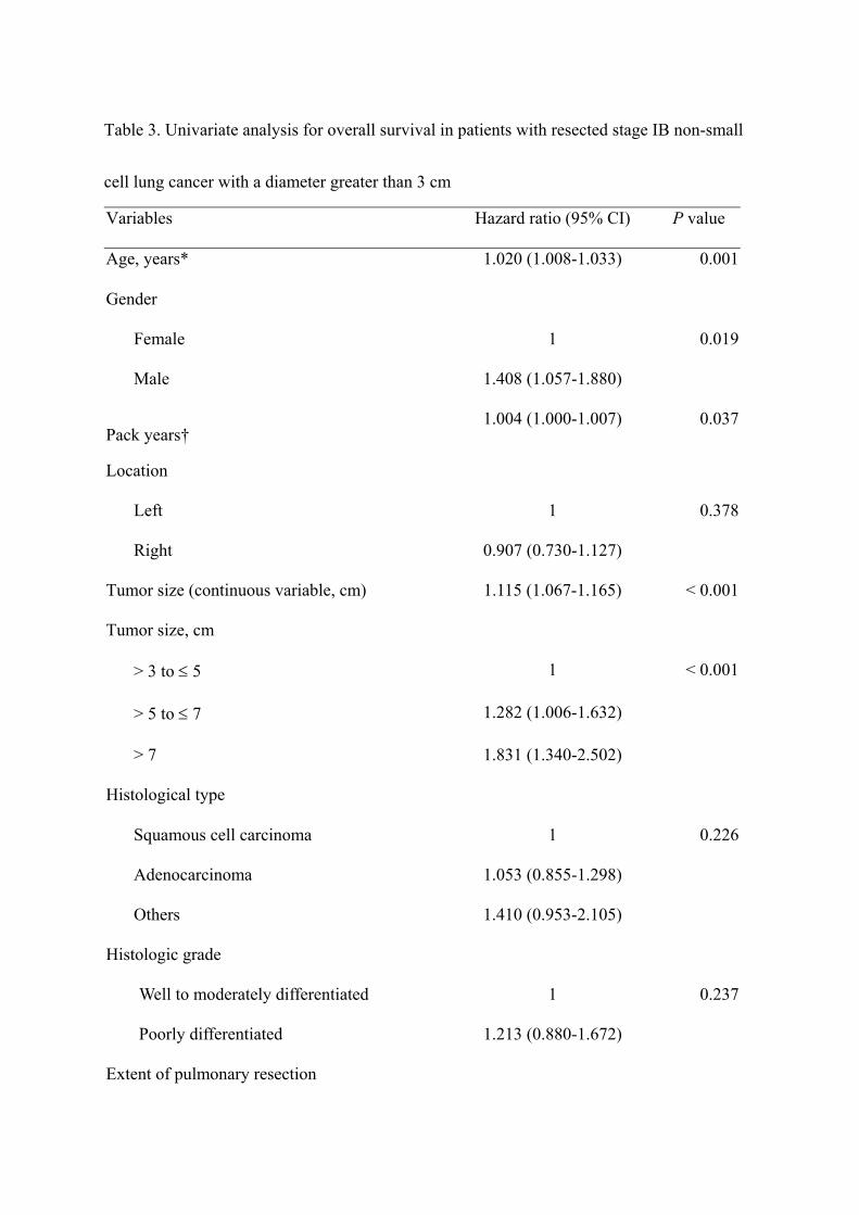
Table 3. Univariate analysis for overall survival in patients with resected stage IB non-small
cell lung cancer with a diameter greater than 3 cm
Variables Hazard ratio (95% CI) P value
Age, years* 1.020 (1.008-1.033) 0.001
Gender
Female 1 0.019
Male 1.408 (1.057-1.880)
Pack years� 1.004 (1.000-1.007) 0.037
Location
Left 1 0.378
Right 0.907 (0.730-1.127)
Tumor size (continuous variable, cm) 1.115 (1.067-1.165) < 0.001
Tumor size, cm
> 3 to ≤ 5 1 < 0.001
> 5 to ≤ 7 1.282 (1.006-1.632)
> 7 1.831 (1.340-2.502)
Histological type
Squamous cell carcinoma 1 0.226
Adenocarcinoma 1.053 (0.855-1.298)
Others 1.410 (0.953-2.105)
Histologic grade
Well to moderately differentiated 1 0.237
Poorly differentiated 1.213 (0.880-1.672)
Extent of pulmonary resection

Sublobar resection or lobectomy 1 0.004
Bilobectomy or pneumonectomy 1.374 (1.109-1.703)
Visceral pleural invasion
Absent 1 0.424
Present 0.902 (0.700-1.162)
Number of LNs dissected/sampled# 0.988 (0.978-0.999) 0.035
CI, confidence interval; LN, lymph node. *The hazard ratio associated with age is that the
increase in hazard is associated with a 1-year increase in age. �The hazard ratio associated with
pack years is an increased hazard per 1 pack-year of additional smoking. #The hazard ratio
associated with number of LNs dissected/sampled is an increased hazard per 1 LN of additional
LN dissection/sampling.

Table 4. Multivariate analysis for overall survival in patients with resected stage IB non-small
cell lung cancer with a diameter greater than 3 cm
Variables Hazard ratio (95% CI) P value
Age, years* 1.028 (1.014-1.043) < 0.001
Gender
Female 1 0.582
Male 1.100 (0.784-1.543)
Pack years 1.003 (0.999-1.007) 0.131
Tumor size, cm
> 3 to ≤ 5 1 0.002
> 5 to ≤ 7 1.377 (1.059-1.790)
> 7 1.724 (1.231-2.415)
Extent of pulmonary resection
Sublobar resection or lobectomy 1 0.002
Bilobectomy or pneumonectomy 1.456 (1.149-1.846)
Histological type
Squamous cell carcinoma 1 0.005
Adenocarcinoma 1.377 (1.059-1.790)
Others 1.724 (1.231-2.415)
Number of LNs dissected/sampled� 0.983 (0.971-0.994) 0.004
CI, confidence interval; LN, lymph node. *The hazard ratio associated with age is that the
increase in hazard is associated with a 1-year increase in age. �The hazard ratio associated with

number of LNs dissected/sampled is an increased hazard per 1 LN of additional LN
dissection/sampling.

Figure legends
Figure 1. Cumulative probability of overall survival in patients with surgically resected stage
IB non-small cell lung cancer with a diameter greater than 3 cm.
Figure 2. Overall survival in patients with resected stage I NSCLC with a diameter greater
than 3 cm grouped according to tumor size (> 3 to ≤ 5, > 5 to ≤ 7 cm, and > 7cm). The
log-rank test was used to compare between groups.

Figure 3. Overall survival in patients with resected stage I NSCLC with a diameter greater
than 3 cm with or without visceral pleural invasion (VPI). P = 0.424 (log-rank test).
