eprints@tamil nadu dr mgr medical university - a...
TRANSCRIPT

A Dissertation on
A DESCRIPTIVE STUDY OF VARIOUS USES OF
COBLATION IN PHARYNGOLARYNGOLOGY
Submitted to the
THE TAMILNADU DR. M.G.R. MEDICAL UNIVERSITY
In partial fulfilment of the requirements
For the award of the degree of
M.S.BRANCH IV (OTORHINOLARYNGOLOGY)
GOVERNMENT STANLEY MEDICAL
COLLEGE & HOSPITAL
THE TAMILNADU DR. M.G.R. MEDICAL UNIVERSITY,
CHENNAI, TAMILNADU
APRIL 2015

DECLARATION
I, Dr. KARTHIK.M, solemnly declare that the dissertation, titled
“A Descriptive study of various uses of coblation in
pharyngolaryngology ” is a bonafide work done by me during the
period of February 2013 to September 2014 at Government Stanley
Medical College and Hospital, Chennai under the expert supervision of
Prof. Dr. N.SEETHALAKSHMI, M.S., D.L.O., D.N.B, Department
Of Otorhinolaryngology , Government Stanley Medical College and
hospitals, Chennai.
This dissertation is submitted to The Tamil Nadu Dr. M.G.R.
Medical University in partial fulfilment of the rules and regulations for
the M.S. degree examinations in Otorhinolaryngology to be held in
April 2015.
Place: Chennai-1
Date: DR. KARTHIK.M

CERTIFICATE
This is to certify that the dissertation - “A DESCRIPTIVE
STUDY OF VARIOUS USES OF COBLATION IN
PHARYNGOLARYNGOLOGY ” presented by DR. KARTHIK.M,
is an original work done in the Department of Otorhinolaryngology,
Government Stanley Medical College and Hospital, Chennai in partial
fulfilment of regulations of the Tamil Nadu Dr. M.G.R. Medical
University for the award of degree of M.S. (Otorhinolaryngology)
Branch IV, under my supervision during the academic period 2012-
2015.
Prof.Dr. AL.Meenakshisundaram
M.D., D.A.,
THE DEAN,
Govt. Stanley medical college,
Chennai-1.
PROF.DR.T.Balasubramanian, M.S., D.L.O.,
PROFESSOR AND HEAD OF THE
DEPARTMENT
Govt. Stanley Medical College and Hospital
Chennai-1
Prof. Dr. N.SEETHALAKSHMI, M.S., D.L.O., D.N.B, Professor of Otorhinolaryngology, Govt. Stanley Medical College and Hospital, Chennai – 600 001. Place : Chennai Date :

ACKNOWLEDGEMENTS
I wish to express my sincere thanks to
Prof. Dr. AL.MEENAKSHISUNDARAM, MD, DEAN, Government
Stanley Medical College and Hospital for having permitted me to utilize
the facilities of the hospital for conducting this study.
My heartfelt gratitude to Prof. Dr. T. BALASUBRAMANIAN,
M.S., D.L.O., Professor and Head of the Department, Department of
Otorhinolaryngology, Government Stanley Medical College and
Hospital for his constant motivation, valuable suggestions, and expert
supervision during the course of this study.
I express my whole hearted gratitude to
Prof. Dr. N.SEETHALAKSHMI, M.S., D.L.O., D.N.B, Professor of
Otorhinolaryngology, and Chief of ENT UNIT II,
PROF.DR.F.ANTONY IRUDHAYARAJAN M.S., D.L.O, Professor
of Otorhinolaryngology, Prof. DR. M.RAMANIRAJ M.S., D.L.O,
Professor of Otorhinolaryngology, for supporting, guiding and
encouraging me in this study.

I wish to thank my Assistant Professors DR.K.ATHIYAMAN
M.S, DR.C.KARUPPASAMYM.S.,D.L.O., R.M.P.CHANDRAMOULI
M.S., DR.C.BHARANIDHARAN D.L.O. Dr.SARAVANA SELVAN
M.S., D.L.O for their valuable tips and guidance.
I also thank Mrs. Radha Kalaiselvan, Audiologist and Speech
Pathologist of ENT Department, Government Stanley hospital for her
expert assistance.
I am grateful to all the other post-graduates who most
willingly helped me during this study period.
I also thank the staff nurses and theatre personnel, Government
Stanley Hospital for their co-operation and assistance in the conduct
of this study.
Last but not the least, I am indebted and grateful to all the
Patients who constitute the backbone of this study, who most
willingly and selflessly subjected themselves to this study for the sake
of the benefit of their community and without whom this study would
not have been possible.

CONTENTS
S. No. Topics Page No.
1. ABSTRACT 1
2. INTRODUCTION 3
3. AIMS AND OBJECTIVE 4
4. REVIEW OF LITERATURE 6
5. MATERIALS AND METHODS 7
6. RESULTS AND OBSERVATION 103
7. DISCUSSION 128
8. SUMMARY 134
9. CONCLUSION 137
10. ANNEXURES
A.BIBLIOGRAPHY
B. PROFORMA
C. ETHICAL COMMITTEE APPROVAL LETTER
D. PATIENT INFORMATION SHEET
E. INFORMED CONSENT FORM
F. PLAGIARISM
G. MASTER SHEET

LIST OF FIGURES
Fig – 1 showing Oropharynx which contains Anterior pillar ,both the Tonsils, Uvula, soft palate
Fig – 2 showing the Anatomy of Larynx – Supraglottis, Glottis and subglottis
Fig – 3 showing the normal vocal cords, interarytenoid area and subglottic region.
Fig – 4 showing Coblator system with Radiofrequency electrosurgical controller
Fig – 5 showing Foot control and Flow control valve unit
Fig – 6 showing Tonsillar wand
Fig – 7 showing laryngeal wand
Fig – 8 showing Chronic Parenchymatous Tonsillitis
Fig – 9 showing Chronic Follicular Tonsillitis
Fig – 10 showing Multiple papillomatous lesions in the supra glottis.
Fig – 11 showing Rose’s position
Fig – 12 showing Dissection of inferior part Adenoid tissue using coblator wand.
Fig – 13 showing Dissection of lateral part Adenoid tissue using coblator wand
Fig – 14 Adenoidectomy is performed under direct endoscopic vision.
Fig – 15 showing Dissection of superior part Adenoid tissue using coblator wand

Fig – 16 showing Dissection of tonsil from its tonsillar bed using Coblator tonsillar wand , tonsil is held medially using tonsil holding forceps
Fig – 17 showing dissection of tonsil from the inferior pole
Fig – 18, 19 showing dissection of enlongated uvula using coblator wand.
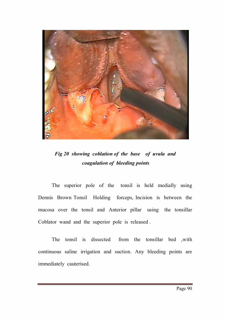
Fig – 20 showing coblation of the base of uvula and coagulation of bleeding points
Fig – 21 showing surgical field following dissection of both the tonsil and uvula using coblation.
Fig – 22 showing Boyce’s position and introduction of
Direct laryngoscopy .
Fig – 23 Now, the Larnygeal Coblator Wand is introduced transorally after setting
Fig –24 showing Dissection of laryngeal papilloma
using coblation.
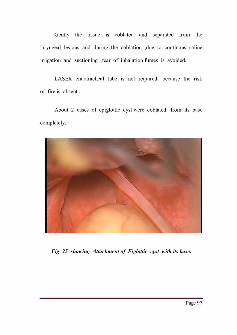
Fig – 25 showing Attachment of Eiglottic cyst with its base.
Fig –26, 27 showing Attachment of Eiglottic cyst with its base being coblated using laryngeal wand .
Fig – 28, 29, 30 showing posterior cordotomy being done using coblator Laryngeal wand

ABSTRACT

Page 1
ABSTRACT
Coblation is controlled and non-heat driven process that
uses bipolar radiofrequency to energise the particles in the
conductive medium, such as saline, to form a precisely focused
plasma field.
Aim:
To study the various uses and safety of Coblator in
Pharyngolaryngology .
Methods:
The study was conducted in the Department of
Otorhinolaryngology of Government Stanley hospital during the
period Feb 2013 to Sept 2014.
About 65 cases which were selected randomly, based on the
inclusion and exclusion criteria, were sub divided based on the
diagnosis.
Out of which 20 were Chronic Adenoid Hypertrophy, 20 were
chronic Tonsillitis, 5 were mild Obstructive sleep apnea syndrome,
20 were Benign Laryngeal lesions underwent Coblation assisted
surgeries respectively.

Page 2
Results:
Coblation Assisted AdenoTonsillectomy ensures complete and
safe removal of tissue, decreased post operative pain ,Early return to
normal diet Coblation Assisted Uvulopalatoplasty ensures complete
relief of symptoms, minimal injury to surrounding normal tissue,
post operative pain, Early return to normal diet. About 3 out of 40
patients had post operative bleeding, following Coblation Assisted
Adenotonsillectomy.

INTRODUCTION

Page 3
INTRODUCTION
Coblation is controlled and non-heat driven process that
uses bipolar radiofrequency to energise the particles in the
conductive medium, such as saline, to form a precisely focused
plasma field.
These energized particles in plasma field have sufficient
energy to disintegrate molecular bonds, providing ablation of soft
tissue, while preserving adjacent normal tissue at a relatively lower
temperature.
Low temperature plasma excision between 40ºC to 70ºC
whereas that of conventional diathermy greater than 100ºC .
The result is a safe, minimally invasive, and virtually painless
technique that can dissect the tissues with minimal injury to the
surrounding healthy tissue and without altering the structure .
Hence healing and recovery is faster compared to the
conventional techniques.

AIMS AND OBJECTIVE

Page 4
AIM AND OBJECTIVES
AIM OF THE STUDY
To study the various uses and safety of Coblator in
Pharyngolaryngology .
OBJECTIVE OF THE STUDY
1. To study the various uses, different operative techniques and
safety of coblator .
2. To assess the primary outcome and secondary outcome in post
coblator Adenoidectomy patients
3. To assess the primary outcome and secondary outcome in post
coblator tonsillectomy patients
4. To assess the primary outcome and secondary outcome in
coblation Assisted Uvulopalatopharyngoplasty
5. To assess the primary outcome and secondary outcome in
coblator Assisted Excision of benign laryngeal lesions and
Dennis Kashima’s procedure.
6. To know the disadvantages of using coblation .

Page 5
INCLUSION CRITERIA
1. Age 10 - 45 years
2. Patients diagnosed chronic Adenotonsillitis ,snoring , benign
laryngeal lesions and Bilateral vocal cord paralysis.
EXCLUSION CRITERIA
1. Age <10 or >45years
2. Systemic infections
3. Patient suspected tonsillar malignancy or laryngeal malignancy
4. Patients with Bronchial asthma/pulmonary Tuberculosis/obesity
5. Patients with bleeding diathesis or on Anticoagulants
6. Immunocomprised patients .

REVIEW OF LITERATURE

Page 6
REVIEW OF LITERAURE
ANATOMY OF PHARYNX AND LARYNX
PHARYNX
The pharynx is a funnel shaped fibromuscular tube extending
from the base of skull to the level of the body of sixth cervical
vertebra .
It is approximately 15cm in length , about 3.5 cm at the
base and 1.5 cm at the level of cricopharynx .
The pharynx is divided into 3 parts
1. Nasopharynx
2.Oropharynx
3.Hypopharynx
The lower end of pharynx is continuous with the
oesophagus. This junction at the cricopharyngeal sphincter is
the narrowest point in the digestive tract .

Page 7
Nasopharynx
Nasopharynx is a cuboidal space and extends from base
of skull to the level of an imaginary line drawn at the level
of hard palate .
Anteriorly it freely communicates with the nasal cavities
through the choana separated by the posterior end of septum.
Posteriorly it is related to upper cervical vertebra C1 and
C2, prevertebral muscles and fascia.
Superiorly, Roof is formed by the basi-sphenoid and basi-
occiput limited by the pharyngeal tubercle posteriorly .
Inferiorly, it communicates with the Oropharynx through the
nasopharyngeal isthmus.
The Lateral wall of the Nasopharynx contains the following
structures -
1. Eustachian tube opening
Situated in the lateral wall on each side of the Nasopharynx
1.25cm posterior to the level of the inferior turbinate. The orifice

Page 8
is bounded postero-superiorly by torus tubaris, an elevation produced
by the cartilagenous part of the Eustachian tube.
Just posterior to this eminence is a recess called Fossa of
Rosenmuller .
2. Adenoids (Luschka’s Tonsils)
These are a sub -mucosal aggregation of lymphoid tissue at
the junction of the roof and posterior wall of the Nasopharynx.
They are composed of vertical ridges of lymphatic tissue separated
by furrows .
The Adenoids are present from birth and increases in size
with age, and undergoes spontaneous regression after the age of 12
years. Very rarely it can persists even after puberty.
The Adenoids are of maximum size at age of 5 years .
Blood supply of Adenoids
1. Ascending palatine branch of facial artery
2. Ascending pharyngeal branch of external carotid artery
3. Pharyngeal branch of the third part of maxillary artery.

Page 9
Lymphatic drainage of adenoids
Upper jugular nodes, retropharyngeal nodes, parapharyngeal
nodes.
3. Tubal Tonsils (Gerlach’s Tonsils )
It is a collection of sub epithelial lymphoid tissue in
relation to the torus tubaris and can be continuous with the
adenoids.
4. Passavant’s ridge
It is a mucosal elevation produced by the palatopharyngeus
muscle at the level of the nasopharyngeal isthmus. It becomes
prominent during the act of swallowing when the soft palate
makes contact with it.
OROPHARYNX
It is space which is in communication with the oral
cavity anteriorly, the nasopharynx superiorly and the hypopharynx
inferiorly. Anatomically, it is the space between the level of hard
palate and the hyoid bone.

Page 10
Oropharynx communicates with the oral cavity through
the oropharyngeal isthmus.
The boundaries of the isthmus are
Superiorly -the soft palate
Inferiorly - Base of the tongue
Laterally -the palatine tonsil and anterior pillar
Posteriorly - Body of C2 ,C3 vertebrae ,prevertebral fascia and
muscles .
Palatine Tonsils
These are bilateral ovoid masses of lymphoid tissue lying in
the mucous membrane of the lateral wall of the Oropharynx in the
tonsillar fossae.
The Tonsillar fossa is triangular in shape .Anteriorly, It is
formed by the anterior pillar enclosing palatoglossus muscle,
posteriorly by the posterior pillar enclosed by the palatopharyngeus
and base is formed by the tongue .

Page 11
A fold of mucous membrane called the plica semilunaris
connects the anterior and posterior pillars at the upper pole of the
tonsil and another fold the plica triangularis connects anterior
pillar to the anteroinferior part of the tonsil.
At birth, the tonsils are small in size during early childhood
They enlarge and then regress in size by the age of 16 -18 years.
The medial surfaces of the tonsil show a number of crypts
(15-20 ). The intratonsillar crypt, Crypta magna is the largest and
lies near the upper pole . It represents remnant of the second
pharyngeal pouch. Tonsillar bed consists of loose areolar tissue ,
Pharyngobasilar fascia, superior constrictor muscle ,para tonsillar
veins and buccopharyngeal membrane. The glossopharyngeal nerve
and styloid process if elongated may lie in relation to the
tonsillar bed.
Internal carotid artery lies 2.5cm postero-lateral to the
tonsil. The Medial surface of the tonsil is covered by stratified
squamous epithelium .
Blood supply of the tonsil .
1. Tonsillar branch of the facial artery

Page 12
2. Branch from the ascending pharyngeal artery
3. Descending palatine artery, a branch of the maxillary artery.
4. Ascending palatine branch of facial artery
5. Dorsalis linguae arteries .
All the arteries except the descending palatine branch
enter the tonsil via the inferior pole where as the descending
palatine branch enters through the upper pole.
Venous drainage - Veins from the tonsil pierce the lateral
surface and the inferior pole. The paratonsillar vein emerges on the
lateral surface ,pierces the superior constrictor muscle to end in
the common facial vein and pharyngeal plexus of veins.
Fig -1 showing Oropharynx which contains Anterior pillar,
both the Tonsils, Uvula, soft palate .

Page 13
Hyopharynx (Laryngopharynx)
The lowest part of the pharyngeal tube which lies partly
posterior and partly lateral in relationship to the larynx on either
side.
It extends from level of the hyoid bone to the level of
lower border of cricoid cartilage. It is related to the C4, C5, C6
cervical vertebrae.
It communicates with Oropharynx superiorly and continuous
with the oesophagus at the level of the cricoids cartilage .
It is divided into three parts .
1.Pyriform fossa
2. Post cricoid region
3.Posterior pharyngeal wall
Pyriform sinus
It is a recess on either side of the larynx .It is an inverted
pyramid like structure .It extends from the pharyngo-epiglottic

Page 14
fold superiorly to its apex inferiorly related to the cervical
oesophagus.
It is bounded by the Aryepiglottic fold medially and the
thyrohyoid membrane and thyroid cartilage laterally .
Post Cricoid region
It extends from the level of arytenoid cartilage and
connecting folds to the inferior border of cricoids cartilage ,thus
forming the anterior wall of hypopharynx .
Posterior Pharyngeal wall
It extends from the level of the hyoid bone o the level of
cricoarytenoid joint.
Layers of the pharyngeal wall
The pharyngeal wall is made up of four layers
Mucous membrane
Nasopharynx is lined by pseudostratified ciliated columnar
epithelium .

Page 15
Oropharynx and laryngopharynx is lined by stratified squamous
epithelium.
Pharyngeal aponeurosis
It lies between the mucosal and muscular layers . It is thickened
in the upper part and is called pharyngobasilar fascia.
Two Muscular layers
Outer layer consists of three constrictors namely superior,
middle and Inferior constrictor .
Inner layer consists of three muscles namely
stylopharyngeus, palatopharyngeus and salphingopharyngeus.
Nerve supply of pharynx
The pharyngeal plexus which supplies to the upper part of the
pharynx including the surface of the tonsil and all the muscles of
the Pharynx, except the stylopharyngeus which is supplied by the
glossopharyngeal nerve.

Page 16
Larynx
Embrology ;
The development of larynx begins from the hypopharynx with the
fusion of lateral structures derived from the tracheobronchial
primordium (arch 4 and 5 ) in the midline.
The larynx, trachea, bronchi and lungs have developed from
the midline ventral respiratory diverticulum namely the
laryngotracheobronchial groove .
Supraglottis develops from the Buccopharyngeal primordium
(Arch 3 and 4 ).Glottis and subglottis develops from the Arches 4
and 6.
The laryngeal framework consists of muscles and their
nerve supply overlying the architectural frame of bones , cartilages.
Framework of the larynx
The cartilages of the larynx are divided into three unpaired
and three paired cartilages .The unpaired cartilages are thyroid
,cricoid, epiglottis .

Page 17
Fig:2 showing the Anatomy of Larynx –
Supraglottis, Glottis and subglottis
TABLE : 1
Thyroid Cricoid Eiglottis
Shape Shield Ring Leaf
Parts
Two lamina joins at an angle of 90
degrees in males and 120Degrees in
females .
Anterior narrower Arch and Posterior
Broader Lamina
Mobile suprahyoid and
Fixed infrahyoid parts
Muscles
Attached
Sternothyroid, thyrohyoid, Inferior
constrictor, thyroarytenoid
Inferior constrictor
Cricothyroid and cricoarytenoid
Aryepiglotticus

Page 18
Ligaments
attached
Thyrohyoid membrane with false
cord .
Cricohyoid membrane
Thyroepiglottic ligament
Functions Protects the larynx Laryngotracheal connection
Guards the laryngeal Inlet
Histology Hyaline cartilage Hyaline cartilage Fibroelastic cartilage
Paired Cartilages :
The paired cartilages are arytenoids, corniculate and cunieform.
The arytenoids form the posterior support for the laryngeal folds.
It has two processes one pointing anteriorly attached to the vocal
cord and forms posterior commisure of glottis and the pointing
laterally called the muscular process giving attachments to the
laryngeal muscles.
The arytenoids articulate with the lamina of the cricoids
cartilage. The corniculate and cunieform cartilages articulate with
the apex of the Arytenoids
Ligaments of larynx
The ligaments are classified as intrinsic ligaments and extrinsic
ligaments.

Page 19
The extrinsic ligments connect the larynx with the hyoid and
trachea.
The thyrohyoid membrane connects the upper border of
thyroid to the hyoid bone .
The membrane is pierced by superior laryngeal nerve and
vessels26. The Cricothyroid Ligament connects the upper border of
the cricoid to The lower border of thyroid cartilage .
The hyoepiglottis ligament connects hyoid to the epiglottis. The
Intrinsic membrane is a fibroelastic membrane which connects the
laryngeal cartilages, strengthens the joints and creates an internal
frame work.
TABLE:2
Upper Quadrilateral Membrane
Lower conus elasticus
Superior attachment Aryepiglottic fold
Thyroid to vocal process
Inferior attachment Vestibular folds Cricoids
Ligaments Inferior border of vestibular ligament
Superior border of vocal ligament

Page 20
Muscles of Larynx :
The muscles of the larynx can be divided into extrinsic and
intrinsic muscles.
TABLE:3
Sl No
Name of the muscle
Attachment Function Nerve
1.
Posterior Cricoarytenoid
Cricoid Lamina to muscular process of arytenoids
Opens the glottis
(Abduction)
Recurrent Laryngeal Nerve
2.
Lateral
Cricoarytenoid
Cricoid Lamina to muscular process of arytenoids
Closes the glottis
(Adduction)
Recurrent Laryngeal Nerve
3.
Interarytenoid
Arytenoid to Arytenoid
Closes the posterior commissure
Recurrent Laryngeal Nerve
4.
Thyroarytenoidis
(Vocalis )
Thyroid to the Arytenoid medial fibres
Internal Tensor
Recurrent Laryngeal Nerve
5.
Cricothyroid
Cricoid to thyroid
External
Tensor
External Laryngeal Nerve

Page 21
Interior of the Larynx
The cavity extends from laryngeal inlet to the beginning of
the lumen of the trachea at the lower border of cricoid cartilage
and is divided by the vestibular and vocal folds into three
compartments.
The superior vestibule is above the vestibular folds .The
Ventricle or sinus of the larynx lies in between vestibular and
vocal folds.
The Inlet is formed by aryepiglottic folds with free margin of
epiglottis and mucosa in between the arytenoids and this is called as
epilarynx.
Glottis is defined area of larynx that lies at level of vocal
cords to level of about 10mm below the vocal cords.
Subglottis is region of larynx extending from 10mm below
the vocal cords to level of lower border of cricoid cartilage.
The sinus of the larynx is situated in between vestibular and
vocal folds elongated in the anterior part as saccule. The saccule
contains mucous glands and hence is known as oil can of larynx.

Page 22
Fig 3 : showing the normal vocal cords ,interarytenoid area and
subglottic region.
Mucous Membrane56 :
The supraglottic and subglottic areas are lined by
pseudostratified ciliated columnar epithelium. The squamous
epithelium is seen over the vocal cords and Transitional epithelium
on few places .The mucous glands are situated all over the
membrane more so over the posterior surface of epiglottis and
saccule but devoid over the vocal cord .The vocal cords are
lubricated by the saccule.

Page 23
Spaces in the Larynx.
Preepiglottic space
Is a Potential space in front of epiglottis. It has a rich
supply of lymphatics.
Boundaries
Anterior - Thyrohyoid membrane and Thyroid cartilage
Posterior- Epiglottis
Lateral – Communicates with the paraglottic space
Inferior – Attachment of epiglottis with thyroid cartilage
Superior – Hyoepiglottic ligament
Paraglottic Space (Space of Tucker)
Lies on the both sides of the larynx lateral to saccule and
anterior to the pyriform fossa.
Boundaries
Anterior - Communicates with pre epiglottic space
Posterior- anterior reflection of the pyriform fossa .

Page 24
Lateral – Thyroid lamina
Medial - Quadrangular membrane and conus elasticus
Reinke’ s space
Sub epithelial space lying over the vocal ligaments
Boundaries
Anterior - Anterior commissure
Posterior- Posterior. commissure
Lateral – Vocalis muscle
Medial - Free margin of vocal cord
Superior – superior arcuate line
Inferior – Inferior arcuate line
Physiology of pharynx56
The act of swallowing is be divided into three phases:
1. The oral phase is the only voluntary phase.
It has preparatory and propulsive stage. The preparatory
phase requires the food to be within the oral cavity and

Page 25
Oropharyngeal sphincter to be closed and process of mastication
is initiated .
The resulting bolus is ready to be propelled by the tongue
into the Oropharynx, here the propulsive stage starts and bolus is
pushed against the hard palate and the palatoglossal sphincter
relaxes, soft palate pulled upwards and base of the tongue falls
forwards and inferiorly
2. The pharyngeal phase is an involuntary phase and consists of
a very complex mechanism.
The laryngeal inlet is closed by superior movement of
larynx and epiglottis closing the inlet and closure of nasopharyngeal
isthmus.
Opening of the cricopharyngeal sphincter , creates negative
suction pressure. Once the bolus enters the oesophagus, aided by the
gravity, pharyngeal constrictors contract from above downwards
and squeezing the remnant food.
3. The oesophageal phase is again involuntary, and propels the
foods.

Page 26
It is an involuntary phase .The food passes through the
oesophagus by peristaltic waves and the cardiac sphincter opens
and food enters the stomach which lasts for 8-20 seconds ,
FUNCTIONS OF THE LARYNX
Sphincteric Action; Protective Mechanism Sphincter actions of
the laryngeal inlet (Epiglottis ) ,Ventricular bands and Vocal cords
prevent the entry of foreign body into the larynx. This is called
the three tier mechanism
Cough Reflex.
It is a important protective mechanism. Cough is produced
by a forced expiration against closed glottis that is suddenly
opened.
Respiratory conduit.
Passage of air through the larynx easily, and mucociliary
clearance of fine particles.
Negative suction pressure which increases the preload

Page 27
Effort closure
During climbing, defecation, micturition, closure of the glottis
prevents escape of air from the chest making it easier to increase
the intraabdominal pressure .
The Biomechanics Of Phonation (53, 55)
Initiation of voice
Initially the sub glottic pressure builds up against the
closed glottis and then the air is forced through the vocal cords
which vibrate at fundamental frequency .
The phases of the vibratory cycle can be classified, into four
stages
1. Closing Phase
2. Closed Phase
3. Opening Phase
4. Open Phase
The Laryngeal tone is modulated and amplified by
resonators such as the mouth, sinus and pharynx.

Page 28
COBLATION — "Controlled Ablation45"
Uses bipolar high frequency electrical energy to excite the
electrolytes in a conductive medium(Normal saline , Ringer lactate )
creating a precisely focused plasma field.
The energized particles in the plasma have sufficient energy
to break organic molecular bonds, excising or dissolving soft tissue
at relatively low temperatures(60 - 70°C) thereby preserving the
integrity of surrounding healthy tissue.
Advantages of Coblation
1. Functions at much cooler temperatures (40-70°C vs. 400-600°C
for non-coblation devices).
2. More precise with minimal effect to the surrounding tissue.
3. Active plasma field is only 100-200µm thick (~thickness of a
sheet of paper)
Advantages of Coblation Technology over other soft tissue removal
options:
1. MIS approach through a small slit allowing 1-2.5mm wand
tipintroduction, no surgical window opening needed.
2. One time entry, no back and forth entry/exit around sensitive
anatomy.

Page 29
COBLATOR
Coblator is a bipolar radiofrequency electrosurgical system
Parts
1. Radiofrequency electrosurgical controller
2. Power cord
3. Foot control
4. Flow control valve unit
5.Flow control cable
6.Sterile wand
Controller is the power source that delivers radiofrequency
energy to the treatment site via the wand.
Coblator is contraindicated in any procedures where a
conductive solution is not used .
Coblator is contraindicated for patients who have
pacemakers or any other electronic implants

Page 30
Fig 4: showing Coblator system with Radiofrequency
electrosurgical controller
Principle of operation of apparatus ;
Coblator system controller is designed to deliver
Radiofrequency energy to the electrodes located at the distal end the
wand .Current flows between the active electrodes element and the
return element providing a localized energy field.
The result of this arrangement is controlled energy delivery
with minimal collateral tissue damage Coblator system works by
passing Radiofrequency energy through a conducive solution (such
as a normal saline) in close proximity to or in contact with the
tissue to be treated .

Page 31
The conductive solution solution forms a thin layer between
the active and return electrode elements .
Fig 5 : showing Foot control and Flow control valve unit
Fig 6 showing Tonsillar wand

Page 32
Fig 7 : showing laryngeal wand
In the coblate mode, when sufficient energy is applied the
conductive solution is converted into a vapour layer containing
energized charged particles. When the high energy charged particles
come in contact with tissue, they cause its disintegration through
molecular dissociation.
This mode of operation results in relatively low treatment
site temperatures when compared to conventional electrosurgical
systems, thus yielding limited collateral thermal damage of the
surrounding untreated tissue.
The function of the device is different when low voltage is
applied between the active electrode and the target tissue .In this
case, the electrical field is below the threshold required to create

Page 33
plasma layer and resistive tissue heating occurs .This mode is
useful when greater thermal is required that is for coagulation of
blood vessel . The appropriate voltage setting will depend on the
wand used tissue type and desired tissue effect .
Voltage outputs
The Coblation and coagulation voltage level settings are
indicated by a single digit LED output display on the front panel
of the controller.
Coblate Mode
Display Output Voltage (Vrms +/_ 10 % )
1 100
2 127 .5
3 155
4 182.5
5 210
6 237.5
7 265
8 290
9 300

Page 34
Coagulation Mode
Display Output Voltage (Vrms +/_ 10 % )
1 65
2 70
3 75
4 81
5 87
6 93
7 100
8 108
9 115
When the controller is first powered on with no wand
attached, the display digit 0 appears on both front panel displays
as default setting.
If a wand is attached a or after power up ,the unit will
adjust he outputs to nominal setting .These will usually provide the
best effect in most situations .

Page 35
Once the wand is connected, the voltage level can be
increased or decreased by pressing the coblate level adjustment or
he coagulation level adjustment buttons located on the front panel.
The conductive media could either be normal saline or
Ringer lactate source must be higher than the patient for proper
operation .
Recommended 1meter/3 feet
System Storage
Temperature range -40 degree C to + 70 degree C .
The relative humidity should be between 10- 85
Pathology and its surgical approaches
Acute Tonsillitis
Acute inflammation of tonsil which usually occurs in
children or Adolescents .Inflammation Tonsil may be associated
with inflammation of the pharynx.

Page 36
Most common organisms are beta haemolytic Streptococci,
Staphylococci Hemophilus influenza, Virus like Adenovirus,
Rhinovirus, Cox sackie virus.
Types of Acute inflammation
1. Acute parenchymatous tonsillitis
2. Acute follicular tonsillitis
3. Acute membranous tonsillitis .
Chronic Tonsillitis
Chronic inflammation of the tonsils usually starts as an
acute inflammation. The crypts are filled with organisms and
chronic inflammatory cells, exudates and debris .
1.Chronic parenchymatous tonsillitis
Fig 8: showing Chronic Parenchymatous Tonsillitis

Page 37
Tonsils are uniformly enlarged and congested. Sometimes the
enlargements is such that they touch each other and are called
kissing tonsils.
2. Chronic Follicular Tonsillitis
Fig 9 : showing Chronic Follicular Tonsillitis
Beads of white discharge are present on the surface of the
tonsils at the entrance to tonsil crypts.
3. Chronic Fibrotic tonsillitis
The Tonsils are small and inflamed .Occurs in the adults.
Anterior pillars are hyperemic.
The most reliable sign is enlarged non-tender jugulo-digastric
lymph nodes.

Page 38
Clinical Features
1. Sore throat – repeated attacks with remission in between
the attacks .
2. Difficulty and pain swallowing
3. Pain around the neck
4. Chronic dry cough and throat irritation
5. Halitosis
6. In hypertrophic tonsillitis - Breathing problems and snoring.
Clinical Examination
Throat Examination -
Clinical Grading of Tonsillar Enlargement (54)
Grade I - Tonsil occupy 25% of the faucial isthmus
Grade II - Tonsil occupy 50% of the faucial isthmus
Grade III - Tonsil occupy 75% of the faucial isthmus
Grade IV- Tonsil occupy 100% of the faucial isthmus

Page 39
Characteristic Signs of chronic tonsillitis are
1. Hypertrophied or atrophied Tonsils
2. Residual congestion of the Anterior pillar
3. Enlarged Non -tender Jugulo -digastric lymph nodes \
4. Extrusion of purulent discharge on pressure over the tonsils
with two tongue depressors .(Irwin Moore’s sign )
Complications of Chronic Tonsillitis
I . Local complications
1. Pharyngitis
2. Laryngitis
3. Peritonsillar abscess
4. Parapharyngeal Abscess
5. Middle Effusion
6. Otitis Media

Page 40
II .Systemic complications
1. Rheumatic arthritis and Rheumatic heart disease
2. Infective Endocarditis
3. Glomerulonephritis
4. Sleep Apnea syndrome
Chronic Adenoid hypertrophy (54)
Aetiology
1. Associaed with recurrent upper respiratory tract infections
2. Physiological hypertrophy in young children
3. Allergic rhinosinusitis
Adenoid facies
A child with adenoid hypertrophy has a characteristic facial
expression
1. Open mouth and mouth breathing
2. Pinch nose
Page 41
3. Crowded teeth and hyperplasia of gums
4. Loss of Nasolabial fold
5. Under slung mandible
6. High arched palate
7. Short upper lip
8. Hypoplasia of maxilla
9. Vacant expression
10. Pectus Excavatum
11. Rounded shoulders
12. Mental condition – subnormal and inability to concentrate and
fix the attention called Guye’s Aprosexia.
Clinical features of Enlarged Adenoids
1.Nasal Obstruction
It can partial or complete obstruction Patient presents with
snoring and mouth breathing .In infants it causes difficulty in feeding
and hence failure to thrive.

Page 42
2.Nasal Discharge
3.Change of voice - Hyponasality
4.Eustachian Tube obstruction
Adenoids cause mechanical obstruction of the Eustachian tube
opening Causing recurrent otitis media and conducive hearing loss.
It leads to Recurrent Acute Otitis media ,chronic suppurative
otitis media and Atelectasis of the tympanic membrane.
5. Snoring and sleep apnea syndrome and secondary
pulmonary hypertension .
6. Infection -
Leads chronic Sinusitis, Chronic Tonsillitis and Chronic
pharyngitis, Chronic otitis media.
7. Growth and Development
Chronic Obstruction to airway and feeding problems leads
failure to thrive in infants .It is due to abnormal regulation of the
growth hormone that occurs in REM sleep.

Page 43
8.Cardio-pulmonary problems
Severe Cases of Obstructive sleep disorders results in cor -
pulmonale, pulmonary vascular hypertension, Alveolar hypoventilation
,hypercapnia, hypoxia, with respiratory academia , Right ventricular
dilatation and finally cardiac failure.
9. Disorders of Masticatory apparatus
Mal -development of upper jaw ,arched or Gothic palate
due o absence of pressure of the tongue on the hard palate ,Dental
malocclusion and undershot jaw.
10. Somatic effects
Flat Chest, Rounded shoulders, thirst and loss of appetite.
Effects on Intelligence, restlessness, apathy, poor academic
performance and mental development.
Clinical Grading of Adenoid Enlargement
Grade I - Adenoids occupy 25% of the Nasopharyngeal lumen
Grade II - Adenoids occupy 50% of the Nasopharyngeal lumen
Grade III - Adenoids occupy 75% of the Nasopharyngeal lumen

Page 44
Grade IV- Adenoids occupy 100 % of the Nasopharyngeal lumen
Surgical Removal of tonsil and adenoid is called
Adenotonsillectomy .
Indications for tonsillectomy
A. Absolute
1. Recurrent attacks of Acute Tonsillitis (More than 6 episodes
in a year ).
2. Tonsillar hypertrophy obstructing respiration .
3. Enlarged tonsil causing snoring and obstructive sleep apnea
syndrome
4. Malignancy of tonsil
B. Relative
1.Chronic Diphtheria carrier
2.As an approach to elongated styloid process excision .
3.Tonsillolith
4.Perionsillar Abscess

Page 45
Contraindications
1.Aneursym of Tonsil
2.Epidemic of poliomyelitis
3.Age below 3 years
4.Cervical Spondylosis
Methods of Tonsillectomy
1.Gullitone method
2.Cold Steel dissection Method
3.Coblation Method
4.Cryosurgery technique
5.LASER Tonsillectomy
6. Harmonic scapel Method .
Main Complications of Tonsillectomy are
1.Hemorrhage -
A. Primary Hemorrhage - Occurs at the time of surgery .

Page 46
B. Reactionary Hemorrhage – Occurs within 24 hours of surgery
C. Secondary hemorrhage -Occurs after 24 hours of surgery
2.Infections
3.Lung Complications – Aspiration, collapse of lung .
Indications for Adenoidectomy
1.Enlarged Adenoids causing Nasal Obstruction and snoring
2.Recurrent Upper Respiratory tract Infection
3.In complications like CSOM and serous otitis media .
Methods of Adenoidectomy
1.Adenoid Currettage using St .Clair Thompson Adenoidectomy
curette.
2.Coblation Assisted Adenoidectomy
3.Microdebrider Assisted Adenoidectomy .

Page 47
Non Malignant Lesions of the Larynx
Is defined as any lesion or mass in the larynx which does
not present with the characteristics of malignancy is called a
Non- Malignant lesion of the larynx .It can divided into 2 types
1.Benign Neoplastic lesion
2.Benign Non Neoplastic lesion
Benign Neoplastic Lesion is classified as follows ;
TABLE:4
Tissue of origin Lesion Epidermal Papilloma Neural
Neural Fibroma Schwannoma Paraganglioma
Glandular Oncocytic tumour Vascular Hemangioma
Arterio-Venous malformation Cartilagenous Chondroma Miscellaneous
Lipoma Fibroma Rhabdomyoma

Page 48
Benign Non Neoplastic lesion
Congenital
Laryngocele Lymphangioma Saccular cyst Laryngeal webs Congenial subglottic stenosis Subglottic Hemangioma Laryngeal and laryngo- tracheal oesophageal cleft
Traumatic
Inflammatory Keratosis Vocal polyp Vocal Nodule Reinke’s Edema Intracordal cyst Contact Ulcer
Granuloma
Bacterial Tuberculosis Leprosy Syphilis Rhinoscleroma Fungal Rhinosporidiosis Candida
Benign Neoplastic lesion 85% of the benign lesions are
papilloma of larynx out of which 25% Were Recurrent
Respiratory papillomatosis.

Page 49
Two Principle out come of these lesion are
1.Phonatory dysfunction
2. Obstructed Airway
Papilloma Larnyx
Fig 10: showing Multiple papillomatous lesions in the supra glottis.
These are squamous in origin, colour ranges from white to
red, are usually sessile, may keratinizing or non keratinizing .
The Adult type and children type are histologically the same
but tumour behaviour is different and they belong to clinically different
groups .

Page 50
Juvenile Papilloma of larynx
Recurrent Respiratory Papillomatosis
This disease has a world wide distribution and involves all
Socioeconomic groups.
In children, there is a female preponderance. Myres and Barnes
found that 84% of the benign laryngeal tumours were
papillomas.
Aetiology of Laryngeal papilloma (53)
Caused by Human papilloma types 6 and 11
Following infection the virus may remain in the basal
layer of the mucous membrane replicating by a process known as
episomal maintenance, where virus is undetectable
but its presence can be determined by DNA hybridization
technique.

Page 51
Characteristics
Occurs in clusters ,multiple non keratinising lesions which
recur even after radical stripping .The symptoms occurs before the
age 4 years .
The symptoms are :
Hoarseness of voice
Abnormal cry
Dyspnea
Later stridor
The papillomas occur more on the anterior aspect of larynx
because of the thick blanket with rapid clearance has a protective
effect on he posterior aspect of the larynx .It can extend up to
trachea and major bronchi.
Natural History
The transmission occurs from the mother with genital warts
to children in about 60% .The remission can take place at any age
unrelated to treatment and relapse can occur at any age.
Page 52
The malignant transformation is very rare and seen after
radiotherapy or in smokers .
Management
The Treatment of choice is microlaryngeal Laserization or
Coblation Assisted Excision.
Adjuvant therapy -Systemic Cis –Retinoids
The recurrence rate is reduced but serious side effects like
skin excoriation occurs.
Indole -3 – carbinol a natural derivative of cruciferous
vegetables.
Interferons -causes only decrease in recurrence or temporary
remission.
Avidano and Singleton reported the use of methotrexate in
three patients
Intralesional Cidofovir(53)

Page 53
Adult papilloma
The papilloma are solitary small and less aggressive in
behaviour than juvenile type .It occurs on the vocal cords. The
histology consist of fronds of connective tissue covered by well
differentiated squamous epithelium with no invasion of stroma or
sub mucosa. Malignant transformation is more frequent.
Laryngeal paraganglioma
It arises from the paired superior and inferior paraganglia
Situated in relation to superior laryngeal nerve and loop of
Gallen.
Clinical features ;
Usually presents a small a red painful mass located on the
aryepiglottic fold. The striking feature is pain radiating to the ear
which resolves after biopsy or excision .They are very vascular
and may bleed profusely during resection, if injured
Treatment : Surgical excision by open surgical technique or
endoscopic removal .

Page 54
NeuroFibroma
Commoner in patients with Von Reckinghausen’s disease or
rarely as solitary lesion . They are bulky slow growing mass over
aryepiglottic fold and false vocal cord.
Voice change, fullness in the throat and slowly developing
dyspnea are the other features .
The treatment is surgical excision.
Haemangioma
The haemangioma should be differentiated from granulation
tissue in all cases. The haemangioma are of two types adult and
congenital.
The congenital type occurs in children in the subglottic area
and undergoes spontaneous remission.
If remission does not occur endoscopic surgical excision
preferably with laser is done.
But in adult due o the bigger diameter of vessels, this less
successful .The adult type lesion occurs in the glottis or

Page 55
supraglottic areas producing hoarseness and stridor and is treated
by open surgical excision.
Miscellaneous Tumours
The Chondroma occurs between the age group of 40- 60
years, with 5: 1 male : female ratio.
About 20% of these tumours can undergo malignant
transformation .They present clinically as a smooth, globular \
encapsulated mass and wide excision is the treatment of choice.
The Lipoma, Fibroma and Rhabdomyoma are very rare .
The granular cell tumours occurs between age group 30-50
years.
The etiology is not clear .
It may be congenital ,Neoplastic or degenerative. They have
PAS positive granular cytoplasm with pseudo epitheliomatous
hyperplasia. Surgical excision is the treatment of choice
Cysts of the larynx are common, Epiglottic cyst,vallecular
cyst Which are fluid filled cavity lined by stratified squamous

Page 56
epithelium ,slow progressive enlargement causing symptoms based
on its location .Diagnosed and confirmed by videolaryngoscopy and
FNA. Treatment involves either complete excision or marsupialisation.
Management of Benign Laryngeal lesions .
1. LASER Assisted excision of the lesions
LASER - is an Acronym for light Amplification by
stimulated emission of Radiation . It is capable of generating an
intense almost parallel beam of electromagnetic energy of a given
wavelength .
In 1961 Johnson developed Neodymium doped (Nd) Yttruim
–Aluminium Garnet(YAG ) that emitted a laser near the infrared
portion of the Spectrum. The Argon Laser was developed by
Bennet et al .
The Carbon dioxide laser was developed by Patel et.al.
2.Microlaryngeal Excision
3. Open surgical techniques like Laryngofissure and lateral
Pharyngotomy.

Page 57
Bilateral Abductor palsy –
It is neurological disorder ,in which both the vocal cords
are paralysed and are located in the midline and which requires
emergency tracheostomy due to involvement of both the recurrent
laryngeal nerve .
Two types -Congenital
Acquired - common causes are total thyroidectomy , Brain
stem injury or lesions
Management - Lateralisation thyroplasty ,Dennis Kashima’s
procedure, Arytenoidectomy.
Snoring and Obstructive sleep apnea syndrome
Snoring is part of the spectrum of Sleep Disordered Breathing
(SDB) from Obstructive Sleep Apnoea/Hypopnoea Syndrome (OSAHS)
at one end to simple snoring at the other3.
The entire spectrum is characterised by changes in the physical
conformation, structural properties and neuromuscular function of
the pharynx.

Page 58
About 50% of the population above age of 30 years snore.
Snoring is more common in obese males .Alcoholism and smoking
increase risk of snoring Sleep is a temporary state of unconsciousness
which can be interrupted by external stimuli . It is organised into
a cyclic pattern of sequential stages .
Two phases of sleep(53) are
1. Quiet or Non Rapid Eye Movement sleep-
EEG waves – progressively slow down and increase in
amplitude
2.Active or Rapid Eye movement sleep
It is further subdivided 4 stages
Stage 1 and 2 EEG – Low voltage and frequency is mixed
Stage 3 and 4 EEG – High voltage and Slow frequency
Predominant sleep stage is stage 2 and accounts for about half of
the adults sleep .
During REM sleep has been characterised by into two
patterns Tonic REM sleep associated with muscle tone depression

Page 59
and phasic REM associated with periods of rapid Eye movements
and twitching movements of face and limbs .
In normal sleep resting muscle tone acts against gravity to
keep the airway patent, however a relative decreased muscle tone
still occurs in the upper airway in comparison to the awake state.
Most dreams happen during REM sleep and there is increased
autonomic activity with marked fluctuation in blood pressure, heart
and respiratory rate.
A typical sleep pattern usually starts with stages 1 and 2 of
non REM sleep which is often alternates with wakefulness before
entering the deeper slow wave sleep (SWS).
REM sleep occurs for the first time approximately ninety
minutes after the onset of sleep and then recurs about four to six
times during a sleep.
REM sleep is about 25 percent of the total sleep time and
as sleep Progresses the REM periods become longer and the non-
REM periods shorter.

Page 60
Respiratory sleep disorders consists four syndromes:
1. obstructive sleep apnoea/hypopnoea (OSAH);
2. central sleep apnoea/hypopnoea;
3. Cheyne-Stokes breathing;
4. sleep hypoventilation.
Snoring is due to obstruction at three levels
1.Nose
2.Oropharynx
3.Hypopharynx
Apnoea - Cessation of Airflow flow at the nostrils and
mouth for atleast
10 seconds .
Hypopnoea - A 50% reduction in the thoraco-Abdominal
movement lasting for 10 seconds in the presence of continued
airflow.

Page 61
Apnoea-Hypopnoea Index – The number of Apnoea and
hypopnoas averaged per hour of sleep
Primary Snoring : patients who snore and in whom
Apnoea-Hypopnoea Index is less than 5 and who do not complain
of excessive day time Sleepiness.
Upper Airway Resistance syndrome : Apnoea-Hypopnoea Index is
less than 5 with snoring and an increased arousal index .
Sleep Apnoea Syndrome :
Apnoea-Hypopnoea Index is less than 5 with snoring and
decreased oxy-hemoglobulin saturation of 90% or less with
excessive day time Sleepiness.
Grading of obstructive sleep apnoea/hypopnoea (OSAH);
It is based on Apnoea - Hypopnoea Index (AHI)
Normal : less than 5
Mild OSAH : 5- 15
Moderate OSAH : 15 -30
Severe OSAH : greater than 30

Page 62
Clinical Symptoms
Patients are usually obese male who present with complains
of Loud snoring
Obstruction to breathing
Excessive Daytime sleepiness
Intellectual deterioration, memory loss
Morning fatigue
Mood and Personality changes
Nocturnal Enuresis
Snoring and Obstructive Sleep Apnea syndrome
Causes for Snoring
1.Enlarged, elongated
2.Prominent Oropharyngeal folds
3.Hyperplastic or Obstructive tonsils
4.Thick soft palate

Page 63
5.Macroglossia
6.Deviated septum
7.Enlarged tongue base
8.Enlarged nasal turbinates
9.Presence of nasal polyps or any other obstructive mass
Continuous Positive Airway Pressure (CPAP)
It is standard mode of treatment for Mild Obstructive Sleep
Apnoea Syndrome.
The Patients wears a face mask over the nose and sleeps
The mask is connected to Ventilator Machine that is kept bedside
The Machine blows air at pressures between 7-15 cm of H2O into
the airway. This helps in maintaining Oxygen Saturation thus
preventing secondary cardiopulmonary complications and acts as a
pneumatic splint.
Surgical Treatment in OSAH
I.Treatment of Obstructive component in Nose

Page 64
1.Septal Correction with Coblation assisted Inferior Turbinoplasty .
II. Surgery in the oral cavity ,Oropharynx and neck
1. Coblation Assissted Tonsillectomy and
Uvulopalatopharyngoplasty6
2. Tongue Base Reduction
3. Mandibular Osteotomy with genioglossus Advancement
4. Infrahyoid Myotomy and Hyoid suspension
5. Maxillary Advancement surgery
6. Permanent Tracheostomy
The specific complications include
1. Severe postoperative pain,
2. Haemorrhage
3. Respiratory events such as laryngospasm,
4. Pulmonary oedema and hypoxia
5. Nasal regurgitation

Page 65
6. Velopharyngeal stenosis,
7. Swallowing problems
8. Voice changes
Preservation of the uvula eliminates nasal regurgitation and
minimizes pharyngeal dryness and swallowing difficulties.
Eliminating the pharyngoplasty part of the UPPP operation,
i.e. not suturing the tonsillar pillars after tonsillectomy, can reduce
morbidity .
Coblation Assisted Uvulopalatopharyngoplasty (CAUP)
The principle usage of lower power and lower tissue
temperatures with a current working at a frequency of 460 kHz
.The device applies thermal injury to specific submucosal sites in
the soft palate resulting in fibrosis of the muscular layer and
volumetric tissue reduction.
The main advantages
1.Day care or outpatient procedure
2.Under local anaesthetic
3.Postoperative pain

Page 66
Multilevel, temperature-controlled radiofrequency therapy of
the palate, base of the tongue and tonsils in adults with OSA was
effective in 33 percent of patients.
Radiofrequency volumetric tissue reduction in' the soft palate
has no adverse impact on voice quality.

MATERIALS AND METHODS

Page 67
MATERIAL AND METHOD OF STUDY
The study was conducted in the Department of
Otorhinolaryngology of Government Stanley hospital during the
period Feb 2013 to Sept 2014.
The hospital caters to a population of about 1.5 lakh and
also receives a number of referral cases from all district
government hospitals .
METHODS OF STUDY
1. HISTORY, CLINICAL EXAMINATION, INVESTIGATIONS
2. SURGERY –COBLATION
3. POST TREATMENT EVALUATION.
Name ; Age /sex : I.P.no :
Address :
CHIEF COMPLAINTS
THROAT PAIN
DIFFICULTY IN SWALLOWING

Page 68
SNORING
MOUTH BREATHING
ANY OTHER ASSOCIATED SYMPTOMS
HOARSENESS OF VOICE
INSOMNIA
INCREASED DAY TIME SLEEPINESS
HISTORY OF PRESENT ILLNESS :
Complete detailed description of each symptoms ,aggrevating
and relieving factors, Associated symptoms .
Negative history
PAST HISTORY :
Any History of Diabetes Mellitus /Epilepsy /Hypertension/
Tuberculosis /Cardiac Disease/thyroid disorders /Asthma/ Bleeding
diathesis.
Any History of previous surgery/Chronic Drug use /Drug allergy

Page 69
FAMILY HISTORY
PERSONAL HISTORY
Diet
Bladder and bowel habits
Addiction .
GENERAL PHYSICAL EXAMINATION
Built, mental status.
Pallor Icterus Cyanosis Clubbing Pedal edema
General Lymphadenopathy .
VITALS
Pulse Rate :
Blood Pressure:
Respiratory Rate:
Saturation :
Jugular Venous Pressure :

Page 70
SYSTEMIC EXAMINATION
CardioVascular System:
Respiratory System;
Per Abdomen:
Central Nervous System:
ENT EXAMINATION
ORAL CAVITY AND OROPHARYNX EXAMINATION
Lips, teeth, gums, buccal mucosa, Buccogingival sulcus, Retro
Molar Trigone, hard palate, Anterior 2/3 rd tongue, floor of mouth,
Anterior pillar, tonsils, soft palate, uvula, posterior pillar, posterior
pharyngeal wall.
Indirect Laryngoscopy :
Examination – Base of Tongue, Vallecula, Epiglottis,
Aryepiglottic fold, Arytenoids, Vestibular bands, Vocal cords, Glottic
chink, tracheal rings Pyriform fossa.

Page 71
NOSE
External Nose
Vestibule
Anterior Rhinoscopy
Right Left
Inferior Meatus
Inferior Turbinate
Middle Meatus
Middle turbinate
Airway patency tests
Right Left
Cottle’s test
Cotton wool test
Cold spatula test
Post nasal Examination

Page 72
EAR EXAMINATION
Right Left
External Ear
External Auditory
Meatus
Tympanic Membrane
TUNING FORK TESTS
Facial Nerve
Examination
Investigations
Complete Blood Count
Renal Function Tests
Liver Function Tests
ELISA –HIV /HbsAg
Blood group and Rh typing
Bleeding Time /Clotting Time
Urine analysis

Page 73
ECG
Chest x ray- PA view
X ray - Nasopharynx soft tissue lateral view
Diagnostic Nasal Endoscopy
It is an Outpatient procedure done under local anaesthesia
using 0 degree endoscope .It consists of 3 passes – 1st pass –
examination of Inferior Meatus , Inferior Turbinate , Choana
,Nasopharynx.
2nd pass – Sphenoethmoidal recess ,superior meatus ,superior turbinate
3rd pass – Middle Meatus, Middle turbinate ,Uncinate ,Bulla
ethmoidalis.
Videolaryngoscopy –
It is an outpatient procedure done under local anaesthesia using
30 degree Endoscope. The following structures are visualised from
above below

Page 74
– Base of Tongue, Vallecula, Epiglottis, Aryepiglottic fold, Arytenoids,
Vestibular bands,Vocal cords, Glottic chink, tracheal rings Pyriform
fossa .
Epworth Sleepiness scale
1. Sitting and reading
2. Watching television
3. Sitting inactive in a public place (e.g. a theatre or a meeting)
4. As a passenger in a car for an hour without a break
5. Lying down to rest in the afternoon when circumstances
permit
6. Sitting and talking to someone
7. Sitting quietly after a lunch without alcohol
8. In a car, while stopped for a few minutes in traffic
Score ; 0 – Never , 1- slight chance ,2- Moderate chance , 3-
high chance of dosing
Nasal Endoscopy during sleep

Page 75
Grading Sleep Nasoendoscopy
Grade 1 – Simple palatal level snoring /palatal flutter
Grade 2 – Single palatal level obstruction
Grade 3 – Palatal obstruction with intermittent Oropharyngeal
involvement
Grade 4 – Sustained Multilevel obsruction
Grade 5– Tongue base level obstruction
Grade 6 – Isolated Epiglottic Involvement
VideoEndoscopy Oesophageal manometry-X- ray Skull and
neck lateral view to determine posterior airway space and site of
airway obstruction .
The Gold standard Investigation is Polysomnography .
It is done in sleep laboratory ,where the patients sleep
pattern is monitored throughout the night .
EEG, ECG, EOG, EMG, Oxygen Saturation level, arousals,
snore, Apnoea And Hypopnoea Episodes are recorded .

Page 76
Description of Various Procedure of Coblation Surgery
I .Description of Coblation Assisted Adenoidectomy
About 20 patients diagnosed as Chronic Adenoid Hypertrophy
were randomly selected between age group between 10- 45 years .
Clinical history, clinical Examination explained above were done
and finding documented .
Adenoid Hypertrophy were graded based on Post Nasal
Examination , DNE and in unco-operative patients X ray Nasopharynx
– Soft tissue Lateral view was taken.
After explaining the procedure and its complications to the
parents and obtaining written consent and fitness for Anaesthesia,
the procedure is undertaken. The procedure is undertaken under
general anaesthesia with the patient placed in supine position with

Page 77
orotracheal intubation
Fig 11 : showing Rose’s position
Patient is placed in Rose’s position and Mouth opened wide
using Boyle Davis Mouth Gag and fixed using Draffin’s bipod
stand.
An infant feeding tube is passed through the one of the
nostril and brought out through the oral cavity and tied , to
retract the soft palate anteriorly and hold it for easy accessibility
to the nasopharyngeal region transorally .
The surgeon stands on the right side of the patient’s head.

Page 78
A 2.7 mm 0 degree rigid scope is introduced through the
opposite nostril through which the infant feeding is not passed .
The entire nasopharynx is visualised .
Now, the Coblator Wand is introduced transorally after
setting 7 ; 5. 7 – coblation and 5 - coagulation , Saline irrigation and
suction apparatus is connected with the tubing .
Fig 12 showing Dissection of inferior part Adenoid tissue using coblator wand.

Page 79
Fig 13 showing Dissection of lateral part Adenoid tissue
using coblator wand
Adenoidectomy is performed under direct endoscopic vision.
Electrodissection of the Adenoids is started inferiorly , and ascends
upwards towards the nasopharynx .
During the electrodissecion ,the tissue are simultaneously
dissected and The dissected tissue is suctioned and if any
bleeding is present it is immediately cauterised .
The lateral limit of dissection is torus tubaris and adenoid
tissue close to torus tubaris is dissected without causing any
injury .

Page 80
Fig 14: showing Dissection of superior part Adenoid tissue
using coblator wand
Adenoid tissue is completely coblated in the roof , so as to
no remnant adenoids is present and if any residual bleeding is
present it is immediately cauterised .
The limitations of the traditional indirect mirror adenoidectomy
are mainly associated to the poor visualization and manoeuvrability.
Thus, the endoscopic transoral approach overwhelms those
limitations. More specifically, by the 30° endoscope and in particular
in patients with small mouth opening, visualization of the nasopharynx,
although better, could be limited.

Page 81
With a 90° endoscope, although the whole nasopharynx could
be effectively visualised , however a closer look into the nasopharynx
would be very difficult, and the endoscope and coblator wand
would be oriented at different axis, thus limiting manoeuvrability.
By using a 45° endoscope the entire nasopharynx can be easily
visualised. In addition, the axis of introduction in both, scope and
coblator , is the same so, bimanual coordination is easily achieved.
Post dissection, no post nasal packing is required.
Post op evaluation
1.Intraoperative bleeding
2.Pain score (day 1,3,5,7)
3.Days reporting pain
4.Liquid diet days
5.Postoperative adenoid grade
6.Post operative haemorrhage .

Page 82
II. Description of Coblation Assisted Tonsillectomy
About 20 patients diagnosed as Recurrent Chronic Tonsillitis
were randomly selected between age group between 10- 45 years .
Clinical history, clinical Examination as explained above were
done and finding documented .
Tonsillar Hypertrophy were graded based on Clinical
Examination and documented .
After explaining the surgical procedure and its complications
to the parents and obtaining written consent and fitness for
Anaesthesia, the procedure was undertaken.
The procedure was done under general anaesthesia with the
patient placed in supine position with Nasotracheal intubation.
Patient is placed in Rose’s position and mouth opened wide
using Boyle Davis’s Mouth Gag and fixed using Draffin’s bipod
stand.
The mouth gag is applied such that the inferior pole of the
Tonsil on both is visible .

Page 83
The surgeon is seated on the cranial side of the patient
with microscope with focal length 400 mm .
Now, the Coblator Wand is introduced transorally after
setting 7 ; 5. 7 – Coblation and 5 - coagulation , Saline irrigation
and suction apparatus is connected with the tubing .

Page 84
Fig 15 and 16 showing Dissection of tonsil from its
tonsillar bed using Coblator tonsillar wand , tonsil is
held medially using tonsil holding forceps
The superior pole of the tonsil is held medially using
Dennis Brown’s Tonsil Holding forceps, Incision is between the
mucosa over the tonsil and Anterior pillar using the tonsillar
Coblator wand and the superior pole is released .
The tonsil is dissected from the tonsillar bed by either of the
methods ,were used intracapsular and extracapsular method along with
continuous saline irrigation and suction. Any bleeding points are
immediately cauterised .

Page 85
Fig 17 showing dissection of tonsil from the inferior pole
.The Dissection is continued inferiorly until the inferior pole
reached and Anteriorly and Posteriorly close to anterior pillar and
posterior pillar respectively .
Now the inferiorly pole is coblated and removed and
bleeding from the Tonsillar branch of the facial artery is
immediately cauterised.
Tonsillar dissection is completed in its entirety ,making sure
not to any residual tonsillar tissue , any bleeding points and a

Page 86
controlled dissection without any injury to uvula ,anterior pillar ,
posterior pillar and tonsillar bed .
Post operative evaluation
1.Intraoperative bleeding
2.Pain score (Day 1,3,5,7)
3.Days reporting pain
4.Liquid diet days
5.Post operative haemorrhage .
III.Coblation assisted Uvulopalatopharyngoplasty
About 5 patients diagnosed as Simple Snoring were randomly
selected between age group between 10- 45 years.
Clinical history ,clinical Examination explained above were
done and finding documented .
Oropharyngeal examination – Tonsillar Hypertrophy ,Elongated
Uvula (more than 1 cm ), base of tongue elevation .

Page 87
Diagnostic Nasal Examination was done to rule out causes
Nasal and Nasopharyngeal Obstruction .
After explaining the procedure and its complications to the
patients and obtaining written consent and fitness for Anaesthesia
,the procedure is undertaken
The procedure is undertaken under general anaesthesia with
the patient placed in supine position with Nasotracheal intubation.
Patient is placed in Rose ‘s position and Mouth opened wide
using Boyle Davis Mouth Gag and fixed using Draffin’s bipod
stand .
The mouth gag is applied such that the inferior pole of the
Tonsil on both is visible .
The surgeon is seated on the cranial side of the patient
with microscope with focal length 400 mm .
Now, the Coblator Wand is introduced transorally after
setting 7 ; 5. 7 – Coblation and 5 - coagulation , Saline irrigation
and suction apparatus is connected with the tubing .

Page 88
Infiltration using 1:80,000 adrenaline : 2% xylocaine is given
in the lateral aspect of the soft palate and uvula.
The Right angled wand is used for the soft palate incision
on both the sides between lateral border of the soft palate and
anterior and posterior pillars and if any bleeding points are present
immediately cauterised and making sure of minimal damage to
the adjacent healthy mucosa .
Incision is carried through the muscular plane. The Uvula is
also resected about 1cm from the distal end of uvula Placement
of the electrode is extremely important. The electrode is Entered
high in the soft palate so that the end point of the electrodes is just
above the uvula but not in the uvula itself.

Page 89
Fig 18 and 19 showing dissection of enlongated uvula using
coblator wand.
Page 90
Fig 20 showing coblation of the base of uvula and
coagulation of bleeding points
The superior pole of the tonsil is held medially using
Dennis Brown Tonsil Holding forceps, Incision is between the
mucosa over the tonsil and Anterior pillar using the tonsillar
Coblator wand and the superior pole is released .
The tonsil is dissected from the tonsillar bed ,with
continuous saline irrigation and suction. Any bleeding points are
immediately cauterised.

Page 91
The tonsillar dissection is done in the plane between
tonsillar capsule and the pharyngobasilar fascia .
The Dissection is continued inferiorly until the inferior pole
reached and Anteriorly and Posteriorly close to anterior pillar and
posterior pillar respectively .
Now the inferiorly pole is coblated and removed and
bleeding from the Tonsillar branch of the facial artery is
immediately cauterised .
Fig 21 showing surgical field following dissection of
both the tonsil and uvula using coblation.

Page 92
Tonsillar dissection is completed in its entirety ,making sure
not to any residual tonsillar tissue , any bleeding points and a
controlled dissection without any injury to uvula ,anterior pillar ,
posterior pillar and tonsillar bed .
1.Snoring Scores as Reported by Patient
The questionnaire is given to subject to score his or her
probability of falling asleep on a scale of increasing probability from 0
to 3 for eight different situations as per Epworth Sleepiness scale .
The Sum of 8 item –score is the Total ESS score (Range 0-24).
Higher the score ,indicates increased daytime sleepinesss
0–9 range - normal
11-15- mild to moderate sleep apnea,
16 and above - severe sleep apnea or narcoleps
2.VAS (Visual Analogue Scales) Scores
Day1
day 3

Page 93
day 5
3.Post operative bleeding
4.Speaking
5.Return to normal swallowing
IV.Description of Coblation Assisted Resection of Benign
Laryngeal Lesions .
About 20 patients diagnosed as Benign Laryngeal Lesion
(Laryngeal papilloma,Ventricular Cyst, Vallecular cyst, epiglottic cyst)
were randomly selected between age group between 10- 45 years .
Clinical history , clinical Examination explained above were done
and finding documented .
Indirect Laryngoscopy and Videolaryngoscopy were done and
findings documented .
After explaining the procedure and its complications to the
patients and obtaining written consent and fitness for Anaesthesia,
the procedure is undertaken.

Page 94
Fig 22 showing Boyce’s position and introduction of
Direct laryngoscopy .
The procedure is undertaken under general anaesthesia with
the patient placed in supine position with orotracheal intubation
using MLE tube.
Patient is placed in Boyce’s position and Mouth opened wide
using Kleinsausar’s Direct Laryngoscope with Chest suspension .

Page 95
The following structures are visualised successively ;
1.Uvula
2.Epiglottis
3.Arytenoids and Aryepiglottic fold
4.Vestibular folds
5.Vocal cords and Anterior commissure
Now the Direct laryngoscope is fixed when the laryngeal
lesions are Visualised completely and MLE tube is pushed
posteriorly.
The surgeon is seated on the cranial side of the patient .
Fig 23 showing Dissection of laryngeal papilloma
using coblation.

Page 96
Fig 24 showing Dissection of laryngeal papilloma
using coblation.
Now, the Larnygeal Coblator Wand is introduced transorally
after setting 5 ; 3 . 5 – Coblation and 3 - coagulation , Saline
irrigation and suction apparatus is connected with the tubing .
Adrenaline soaked cotton balls are placed over the laryngeal
lesions for about few minutes.
The Laryngeal papilloma is held medially using laryngeal
Cup forceps, Incision is between the mucosa over the Vestibular
fold and Papilloma.
Page 97
Gently the tissue is coblated and separated from the
laryngeal lesions and during the coblation ,due to continous saline
irrigation and suctioning ,fear of inhalation fumes is avoided.
LASER endotracheal tube is not required because the risk
of fire is absent .
About 2 cases of epiglottic cyst were coblated from its base
completely.
Fig 25 showing Attachment of Eiglottic cyst with its base.

Page 98
Fig 26,27 showing Attachment of Eiglottic cyst with its base being
coblated using laryngeal wand .
Page 99
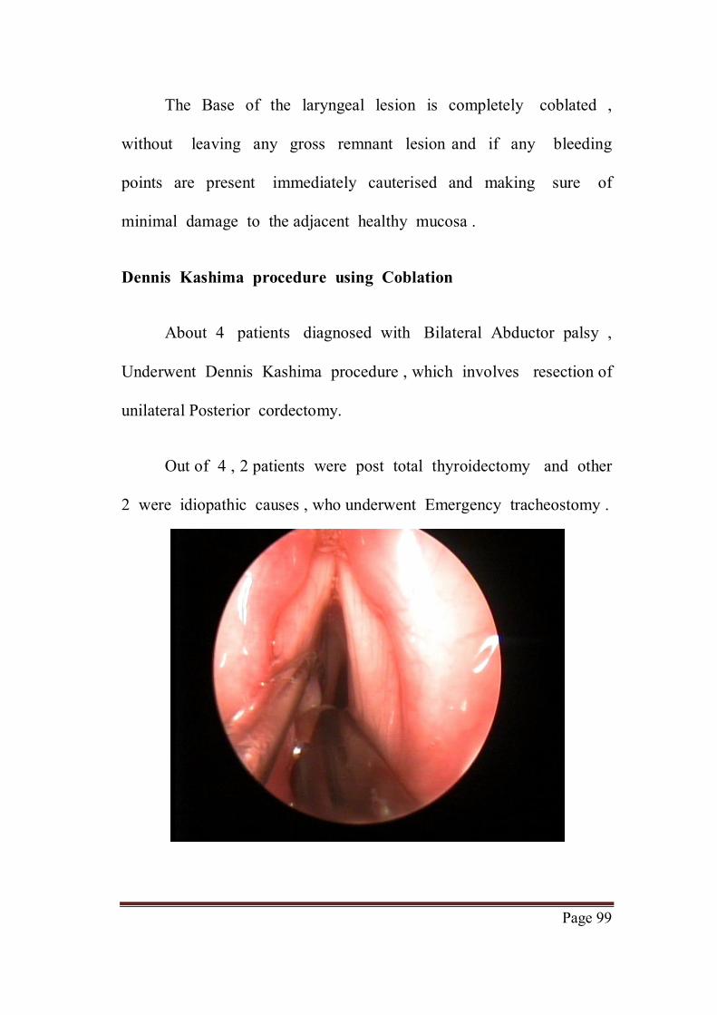
The Base of the laryngeal lesion is completely coblated ,
without leaving any gross remnant lesion and if any bleeding
points are present immediately cauterised and making sure of
minimal damage to the adjacent healthy mucosa .
Dennis Kashima procedure using Coblation
About 4 patients diagnosed with Bilateral Abductor palsy ,
Underwent Dennis Kashima procedure , which involves resection of
unilateral Posterior cordectomy.
Out of 4 , 2 patients were post total thyroidectomy and other
2 were idiopathic causes , who underwent Emergency tracheostomy .

Page 100
Fig 28,29,30 showing posterior cordotomy being done using
coblator Laryngeal wand .

Page 101
The procedure is undertaken under general anaesthesia
through the Tracheostomy tube with the patient placed in supine
position .
Patient is placed in Boyce’s position and Mouth opened wide
using Kleinsausar’s Direct Laryngoscope with Chest suspension .
The following structures are visualised successively ;
1.Uvula
2.Epiglottis
3.Arytenoids and Aryepiglottic fold
4.Vestibular folds
.Vocal cords and Anterior commissure
Now the Direct laryngoscope is fixed when both vocal cords
are visualised completely and MLE tube is pushed posteriorly.
Now, the Laryngeal Coblator Wand is introduced transorally
after setting 5 ; 3 .5 – Coblation and 3- coagulation, Saline irrigation
and suction apparatus is connected with the tubing .

Page 102
Adrenaline soaked cotton balls are placed below in the
subglottic region.
Either of the cord is selected and using coblator wand the
posterior 1/3rd of the vocal cord is resected laterally upto vocalis
muscle plane posteriorly upto the vocal process and anteriorly upto
the mid 1/3rd of the vocal cord and if any bleeding points are
present immediately cauterised and making sure of minimal
damage to the adjacent healthy mucosa .
Post operative evaluation
1.Intraoperative bleeding
2.Pain score (Day 1,3,5)
3.Days reporting pain
4.Liquid diet days
5.Post operative haemorrhage
Patient was decannulated on Day 2, after 24 hours of
tolerating spigotting .

RESULTS AND OBSERVATION

Page 103
RESULTS AND OBSERVATION
Chi square test – The chi square test for independence is used
to determine the relationship between two variable of a sample.
Independence means that the two factors are not related .
Random sampling – a random sampling of the data from a fixed
distribution or population
Fisher Exact test – The Fisher Exact test looks at a contigeny
table which displays how different interventions have produced
different outcomes. Its null hypothesis is that intervention do not affect
the outcomes – that the two are independent. P- value significant if less
than 0.05
Statistical software – The statistical software used are namely –
SAS9.2, SPSS Systat 12.0 . Graphs ,bar charts and tables were done
using Microsoft Excel.
In this study, following Coblation assisted Adenoidectomy
,patient was treated with IV antibiotics, analgesics and liquid fluids
were started initially and then gradually oral soft diet was given.

Page 104
In this study, 20 randomly selected patients, diagnosed Chronic
Adenoid Hypertrophy, who underwent Coblation Assisted
Adenoidectomy.
The following results were obtained
Ratio of Male : Female - 3:1
Cumulative percent was 75 and 25 percent for male and female
respectively.
sex
Total female male
Adenoid Grade II 1 2 3
Grade III 4 13 17
Total 5 15 20
Female, 5
Male, 15
Gender Distribution

Page 105
Value df
Asymp.Sig.
(2-sided)
Exact Sig.
(2-sided)
Pearson Chi-
Square
.131a 1 .718
Continuity
Correctionb
0.000 1 1.000
Likelihood Ratio .124 1 .725
Fisher's Exact Test 1.000
No. of
Valid Cases
20
Since the p value is greater than 0.05 ,it is not significant.
In this study ,about 17 patients out of 20 patients presented
with Aural fullness, 3 patients did not have ear complaints.
3
17
02468
1012141618
No Yes
Aural fullness

Page 106
In this study ,about 17 patients out of 20 patients presented
with Grade 3 Adenoid Hypertrophy , 3 patients had Grade 2 Adenoid
hypertrophy.
Valid percent was 15and 85 percent for Grade I and II
Adenoid hypertrophy respectively.
3
17
02468
1012141618
Grade II Grade III
Adenoid
19
1
02468
101214161820
No Yes
Post OP Bleeding

Page 107
In this study ,about 19 patients out of 20 patients had no
post operative bleeding , only 1 patients had post operative bleed
which was managed with post Nasal packing and Blood transfusion
,who recovered after 2 days .
About 95 percent was patient had no significant post operative
bleeding. Rest 5 percent had post operative bleed which was
managed with post Nasal packing and Blood transfusion
Sex
Total Female Male
Post op bleeding
No 4 15 19
yes 1 0 1
Total 5 15 20
Value df Asymp. Sig.
(2-sided) Exact Sig. (2-sided)
Pearson Chi-Square
3.158a 1 .076
Continuity Correctionb
.351 1 .554
Likelihood Ratio
2.937 1 .087
Fisher's Exact Test
.250
N of Valid Cases
20
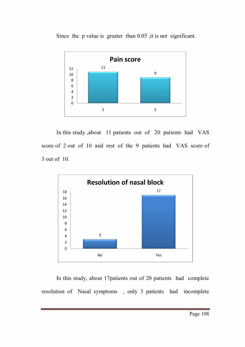
Page 108
Since the p value is greater than 0.05 ,it is not significant.
In this study ,about 11 patients out of 20 patients had VAS
score of 2 out of 10 and rest of the 9 patients had VAS score of
3 out of 10.
In this study, about 17patients out of 20 patients had complete
resolution of Nasal symptoms , only 3 patients had incomplete
119
02468
1012
2 3
Pain score
3
17
02468
1012141618
No Yes
Resolution of nasal block

Page 109
resolution of nasal block as these patients also had associated
mild septal deviation.
About 85 percent was patient had significant resolution of
Nasal Obstruction and rest of the 15 percent had partial reduction
of the Symptom.
Sex
Total Female Male
Resolution of nasal block
No 1 2 3 Yes 4 13 17
Total 5 15 20
Value df Asymp.Sig.
(2-sided) Exact Sig. (2-sided)
Pearson Chi-Square
.131a 1 .718
Continuity Correctionb
0.000 1 1.000
Likelihood Ratio .124 1 .725
Fisher's Exact Test 1.000
N of Valid Cases 20
Since the p value is greater than 0.05 ,it is not significant.

Page 110
Coblation Assisted Tonsillectomy
Ratio of Male : Female - 4:1
Cumulative percent was 80 and 20 percent for male and female
respectively.
Female, 4
Male, 16
Gender Distribution
3
17
0
2
4
6
8
10
12
14
16
18
No Yes
Sore throat

Page 111
In this study, about 17 patients out of 20 patients presented
with Sore throat, 3 patients presented mainly with complaints of
difficulty in swallowing .
In this study ,about 17 patients out of 20 patients presented
with Grade 3 Tonsillar Hypertrophy , 3 patients had Grade 2 Tonsillar
hypertrophy.
Valid percent was15and 85 percent for Grade II and III Tonsillar
hypertrophy respectively.
Sex
Total female male Tonsil size Grade
II 2 1 3
Grade III
2 15 17
Total 4 16 20
3
17
02468
1012141618
Grade II Grade III
Tonsil size

Page 112
Value df
Asymp. Sig. (2-sided)
Exact Sig. (2-sided)
Exact Sig. (1-sided)
Pearson Chi-Square
4.804a 1 .028
Continuity Correctionb
1.985 1 .159
Likelihood Ratio
3.882 1 .049
Fisher's Exact Test
.088 .088
N of Valid Cases
20
Since the p value is less than 0.05 it is Significant
In this study ,about 19 patients out of 20 patients presented
with difficulty in swallowing .
1
19
0
5
10
15
20
No Yes
Difficulty in swallowing

Page 113
About 95 percent presented with complaints of difficulty in
swallowing.
In this study, about 18 patients out of 20 patients had no post
operative bleed , only 2 patients had post operative bleed which was
managed with IV antibiotics and Blood transfusion ,who recovered
after 2 days.
About 93 percent had no post op bleeding only about 7 percent
had bleed which is significant .
Sex
Total female male
Post op bleed
No 3 15 18
Yes 1 1 2
Total 4 16 20
18
2
0
5
10
15
20
No Yes
Post OP Bleed

Page 114
Value df Asymp. Sig. 2-sided)
Exact Sig. (2-sided)
Pearson Chi-Square
1.250a 1 .264
Continuity Correctionb
.035 1 .852
Likelihood Ratio 1.023 1 .312
Fisher's Exact Test
.368
N of Valid Cases 20
Since the p value is greater 0.05 ,it is not significant.
In this study, about 11 patients out of 20 patients had VAS score
of 2 out of 10 and rest of the 9 patients had VAS score of 3 out
of 10.
Over all on the average pain score was about 2.4
11
9
0
2
4
6
8
10
12
2 3
Pain score D1

Page 115
Sex
Total female male
Pain score D1
2 3 8 11
3 1 8 9
Total 4 16 20
Value df
Asymp. Sig.
(2-sided) Exact Sig. (2-sided)
Pearson Chi-Square
.808a 1 .369
Continuity Correctionb
.114 1 .736
Likelihood Ratio .846 1 .358
Fisher's Exact Test
.591
N of Valid Cases 20
14
6
0
2
4
6
8
10
12
14
16
3 4
Normal diet (day)

Page 116
In this study, about 14 patients out of 20 patients had returned to
normal diet with minimal throat discomfort on post operative Day 3.
Rest of the 6 patients had returned to normal diet on post
operative Day 4. About 70 percent of patient s returned to normal diet
on Day 3.
Descriptives
Descriptive Statistics
N Mean Std.
Deviation
Age 20 13.20 2.093
Time taken(mins)
20 21.80 3.488
Intra op bleed (ml)
20 12.80 3.286
Valid N (listwise)
20
Snoring
Female, 1
Male, 4
Gender Distribution

Page 117
Ratio of Male : Female - 4:1
Cumulative percent was 80 and 20 percent for male and female
respectively.
In this study, about 3 patients out of 5 patients presented with
Grade II Tonsillar Hypertrophy, 1 patient each had Grade I and III
Tonsillar hypertrophy respectively.
Valid percent was 20, 60, 20 percent for Grade I, II and III
Tonsillar hypertrophy respectively
female male
Tonsillar hypertropy
Grade I 3 4 Grade II 2 7 Grade III 1 3
Total 6 14
1
3
1
0
2
4
6
8
10
Grade I Grade II Grade III
Tonsillar Hypertropy

Page 118
Chi-Square Tests
Value df Asymp. Sig.
(2-sided)
Pearson Chi-Square
.858a 2 .651
Likelihood Ratio
.840 2 .657
N of Valid Cases
20
In this study, about 4 patients out of 5 patients presented with
complaints of snoring, only 1 patient presented with Increased day
time sleepiness.
1
4
0
5
10
15
20
No Yes
Snoring

Page 119
In this study, about 4 patients out of 5 patients had complete
resolution of symptom of snoring, only 1 patient had minimal snoring.
About 80 -90 percent was patient had significant resolution
of snoring. Rest 10-20 percent, had only partial reduction in the
symptom due to multilevel obstruction.
Sex
Total female male
snoring No 0 3 3
Yes 6 11 17
Total 6 14 20
1
02468
101214161820
Absent Yes
Post OP Snoring

Page 120
Value df
Asymp. Sig. (2-sided)
Exact Sig. (2-sided)
Pearson Chi-Square
1.513a 1 .219
Continuity Correctionb
.299 1 .585
Likelihood Ratio 2.360 1 .124
Fisher's Exact Test
.521
Descriptive Statistics
N Minimum Maximum Mean Std. Deviation
Age 20 28 47 37.40 5.960
T-Test
20.50
6.35
0.00
5.00
10.00
15.00
20.00
25.00
Pre OP Post OP
ESS

Page 121
Paired Samples Statistics
Mean N Std. Deviation
Std. Error Mean ESS
Pair 1 ESS pre op 20.50 20 2.164 .484 Pre OP
20.50
ESS post op
6.35 20 1.843 .412 Post OP
6.35
Paired Samples Test
Pair 1
ESS pre op - ESS post op
Paired Differences
Mean 14.150
Std. Deviation 2.870
Std. Error Mean .642
95% Confidence Interval of the Difference
Lower 12.807
Upper 15.493
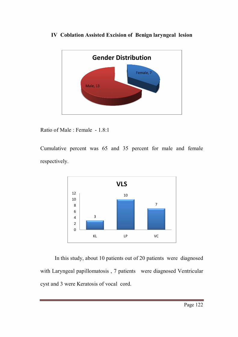
Page 122
IV Coblation Assisted Excision of Benign laryngeal lesion
Ratio of Male : Female - 1.8:1
Cumulative percent was 65 and 35 percent for male and female
respectively.
In this study, about 10 patients out of 20 patients were diagnosed
with Laryngeal papillomatosis , 7 patients were diagnosed Ventricular
cyst and 3 were Keratosis of vocal cord.
Female, 7
Male, 13
Gender Distribution
3
10
7
02468
1012
KL LP VC
VLS

Page 123
Valid percent was 35, 50, 15 percent for Keratosis, Laryngeal
papillomatosis and Ventricular cyst respectively.
Sex
Total female male
snoring No 0 3 3 Yes 6 11 17
Total 6 14 20
Value df Asymp.Sig. (2-sided)
Exact Sig. (2-sided)
Pearson Chi-Square
1.513a 1 .219
Continuity Correctionb
.299 1 .585
Likelihood Ratio
2.360 1 .124
Fisher's Exact Test
.521
Since p-value is greater 0.05 ,it is not significant.

Page 124
In this study, about 16 patients out of 20 patients had localised
disease, glottis chink was adequate and were not in stridor and rest
of the 4 patients were in stridor who required elective
Tracheostomy.
On the average of about 80 percent did not require
Tracheostomy and 20 percent required .
Sex
Total Female Male
stridor Absent 4 12 16 Present 3 1 4
Total 7 13 20
16
4
02468
1012141618
Absent Present
Stridor

Page 125
Chi-Square Tests
Value df
Asymp. Sig. (2-sided)
Exact Sig. (2-
sided)
Exact Sig. (1-
sided)
Pearson Chi-Square
3.516a 1 .061
Continuity Correctionb
1.662 1 .197
Likelihood Ratio 3.404 1 .065
Fisher's Exact Test
.101 .101
N of Valid Cases
20
In this study ,about 10 patients out of 20 patients had VAS
score of 3 and below and rest of the patients had VAS score of
5. About 50 percent of the patients had VAS score of less than 4.
3
7
5 5
012345678
2 3 4 5
Pain score

Page 126
In this study, about 16 patients out of 20 patients had localised
disease, glottis chink was adequate and were not in stridor and rest
of the 4 patients were in stridor who required elective tracheostomy
In this study, about 17 patients out of 20 patients had no disease
recurrence during the study period of 2 years and rest 3 patients
diagnosed laryngeal papillomatosis, had recurrence who were treated
with revision surgery with coblation .
16
4
0
5
10
15
20
No Yes
Tracheostomy needed
17
3
02468
1012141618
No Yes
Recurrence

Page 127
About 85 percent had no significant recurrence of the
disease, Three out of the 10 patients of laryngeal papillomatosis
patient had Local recurrence of the disease and required revision
surgery.
Descriptives
Descriptive Statistics
N Mean Std.
Deviation
Age 20 30.75 9.547 Blood loss in ml 20 12.70 2.430
Valid N (listwise) 20

DISCUSSION
Page 128
DISCUSSION
A cross sectional descriptive study was conducted from a period
of February 2013 to September 2014 in our department of
Otorhinolaryngology.
In this study, 40 randomly selected patients, diagnosed
Chronic Adenotonsillitis, underwent Coblation Assisted
AdenoTonsillectomy ,
Subjects who had past history co morbid illness like diabetes
mellitus, hypertension, ischemic heart disease, pulmonary tuberculosis,
obesity, malignancy,bleeding disorder were excluded from the study.
In this study all subjects were in the age group between 10 to 45
years. None had significant past or family history .
In our study ,Cumulative percent was 75 and 25 percent for
male and female respectively. Valid percent was 15 and 85 percent
for Grade II and III Tonsillar hypertrophy respectively. About 70
percent of patient s returned to normal diet on Day 3.About 93 percent
was patient had no significant post operative bleeding .Rest 7 percent

Page 129
had post operative bleed .Over all on the average pain score was
about 2.4.
The coblation tonsillectomy took on an average of 32
minutes. The study protocol was approved by the UCLA
Institutional Review Board.
Children ages 2 to 16 years undergoing outpatient
adenotonsillectomy were offered participation in this prospective trial.
Postoperative outcome measures included postoperative pain, time
taken In conclusion, the overall advantages of Coblation
adenoidectomy, are the decrease in intra- and post-operative bleeding,
better safety, precision of adenoid removal and less injury to adjacent
tissues.
Shah et al (27) National Prospective Tonsillectomy Audit study
showed. Mean pain level on postoperative day (POD) 1 based on a
visual analogue scale of 1 to 10. Average pain score was 3 in their
study The coblation tonsillectomy took on an average of 20-24
minutes.

Page 130
The studies have shown that the standard operation with
blood vessels tied off was most likely to result in someone
needing more surgery because of bleeding in the first 24 hours
A recent national audit (called the National Prospective
Tonsillectomy Audit) collected information concluded that coblation
almost 1 in 100 patients had reactionary haemorrhage due to
coblation.
Chang et al study(30) done in 2004 showed in children mean
pain score was 2 post coblation tonsillectomy percentage of children
return back to normal diet was 80% by D4 post coblation
tonsillectomy.
Stoker et al(27), 2004 performed similar study
Snoring
In this study, 5 randomly selected patients, diagnosed Mild
Obstructive sleep Apnea syndrome , underwent Coblation Assisted
uvulopalatopharyngoplasty .

Page 131
Subjects who had past history co morbid illness like diabetes
mellitus, hypertension, ischemic heart disease, pulmonary tuberculosis,
obesity, malignancy, bleeding disorder were excluded from the study.
Cumulative percent was 80 and 20 percent for male and
female respectively.
About 80 percent had history of significant snoring ,rest of 20
percent presented with increased day time sleepiness.
About 80 -90 percent was patient had significant resolution
of snoring .
Rest 10-20 percent , had only partial reduction in the symptom
due to multilevel obstruction.ESS score from 20 it decreased to 6.
In 1997, Finkelstein and et al(57)studied in about 39 randomly
selected patients, diagnosed as Mild Obstructive sleep apnea syndrome
showed significant improvement ESS score from 18 it decreased to
7 and Symptomatic decrease in snoring .
In 2000, Madani. M and et al (58) studied in about 63 randomly
selected patients, diagnosed as Mild Obstructive sleep apnea syndrome
showed significant improvement ESS score and decrease in snoring

Page 132
Rapid Recovery .
Benign laryngeal lesions
In this study, 20 randomly selected patients , diagnosed Benign
laryngeal Lesions.Subjects who had past history co morbid illness like
diabetes mellitus, hypertension, ischemic heart disease, pulmonary
tuberculosis,obesity, malignancy, bleeding disorder were excluded
from the study.
Valid percent was 35,50,15 percent for Keratosis, Laryngeal
papillomatosis and Ventricular cyst respectively. On the average of
about 80 percent did not require Tracheostomy and 20 percent
required .About 85 percent had no significant recurrence of the
disease, About 50 percent of the patients had VAS score of less
than 4.Operation time 18 min . Blood loss was about 15 mL.
Three out of the 10 patients of laryngeal papillomatosis
patient had Local recurrence of the disease and required revision
surgery.
In 2011 , In china Qingfeng Zhang, DeLong Liu, Cuiping She
(20) Studied 21 cases with laryngeal papilloma treated by Coblation.

Page 133
Operation time 11 min . Blood loss was about 13 mL The
follow-up was carried out from 1 to 3 years and 9 months and there
were 4 cases of recurrence with the average recurrence interval of seven
months In 2007 , Timms MS ,Bruce IA and et al (18)Studied 21 cases
with Benign laryngeal lesion treated by Coblation. Operation time 20
min . Blood loss was about 24 mL
The follow-up was carried out from 1 to 3 years and had
recurrence cases .

SUMMARY

Page 134
SUMMARY
A cross sectional descriptive study was conducted from a period
of February 2013 to September 2014 in our department of
Otorhinolaryngology.
About 65 cases which were selected randomly, based on the
inclusion and exclusion criteria, were sub divided based on the
diagnosis .
Out of which 20 were Chronic Adenoid Hypertrophy, 20 were
chronic Tonsillitis, 5 were mild Obstructive sleep apnea syndrome,
20 were Benign Laryngeal lesions.
Each of these, underwent their respective surgeries Coblation
assisted Adenoidectomy ,Coblation Assisted Tonsillectomy ,Coblation
Assissted Uvulopalatopharyngoplasty and Coblation assisted excision
of Benign Laryngeal lesions Coblation Assisted Adenoidectomy
ensures complete and safe removal of adenoid tissue ,decreased post
operative pain ,Early return to normal diet Coblation Assisted
Tonsillectomy ensures complete Extracapsular issection of tonsil
decreased post operative pain ,Early return to normal diet .

Page 135
The main advantages are:
i) Bleeding is minimal intra operatively and post operatively
and which can be immediately coagulated in case of any active
bleeding .
ii) Direct endoscopic visualisation ensures complete excision.
iii) Able to access all the areas due usage small tip used for
dissection.
iv) Absent significant residual tissue.
v) fewer complications – injury to surrounding tissues.
vi) No age limitation
vii) requirement of post-operative drug consumption is
decreased
viii) a reduction in the loss of academic performance due to
faster post-surgical healing .
The main disadvantages are
1.Cost of the Instrument and wand

Page 136
2. Single Use Wand
3 .Separate Wand for each surgery - Tonsillar wand ,Laryngeal
wand .
4.Time taken for surgical dissection is more .
5.Post operative bleed is about 6-8 percent .
6.Nasal decongestion is required for Coblation assisted
Adenoidectomy

CONCLUSION

Page 137
CONCLUSION
In this study ,about 65 cases were selected randomly, Out of
which 20 were Chronic Adenoid Hypertrophy ,underwent Coblation
assisted Adenoidectomy ,20 were chronic Tonsillitis who underwent
Coblation Assisted Tonsillectomy ,5 were mild Obstructive sleep
apnea syndrome underwent Uvulopalatopharyngoplasty, 20 were
Benign Laryngeal lesions who underwent Coblation assisted excision
had complete excision of the lesions with safety , with minimal post
operative pain,with minimal injury to the surrounding healthy
tissue, intraoperative bleed, early recovery to normalcy.
About 3 out of 40 patients had post operative bleeding,
following Coblation Assisted Adenotonsillectomy, Time consuming
,requires expertise and less cost effective
However , the sample size is too small to come to definitive
conclusion.

ANNEXURES

A.BIBLIOGRAPHY

A. BIBILOGRAPHY
1. Stradling J, Crosby J. Predictors and prevalence of obstructive Sleep apnoea and snoring in 1001 middle-aged men .Thorax 1991; 46: 85—90.
2. De Backer W et al. Intraluminal pressures during central apnea compared to critical closing pressures. Sleep Research 1996; 25: 231.
3. Gavriely N, Jensen O. Theory and measurement of snores. Journal of Applied Physiology 1993; 74: 2828—2837.
4. Croft C, Pringle M.Sleep nasoendoscopy: a technique of assessment in snoring and obstructive sleep apnoea. Clinical Otolaryngology 1991; 16: 504—509.
5. Schwartz A-R et al. Effect of positive nasal pressure on upper airway pressure—flow relationships. Journal of Applied Physiology 1989; 66: 1626—1634.
6. Stoohs R, Guilleminault C. Snoring during NREM sleep: respiratory timing, esophageal pressure and EEG arousal. Respiratory Physiology 1991; 85: 151—167.
7. Skatrud J, Dempsey J. Airway resistance and respiratory muscle function in snorers during NREM sleep. Journal of Applied Physiology 1985; 59: 328—335.
8. Perez-Padilla J, West P, Kryger M. Snoring in normal young adults: prevalence in sleep stages and associated changes in oxygen saturation, heart rate and breathing pattern. Sleep 1987; 10: 249—253.
9. Lugaresi E et al. Breathing during sleep in man in normal and pathological conditions. Advances in Experimental Medicine and Biology 1978; 99: 35—45.
10. Waller P, Bhopal R. Is snoring a cause of vascular disease: an epidemiological review. Lancet 1989; (1): 143—146.

11. Schmidt-Nowara W et al. Snoring in a Hispanic-American population: risk factors and association with hypertension and other morbidity. Archives of Internal Medicine 1990; 50: 597—601.
12. Exar E, Collop N. The upper airway resistance syndrome. Chest 1999; 115(4): 1127—1139.
13. Urschitz M et al. Snoring, intermittent hypoxia and academic performance in primary schoolchildren. American Journal of respiratory and critical care medicine 2003; 168: 464—468.
14. Wiggins CL et al. Comparison of self and spouse reports of snoring and other symptoms associated with sleep apnea syndrome. Sleep 1990; 13
15. Hoffstein V, Mateika S, Anderson D. Snoring: is it in the ear of the beholder? Sleep 1994; 17: 522—526.
16. Scott S et al. A comparison of physician and patient perception of the problems of habitual snoring. Clinical Otolaryngology 2003; 28(1): 18—21.
17. Shehab N, Sweet BV, Hogikyan N D. Cidofovir for the treatment of recurrent respiratory papillomatosis: a review of the literature [J]. Pharmacotherapy, 2005, 25 (7): 977-989.
18. Timms MS, Bruce IA, Patel N K. Radiofrequency ablation (coblation): a promising new technique for laryngeal papillomata [J]. J Laryngol Otol, 2007,121 (1) :28-30.
19. Carney AS, Evans AS, Mirza S, et al. Radiofrequency coblation for treatment of advanced laryngotracheal recurrent respiratory papillomatosis Laryngol Otol, 2010, 124 (5) :510-514.
20. She Cuiping, ZHANG Qing-feng, Cheng Chenjing low temperature radiofrequency treatment of adult laryngeal papilloma [J]. Otorhinolaryngology Head and Neck Surgery, 2011,46 (4) :336-338.
21. Anthony R, Wallace H, Frewer J, Varlow J, Smelt GJC. Coblation tonsillectomy compared with conventional dissection and tie technique - a randomised trial.

22. Back L, Paloheimo M, Ylikoski J. Traditional tonsillectomy compared with bipolar radiofrequency thermal ablation tonsillectomy in adults: a pilot study.
23. Jayasinghe H, Lee P, Williams A, Kerr AIG. Randomised single blinded trial of outcome for coblation versus cold steel tonsillectomy. Emailed presentation 2005.
24. Parsons SP, Cordes SR, Comer B. Comparison of posttonsilectomy pain using the ultrasonic scalpel, coblator, and electrocautery. 2006;134:106–13.
25. Philpott CM, Wild DC, Mehta D, Daniel M, Banerjee AR. A double-blinded randomized controlled trial of coblation versus conventional dissection tonsillectomy on postoperative symptoms. Clinical Otolaryngology 2005;30(2): 143–8.
26. Shah UK, Galinkin J, Chiavacci R, Briggs M. Tonsillectomy by means of plasma-mediated ablation: prospective, randomized, blinded comparison with monopolar electrosurgery. Archives of Otolaryngology - Head and Neck Surgery 2002;128(6):672–6.
27. Stoker KE, Don DM, Kang DR, Haupert MS, Magit A, Madgy DN. Pediatric total tonsillectomy using coblation compared to conventional electrosurgery: a prospective, controlled single-blind study. Otolaryngology - Head and Neck Surgery 2004;130(6):666–75.
28. Temple RH, Timms MS. Paediatric coblation tonsillectomy. International Journal of Pediatric Otorhinolaryngology 2001; 61(3):195–8.
29. Arya A, Donne AJ, Nigam A. Double-blind randomized controlled study of coblation tonsillotomy versus coblation tonsillectomy on post-operative pain. Clinical Otolaryngology 2003;28:503–6.
30. Chan KH, Friedman NR, Allen GC, Yaremchuk K, Wirtschafter A, Bikhazi N, Bernstein JM, Kelley PE, Lee KC. Randomized, controlled, multisite study of intracapsular tonsillectomy using low-temperature plasma excision. Archives of Otolaryngology - Head and Neck Surgery 2004;130(11):1303–7.

31. Chang KW. Randomized controlled trial of Coblation versus electrocautery tonsillectomy. Otolaryngology – Head and Neck Surgery 2005;132(2):274–80.
32. Hall DJ, Littlefield PD, Birkmire-Peters DP, Holtel MR. Radiofrequency ablation versus electrocautery in tonsillectomy. Otolaryngology - Head and Neck Surgery 2004;130(3):300–5.
33. Littlefield PD, Hall DJ, Holtel MR. Radiofrequency excision versus monopolar electrosurgical excision for tonsillectomy. Otolaryngology - Head and Neck Surgery 2005;133:51–4.
34. Noordzij JP, Affleck BD. Coblation versus unipolar electrocautery tonsillectomy: a prospective, randomized, single-blind study in adult patients. The Laryngoscope 2006;116: 1303–9.
35. Patel J, Mandal S, Rachmanidou, A. Paediatric coblation tonsillectomy versus dissection tonsillectomy: a comparative study of post-operative pain and complications. International Journal of Paediatric Otorhinolaryngology 2004; 68(5):725-6 (Abstract No. L.5.2)
36. Polites N, Joniau S, Wabnitz D. Fassina R, Smythe C, Varley P, Carney AS. Postoperative pain following coblation tonsillectomy: randomized clinical trial. ANZ Journal of Surgery 2006;76(4):226–9.
37. Saengpanich S, Kerekhanjanarong V, Aramwatanapong P,Supiyaphun P. Comparison of pain after radiofrequency tonsillectomy compared with conventional tonsillectomy: a pilot study. Journal of the Medical Association of Thailand 2005;88(12):1880–3.
38. Timms MS, Temple RH. Coblation tonsillectomy: a double blind randomized controlled study. The Journal of Laryngology and Otology 2002;116(6):450–2.
39. Di Rienzo Businco L, D’Emilia M, Lauriello M, Coen Tirelli G. Coblation tonsillectomy versus dissection in adults: comparison of techniques. XVIII IFOS World Congress, Rome, Italy, 2005, 25-30 June. XVIII IFOS World Congress. 2005.

40. Trotter M. The effect of different surgical techniques on postoperative morbidity following tonsillectomy. Laryngology and Otology 2003;117(29 (Suppl)):45 (Abstract No. HN08).
41. Walner DL. Pediatric tonsillectomy: coblation vs electrocautery. Otolaryngology - Head and Neck Surgery 2004;131 (Suppl 2):P83.
42. Zhong Z, Xiao SF, Wang C, Wang H, Wang G. Coblation tonsillctomy versus blunt dissection tonsillectomy. Journal of Clinical Otorhinolaryngology 2006;2(9):391–5.
43. Parker D. Comparative study of pain levels following paediatric tonsillectomy using either conventional tonsillectomy instruments or the coblator device for tonsil removal. National Research Register 2006. [: ISRCTN07513663]
44. Ballantyne John C, Harrison DFN (eds). Rob & Smith’s Operative Surgery: Nose and Throat. 4th Edition. London: Butterworths, 1986.
45. British Association of Otorhinolaryngologists - Head and Neck Surgeons Comparative Audit Group & Clinical Effectiveness Unit, The Royal College of Surgeons of England. National Prospective Tonsillectomy Audit: FINAL REPORT of an audit carried out in England and Northern Ireland between July 2003 and September 2004. http://www.rcseng.ac.uk/publications/docs/national_prospective.html/view?searchterm=tonsillectomy%20auditMay 2005.
46. Bergler W, Huber K, Hammerschmitt N, Hormann K. Tonsillectomy with argon plasma coagulation (APC): evaluation of pain and hemorrhage. The Laryngoscope 2001; 111(8):1423–9.
47. Cowan DL, Hibbert J. Acute and chronic infections of the pharynx and tonsils. Scott-Brown’s Otolaryngology. 6th Edition. Vol. 5, Oxford, Boston: Butterworth-Heinemann, 1997.
48. Hannington-Kiff JG. The need for analgesic cover after ENT surgery - comparison of nefopam and papaveretum. Anaesthesia 1985;40(1):76–8.
49. Higgins JPT, Green S (eds). Cochrane Handbook for Systematic Reviews of Interventions 4.2.6 [updated September 2006]. In: The

Cochrane Library, Issue 4, 2006. Chichester, UK: John Wiley & Sons, Ltd., 2006.
50. Husband AD, Davis A. Pain after tonsillectomy. Clinical Otolaryngology and Allied Sciences 1996;21(2):99–101.
51. Maddern BR. Electrosurgery for tonsillectomy. The Laryngoscope 2002;112(8 Pt 2 Suppl 100):11–3.
52. Pinder D, Hilton M. Dissection versus diathermy for tonsillectomy. Cochrane Database of Systematic Reviews 2004, Issue 3. [Art. No.: CD002211. DOI: 10.1002/14651858.CD002211]
53. Scott brown 7th edition otorhinolaryngology, head and neck surgery.
54. Cummings otorhinolaryngology 5th edition Bailey, Byron, Johnson, Newslands – Head and neck surgery – Otorhinolaryngology 4th Edition volume 1 .
55. Coblation tonsillectomy by Dr.T.Balasubramanian M.S.D.L.O – drtbalu otolaryngology online
56. Phipott CM ,Wild DC ,Mehta D ,Banerjee , A double blinded Randomised control trial ,coblation tonsillectomy verus conventional dissection tonsillectomy .
57. Finkelstein Y, Shapiro-Feinberg M, Stein G, Ophir D. Uvulopalatopharyngoplasty vs. laser-assisted uvulopalatoplasty: anatomical considerations. Arch Otolaryngology
58. Madani M. Radiofrequency somnoplasty: a new treatment for snoring and sleep apnea. Int J Oral and Maxillofac Surg 1999;28(1).

B. PROFORMA
B. PROFORMA
Name ; Age /sex : I.P.no : D.O.A: D.O.S D.O.D: Address : CHIEF COMPLAINTS THROAT PAIN DIFFICULTY IN SWALLOWING SNORING MOUTH BREATHING ANY OTHER ASSOCIATED SYMPTOMS HOARSENESS OF VOICE INSOMNIA INCREASED DAY TIME SLEEPINESS HISTORY OF PRESENT ILLNESS : Complete detailed description of each symptoms, aggravating and relieving factors, Associated symptoms. Negative history

PAST HISTORY : Any H/O DM /Epilepsy /HTN /PTB /Drug allergy /bleeding diathesis /Cardiac disease /Asthmatic Chronic Drug use /any other medical comorbidities . FAMILY HISTORY PERSONAL HISTORY Diet Bladder and bowel habits Addiction . GENERAL PHYSICAL EXAMINATION Built , mental status . Pallor Icterus Cyanosis Clubbing Pedal edema General Lymphadenopathy . VITALS Pulse Rate : Blood Pressure: Respiratory Rate: Saturation : Jugular Venous Pressure : SYSTEMIC EXAMINATION Cardio Vascular System: Respiratory System;

Per Abdomen: Central Nervous System: ENT EXAMINATION ORAL CAVITY AND OROPHARYNX EXAMINATION Lips, teeth, gums, buccal mucosa, Buccogingival sulcus , Retro Molar Trigone , hard palate, Anterior 2/3 rd tongue , floor of mouth, Anterior pillar, tonsils, soft palate, uvula, posterior pillar, posterior Pharyngeal wall. Indirect Laryngoscopy – Base of Tongue , Vallecula , Epiglottis , Aryepiglottic fold ,
Arytenoids, Vestibular bands, Vocal cords, Glottic chink, tracheal
rings, pyriform fossa.
NOSE
External Nose
Vestibule
Anterior Rhinoscopy
Right Left
Inferior Meatus
Inferior Turbinate
Middle Meatus
Middle turbinate

Airway patency tests
Right Left
Cottle’s test
Cotton wool test
Cold spatula test
Post nasal Examination
EAR EXAMINATION
Right Left
External Ear
External Auditory Meatus
Tympanic Membrane TUNING FORK TESTS
Facial Nerve Examination
Diagnostic Nasal Endoscopy
Videolaryngoscopy
Investigations
Complete Blood Count
Renal Function Tests
Liver Function Tests
ELISA –HIV /HbsAg
Blood group and Rh typing
Bleeding Time /Clotting Time
Urine analysis
ECG
Chest x ray- PA view
X ray - Nasopharynx soft tissue lateral view

B. PROFORMA

C. ETHICAL COMMITTEE
APPROVAL LETTER

E. INFORMED CONSENT FORM

C. ETHICAL COMMITTEE APPROVAL LETTER

D. PATIENT INFORMATION
SHEET

D. PATIENT INFORMATION SHEET
ப ேக பவ ெபய ம வலாச :
ஆ வாள ைகெயா ப : நா :
ஆ வாள ெபய : இட :
தகவ ப வ
த க ெச த ப ேசாதைனக ல த க
ேநா உ ள ெத யவ ள . இ த ேநாைய
ண ப த பலவைக அ ைவசிகி ைச ைறக உ ளன.
அதி உ க .
ைற பய ப த பட ள . இ த அ ைவ சிகி ைசய
(coblation) பய ப த பட ள . ேம இ த ைறய
ல ஏ ப வைள ைள ஒ பட ஆ ேம ெகா ள பட
உ ள . இ றி த உ கள ேநா ம அத
வவர கைள ஆ வ பய ப த வ கிேறா . தா க
வ பனா ம வ ஆ வலி எ ெபா
ேவ மானா வலகி ெகா ளலா . எ த ச டசி க
இடமி லாம எ ெபா ேவ மானா த க
ஆ வலி வலகி ெகா ளலா . இ த ஆ வ ல
கிைட தகவ க ப ேசாதைன க த கள
ஒ தலி ல ம ஆ வ பய ப த ப .
ஆ வாள ைகெயா ப :
ஆ வாள ெபய :
இட :
நா :

E. INFORMED CONSENT FORM
ஆரா சி நிைலய : கா , , ெதா ைட ப
டா லி அர ெபா ம வமைன ம ம வ
க
ப ெப பவ ெபய :
ப ெப பவ எ :
ம வ ஆ வ வவர க என
வள க ப ள . என ேநா
ம அத நிைல ெத யப த ப ட . என ேநா ப றிய
ச ேதக கைள ேக க அத கான த த வள க கைள
ெபற வா பள க ப ட . இ த ேநாைய ண ப
அ ைவ சிகி ைச ைறக .
வள க ப ள . அ த அ ைவ சிகி ைசய (coblation)
பய ப த ப வ ப றி வள க ப ட . இைத என அ ைவ
சிகி ைசய ேபா பய ப த யநிைன ட ச மதி கிேற .
இ த அ ைவ சிகி ைசய வைள கைள ஆ வ
பய ப த த ன ைசயாக ச மதி கிேற .
எ காரண தினா எ த க ட தி எ த ச ட சி க
உ படாம இ வா வ இ வலகி ெகா ளலா எ
அறி ெகா ேட .
இ த ஆ வ ல கிைட தகவ கைள
ப ேசாதைன கைள ம வ ேம ெகா
ஆ வ பய ப தி ெகா ள அைத ப ர க
ேதைவ ப டா எ ைன என நட அ ைவ
சிகி ைசைய ைக பட எ க நா மன ட
ச மதி கிேற .
ப ேக பவ ைகெயா ப : நா :
க ைட வர ஒ ப இட :

F. PLAGIARISM

F. PLAGIARISM

G. MASTER SHEET

MASTER CHART
COBLATION ASSISSTED ADENOIDECTOMY Sl No
Age Sex IP number
Adenoid
hypertropy
Nasal
block
Aural
fullness
Post OP
Bleeding
Pain score
Resolution of nasal block
Recurrence
1. 11 male 156902 Grade III Yes Yes No 3 Yes No
2. 12 female 165527 Grade III Yes Yes No 2 Yes No
3. 11 male 140315 Grade III Yes Yes No 3 Yes No
4. 13 male 373341 Grade III Yes Yes No 2 Yes No
5. 11 male 179220 Grade III Yes Yes No 3 Yes No
6. 13 male 140270 Grade III Yes Yes No 2 Yes No
7 12 female 373341 Grade III Yes Yes No 2 Yes No
8 11 male 160027 Grade III Yes Yes No 3 Yes No

9 13 male 199628 Grade II No No No 2 Yes No
10 12 male 280918 Grade III Yes Yes No 2 Yes No
11 12 male 164950 Grade III Yes Yes No 2 Yes No
12 11 male 108697 Grade III Yes Yes No 3 Yes No
13 11 male 192290 Grade II No Yes No 3 Yes No
14 12 female 168747 Grade III Yes Yes No 3 Yes No
15 13 female 190172 Grade III Yes Yes No 2 Yes No
16 11 male 164783 Grade II No Yes No 3 Yes No
17 12 male 132992 Grade III Yes Yes No 2 Yes No
18 12 male 742301 Grade III Yes Yes No 2 Yes No
19 11 male 167663 Grade III Yes Yes No 3 Yes No
20 13 female 161593 Grade III Yes Yes No 2 Yes No

COBLATION ASSISSTED TONSILLECTOMY
Sl
No
Age Sex IP number
Tonsillar
hypertropy
Sore
throat
Difficulty in
Swallowing
Post OP
Bleeding
Pain score
Return to
Normal diet (day)
Time
Taken
(min)
1. 14 Male 142970 Grade III Yes Yes No 3 4 18
2. 15 Female 101699 Grade III Yes Yes No 2 3 22
3. 11 Male 140393 Grade III Yes Yes No 3 3 19
4. 16 Male 307861 Grade III Yes Yes No 2 3 23
5. 11 Male 307932 Grade III Yes Yes No 3 3 20
6. 13 Male 307881 Grade III Yes Yes No 2 3 18
7 12 Male 186337 Grade III Yes Yes No 2 3 20
8 11 Male 307908 Grade III Yes Yes No 3 4 22
9 15 Male 168633 Grade II No No No 2 3 29

10 12 Female 307877 Grade III Yes Yes No 2 4 18
11 12 Male 117753 Grade III Yes Yes No 2 3 24
12 16 Male 230217 Grade III Yes Yes No 3 3 22
13 11 Male 134062 Grade II No Yes No 3 4 24
14 17 Female 1403170 Grade III Yes Yes No 3 3 19
15 13 Male 221380 Grade III Yes Yes No 2 3 25
16 17 Male 161721 Grade II No Yes No 3 3 30
17 12 Male 183941 Grade III Yes Yes No 2 3 23
18 12 Male 198447 Grade III Yes Yes No 2 3 18
19 11 Male 157930 Grade III Yes Yes No 3 4 23
20 13 Female 148103 Grade III Yes Yes No 2 3 24

COBLATION ASSISTED UVULOPALATOPLASTY
Sl No
Age Sex IP Number
Tonsillar
hypertropy
Snoring Difficulty in
Swallowing
Post op
Snoring
Pain score
Return to
Normal diet (day)
scarring ESS Score (Pre OP)
ESS Score (Post OP)
1. 34 Male 962120 Grade I Yes Yes Absent 3 4 No 18 6
2. 32 Female 396186 Grade II Yes Yes Absent 2 5 No 19 7
3. 45 Male 178729 Grade III Yes Yes Absent 3 5 No 18 6
4. 28 Male 308120 Grade I Yes Yes Absent 4 6 No 24 7
5. 40 Female 205551 Grade I Yes Yes Absent 3 3 No 21 6

COBLATION ASSISSTED EXCISION OF BENIGN LARNYGEAL LESION
Sl No
Age Sex IP Number Change of voice
VLS stridor Tracheostomy
needed
Pain score
Voice improvement
Recurrence
1. 33 Male 141436 present Larngeal
Papilloma
Absent No 3 Present No
2. 40 female 242361 Present Ventricular cyst
Present Yes 2 Present No
3. 42 male 280558 Present Larngeal
Papilloma
Present Yes 3 Present No
4. 16 male 243201 Present Larngeal
Papilloma
Absent No 5 Present No
5. 28 male 196502 Present Ventricular cyst
Absent No 3 Present No

6. 33 female 177376 Present Larngeal
Papilloma
Absent No 5 Present yes
7 41 male 5228443 Present Ventricular cyst
Absent No 4 Present No
8 45 female 229905 Present Ventricular cyst
Absent No 3 Present No
9 28 male 250892 Present Valleculacyst Absent No 5 Present No
10 29 female 155001 Present Larngeal
Papilloma
Present Yes 4 Present No
11 18 male 205840 Present Ventricular cyst
Absent No 2 Present No
12 36 male 144198 Present Valleculacyst Absent No 3 Present No
13 19 male 312116 Present Larngeal
Papilloma
Absent No 4 Present yes

14 17 female 464421 present Larngeal
Papilloma
Absent No 3 Present No
15 28 male 997212 Present Ventricular cyst
Absent No 5 Present No
16 17 male 731091 Present Larngeal
Papilloma
Absent No 3 Present No
17 29 female 159073 Present Vallecular cyst
Absent No 4 Present No
18 33 male 170482 Present Larngeal
Papilloma
Absent No 2 Present No
19 39 male 724081 Present Ventricular cyst
Absent No 4 Present No
20 44 female 315730 present Larngeal
Papilloma
Present Yes 5 Present No