epipsoc·etae (psocoptera) from the reserva ducke, amazonas
TRANSCRIPT

Epipsoc·etae (Psocoptera) from the Reserva Ducke, Amazonas
Abstract
31 species of Epipsocetae are recorded from the Reserva Ducke, near Manaus (Brazil), of which 27 are described as new, and illustrated . The new taxa repre. sent the genera Ptiloneura (1 ), Triplocania (8), Euplocania (2), lsthmopsocus (1), Neurostigma (4), Dicropsocus (1), Eplpsocus (10), and thelr affinlties are dls. cussed.
!NTRODUCTION
This paper is a taxonomic tratment of part of a large collection of Psocoptera made in the Reserva Ducke, near Manau~ (Amazonas) during 1977-78 by Drs. J. Arias and N. D. Penny, and deals with the Epipsocetae . ,. ~ - r .
The psocopteran group Epipsocetae is well· represented In Brazil and other parts of tropical South Ame rica. Thirty eight species, repre· senting four families, have been described from Brazii, and none of these have yet been re· corded from other parts of the continent. They were described by Banks (1920), Roesler (1940), New (1972, 1974), Eertmo~d (1973) and Badonnel (1974); most are known only from one sex and from few specimens.
The coiiP,cti-:>n torming the subject thi s paper comprises 31 species of Epipsocetae, of which 27 are described as new: the remaining four are already recorded from Brazil. The o.:currence of this number of species at one site, and of 21 species at a small Mato Grosso site (New, 1972) - the only other Brazilian site so far sampled for psocids over a period - together with the fact that only three species occurred in both collections, indicates that the Brazilian fauna of Epipsocetae must be very large, and that considerable radiation has occurred. Large areas of the country have not been surveyed for psocids and, as single-
T. R. New (")
tons only are known for many species, it seems that many ot them may be rare. This collection adds cons1derably to our knowledge of severa( genera, and providas the first South American record of a genus hitherto known only from Melanesia. Many of the spe<;ies are described below from single specimens. sometimes incomplete, but in ali cases geni· tal ia are present. fhe distinctive nature of mê.le genitalia, in particular, renders thc likelihood of confusion between species small and the two se:xes of Epipsocetae are com· monly associable on venational features or details of body and wing coloration .
Most spec1mens were taken in Malaise traps or light traps, and are males: in general this sex is more commonly attracted to light and many Ptiloneuridae (In particular) are known only from singletons captured by this method.
For the sake of brevity, only the collecting method and date is given for the specimens. Ali are from the Reserva Ducke (03°08'S, 60°02'W). 26 km along the Manaus · ltacoatiara Highway (Am-01 O) to the N . E. ot Manaus, Amazonas, Brazil . The vegetation ot the area is primary rain forest . Types of the new species will be deposited in the collections of INPA (Manaus) and, where possible, paratypes in the British Museum (Natural History) . London. Measurements, other than for body length (8). are trom slide-mounted specimens. Ali are in mm, and the following abbreviations are used: FW (forewing length); HW (hind· wing length); f t, f~ (first and second flagellar segment lengths); F, T, t1·fJ (lengths of hind femur, tibia and tarsal segments 1·3), ct (number of ctenidia on hind tarsal segments 1 . 3) .
(•) - Department of Zoology, La Trobe University, Bundoora, Victoria 3083, Australla.
ACTA AMOZONICA 10(1) : 179·206. 1980 - 179
PTILONEURIDAE
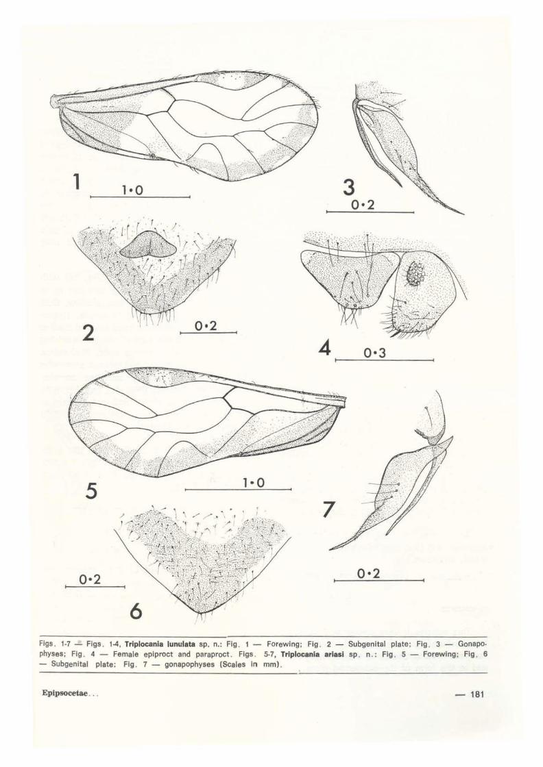
Ptiloneura (Loneura) amazonica sp. n. (Figs. 12-14, 97J
FEMALE. Unknown. MALE. Coloration. Pale brown; heod darker. Eyes black. Ocelli on black tubercle. Vertex betwéen eyes and ocelli dark brown; frons, except median anterior region, dark brown; genae dark brown; postclypeus with traces of 5 or 6 broad striae each side of midline; anteclypeus and labrum dark brown; maxtllary palpi dark brown; antennae paler. Thorax with slight dark brown markings on dorsum and pleura. Legs : femora with 2 dark brown bands, tibiae darkened near apex, tarsi wholly darK brown. Forewing (Fig. 12) marked with brown: apices of veins darkened; base and apex of pterostigma dark brown; a series of dark ares near margin between ali veins from R4+5 -Cuia; nodulus and adjacent area of cell Culb darkened. Hindwing pai e, except for ~lighi.
darkening at apices of veins.
Abdomen irregularly darkened on ali tergi-tes.
Morphology. Lacinial apex as in Fig. 97. Forewing venation (Fig. 12) ; M 5-branchea. Hindwing with M 3-branched. Hypandrium (Fig . 13) heavily sclerotised, w ith 2 elongate pointed lateral processes. Phallosome (Fig . 14) complex, trame closed anteriorly, and with complex heavi ly-sclerotised radular rods. Epiproct bluntly rounded, with group of about 8 lateral setae and small apical spiculate area. Paraproct with field of about 24 trichobothria.
Dimensions . B 3.10, FW 3.28, HW 2.25, f, O. 555, fz O. 450, t, / f2 1 . 233, F O. 870, T 1 470, t, o 645, t2 0.060, t3 0 . 120, t, / t2 10.750, t2/t3 O. 500, ct 22 . 1 . 2.
Holotype ô , Malaise trap 14.iii . 1978; paratypes (ali Malaise trap) 1 ~ 16.v.1978, 2<S<S8. viii.1978, 1 t 11.viii.1978, 1 ~ 25.viii.
1978, 1 6 13. ix. 1978.
COMl\'IENTS
This species is clearly refcrable to Ptilo· neura Enderlein (1900), and the forewing markings are rather similar to those of P.
180-
(Loneura) splendida Mockford (1957) known from Guatemala anel Mexico, male genitalta ol which were figured in part by Eertmoed (1973) Eertmoed (unpublished data supplement to his paper) gives the following hypandrial char3cters for sp/endida: (a) distai margin distinctly bilobed, lobes heavily sclerotiscd, (b) heavily sclerotised, (c) lateral lobes cylindrical. The two taxa are thus closely rel :lted and diffe1 mainly in the different forewing markings in cell Culb and in details of the phallosome: the radular sclerites of splendida are more dtscretE> than in amazonica, and the anterior of the trama is relatively broad.
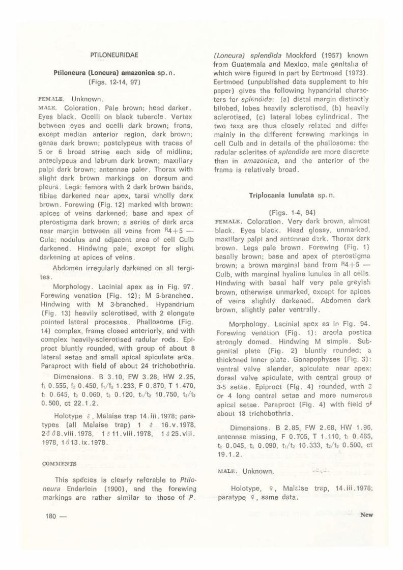
Triplocania lunulata sp. n.
(Figs. 1-4, 94) FEMALE. Coloration. Very dark brown, almost black. Eyes black. Head glossy. unmarked, maxillary palpi and antennae d~rk. Thorax dark brown. Legs pale brown. Forewing (Fig. 1) basally brown; base and apex of pterostigma brown; a brown marginal band from A4+5 -Culb, with marginal hyaline lunules in ali cells Hindwing with basal half very pale greyish brown, otherwise unmarked, except for apices of veins slightly darkened. Abdomen dark brown, slightly paler ventr:tlly.
Morphology. Lacinial apex as in Fig. 94. Forewing venation (Fig. 1): areo!a postica strongly domed. Hindwing M simple. Subgeniial plate (Fig. 2) bluntly rounded; <~ thlckened inner plate. Gonapophyses (Fig. 3): ventral valve slender, spiculate near apex; dorsal valve spiculate, with central group ot 3-5 setae. Epiproct (Fig. 4) rounded, wtth 3 or 4 long central setae and more numerous apical selae. Paraproct (Fig . 4) with field o f about 18 trichobothri a.
Dimensions. B 2.85, FW 2.68, HW 1.96, antennae missing, F 0.705, T 1.110, t, 0.465, t2 0.045, t~ 0.090, t J tz 10. 333, f2/ l! 0.500, C'(
19.1.2.
MALE. Unknown. -... -
Holotype, ~ , MaiE-;se trG:p, 14. iii.1978; paratype ~ , same data .
New
3 0•2
2 4 0•3
5
0•2 0•2
6 Figs . 1-7- Figs . 1-4, Trlplocanla lunulata sp. n.: Flg. 1 - Forewing; Flg. 2 - Subgenltal plate; Flg . 3 - Gonapophyses; Flg. 4- Female eplproct and paraproct . Flgs . 5-7, Triplocania arlasl sp . n.: Flg. 5- Forewlng; Fig . 6 - Subgenltal plate; Flg . 7 - gonapophyses (Scales In mm) .
Epipsocetae... - 181

COMMENTS
This, and the · next two species, are superficially similar in having a broad brown marginal or premarginal band around the mediai border of the forewing and the wing base darkened. T. reflexa Roesler ( 1940) also shows this feature.
r. lunulata is most similar to the next species, and differs from it by having distinct shallow hyaline lunules in cells AS - Cuia and by having a pronounced inner plate to the subgenital plate.
Triplocania ar iasi sp. n. (Figs. 5-7, 95)
FEMALE. Coloration. Dark greyish brown. Eyes black. Ocelli on black tubercle. Face unmarked. Maxillary palpi pai e brown. Thorax dark dorsally, paler laterally. Legs pale brown. Forewing (Fig. 5): base and apex of pterostigma dark brown; marginal band from R2+3 posteriorly, without hyaline lunules, but paler in central region of mediai cells. Hindwing hyaline. Abdomen pai e brown, darkened middorsally.
Morphology. Lacinial apex as in Fig. 95. Forewing venation as in Fig. 5. Hindwing M simple. Subgenital plate (Fig. 6) tapered, sclerotised area distinctly Y-shaped. Gonapophyses (Fig. 7): ventral valve slender; dorsal valve with elongate spiculate apex and central group of 2 or 3 setae. Epiproct deep, rounded , strongly setose on posterior half. Paraproct deep, with field of about 25 trichobothria.
Dimensions . B 3.20, FW 3.16, HW 2.20, antennae and hind legs missing. MALE. Unknown.
Holotype, 9, Malaise trap 25.x. 1977.
COMMENTS
This species if very similar to T. lunulata, from whfch it differs mainly by lacking marginal hyaline lunules in the forewing band, and m the form of the subgenital plate.
182-
Triploc.ania caudata sp. n. (Figs. 8-11, 96)
FEMALE. Unknown.
MALE . Coloration. Pai e to mid-brown. Eyes black. Ocelli on black tubercle. Postclypeus with traces of narrow striae, face otherwise unmarked. Thorax slightly darkened dorsally. Legs pai e. Forewing (Fig. 8) with browr. marginal band posteriorly from antenor to R4+5; brown markings at apex of pterostigma and of R2+3, along basal Rs, behind Culb ano at wing base. Hindwing with trace of pale greyish brown marginal shading behind wing apex . Abdomen pai e.
Morphology. Lacinial apex (Fig. 96) wi th outer tine enlarged . Forewing venation as 1n F1g . 8: pterostigma long and shallow, Cuia strongly sinuous. Hindwing M simple. Hypan· drium (Fig. 9) with prominent trilobed median tongue bearing 2 short apical s9tae, this arising from emarginate transverse apex. Phallosome (Fig. 1 O) trame closed and tapered antenorly; open posteriorly; radular sclerites complex and toothed. Epiproct (Fíg. 11) rounded, with few shorl central setae and apical field or sp icules and short setae. Paraproct with field of about 20 trichobothria.
Dimensions . B 3.40, FW 3.26, HW 2.20, f1 O. 570, f2 O. 450, f1 / f2 1. 267, F O. 900, T 1. 395. t l o. 600, t, o. 050, Í3 o. 120, t t!t2 12 .000, t2 t o. 417, ct 21 . 1 . 2.
Holotype, ô, light trap, 20.xii.1977.
COMMENTS
Forewing venation ot this species, especially the unusual shape of the areola postica, is closely similar to that of T. reflexa Roesler (1940). The two taxa differ markedly on the form of t he hypandrium (that of reflexa lacking the median tongue prominent in caudata) and on details of the phallosome, as well as in differences in pigmentation ot the basa: half of the forewing.
New
10
11 0•3
12 14
13 0·2 0·3
flgs. 8-14 - Figs . 8-11. Triplocania caudata sp. n .: Fig . r; - Forewlng; Fig . 9 - Hypandrium; Fig . 10 - Phallosome; Fig . 11 - Male epiproct and paraproct . Fígs . 12-14. Ftiloneura (loneura) amazonica sp. n.: Fig. 12- Forewing; Fig. 13- Hypandrium; Fig. 14 - Phallosome. fScales in mm) .
Eplpsocetae ... - 183

Triplocanla umbrata sp. n. (Figs 19-23, 98)
FEMALE. Coloration. Pai e brown . Eyes black . Ocelli on small black tubercle. Vertex with pale, but distinct, brown lines each side of midline; frons pale; postclypeus with traces of 5-7 narrow striae each side ot midline; anteclypeus, labrum, maxillary palpi and antennae pale brown; genae darkened ventrally. Thorax pale, except for narrow dark brown pleural stripe. Legs pai e, apex of tibiae and whole of tarsi darkened. Forewing (Fig. 19) with ali veins heavily shaded with tawny brown, and similar pigmentation around margin of wing; branches of As , of M , and Cul with dark greyish brown marginai spot; base of pterostigma dark greyish brown; setae on veins arising from dark spots. Hindwing hyaline. Abdomen witb traces of darker dorsal annuli.
Morphology . Lacinial apex (Fig. 98) rela· tively narrow. Forewing venation (Fig. 19): basal half of Gula sinuous; stem of As long. Subgenital plate (Fig. 20) tapered, with deep U-shaped sclerotised area. Gonopore pia te (Fig . 21 J with lateral heavily sclerotised projections. Gonapophyses (Fig. 22): ventral valve relatively short, dorsal valve with long spiculate apex. a central group of 5 setae . Epiproct (Fig. 23) tapered, with few central setae, and apical setae. Paraproct (Fig. 23) tapered, with a field of about 24 trichobothria and 1 or 2 central setae without basal rosettes.
Dimensions. B 3.50, FW 3.21, HW 2.25, f , 0.495, f2 0.360, f, / f2 1.375, F 0.870, T 1.440, t , 0.915, t2 0.060, t3 0.105, t, / t2 15.250, t2/!3 0.571, ct 23.1.2.
MALE. Unknown.
Holotype, 9 , Malaise trap 20. ix. 1978.
Paratype, 9 , same data.
The unusual forewing markings of T. um brata immediately differentiate it from ali other described Ptiloneuridae. The thickened gonopore plate with lateral processes is also anomalous.
184-
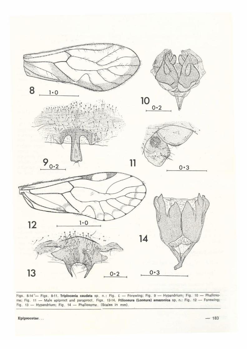
Triplocania immaculata sp. n. (Figs. 28-31 , 100)
FEMALE. Unknown.
MALE. Coloraticm. Pai e brown. Eyes black . Ocelli on small black tubercle. A broad black interocular band dorsal to ocelli, and a similar band extending to frons border and eyes immediately dorsal to antennae. Postclypeus, anteclypeus, labrum, maxillary palpi darK brown. Genae b lackened ventrally. Antennae: b::.tse very dark brown, flagellum paler. Thorax mainly pale, with slight darkening on dorsal lobes . Legs pale, except apex of femora dark. Forewing hyaline, veíns brown. Hindwing hyaline. Abdomen pale.
Morphology. L~cmial apex as in Fig . 100. Forewi ng venati on as in Fi g. 28: pterostigma very shallow. H indwing M simple. Hypandriun, (Fig . 29) broad, heavily sclerotised, with lateral lobes and a small thickened medi~n lobe . Phallosome (Fig . 30) complex: frame rounded and closed anteriorly; with divergent hooked lobes posteriorly; complex radular sclerites. Epiproct (Fig. 31) shallow, rounded. Paraproct (Fig . 31) rounded. with field of about 20 small t richobothria.
Dimensions. B 3. 85, FW 4. 07, HW 2. 78, f 1 1.125, f2 1.200, f ,/f2 0.938, F. 1.155, T 2.070 t, 0.900, t2 0.060, tl 0.135, tl / t? 15 .000, t2/ t3 O. 444, ct 34. 1 . 2.
Holotype, ~. light trap 20 .ix. 1978.
COMMENTS
The only described species of Triplocania with unmarked wings is T. dolosa Aoesler ( 1940) . known only from the female. From Aoesler's figure, thc two species differ considerably in the shapes of the pterostigma and areola postica. Thesg features also separate dolosa from the next species.
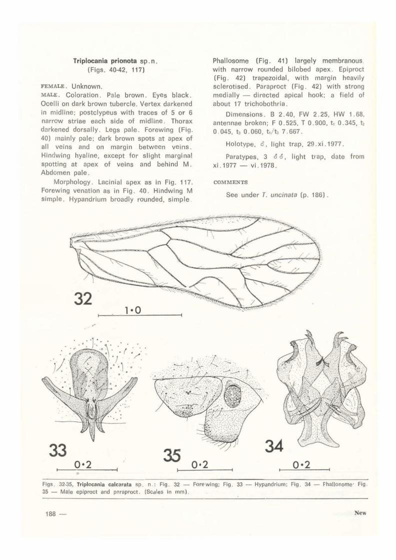
Triplocania calcarata sp. 11· (Figs. 32-35, 101)
.J '
FEMALE . Unknown.
MALE . Goloration . Dark brow~ . Eyes black. Ocelli on dark tubercie. Frons mainly very dark: postclypeus with about 6 narrow d,ar~
New

15 2·0 17
0·5
18 0·3
16 0·3
0·2
19 1• o 22
21 0·2
Flgs. 15·23- Figs . 15-18, Euolocania plcta sp. n .: Fig. 15 - Forewlng; Fig. 16- Hypandrium; Fig. 17- Phalloso. me; Frg. 18- Male epiproct and paraproct. Figs. 19-23. Trlplocania umbrata sp. n . : Flg . 19- Forewing; Flg. 20-~ubgenital plate; Flg. 21 - Gonopore plate; Fig 22 - Gonapophyses; Fig. 23 - Female epiproct and paraproct . (Scales In mm).
Epipsocetae ... - 185

stríae each síde of m1dlíne; anteclypeus and labrum dark, maxillary pa!pi and antennae paler. Thorax írregulsrly darkened dorsally and laterally. Legs pai e. Forewíng hyaline, vein.:pale brown. Htndwing hyaline. Abdomen pale
Morphohlogy. Lacinia l apex as in Fig. 101. Forewing venation as in Fig. 32. Hindwing M simple. Hypandrium (Fig . 33) heavily sclerotised; two outwardly curved lateral processes flanking an elongate pointed median projection; a deep narrow anterior sclerotised reglon . Phallosome (Fig. 34) complex; frame closed and broadened anteriorly; dívergent apical hooks and complex radular sclerites. Epiproct (Fig . 35) shallow, rounded . Paraproct (Fig . 35) with field of about 25 small trichobothria.
Dimensions. B 3.15, FW 3.07, HW 2.16, f, 0.465, f2 0.435, f ,/f2 1.069, F 0.825, T 1.380, ti 0.615, t2 0.045, t3 0.120, t, 1t2 13.667, t2/t~ 0.375, ct 21 .1.1.
Holotype, d' . I ight trap 31 . i. 1970. Paratypes, 1 c3 light trap, 13.ix.1977; 1 ô
light trap 1 . xi. 1977; 1 c3 , light trap, 3. i. 1978.
COMMENTS
The generir placement of this species is tentative: ali specimens seen have the media of the forewit;g 4-branched, but in ali. the postericr two branches arise trom a common stem. This contrasts with the taxa conventionally placed in Eup/ocania, in which the media has 4 branches arising independently from the stem (see Fig. 15) . The species somewhat resembles the last-described on gross form of the phallosome and in having the hypandrium trilobed, and ís distinct on the greater development of the median hypandrial lobe, and on numerous other details of genitalia. Although reassessment of its generic placement may later be necessary, it is here placed in Triplocania on account of its re· semblance to T. immaculata.
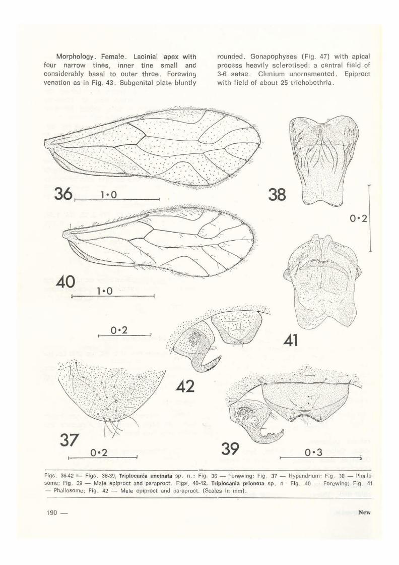
Triplocania uncinata sp. n. (Figs. 36-39, 116)
FEMALE. Unknown.
MALE. Coloration. Head very dark brown, unmarked. Eyes black. Ocelli on black tubercle.
186 -
Thorax darkened dorsally. Legs {I & liJ pale. Forewing (Fig. 36) with most veins d3rk greyish brown; a marginal spot between each pair of veins from R2+3 - Culb. Hindwing hyaline. Abdomen pai e brown.
Morphology. Lac inia l apex as in Fig. 116. Forewing venation as in Fig. 36: R4+ 5, branches of M, Culb with minute spurvein near margin of wing. Hindwing M simple. Hypan· drium (Fig. 37) deep, rounded, simple . Phallosome frame (Fig. 38) membranous. without well-defined sclerites; bilobed posteriorly. Epiproct (Fig. 39) broad, shalloN, emarginate apically. Paraproct (Fig. 39) wittt strong apical spine bearing rugose processes; a smsll field of 14 trichobothria.
Dimensions. 8 2.60, FW 2.49, HW 1.87, antennae and hind legs missing.
Holotype, ~ , light trap 13 . ix. 1977.
COMl\IENTS
This, and the next species, are related to two species referred to Triplocania by New (1972) -- namely T. fusca New ( ç only known) and T. domestica New. 8oth slnre with fusca the marginal spots between many of the forewing veins, and with domestica the hooked mal e paraprocts. T. domestica has unmarked forewings.
Males differ from other Trip/ocania species in having a very simple hypandr ium and hooked paraprocts, and these strongly suggest eventual generic separation. The minute side branches (spurveins) anstng from some forewing veins suggest simil3rity to Cladiop· socus Roesler (1940) (as figured by Eertmoed, 1973, Fig. 12), but are extremely weakly developed. On genita!lc features, the two specics described here a;e clearly clo5ely related, sugaesting that the presence of spurve ins may not alone be of generic value. They differ on details of the phallosome and of om::~mentation of the paraproct hook and on epiproct shape. T. uncinata may prove to be the male of fusca. but differences in areo1a postica shape and forewing pigmentation render this associatton currently unwise.
24 26
. . .
0·2 25 27 0•3
28 2·0 30 0·5
31 29 0•3
0·3
Flgs . 2~·31 - Figs . 24-27. Euplocania cerata sp. n .: Fig . 24 - Forewlng; Fig . 25 - Hypandrium; Flg . 26 - Phallosome; Fig . 27 - Male epiproct and paraproct . Figs . 28-31. Triplocanla lmmaculata sp . n .: Fig . 28 - Forewlng ; Fig 29 - Hypandrium; Flg . 30 - Phallosome; Flg . 31 - Male eplproct and paraproct. (Scales in mrro).
Epipsocetae . . . - 187
Triplocania prionota sp. n . (Figs. 40-42, 117)
FEMALE. Unknown. MALE. Coloration. Pale brown. Eyes black. Ocelli on dark brown tubercle. Vertex darkened in midline; postclypeus with traces of 5 or 6 narrow striae each side of midline. Thorax darkened dorsally. Legs pai e. Forewing (Fig. 40) mainly pale; dark brown spots at apex of ali veins and on margin between veins. Hindwing hyaline, except for slight marginal spotting at apex of veins and behiml M. Abdomen pai e.
Morphology. Lacinial apex as in Fig. 117. Forewing venation as in Fig. 40. Hindwing M simple. Hypandrium broadly rounded, simple
32 1·0
.-
33 35 0•2
Phallosome (Fig. 41) largely membranous with narrow rounded bilobed apex . Epiproct (Fig. 42) t rapezoidal, with margin heavily sclerotised. Paraproct (Fig. 42) with strong medially - directed apical hook; a field of about 17 trichobothria.
Dimensions. B 2 . 40, FW 2. 25, HW 1 . 68, antennae broken; F 0.525, T 0 .900, t 1 0.345, t, o 045, Í3 o. 060, tJ!t, 7 . 667.
Holotype, 6 , light trap, 29.xi.1977.
Paratypes, 3 ô ô, light trap, date from xi .1977 - vi .1978.
COMMENTS
See under T. uncinata (p. 186) .
0•2
Fígs. 32-35, Triplocania calcarata sp . n : Fig. 32 - Forewlng; Fig. 33 - Hypandrlum; Flg . 34 - Fhallosome· Fig 35 - Male epiproct and pPraproct. (Scales in mm) .
188 -

Euplocania picta sp . n. (Figs . 15-18)
FEMALE . Unknown . MALE. Coloration. Brown. (Head missing) . Thorax darkened dorsally, pleura with traces of stripe . Legs: coxae with dark bar, femara pale, tibiae and tarsi dark brown . Forewing (Fig. 15) strongly marked with very dark brown black: a marginal band interrupted by white ares flanking apices of veins . Hindwing pale grey basally; apices of veins browned. Abdomen with traces of darker brown dorsal markings .
Morphalogy . Forewing venation as in Fig. 15: M 4-branched, Cuia sinuous. Hindwing M simple. Hypandrium (Fig. 16) heavily sclerotised; with bifurcate lobes flanking a short median are, ridged longitudinally each side of midline . Phallosome trame (Fig. 17) closed and tapered anteriorly, open posteriorly; radular sclerites complex . Epiproct (Fig. 18) deep, rounded , with few setae . Paraproct (Fig. 18) with small field of about 25 trichobothria.
Dimensions . FW 4.12, HW 2 .92 . Holot ype, é' , Malaise trap 6. ix . 1978.
COMME.NTS
Few species of Eup/ocania have been described: this specimen, although incompleta differs markedly on forewing markings from any of these and its description as a new species is facilitated by the distinctive mate genital ia . Gen1talia are otherwise known only for E. greeni New ( 1972) and for the next species. The form of the hypandrium is grossly similar to that of greeni in being centrally bifurcate and with st rong lateral sclerites. bu t the phallosomes of the two species differ considerably.
Euplocania cerata sp. n . (Figs. 24·27, 99)
FEMALE . Unknown. MALE. Coloration . Dark brown. Eyes black. Ocell i on black tubercle . Anterior o f vertex , lateral regions of frons very dark; postclypeus wíth traces of 3 or 4 striae each side of midl ine; anteclypeus and labrum dark brown; palpí dark. Thorax with irregular dark pleur31
Eplpsocetac ...
stripe, dorsum dark . Legs brown, femora darker than rest of Jeg. Forewing (Fig. 24) with slight brown markings at apices of veins A2+3 .. Culb, at base and apex of pterostígma, and at nodulus; a brown spot behind Cu, shortly after separation from M. Hindwing unmarked. Abdomen pai e, except for dark pleural stripe along anterior half.
Morphology. Lacinial apex as in Fig . 99 . Forewing venation as in Fig . 24 . Hmdwing M simple . Hypandrium (Fig. 25) heavily sclerotised, with twó short tapered apical processes. Phallosome (Fig. 26) closed and slightly tapered anteriorly, open posteriorly, with elong:lte central sclerites and complex anterior racJular scle:-ites. Epiproct (Fig . 27) rounded, with apical spiculate field. Paraproct with field of about 21 trichobothria .
Dimensions. B 2 .90, FW 2.68, HW 1 .96. F O. 705, T 1. 110, t , 0 .465, t2 0.045, t3 0 .090, t ,ftz 10.333, t2/t3 0.500, ct 19.1 .2 .
Holotype, e, light trap, 6. xii.1977 . Paratype. ô same data.
COMMENTS
E. cerata differs from other members of the genus on forewing markings and on the shape of the areola postica, as well as on genit:llic featu res .
DOLABELLOPSOCIDAE
Dolabellopsocus ctenatus (New)
Eplpsocus ctenatus New, 1972: 480, figs. 87·90 ( ~ , Ma· to Grosso).
Dolabellopsocus ctenatus (New) . Eertmoed, 1973: 397.
Material examined . 3 ~é' ; 2, light trap 6.xii . 1977, 1, Malaise trap 14. ii . 1978.
COMME.NTS
This characteristic species is now known from both Mato Grosso and Amazonas, and males only have been found.
lsthmopsocus luridus sp. n . (Figs. 43-47)
BOTH sgxEs. Coloration . Pai e brown. Eyes black. Ocell i on dark tubercle. Body unmarked Wings very pai e tawny .
- 189
Morphology. Female. Lacinial apex with four narrow tines, inner tine small and considerably basal to outer three. Forewing venation as in Fig. 43. Subgenital plate b!untly
36 t------=-1 ___:::... 0_---t
40
0·2
37 0•2
rounded. Gonapophyses (Fig. 47) with apical process heavily sclerotised; a central field of 3-6 seta e. Clun i um unornamented. Epiproct with field of about 25 trichobothria.
38 0·2
41
39 0·3
Figs . 36-42- Figs. 36-39, Triplocanla U'llcinata sp. n . : Fig. 36- l=orewing; Fig . 37- Hypandrlum: F:g . '38- Phallo some; Fig. 39- Male epiproct and paraproct. Figs. 40-42, Triplocania prionota sp . n · Fig . 40 - Forewing; Fig 41 - Phal losome; Fig . 42 - Mal e epiproct and paraproct. (Scales in mm).
190- New

MALE. Lacinia and wing venation as female. Hypandrium transverse. Phallosome frame (Figs. 44, 45) elongate. with apica l hooks and complex radular sclerites; a iong denticulate anterior radular process. Clunium (Fig. 46) slightly ornamented in central region. Epiproct (Fig. 46) rounded. Paraproct (Fig. 46) with field of about 25 trichobothria.
Dimensions . ~, B 2.60, FW 2.49, HW 1.87. f, O. 405, f2 O. 070, f, / f2 1 . 500, F O. 645, T 1 . 050, t , 0.390, t2 0.135, t ./t2 2.889, ct 19.4.
& , B 2. 50, FW 2. 30 HW 1 . 78, f2 O. 760, F 0.660, T 0.990, t , 0.375, t2 0.135, t ,!t2 2 .778, ct 21.4.
43 1· o
44 0·2 45
Holotype, d, Emergence trap, 11.x. 1977. Paratypes, 1 O c! ó, 1 O 9 9 , Emergence
traps , various dates from x .1977-vi .1978.
COMMENTS
On forewing venation. most specimens of this species are clearly referable to lsthmopsocus Eertmoed . In severa!, however, vein A, terminates free in the membrane rather than joining A·; genitalia of these specimens are identical with 'typical' specimens, and it now appears that variation in this wing character may transcend the convent1onal boundary between /sthmopsocus and Dolabellopsocus.
46 0•2
47 0·2
Figs. 43·47, lsthmopsocus luridus sp. n.: Fig. 43- Forewing; Figs. 44, 45- Phallos:ome; Flg . 46- Male clunium opiproct and paraproct; Flg 47 - Gonapophyses . (Scales In mm) .
Epipsocetae ... - 191

The phallosome form is most similar to that of I. hylonomus Eertmoed in having a bifurcate apex, but details of radular sclerites differ considerably. I. hylonomus is known only from Panama .
This was by far the most common species of Epipsocetae captured during the survey: the paratype series represents only a small proportion of numbers seen. .>everal hundred specimens were captured in emergence traps, in Malaise traps and in light traps.
EPiPSOCIDAE
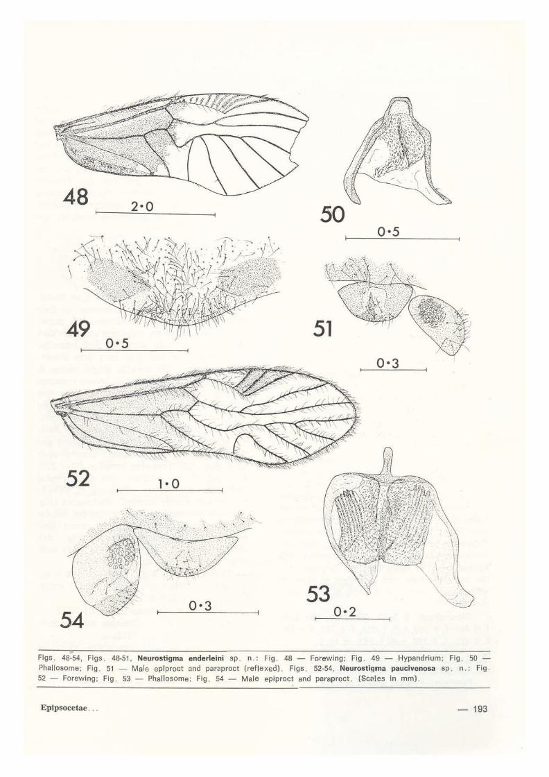
Neurost igma enderleini sp. n. (Figs. 48-51, 104)
FEMALE . Unknown.
MALE. Goloration. Dark brown . Eyes black. Head glossy, central regions of vertex and frons darkened; postclypeus with traces o f narrow striae; anteclypeus and labrum dark brown; palpi dark. Thorax almost black. L6gs dark, except pale apex of femora and base of tibiae. Forewing (Fig. 48) basally very dark brown, stigmal crossveins similar: ali other veins almost black. Hindwing with basal half greyrsh brown. Abdomen pai e, central dorsal region dark brown.
Morphology: Lacinial apex as in Fig. 104. Forewing venation as in Fig. 48; basal stigmal crossveins not shaded to wing margin; M strongly flexed basal to areola postica; Gula fused with M, mediai cells narrow. Hindwing: A8 forked. M simple. Hypandrium (Fig. 49) broadly rounded, with strongly sclerotised lateral regions. Phallosome (Fig. 50): apex trapezoidal, frame open anteriorly, radular sclerites weakly developed. Epiproct (Fig. 51) rounded, with central and apical setae. Paraproct (Fig. 51) with field of about 44 small trichobothria.
Dimensions. B 4.45, FW 4.31, HW 2.87, f1 1. 02, remainder of antennae, whole of hind leg missing.
Holotype, S Malaise trap 29. viii. 1978.
192 -
COMMENTS
This species is most similar to N. chaetocephalum Enderlein (1900, Peru), the type of which has not been seen. lt resembles chaetocephalum in (a) the shape of the pterostigma, and (b) the pronounced flexure of M before linking with Gula. and apparently differs from it in (a) smaller size (FW 4.3 cf '6%' of Enderlein), (b) the basal forewing pigmentation heing more pronounced and (c) the basal pterostigmal crossveins not being pigmented to the wing margin. Genital ia of chaetocephalum are unknown .
Neurostigma paucivenosa sp. n . (Figs. 52-54, 1 05)
FEMALE. Unknown .
MALE. Goloration. Brown. Eyes black. Ocelli on dark brown tubercle . Posterior of vertex very dark; face unmarked except for irregular traces of dark brown postclypeal striae; maxillary palpi and antennae brown. Thorax dark glossy brown, almost black dorsally;
pleura darkened i mmediately dorsal to coxa e. Legs with tibiae and tarsi da;k brown . Forewing (Fig. 52): basal hatf pai e brown, apical half of pterostigma plnkish brown, ali other veins dark brown . Hindwing pale greyish brown basally. Abdomen pai e.
Morphology. Vertex strongly se tose. eyes almost reaching vertex . Lacinial apex as in Fig. 105. Forewing venation (Fig. 52): pterostigma with 2 crossveins, areola postica not linked with M . Hindwing A5 simple, unbranr;hed. Hypandrium broadly rounded. Phallosorne (Fig. 53) with elongate rounded meóian apex; broad anteriorly; radular sele rites represented by rows of sclerotiseó denticles. Epiproct (Flg. 54) broad, shallow. with setae on central region. Paraproct (Fig. 54) simple, with field of about 27 trichobotht ia
Dimensions. B 3.00, FW 3.02, HW 2.20, f1 O. 585, f2 O. 360, f1 / f2 1 . 625, hindleg missing.
Holotype, o, light trap, 13. xii. 1977.
New
48 2·0 50 0•5
49 51 0·5
0•3
52 1· o
0•3 53
0·2
Figs. 48-54, Figs. 48-51, Neurostigma enderleini sp. n .: Fig. 48 - Forewing; Fig. 49 - Hypandrium; Fíg. 50 -Phallosome; Fig . 51 - Mal e epiproct and paraproct (reflexed). Fígs . 52-54, Neurostigrna paucivenosa sp . n.: Fíg . 52 - Forewing; Fíg . 53 - Phallosome; Fig . 54 - Male epíproct and paraproct . (Scales in mm) .
Epipsocetae ... - 193

COMMENTS
This species differs markedly from other species of Neurostigma in having a relatively narrow forewing with very few pterostigmai crossveins, but is clearly referable to this genus on other venation features. The phallosome differs from that ot other species in the more pronounced apical projection and the better-differen tiated radular sclerites.
Neurostigma dispositum Roesler (Fig. 55)
Neurostigma dispositum Roesler, 1940: 130, figs. 40·48. Neurostlgma dispositum Roesler. New, 1972: 484.
Material examined. 1 éS, light trap, 20.xii.1977.
Neurostigma roesleri sp. n. (Figs. 56-58, 102)
FEMALE. Coloration. Pai e brown. Eyes dark grey. Body unmarked except for traces of broken dark brown pleural stripe along pterothorax and abdomen. Legs with apex of coxae, dorsal edge of femora and apex ot tibiae with dark brown mottling. Forewing (Fig . 56) pale, stigmal crossveins very dark brown, most other veins pale brown, branches of R 1. R5 , M, Cuia shaded towards apex. Hindwing unmarked.
Morphology. Head and thoracic dorsum conspicuously hairy. Lacinial apex ús in Fig. 102, with outer edge strongly produced. Fore· wing venation (Fig. 56): Cuia strongly sinuous, not contacting M; apical two veins of ptcrostigma paler than basal crossveins; pterostigm<l very large. Hindwing Rs forked, M simple. Subgenital plate (Fig. 57) simple, rounded. Gonapophyses (Fig. 58) bluntly tapered, with central group of about 6 setae. Epiproct (Fig. 58) rounded. Paraproct with field of about 20 trichobothria and 1 or 2 setae without basal rosettes.
Dimensions. B 3.80, FW 3.59, HW 2.59, f 1 0.405, f2 0.300, f J f2 1.350, F O. 765, T 1.245, t , 0 .465, h 0.180, t,/~ 2.583, ct 19.2.
MALE. Unknown.
Holotype, ç, light trap, 20. xii. 1977.
Paratype, <i?, light trap, 7.ii. 1978 .
194 -
COMMENTS
N. roesleri resembles N. dispositum (fore· wing, Fig. 55) , but differs from it on the different lacinial form and in a number of forewing characters: (a) more dispersed pigmentation in the basal half of the wing and overall paler, (b) the paler apical veins of the pterostigma, which is rather l<lrger, (c) the shaded apical veins and (d) the different shape of the areo la postica. The latter feature is somewhat variable in species of Neurostigma, but the two specimens available are alike.
Neurostigma xanthoptera sp. n. (Figs. 59·61, 103)
FEMALE. Unknown. MALE. Coloration . Pai e brown. Eyes black. Ocelli small, wi th black crescents on their inner margins . Fa~e with dense dark setae; pedicel and maxillary palpi marked with dark brown. Thorax pai e dorsally, dark laterally. Legs with tibiae and tarsi very dark brown. Forewing (Fig. 59) pai e to bright yellow in basal half, with pale greyish brown markings flanking basal veins; pterostigmal crossveins dark brown; venation mainly brown. Hindwing with basal half largely pai e greyish brown. Abdomen laterally darkened on anterior half.
Morphology. Vertex narrowed, raised and distinctly emarginate medially. Lacinial apex as in Fig. 103. Forewing venation (Fig. 59): M flexed before junction with Cuia; mediai cells deep and narrow. Hindwing R5 forked. Hypandrium bluntly rounded. Phallosome (Fig. 60) with trapezoidal apical projection; radular sclerites with small denticles, more pronounced in midline. Epiproct (Fig. 61) shallow, rounded. Paraproct (Fig. 61) with large field of about 40 trichobothria.
Dimensions. B 4.50, FW 4.41, HW 2.91, f, 0.885, f2 0.615, fJ f2 1.439, F 0.900, T 1.695, t, 0.600, t2 0.210, t 1/t2 2.857, ct 23.5.
Holotype, a, Malaise trap, 14.ii. 1978. Paratypes, 11 a 5, Malaise trap, various
dates from i i. 1978- vi i i. 1978. This large species resembles chaetoce
phalum in some venation features, but differs from it on wing colour. The excavated vertex appears to be unusual in this genus.
New

55
56
58 3·7
59 2·0
60 61
Flgs. 55..61, Fig. 55, Forewlng of Neurostgma dispositum Roesler. Flgs. 56-58, Neurostlgma roesleri sp. n . : Fig. 56 - Forewing; Flg . 57 - Subgenital plate; Fig. 58 - Gonapophyses, eplpt·oct and paraproct. Figs. 59-61, Neurostigma xanthoptera sp. n . : Fig. 59 - Forewing; Fig. 60 - Phallosome; Flg. 61 - Male eplproct and paraproct. (Scates tn mm) .
Epipsocetae ... - 195

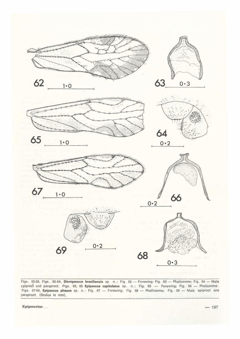
Dicropsocus brasi liensis sp. n. (Figs. 62-64, 106)
FEMALE. Unknown. MALE. Coloration. Dark greyish brown. Eyes black. Face largely unmarked; postclypeus with traces of 3 or 4 broad striae each side of midline; maxillary palpi pai e brown. Thorax very dark dor .>ally, paler laterally. Legs with coxae and femora pale, tibiae and tarsi dark brown. Forewing (Fig. 62) marked with dark brown; setae on basal veins sited on dark spots; apex of pterostigma dark; much of margin from As- Culb dark, with small hyaline lunules in most cells. Hindwing with brown shading at vein apices. Abdomen mainly dark brown, paler ventrally.
Morphology. Lacinial apex as in Fig. 106, narrow. Forewing (Fig. 62) with extensive secondary venation: As 4-branched, M 4-or 5-branched. Hindwing As forked, M simple. Hypandrium transverse, lightly sclerotised. Phallosome trame (Fig. 63) simple, open anteriorly and with short blunt median projection. Epiproct (Fig. 64) rounded, shallow, with short setae on central region and central marginal setae. Paraproct with field of about 32 trichobothria.
Dimensions. B 2. 85, FW 2. 73, HW 1 . 96, antennae missíng, F 0.705, T 1. 215, hínd tarsus missing.
Holotype, ~, I ight trap 25. i v. 1978.
(JOM:VIENTS
Forewing ch:aracters of this unusual species correspond closely to those of species of Dicropsocus Smithers and Thornton ( 1977). a genus hitherto known only from Melanesia. However, the lacinia differs markedly: that of Dicropsocus s. str. h as an elongate ou ter projection rather then severa! small teeth as in the present species. Males are known only of O. montanus Smithers and Thornton (New Guinea) . The central lobe of the phallosome is extended and truncate, as in brasiliensis, but the radula of montanus appears to be more strongly developed. Nane of the described species of Dicropsocus has a pterostigmal crossvein.
196 -
The form of the lacinia is thus the major teature in which this species difters from Melanesian Dicropsocus species. Although this teature is generally accepted as of generic value in Epipsocidae, to raise a new genus on this teature alone, and from a single specimen, is premature: brasiliensts is thus tentatively reterred to Dicropsocus, although it may be transterred to a new genus when more material becomes available for study. No similar Neotropical Epipsocidae are known.
Epipsocus capitulatus sp. n. (Figs. 65, 66, 107)
FEMALE. Unknown.
MALE. Coloration. Pai e brown. Eyes black . Ocelli on dark brown tubercle. Face unmarked. Thorax with dorsum darkened and a pronounced dark brown pleural stripe, continued along anterior halt o f abdomen. Legs pai e brown . Forewing (Fig 65) extensively marked with pale greyish brown; setae on ali veins sited on dark spots; small hyaline marginal lunules in mediai cells. Hindwing hyaline, slight darkening at apex ot veins.
Morphology. Lacinial apex as in Fig . 107 . Forewing venation as in Fig. 65. Hindwing As forked near apex. Hypandrium transverse. Phallosome trame (Fig. 66) open anteriorly, simple, with expanded median posterior projection; radular scleri'~es scarcely developed . Epiproct small , trapezoidal. Paraproct with field of about 38 small trichobothria.
Dimensions. B 2.70, FW 2.59, HW 2.16, antennae missing, F O. 795, T 1 . 320, t, O. 630, t2 0.150, t,! t2 4.200, ct 31.4.
Holotype, & , light trap, 6.xii .1977.
COMMEN'l'S
This, and the next species, both apper to be related to E. roncadorensis New and E. slnuatus New on forewing pattern. E. capitu/atus is very similar to roncadorensis on the form of the phallosome and is differenriated on the longer shallower pterostigma and areola postica. The phallosome also separates it from phaeus sp. n., in which the phallosome has a smaller apex and slightly more pro-
New
62 1·0 63 0·3 1--------------l
65 1•0 64
0•2
67 1·0
0•2 66
69 0·2
68 0•3
Flgs o 62°69, Flgs o 62-64, Dlcropsocus brasiliensis sp no: Flg o 62 - Forewing; Flg o 63 - Phallosome; Fig o 64 - Mal e cplproct c.nd paraprocto Figso 65, 66 Eplpsocus capitulatus spo n o: Figo 65 - Forewlng; Fig o 66 - Phallosome o Figs o 67-69, Epipsocus phaeus sp o n o: Fig o 67 - Forewlng; Figo 68- Phallosome; Fig o 69- Male eplproct ano paraproct o (Scales In mm) o
Eplpsocetae o o o - 197

nounced radular ornamentation. Males ot sinuatus are not known, but the more ~xtensive basal pigmentation in the forewing, as well as the more elongate pterostigma and areola postica, strongly imply that capitulatus is distinct.
Epipsocus phaeus sp.n. (Figs. 67-69, 108)
FEMALE. Unknown. MALE. Coloration. Pai e brown. Eyes black; a broad interocular dark brown band enclosing ocelli and continued behind eyes; postclypeus pale; anteclypeus dark; labrum paler medially. Thorax dark brown in dorsal midline, and with broad dark pleural band. Legs: femora with dark spot on outer edge of apex, tibiae with 2 dark bands; base of t. darkened. Forewing (Fig. 67): setae on ali veins sited on dark spots; extensive pale brown patches near margin in mediai cells and around Cuia; much of basal half of wing pale brown; apices of ali veins darkened. Hindwing hyaline, except for slight darkening at apices of main veins. Abdomen dark brown laterally, irregularly darkened dorsally.
Morphology. Lacinial apex as in Fig. 108. Forewing venation as in Fig. 67. Hindwing As forked near apex. Hypandrium shallowly rounded, incipiently bilobed. Phallosome frame (Fig. 68) open, broad; apex small and truncate; radular sclerites weakly developed. Epiproct (Fig. 69) rounded, with apical spicules and scattered setae. Paraproct with field of about 38 trichobothria.
Dimensions. B 3.00, FW 2.92, HW 2.20, f1 1.170, F 0.825, T 1 .410, t1 0 .645, f2 0.150, t 1/t2 4.300, ct 38.4.
Holotype, &, light trap, 4.x.1977.
COMMENTS
See under E. capitulatus (p. 196).
Epipsocus roesleri New (Fig. 119)
Eplpsocus roesleri New, 1972: 469 ( g, ~, figs, 42-49).
Material examined: 1 & , light trap, 3.1.1978.
198-
COMMENTS
Phallosome and wing features of this specimen correspond closely with those of roesleri and leave no doubt of its identity.
' The lacinia (Fig. 119) has not previously been figured.
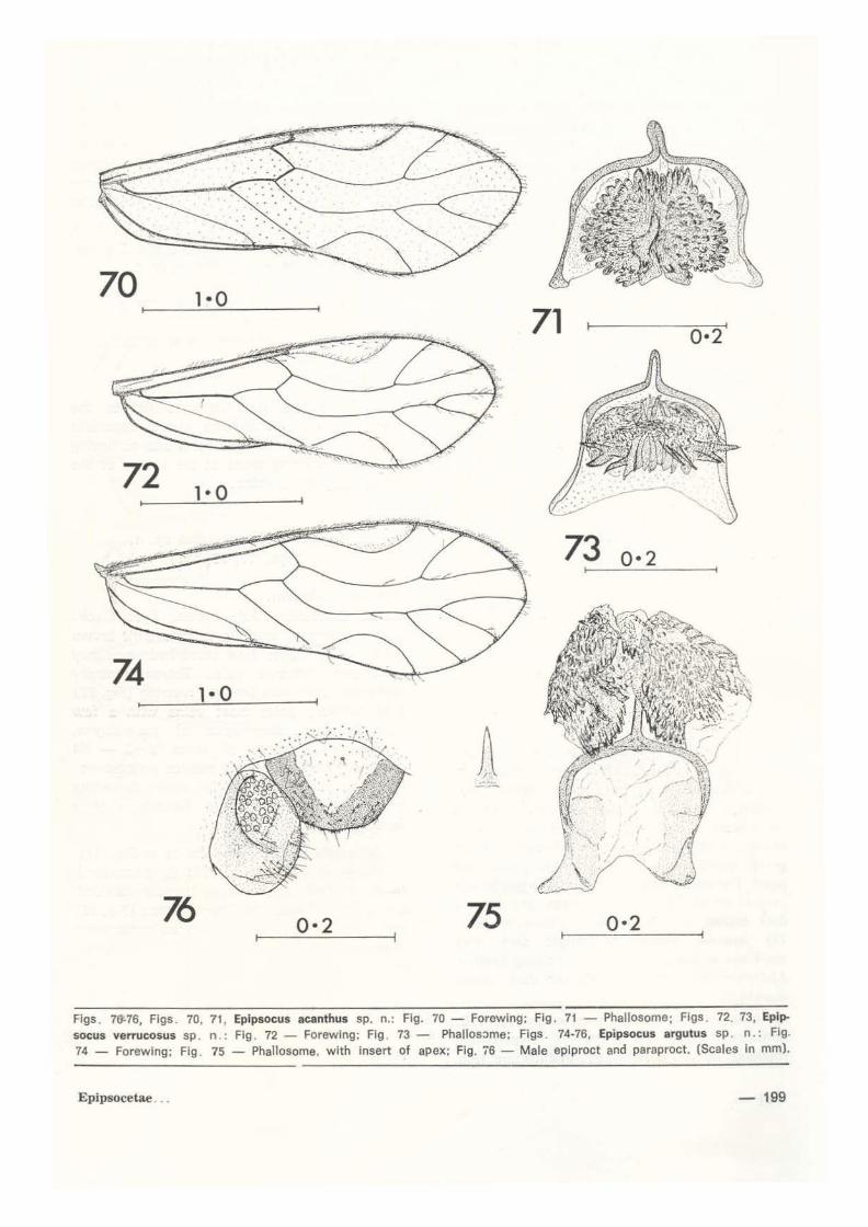
Epipsocus verrucosus sp. n. (Figs. 72, 73, 109)
FEMALE. Unkhown. MALE. Coloration . Dark brown. Eyes black. Ocelli on large black tubercle. Postclypeus with 3 or 4 narrow dark striae each side of midline; central region of anteclypeus dark. Thorax darker dorsally. Legs brown, uniform. Forewing (Fig. 72) unmarked, except for slight darkening at nodulus . Hindwing hyaline Abdomen slightly darkened dorsally.
Morphology. Lacinial apex as in Fig. 109. Forewing venation as in Fig. 72. Hypandrium incipiently bilobed, heavily sclerotised. Phal· losome (Fig. 73) broad, open with arms divergent anteriorly; an elongate tapered median posterior process; radula with laterally directed anterior spines, numerous smaller pointed or rounded sclerites posteriorly. Epiproct broadly rounded, with border heavily sclerotised. Paraproct with field of about 24 trichobothria.
Dimensions. B 2. 45, FW 2. 25, HW 1 . 63. f1 0.525, f2 0.375, f./ f2 1.400, F 0.615, T 1.020, t 1 0.510, f2 0.135, t1 / t2 3.778, ct 27.4.
Holotype, ô, light trap 1.x.1977.
COMMENTS
This species appears to be mcst similar to E. quurcus Roesler (1940), genitalia of which are unknown. Pending reexamination of the types of quurcus, it is unwise to assign the present specimen to that species, as what are apparently minor differences in forewing venation may prove to be of specific value. The phallosome frame implies relationship with the following two species, and ali three have the male epiproct border thickened as in Fig. 76. They are separable on details of phallosome shape, of radular sclerite and of the forewing.
New
70 1•0 71 0•2
72
73 0• 2
74 1· o
76 0·2 75 0·2
Flgs . 7&-76, Figs . 70, 71 , Epipsocus acanthus sp. n. : Flg. 70- Forewing: Fig . 71 - Phallosome; Figs . 72. 73, Epipsocus verrucosus sp . n .: Flg . 72- Forewing; Fig . 73- Phallosome: Figs. 74-76, Epipsocus argutus sp . n.: Fig. 74 - Forewlng; Fig . 75 - Phallosome, with insert of apex; Fig. 'i6 - Male eplproct and paraproct. (Scales in mm).
Epipsocetae ... - 199

[;pipsocus acanthus sp. n. (Figs. 70, 71)
FEMALE . Unknown . MALE . Colorat ion . Pai e brown. Eyes black . Ocellar tubercle darkened. Central region of face and of thoracic dorsum slightly darkened; body otherwise unmarked. Legs pai e. Forewing (Fig . 70) very pai e brown . Hindwing hyaline .
Morphology . Forewing venation as in Fig. 70. Hypandrium incipiently bilobed. Phallosome frame (Fig. 71) broad, open anteriorly, elongate rounded median projection; radular sclerites complex: many small rounded lateral dent ic les and central poste1ior more elongate sclerites . Epiproct rounded, with border strongly sclerotised . Paraproct with oval field ::>f about 26 trichobothria.
Dimensions. B 2.60, FW 2.54, HW 1.87, f , 0 .630, F 0 .675, T 1.170, t, 0.525, "t2 0. 135, t , t2 3.889, ct 29.4 .
Holotype, ô , light trap , 31 . i . 1978.
COMMENTS
See comment following the prevlous species.
Epipsocus argutus sp. n . (Figs. 74-76, 110)
FEMALE. Unknown.
MALE . Coloration. Dark brown . Eyes black . Ocelli on black tubercle . Vertex blackened dorsal to eyes and in m1dline; frons dark; postclypeus with 3 posterior striae converging on midline trom each side, anteriorly dark brown; anteclypeus very dark brown; labrum paler; maxillary palpi dark brown; antennae paler Thorax darkened dorsally; a broad brown pleural stripe . Legs with coxae and femora dark brown, otherwise pale. Forewing (Fig. 74) hyaline, except for slight dark grey markings at apices of veins . Hindwing hyaline. Abdomen with irregular dorsal dark brown markings .
Morphology. Lacinial apex as in Fig. 11 O. Forewing venation as in Fig . 74. Hypandrium rounded, heavily sclerotised. Phallosome (Fig.
200-
75) broad, open anteriorly; a long pointed median posterior process; radular sclerites complex, but ali pointed rather than rounded , and ali small. Epiproct (Fig . 76) bluntly rounded, with borders thickened . Paraproct (Fig. 76) with field of about 24 trichobothria .
Dimensions. B 2.65, FW 2 .54, HW 1 .87, f , 0.675, f2 0 .570, f,/f2 1. 184, F 0.675, T 1.155, t , 0.525, t2 0 . 135, t ./ t2 3 .889, ct 30.3 .
Holotype, ó, light trap, 11. x. 1977 .
Paratype, d , light trap, 15. xi. 1977 .
COMMENTS
This species is clearly related to the preceding two new species, but is separable from them on phallosome form and on having darkened forewing areas at the apices of the radial and media i veins.
Epipsocus fuscareolatus sp. n. (Figs. 77, 78, 111)
FEMALE. Unknown.
MALE . Coloration. Pai e brown. Eyes black . Ocelli darkened, sited in narrow dark brown band across frons; face unmarked; maxillary palpi and antennae pai e. Thorax ventratly darkened . Legs dark brown. Forewing (Fig. 77) predominantly pale; most veins with a few dark brown spots; apex of pterostigma, membrane at apices of veins R2+ 3 - M4 darkened; whole of areola postica dark brown. Hindwing hyaline, except for slight darkening at nodulus. Abdomen pai e dorsally, slightly darkened laterally.
Morphology. Lacinial apex as in Fig . 111 . Forewing venation (Fig . 77): R5 3-branched M 4-branched . Hypandrium broadly rounded, lightly sclerotised. Phallosome frame (Fig. 78) open anteriorly, sinuously rounded posteriorly; radular sclerites scarcely evident. Epiproct shallow, rounded. Paraproct with field of about 23 trichobothria .
Dimensions. B 2.45, FW 2.30, HW 1 . 77, f, o. 450, f2 O. 345, f, / f2 1 . 304, hind leg missing
Holotype, ô, lighttrap,18.x.1977.
New

COMMENTS
The dark brown areola postica, w ith no similar darkening of the mediai cells, im-
77 1· o
79 1· o
81 1•0
83
mediately differentiates fuscareolatus from ali other South American Epipsocidae. lt is unknown whether the additional branches to R5 and M in the forewing are aberrant.
78 0•2
80 0 · 2
Flgs . 77·83, Flgs . 77, 78, Epipsocus fuscareolatus sp . n .: Flg . 77 - Forewlng; Fig . 78- Phaltosome. Fig . 79, 80, Epipsocus pennyl sp . n .: Fig . 79, Forewlng; Fig . 80- Phaltosome. Figs . 81-83 . Epipsocus maculithorax sp . n .: Fig. 81 - Forewing; Fig . 82 - Phallosome; Flg . 83 - Mal e epiproct and paraproct. (Sc~Jes In mm).
Epipsocetae ... - 201

84 1•0
85 0•2
8 7 t---- 1'---'· 0::....___-----1
0 •2
86 88
0 · 2
Flgs . 84-88, Figs. 84-86, Epipsccus atratus sp n . : Fig . 84 - Forewing; Fig . 85 - ?hallosome; Fig . 86 - Male cplproct and paraproct . Figs . 87, 88, Epipsocus pereirai eadonnel : Fig . 87 - Forewlng ; Fig. 88 - Phallosome. rScales In mm) .
Epipsocus pennyi sp. n. (Figs. 79, 80, 112)
FEMALE. Unknown.
MALE. Coloration . Pai e brown, head overall darker . Eyes black . Ocelli on black tubercle ; postclypeus and anteclypeus darkened. Thorax pale, slightly darkened dorsally . Legs pale brown. Forewing (Fig. 79) pale greyish brown, with darker central areas in radial and mediai cell ; apices of most veins darkened. Hindwing very pale greyish brown . Abdomen unmarked.
Morphology. Laciniaf apex as in Fig. 112. Forewing venation as in Fig. 79. Hypandrium
202-
tra,,sverse . Phall o some (Fig. 80) broad, open anteriorly; a narrow rounded median posterior pracess; radula scarcely sclerotised . Epiproct rounded. Paraproct with field of about 26 widely-spaced trichobothria.
Dimensions. B 2.50, FW 2.44, HW 1.82, antennae and hind legs missing.
Holotype, d , Malaise trap, 2. viii .1977 .
COMMENTS
This, and the next species, resemble E. nepos Enderlein (1900, Peru), E. plaumanni Roesler ( 1940, Braz li) and E. latistigma
New
Roesler (1940. Brazil) in having relatively discrete dark patches in the radial and mediai forewing cells. Genitalia of nepos are unknown but (from Enderlein's figures), the areola postica is considerably longer and shallower than in either of the present species or in Roesler's species.
E. pennyi differs from other members of this group in the ground colour of the forewing beinÇJ darker and in the narrow elongate median phallosome process. The phallosome of latistigma is also relatively simple, but the apex broader and bluntly rounded. That of plaumanni is considerably more complex (Roesler 1940, fig. 56) . E. pennyi and macu-
89 1·0 ~----~~~------~
...
91 0·3
lithorax sp . n . ate readily separable on phallosome and forewing features: the three discrete groups of radular denticles found in maculithorax also separate it from ali other known species.
Epipsocus maculithorax sp. n. (Figs. 81-83, 113)
FEMALE. Unknown.
MALE. Coloration. Pale tawny brown. Eyes black. Head unmarked except for small dark brown spot behind eyes, contiguous with narrow pleural stripe along cervix and pro
90 0·2
92 0·2
93 Figs. 89-93, Epipsocus meruleus sp. n.: Fig. 89 - Forewing; Fig . 90 - Phallosome; Fig. 91 - Subgenltal plate; Flg. 92 - Gonapophyses; Flg. 93 - Male epiproct . and paraproct. (Scales In mm).
Epipsocetae - 203
thorax . Thorax ais o with small black spot at base of each wing, and a larger spot above hind coxa. Legs: apex of tibiae, whole of tarsi darkened. Forewing (Fig. 81) yel lowish brown, with dark brown markings at apex of veins R 2, 3 - Cu2; slight darker patches in radial and mediai cells. Hindwing hyaline, except fo r slight da1·kening at apices of As and M.
Morphology. Lacinial apex (Fig. 113) strongly produced on outer si de . Forewing venation (Fig. 81) . Hypandrium transverse. Phallosome (Fig. 82) broad, open anteriorly; median posterior projection short, emarginate at apex; radula with three patches of dark denticles. Epiproct (Fig. 83) deep, rounded. Paraproct (Fig . 83) with elongate f ield of about 20 trichobothria .
Dimensions. B 3 .85, FW 3 .64, HW 2 .68, f, 1.245, F 0.975, T 1.905, t , 0 .870, t2 0 . 180. t,!t2 4 .833, ct 42.4 .
Holotype, ~ . light trap, 25.v.1978.
Paratypes, 6 ~ ~ . light trap, 1 on each of following dates: 7 . iii .1978, 2.v.1978, 9.v.1978, 27 . vi . 1978, 6. ix. 1978, 27. ix . 1978.
COMMENTS
See under preceding species . E. macuflthorax is further separable by its colour and relatively large size.
Epipsocus atratus sp . n. (Figs. 84-86, 114)
FEMALE. Unknown.
MALE. Coloration . Dark glossy brown, unmarked except for blackening of central region of the vertex. Eyes black. Legs dark brown. Forewing (Fig. 84) uniform dark greyish brown. Hindwing very pale greyish brown . Abdomen predominantly pai e, darkened dorsally.
Morphology. Lacinial apex as in Fig. 114. Forewing venation as in Fig. 84. Hypandrium transverse. incipiently bilobed . Phallosome trame (Fig. 85) open anteriorly; a rounded median posterior process; lightly sclerotised 'mushroom - shaped' radular patches. Epiproct
204-
(Fig. 86) deep, rounded. Paraproct (Fig. 86) with large field of about 32 trichobothria .
Dimensions. B 2.80, FW 2.73, HW 2.15, f , 0.465, f2 0.345, f, l f2 1 .348, F O. 705, T 1.215, t , 0.510, t2 0 . 180, t ,/ t2 2.833, ct 20 .5.
Holotype, ~ . Malaise trap, 29.xi.1977. Paratype, o, Malaise trap , 9 .viii . 1977 .
COMMENTS
The darkwinged species is superficially similar to E. niger New (1972), E. fuscatus New (1972) and to Mesepipsocus newi Badonnel
'\l 94 95 96 97
98 99 100
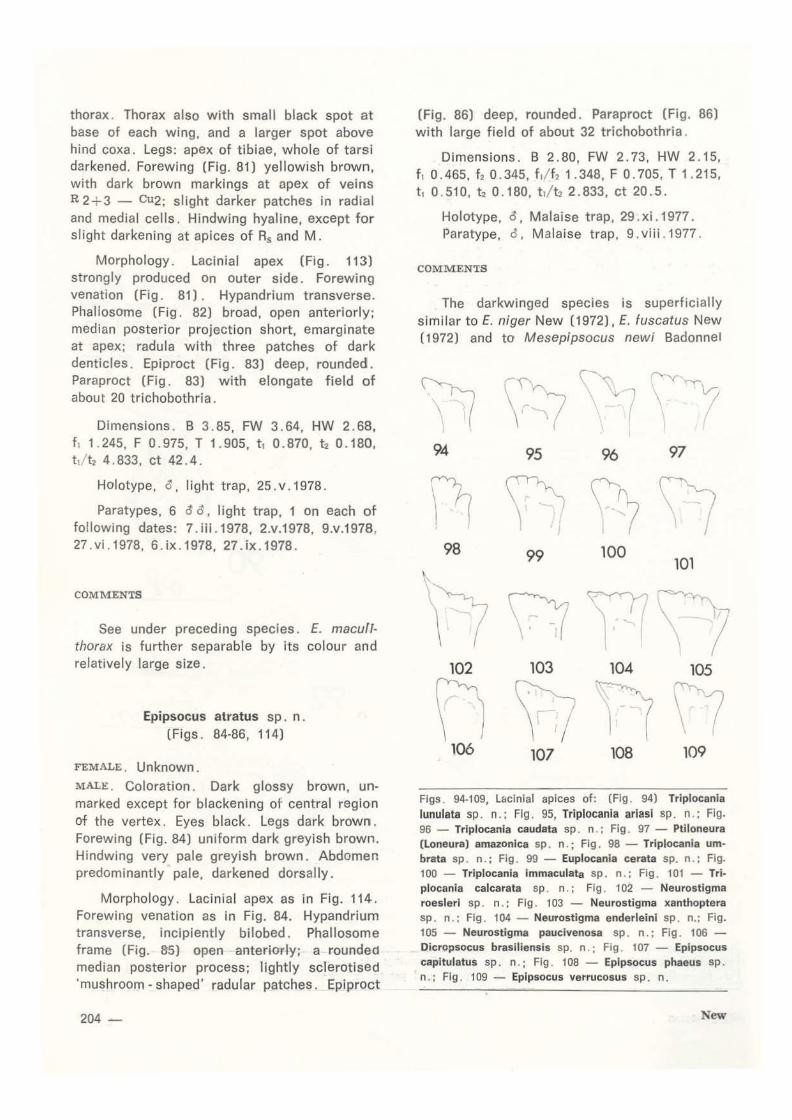
Flgs . 94-109, Lacinlal apices of: (Fig . 94) Triplocania
lunulata sp . n.; Fig . 95, Triplocania ariasi sp . n . ; Fig. 96 - Triplocania caudata sp . n . : Fig . 97 - Ptiloneura (Loneura) amazonica sp. n.; Flg . 98 - Triplocania umbrata sp . n . ; Fig . 99 - Euplocania cerata sp. n . ; Fig. 100 - Triplocania immaculata sp . n .; Fig . 101 - Triplocania calcarata sp . n. : Flg . 102 - Neurostigma roesleri sp. n . : Flg . 103 - Neurostigma xanthoptera sp . n . ; Fig . 104 - Neurosti gma enderleini sp . n.; Fig. 105 - Neurostigma paucivenosa sp . n .; Fig . 106 -Dicropsocus brasiliensis sp. n .; Fig . 107 - Epipsocus capitulatus sp. n .; Fig . 108 - Epipsocus phaeus sp . n . : Flg . 109 - Epipsocus verrucosus sp. n.
New
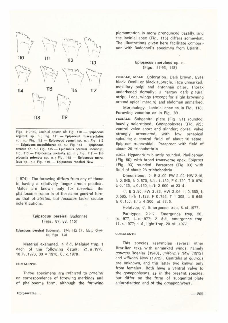
114 115 116 117
fl PI 118 119
Flgs. 110-119, laclnlal apices of: Fig. 110 - Epipsocus argutus sp. n.; Flg . 111 - Epipsocus fuscareolatus sp . n . ; Fig. 112- Epipsocus pennyi sp. n.; Fig. 113 - Epipsocus macullthorax sp. n.; Fig. 114 - Epipsocus atratus sp. n.; Flg. 115 - Epipsocus pereirai Badonnel; Flg . 116- Triplocania uncinata sp. n . ; Flg. 117- Trlplocanla prionota sp. n.; Fig. 118 - Epipsocus meruJeus sp. n.; Flg. 119 - Epipsocus roesleri New.
(1974). The forewing differs from any of these in having a relatively longer areola postica. Males are known only for fuscatus: the phallosome trame is of the same general form as that of atratus, but fuscatus lacks radular sclerifications.
Epipsocus pere1ra1 Badonnel (Figs. 87, 88, 11 5)
Eplpsocus pereiral Badonnel, 1974: 192 ( ~ , Mato Grosso, figs . 1-3)
Material examined . 4 ~ ~, Malaise trap, 1 each of the following dates: 21. ii. 1978, 18.iv.1978, 30.v.1978, 6.ix. 1978.
COMMENTS
These specimens are referred to perelrai on correspondence of forewing markings and of phallosome form, although the forewing
Epipsocetae ..
pigmentation is more pronounced basally, and the lacin1al apex (Fig. 115) differs somewhat. The illustrations given here facilitate compari· son with Badonnel's specimen from Utiariti.
Epipsocus meruleus sp. n. (Figs. 89-93, 118)
FEMALE, MALE. Coloration. Dark brown. Eyes black. Ocelli on black tubercle. Face unmarked; maxillary palpi and antennae paler. Thorax undarkened dorsally; a narrow dark pleural stripe. Legs, wings (except for slight browning around apical margin) and abdomen unmarked.
Morphology. Lacinial apex as in Fig. 118. Forewing venation as in Fig . 89 .
FEMALE. Subgenital piate (Fig. 91) rounded , heavi ly sclerotised. Gonapophyses (Fig. 92): ventral valve short and slender; dorsal valve strongly attenuated, with few preapical spicules; a central field of about 1 o setae . Epiproct trapezoidal. Paraproct with field of about 26 trichobothria.
MALE. Hypandrium bluntly rounded. Phallosome (Fig. 90) with broad t ransverse apex. Epiproct (Fig. 93) rounded. Paraproct (Fig. 93) with field of about 28 trichobothria.
Dimensions. !i' . B 3.00, FW 2.92, HW 2.16, f, 0.645, f2 0.570, f, / f2 1.132, F O. 720, T 0.870. t, 0.435, t,. 0.150, t ,lt2 2.900, ct 23.4.
a, B 2.90, FW 2.83, HW 2.06, f, 0.660, f2 0.585, f ,/ f2 1.128, F O. 795, T 1.305, t, 0 . 645, Í2 0.150, t,! t2 4.300, ct 33.5 .
Holotype, ô . Emergence trap, 8. xi . 1977 .
Paratypes. 2 <? ~ . Emergence t rap, 20. ix. 1977, 4.x.1977; 2 aa, emergence trap, 11.x. 1977; 1 a,lighttrap, 20.xli . 1977.
COMMENTS
This species resembles severa! other Brazilian taxa with unmarked wings, namely quurcus Roesler (1940), uniformis New (1972) and willineri New (1972). Genitalia of quurcus are unknown, and the latter two known only from fema les. Both have a ventral valve to the gonapophyses, as in the present species, but differ on the f orm ot subgenital plate sclerot isation and of t he gonapophyses.
-205

ACKNOWLEDGMENTS
I am very grateful to Drs. Jorge Arias and Norman D. Penny for allowing me to examine the Psocoptera from the Reserva Ducke Survey.
Resumo
Trinta e uma (31) espécies de Epipsocetae são citadas da Reserva Ducke, localizada perto de Manaus (Brasil). das quais vinte e sete (27) são descritas como novas e ilustradas . Os novos taxa representam os gêneros Ptiloneura (1). Trlplocanla (8), Euplocania (2), lsthmopsocus (1), Neurostigma (4), Dicropsocus (1),
Epipsocus ( 1 O). sendo as suas afinidades discutidas.
LlTERATURE
BADONNEL, A .
1974 - Sur quelques espaces brésiliennes du groupe des Epipsocetae (Psocoptera Psocomorpha) . Buli. Soe. ent. Franee, 79 : 192-197.
BANKs, N .
1920- New neuropteroid insects . Buli. Mus. eomp. Zool. Harvard, 64: 299-362 .
•
206-
E ERTMOED, G . E . 1973 - lhe phenetic relationships of the Epipso
cetae (Psocoptera): the hgher taxa and the specles of two new families. Trans. Amer. Ent. Soe., 99 : 373-414.
ENDERLErN, G . 1900 - Di e Psocidenfauna Perus. Zool. Jahrb. Abt.
Syst., 14 : 133-160.
Mocxroao, E .L . 1957 - Some Psocoptera from Tikal, Guatemala.
Ent. News, 68 : 197-205.
NEW, T . R . 1972 - Some Epipsocetae (Psocoptera) from cen
tral Brazil. Trans. R. ent. Soe. Lond., 123: 455·497.
1974 - Two species of Epipsocetae (Psocoptera) from cacao litter in Brazll. Rev. Bras. Ent., 18 : 101-105.
ROESLER, R . 1940 - Neue und wenig bekannte Copeognathen
gattungen, i, li . Zool. Anz., 129 : 227-243; 130, 1·25 .
SMITHERS, C . N . & TKORNTON, I.W. B. 1977- A new genus and some new species of
Epipsocidae (Psocoptera) from the Melanesian are . Proe. Linn. Soe. N.S.W., 102: 60-75 .
(Aceito para publicação em 27/ 07 / 79)
New