“epidermoid cyst of hard palate and ... platelet-rich fibrin (prf): a second-generation platelet...
TRANSCRIPT

3serve as a resorbable membrane.
Choukroun and his associates were
amongst the pioneers for using PRF
protocol.
MTA is a calcium silicate –based
hydraulic cement that possesses
favourable biological properties
including seal ing abi l i ty, good
biocompatibility and hard tissue 4
–inducing effects.
Ultrasound as a diagnostic tool has
been widely used in many medical fields,
and its applications in dentistry have not
been sufficiently explored. US imaging
provides sufficient information regarding
the nature of the periapical lesions, and is
a reliable diagnostic technique for
differentiating periapical lesions. (Cotti 5
et.al., 2006)
“EPIDERMOID CYST OF HARD PALATE AND EVALUATION OF HEALING BY COLOR POWER DOPPLER ULTRASONOGRAPHY: A CASE REPORT
1 2 3 4Sanjeev Chauhan , Ashu Gupta , Vishal Sharma , Neeti Aggarwal1Post graduate Student, Deptt. of Conservative Dentistry and Endodontics, H.P.G.D.C., Himachal Pradesh, India2Professor and Head , Deptt. of Conservative Dentistry and Endodontics, H.P.G.D.C., Himachal Pradesh, India3Professor Deptt. of Conservative Dentistry and Endodontics H.P.G.D.C., Himachal Pradesh, India4Assistant Professor, Deptt. of Radiodiagnosis, I.G.M.C. Himachal Pradesh, India
Corresponding Author: Sanjeev Chauhan
E-mail: [email protected]
thReceived: 30 December 2016ndAccepted: 2 April 2017th Online: 20 May 2017
CASE REPORTwww.djas.co.in ISSN No-2321-1482
DJAS 5(I), 58-62, 2017All rights are reserved
Dental JOURNAL of A d v a n c e S t u d i e s
ABSTRACT
Epidermoid cyst is a rare developmental cyst of the oro-facial region which results from entrapped epidermal elements with an incidence of 6.9-7% and represents less than 0.01% of all oral cavity cysts. PRF, as a physiologic fibrin matrix, serves as a net to stem cells, especially when an accelerated angiogenesis develops in the fibrin membrane .This aspect is of particular interest in the case of wide osseous defects. The Color Power Doppler detects minute areas of blood flow and the velocity of blood flow in healing bone by recording the change in frequency caused by the moving red blood cells. It demonstrates the progressive formation of new vessels in bone during the initial healing period. As the bone remodelling proceeds, there is a decrease in flow signals.
Keywords: Epidermoid Cyst, PRF, MTA, Color Power Doppler Ultrasonography
INTRODUCTION
Epidermoid cysts are rare lesions of
non-odontogenic nature inclusion cysts
lined by ectoderm and derived from
germinal epithelium and can be
encountered throughout the body, in areas 1where embryonic elements fuse together.
Most cases have been reported in ovaries
and the testicles, with 7% occurring in the
oro-facial area and 1.6% in the oral cavity,
representing 0.01% of all oral cavity 2cysts.
In recent years autologous platelet
concentrates have been used with the aim
of enhancing the healing rate in bone
regeneration procedures. Autologous
Platelet rich fibrin (PRF) is a fibrin matrix
in which platelet cytokines, growth
factors, and cells are trapped which may
be released after a certain time and can
62
CASE REPORT
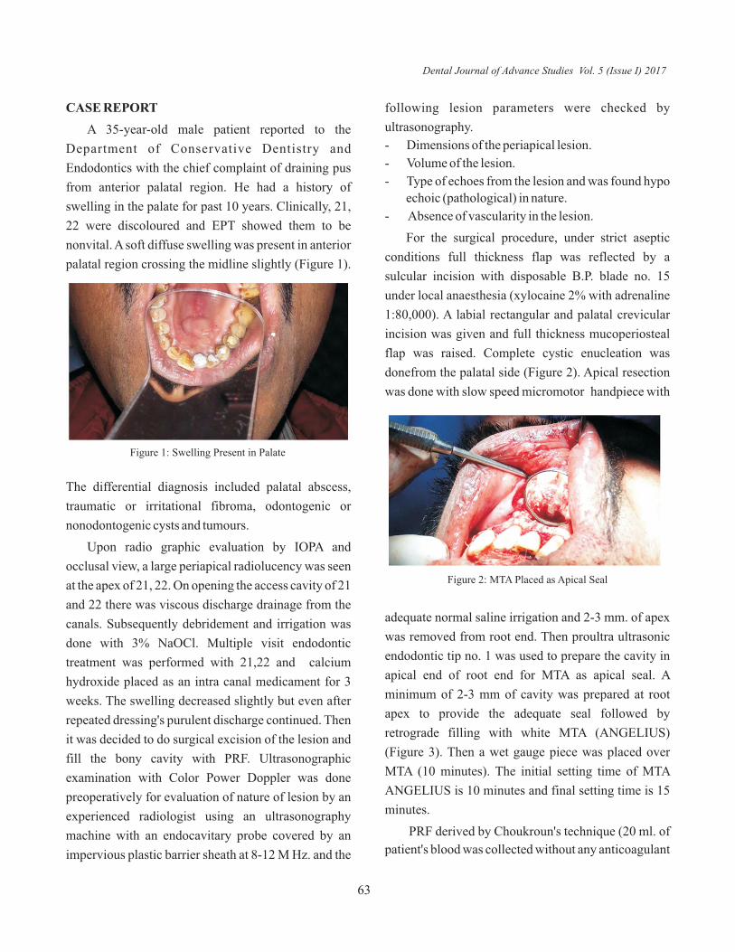
A 35-year-old male patient reported to the
Department of Conservative Dentistry and
Endodontics with the chief complaint of draining pus
from anterior palatal region. He had a history of
swelling in the palate for past 10 years. Clinically, 21,
22 were discoloured and EPT showed them to be
nonvital. A soft diffuse swelling was present in anterior
palatal region crossing the midline slightly (Figure 1).
following lesion parameters were checked by
ultrasonography.
- Dimensions of the periapical lesion.
- Volume of the lesion.
- Type of echoes from the lesion and was found hypo
echoic (pathological) in nature.
- Absence of vascularity in the lesion.
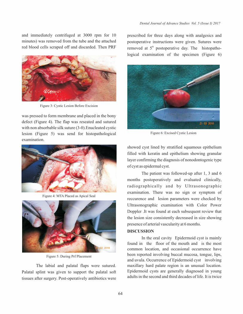
For the surgical procedure, under strict aseptic
conditions full thickness flap was reflected by a
sulcular incision with disposable B.P. blade no. 15
under local anaesthesia (xylocaine 2% with adrenaline
1:80,000). A labial rectangular and palatal crevicular
incision was given and full thickness mucoperiosteal
flap was raised. Complete cystic enucleation was
donefrom the palatal side (Figure 2). Apical resection
was done with slow speed micromotor handpiece with
Figure 1: Swelling Present in Palate
The differential diagnosis included palatal abscess,
traumatic or irritational fibroma, odontogenic or
nonodontogenic cysts and tumours.
Upon radio graphic evaluation by IOPA and
occlusal view, a large periapical radiolucency was seen
at the apex of 21, 22. On opening the access cavity of 21
and 22 there was viscous discharge drainage from the
canals. Subsequently debridement and irrigation was
done with 3% NaOCl. Multiple visit endodontic
treatment was performed with 21,22 and calcium
hydroxide placed as an intra canal medicament for 3
weeks. The swelling decreased slightly but even after
repeated dressing's purulent discharge continued. Then
it was decided to do surgical excision of the lesion and
fill the bony cavity with PRF. Ultrasonographic
examination with Color Power Doppler was done
preoperatively for evaluation of nature of lesion by an
experienced radiologist using an ultrasonography
machine with an endocavitary probe covered by an
impervious plastic barrier sheath at 8-12 M Hz. and the
Figure 2: MTA Placed as Apical Seal
adequate normal saline irrigation and 2-3 mm. of apex
was removed from root end. Then proultra ultrasonic
endodontic tip no. 1 was used to prepare the cavity in
apical end of root end for MTA as apical seal. A
minimum of 2-3 mm of cavity was prepared at root
apex to provide the adequate seal followed by
retrograde filling with white MTA (ANGELIUS)
(Figure 3). Then a wet gauge piece was placed over
MTA (10 minutes). The initial setting time of MTA
ANGELIUS is 10 minutes and final setting time is 15
minutes.
PRF derived by Choukroun's technique (20 ml. of
patient's blood was collected without any anticoagulant
Dental Journal of Advance Studies Vol. 5 (Issue I) 2017
63
and immediately centrifuged at 3000 rpm for 10
minutes) was removed from the tube and the attached
red blood cells scraped off and discarded. Then PRF
prescribed for three days along with analgesics and
postoperative instructions were given. Sutures were thremoved at 5 postoperative day. The histopatho-
logical examination of the specimen (Figure 6)
Figure 3: Cystic Lesion Before Excision
was pressed to form membrane and placed in the bony
defect (Figure 4). The flap was reseated and sutured
with non absorbable silk suture (3-0).Enucleated cystic
lesion (Figure 5) was send for histopathological
examination.
Figure 4: MTA Placed as Apical Seal
Figure 5: During Prf Placement
Figure 6: Excised Cystic Lesion
showed cyst lined by stratified squamous epithelium
filled with keratin and epithelium showing granular
layer confirming the diagnosis of nonodontogenic type
of cyst as epidermal cyst.
The patient was followed-up after 1, 3 and 6
months postoperatively and evaluated clinically,
radiographically and by Ultrasonographic
examination. There was no sign or symptom of
reccurence and lesion parameters were checked by
Ultrasonographic examination with Color Power
Doppler .It was found at each subsequent review that
the lesion size consistently decreased in size showing
presence of arterial vascularity at 6 months.
DISCUSSION
In the oral cavity Epidermoid cyst is mainly
found in the floor of the mouth and is the most
common location, and occasional occurrence have
been reported involving buccal mucosa, tongue, lips,
and uvula. Occurrence of Epidermoid cyst involving
maxillary hard palate region is an unusual location.
Epidermoid cysts are generally diagnosed in young
adults in the second and third decades of life. It is twice
The labial and palatal flaps were sutured.
Palatal splint was given to support the palatal soft
tissues after surgery. Post-operatively antibiotics were
Dental Journal of Advance Studies Vol. 5 (Issue I) 2017
64

as common in men as in women with a male to female
ratio of 3:1.
Depending on the pathogenesis, Epidermoid
cyst can be divided into:
1) Congenital
2) Acquired
Acquired cysts are derived from traumatic or
iatrogenic inclusion of epithelial cells or from
occlusion of sebaceous gland duct, it was first
recognized by Werhner in 1855 and Sutton in 1895 6referred it to be “Implantation cyst”.
PRF contains all key immune cytokines such as
interleukins IL1, IL-6,IL-4 and tumour necrosis factor.
PRF has a supportive effect on the immune system and
acts by stimulating defense mechanisms. This could be 8
important in the case of wound infection. Acc. to
Joseph Choukroun, et. al. PRF permits a rapid
angiogenesis and an easier remodelling of fibrin in a
more resistant connective tissue.
In PRF platelets and leukocyte cytokines play an
important part and the fibrin matrix supporting them is 7responsible for the therapeutic potential of PRF. A
progressive polymerization mode indicates increased
incorporation of the circulating cytokines in the fibrin
meshes. PRF, thus would be able to release cytokines
during fibrin matrix remodelling and has a long term 9effect.
In the present study the healing potential of PRF
was evaluated at varying intervals both qualitatively
and quantitatively by means of color power doppler
ultrasonography using MTA as apical seal.
Jessie F. Reyes-Carmona, et. al., studied ability of
mineral trioxide aggregate (MTA) to promote hard-
tissue deposition and wound healing and concluded
that MTA induced a pro inflammatory and pro wound
healing environment. The bio mineralization process
occurred simultaneously and promoted the integration
of the biomaterial into the environment.
Namita Raghav,et.al., evaluated the efficacy of
conventional radiography, digital radiography and
ultrasound imaging in diagnosing periapical lesions.
The percentage accuracy of diagnosing periapical
lesions using conventional radiography was 47.6%,
digital radiography 55.6%, and ultrasound 95.2%
according to them Ultrasound had the highest
sensitivity and specificity.
Smita Singh,et.al.,(2013) in their case study also
have observed and stated that it requires around 1 year
for complete healing to occur after the periapical
surgery while with the use of PRF, healing is fastened
and requires approx 6 months for complete
regeneration of bone similar to observations of our
study. They also used PRF as matrix for faster bone
formation and observed healing by ultrasonography.
The color power Doppler detects minute areas of
blood flow and the velocity of blood flow in healing
bone by recording the change in frequency caused by
the moving red blood cells. In the present study
vascularity (arterial) was absent in the lesion
preoperatively but was evident at 6 months and peak
systolic velocity was found to be 13.4cm/s (Figure 7-
11).
In the study of healing in this case study it was
observed that there was a consistent decrease in volume
of lesion at all subsequent reviews postoperatively.
Dental Journal of Advance Studies Vol. 5 (Issue I) 2017
65
Figure 7: Ultrasonographic examination showing presurgical lesion size
Moreover as epidermoid cysts are kerain filled cystic
lesions the ultrasonographic examination showed
preoperative lesion to be hyperechoic in nature.
Postoperatively at one month review the nature of

CONCLUSION
As per our study conclusion Epidermal cyst is
a rare lesion of hard palate and PRF can be used for
faster healing of such cases and Color Power Doppler
Ultrasonography is an important and reliable tool to
measure healing of such lesion using different
parameters.
REFERENCES
1) Varun Rastogi, Naveen Puri, Geetpriya Kaur, Lalita Yadav,
Rachna Sharma;Unusual Cases of Epidermoid cyst: Case
Series, International Journal of Scientific Study , 2013; 01;
(02); 72-75
2) Ozan F, Polat HB, Ay S, Goze F. Epidermoid cyst of the buccal
mucosa: A case report. J Contemp, Dent Pract 2007;8(3):1-6.
(3) Joseph Choukroun, David M. Dohan, Antoine Diss, Steve L.
Dohan, Anthony J. J. Dohan, Jaafar Mouhyi, and Bruno
Gogly, Platelet-rich fibrin (PRF): A second-generation platelet
concentrate. Part II: Platelet-related biologic features Oral
Med Oral Pathol Oral Radiol Endod 2006;101:E45-50
(4) Jessie F. Reyes-Carmona, Host–Mineral Trioxide Aggregate
Inflammatory Molecular Signaling and Biomineralization
Ability; JOE; 2010; 36, (8);1347-1353
5) Namita Raghav, Sujatha S. Reddy, A. G. Giridhar, Srinivas
Murthy, Yashodha Devi B. K, N. Santana, N. Rakesh, and
Atul Kaushik, India Comparison of the efficacy of
conventional radiography, digital radiography, and ultrasound
in diagnosing periapical lesions Oral Surg.Oral Med. Oral
Patho. Oral Radiol. Endod. 2010; 110:379-385
6) Noffke CE. Implantation-type epidermoid cyst of the
mandible. Dento maxillofac Radiology 1999; 28: 383-385.
7) Choukroun J, Diss A, Simonpieri A, Girard MO, Schoeffler C,
Dohan SL, Platelet-rich fibrin (PRF): A second-generation
platelet concentrate. Part IV: Clinical effects on tissue healing.
Oral Surg Oral Med Oral Pathol Oral Radiol Endod,
2006;101:e56-60.
8) Dohan DM, Choukroun J, Diss A, Dohan SL, Dohan AJ,
Mouhyi J. Platelet-rich fibrin (PRF): A second-generation
platelet concentrate. Part II: Platelet-related biologic features.
Oral Surg Oral Med Oral Pathol Oral Radiol Endod
2006;101:e45-50.
9) Choukroun J, Diss A, Simonpieri A, Girard MO, Schoeffler C,
Dohan SL, Dohan AJ, Mouhyi J, Dohan DM. Platelet-rich
fibrin (PRF): A second-generation platelet concentrate. Part V:
histologic evaluations of PRF effects on bone allograft
maturation in sinus lift. Oral Surg Oral Med Oral Pathol Oral
Radiol Endod 2006;101:299-303.
Dental Journal of Advance Studies Vol. 5 (Issue I) 2017
Source of Support: Nil, Conflict of Interest: None Declared
66
Figure 8: Lesion size at one month postoperatively
Figure 9: Lesion size at three months postoperatively
Figure 10: Lesion size at six months postoperatively
Figure 11: Presence of vascularity at six months
lesion was hypoechoic subsequently changing to
predominantly hyperechoic nature at 6 months review
as the healing progressed.