epa - california office of environmental health hazard … · 12 toxicity studies (u.s. epa,...
TRANSCRIPT

July 2000 DRAFT-DO NOT QUOTE OR CITE
EPA
TOXICOLOGICAL REVIEW
of
METHYL CHLORIDE
(CAS No. 74-87-3)
In Support of Summary Information on theIntegrated Risk Information System (IRIS)
July 2000(DRAFT)
This document is an external peer review draft. It has not been formally released by theU.S. Environmental Protection Agency and should not at this stage be construed to representAgency position on this chemical. It is being circulated for review of its technical accuracy andscience policy implications.
U.S. Environmental Protection AgencyWashington, DC

July 2000 DRAFT-DO NOT QUOTE OR CITEii
DISCLAIMER
This document has been reviewed in accordance with U.S. Environmental ProtectionAgency policy and approved for publication. Mention of trade names or commercial productsdoes not constitute endorsement or recommendation for use.
Note: This document may undergo revisions in the future. The most up-to-date versionwill be available electronically via the IRIS Home Page at http://www.epa.gov/iris.

July 2000 DRAFT-DO NOT QUOTE OR CITEiii
CONTENTS—TOXICOLOGICAL REVIEW OF METHYL CHLORIDE(CAS No. 74-87-3)
FOREWORD . . . . . . . . . . . . . . . . . . . . . . . . . . . . . . . . . . . . . . . . . . . . . . . . . . . . . . . . . . . . . . . . . v
AUTHORS, CONTRIBUTORS, AND REVIEWERS . . . . . . . . . . . . . . . . . . . . . . . . . . . . . . . . vi
1. INTRODUCTION . . . . . . . . . . . . . . . . . . . . . . . . . . . . . . . . . . . . . . . . . . . . . . . . . . . . . . . . . . . 1
2. CHEMICAL AND PHYSICAL INFORMATION RELEVANT TO ASSESSMENTS . . . . . 3
3. TOXICOKINETICS RELEVANT TO ASSESSMENTS . . . . . . . . . . . . . . . . . . . . . . . . . . . . . 43.1 ABSORPTION . . . . . . . . . . . . . . . . . . . . . . . . . . . . . . . . . . . . . . . . . . . . . . . . . . . . . . . . . . 53.2 DISTRIBUTION . . . . . . . . . . . . . . . . . . . . . . . . . . . . . . . . . . . . . . . . . . . . . . . . . . . . . . . . 93.3 METABOLISM . . . . . . . . . . . . . . . . . . . . . . . . . . . . . . . . . . . . . . . . . . . . . . . . . . . . . . . . 123.4 EXCRETION . . . . . . . . . . . . . . . . . . . . . . . . . . . . . . . . . . . . . . . . . . . . . . . . . . . . . . . . . . 24
4. HAZARD IDENTIFICATION . . . . . . . . . . . . . . . . . . . . . . . . . . . . . . . . . . . . . . . . . . . . . . . . 264.1 STUDIES IN HUMANS—EPIDEMIOLOGY, CASE REPORTS, CLINICAL
CONTROLS . . . . . . . . . . . . . . . . . . . . . . . . . . . . . . . . . . . . . . . . . . . . . . . . . . . . . . . . . . 264.1.1 Mortality and Noncancer Effects . . . . . . . . . . . . . . . . . . . . . . . . . . . . . . . . . . . . . 264.1.2 Cancer Effects . . . . . . . . . . . . . . . . . . . . . . . . . . . . . . . . . . . . . . . . . . . . . . . . . . . 33
4.2 PRECHRONIC AND CHRONIC STUDIES AND CANCER BIOASSAYS INANIMALS—ORAL AND INHALATION . . . . . . . . . . . . . . . . . . . . . . . . . . . . . . . . . . . 354.2.1 Subchronic and Chronic Oral Studies . . . . . . . . . . . . . . . . . . . . . . . . . . . . . . . . . 354.2.2 Subchronic and Chronic Inhalation Studies . . . . . . . . . . . . . . . . . . . . . . . . . . . . . 36
4.3 REPRODUCTIVE/DEVELOPMENTAL STUDIES—ORAL AND INHALATION . . 464.4 OTHER STUDIES . . . . . . . . . . . . . . . . . . . . . . . . . . . . . . . . . . . . . . . . . . . . . . . . . . . . . . 57
4.4.1 Neurological Studies . . . . . . . . . . . . . . . . . . . . . . . . . . . . . . . . . . . . . . . . . . . . . . 574.4.2 Acute and Subacute Inhalation Studies . . . . . . . . . . . . . . . . . . . . . . . . . . . . . . . . 664.4.3 Genotoxicity Studies . . . . . . . . . . . . . . . . . . . . . . . . . . . . . . . . . . . . . . . . . . . . . . 794.4.4 Mode of Action in Relation to Toxicity . . . . . . . . . . . . . . . . . . . . . . . . . . . . . . . . 83
4.5 SYNTHESIS AND EVALUATION OF MAJOR NONCANCER EFFECTSAND MODE OF ACTION (IF KNOWN)—ORAL AND INHALATION . . . . . . . . . . . 85
4.6 WEIGHT-OF-EVIDENCE VALUATION AND CANCERCHARACTERIZATION—SYNTHESIS OF HUMAN, ANIMAL, AND OTHERSUPPORTING EVIDENCE, CONCLUSIONS ABOUT HUMANCARCINOGENICITY, AND LIKELY MODE OF ACTION . . . . . . . . . . . . . . . . . . . . 91
4.7 SUSCEPTIBLE POPULATIONS . . . . . . . . . . . . . . . . . . . . . . . . . . . . . . . . . . . . . . . . . . 944.7.1 Possible Childhood Susceptibility . . . . . . . . . . . . . . . . . . . . . . . . . . . . . . . . . . . . 954.7.2 Possible Gender Differences . . . . . . . . . . . . . . . . . . . . . . . . . . . . . . . . . . . . . . . . 95
5. DOSE-RESPONSE ASSESSMENTS . . . . . . . . . . . . . . . . . . . . . . . . . . . . . . . . . . . . . . . . . . 965.1 ORAL REFERENCE DOSE (RfD) . . . . . . . . . . . . . . . . . . . . . . . . . . . . . . . . . . . . . . . . . 965.2 INHALATION REFERENCE CONCENTRATION (RfC) . . . . . . . . . . . . . . . . . . . . . . 96

July 2000 DRAFT-DO NOT QUOTE OR CITEiv
CONTENTS (cont’d)—TOXICOLOGICAL REVIEW OF METHYL CHLORIDE(CAS No. 74-87-3)
5.2.1 Choice of Principal Study and Critical Effect—with Rationaleand Justification . . . . . . . . . . . . . . . . . . . . . . . . . . . . . . . . . . . . . . . . . . . . . . . . . . 96
5.2.2 Methods of Analysis—No-Observed-Adverse-Effect Level/Lowest-Observed-Adverse-Effect Level . . . . . . . . . . . . . . . . . . . . . . . . . . . . . . . . . . . . . . . . . . . . . . 96
5.2.3 RfC Derivation—Including Application of Uncertainty Factors (UF) andModifying Factors (MF) . . . . . . . . . . . . . . . . . . . . . . . . . . . . . . . . . . . . . . . . . . . 97
5.3 CANCER ASSESSMENT . . . . . . . . . . . . . . . . . . . . . . . . . . . . . . . . . . . . . . . . . . . . . . . . 98
6. MAJOR CONCLUSIONS IN THE CHARACTERIZATION OF HAZARD ANDDOSE RESPONSE . . . . . . . . . . . . . . . . . . . . . . . . . . . . . . . . . . . . . . . . . . . . . . . . . . . . . . . . . 986.1 HUMAN HAZARD POTENTIAL . . . . . . . . . . . . . . . . . . . . . . . . . . . . . . . . . . . . . . . . . 986.2 DOSE RESPONSE . . . . . . . . . . . . . . . . . . . . . . . . . . . . . . . . . . . . . . . . . . . . . . . . . . . . . 99
7. REFERENCES . . . . . . . . . . . . . . . . . . . . . . . . . . . . . . . . . . . . . . . . . . . . . . . . . . . . . . . . . . . 100
APPENDIX A. External Peer Review—Summary of Comments and Disposition . . . . . . . . . 110

July 2000 DRAFT-DO NOT QUOTE OR CITEv
FOREWORD
The purpose of this Toxicological Review is to provide scientific support and rationale forthe hazard identification and dose-response assessment in IRIS pertaining to chronic exposure tomethyl chloride. It is not intended to be a comprehensive treatise on the chemical ortoxicological nature of methyl chloride.
In Section 6, EPA has characterized its overall confidence in the quantitative and qualitativeaspects of hazard and dose-response. Matters considered in this characterization includeknowledge gaps, uncertainties, quality of data, and scientific controversies. This characterizationis presented in an effort to make apparent the limitations of the assessment and to aid and guidethe risk assessor in the ensuing steps of the risk assessment process.
For other general information about this assessment or other questions relating to IRIS, thereader is referred to EPA’s Risk Information Hotline at 513-569-7254.

July 2000 DRAFT-DO NOT QUOTE OR CITEvi
AUTHORS, CONTRIBUTORS, AND REVIEWERS
Chemical Manager
Mark GreenbergNational Center for Environmental AssessmentU.S. Environmental Protection AgencyResearch Triangle Park, NC
Authors
James Riddle, Ph.DSciences International, Inc.Alexandria, VA
Mark GreenbergNational Center for Environmental AssessmentU.S. Environmental Protection AgencyResearch Triangle Park, NC
Internal EPA Reviewers
Hugh Tilson, Ph.DNational Health and Environmental Effects Research LaboratoryU.S. Environmental Protection Agency
Karl Jensen, Ph.DNational Health and Environmental Effects Research LaboratoryU.S. Environmental Protection Agency
John Lipscomb, Ph.DNational Center for Environmental AssessmentU.S. Environmental Protection Agency

July 2000 DRAFT-DO NOT QUOTE OR CITE1
1. INTRODUCTION1
2
This document presents background information and justification for the hazard and3
dose-response assessment summaries in EPA’s Integrated Risk Information System (IRIS). IRIS4
summaries may include an oral reference dose (RfD), inhalation reference concentration (RfC),5
and a carcinogenicity assessment.6
7
The RfD and RfC provide quantitative information for dose-response assessments of8
potential noncancer health effects. The RfD is based on the assumption that thresholds exist for9
many toxic effects such as cellular necrosis, although they may not exist for other toxic effects10
such as some carcinogenic responses. It is expressed in exposure units of mg/kg-day, and11
generally represents an estimate (with an uncertainty spanning perhaps an order of magnitude) of12
a daily exposure to the human population (including sensitive subgroups) that is likely to be13
without an appreciable risk of deleterious noncancer effects during a lifetime following long-term14
or chronic exposure (e.g., seven years to a lifetime). The inhalation RfC is analogous to the oral15
RfD, but provides a continuous inhalation exposure estimate that permits the same degree of16
anticipated protection. This inhalation RfC considers toxic effects that may occur either in the17
respiratory system itself (portal-of-entry effects) and for effects peripheral to the respiratory18
system (extrarespiratory or systemic effects). It is expressed in units of mg/m3.19
20
The carcinogenicity assessment provides information on the carcinogenic hazard potential21
of the substance in question and quantitative estimates of risk from oral exposure and inhalation22
exposure. This information includes a weight-of-evidence judgment of the likelihood that the23
agent is a human carcinogen and the conditions under which any carcinogenic effects may be24
expressed. Quantitative risk estimates are presented in three ways. The slope factor results from25
the application of a low-dose extrapolation procedure and is presented as the cancer risk per26
mg/kg-day of exposure. The unit risk is the quantitative estimate of cancer risk expressed either27
per µg/L drinking water consumed or per µg/m3 air breathed. Another form in which risk is28
presented is as a series of drinking water or air concentrations resulting in cancer risks of 1 in29
10,000; 1 in 100,000; or 1 in 1,000,000.30
31

July 2000 DRAFT-DO NOT QUOTE OR CITE2
Development of these hazard identification and dose-response assessments for methyl1
chloride has followed the general guidelines for risk assessment as set forth by the National2
Research Council (1983). EPA guidance documents that may have been considered in the3
development of this assessment include the following: the Guidelines for Carcinogen Risk4
Assessment (U.S. EPA, 1986a); Guidelines for the Health Risk Assessment of Chemical Mixtures5
(U.S. EPA, 1986b); Guidelines for Mutagenicity Risk Assessment (U.S. EPA, 1986c); Guidelines6
for Developmental Toxicity Risk Assessment (U.S. EPA, 1991); Proposed Guidelines for7
Carcinogen Risk Assessment (U.S. EPA, 1996a); Reproductive Toxicity Risk Assessment8
Guidelines (U.S. EPA, 1996b); Guidelines for Neurotoxicity Risk Assessment (U.S. EPA, 1998a);9
Recommendations for and Documentation of Biological Values for Use in Risk Assessment (U.S.10
EPA, 1988); Interim Policy for Particle Size and Limit Concentration Issues in Inhalation11
Toxicity Studies (U.S. EPA, 1994a); Methods for Derivation of Inhalation Reference12
Concentrations and Application of Inhalation Dosimetry (U.S. EPA, 1994b); Peer Review and13
Peer Involvement at the U.S. Environmental Protection Agency (U.S. EPA, 1994c); The use of14
the Benchmark Dose Approach in Health Risk Assessment (U.S. EPA, 1995a); Science Policy15
Council Handbook: Peer Review (U.S. EPA, 1998b); and Policy for Risk Characterization at the16
U.S. Environmental Protection Agency (Memorandum of Administrator, Carol M. Browner17
(U.S. EPA, 1995b).18
19
Literature search strategies employed for this compound were based on the CASRN and at20
least one common name. At a minimum, the following databases were searched: RTECS,21
HSDB, TSCATS, CCRIS, GENETOX, EMIC, EMICBACK, DART, ETICBACK, TOXLINE,22
CANCERLIT, MEDLINE, and MEDLINE backfiles. Any pertinent scientific information23
submitted by the public to the IRIS Submission Desk was also considered in the development of24
this document.25
26
27

July 2000 DRAFT-DO NOT QUOTE OR CITE3
2. CHEMICAL AND PHYSICAL INFORMATION RELEVANT TO ASSESSMENTS1
2
Methyl chloride’s CAS Registry Number is 74-87-3. It is also referred to as chloromethane3
or monochloromethane, and is or has been supplied in several purity grades: pure ($ 99.5%),4
technical, and two “refrigerator” grades (HSDB, 1999; Lewis, 1993). Selected chemical and5
physical properties of methyl chloride are presented in Table 2-1.6
7
8
Table 2-1. Selected physical and chemical properties of methyl chlorideParameter Data Reference
Empirical formula CH3Cl Lewis (1993)Molecular weight 50.49 Budavari et al. (1989)Melting point -97.6 EC Lewis (1993)Boiling point -23.7 EC Lewis (1993)Vapor pressure 3670 mm Hg at 20 EC
4310 mm Hg at 25 EC
ATSDR (1998)
Solubility in water 6263-7400 mg/L at 20 EC
4800-5325 mg/L at 25 EC
U.S. EPA (1986d)
ATSDR (1998)Log KOW 0.91 (experimental) ATSDR (1998)Odor threshold 100 ppm Sittig (1991)Blood-air partition coefficient
(modeled human data)
2.12-2.49, 10 ppm exposure
1.71-1.83, 50 ppm exposure
Nolan et al. (1985)
Conversion factors
(in air at 25E C)
1 ppm (v/v) = 2.064 mg/m3
1 mg/m3 = 0.4845 ppm (v/v)
ATSDR (1998)
Methyl chloride is a colorless gas that compresses to a colorless liquid, both of which have1
been described as having a difficult-to-detect, nonirritating ethereal odor that is faintly sweet to2
the taste (Budavari et al., 1989; Lewis, 1993). It is moderately soluble in water, by which it is3
decomposed to methanol and hydrogen chloride; it is generally synthesized from these same two4
chemicals, and to a lesser extent from the chlorination of methane (ATSDR, 1998; Lewis, 1993;5
U.S. EPA, 1986d).6
7

July 2000 DRAFT-DO NOT QUOTE OR CITE4
Methyl chloride is abundant in nature; the vast majority comes from natural sources such as1
the oceans, microbial fermentation, and biomass fires. Natural sources are thought to exceed2
anthropogenic sources by at least an order of magnitude, with much of the latter being produced3
and consumed industrially and not released into the environment (ATSDR, 1998; Farber and4
Torkelson, 1989; U.S. EPA, 1986d). Methyl chloride is principally used in the manufacture of5
silicones (72%), agrichemicals (8%), methyl cellulose (6%), quaternary amines (5%), butyl6
rubber (3%), and miscellaneous products (2%) such as tetramethyl lead and fluids used in7
thermometric and thermostatic equipment; it is no longer used significantly as a refrigerant or an8
anesthetic (ATSDR, 1998; Farber and Torkelson, 1989; Lewis, 1993; U.S. EPA, 1986d). Methyl9
chloride has a time-weighted average threshold limit value (TLV-TWA) of 50 ppm and a10
short-term exposure limit (STEL) of 100 ppm (ACGIH, 2000; ATSDR, 1998; OSHA, 1999); the11
OSHA permissable exposure limit (PEL) values are a 100 ppm TWA, and a 200 ppm ceiling12
with a 5-min maximum peak of 300 ppm in any 3-h period (OSHA, 1999; NIOSH, 1999).13
14
3. TOXICOKINETICS RELEVANT TO ASSESSMENTS15
16
The principal route of human exposure to methyl chloride is inhalation. Methyl chloride is17
readily absorbed by the lungs and rapidly reaches a steady-state concentration in the blood, from18
which it is presumably distributed to most organs and tissues. Precise determinations of tissue19
distribution and equilibrium concentrations are complicated by methyl chloride’s high volatility20
and rapid metabolism. Although cytochrome P-450-mediated oxidation seems to be involved to21
some degree in the metabolism of methyl chloride, substantial evidence indicates that22
conjugation with glutathione (GSH) is likely to be the principal first step in vivo. Direct23
dehalogenation of methyl chloride to formaldehyde by P-450 may occur, but P-450 oxidation of24
GSH-methyl chloride conjugation intermediates (e.g., S-methylcysteine or methanethiol) also25
could be involved in the formation of formaldehyde. The resulting formaldehyde then enters the26
“one-carbon pool,” either directly or through conversion to formic acid, where it becomes27
available for macromolecular synthesis or is expired as CO2. Very little methyl chloride is28
excreted unchanged, and most of that which is not used in various anabolic pathways or expired29
as CO2 appears to be excreted in the urine. The various reported or hypothesized urinary30

July 2000 DRAFT-DO NOT QUOTE OR CITE5
metabolites comprise several sulfur-containing compounds, all thought to be derived from the1
initial GSH conjugate (S-methylglutathione).2
3
3.1 ABSORPTION4
5
As noted, human absorption of methyl chloride is likely to occur through inhalation,6
although dermal absorption could constitute a minor contributing route of exposure. Ingestion is7
highly improbable. Data on absorption of methyl chloride are available only for inhalation8
exposure. Nolan et al. (1985) determined an in vivo blood:air partition coefficient for humans in9
the range of 2.12 to 2.49 at 10 ppm. Gargas et al. (1989) found a similar value (2.47) for the rat10
using an in vitro technique.11
12
In a human behavioral study designed to explore the effects of methyl chloride and13
diazepam, administered singly or in combination, volunteers (sex unspecified) were exposed for14
3 h to methyl chloride concentrations of 200 ppm (n = 24) or 100 ppm (n = 8) (Putz-Anderson15
et al., 1981). Levels of methyl chloride expired in alveolar breath reached equilibrium within the16
first hour of exposure, and were highly correlated with venous blood levels (r = 0.85, n = 29,17
p < 0.01). Blood samples were taken prior to and approximately half-way through (90 min) the18
exposure period. For the 200 ppm exposure group, the mean alveolar breath concentration19
(± standard deviation) of methyl chloride was 63 ± 23.6 ppm, while that in the blood was20
11.5 ± 12.3 ppm. For the 100 ppm exposure group, breath concentrations were 36 ± 12 ppm and21
blood concentrations were 7.7 ± 6.3 ppm. These data reflect large inter-individual differences in22
apparent body burden, and the study authors noted that 3 of the 24 participants in the 200 ppm23
exposure group had methyl chloride breath concentrations of over 100 ppm, substantially higher24
than the remainder. Under these conditions, mean alveolar breath levels were proportionate to25
exposure concentrations, while blood levels were somewhat less so.26
27
In another human study that utilized a complex exposure scheme, groups of 2-4 males were28
exposed for 7.5 h (Group I), 3 h (Group II) or 1 h (Group III) to concentrations of 0, 20, 100,29
“100f” (fluctuating among 50, 100, and 150 ppm for equivalent periods of time during the30
exposure period) or 150 ppm methyl chloride, 2-5 days/wk over the course of 6 weeks (Stewart31

July 2000 DRAFT-DO NOT QUOTE OR CITE6
et al., 1980). Only a single concentration (counting 100f as a single concentration) was utilized1
in any given week, except the final week when two days of exposure to 150 ppm were followed2
by a single day of exposure to 0 ppm. Similarly, three groups of women were exposed for 7.5,3
3 or 1 h (Groups I, II, and III, respectively) to 0 ppm methyl chloride for one day during4
Weeks 1 and 3, and to 100 ppm for five days during Week 2. For various reasons, including5
study drop-out, data are not available for every time point for each subject initially in the study,6
thus compromising the study’s quality.7
8
Breath samples obtained prior to, and 1 min, 15 min, 30 min, 1 h, 2 h, and 3 h after each9
exposure were analyzed for methyl chloride. Blood samples, taken pre-exposure, immediately10
“pre-exit” (from the exposure chamber), and 15 and 30 min post-exposure on Days 1 and 4 of11
each exposure week were also analyzed. Collectively, these data indicated that mean methyl12
chloride levels in expired alveolar breath were directly correlated with exposure concentrations13
immediately after exposure, although the proportionality tended to degrade at subsequent time14
points. One-minute post-exposure alveolar breath levels were approximately 40-55% of15
exposure concentrations. Of the 11 male and 9 female subjects, 3 males and 1 female were16
identified as “high responders” (or more appropriately, “slow metabolizers”) whose alveolar17
breath levels of methyl chloride were 60-110% higher than their identically exposed peers18
immediately after exposure, and 3-6 fold higher at 1 h post-exposure. No inference was made to19
associate high blood levels with increased toxicity. Blood concentrations of methyl chloride20
were low even just prior to exiting the exposure chamber, declined rapidly, and were difficult to21
measure, especially for the 20 ppm exposure and for most time points other than pre-exit. 22
Because of inter-individual variation and lack of complete data, generalizations are difficult to23
make. However, blood concentrations appeared to generally increase with exposure24
concentration (20 ppm < 100 ppm < 100f ppm < 150 ppm); for 7.5 h exposures to 100f or25
150 ppm, pre-exit blood concentrations ranged from 3.2 to 8.2 ppm.26
27
Methyl chloride concentrations were also measured in blood, expired breath, and alveolar28
breath samples taken from 6 male volunteers who were each exposed for 6 h to 50 and 10 ppm29
methyl chloride, on different days separated by 2 weeks (Nolan et al., 1985). Methyl chloride30
rapidly reached equilibrium levels in both expired air and blood during the first hour of exposure,31

July 2000 DRAFT-DO NOT QUOTE OR CITE7
and these levels were proportional to exposure concentration. Of particular note, the authors1
reported no consistent relationship between methyl chloride concentrations in expired air and2
those in alveolar air. As in the previous study, a subpopulation of apparent high responders (or3
more appropriately, “slow metabolizers or rapid eliminators”) was noted; two of the subjects4
were observed to have methyl chloride concentrations in expired air that were 1.5-2 times higher5
and venous blood levels 3 times higher than those of the remaining four subjects. Combining the6
respiratory minute volume (RMV) from a two-compartment pharmacokinetic model with the7
differences between inspired- and expired-air methyl chloride concentrations, the study authors8
calculated that the rapid and slow metabolizers, equivalent to the low and high responders9
identified previously by Stewart et al. (1980), absorbed 3.7 and 1.4 µg CH3Cl/min/kg,10
respectively.11
12
The pharmacokinetics of methyl chloride absorption and metabolism have also been13
studied in rats and dogs (Andersen et al., 1980; Landry et al., 1983a,b). Groups of 6-9 male14
Fischer 344 rats were exposed for approximately 2-3 h in a closed, atmosphere-recirculating15
chamber to various concentrations of different gases having low water solubility, including16
methyl chloride (Andersen et al., 1980). Disappearance of methyl chloride from the chamber17
was monitored, and the data were analyzed using a four-compartment (gas phase, blood and18
richly perfused tissues [RPT], poorly perfused tissues [PPT], and production of metabolites),19
steady-state pharmacokinetic model. Methyl chloride was characterized as having mixed uptake20
kinetics, comprised of both a slow first-order process and a rapid, but saturable, process. The21
former, thought to largely represent uptake into PPT, was fairly negligible for methyl chloride22
(rate constant = 0.027 kg-1 hr-1), while the latter drove the overall uptake rate and was accounted23
for by the Km describing generation of metabolites in the RPT (Km = 630 ppm; Vmax =24
120 ppm/kg/h = 7.7 mg/kg/h). Thus, at low concentrations, absorption of methyl chloride would25
be essentially organ perfusion-limited and appear nearly first-order.26
27
Using a dynamic, non-recirculating exposure chamber, Landry et al. (1983a) exposed male28
Fischer 344 rats for 6 h and male beagle dogs for 3 h to methyl chloride concentrations of 50 or29
1,000 ppm (3 animals/species/concentration). Blood levels of methyl chloride were monitored30
during the exposure period and for several hours thereafter, and the data were found to be31

July 2000 DRAFT-DO NOT QUOTE OR CITE8
adequately described by a linear, two-compartment open pharmacokinetic model (zero-order1
uptake, first-order compartmental input, blood included in the first compartment). In both rats2
and dogs, apparent steady-state blood levels were achieved within the first hour of exposure and3
were proportionate to exposure concentration. End-exposure methyl chloride blood levels were4
very similar for both species at exposure concentrations of 50 ppm (rat mean = 194 ng/g;5
individual dogs = 135, 171, and 177 ng/g) and 1,000 ppm (rat mean = 3,930 ng/g; individual6
dogs = 3,220, 3,760, and 4,080 ng/g). Methyl chloride blood levels in both species at the two7
exposure concentrations closely approximated the 20-fold ratio (1,000 ppm/50 ppm) expected for8
dose-independent uptake kinetics over the tested concentration range (rat ratio = 20.3; ratios for9
the two dogs exposed to both concentrations = 19 and 21). In a final expression of species10
similarity and relative concentration independence, mean steady-state blood:gas concentration11
ratios at 50 and 1,000 ppm were 1.8 and 1.9 for rats, respectively, and 1.5 and 1.8 for dogs.12
13
In a follow-up study, Landry et al. (1983b) utilized another dynamic, non-recirculating14
exposure apparatus in conjunction with a whole-body plethysmography chamber to expose male15
Fischer 344 rats to methyl chloride concentrations of 50 or 1,000 ppm. The collected, digitized16
airflow data were used to calculate respiratory parameters (inhalation frequency, tidal volume,17
RMV). Methyl chloride uptake was calculated at 1.5 and 2 h of exposure using measurements in18
chamber supply and exhaust air, as well as chamber airflow rate. Incorporating RMV values19
permitted calculation of the ratios of methyl chloride concentrations in expired to inspired air. 20
Virtually identical uptake rates at both time points indicated that apparent steady-state conditions21
had been achieved by 1.5 h of exposure for both test concentrations. Methyl chloride uptake22
rates of 0.20 and 3.27 nmol/min/g body weight (bw) for exposure concentrations of 50 and23
1,000 ppm, respectively, displayed an approximate 16.5-fold difference, that was marginally24
different (0.05 < p < 0.1) from the 20-fold difference expected from a linear, dose-independent25
model of uptake. However, the ratios of methyl chloride concentrations in expired to inspired air26
were equivalent (0.86) for both exposure concentrations, apparently accounted for by a27
significant (p < 0.05) 20% reduction in RMV for the 1,000 ppm rats (143 mL/min) compared to28
the 50 ppm rats (179 mL/min).29
30

July 2000 DRAFT-DO NOT QUOTE OR CITE9
The authors utilized these data, the methyl chloride blood concentration data from their1
earlier study (Landry et al., 1983a), and the addition of a metabolism component to their earlier2
two-compartment pharmacokinetic model, to suggest that under steady-state conditions, uptake is3
not strictly ventilation-limited. According to their model, in addition to a reduction in RMV at4
the higher exposure concentration, reductions in the first-order rate constants for metabolism (km,5
from 0.19 min-1 to 0.14 min-1) and expiration (k1e, from 1.38 min-1 to 0.96 min-1) would also be6
expected. Thus the model estimated that for a 6-h exposure to 50 or 1,000 ppm of methyl7
chloride, the corresponding effective doses (the amounts remaining in the body at the end of8
exposure, plus the amounts metabolized) would be 3.8 and 67 mg/kg, respectively, or a9
fold-increase of 17.6. This increase is less than the 20-fold increase expected for linear,10
dose-independent uptake kinetics.11
12
In summary, it appears that methyl chloride is readily absorbed during inhalation exposure13
and rapidly reaches steady-state levels in both expired air and in the blood of humans and14
laboratory animals. Of particular interest is the observation of two identifiable groups of humans15
who differ in their rate of metabolism: apparent slow metabolizers in contrast to apparent rapid16
metabolizers. Furthermore, in humans, rats, and dogs, absorption from the lungs at relatively low17
exposure concentrations (possibly up to 500-1,000 ppm) seems closely proportional to the18
exposure concentration. In addition to the studies with dogs described above (Landry et al.,19
1983a,b), older studies reviewed in ATSDR (1998) suggest that methyl chloride uptake in dogs20
may be proportional to exposure up to concentrations as high as 15,000 and 40,000 ppm.21
22
3.2 DISTRIBUTION23
24
A number of studies have directly or indirectly investigated methyl chlorides’s distribution25
to tissue in Fischer 344 rats and dogs (Bus et al., 1980; Kornbrust et al., 1982; Landry et al.,26
1983a; Xu et al., 1990). As in humans, rapid and biphasic blood clearance was found in both27
Fischer 344 rats and beagle dogs after exposures to 50 or 1,000 ppm for 6 h (dogs) or 3 h (rats)28
(Landry et al., 1983a). Rapid and slower phase half-times were 4 and 15 min, respectively, for29
rats, and 8 and 40 min, respectively, for dogs.30
31

July 2000 DRAFT-DO NOT QUOTE OR CITE10
Citing their earlier unpublished observations, Kornbrust et al. (1982) noted that after1
exposing rats to an unspecified inhalation dose of radio-labelled methyl chloride, over 45% was2
exhaled as 14CO2 and only 6.6% as parent compound, while >14% of the radioactivity was still3
associated with the carcass after two days and nearly as much after five days. Persistent4
radioactivity was also observed in several major organs. Experiments were then conducted to5
determine whether these findings resulted from direct binding of methyl chloride to long-lived6
macromolecules, or from metabolic incorporation through normal anabolic pathways. Following7
exposure of male Fischer 344 rats for 6 h to 500 ppm of 14C-labeled methyl chloride, uptakes of8
radioactivity into various tissues immediately post-exposure (expressed as µmole of CH3Cl9
equivalents per gram wet weight of tissue; mean ± SD) were reported as: 4.13 ± 0.65 (liver),10
3.43 ± 0.53 (kidney), 2.42 ± 0.24 (intestine), 2.29 ± 0.19 (testes), 1.21 ± 0.25 (lung), 0.71 ± 0.0511
(muscle), and 0.57 ± 0.08 (brain).12
13
In each case, 80% or more of the recovered radioactivity was acid-soluble, with the14
remainder variously distributed among deoxyribonucleic acid (DNA), ribonucleic acid (RNA),15
protein, and lipid macromolecules (Kornbrust et al., 1982). Increasing the exposure16
concentration to 1,500 ppm did little to alter the tissue distribution pattern, and on average17
increased the proportion of radioactivity found in the nucleotide fractions by less than 1.5-fold. 18
While the total amounts of radioactivity associated with protein and lipid were generally an order19
of magnitude greater than those associated with nucleic acid, the specific activity (per nucleotide20
or amino acid) was much higher in nucleic acid in each tissue examined. In the liver, compared21
to immediately after exposure, the 24-h percentage of tissue radioactivity found associated with22
protein had increased from 9 to 48%, whereas acid-soluble radioactivity had decreased from 7923
to 26%; smaller absolute increases were observed for lipid (9.5 to 13.1%), RNA (1.4 to 4.2%)24
and DNA (0.4 to 9.0%). Similar observations were reported for the other tissues examined, with25
the exception of the testes, in which acid-soluble radioactivity was retained to a much greater26
extent.27
28
The study authors noted that this apparent shift of label to the macromolecular fractions29
was not due to their continued accumulation of radioactivity, but rather to a relatively more rapid30
loss of radioactivity in the acid soluble fraction. For liver, lung, kidney, and testes there were no31

July 2000 DRAFT-DO NOT QUOTE OR CITE11
great changes from 0 to 24 h post-exposure in the amount of radioactivity associated with RNA1
or protein, but significant decreases with lipid and increases with DNA were noted, especially in2
liver. Methyl chloride’s simple structure and the ubiquitous distribution of its 14C atom3
suggested that metabolic incorporation, rather than direct alkylation, could be responsible for the4
label uptake into macromolecules. Therefore, several additional experiments involving5
macromolecular synthesis inhibitors and chromatography were conducted (Kornbrust et al.,6
1982). The results, discussed more fully in Sections 3.3 and 4.4.3, indicated that label7
incorporation into DNA, lipid, and protein macromolecules was largely dependent on their8
uninhibited biosynthesis. Furthermore, label in DNA and lipid appeared to be anabolically9
incorporated into purine bases and phospholipids.10
11
Thus, the observed distribution of methyl chloride’s carbon atom into macromolecules of12
the various organs and tissues examined appears mainly to result from its entry into the13
one-carbon pool used for biosynthesis, rather than from methyl chloride’s direct interaction with14
cellular macromolecules. As the fraction of acid-soluble radioactivity attributable to15
unmetabolized methyl chloride was not determined in the examined tissues, the degree to which16
distribution of radioactivity reflects actual distribution of methyl chloride versus one-carbon pool17
metabolites is uncertain from these data.18
19
Xu et al. (1990) compared the alkylation of hemoglobin (Hgb) in Fischer 344 rats by 14C-20
labelled methyl chloride, methyl bromide, and methyl iodide. Following 24 h of inhalation21
exposure in a closed system injected with 45 µmol of test agent, the investigators found a22
“covalent binding index” (pmol bound/mg Hgb per µmol administered/kg bw) for methyl23
chloride of 145 ± 16 (SD, n = 5), only about 11 and 15% of the comparable indices for methyl24
bromide and methyl iodide, respectively. Although these results were presented in terms of25
alkylation of Hgb, it appears likely, given the findings of Kornbrust et al. (1982) discussed above,26
that much of the relatively small amount of methyl chloride-derived “covalently bound”27
radioactivity may be the result of anabolic incorporation of label from the one-carbon pool.28
29
In another study, male Fischer 344 rats (6/dose) were exposed for 6 h to 0, 50, 225, 600, or30
1,000 ppm of 14C-labeled methyl chloride (Landry et al., 1983a). Total (nonvolatile) and31

July 2000 DRAFT-DO NOT QUOTE OR CITE12
nonextractable 14C activities were quantified in liver, kidneys, and testes (low activity or1
inadequate tissue supply precluded measurement in other tissues), and nonprotein sulfhydryl2
(NPSH) levels were measured in these tissues and in epididymis, blood, brain, and lung. On an3
activity per mg tissue basis, the tissue order of 14C association was liver > kidney > testes. 4
Accumulation of nonvolatile activity was closely proportionate to exposure concentration. 5
In contrast, accumulation of nonextractable activity clearly demonstrated evidence of saturation6
in liver and kidney at exposures of 600 and 1,000 ppm, and in the testes at 1,000 ppm. This7
nonlinearity likely reflects the saturation of a metabolic process, perhaps for the incorporation of8
methyl chloride-derived one-carbon pool elements into macromolecules. Levels of NPSH were9
reduced in a dose-dependent manner in liver, kidney, testis, and epididymis tissues at exposure10
concentrations of 225, 600, and 1,000 ppm, suggesting that methyl chloride was distributed to11
these tissues and subsequently metabolized via a postulated glutathione conjugation pathway (see12
Section 3.3).13
14
In a study described only in abstract (Bus et al., 1980), maternal and fetal tissue from15
pregnant Fischer 344 rats that had been exposed for 6 h to 1,500 ppm of methyl chloride on16
Gestation Day 19 were examined for NPSH depletion at 0, 2, 4, and 8 h after termination of17
exposure. Relative to control values, maternal liver and kidney NPSH levels were maximally18
depressed immediately after exposure (to 14.9 and 27.4% of control values, respectively),19
returning to normal by 8 h; fetal placental NSPH, also maximally depressed at 0 h (to 87.5% of20
the control value), returned to normal within 4 h. In contrast, fetal liver and carcass NPSH levels21
were not maximally depressed until 2 h post-exposure (to 66.8 and 71.0% of control values,22
respectively), and had not returned to normal levels by 8 h (to 86.5 and 92.6% of control values,23
respectively). These data suggest that methyl chloride can be distributed transplacentally to24
various fetal tissues. However, the details of this study were not located in a peer-reviewed25
report.26
27
3.3 METABOLISM28
29
Several early studies concerning the metabolism of methyl chloride demonstrated increased30
levels of formaldehyde in the blood of rats exposed by inhalation, as well as the in vitro reaction31

July 2000 DRAFT-DO NOT QUOTE OR CITE13
of methyl chloride with human plasma albumin to form S-methylcysteine upon hydrolysis, and1
with NPSH (principally GSH) in human erythrocytes and in rat tissue homogenates of liver,2
kidney, and brain, to form S-methylglutathione and S-methylcysteine (reviewed in Farber and3
Torkelson, 1989; U.S. EPA, 1986d). The rat tissue conversions were shown to be enzyme4
catalyzed.5
6
In a study by van Doorn et al. (1980), S-methylcysteine was detected in the urine of six7
chemical workers who were exposed to methyl chloride for 8 h a day during a 7-day shift,8
although the levels were very low in two who had the highest exposure. The researchers9
calculated that in four of the workers nearly all the “retained” methyl chloride (i.e., that not10
exhaled unchanged or as CO2) was excreted as S-methylcysteine, whereas the other two excreted11
less than 10% of retained methyl chloride. No methylmercapturic acid was detected in the urine. 12
Citing unpublished data from their laboratory, Kornbrust and colleagues noted, based upon13
recovery of activity from rats exposed by inhalation to radiolabelled methyl chloride for 6 h, the14
major (45-50%) final metabolite was CO2 (Kornbrust et al., 1982; Kornbrust and Bus, 1983). 15
Collectively, such observations suggested the potential involvement of both oxidation (possibly16
cytochrome P-450 mediated) and GSH conjugation pathways in the metabolism of methyl17
chloride.18
19
As previously described (Section 3.2), inhalation exposure to methyl chloride has been20
found to cause in vivo depletion of NPSH in various tissues of male, pregnant female, and fetal21
Fischer 344 rats (Bus et al., 1980; Dodd et al., 1982; Landry et al., 1983a). Levels of NPSH were22
maximally depressed in maternal liver, maternal kidney, and fetal placental tissue immediately23
after an exposure of the dams to 1,500 ppm for 6 h, but not until 2 h later in fetal liver and24
carcass (Bus et al., 1980). Depressed values returned to control levels within 4-8 h after25
exposure, and alteration of maternal blood NPSH was not observed. Landry et al. (1983a)26
exposed male Fischer 344 rats for 6 h to methyl chloride concentrations of 0, 50, 225, 600, or27
1,000 ppm, and observed dose-related depletions of NPSH in liver, kidney, testis, and epididymis28
of 225-1,000 ppm. Relative to control levels, residual NPSH was lowest in the liver and highest29
in the testis (13 and 70%, respectively, after 1,000 ppm). Examined only at the lowest and30
highest exposure concentrations, NPSH depletion was not observed in the blood, brain, or lung. 31

July 2000 DRAFT-DO NOT QUOTE OR CITE14
Kornbrust and Bus (1984) found that brain NPSH was reduced in male F344 rats when exposed1
for 6 h to methyl chloride levels of 1,500 ppm and greater.2
3
In a similar study, Dodd et al. (1982) exposed male Fischer 344 rats for 6 h to methyl4
chloride concentrations of 0, 100, 500, or 1,500 ppm. Tissue levels of NPSH were not5
significantly reduced at 100 ppm, nor was blood NPSH at any concentration. However,6
concentration-dependent depleted levels relative to control values were observed in liver (41 and7
17%), kidney (59 and 27%), and lung (55 and 30%) at 500 and 1,000 ppm, respectively. 8
Depletion of NPSH was dependent on duration of exposure, with levels being successively lower9
after 1, 2, 4 and 6 h of exposure; even at 1,500 ppm, levels had recovered to approximately10
90-95% of control values within 8 h after exposure in the liver and kidney, and to 80-85% in the11
lungs. Pretreatment of the rats with Aroclor-1254 did not significantly alter the methyl12
chloride-induced depletions of NPSH, despite significantly increasing activity levels in the liver13
of glutathione-S-alkyltransferase (GSH-SAT) and ethoxycoumarin O-deethylase (EOD). 14
Similarly, pretreatment with SKF-525A, an inhibitor of microsomal enzymes, did not15
significantly change methyl chloride depletion of NPSH in liver or lung, although it appeared to16
ameliorate the effect in kidney tissue (p < 0.01). In general, these findings suggest that17
enzymatic formation of reactive metabolites of methyl chloride is not responsible for the18
observed NPSH depletion, and that GSH-SAT, if involved, is not the rate-limiting enzyme in the19
process.20
21
As previously discussed (Section 3.2), when male Fischer F344 rats were exposed for 6 h to22
500 or 1,500 ppm of 14C-labeled methyl chloride, 8-20% of the radioactivity recovered from23
whole tissue homogenates of lung, liver, kidney, testes, brain, muscle, and intestine was24
associated with acid-insoluble material (Kornbrust et al., 1982). While the majority of label was25
found in protein and lipid, on a per residue basis the concentration of 14C was generally an order26
of magnitude higher in RNA and DNA. Chromatographic analysis of extracted DNA and lipid27
indicated that label was detectably associated only with unmodified purine bases and28
phospholipids, and most association of label with protein was prevented by pretreatment of the29
rats with the protein synthesis inhibitor, cycloheximide. Similarly, the folic acid antagonist30
methotrexate (MTX) inhibited 14C uptake into DNA, RNA, non-lipid acid-insoluble, and lipid31

July 2000 DRAFT-DO NOT QUOTE OR CITE15
fractions relative to control values (by 94, 65, 64, and 47%, respectively), while increasing that1
associated with acid-soluble material by 55%. Pretreatment of the rats with methanol was2
reported to inhibit association of label with acid-insoluble material and expired CO2 by3
approximately 66%, although pretreatment with ethanol (a competitive inhibitor of methanol4
metabolism), 3-amino-1,2,4-triazole (a specific inhibitor of catalase-dependent methanol5
oxidation), or 4-methylpyrazole (an alcohol dehydrogenase inhibitor) were without significant6
effect. Collectively, these results suggest that the association of methyl chloride carbon atoms7
with tissue macromolecules results primarily not from alkylation or other direct processes, but8
rather through metabolism to the one-carbon pool. Methyl chloride did not appear to be9
converted to methanol, although metabolism to formaldehyde or formate remained a plausible10
fate; methanol’s metabolism to these same two compounds could explain it’s inhibitory effect on11
the incorporation of methyl chloride-derived carbon into macromolecules and CO2 (i.e., through12
dilution of methyl chloride’s contribution to the one-carbon pool).13
14
When male Fischer rats were exposed for 3 h to 4,000, or 10,000 ppm of methyl chloride,15
formate accumulation was not observed in the blood, and only a nonsignificant (p > 0.05)16
increase was observed in urine at the high dose (Kornbrust and Bus, 1982). In contrast, as little17
as 50 mg/kg bw of methanol injected intraperitoneally (ip) produced significant (p < 0.05)18
formate increases in both blood and urine. Pretreatment of the rats with nitrous oxide (which19
inhibits methionine synthetase and blocks the regeneration of tetrahydrofolate [THF] necessary20
for formate oxidation) or with MTX (which blocks the synthesis of THF from folate by binding21
to the enzyme dihydrofolate reductase) resulted in significantly elevated blood and urine levels of22
formate. These pretreatments were also observed to inhibit the incorporation of radioactivity into23
cellular macromolecules from either 14C-labeled methyl chloride or formate. Furthermore, when24
rats were pretreated with nitrous oxide and injected ip with sodium formate (to dilute formate25
derived from methyl chloride), then exposed to 14C-labeled methyl chloride, the amount of label26
exhaled as CO2 was approximately halved, while that excreted in the urine was approximately27
doubled. The study authors thus concluded that methyl chloride is metabolized to formate, with28
subsequent incorporation into macromolecules or conversion to CO2 via folate-dependent29
single-carbon pathways, and that this is likely to be a quantitatively significant in vivo process. 30
Since exposure to methyl chloride alone does not result in elevated formate levels, but can31

July 2000 DRAFT-DO NOT QUOTE OR CITE16
deplete tissue NPSH (Bus et al., 1980; Dodd et al., 1982; Landry et al., 1983a) and thus1
presumably inhibit the GSH-requiring enzyme formaldehyde dehydrogenase that oxidizes2
formaldehyde to formate, Kornbrust and Bus (1982) speculated that methyl chloride’s toxicity3
might in part result from an accumulation of formaldehyde.4
5
In further studies, Kornbrust and Bus (1983) showed that metabolism of methyl chloride to6
formaldehyde by rat liver microsomes occurred at a relatively low rate, was dependent on the7
presence of NADPH (or to a lesser degree, NADH), was approximately doubled by pretreating8
the rats with the microsomal enzyme inducers phenobarbital or 3-methylcholanthrene (3-MC),9
and was inhibited by addition to the microsomal mix of the P-450 inhibitors SKF-525A, carbon10
monoxide, metyrapone, or hexobarbital, suggesting an in vitro role for cytochrome P-450.11
Pretreatment of rats with SKF-525A, Aroclor 1254, or 3-MC did not significantly alter the12
in vivo uptake of label from 14CH3Cl into liver macromolecules, but phenobarbital increased13
uptake into liver lipid and acid-insoluble material by 35 and 28%, respectively. In vivo14
metabolism of 14CH3Cl to 14CO2 was increased 19% by phenobarbital pretreatment and decreased15
30% by SKF-525A pretreatment, but neither agent had any effect on urinary metabolite16
radioactivity. Pretreatment with diethylmaleate, which depletes GSH, inhibited the incorporation17
of label into liver macromolecules by 70-85% and reduced the occurrence of labeled CO2 and18
urinary metabolites by 50-60%. In conjunction with data from studies discussed previously,19
these results indicate that in vivo metabolism of methyl chloride to formate is GSH-dependent,20
and not principally via direct P-450 mediated oxidation, although P-450 may be involved in a21
later, nonrate-limiting step. When S-methylcysteine was administered prior to 14CH3Cl exposure,22
the amount of label expired as CO2 was decreased by 65%, but the amount excreted in urine was23
nearly doubled. This suggests that S-methylcysteine is an early intermediate in the metabolism24
of methyl chloride (exogenously added S-methylcysteine would dilute the 14C-labeled S-25
methylcysteine derived from 14CH3Cl, thus reducing the amount of label available for metabolism26
to formate and CO2, and increasing the amount available for urinary excretion). The scheme for27
methyl chloride’s metabolism presented in Figure 1 is based upon an original diagram by28
Kornbrust and Bus (1983) and later slightly modified by Hallier et al. (1990).29
30

July 2000 DRAFT-DO NOT QUOTE OR CITE17
Liver and kidney cytosolic enzymes from several strains of mice (C3H, C57B16, B6C3F1,1
C3B6F1) and from Fischer 344 rats were incubated in head-space vials with 1,000 ppm of methyl2
chloride, and the decline in methyl chloride concentration monitored by gas chromatography as a3
measure of in vitro metabolism of the compound, presumably mediated via glutathione-S-4
transferase (GST) (Hallier et al., 1990). The metabolic turnover in kidney enzyme preparations5
from either rats or mice was found to be approximately 40-50% of that in the liver enzyme6
preparations, while the liver or kidney rates found for female mouse preparations were generally7
20-50% higher than those for males (Hallier et al., 1990). Turnover in male rat preparations was8
significantly lower, about 10-30% of the rates seen in those from male mice. Hepatic and renal9
glutathione (GSH + GSSG) levels were monitored in B6C3F1 mice during the course of a 6-h10
exposure to 1,000 ppm of methyl chloride (Hallier et al., 1990). Glutathione depletion occurred11
rapidly in both tissues, but was more rapid in liver than in kidney (e.g., down to approximately 12
5-10% versus 45-60% of control values, respectively, after 1 h of exposure; levels had fallen to13
approximately 1-5% after 6 h of exposure in both tissues). In contrast to earlier work reporting14
the conjugation of methyl chloride to GSH in human erythrocytes, these researchers also15
demonstrated that mouse and rat erythrocyte cytoplasm did not metabolize methyl chloride under16
conditions of in vitro exposure to 1,000 ppm. Finally, hepatic and renal levels of cytochromes17
P-450, P-420 and b5 were determined in the unexposed four strains of mice (Hallier et al., 1990). 18
Levels of all three cytochromes in liver, as well as P-452 in kidney, were similar in males and19
females. However, kidney levels of P-450 and b5 were substantially lower in females than in20
males.21
22
In contrast to the in vitro findings that methyl chloride metabolism was greater in female23
than male B6C3F1 mice, Jäger et al. (1988) found that GST activity in liver from male B6C3F124
mice (controls and those exposed to 100 or 1,000 ppm) was over 2-fold higher than that in liver25
from female mice or Fischer 344 rats of either sex, while kidney GST activities displayed26
species, but not sex, differences (activities not reported). Activities of FDH, potentially involved27
in methyl chloride’s metabolism (i.e., in the conversion of formaldehyde to formate, see28
Figure 1), were reported to be twice as high in mouse liver (12 nmol/min/mg protein) as in rat29
liver, with no differences by sex observed (Jäger et al., 1988). Kidney FDH activities were30
similar in both sexes of both species (approximately 3.5 nmol/min/mg protein).31

July 2000 DRAFT-DO NOT QUOTE OR CITE18
Figure 1. Scheme for metabolism of methyl chloride (after Kornbrust and Bus, 1983;Hallier ea al., 1990; Landry et al., 1983a)

July 2000 DRAFT-DO NOT QUOTE OR CITE19
A variety of isoenzymes constitute the GSTs, which catalyze the conjugation of GSH with a1
wide range of electrophilic, mainly hydrophobic compounds, and four multigene classes of2
soluble GSTs have been recognized in humans (Kempkes et al., 1996; Warholm et al., 1994). 3
Human erythrocytes have been demonstrated to have GST activity toward methyl chloride, likely4
that of one of the two theta (2) class isoenzymes, GSTT1-1 (Kempkes et al., 1996; Peter et al.,5
1989a,b; Warholm et al., 1994). In contrast, such erythrocyte GST activity (or any erythrocyte6
metabolic activity towards methyl chloride) has not been demonstrable in rats (Fischer 344,7
Sprague Dawley, or Wistar), mice (B6C3F1), bovines, pigs, sheep, or rhesus monkeys (Peter8
et al., 1989b; Hallier et al., 1990). The GST activity towards methyl chloride in erythrocyte9
preparations from 45 German volunteers (20 male, 25 female) was examined by incubating them10
in head-space vials and monitoring the disappearance of methyl chloride and the formation of S-11
methlyglutathione (GS-CH3) (Peter et al., 1989a,b). Two populations were distinguished by the12
results: 60% (12/20 males, 15/25 females), referred to as “conjugators,” displayed GST(2)13
activities of 0.45-5.26 nmol/min/1.5 × 1010 erythrocytes; the remaining 40% (8/20 males,14
10/25 females), or “non-conjugators,” displayed no detectable activity. Furthermore, no activity15
was observed with eight other substrates, including vinyl chloride, chloroethane, and methylene16
chloride (Peter et al., 1989a), nor was any GS-CH3 generated when GST(B) from human placenta17
was incubated with methyl chloride (Peter et al., 1989a,b).18
19
In the study by Kempkes et al. (1996), 15% (6/40) of the subjects tested were classified20
genotypically (using polymerase chain reaction [PCR] methodology) as non-conjugators (i.e.,21
were homozygous for a deletion in the erythrocyte GSTT1 gene), whereas 17.5% (7/40) were22
classified phenotypically on the basis of erythrocyte GSTT1-1 enzyme activity towards methyl23
chloride. Distribution of erythrocyte GST(2) activity in 208 Swedes (177 males, 31 females)24
towards methyl chloride indicated that 88.9% (185/208) were conjugators, while 11.1% (23/208)25
were non-conjugators (Warholm et al., 1994). Further analysis indicated that 42.8% (89/208)26
were “high activity” conjugators and 46.2% (96/208) were “low activity” conjugators. These27
groups were interpreted to represent individuals as either homozygous (+/+) or heterozygous28
(+/-), respectively, for the functional GST(2) allele (i.e., of the GSTT1 gene). Allelic frequencies29
of the functional and defective alleles in this population were calculated to be 0.659 and 0.341,30
respectively. Results were not gender-dependent, and the distribution of GSTM1, the gene for31

July 2000 DRAFT-DO NOT QUOTE OR CITE20
another polymorphic glutathione-S-transferase, GST(µ), was unrelated to GSH conjugation with1
methyl chloride. These authors cite unpublished results from their laboratory that indicate2
GST(2) is probably also expressed in human liver. Collectively, these studies reinforce those3
discussed elsewhere (Sections 3.1, 3.2, and 3.4) that describe two human subpopulations in terms4
of methyl chloride absorption/distribution and excretion, although the implications of these5
findings for human toxicological risk are not clear. In a study designed to determine if6
erythrocytes from individuals with a homozygous deletion of the GSTT1 gene (null genotype)7
were more susceptible to oxidant challenge than controls, Onaran et al. (2000) separated8
64 Turkish men and women into two groups on the basis of presence or absence of the GSTT19
genes. When collected erythrocytes were exposed to cumene hydroperoxide, levels of10
malondialdehyde and carbonyl levels were used for evidence of lipid peroxidation. Results11
indicated that those with the null genotype were no more susceptible to oxidant challenge than12
controls.13
14
Peter et al. (1989c) demonstrated a possible role for intestinal microflora in the metabolism15
and toxicity of methyl chloride, at least in rodents. They found that feces taken from various16
sections of male Fischer 344 rat intestine, when incubated with S-methylcysteine or to a lesser17
degree with GS-CH3 (early intermediates in methyl chloride’s proposed metabolic pathway; see18
Figure 1), were able to later generate the toxic intermediate compound, methyl mercaptane19
(methanethiol). Feces from male B6C3F1 cecum or colon were able to carry out this conversion20
only to a limited extent using S-methylcysteine as substrate, and not at all using GS-CH3. The21
authors speculated on the possibility that methyl chloride’s acute toxicity might in part result22
from biliary excreted GS-CH3, perhaps mediated via methanethiol.23
24
Of the more than 30 enzyme forms of cytochrome P-450 present in most species, in vitro25
studies have indicated that oxidation of methyl chloride to formaldehyde is mediated by26
cytochrome P-450 IIE1 (CYP2E1) in humans (Guengerich et al., 1991) and rodents (Dekant27
et al., 1995). CYP2E1 is constitutively expressed in human and rodent liver, is inducible by28
ethanol and other small molecule substrates for its activity, has a catalytic specificity that appears29
generally conserved among species, and, in humans, appears to be found extrahepatically in30
significant amounts only under conditions of induction or diabetes (Dekant et al., 1995; Gonzalez31

July 2000 DRAFT-DO NOT QUOTE OR CITE21
and Gelboin, 1994; Guengerich and Shimada, 1991; Guengerich et al., 1991). Using human liver1
microsomal enzyme preparations in conjunction with selective indicator substrates and inhibitors2
of CYP2E1, as well as specific antibodies raised against human CYP2E1, Guengerich et al.3
(1991) concluded that this cytochrome P-450 isozyme was a major, if not the principal, catalyst4
involved in the oxidation of methyl chloride in humans. Citing Kornbrust and Bus (1983),5
however, these authors acknowledged that in the case of methyl chloride, CYP2E1-mediated6
direct oxidation to formaldehyde might constitute a detoxication reaction. As noted by Gonzalez7
and Gelboin (1994), while CYP2E1 levels have demonstrated some differences among individual8
human liver specimens, the variability is not as great as with other P-450s, making it unlikely9
that a polymorphism with respect to CYP2E1 expression exists in humans. Therefore, CYP2E110
levels would not appear to significantly contribute to differentiating the two human subgroups11
that have been identified with respect to methyl chloride metabolism (Nolan et al., 1985; Stewart12
et al., 1980; van Doorn et al., 1980).13
14
Dekant et al. (1995) demonstrated a time, NADPH, and protein concentration-dependent15
oxidation of methyl chloride to formaldehyde in renal microsomes from the male Swiss-Webster16
CD-1 mouse. Citing earlier work by Hu et al. (1990) that identified an androgen-dependent17
CYP2E1 in male mouse kidney, Dekant and co-workers investigated microsome preparations18
from female or castrated male mice and found they had only 20-25% the activity of those from19
naïve males, while those from testosterone-treated females had 84%. These results, in20
conjunction with parallel findings for oxidation of the CYP2E1-specific substrate chlorzoxazone21
and for amounts of microsomal CYP2E1 protein (determined by Western blot analysis using22
rabbit anti-rat CYP2E1 IgG antibody), implicated a mouse renal CYP2E1 in the observed23
microsomal oxidation of methyl chloride. By contrast, methyl chloride- and chlorzoxazone-24
oxidizing activities in liver microsomes from male, female, and testosterone-treated females were 25
similar, and over twice that observed in naïve male mouse renal preparations. Hepatic activity26
was inducible by ethanol pretreatment of the mice, whereas renal activity apparently was not. 27
Additional experimentation demonstrated a nearly three-fold range in renal methyl chloride28
oxidation activities over the four male mouse strains (CD-1, C3H, C57BL/6J, and NMRI)29
examined. The C57BL/6J had the lowest activity and the CD-1 the highest. Sprague-Dawley30
rats displayed no detectable renal microsome activity, but did have an ethanol-inducible, sex-31

July 2000 DRAFT-DO NOT QUOTE OR CITE22
independent activity in liver microsomes less than half that observed in CD-1 mice. The1
researchers discuss these findings in terms of a possible explanation for methyl chloride’s renal2
tumorigenicity in male mice, but not in female mice or rats, and in terms of this tumorigenicity’s3
relevance to human risk assessment given the lack of demonstrable CYP2E1 in humans to date.4
5
While it appears established that methyl chloride is principally metabolized via a GSH-6
conjugation mediated pathway in rodents, the precise contribution of CYP2E1 to methyl7
chloride’s in vivo metabolism and toxicity is less well understood (e.g., Kornbrust and Bus,8
1983; Hallier et al., 1990; Dekant et al., 1995). Monitoring the declines of test agent compound9
concentrations in the exposure chamber, Ottenwälder et al. (1989) showed that uptake and10
metabolism of methylene chloride in mice was greatly reduced by pretreating the animals with11
specific inhibitors of cytochrome P-450; in contrast, they cite a personal communication from12
one of the authors indicating that neither inhibitor significantly affected the pharmacokinetics of13
methyl chloride. They also demonstrated that the uptake kinetics of 1,000 ppm methyl chloride14
was not significantly changed in the presence of 1,000 ppm methylene chloride, suggesting again15
that these two substances are principally metabolized by different pathways. However, after16
approximately 3 h of exposure (when the chamber concentration of methyl chloride had declined17
to 300-400 ppm), the data show a slight but consistent lag of methyl chloride uptake in the18
presence of methylene chloride. This small competitive effect could be another indication of a19
relatively minor (at least in terms of being rate limiting) in vivo role for CYP2E1 in the20
metabolism of methyl chloride.21
22
The correlation of methyl chloride-induced GSH depletion and consequent lipid23
peroxidation was investigated in brain, liver, and kidney tissues of male B6C3F1 mice and F-34424
rats (Kornbrust and Bus, 1984). Over a range of 100-2,500 ppm, single 6-h exposures to methyl25
chloride induced substantial concentration-dependent NPSH decreases in all three tissues of both26
species, although the reductions were significantly more pronounced in mice than in rats. 27
Depletion was greatest in liver and least in brain; relative to controls, NPSH levels at 2,500 ppm28
were approximately 2 and 15% in liver and 30 and 61% in brain for mice and rats, respectively. 29
At 100 ppm, mouse liver NPSH was approximately 55% that of controls. Most of the depletion30
occurred during the first 30 min of exposure, with 2,500 ppm mouse liver levels at 7.8 and 2.1%31

July 2000 DRAFT-DO NOT QUOTE OR CITE23
after 0.5 and 6.0 h, respectively. After a 6-h exposure to 1,500 ppm, NPSH had recovered to1
levels equal to or greater than controls in mouse liver and kidney, as well as rat liver (rat kidney2
data not presented), by 4 h post-exposure. However, recovery took much longer in brain,3
reaching only approximately 70% (mice) or 90% (rats) of control levels 18 h after the end of4
exposure. Repeated exposure to 2,000 ppm (6 h/day, 5 consecutive days) produced a steady5
decline in mouse brain NPSH when measured immediately after each exposure period, but not in6
kidney or liver, where complete recovery occurred during the 18-h intervals between each7
exposure episode.8
9
When 9,000g supernatant fractions from the tissues were examined for their capacities to10
undergo lipid peroxidation (measured as thiobarbituric acid-reactive material, TRM), none was11
detected in the liver fractions from control mice or rats; however, substantial TRM levels were12
observed in the brain and kidney fractions of mice, and to a lesser extent, from rats. Exposure of13
rats to 2,000 ppm of methyl chloride for 6 h did not induce additional TRM in brain or kidney14
fractions, although a slight increase was seen in the liver fraction. In contrast, when mice were15
similarly exposed to 2,000 ppm, TRM was significantly increased in kidney (by 77%) and brain16
(by 11%) fractions, and was elevated in liver fraction to near that found in the brain fraction17
(~1.6 µmole/g wet weight). In mouse liver, when levels of NPSH were reduced by methyl18
chloride exposure to 20-30% or less of the control value, they were closely and inversely19
correlated with the in vitro levels of lipid peroxidation measured in liver 9,000g fractions. For20
comparison, it was observed that diethyl maleate (DEM) also decreased liver NPSH, but a near-21
lethal injection (2 mL/kg) was required to lower the level to 10%, and it was less effective at22
inducing in vitro lipid peroxidation (liver fraction TRM). Following a 6-h exposure to23
1,500 ppm of methyl chloride, formation of liver fraction TRM was inhibited in a concentration-24
dependent manner to nearly non-detectable levels by the addition of 0.2-2.0 mM exogenous25
GSH. It was also inhibited by the addition of 2.0 mM EDTA, perhaps due to a sequestration of26
free iron (a good initiator of lipid peroxidation), and was not observed in animals that were27
allowed to recover for 4 h. Finally, using exhalation of ethane as an indicator, in vivo lipid28
peroxidation during a 6 h exposure to 2,500 ppm appeared to increase linearly during the last 3 h29
of exposure to a level over 25-fold greater than control levels, then to return to control levels by30
1-h post-exposure. This study provides further evidence that depletion of GSH levels in target31

July 2000 DRAFT-DO NOT QUOTE OR CITE24
tissues could be a significant aspect of methyl chloride’s mechanism of toxicity, perhaps1
involving increased levels of lipid peroxidation, especially in the liver. The degree of GSH2
depletion and associated lipid peroxidation found in rat liver was less severe than that observed3
in mouse liver, which parallels the susceptibility to liver toxicity seen in these two species.4
5
3.4 EXCRETION6
7
Most of the limited number of studies providing information on the excretion of methyl8
chloride have been previously discussed to some extent in Sections 3.1-3.3. Relevant data from9
human studies is sparse. Stewart et al. (1980) exposed adults of both sexes to methyl chloride10
concentrations of 20-150 ppm according to various repetitive exposure regimens, and found that11
methyl chloride concentrations in alveolar breath had declined to less than several ppm within12
the first hour after cessation of exposure. Contrary to some reviews (e.g., ATSDR, 1998), this13
study did not analyze the subjects’ urine for the presence of methyl chloride; rather, methanol14
(considered at the time a potentially significant metabolite of methyl chloride) was analyzed for,15
but not detected. This result is not surprising given the metabolic scheme for methyl chloride16
that is presented in Figure 1.17
18
In a study of six workers exposed to average methyl chloride concentrations of 30-90 ppm19
during the course of a seven day work week, van Doorn et al. (1980) were able to demonstrate20
significantly elevated, yet variable, levels of urinary S-methylcysteine in four of the six subjects. 21
Much lower levels were observed in the other two workers, a finding consistent with the22
conjugator and non-conjugator subpopulations discussed in Sections 3.1–3.3. Urinary excretion23
of methylmercapturic acid was not detected. As discussed earlier, upon exposure of six24
volunteers to 10 or 50 ppm of methyl chloride for 6 h, Nolan et al. (1985) were also able to25
distinguish two groups based upon steady-state levels of methyl chloride measured in blood and26
expired air, and rates of biphasic elimination from blood and expired air after cessation of27
exposure. The half-life of the slower, $-phase in blood was 90 min for the slow metabolizers28
(high steady-state levels), compared to 50 min for the rapid metabolizers (low steady-state29
levels). However, the investigators were not able to demonstrate a relationship between methyl30
chloride exposure and the level of S-methylcysteine measured in the urine.31

July 2000 DRAFT-DO NOT QUOTE OR CITE25
Citing unpublished results from their laboratory, Kornbrust and colleagues reported that in1
rats after an inhaled dose (amount not specified) of 14CH3Cl, only 6.6% was expired unchanged,2
while 45-50% was exhaled as CO2 (Kornbrust et al., 1982; Kornbrust and Bus, 1983). Although3
these authors demonstrated that formate was generated during the metabolism of methyl chloride,4
increased urinary formate levels were not found in rats after a 3 h exposure to 4,000 ppm, and5
only a nonsignificant (at p < 0.05) doubling of urinary formate was seen after a 3-h, 10,000 ppm6
exposure. Increased urinary formate after methyl chloride exposure was only observed under7
conditions that inhibited folate-dependent formate metabolism or depleted GSH levels. Bus8
(1978) demonstrated that in rats exposed for 6 h to 1,500 ppm of 14CH3Cl, of the total excreted9
radioactivity, 63.9, 32 and 3.9% were recovered in exhaled air, urine and feces, respectively10
(U.S. EPA, 1989, 1986d).11
12
As discussed in Sections 3.1 and 3.2, Landry et al. (1983a) demonstrated that rats and dogs13
rapidly achieved steady-state blood concentrations of methyl chloride after exposures to 50 or14
1,000 ppm, then underwent biphasic elimination of methyl chloride from the blood with " and $15
phase half-lives of 4 and 15 min, respectively, in the rat, and 8 and 40 min, respectively, in the16
dog. When rats were exposed for 6 h to 50-1,000 ppm of 14CH3Cl, three urinary metabolites17
likely to be the product of GSH conjugation reactions were detected: N-acetyl-S-methylcysteine,18
methylthioacetic acid sulfoxide, and N-(methylthioacetyl)glycine. Excretion was most rapid19
during the first 5-h post-exposure, and was approximately proportional to exposure20
concentration. The amount of S-methylcysteine found in the urine of dogs was significantly21
(p < 0.01) greater after exposure to 1,000 ppm, but not 50 ppm, of methyl chloride.22
23
In summary, much of the methyl chloride that is absorbed appears metabolized through a 24
GSH-mediated pathway via formate to the one-carbon pool, from which it is either incorporated25
into tissue macromolecules or exhaled as CO2. Under normal exposure conditions, little excess26
formate appears to be excreted in the urine, although several metabolites resulting from methyl27
chloride’s conjugation with GSH are excreted in the urine. Little, if any, methyl chloride is28
excreted in the feces. Cytochrome P-450 oxidation of methyl chloride also occurs, but to a lesser29
extent than GSH-mediated reactions. The significance of polymorphism of GST-2 in human30
subpopulations to potential effects of methyl chloride is unclear.31

July 2000 DRAFT-DO NOT QUOTE OR CITE26
4. HAZARD IDENTIFICATION1
2
4.1 STUDIES IN HUMANS—EPIDEMIOLOGY, CASE REPORTS, CLINICAL3CONTROLS4
5
4.1.1 Mortality and Noncancer Effects6
7
Methyl chloride acts principally as a depressant of the central nervous system (CNS),8
producing acute effects similar to, but less severe than, those of methyl bromide and methylene9
chloride. These effects result in a state somewhat like alcohol-induced drunkenness, although10
more persistent (ATSDR, 1998; Ellenhorn and Barceloux, 1988; Farber and Torkelson, 1989;11
Sittig, 1991). Typical signs and symptoms of intoxication have been described as appearing12
within 2-3 h of exposure and including headache, nausea, vomiting, painful neck, loss of13
appetite, diarrhea, dizziness, giddiness, blurred vision, ataxia, confusion, slurred speech, diplopia14
(double vision), tremors of the hands and lips, drooping eyelids and eye twitch, muscle spasms,15
convulsions and opisthotonus, cold and clammy skin, loss of memory, hallucinations, respiratory16
depression, unconsciousness, coma, and death (ATSDR, 1998; Ellenhorn and Barceloux, 1988;17
Farber and Torkelson, 1989; WHO, 1999; Sittig, 1991). Effects of longer-term, low-level18
exposure are thought to be generally, but not always, mild and reversible after a recovery period19
of days to months, and include fatigue or malaise, loss of appetite, headache, disequilibrium,20
blurred vision, confusion, anxiety, personality changes, short-term memory loss, vertigo, loss of21
coordination, weakness, pale skin, nausea, and vomiting (Ellenhorn and Barceloux, 1988; Sittig,22
1991). These CNS effects are further discussed in Section 4.4.1.23
24
As cited by Farber and Torkelson (1989), a review of the literature prior to 1955 by25
von Oettingen (1955) found 19 case reports describing fatalities as a result of a single or only a26
few severe exposures, and over 200 nonfatal cases. Typically these exposures resulted from27
leaking refrigerators or refrigeration systems, and probably involved very high concentrations of28
methyl chloride, perhaps in the vicinity of 30,000 ppm or greater (ATSDR, 1998; WHO, 1999). 29
Citing Morgan Jones (1942), ATSDR (1998) indicates that short exposures to extremely high30
concentrations (600,000 ppm), while causing neurological effects, are not necessarily lethal.31

July 2000 DRAFT-DO NOT QUOTE OR CITE27
Evidence suggests that in persons exposed to doses of methyl chloride sufficient to cause1
serious CNS effects, other organ systems including the heart, gastrointestinal tract, liver, kidneys,2
and lungs can also be adversely affected, although the cardiovascular and gastrointestinal effects3
may be secondary to CNS toxicity (ATSDR, 1998; WHO, 1999; Farber and Torkelson, 1989). 4
Reported cardiovascular effects include tachycardia, increased pulse rate, low blood pressure,5
and electrocardiogram abnormalities; reported hepatic effects include clinical jaundice, cirrhosis6
of the liver, and impaired performance on the laevulose tolerance test; and reported renal effects7
include albuminuria, increased serum creatinine and blood urea nitrogen, proteinuria, anuria, and8
hematuria (ATSDR, 1998; WHO, 1999). In a review on the effects of environmental pollutants9
on taste and smell, olfactory loss (permanent anosmia or hyposmia) was identified as a result of10
chronic exposure to the chloromethanes, including methyl chloride (Schiffman and Nagle, 1992).11
12
In a report submitted to the U.S. EPA, Dow Chemical Company (1992) presented a study13
of six cases of methyl chloride intoxication in workers who were chronically exposed to low14
levels (200-400 ppm) for at least 2-3 weeks prior to the onset of symptoms. Cases 1 and 2 were15
methyl chloride workers who worked together and were exposed to an 8-h time-weighted average16
(TWA) of up to 300 ppm. Case 1, a 54-year-old male, was admitted to the hospital with a17
diagnosis of symptomatic hypertension, and, according to his wife, had been displaying peculiar18
behavior for about 10 days (confusion, erratic driving, blurry vision, eating and swallowing19
difficulties, headaches, disturbed balance). Moderate hypertension (160/120) was the only20
abnormality observed upon hospital work-up. Three weeks after hospitalization, the patient still21
complained of headache and staggered upon first getting up from a chair. Some short-term22
memory deficit, balance instability, and hand tremor were noted two months after hospitalization. 23
At three months the patient had improved, but continued showing hand tremor and displayed24
heightened nervousness. Case 2, a 58-year-old male who had been working 12-h shifts due to the25
absence of Case 1, was admitted to the hospital two weeks later with symptoms of delirium,26
confusion, disorientation, and combativeness. His hospital work-up was normal. Two months27
later, Case 2 continued complaining of short-term memory loss and increased nervousness, but28
after three months felt well enough to return to work.29
30

July 2000 DRAFT-DO NOT QUOTE OR CITE28
Cases 3-6 were foam workers exposed for 2-3 weeks to an 8-h TWA of approximately1
265 ppm methyl chloride (Dow Chemical Company, 1992). Case 3, a 33-year-old male,2
developed blurred vision, increased tiredness, nervousness, and stuttering, all of which resolved3
within six weeks after removal from work exposure to methyl chloride. Case 4, a 34-year-old4
male, experienced dizziness, weakness and numbness of his right arm, and a feeling of5
drunkenness. He felt improved after two days away from work, but after an additional week of6
work the symptoms returned, along with slurring of speech, difficulty in remembering what he7
wanted to say, loss of concentration, and staggering to the right. When work exposure to methyl8
chloride ceased, these symptoms resolved over the course of approximately one month’s time. 9
Case 5, a 24-year-old (presumably male), exhibited diplopia, dizziness, difficulty driving,10
staggering gait, and stuttering; his physical exam was normal except for mild hypertension11
(150/90). After his removal from work exposure to methyl chloride, the symptoms resolved12
within approximately two months. Case 6, a 29-year-old male, developed diplopia, nervousness,13
anorexia, vertigo, staggering gait, diarrhea, slurred speech, difficulty in remembering what he14
wanted to say, and feelings of a swollen tongue, and abnormal coordination. Blood pressure was15
slightly elevated when compared with his pre-employment status (150/100 versus 130/80), and16
his EEG revealed a predominance of slow beta waves that persisted for at least one month. 17
Removal from work-place exposure to methyl chloride led to symptoms resolving within18
approximately two-and-one-half months.19
20
As previously discussed (Section 3.1), Stewart et al. (1980) used a complex exposure21
regimen to expose groups of 2-4 male volunteers for 7.5 h (Group I), 3 h (Group II), or 1 h22
(Group III) to concentrations of 0, 20, 100, 100f (fluctuating among 50, 100 and 150 ppm for23
equivalent periods of time during the exposure period), or 150 ppm of methyl chloride, 2 to24
5 days/week, over the course of 6 weeks. Only a single concentration (counting 100f as a single25
concentration) was utilized in any given week, except for the final week in which two days of26
exposure to 150 ppm were followed by a single day of exposure to 0 ppm. Similarly, three27
groups of women were exposed for 7.5, 3 or 1 h (Groups I, II, and III, respectively) to 0 ppm28
methyl chloride for one day during Weeks 1 and 3, and to 100 ppm for five days during Week 2. 29
Each subject received a comprehensive medical exam prior to the beginning and after the study30
that inclued a complete history and physical exam, a complete blood count (CBC) and panel of31

July 2000 DRAFT-DO NOT QUOTE OR CITE29
clinical chemistries, and a 12-lead electrocardiogram (EKG). The CBC and clinical chemistries1
were also performed at least weekly during the study, and brief medical exams (blood pressure,2
temperature, subjective signs/symptoms, urinalysis) were given each day prior to exposure. 3
During exposure, EKGs were continuously monitored and recorded hourly. Blood, breath, and4
urine sampling analyses and results (for detection of methyl chloride and methanol) have been5
described in Sections 3.1 and 3.4. Blood carboxyhemoglobin was monitored at least weekly. 6
Several neurological and cognitive tests were conducted, and are described later in Section 4.4.1. 7
Cardiopulmonary function parameters that were monitored included RMV, vital capacity,8
functional residual capacity, metabolic rate, alveolar-capillary gas exchange, heart rate, and blood9
pressure, both at rest and, for some parameters, at two levels of dynamic muscular exercise. 10
Electromyograms (EMGs) of the electrically-elicited monosynaptic reflex of the gastrocnemius11
muscle were obtained for Group I males and females and were subjected to visual analysis and12
measurement of stimulus-response latencies. Finally, individualized forms were provided for13
each volunteer to record any subjective responses that occurred during exposures or within 3 h14
thereafter.15
16
All subjects were found to be in good health prior to and after the study, and daily medical17
surveillance revealed no abnormalities of temperature, blood pressure, clinical blood chemistries,18
CBCs, urinalyses, or EKGs that were attributable to any methyl chloride exposure. Blood19
carboxyhemoglobin levels gave no indication that methyl chloride was being converted to carbon20
monoxide in the body, nor was elevated methanol detected in any urine sample. Methyl chloride21
exposure had no major effect upon pulmonary mechanics as evidenced by generally normal22
pulmonary ventilation rates, and maximum and partial expiratory volumes and flow rates. As23
cardiopulmonary function is sensitive to metabolic rate, the data were also analyzed as a function24
of metabolic rate during resting or muscular exercising conditions. With the possible exceptions25
of arterial pH and PCO2, cardiopulmonary functions remained within normal limits throughout the26
study. A slight respiratory acidosis was noted during some exposures, especially at 100f ppm,27
likely caused by an accompanying tendency toward alveolar hypoventilation. EMGs obtained28
during methyl chloride exposures were not significantly altered from those of controls, nor were29
subjective responses significantly different between control days and methyl chloride exposure30
days. The authors concluded that exposure to 20 to 150 ppm methyl chloride for up to 7.5 h/day31

July 2000 DRAFT-DO NOT QUOTE OR CITE30
for 2-5 consecutive days produced no detrimental effects on the various physiological and1
clinical parameters measured in the study. As noted, the neurological and cognitive aspects of2
this study are discussed in Section 4.4.1.3
4
In a cohort study by Rafnsson and Gudmundsson (1997), mortality and cancer patterns5
were investigated among a group of men exposed acutely to toxic levels of methyl chloride6
32 years earlier in a boating incident. In 1963, methyl chloride leaked from a refrigerator aboard7
a fishing trawler, which was located under the quarters of 17 crewmen (deckhands); all but two8
exhibited symptoms of intoxication. Eleven other crewmen, including seven officers, had9
quarters farther astern and were exposed only while checking upon their sick crewmates in their10
quarters, or during repair of the refrigerator. Methyl chloride may have leaked from the11
refrigerator for 4 days prior to discovery and repair, and the first “incomprehensible” symptoms12
of intoxication appeared within the next 2 days. Earlier published studies documented the13
deckhands’ illness, neurological signs, and clinical status after follow-up periods of 20 months14
and 13 years. One deckhand died within 24 h of the exposure, two others developed severe15
depression and committed suicide within the following 18 months, and one officer fell off the16
trawler 11 months later and was lost. This study’s cohort consisted of 24 crew members,17
stratified into 18 deckhands and 6 officers, while the 120 referents, chosen with substantial care18
and controlled for age, occupation, social class and lifestyle factors, consisted of 90 deckhands19
and 30 officers. Various Icelandic statistics registers permitted a determination of the vital status20
of all the subjects, as well as causes of death for the deceased. Comparisons between crew and21
referent groups were based upon person-years at risk, and were expressed as either risk ratios22
(RR) or Mantel-Haenszel test point estimates (M-H), along with the corresponding 95%23
confidence intervals (CI).24
25
Comparing the entire crew with all the referents, an excess in mortality from all causes was26
found: 2.2 (RR, CI = 1.1-4.2), or 2.0 (M-H, CI = 1.3-3.1). When stratified into deckhand and27
officer groups, RRs were 2.5 (CI = 1.0-5.7) and 2.2 (CI = 0.6-6.4), respectively. The crew also28
demonstrated an excess of mortality because of cardiovascular disease: 2.3 (RR, CI = 0.9-5.5),29
or 2.1 (M-H, CI = 1.2-3.8). Furthermore, this excess appeared more prominent in the more30
highly exposed deckhands (RR = 3.9, CI = 1.0-14.4) than in officers (RR = 1.7, CI = 0.3-6.4). 31

July 2000 DRAFT-DO NOT QUOTE OR CITE31
The overall crew-referent comparisons for mortality due to all cancers and to lung cancer were1
1.6 (RR, CI = 0.3-6.0) or 1.5 (M-H, CI = 0.3-5.6), and 2.9 (RR, CI = 0.1-55.6) or 2.7 (M-H, CI =2
0.1-52.6), respectively. Few cases of cancer were observed in the small crew cohort, with no3
cases of lung cancer occurring in the crew officer stratum. According to the International Cause4
of Diseases classification scheme, of the 14 deaths occurring in the exposed crew, 6 were due to5
myocardial infarction, 1 to hypertensive heart disease, and 1 to cerebral hemorrhage. The authors6
concluded that the long-term sequelae of severe, acute exposure to methyl chloride may include7
elevated mortality from cardiovascular disease. They offered some speculative discussion on8
alkylation by methyl chloride as a biological basis for this phenomenon, although other evidence9
suggests this explanation to be unlikely (Sections 3.2, 3.3, 4.4.3, and 4.4.4). The data may also10
be weakly suggestive of an elevated cancer mortality risk, although the RR and M-H confidence11
intervals were quite wide and included unity.12
13
Holmes et al. (1986) conducted a retrospective cohort study of workers in a synthetic butyl14
rubber manufacturing plant to determine whether there was indication of increased mortality15
overall or from specific causes. The principal impetus for the study was worker exposure to16
methyl chloride, which was used in the manufacturing process. The cohort consisted of 852 male17
process workers (661 white, 191 non-white) who were employed at the facility for at least one18
month during the period from startup in 1943 to 1978, and was divided into groups identified (by19
job title, duties, location, etc.) as having the potential for high (H), medium (M) or low (L)20
exposure. Because of process changes that could have significantly affected (i.e., reduced)21
exposure potential, three time periods (1943-1950, 1951-1960, and 1961-1978) were also22
analyzed. The causes of death were coded by a trained nosologist according to the 8th Revision23
of the International Classification of Diseases. Standardized mortality ratios (SMRs) were24
calculated ([observed/expected deaths] × 100), along with 95% CIs, and a computer program was25
used to generate the expected numbers of death based upon application of United States age,26
race, sex, and cause-of-death rates to appropriately categorized cohort groups. Overall, in white27
male workers no excess mortality was found for all causes (120/146.1, SMR = 82, CI = 68-98),28
cancer (19/28.8, SMR = 66, CI = 40-103), circulatory system diseases (71/72.9, SMR = 97, CI =29
76-123), or external causes of death, including accidents (15/16.2, SMR = 93, CI = 52-153). 30
In general, similar results were found for non-white male workers, white male workers stratified31

July 2000 DRAFT-DO NOT QUOTE OR CITE32
by employment period, and white males first employed during 1943-1950 further stratified by1
duration of exposure or potential degree (H, M, L) of exposure. In the 1943-1950 first employed2
white male group, while no individual SMRs were statistically significant, visual inspection of3
the limited data suggests some evidence of trending in SMRs by exposure potential for all causes4
(L = 56, M = 72, H = 89), cancer (L = 42, M = 45, H = 65), and circulatory system diseases (L =5
48, M = 91, H = 108). Analysis for the statistical significance of these possible trends was not6
reported.7
8
The researchers concluded that their data provide no evidence that chronic exposure to9
methyl chloride results in excess mortality from all causes of death, or from any specific cause. 10
They noted that for the broad disease categories utilized, cohort RRs down to at least 2-3 would11
have been detectable, but that the study’s power would be substantially reduced for cohort12
subgroups and rare (or more specific) causes of death. They also note that the total number of13
deaths observed in the cohort was significantly less than expected, a finding in agreement with14
most occupational cohort studies and attributable to the “healthy worker effect.” Inspection of15
this study’s data indicates that in the present instance, this healthy worker effect was largely16
accounted for by lower-than-expected cancer mortality, rather than by reduced cardiovascular17
mortality as is often the case. This fact was observed by Rafnsson and Gudmundsson (1997)18
when concluding that their findings of an association between methyl chloride exposure and19
excess cardiovascular mortality were not directly contradicted by this study. Given the possible20
trending of SMRs with degree of exposure potential discussed above, and the fact that21
cardiovascular SMRs for white male workers tended to approximate 100, this study appears not22
to preclude a possible methyl chloride effect on cardiovascular mortality that is superimposed on23
a healthy worker phenomenon (resulting from reduced cancer mortality). Finally, the exposure in24
Rafnsson and Gudmundsson (1997) was acute rather than chronic, as in Holmes et al. (1986),25
and appears to have involved substantially higher concentrations of methyl chloride.26
27
The mortality of a cohort of 2,610 Louisiana chemical plant workers were analyzed by28
Olsen et al. (1989). With respect to non-cancer mortality, no excess mortality was observed29
when compared with United States, Louisiana, or local mortality rates for arteriosclerotic heart30
disease (SMRs = 39-46), cerebrovascular disease (SMRs = 92-124) or all external causes of31

July 2000 DRAFT-DO NOT QUOTE OR CITE33
death, including accidents (SMRs = 38-48). These findings again demonstrate the healthy1
worker effect, along with the frequently seen contribution of reduced cardiovascular mortality. 2
However, they are of minimal value in assessing the risks of methyl chloride exposure because3
the cohort was exposed to many other chemicals in addition to methyl chloride and no effort was4
made to stratify workers by exposure to specific chemicals or by level of potential exposure.5
6
The literature contains other, mostly older, case reports and human studies that have been7
previously summarized (ATSDR, 1998; Farber and Torkelson, 1989; WHO, 1999). They8
provide additional descriptions of the CNS, cardiovascular, hepatic, and renal effects that can be9
caused in humans by exposure to methyl chloride. Many exposures appear to have been acute or10
of unknown duration; methyl chloride concentrations may have generally been known to be high11
or low, but rarely were defined with any specificity. Signs and symptoms appeared typically to12
have become noticeable within hours or a day or two of exposure, and generally the effects13
resolved within days or several months of the cessation of exposure. In some cases, however, the14
effects appeared to persist for years and rarely, perhaps, for a lifetime.15
16
4.1.2 Cancer Effects17
18
Few data were located pertaining to the carcinogenicity of methyl chloride in humans. 19
As noted in Section 4.1.1, only very weakly suggestive evidence for an effect of acute, severe20
exposure to methyl chloride on mortality from all cancers or from lung cancer was seen in the21
trawler cohort study of Rafnsson and Gudmundsson (1997). As indicated by the large CIs that22
included unity, cohort and group sizes were just too small to generate reliable indications of23
carcinogenic potential. The butyl rubber worker cohort study (Holmes et al., 1986) that dealt24
with chronic exposure, demonstrated lower-than-expected incidences of cancer at all sites and at25
certain specified sites. For all categories but two, SMRs ranged from 25 to 75; the SMR of26
malignant neoplasms of the lung for non-white workers was 120, while that for all malignant27
neoplasms in white workers first employed during the period from 1951 to 1960 was 107. Both28
increases were statistically non-significant.29
30

July 2000 DRAFT-DO NOT QUOTE OR CITE34
The deficiencies (exposure to multiple chemicals in addition to methyl chloride, lack of1
cohort stratification, etc.) of the Louisiana chemical worker study (Olsen et al., 1989), with2
respect to defining any effects specifically attributable to methyl chloride exposure, have also3
been noted in the previous section. When compared with United States, Louisiana, or local4
reference populations, the study did identify an increase in mortality due to cancers of the brain5
and CNS (SMRs = 333, 354, and 322, respectively), but CIs were large and the results were6
statistically non-significant. Similarly, mortality due to leukemia and aleukemia was non-7
significantly elevated versus United States and Louisiana populations (SMRs of 331 and 356,8
respectively), although the increase was statistically significant when compared with the local9
five-parish area (SMR = 492, CI = 101-1,437). However, there were only three observed cases 10
and they did not share a common histology (chronic granulocytic leukemia, acute lymphoblastic11
leukemia, acute aleukemic myeloid leukemia); their respective durations of employment at the12
facility were 9.8, 1.2, and 2.8 years, and their job titles were different. Considering these facts,13
the researchers concluded that the leukemia deaths were not likely to be job-related. In any case,14
the study did not assess the degree of methyl chloride exposure or exposure to any of 21 other15
specifically identified chemicals used or made at the facility.16
17
Dow Corning Corporation (1992) reported a follow-up case control study of respiratory18
cancers at one of its silicone production plants. Earlier retrospective mortality studies had19
established a non-significant excess of respiratory cancers for a cohort of 1,942 plant employees20
(30 observed/24.9 expected; SMR = 120.5, CI = 81.3-172.0), and a significant excess among21
long-term plant employees having $20 years of service (18 observed/8.5 expected; SMR = 211.9,22
CI = 131.9-351.6). The 41 index cases comprised all regularly employed persons at the plant23
from 1943 to 1980 who were identified as having primary respiratory cancer (i.e., cancers of the24
trachea, bronchus, lung, or pleura), while the referent group consisted of 4 matched controls for25
each case. Exposure to the following groups of substances were considered: (1) substances26
present in the plant known to cause pulmonary neoplasms (asbestos, chromium, cadmium,27
radiation from a cobalt-60 catalyst, nickel, arsenic compounds, and crystalline silica; radiation28
and arsenic compounds were subsequently excluded due to lack of index case exposure, and29
benzene was added because it was a known human carcinogen); (2) suspected respiratory30
carcinogens (dimethyl sulfate, formaldehyde, vinyl chloride, talc dust, and acetonitrile); and31

July 2000 DRAFT-DO NOT QUOTE OR CITE35
(3) potential respiration hazards having significant plant exposure potential and/or limited1
evidence of safety in humans (chlorosilanes, methoxysilanes, amorphous silica, and methyl2
chloride). For each substance, each plant job was classified as having no contact, incidental3
contact, or routine contact. The plant was also divided into process areas and activity zones, and4
jobs were categorized by standardized titles for subsequent risk analysis.5
6
When employment of any duration was considered, no plant zone had significantly elevated7
odds ratios (ORs) for respiratory cancer, although non-significant increases were seen in several8
zones. Despite the sparseness of available data, among employees with 5 or more years of9
service within any given zone, a significant excess was found in the Elastomers and Rubber10
Production zone (OR = 3.7, CI = 1.4-9.8), with lesser non-significant excesses found in several11
other areas. No significant excess, however, was found for any zone among employees having12
10 or more years of service in any zone, although the data were very limited (<10% of the cases13
had $10 yr of service in any specific plant zone). Few cases held common job titles, and only14
several of the many job titles reached statistical significance (p < .05) or near significance; these15
associations were not considered etiologically relevant, however, because long-term employment16
in such positions was uncommon (often only 2-10 months). Statistically elevated ORs were17
found for plant exposures to several specific materials (e.g., acetonitrile, talc dust, dimethyl18
sulfate), but these were typically based upon incidental rather than routine contact, and upon19
more recent rather than the earlier time-frame exposures (i.e., within the past 5 years, rather than 20
$20 years earlier) generally thought to be etiologically most relevant to respiratory cancer. 21
Because methyl chloride exposure was fairly specific to silicones production plants, it was22
specifically focused on for any indication of association with increased respiratory cancer risk; no23
such association was found, and the authors concluded that the prevalence of methyl chloride24
exposure among the work force appeared sufficient to exclude any significant level of risk.25
26
27
28
29
30
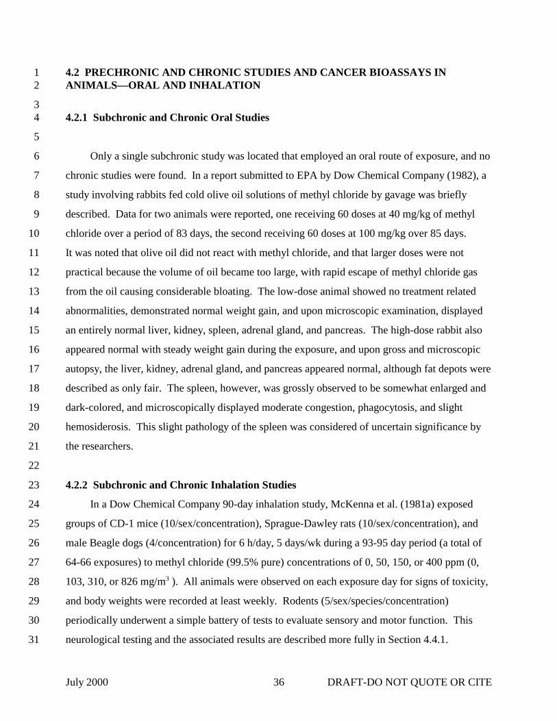
July 2000 DRAFT-DO NOT QUOTE OR CITE36
4.2 PRECHRONIC AND CHRONIC STUDIES AND CANCER BIOASSAYS IN1ANIMALS—ORAL AND INHALATION2
34.2.1 Subchronic and Chronic Oral Studies4
5
Only a single subchronic study was located that employed an oral route of exposure, and no6
chronic studies were found. In a report submitted to EPA by Dow Chemical Company (1982), a7
study involving rabbits fed cold olive oil solutions of methyl chloride by gavage was briefly8
described. Data for two animals were reported, one receiving 60 doses at 40 mg/kg of methyl9
chloride over a period of 83 days, the second receiving 60 doses at 100 mg/kg over 85 days. 10
It was noted that olive oil did not react with methyl chloride, and that larger doses were not11
practical because the volume of oil became too large, with rapid escape of methyl chloride gas12
from the oil causing considerable bloating. The low-dose animal showed no treatment related13
abnormalities, demonstrated normal weight gain, and upon microscopic examination, displayed14
an entirely normal liver, kidney, spleen, adrenal gland, and pancreas. The high-dose rabbit also15
appeared normal with steady weight gain during the exposure, and upon gross and microscopic16
autopsy, the liver, kidney, adrenal gland, and pancreas appeared normal, although fat depots were17
described as only fair. The spleen, however, was grossly observed to be somewhat enlarged and18
dark-colored, and microscopically displayed moderate congestion, phagocytosis, and slight19
hemosiderosis. This slight pathology of the spleen was considered of uncertain significance by20
the researchers.21
22
4.2.2 Subchronic and Chronic Inhalation Studies23
In a Dow Chemical Company 90-day inhalation study, McKenna et al. (1981a) exposed24
groups of CD-1 mice (10/sex/concentration), Sprague-Dawley rats (10/sex/concentration), and25
male Beagle dogs (4/concentration) for 6 h/day, 5 days/wk during a 93-95 day period (a total of26
64-66 exposures) to methyl chloride (99.5% pure) concentrations of 0, 50, 150, or 400 ppm (0,27
103, 310, or 826 mg/m3 ). All animals were observed on each exposure day for signs of toxicity,28
and body weights were recorded at least weekly. Rodents (5/sex/species/concentration)29
periodically underwent a simple battery of tests to evaluate sensory and motor function. This30
neurological testing and the associated results are described more fully in Section 4.4.1. 31

July 2000 DRAFT-DO NOT QUOTE OR CITE37
Hematological parameters (RBC, PCV, Hgb, total and differential WBC) were evaluated in all1
rats and dogs immediately prior to the initiation of exposure and at study termination, as were2
urinalyses (specific gravity, pH, sugar, protein, ketones, bilirubin, urobilinogen, occult blood). 3
Various clinical chemistry determinations were conducted at necropsy on all rats and dogs (blood4
urea nitrogen or BUN, alanine aminotransferase or ALT, aspartate aminotransferase or AST5
[dogs only], and alkaline phosphatase or AP). Two rats and 4 mice that died during exposure or6
were killed moribund were subjected to gross necropsy; 2 of the mice were also examined7
microscopically. At the study’s end, gross necropsy was performed on 40 male and 38 female8
rats, 36 male and 38 female mice, and 16 male dogs; the rats and dogs were fasted overnight,9
mice were not. Eye examinations were conducted, and organ weights for liver, kidneys, heart,10
and testes were recorded. Representative specimens from most major organs and tissues11
(somewhat fewer from mice) were obtained for microscopic evaluation from 0 and 400 ppm12
groups for all dogs and most rodents. Cerebrum, cerebellum, spinal cord, peripheral nerves, and13
brain stem were examined. Liver specimens were also obtained from low- and intermediate-14
dosed animals. Statistical significance (using analysis of variance and Dunnett’s test) was15
defined as p < 0.05.16
17
The daily clinical observations failed to reveal any signs or symptoms in any animal18
exposure group attributable to methyl chloride. However, statistical analyses of the rodent19
sensory and motor function tests revealed various differences between control and methyl20
chloride-exposed rodents; these statistical differences were judged either unrelated to methyl21
chloride exposure or of suspect toxicological significance, and are further discussed in22
Section 4.4.1. Some statistical differences in mean body weights between control and exposed23
groups were observed, but these were not dose-dependent or consistent with exposure duration,24
and were thus considered incidental effects unrelated to methyl chloride exposure. The only25
mean or relative organ weight findings that appeared consistent with a methyl chloride exposure26
effect were small, statistically significant increases in mean relative liver weights for male rats27
and female mice at 400 ppm. There were no gross or histopathological observations supportive28
of hepatotoxicity in either group, but statistically non-significant trends toward increased mean29
relative liver weights were noted in 400 ppm male mice and in 150 ppm male and female mice. 30
These observations, combined with the presence of equivocal histological evidence for liver31

July 2000 DRAFT-DO NOT QUOTE OR CITE38
toxicity in male mice, suggested that the slight liver weight effects may have been attributable to1
methyl chloride exposure.2
3
Upon hematological examination, 400 ppm male rats and male dogs each displayed small4
but statistically significant reductions in mean RBC. In rats, although this reduction was5
accompanied by a non-statistically significant decline in mean Hgb, similar findings were not6
seen in female rats; there were no indications of dose-response, and the mean RBC value was7
within the laboratory’s historical range. In dogs, the reduced mean RBC relative to controls was8
also observed in the groups prior to the initiation of exposure, with no significant change found9
in pre- versus post-exposure RBC values for the 400 ppm dogs. Thus, the statistical RBC10
reductions seen in 400 ppm rats and dogs were both considered within the range of normal11
variability, apparently unrelated to methyl chloride exposure and of no toxicological significance. 12
Similarly, the statistically significant reductions in mean AP activity noted in all methyl chloride-13
exposed dog groups relative to controls were also observed pre-exposure, and were therefore14
judged unrelated to methyl chloride exposure. No other clinical chemistry effects were noted. 15
Urinary specific gravities were significantly reduced in 400 ppm male rats and in 6 of 10 of the16
150 ppm female rats, but not in 400 ppm female rats. No other significant alterations in urinary17
parameters were observed, nor were any gross or microscopic pathological effects found in the18
kidneys. As a result, the authors considered the reduced urinary specific gravity an exposure-19
related effect, but were uncertain if it represented an adverse or simply a sub-clinical20
pharmacologic effect of methyl chloride.21
22
Pathological examination of rodents dying or sacrificed prior to study termination revealed23
no indication that the deaths were related to methyl chloride exposure, nor were any of the gross24
or microscopic pathological changes observed in rats sacrificed at study termination—all were25
considered incidental and typical of spontaneously occurring natural lesions commonly observed26
in rats of that age and strain. Similarly, most lesions observed in the mice were considered27
incidental and unrelated to methyl chloride exposure. However, subtle changes were noted in the28
appearance of some hepatocytes from the livers of 5 of 10 male mice in the 400 ppm group. 29
These changes involved altered tinctorial properties, perhaps due to altered vacuolation and30
basophilia, and were considered reversible. Similar changes were also observed in 2 of 9 control31

July 2000 DRAFT-DO NOT QUOTE OR CITE39
mice, and in 1 of 7 of the 150 ppm male mice. All dogs had lesions in the lungs suggestive of1
parasitic nematode infection, as well as other nonspecific lesions not attributable to methyl2
chloride exposure. Slight hepatocytic swelling was found in 2/4 of the 400 ppm dogs, 1/4 of the3
150 ppm dogs and 2/4 of the 50 ppm dogs, but not in control dogs. Because of the lack of an4
apparent dose response or any correlation with appreciably altered liver weights or clinical5
chemistry findings in individual dogs, this effect was also not considered exposure related. No6
clinically observable neuromuscular effects were observed nor were there any adverse effects on7
the CNS. In summary, this study failed to reveal any unequivocal evidence of toxicity related to8
methyl chloride exposure in mice, rats, or Beagle dogs, and suggests a NOAEL of 400 ppm for9
intermittent subchronic exposure.10
11
A 90-day inhalation study was also conducted by Battelle for the Chemical Industry12
Institute of Toxicology (CIIT) in F-344 rats and B6C3F1 mice (Mitchell et al., 1979a). This was13
conducted as a pilot study to select exposure levels for the 2-year chronic study described later. 14
Animals (10/sex/species/concentration) were exposed for 13 weeks, 5 days/wk, 6 h/day to methyl15
chloride (99.5% pure) at concentrations of 0, 375, 750, or 1,500 ppm (0, 774, 1,549, or16
3,098 mg/m3 ). All animals were observed daily and body weights were recorded weekly. Pre-17
and post-exposure ophthalmoscopic exams were performed, and organ weights (heart, adrenals,18
brain, testes/ovaries, spleen, liver, kidneys, lungs, and pancreas) were taken at necropsy. Urine19
was collected during Week 13 for analysis (specific gravity, pH, glucose, ketones, occult blood,20
protein, urine sediment), and just prior to necropsy, blood samples were collected for hematology21
(RBC, MCV, Hgb, total and differential WBC, reticulocytes, mean corpuscular hemoglobin22
[MCH], MCH concentration [MCHC], bone marrow myeloid:erythroid [M:E] ratio) and clinical23
chemistry (glucose, BUN, ALT, AST, AP, creatinine phosphokinase [CPK]) determinations. 24
Pathology examinations were conducted on tissues (including brain) from control and 1,500 ppm25
animals first; if a methyl chloride-related lesion was seen at the high concentration, lower26
concentrations were successively examined until the lesion was not found.27
28
The study’s summary reported that significantly lower body weights were observed in male29
and female 1,500 ppm rats during Weeks 3-13, and in male and female 750 ppm rats during30
Weeks 6-12. Increased relative liver weights were noted for male and female mice in the 75031

July 2000 DRAFT-DO NOT QUOTE OR CITE40
and 1,500 ppm groups. Generally, any observed changes in hematological or clinical chemistry1
parameters were considered within normal expected ranges, or were not clearly dose response-2
related. However, increased AST levels were observed in male mice of the 1,500 ppm group. 3
Vacuolar changes in the cytoplasm of hepatocytes were noted in the livers from 9 of 14 rats in4
the 1,500 ppm group (severe in 1, moderate in 5, mild in 2), from 7 of 18 rats in the 750 ppm5
group (severity distribution similar to control rats), and from 7 of 19 control rats (moderate in 1,6
mild in 6), and was approximately five times more prevalent in females than males. 7
Additionally, hepatic infarctions were observed in 1 male mouse and 1 female rat at 1,500 ppm. 8
Thus, effects on body weight (rats) and relative liver weight (mice) at 750 and 1,500 ppm, as well9
as on hepatic histology (mice and rats) at 1,500 ppm, were considered likely or potentially related10
to methyl chloride exposure. No histopathological effects in the brain in either the rat or mouse11
were observed.12
13
Battelle subsequently conducted a 24-month, chronic inhalation study in F-344 rats and14
B6C3F1 mice for CIIT (1981). Groups of animals (120/sex/species/concentration) were exposed15
6 h/day, 5 days/wk, for up to 24 months to concentrations of 0, 50, 225, or 1,000 ppm (0, 103,16
465, or 2,065 mg/m3) of 99.97% pure methyl chloride. These concentrations were based in part17
on the results of the 90-day pilot study previously described (Mitchell et al., 1979a). Interim18
sacrifices and toxicological evaluations were scheduled for 6, 12, and 18 months after initiation19
of the study. Mortality records were kept, and clinical observations, ophthalmic examinations,20
body and organ weight determinations, hematology, clinical chemistry and urinalysis21
determinations, and necropsy and histopathological examinations were conducted on the same22
battery tissues described for the 90-day subchronic study. These tissues included spinal cord,23
brain, liver, lung, testes, epididymis, and spleen. Animals were first subjected to a complete24
gross pathology examination, then a pre-selected battery of tissues (including brain) were25
examined by light microscopy and pre-selected organs weighed. Porphyrin fluorescence26
evaluations and neurofunction examinations were also performed.27
28
A six-month interim status report of this study was prepared by Mitchell et. al. (1979b). 29
Observations were based upon animals (10/sex/species/concentration) sacrificed after 6 months30
of exposure. Biologically significant increases in ALT levels were observed in male and female31

July 2000 DRAFT-DO NOT QUOTE OR CITE41
mice, but were not well correlated with individual animal histopathology. Similarly, decreased1
BUN values in some mice were not correlated with the presence of severe hepatic disease, and so2
were not considered of clinical significance. All other hematological variations were judged to3
be without clinical significance, and most other parameters were within normally expected4
ranges or did not exhibit convincing dose-response relationships. Significant decreases in5
absolute and relative kidney weights were found in 1,000 ppm male mice. Relative liver and6
kidney weights were significantly increased in male rats, as were relative lung weights in males7
at all concentrations and females at 1,000 ppm. In rats, chronic inhalation of methyl chloride8
appeared associated with testicular germ cell degeneration and sperm granulomas, interstitial9
pneumonia (1 male, 4 females), and subacute tracheitis in females. The levels at which these 10
effects occurred were not stated. In mice, associations with focal acute scleritis, hepatocellular11
degeneration, splenic lymphoid depletion, and multifocal thymic lymphoid necrosis were found. 12
The levels that these effects occurred in mice were not stated although it was indicated in the13
final report (CIIT, 1981) that they occurred at 1,000 ppm. Body weights were significantly14
depressed in 1,000 ppm male and female rats, in 1,000 ppm male mice, and in all concentration15
groups of female mice. No brain lesions were reported in the 6-month evaluation.16
17
The overall results of this chronic study were presented in the final report by CIIT (1981). 18
For mice, one exposure mistake was noted: on three consecutive days, the 50 ppm and 1,00019
ppm groups were switched and received each other’s exposure; although the effect of this error20
was considered negligible by the study authors, it may have affected the pathology results for21
spinal cord lesions (see below) in the 50 ppm group. Mouse mortality was increased in both22
sexes at the 1,000 ppm concentration, but was unaffected at 50 and 225 ppm; it was also elevated23
in males when compared with females as a result of male dominance fighting, which led to24
wounds around the prepuce and penile orifice that resulted in infection, urinary stasis, and death. 25
Clinical signs suggestive of CNS toxicity (e.g., tremor, paralysis) were noted only in 1,000 ppm26
mice, consistent with the neurofunctional impairment and cerebellar lesions described below. 27
No ophthalmic effects were observed in mice of either sex at any concentration, but28
neurofunctional impairment (clutch response) was found in nearly all 1,000 ppm mice of either29
sex after 18-22 months of exposure. This finding was supported by the histopathological30
observation of cerebellar lesions that first appeared in 1,000 ppm male and female mice at the31

July 2000 DRAFT-DO NOT QUOTE OR CITE42
18-month sacrifice. The lesion was characterized by degeneration and atrophy of the cerebellar1
granular layer and was seen in 3 of 7 males and 6 of 8 females, and in 17 of 18 females sacrificed2
at 22 months. The lesion was also found in 1,000 ppm mice that died spontaneously between3
0 and 17 months (15/24 males, 9/20 females), or between 18 and 22 months (45/47 males,4
35/37 females). It did not occur in the 0, 50, or 225 ppm groups. In the 18-24 month spontaneous5
death category, 35/37 females and 45/47 males in the 1,000 ppm group had cerebellar granular6
cell atrophy that was more extensive at 24 months than at 18.7
8
Thirteen of 18 females had minimal to moderate axonal swelling and degeneration of the9
lumbar spine nerves at the 22-month necropsy (only 1,000 ppm females examined). Similar10
lesions were found in 12/18 females in the thoracic spinal cord and 5/18 in the cervical spinal11
cord, lesions that were not present in 1,000 ppm animals at 18 months. At the 18-month interim12
necropsy, multifocal axonal swelling and degeneration (dorsal root of spinal cord) occurred in13
7/10 female mice (1/5 in males) at 50 ppm and 7/10 in females (2/5 in males) at 225 ppm, but14
none at 1,000 ppm; this was not observed in controls. Similar lesions were seen in the cauda15
equina of the spinal cord at 18 months. Tabular incidence was 3/5 (males) and 3/10 (females) at16
both 50 and 225 ppm, respectively [incidence in text ranged from 4/5 males and 10/10 females at17
50 and 225 ppm]; it also was observed in 3/7 males at 1,000 but not in any of the 8 females. This18
lesion occurred in 1/5 control males and 2/10 control females. It was indicated that axonal19
swelling and degeneration of minimal to mild severity was observed in all groups (not clear if20
this included controls) at 12 months.21
22
The growth rate of male 1,000 ppm mice was significantly depressed through the first23
18 weeks of exposure, with necropsy body weights significantly decreased in 1,000 ppm females24
at 6 and 12 months, and in 1,000 ppm males at 12 months. Among the statistically significant25
organ weight changes observed in male mice were: brain, absolute decrease (1,000 ppm, 6, and26
12 mo); kidney, absolute decrease (1,000 ppm, 6 and, 12 mo); and liver and lungs, relative27
increase (1,000 ppm, 12 mo). Those in female mice included: heart, absolute increase28
(1,000 ppm, 12 mo) and relative increase (1,000 ppm, 12 and, 18 mo; 225 ppm, 24 mo); brain,29
absolute decrease (1,000 ppm, 6, 12, and 18 mo); liver, absolute increase (1,000 ppm, 18 mo)30
and relative increase (1,000 ppm, 6, 12, and 18 mo); and kidneys, relative increase (1,000 ppm,31

July 2000 DRAFT-DO NOT QUOTE OR CITE43
12, and 18 mo). The relationship between brain weight decreases and the previously described1
cerebellar lesions was not known, nor were the reasons for the apparent sex-related difference in2
kidney weight response (although renal tumors were found only in males). Increased liver3
weight values, more prominent in female mice, did not correlate well with hepatic lesions, which4
were found more frequently in males.5
6
Significantly elevated ALT levels were observed in male mice (1,000 ppm at 6, 12, and7
18 mo; 50 and 225 ppm at 6 mo), which were consistent with histologically observed8
hepatocellular degeneration and necrosis at the 1,000 ppm concentration—lesions considered9
induced by methyl chloride exposure. Elevated ALT levels were also found in female mice at all10
methyl chloride concentrations at 6 and 12 months, but these were not correlated with any11
specific histopathological changes, and values were normal at 18 and 24 months. Significant12
reticulocytosis was observed in 1,000 ppm males at 6 months, and hypoglycemia in 50 and13
1,000 ppm males at 12 months; all groups, including controls, were hypoglycemic at 24 months. 14
These data were not correlated with any specific histopathological alterations. No other15
hematological, clinical chemistry, or urinalysis findings in mice could be related to methyl16
chloride. Porphyrin fluorescence evaluations of fresh liver impression smears made at necropsy17
were negative.18
19
The hepatocellular lesions (vacuolization, karyomegaly, cytomegaly, multinucleation,20
degeneration) first noted at 6 months in 1,000 ppm male mice were found with increasing21
frequency at 12 and 18 months, and were seen in the majority of males suffering unscheduled22
deaths. In less severe form, they also became apparent in approximately one third of the23
1,000 ppm females that died early during months 18-22. Renal tubuloepithelial hyperplasia and24
karyomegaly appeared in 1,000 ppm males at 12 months, subsequently increasing in incidence25
and severity until the last males in this group were sacrificed at 21 months. Renal tumors were26
significantly increased in 1,000 ppm males during months 12-21; 17 renal neoplasms were found27
in 13 animals (8 renal cortical adenomas, 4 adenocarcinomas, 2 papillary cystadenomas,28
2 tubular cystadenomas, and 1 papillary cystadenocarcinoma). These were considered induced29
by methyl chloride exposure, as were 2 adenomas (statistically nonsignificant in and of30
themselves) occurring in 225 ppm males at 24 months. A statistically significant increase in31

July 2000 DRAFT-DO NOT QUOTE OR CITE44
renal cortical cysts was seen at 18-22 months in 7 males and 1 female from the 1,000 ppm group,1
as well as in 1 male and 1 female from the 225 ppm group at 24 months. Also at 24 months,2
microcysts were observed in 6 males from the 50 ppm group, and 1 control male had a cyst. 3
Although their precise relationship to each other and to the other renal lesions was not clear,4
renal cyst and microcyst formation was considered by the investigators to be methyl chloride-5
related. However, the low incidence in the 225 ppm group suggests that it may be a spontaneous6
lesion. Unpublished data (Johnson, 1988) from eight 2-year mouse studies indicate that the7
incidence values for renal microcysts from the CIIT (1981) study fall within Dow Chemical8
Company’s historical control incidence for this strain. Seminiferous tubule atrophy and9
degeneration were also statistically significant and considered exposure-related in 1,000 ppm10
males. Finally, 1,000 ppm mice developed splenic atrophy and lymphoid depletion during11
months 6-22 that was considered related to methyl chloride exposure. Mice exposed to 225 ppm12
or less of methyl chloride developed little splenic lymphoid depletion or atrophy, but along with13
controls developed a high incidence of non-exposure-related splenic lymphoid hyperplasia, a14
relatively common condition in laboratory mice.15
16
In rats, mortality was not significantly affected at any concentration of methyl chloride. 17
Ophthalmologic examination revealed sporadic corneal cloudiness and opacity across all groups18
at 6 months that developed into a significant pattern in females, but not males, at 18 months. 19
Twelve-month observations were complicated by a different lesion, “corneal haze,” that appeared20
in control and 1,000 ppm rats, possibly a result of SDA virus outbreaks. By 24 months corneal21
cloudiness and opacity were similar in exposed and control rats of both sexes. Lenticular22
changes (prominent anterior lens sutures) were significant in both sexes at 18 months, although23
more males than females appeared affected at all concentrations. Both the corneal and lenticular24
lesions were considered possibly related to methyl chloride exposure. No neurofunctional25
impairments and no statistically or biologically significant alterations in hematological, clinical26
chemistry, or urinalysis parameters that could be attributed to methyl chloride exposure were27
found in rats. No clinical observations pertaining to neurotoxicity were recorded for rats, and as28
in mice, there was no indication that methyl chloride-exposed rats became porphyritic.29
30

July 2000 DRAFT-DO NOT QUOTE OR CITE45
Growth rates for 1,000 ppm male and female rats, and for 250 ppm females, were1
significantly reduced during the first 24 weeks of exposure. Necropsy body weights were2
reduced for 1,000 ppm males and females at all sacrifice periods, and for 225 ppm males at3
18 months; conversely, body weights were increased for 50 ppm males at the 24-month terminal4
sacrifice. Among the statistically significant organ weight changes observed in male rats were: 5
heart, absolute increase (1,000 ppm at 12 mo) and relative increase (1,000 ppm at 12 and 18 mo,6
225 ppm at 24 mo); brain, absolute decrease (1,000 ppm at 6, 18, and 24 mo) and relative7
increase (1,000 ppm at 12 and 18 mo, 225 ppm at 18 mo); testes, absolute decrease (1,000 ppm at8
18 and 24 mo) and relative decrease (1,000 ppm at 24 mo); lungs, absolute increase (50 and9
225 ppm at 6 mo) and relative increase (50, 225, and 1,000 ppm at 6 mo); adrenals, absolute10
decrease (1,000 ppm at 18 mo); liver, relative increase (1,000 ppm at 6 and 24 mo, 225 ppm at11
12 mo); and kidneys, relative increase (1,000 ppm at 6, 12, 18 [right only], and 24 [left only] mo)12
and relative decrease (50 ppm at 24 mo). Those in female rats included: brain, absolute decrease13
(1,000 ppm at 18 and 24 mo), relative increase (1,000 ppm at 12 and 24 mo); liver, absolute14
decrease (1,000 ppm at 12 and 24 mo), relative decrease (50 ppm at 24 mo); kidneys, absolute15
decrease (1,000 ppm at 24 mo), relative decrease (50 ppm at 24 mo), and relative increase16
(1,000 ppm [left only] at 6 and 12 mo); lungs, absolute decrease (1,000 ppm at 24 mo) and17
relative increase (1,000 ppm at 6 mo); spleen, absolute decrease (1,000 ppm at 12 mo); and heart,18
relative increase (1,000 ppm at 12 and 24 mo). Decreased body weights appeared to be the major19
contributing factor in most instances where changes in absolute weights did not correlate with20
those in relative weights. No body or organ weight changes occurring at exposure concentrations21
of less than 1,000 ppm were considered to be biologically relevant by the authors. It was noted22
that weight alterations in the brain, kidney, and heart were not reflected in any clinical or23
pathological findings, although the decreased brain weights could be a reflection of a methyl24
chloride effect similar to that seen in mice. No explanation was apparent for the complex25
changes in liver weights, although they generally paralleled those of the kidneys in both males26
and females. Heart weight changes were considered probably not biologically significant, and27
appeared in a number of instances to have reflected low heart weights in the controls and/or low28
final body weights.29
30

July 2000 DRAFT-DO NOT QUOTE OR CITE46
The testes were the only target organs examined in the rat that were considered to have1
significant gross or histopathological lesions related to methyl chloride exposure. Bilateral,2
diffuse degeneration and atrophy of the seminiferous tubules were only observed in 1/10 of3
1,000 ppm males at 6 months, 4/10 of 1,000 ppm at 12 months, and 10/20 at 18 months (uni- and4
bilateral and signs of age-related interstitial cell hyperplasia and adenomas). In contrast, no5
testicular lesions were observed in the other exposure groups at 6 or 12 months. However,6
beginning with the 18-month interim sacrifice, interstitial hyperplasia and/or adenomas was7
apparent, particularly in the control and 225 ppm group, such that there was no exposure-8
response based on incidence.9
10
At 24 months, all animals including controls had a background of interstitial cell11
hyperplasia and adenoma. Some histopathological observations suggested that methyl chloride12
exposure might increase interstitial cell hyperplasia while reducing the size of interstitial cell13
tumors. Epididymal sperm granulomas were seen in 2 male rats of the 1,000 ppm group at 614
months, and in 1 at 24 months. The small number of lesions, their time-course of appearance,15
and their observation in the control groups of other studies conducted in that laboratory16
precluded directly attributing sperm granuloma formation to methyl chloride exposure.17
18
The testicular results in rats are consistent with a LOAEL of 1,000 ppm, based on early19
signs of seminiferous tubule degeneration and atrophy in the absence of age-related degeneration.20
A NOAEL of 225 ppm appears reasonable since tubule degeneration and atrophy at this exposure21
level occurred upon onset age-related hyperplasia and compressive adenomas. In mice, a22
LOAEL of 50 ppm, the lowest concentration tested, was identified based on an exposure-related23
increased incidence of axonal degeneration and atrophy in areas of the spinal cord.24
25
4.3 REPRODUCTIVE/DEVELOPMENTAL STUDIES—ORAL AND INHALATION26
27
No human or animal studies on the reproductive or developmental effects of methyl28
chloride by the oral route of exposure were located.29
30

July 2000 DRAFT-DO NOT QUOTE OR CITE47
No studies were located in which reproductive or developmental effects were examined in1
humans after inhalation exposure to methyl chloride that occurred in the absence of significant2
exposure to other chemicals. A report by Kucera (1968) has been cited as describing a single3
case of an infant born with sacral agenesis to a mother exposed during pregnancy to methyl4
chloride and ammonia (John et al., 1984), and as describing an association of sacral agenesis in5
five infants born to mothers having close contact during pregnancy to “trichloroethylene and6
methyl chloride, among other industrial chemicals…” (Schardein, 1993).7
8
Huel et al. (1990) attempted to assess the potential adverse reproductive outcomes among9
former female workers in a New Mexico microelectronics assembly plant. After a matching10
process, 90 former worker-referent pairs were obtained that represented 302 and 32411
pregnancies, respectively. The study appeared to identify an increased risk of spontaneous12
abortion after the beginning of employment among the female workers when compared to13
referents (OR = 5.6, p < 0.01; OR = 4.0, p < 0.05 after taking into account the increased risk for14
subsequent spontaneous abortions in women who suffered them during previous pregnancies15
prior to employment). However, methyl chloride was only one chemical among many to which16
the women were exposed (e.g., chlorofluorocarbons, chlorinated hydrocarbons, glycol ethers,17
isopropanol, acetone, toluene, xylene, “alcohol”). Women reportedly suffered symptoms of18
intoxication on a daily basis, but the layout and manufacturing procedures of the plant did not19
permit any firm conclusions regarding the association of this apparent elevated risk of20
spontaneous abortion with exposure to particular organic solvents, including methyl chloride, and21
therefore is of limited value.22
23
In studies described elsewhere in greater detail (Sections 4.2.2 and 4.4.2), exposure of24
Sprague Dawley rats for 48-72 h to 500, 1,000, or 2,000 ppm (1,032, 2,065, or 4,130 mg/m3) of25
methyl chloride resulted in epididymal toxicity (e.g., degeneration, inflammation, interstitial26
edema, decreased sperm counts, sperm granulomas, scarring, and obstructive changes) that was27
dose-related, persistent through 12 days of recovery, and in some animals accompanied by28
testicular atrophy and degeneration (Burek et al., 1981). Clearly dose-dependent testicular29
degeneration (reduced numbers of late-stage spermatids, separation and luminal sloughing of30
spermatocytes and early-stage spermatids, vacuolated germinal epithelium, multi-nucleated giant31

July 2000 DRAFT-DO NOT QUOTE OR CITE48
cells) and accompanying epididymal lesions (e.g., reduced sperm counts, sloughed1
spermatocytes, giant cells, and cellular debris in tubules) were found in male F-344 rats exposed2
for 6 h/day on 9 of 11 days to 0, 2,000, 3,500, or 5,000 ppm (4,130, 7,228 mg/m3) of methyl3
chloride (Morgan et al., 1982). During the course of the CIIT chronic inhalation study4
(Section 4.2.2) (Mitchell et al., 1979b; CIIT, 1981), degeneration and atrophy of the seminiferous5
tubules were observed in the testes of F-344 rats and B6C3F1 mice at 1,000 ppm. The increased6
incidence of sperm granulomas in rats, especially after six months of exposure, was not7
considered by the investigators to be directly attributable to methyl chloride.8
9
A number of additional studies have also explored the effects of subacute exposure to10
methyl chloride on the reproductive physiology and performance of adult male F-344 rats11
(Chapin et al., 1984; Working et al., 1985a,b; Working and Bus, 1986; Chellman et al., 1987). 12
To characterize testicular and epididymal lesions and any associated effects on reproductive13
hormones, Chapin et al. (1984) exposed male F-344 rats to 3,500 ppm of methyl chloride,14
6 h/day, for 5 days, and after an interim of 3 days, for an additional 4 days. By Day 9 after the15
initiation of exposure, delay of spermiation (suggesting late-stage spermatids or Sertoli cells may16
be initial targets) and the occurrence of periluminal bodies were observed in the testes to a17
minimal degree; these lesions increased in severity through Day 19, with degeneration (cellular18
vacuolation and exfoliation) of the seminiferous epithelium beginning on Day 13, and the19
occurrence of a few giant cells on Day 19. The occurrence of round, periodic acid-Schiff stain20
(PAS) positive bodies in the tubular lumina of regions 1 and 2 of the epididymis was noted on21
Day 9, and decreased luminal sperm density on Day 11; these lesions were also observed in22
epididymal region 3, but with a lag period of approximately 2 days, and their severity increased23
for about 4 days before beginning to moderate. Bilateral sperm granulomas were first observed24
in epididymal regions 5 and 6 on Day 7 (in 1 of 8 rats), increasing in frequency through Day 1925
when they were found in all animals (6/6). The primary inflammatory response appeared to be26
directed against tubular epithelium, rather than extravasated sperm. After 5 days of exposure,27
circulating levels of testosterone were reduced from 120 ± 31 ng/mL in controls to <6 ng/mL. 28
However, when challenged with human chorionic gonadotropin (hCG) to test Leydig cell29
function, or luteinizing hormone releasing hormone (LHRH) to test pituitary function, methyl30
chloride-exposed and control rats responded similarly (although somewhat less strongly in the31

July 2000 DRAFT-DO NOT QUOTE OR CITE49
case of hCG) in terms of induced serum testosterone levels. Methyl chloride exposure for 6 h1
was also found to reduce NPSH levels in the testes, epididymis, and liver (relative to 0 h levels)2
by approximately 66, 79-87, and 92%, respectively, but not in blood, supporting a mechanism of3
enzyme-mediated conjugation of GSH with methyl chloride. After 8 weeks of recovery, about4
80% of the tubules in 4/5 rats still showed no evidence of spermatogenesis.5
6
Working et al. (1985a,b) conducted a two-part study on male Fischer 344 rats (80/group)7
that were exposed 6 h/day for 5 days to 0, 1,000, or 3,000 ppm (0, 2,065, or 6,195 mg/m3) of8
methyl chloride, or were injected with 0.2 mg/kg bw of triethylenemelamine (TEM, positive9
control). Each of forty males per exposure group was bred weekly to a single, untreated female10
for 8 wk in order to assess mating performance and dominant lethality (Working et al., 1985a),11
while the remaining 40 males/group were used to evaluate sperm quality and testicular12
histopathology (Working et al., 1985b). In a smaller second experiment, F-344 males (40/group)13
were exposed 6 h/day for 5 consecutive days to either 0 or 3,000 ppm methyl chloride, then bred14
as described for just 4 wk prior to sacrifice. At the end of the 5-day exposure period, body15
weights were significantly reduced (p < 0.05) relative to controls in the 1,000 and 3,000 ppm16
groups (2 and 16%, respectively), but returned to control levels by post-exposure Weeks 3 and 4,17
respectively. The percentages of females mated by males in the 1,000 ppm and TEM groups18
were not significantly different from controls at any week post-exposure, but were reduced for19
experiment-one 3,000 ppm males during the first 2 weeks post-exposure (Week 2, p < 0.05). In20
the second experiment, the percentages of mated females for control and 3,000 ppm groups were21
not significantly different (4-week averages: controls, 85.6%; 3,000 ppm males, 91.0%). 22
Fertility was not significantly reduced in 1,000 ppm males, but was significantly reduced in TEM23
(Week 2) and 3,000 ppm (Weeks 2 and 3, approximately 35-65% of control values) males; a24
small, statistically non-significant reduction persisted in the latter group through Week 16. 25
At termination (Week 17), unilateral or bilateral sperm granulomas were observed in the26
epididymides of 30% (12/40) of the 3,000 ppm males, but not in any of the control, 1,000 ppm,27
or TEM males.28
29
The dominant lethal test measures embryo loss in untreated female animals that have been30
mated with exposed males; this embryonic death results from genetic lesions (primarily structural31

July 2000 DRAFT-DO NOT QUOTE OR CITE50
and numerical chromosome abnormalities) or, in some cases, cytotoxic effects that were induced1
in the male germ cells (Olshan and Faustman, 1993; Working and Chellman, 1989). Embryonic2
or fetal loss occurring after implantation (postimplantation loss) is considered a direct measure of3
genotoxic effects, whereas conceptus loss prior to implantation (preimplantation loss) may reflect4
either genotoxicity or cytotoxicity that adversely affects sperm function. In this study (Working5
et al., 1985a), exposure to 1,000 ppm methyl chloride did not cause any consistently significant6
change in dominant lethality parameters relative to controls. However, 3,000 ppm produced a7
small but significant (p < 0.05) increase in postimplantation loss of 9.5% for post-exposure8
Week 1 matings; thus, the relevant genetic lesions appeared to be induced only in sperm residing9
in the vas deferens and epididymis during methyl chloride exposure. In contrast, increases of10
preimplantation loss (approximately 14-94% over controls) were observed for all 8 weeks of11
mating; the maximum increase was for Week 2, and increases were statistically significant for12
Weeks 2-4, 6, and 8. Significantly elevated rates of preimplantation loss were observed for13
matings of TEM-treated males during post-exposure Weeks 2 and 3, and of postimplantation loss14
during Weeks 1-5. Dominant lethal genetic effects typically demonstrate a coupling of increased15
postimplantation loss with decreased preimplantation loss as matings progress through the post-16
exposure period; this pattern was observed with TEM, but not with methyl chloride.17
18
In the companion study (Working et al., 1985b), no consistent differences in sperm quality19
or testicular histopathology parameters relative to controls were observed in males from the20
1,000 ppm methyl chloride or TEM groups. In the 3,000 ppm group, testicular weight was21
significantly depressed (p < 0.05) at Weeks 3-8 post-exposure, recovering to near-control levels22
by Week 16. Beginning at Week 2, just over 50% (20/39) of the animals displayed gross or23
microscopic sperm granulomas in the caudal epididymis; these increased in size and severity24
over time, and were unilateral in some cases, bilateral in others. Histologically, the testes25
evidenced a delay in spermiation, chromatin margination in round spermatids, epithelial26
vacuolation, luminal exfoliation of spermatogenic cells, and multinucleated giant cells. 27
Testicular spermatid head count was significantly depressed during Weeks 2-8. While sperm28
count in the vas deferens began declining by Week 2, it was significantly reduced during29
Weeks 3-8, and still appeared somewhat depressed at Week 16. The frequencies of intact and30
motile sperm observed during Weeks 1-6 were generally significantly depressed (3.5-69% of31

July 2000 DRAFT-DO NOT QUOTE OR CITE51
controls), but had recovered by the end of 16 weeks. Furthermore, increases in morphologically1
abnormal sperm isolated from the vasa deferentia were seen during the first 5 weeks post-2
exposure; these increases were significant (p < 0.05) for Weeks 1-3, dramatically peaking at3
approximately 70% for Week 3. These companion studies indicate that exposure for 6 h/day on4
5 consecutive days to 3,000 ppm of methyl chloride resulted in toxicity to sperm located in the5
epididymis and vas deferens, which resulted in a small increase in dominant lethality expressed6
as postimplantation loss, presumably due to genetic lesions. Exposure also caused toxic effects7
to spermatids, primary spermatocytes, and spermatogonial stem cells in the testes, as well as8
chronic epididymal inflammation. Because of this inflammation, the resulting sperm9
granulomas, and the atypical pattern of pre- and postimplantation fetal loss induced by methyl10
chloride, the authors speculated that the observed increases in infertile matings and11
preimplantation loss may be largely the result of failure of fertilization because of low sperm12
number and poor sperm quality (i.e., cytotoxic rather than genotoxic mechanisms). The13
reproductive NOAEL and LOAEL, based upon the parameters described in these companion14
studies (Working et al., 1985a,b), are 1,000 and 3,000 ppm, respectively.15
16
To further explore the potential role of cytotoxicity in the preimplantation losses induced by17
methyl chloride, Working and Bus (1986) conducted a similar study in which male F-344 rats18
were exposed to 0 (n = 30), 1,000 (n = 10), or 3,000 ppm methyl chloride (n = 20), 6 h/day for19
5 consecutive days. Positive control males (n = 10) received a single ip injection of 0.2 mg/kg20
TEM on exposure Day 5. Following the exposure period, each male was bred weekly for up to21
8 weeks to 2 untreated females. Twelve hours post-mating, females were sacrificed and their ova22
recovered and scored as fertilized or unfertilized. The fertilization rate from control matings was23
approximately 88% (602/683), and rates were not significantly depressed from this level in the24
1,000 ppm (80%) or TEM (77%) groups. However, fertilization rate was significantly depressed25
(p < 0.05) in the 3,000 ppm group through the 8 weeks of mating, ranging from 3.4% (14/415,26
Week 2) to 72.3% (159/221, Week 8). Furthermore, the 8-week patterns of unfertilized ova in27
this study and preimplantation loss in the dominant lethal assay coincided, with the percentage of28
unfertilized ova equaling or exceeding that of preimplantation loss for each post-exposure week29
of mating. By contrast, unfertilized ova could account for only a little more than a third of the30
preimplantation losses induced by the genotoxicant, TEM. When fertilized ova were cultured31

July 2000 DRAFT-DO NOT QUOTE OR CITE52
in vitro for 24 h, only the TEM-exposed ova displayed a significant decrease from controls (851
versus 96%) in the percentage of ova that underwent cleavage. Collectively, these data support2
the authors’ conclusion that methyl chloride-induced preimplantation loss in the dominant lethal3
assay results from cytotoxicity-mediated failure of fertilization, rather than from genotoxic4
effects.5
6
An additional study conducted by the same laboratory (Chellman et al., 1987) explored7
whether the observed cytotoxic effects on sperm were the result of methyl chloride’s effects on8
the testes or on the epididymides. Groups of male F-344 rats (18/group) were exposed 6 h/day9
for 5 consecutive days to 0 or 3,000 ppm methyl chloride, with or without concurrent exposure to10
the anti-inflammatory agent 3-amino-1-[m-(trifluoromethyl)phenyl]-2-pyrazoline (BW755C),11
10 mg/kg by ip injection, 1 h pre- and post-exposure. Earlier studies indicated that co-treatment12
with BW755C prevented methyl chloride’s induction of both epididymal inflammation and the13
increased postimplantation loss observed in the dominant lethal assay for matings at 1 and 214
weeks post-exposure (Chellman et al., 1986a). These findings suggested that methyl chloride’s15
induction of epididymal inflammation may be responsible for producing the DNA damage that16
results in postimplantation loss, perhaps via the generation of reactive oxygen intermediates17
(Chellman et al., 1986a, 1987; Working and Chellman, 1989). In contrast, BW755C did not18
mitigate the methyl chloride-induced increases in preimplantation loss that were observed19
beginning at Weeks 2 and 3 post-exposure.20
21
In this study (Chellman et al., 1987), exposure to 3,000 ppm methyl chloride significantly22
reduced the post-exposure relative organ weights of the seminal vesicles (Week 1), epididymides23
(Weeks 2 and 3) and testes (Week 3); similar reductions also were observed in rats co-exposed to24
methyl chloride and BW755C, but not in controls or animals treated only with BW755C (except25
for Week 2 seminal vesicles in the latter). Exposure to methyl chloride resulted in testicular26
histopathology characterized by delayed spermiation, disorganization, and destruction of the27
seminiferous epithelium, and marked, persistent decreases in the number of mid- to late-stage28
spermatids in the testes; again, co-exposure to BW755C did not prevent these degenerative29
changes. Daily sperm production in the testes was significantly reduced during Weeks 1-3 by30
methyl chloride exposure, with or without BW755C. Methyl chloride-exposed males displayed31

July 2000 DRAFT-DO NOT QUOTE OR CITE53
visible uni- or bilateral epididymal sperm granulomas (4/18 animals), and inflammatory cells,1
debris, and nucleated cells (8/18 animals) in the epididymal tubular lumina; no such effects were2
observed in the methyl chloride + BW755C group. Sperm numbers were normal in the vas3
deferens of the methyl chloride-exposed group at Weeks 1 and 2, but were drastically reduced at4
Week 3; percentages of motile sperm were significantly reduced at Weeks 1 and 2, severely so at5
Week 3, when the percentage of morphologically abnormal sperm was also greatly increased6
(99 versus 2% in controls). Co-exposure to BW755C did not prevent these adverse effects on7
sperm. Given these findings, in conjunction with known epididymal transit times for sperm in8
F-344 rats, the authors concluded that the preimplantation losses observed at Weeks 2 and 3 post-9
exposure (and later) in dominant lethal experiments likely resulted from methyl chloride’s10
cytotoxic effects on sperm located in the testes during exposure, rather than in the epididymides.11
12
A two-generation reproduction study using F-344 rats was conducted by Hamm et al.13
(1985). Animal groups (40 males, 80 females/concentration) were exposed by inhalation to 0,14
150, 475, or 1,500 ppm ( 0, 310, 981, or 3,098 mg/m3) methyl chloride, 6 h/day, 5 days/wk for15
10 weeks. Exposure was then increased to 7 days/wk, and each F0 male was mated with two F016
females during the course of 2 weeks (1 female/wk). After the two-week breeding period, 10 F017
males/group were necropsied and the remaining 30/group were each bred for 2 weeks to two18
unexposed females; after an additional 10 weeks of recovery, 10 more F0 males/group were19
necropsied and the remaining 20/group were each bred to two unexposed females; finally, after20
an additional 18 weeks of recovery, another 10 F0 males/group were necropsied and the21
remaining 10/group were each bred to two unexposed females. Pregnant F0 females, bred to F022
males during Weeks 10-12, continued to be exposed to methyl chloride, except from Gestation23
Day 18 through Postnatal Day 3; exposure was resumed 6 h/day, 7 days/wk during Postnatal24
Days 4-28, when pups were weaned. Pups were never directly exposed to methyl chloride. After25
weaning, 15 F1 pups/sex and 25 F0 females were necropsied. The same exposure regimen, but26
without the recovery breeding protocol, was utilized for F1 matings (40 males, 80 females for 027
and 150 ppm; 23 males, 46 females for 475 ppm).28
29
Body weight gains were depressed (10-20%) at all weekly weighings after 2 weeks of30
exposure for both sexes in the 1,500 ppm group, and in the 475 ppm group (5-7%) after Day 57. 31

July 2000 DRAFT-DO NOT QUOTE OR CITE54
Exposure-related lesions were observed only in 1,500 ppm F0 males, and included degeneration1
and atrophy of the seminiferous tubules (10/10 males examined), as well as epididymal sperm2
granulomas (3/10); the testes of these animals weighed only approximately half as much as those3
of control and lower-dose groups. The number of females that mated (as evidenced by the4
presence of copulation plugs) did not vary by exposure group, but no litters were produced in5
exposed or unexposed females that were sired by 1,500 ppm males. The number of males proven6
fertile was also statistically lower in the 475 ppm group. No consistently significant differences7
in litter size, sex ratio, pup viability, survival, or growth were observed in the 150 or 475 ppm8
groups relative to controls. Body weight for the 1,500 ppm males remained significantly lower9
than for controls during the first 9 weeks of recovery, when it then became comparable. After10
10 weeks of recovery, the percentage of fertile 475 ppm males was not significantly different11
from controls, and that of 1,500 ppm males had partially recovered (25 versus 75% for controls);12
further improvement in the 1,500 ppm group was not seen after an additional 18 weeks of13
recovery. Also, testicular weights in the 1,500 ppm group remained less than in controls, but no14
epididymal sperm granulomas were observed. Methyl chloride exposure (150 or 475 ppm) did15
not cause statistically significant effects on F1-generation fertility, although 475 ppm litters had a16
decreased percentage of males (41 ± 16% versus 51 ± 18% in controls, 53 ± 13% 150 ppm17
litters) and reduced pup weights for Days 14-21 (but not earlier or later). Based upon a marginal18
reduction in the number of males proven fertile, the two-generation reproductive LOAEL in this19
study was 475 ppm with a corresponding NOAEL of 150 ppm. As noted by the authors, the20
actual NOAEL may in fact have been closer to the 475 ppm concentration, where the reduction in21
number of fertile males was only statistically significant when matings with exposed (2) and22
unexposed (2) females were combined, and not when considered separately.23
24
Wolkowski-Tyl et al. (1983a,b) conducted evaluations of the structural teratogenicity of25
methyl chloride in rats and mice after inhalation exposure. Groups of bred female F-344 rats26
(25/concentration) and female C57BL/6 mice bred to C3H males (33/concentration) were27
exposed 6 h/day to methyl chloride concentrations of 0, 100, 500, or 1,500 ppm ( 0, 206, 1,032 or28
3,098 mg/m3). Rats were exposed from Gestation Days 7 through 19, then were sacrificed on29
Gestation Day 20; mice were exposed from Gestation Days 6 through 17 with sacrifice on30

July 2000 DRAFT-DO NOT QUOTE OR CITE55
Gestation Day 18, except for the 1,500 ppm group, in which the animals were sacrificed in1
extremis during Gestation Days 10-14. In rats, no behavioral toxicity in the dams was seen at2
any concentration of methyl chloride, but at 1,500 ppm there was evidence of both maternal3
toxicity (depressed weight gain during exposure and body weight at sacrifice) and fetal toxicity4
(reduced male and female fetal body weights, reduced female crown-rump length at Gestation5
Day 20). There were no exposure-related effects on implantations, resorptions, dead or live6
fetuses, or sex ratio; the authors speculated that methyl chloride’s effects were expressed late in7
gestation and were probably secondary to maternal toxicity. No exposure-related fetal8
malformations were observed (external, visceral, or skeletal), although some evidence of delayed9
ossification was observed in the 1,500 ppm fetuses. The study authors concluded that methyl10
chloride was not teratogenic in pregnant female F-344 rats exposed to maternally and fetotoxic11
concentrations during the critical periods of fetal and embryo development.12
13
Pregnant mice of the 1,500 ppm group displayed urogenital bleeding and CNS dysfunction14
(hunched posture, walking on “tip-toes”, tremors, imbalance) commencing on Gestation Days915
(the fourth day of exposure), of severity that required premature sacrifice on Gestation16
Days 10-14 (exposure Days 5-9). Upon necropsy, these animals exhibited necrosis of neurons in17
the internal granular layer of the cerebellum. No reproductive parameters were significantly18
affected by methyl chloride exposure. Statistically non-significant trends for increasing body19
weights and crown-rump lengths with increasing concentration were seen in male and female20
fetuses; the only significant finding (p < 0.05) was increased crown-rump length in male fetuses21
of the 100 ppm group. No significant, exposure-related external or skeletal anomalies were22
observed in the fetuses of either the 100 or 500 ppm group. However, visceral examination of23
50% of the mouse fetuses revealed a small but statistically significant increase in the incidence of24
a heart anomaly in the 500 ppm group. This lesion involved the tricuspid valve, chordae25
tendineae, and papillary muscles in the right atrioventricular septum (6 fetuses), or the bicuspid26
valve, chordae tendineae, and papillary muscles in the left atrioventricular septum (3 fetuses). 27
The lesion was found in 6 of 17 litters and in both sexes (3 males, 6 females), with no single28
fetus having both sides involved; 1 litter had right and left side involvement, and 5 of 6 affected29
litters also had fetuses with normal appearing hearts.30
31

July 2000 DRAFT-DO NOT QUOTE OR CITE56
To further explore this apparent teratogenic effect in B6C3F1 fetuses, 74-77 female1
C57BL/6 mice impregnated by C3H males were exposed 6 h/day from Gestation Days 6 through2
17 to 0, 250, 500, or 750 ppm (0, 516, 1,032, or 1,548 mg/m3) of methyl chloride (Wolkowski-3
Tyl et al., 1983b). Beginning on the seventh day of exposure (Gestation Day 12), females in the4
750 ppm group displayed ataxia, tremors, convulsions, and hypersensitivity to touch or sound;5
prior to the scheduled sacrifice, 6 of these animals died and 1 was killed in extremis. When6
compared with controls, survivors of this group were the only ones to exhibit significant7
reductions in body weight gain during gestation, body weight on Gestation Day 18, and absolute8
weight gain (weight gain minus gravid uterine weight). No exposure-related differences were9
observed in pregnancy rate, gravid uterine weight, maternal liver weight, numbers of10
implantations, resorptions, dead fetuses, nonlive (dead plus resorbed) fetuses, live fetuses, sex11
ratio, or mean fetal weight per litter. However, statistically significant (p < 0.05) and12
concentration-related trends were observed for the numbers and percents of malformed or13
affected (nonlive plus malformed) fetuses per litter. All but one malformation (an umbilical14
hernia, 250 ppm group) were found in the heart and included reductions in the number of15
papillary muscles and/or chordae tendineae on the right associated with the tricuspid valve,16
globular hearts, tricuspid valve defects, small right ventricle, and white deposits (calcium-17
containing) in the left ventricular wall. Defects were more common in females (23) than males18
(14), with 1 fetus in the 500 ppm group and 3 in the 750 ppm group exhibiting multiple heart19
malformations. Incidences of malformations were: 0 ppm (3/433 fetuses, 3/58 litters), 250 ppm20
(7/458 fetuses, 6/62 litters), 500 ppm (11/444 fetuses, 7/62 litters), and 750 ppm (17/400 fetuses,21
14/56 litters). Relative to controls, there were statistically significant increases in the numbers of22
750 ppm litters, male fetuses, and female fetuses with malformations; increases across all groups23
were seen for the numbers of fetuses with any malformations (p < 0.025) and with heart24
malformations (p < 0.005). The researchers concluded that in pregnant C57BL/6 female mice25
and their B6C3F1 conceptuses, methyl chloride exposure during gestation (Gestations Days 6-17)26
was maternally toxic at 750 ppm and teratogenic at 750 and 500 ppm, causing heart27
malformations. No maternal, embryo or fetotoxicity, nor any teratogenicity, was observed at the28
250 ppm concentration.29
30

July 2000 DRAFT-DO NOT QUOTE OR CITE57
Findings of teratogenicity in the mouse have been questioned (John-Greene et al., 1985). 1
These authors discuss the unusual nature and rarity of the heart lesion described by Wolkowski-2
Tyl et al. (1983a,b), its apparent dissimilarity to lesions reported in human and hamster hearts, its3
relatively low incidence in methyl chloride-exposed animals and its occurrence in controls, the4
relatively uncommon use of the B6C3F1 hybrid in teratogenicity testing, the technical difficulties5
involved in detecting the lesion without introducing artifacts, and the interanimal variability in6
appearance of the papillary muscles. Furthermore, when conducting blindly-scored studies on7
animals exposed continuously to 300 ppm for 24 h (Gestation Days 11.5-12.5, the developmental8
stage they considered the most critical for cardiac structures), or 1,000 ppm for 12 h (Gestation9
Days 11.5-12), John-Greene et al. (1985) were unable to demonstrate the lesion’s occurrence. 10
In response, Tyl (1985) acknowledged and shared some of these concerns, but pointed out that11
the lesion was found in two studies using different personnel and examination procedures, and in12
one study was confirmed by another pathologist and staff, and that great care was used in cutting13
the hearts for examination. Tyl (1985) also pointed out the differences in exposure regimen,14
6 h/day for Gestation Days 6-17 versus 12-24 continuous hours on Gestation Day 11.5 to15
Gestation Day 12-12.5, discussing in particular why exposure beyond Gestation Day 12-12.516
could be critical for generation of the lesion. Until further work is reported, methyl chloride17
should prudently be considered a mouse teratogen.18
19
Finally, the report by Bus et al. (1980) previously discussed in Sections 3.2 and 3.320
indicates that methyl chloride can cross the placenta and cause potentially adverse effects in the21
fetus. Pregnant F-344 rats were exposed on Gestation Day 19 for 6 h to 1,500 ppm methyl22
chloride. Maternal liver and kidney NPSH levels were maximally depressed immediately after23
the end of exposure, but had returned to normal by 8 h. Fetal placental NPSH, also maximally24
depressed at 0 h post-exposure, returned to normal within 4 h, but fetal liver and carcass NPSH25
decreases were not maximal until 2 h post-exposure. These decreases were substantial (66.8 and26
71.0%, respectively) and had not yet returned to normal by 8 h post-exposure.27
28
29
30
31

July 2000 DRAFT-DO NOT QUOTE OR CITE58
4.4 OTHER STUDIES1
24.4.1 Neurological Studies3
4
The study on male and female volunteers by Stewart et al. (1980), previously discussed in5
Sections 3.1-3.4 and 4.1.1, also included several neurological and cognitive tests on its subjects,6
who were exposed sedentarily for up to five consecutive days, up to 7.5 h/day, to methyl chloride7
concentrations of 0, 20, 100, “100f” (fluctuating 50-150), or 150 ppm. During the first 5 and last8
10 min of exposure, each subject performed modified Romberg and heel-to-toe equilibrium tests9
(standing on one leg with arms at sides for $3 sec, and walking heel-to-toe in a straight line for10
~5 ft, first with eyes open and then with eyes shut) that were videotaped. Group I subjects also11
had spontaneous electroencephalograms (EEGs) and visual evoked responses (VERs) recorded 412
times/day, 3 days/wk during their 7.5 h/day, 5 day/wk exposure regimen. EEGs were visually13
inspected, and VER wave complexes were analyzed for amplitude, latency, and wave form; data14
from the three methyl chloride exposure concentrations were compared with those from the15
0 ppm control sessions. Groups I and II also received a battery of five cognitive tests on Days 1,16
3, and 5 of each exposure week, beginning 2-3 h after the commencement of exposure. In order17
of performance, the tests included: (1) a 10- and 30-sec time estimation test; (2) the Marquette18
Time Estimation Test, involving a series of light and tone stimuli; (3) a coordination test19
(Flanagan Aptitude Classification Tests, 7A, Coordination); (4) a two-part arithmetic test20
involving addition and subtraction, then multiplication and division; and (5) a visual inspection21
test involving spotting the number “3” from a page containing rows of random numbers.22
23
No significant neurological abnormalities were observed in any of the male or female24
individuals exposed to methyl chloride, even at 150 ppm for 7.5 h/day, 2 days/1 wk, or 100 ppm25
(or 100f ppm) for 7.5 h/day, 5 days/1 wk. Equilibrium remained subjectively normal during26
exposures, as did EEG recordings and VER tracings. No statistically significant effects of27
methyl chloride exposure on the cognitive tests were apparent, with one exception. Group I28
males (exposed for 7.5 h/day) changed from overestimating the length of the light stimulus on29
the Marquette test, to underestimating it with increasing methyl chloride concentration. 30
However, the researchers considered this finding to be spurious because the same shift was not31

July 2000 DRAFT-DO NOT QUOTE OR CITE59
observed for the sound stimulus, nor did it occur in the Group II (3-h exposure) male subjects. 1
They also drew attention to the lack of any observed decrement in arithmetic task performance, 2
noting that while this result appeared to differ from the findings of Repko and colleagues (see3
below), the arithmetic tasks in this study were not part of a multi-tasking regimen as they were in4
the Repko study. The researchers concluded that several days exposure of male or female5
volunteers to as much as 150 ppm methyl chloride for 7.5 h/day did not result in any biologically6
significant decrements in their neurological or cognitive functioning.7
8
The Dow Chemical Company (1992) report on six cases of methyl chloride intoxication in9
humans, previously discussed in Section 4.1.1, is one of numerous case history studies reviewed10
by Repko (1981), although the published version was reviewed (Scharnweber et al., 1974). 11
Collectively, these case histories are summarized to demonstrate that symptoms of methyl12
chloride exposure indicative of CNS toxicity in humans include headache, nausea, double vision,13
mental confusion, vomiting, anuria, nervousness, emotional instability, ataxic gate, low blood14
pressure, insomnia, anorexia, vertigo, tremor, convulsions, increased pulse and respiration rates,15
coma, and eventual death. As previously noted, generally (but perhaps not always) these16
symptoms seem completely reversible, although some memory loss of events during exposure17
may remain. Repko (1981) also notes that several case histories and medical reports have18
described specific degeneration of portions of the CNS (cerebral cortex to the sacral part of the19
spinal cord), frontal and parietal atrophy, lipoid-filled histocytes in the leptomeninges of the20
cerebral hemispheres, generalized hyperemia of the cerebral cortex, nerve cell degeneration, etc.21
22
Also reviewed by Repko (1981) is an earlier study (Repko et al., 1976) that attempted to23
quantitatively investigate the neurological and behavioral effects of low-level occupational24
exposure to methyl chloride. A group of 122 methyl chloride workers was compared with25
another group of 49 workers presumably not exposed to methyl chloride. The mean ambient air26
concentration of methyl chloride was determined to be 33.6 ppm. Both groups were reported to27
be asymptomatic with respect to either acute or chronic methyl chloride intoxication, and28
neurological examinations revealed no significant differences between them, including29
dysrhythmias determined from EEG tracings. Quantitative test results were reported to be30
correlated with ambient air levels of methyl chloride and with urine pH, but not with breath31

July 2000 DRAFT-DO NOT QUOTE OR CITE60
levels of methyl chloride. Group comparison suggested that methyl chloride exposure adversely1
affected performance on cognitive time-sharing tasks and magnitude of finger tremor, but not on2
various psychological and personality tests. These results implied that methyl chloride exposure3
can cause “a decrease in speed and accuracy of simple tasks and a dimintion [sic] of the ability to4
time-share in more complex performance or work situations…at ambient levels of methyl5
chloride below 100 ppm” (Repko, 1981). However, the study has been severely criticized6
(Farber and Torkelson, 1989) on the grounds that the control group was much younger than the7
exposed group, the controls were measured later in the study and at a different location, and the8
methyl chloride workers had previously been exposed to higher concentrations of methyl9
chloride, thus confounding the relationship of any observed effects to the reported low-level10
exposure (33.6 ppm). Anger and Johnson (1985) also suggested that the subjective opinion of11
the neurologist that 58% of the control subjects (versus 57% of the exposed group) were12
abnormal in some way was problematic, and they expressed concern that the data interpretation13
was based upon t-tests that were uncorrected for the total number of dependent variables. 14
Nonetheless, given the large number of significant differences, these reviewers considered it15
likely that chronic exposure to methyl chloride at concentrations approximating 35 ppm resulted16
in some worker behavioral changes.17
18
Putz-Anderson et al. (1981) studied the behavioral effects on 56 humans (39 males,19
17 females; 8 or 12/group) of inhaling 0, 100, or 200 ppm of methyl chloride for 3 h, with or20
without concurrent ingestion of 10 mg of diazepam (Valium). Participants were evaluated on21
three performance tests: a visual vigilance task (similar to an automated version of Mackworth’s22
Clock Test), a dual task (tone detection and eye-hand compensatory tracking), and a time23
discrimination task. Three performance indices were obtained: threshold performance level24
(TPL), which approximated a 70% accuracy level; response time (RT); and number of response25
blocks (RB), defined as a longer-than-usual RT (i.e., greater than the mean RT plus two SDs of26
the baseline data). The net decline in mean performance across the three tasks was 4% for27
200 ppm methyl chloride (marginally significant, p < 0.053), while for diazepam it was a28
significant 10.1% (p < 0.01); no potentiation was observed, as the performance decrement for29
exposure to 200 ppm methyl chloride plus10 mg diazepam was 13.5%, or essentially additive. 30
Only the visual vigilance TPL and the time discrimination RB indices were sensitive indicators31

July 2000 DRAFT-DO NOT QUOTE OR CITE61
of methyl chloride impairment, whereas all indices except the time discrimination RB were1
significantly affected by diazepam exposure. In line with other studies described in the2
toxicokinetic sections, these authors found that 3 of 24 subjects exposed to 200 ppm had3
significantly higher breath levels (> 100 ppm) of methyl chloride than did the remainder, and that4
blood levels were highly correlated with breath levels. This study indicates a marginal5
behavioral effect of a 3-h exposure to 200 ppm of methyl chloride (behavioral testing of the6
100 ppm group was not performed), an effect that was not potentiated by concurrent exposure to7
the commonly prescribed CNS depressant diazepam (effects were merely additive).8
9
Neurotoxic effects resulting from inhalation exposure to methyl chloride have also been10
described in a number of animal studies. As described in Section 4.4.2, Morgan et al. (1982)11
exposed groups of F-344 rats (10/sex/concentration) 6 h/day to 0, 2,000, 3,500, or 5,000 ppm (0,12
7,228 or 10,325 mg/m3) methyl chloride for 5 days, then for another 4 days after a 2-day break13
(i.e., on Days 1-5 and 8-11). Among other effects in the 5,000 ppm group, forelimb ataxia was14
observed in most females and in about half the males on Day 3, hindlimb paralysis developed in15
2 males and 1 female on Day 5, and 1 female suffered convulsions during the Day 11 exposure. 16
Following the Day 5 exposure, 13 rats were sacrificed in extremis (5,000 ppm; 6 males and17
5 females; 3,500 ppm; 2 females), but the survivors seemed to tolerate the methyl chloride18
exposure much better during the second week (Days 8-11). At the high concentration, 3 of 1019
males and 2 of 9 females (Table 4-1) displayed minimal histopathological evidence of cerebellar20
degeneration (foci of internal granular layer cells having pyknotic nuclei with distended, hydropic21
perikarya). This study demonstrated a NOAEL and LOAEL for neurotoxicity in F-344 rats22
following subacute exposure to methyl chloride of 3,500 and 5,000 ppm, respectively.23
24
Jiang et al. (1985) conducted an ultrastructural study of lesions induced in the cerebella of25
C57BL/6 mice (10 females/concentration) exposed 6 h/day, 5 days/wk for 2 weeks to 0 or26
1,500 ppm (0 or 3,098 mg/m3) methyl chloride. Two mice died during the second week of27
exposure, and several displayed ataxia; no clinical signs of CNS disturbance were noted in the28
other animals, including those displaying histological evidence of severe cerebral degeneration,29
and no gross abnormalities of the brain or other organs were observed. Under light microscopy,30
two types of lesions were found in inner granular layer cells of the cerebellum (most frequently in31

July 2000 DRAFT-DO NOT QUOTE OR CITE62
the ventral paraflocculus: a coagulative necrosis (also seen in controls, but in milder form and in1
substantially fewer cells) involving nuclear and cytoplasmic condensation; and a focal malacia2
involving edema in groups or extensive areas of cells, with nuclear condensation, karyorrhexis,3
necrosis, separation of myelinated axons, and microvacuolation. Electron microscopy confirmed4
the type one lesion, showing pyknotic nuclei without cytoplasmic edema, but with variable5
disruption of organelles. Areas of malacia exhibited characteristics ranging from perikarya6
edema of granule cells to near complete destruction of all tissue components with the exception7
of blood vessels (nuclear pyknosis and condensation, karyorrhexis, organelle remnants, etc.). 8
Few abnormalities were observed in the kidneys of treated females (slight degeneration of9
proximal tubules with some proteinaceous material in tubular lumina was seen in two animals),10
leading the researchers to conclude that the brain lesions were most likely not a secondary effect11
of renal toxicity (these types of brain lesions had been associated with renal insufficiency in12
humans).13
14
In a study more fully described in Section 4.4.2, Landry et al. (1985, 1983c) exposed15
female C57BL/6 mice (12/group) continuously (22-22.5 h/day) to 0, 15, 50, 100, 150, 200, or16
400 ppm (0, 31, 103, 206, 309, 412 or 824 mg/m3) or intermittently (5.5 h/day) to 0, 150, 400,17
800, 1,600, or 2,400 ppm (0, 309, 824, 1,648, 3,296 or 4,956 mg/m3) of methyl chloride for18
11 days. Continuous exposure was interrupted once in the morning and once in the afternoon,19
and animals were observed twice daily. Neurofunctional testing was conducted during the course20
of the study that consisted of monitoring mice (previously trained on the apparatus) for their21
abilities to stay on an accelerating rod (acceleration = 1 rpm/sec, from 10 rpm up to 70 rpm) 2 to22
2.5 h post-exposure after 4, 8 and 11 days of exposure. Upon termination, all mice were23
subjected to gross and histopathological examination. Body and organ weights were obtained, as24
were samples of most major organs and tissues (e.g., areas of the spinal cord), including tissue25
samples for examination by electron microscopy from the cerebella of three pre-selected mice26
from each of the 0 and 150 ppm continuously-exposed groups after 1, 2, 4, 6, 8, or 10.5 days of27
exposure. In addition to other effects described in Section 4.4.2, various indications of28
neurotoxicity were observed in both the continuously (C) and intermittently (I) exposed groups. 29
Exposure to 400-C or 200-C ppm was lethal after 4 or 5 days, respectively. Mice exposed to30
150-C to 400-C ppm developed poor motor coordination and deteriorated to a moribund31

July 2000 DRAFT-DO NOT QUOTE OR CITE63
condition at a rate that was dose-dependent. After 3 days, 200-C mice were severely affected,1
displaying loss of appetite and ataxia with frequent falling, but were not yet moribund and2
retained good muscle tone and responsiveness to stimuli such as touch. In intermittently exposed3
mice, a transient (i.e., at 0.5 h, but not 3 h, post-exposure) sedation was observed in 1,600-I and4
2,400-I ppm groups at 4-7 days of exposure, but not after 8 days. Slow movement was apparent5
in 2,400-I ppm mice after 5 days of exposure, and some hind-limb extensor rigidity by 8 days;6
these animals deteriorated and were sacrificed moribund on Days 8-9. Mice exposed to 1,600-7
I ppm displayed less severe effects, including slightly rigid hind limbs and some tendency to rear8
on hind legs (2/12) and be more excitable than controls; these effects lessened during overnight9
periods of nonexposure. No significant decrements in rotating rod performance were noted for10
the control, 15-C to 100-C ppm, and 150-I and 400-I ppm groups. Substantially diminished11
performance was seen in the 150-C ppm group after 4 and 8 days, with animals moribund or dead12
by Day 11. Animals in the 200-C ppm group were essentially nonfunctional after 4 days and13
moribund or dead at later times, while those in the 400-C ppm group were moribund by Day 4. 14
Significant decrements were observed in the 800-I and 1,600-I ppm groups only on Day 4, and in15
the 2,400-I ppm group only on Days 4 and 8 (animals had multiple manifestations of toxicity on16
Day 8 and were moribund or dead by Day 11). Histopathologically, continuous exposure to 10017
or more ppm resulted in concentration- and duration-dependent degenerative changes to the18
cerebellum, principally in the granule cells, that was characterized by nuclear pyknosis and19
karyorrhexis. Transient intra- and extracellular vacuolation in the purkinje and/or molecular cell20
layer and in the white matter were also noted. Only slight granule cell degeneration was21
observed in one-third of the 400-I and two-thirds of the 800-I and 1,600-I ppm animals, whereas22
all of the 2,400-I ppm animals expressed the lesion in slight-to-moderate degree. Electron23
microscope observations were consistent with those obtained through light microscopy (Landry24
et al., 1983c). There was no evidence of damage to spinal or peripheral nerves. This study’s25
neurotoxicity NOAEL and LOAEL for continuous subacute exposure were 50 and 100 ppm,26
respectively, and for intermittent exposure were 150 and 400 ppm, respectively. These NOAELs27
were nearly proportionate to the product of concentration times exposure duration, although the28
dose-response curve for continuous exposure was much steeper than that for intermittent29
exposure. It is also noteworthy that the cerebellar lesions were observed at lower concentrations30
than were the decrements in rod running performance.31

July 2000 DRAFT-DO NOT QUOTE OR CITE64
In a study by Morgan et al. (1982) (detailed in Section 4.4.2), neurotoxic effects were1
observed to varying degrees in three strains of mice (C3H, C57BL/6, B6C3F1) that were exposed2
(5/sex/strain/concentration) 6 h/day for 12 consecutive days to 0, 500, 1,000, or 2,000 ppm (0,3
1,032, 2,065, or 4,130 mg/m3) of methyl chloride. At the 2,000 ppm concentration, all male4
B6C3F1 mice were moribund or dead by Day 2, a single C57BL/6 male died on Day 2, and all5
remaining mice were moribund by Day 5. All mice in the remaining groups survived the 12 day6
exposure regimen, with the exception of one 1,000 ppm C3H male that died on Day 11. Prior to7
death, some mice were described as exhibiting moderate to severe ataxia. In addition to renal8
and hepatic effects, Table 4-1 (see page 84) summarizes the occurrence of histopathological9
changes that were observed in the brains of exposed mice. The lesion was described as focal10
degeneration of the cerebellar internal granular layer, consisting of small foci of granule cells11
having dense pyknotic nuclei that were surrounded by what appeared to be distended, hydropic12
perikarya; the effect was observed only in granule cells, and most frequently in the dorso-medial13
cerebellar folia. The authors speculated on the possible involvement of hepatic or renal toxicity14
in the formation of this lesion, although the literature did not strongly support such an15
association, nor did the findings of Jiang et al. (1985) discussed above. From Table 4-1, it is16
readily apparent that the lesion demonstrated a marked strain and sex specificity, occurring17
principally in C57BL/6 females. Based on this lesion and the reported ataxia observed in some18
animals, this study suggests a NOAEL and LOAEL for neurotoxic effects in mice after19
intermittent subacute exposure to methyl chloride of 500 and 1,000 ppm, respectively. Note that20
hepatocellular (and minimal renal) toxicity was observed in these same groups of mice at21
500 ppm (see Section 4.4.2).22
23
Similar neurotoxic effects have been reported in guinea pigs (Kolkmann and Volk, 1975). 24
The English abstract of this German study indicates that 19 guinea pigs of both sexes were25
exposed 6 times per week for 10 min to 2% (v/v) methyl chloride in air. Total exposure ranged26
from 6 to 61 inhalations over the course of 7 to 70 days. Normal behavior was observed for 927
animals after 32 exposures in 37 days, while 6 animals displayed staggering, ataxia of the head,28
and retarded spontaneous reactions and mobility, with ataxia and paresis of the hind legs first29
occurring after 17 exposures. These symptoms were reported to have occurred in 4 animals30
(presumably the remaining ones) only after 25 days. Gross pathological examination revealed31

July 2000 DRAFT-DO NOT QUOTE OR CITE65
necrosis in the cerebellum (mainly in the lower vermis) that was accompanied by tissue swelling1
in several regions. Light and electron microscopy observations indicated histopathological2
changes in the cerebella; these were progressive in nature (first noted after 10 days of exposure),3
were confined principally to granular layer cells, and included pyknotic nuclei, cytoplasmic4
edema and homogenization, necrosis, vacuolar degeneration, chromatin fragmentation, organelle5
disorganization with cellular debris, and cell membrane rupture. These neurotoxic findings in6
guinea pigs after repeated, acute exposure to a high concentration of methyl chloride7
substantially agree with the available data for rats and mice.8
9
In the Dow Chemical Company 90-day inhalation study detailed in Section 4.2.2, McKenna10
et al. (1981a) exposed groups of CD-1 mice (10/sex/concentration), Sprague-Dawley rats11
(10/sex/concentration), and male Beagle dogs (4/concentration) for 6 h/day, 5 days/wk during a12
93-95 day period (a total of 64-66 exposures) to methyl chloride concentrations of 0, 50, 150, or13
400 ppm (0, 103, 310 or 824 mg/m3). Weekly for the first 4 weeks, then bi-weekly thereafter (for14
a total of 9 intervals), rodents (5/sex/species/concentration) were administered a simple battery of15
tests to evaluate sensory and motor function. These included tests for body position, respiration,16
piloerection, exophthalmos/palpebral closure, tremor, corneal reflex, pinna reflex, tail pinch, toe17
pinch, righting reflex, visual placing, wire maneuver, hind limb clamping, and grasp irritability. 18
To simplify data analysis, the exposure period was divided into three testing periods, each19
consisting of 3 intervals, with scores averaged for each period and then compared with control20
values using the Mann-Whitney U-test (p < 0.05). Routine clinical observations failed to reveal21
any signs or symptoms related to methyl chloride exposure in any of the species, including CNS22
effects in the dogs. Statistically significant differences from control values were noted for pinna23
reflex (400 ppm male mice, Days 16-39) and hind limb clamping (150 and 400 ppm male rats,24
Days 16-39; 400 ppm female rats, Days 40-66), but because these were not convincingly25
concentration- or exposure duration-related in nature, they were considered by the researchers to26
be sporadic and/or incidental effects unrelated to methyl chloride exposure. The only other27
statistically significant decrement observed was in the wire maneuver test, which demonstrated28
some dependence on methyl chloride concentration and exposure duration (50 ppm male mice,29
Days 40-66; 150 ppm female rats, Days 40-56; 400 ppm female rats, days 16-39 and 40-66). 30
However, this performance decline was not associated with any discernible neuromuscular31

July 2000 DRAFT-DO NOT QUOTE OR CITE66
incoordination or other deficit, was interpreted as a general muscular weakness characterized by1
the animals’ inability to raise their hind quarters to the wire while gripping it with their2
forelimbs, and was complicated by a general decline in performance of this test over time in all3
groups, perhaps as a result of increasing body weight. Hence, the authors felt the toxicological4
significance of this finding was suspect, and identification of an unequivocal NOAEL/LOAEL5
for neurotoxicity from this study is questionable, although the LOAEL may have been in the6
vicinity of 400 ppm or somewhat higher. However, it is noteworthy that the 90-day inhalation7
study conducted by Battelle (Mitchell et al., 1979a), summarized in Section 4.2.2, did not report8
neurotoxic effects in F-344 rats or B6C3F1 mice at methyl chloride concentrations of 375, 750, or9
1,500 ppm. The subsequent 24-month chronic inhalation study (CIIT, 1981) noted clinical signs10
of CNS toxicity, with associated neurofunctional impairment and cerebellar lesions, only in mice11
at the highest concentration of 1,000 ppm (Section 4.2.2) and histopathology indicated axonal12
swelling and degeneration occurred in areas of the spinal cord. The spinal cord lesions in13
exposed animals were similar in incidence to controls at terminal necropsy. At earlier time14
points, incidence over controls was elevated in lower dose groups, but was not dose-related.15
16
4.4.2 Acute and Subacute Inhalation Studies17
18
As described in Sections 3.2 and 3.3, two studies (Dodd et al., 1982; Landry et al., 1983a)19
reported an effect on tissue levels of NPSH following acute inhalation exposure to methyl20
chloride. Dodd et al. (1982) exposed male F-344 rats for 6 h to 0, 100, 500, or 1,500 ppm (0,21
206, 1,032, or 3,098 mg/m3) observing time- and concentration-dependent depletions of NPSH22
relative to controls. After 6 h of exposure, NPSH levels in liver, kidney and lung were reduced23
in the 500 ppm group to 41, 59, and 55%, respectively, and in the 1,500 ppm group to 17, 27, and24
30%, respectively. Levels were not significantly affected in any tissue by exposure to 100 ppm,25
nor in the blood by any concentration, and by 8 h after exposure NPSH levels had recovered to26
80-95% of control values in the liver, kidney, and lungs. When Landry et al. (1983a) exposed27
male F-344 rats for 6 h to methyl chloride concentrations of 0, 50, 225, 600, or 1,000 ppm, they28
also observed concentration-related depletions of NPSH in the liver, kidney, testis and29
epididymis (but not in the blood, brain or lung) at 225-1,000 ppm. After exposure to 1,000 ppm,30
residual NPSH levels relative to controls were lowest in the liver (13%) and highest in the testis31

July 2000 DRAFT-DO NOT QUOTE OR CITE67
(70%). The potentially toxic consequences of NPSH (i.e., principally GSH) depletion are not1
precisely known, but may include enhancement of the toxicity of other chemicals that are2
normally detoxified by conjugation with GSH, as well as reductions in the capacities of GSH to3
buffer against excessive lipid peroxidation, free radical generation, and thiol oxidation, to4
transport amino acids, and to serve as a cofactor in enzymatic reactions (e.g., with formaldehyde5
dehydrogenase).6
7
Chellman et al. (1986b), using male B6C3F1 mice, examined the role of GSH in mediating8
the acute toxicity of methyl chloride exposure in brain, liver, and kidney target tissues. Groups9
of mice (3-5/group) were exposed for 6 h to 2,500 ppm methyl chloride, ± pretreatment (at10
-1.5 h) with 4 mmol buthionine-S,R-sulfoximine (BSO), a potent and specific inhibitor of (-11
glutamylcysteine synthetase. To determine the LC50, groups of 5 mice were exposed to12
concentrations of methyl chloride which were increased in 500 ppm increments from nonlethal13
concentrations to a concentration that caused 100% mortality. Relative to control (-BSO) values14
at 0, 3, and 6 h of methyl chloride treatment, GSH (measured as NPSH) in +BSO mice was15
substantially depleted in kidney (to 25, 28, and 32%) and liver (to 19, 35, and 65%), and was16
steadily declining in the more BSO-resistant brain tissue (to approximately 90, 70, and 58%). 17
This BSO pretreatment reduced the methyl chloride exposure’s 18-h lethality from 93% (14/15)18
to 0 (0/10), and increased the approximate LC50 from 2,200 to 3,200 ppm. Male mice were also19
exposed to 1,500 ppm methyl chloride 6 h/day, 5 days/wk for 2 weeks, ± daily pretreatment with20
2 mmol BSO (dose reduced to eliminate toxic side-effects seen during the second week with21
4 mmol; NPSH depletion was similar to higher dose, but somewhat less in magnitude at 0, 3, and22
6 h for kidney [approximately 40, 30, and 35%] and liver [approximately 72, 61, and 83%]). 23
It was observed that BSO protected against both methyl chloride-induced lethality (28% [10/36]24
and 11% [5/45] without BSO in two trials, versus 0 deaths in BSO pretreated animals) and the25
induction of characteristic histopathological lesions in the brain (multiple degenerative/necrotic26
foci in the granular cell layer).27
28
Hepatic toxicity in male mice exposed for 6 h to 1,500 ppm methyl chloride was reflected29
in hepatocellular necrosis and cytoplasmic vacuolation, as well as nearly a 50-fold increase in30
serum ALT activity (from 46 IU/liter in controls to 2,147 IU/liter in 1,500 ppm animals). 31

July 2000 DRAFT-DO NOT QUOTE OR CITE68
Pretreatment of the animals with 8 mmol BSO, 0.25 mL/kg DEM or fasting for 18 h was found1
both to substantially deplete hepatic NPSH (to 26, 40, or 50% of controls, respectively) and to2
virtually eliminate hepatotoxicity as measured by serum ALT levels (43, 42, and 100 IU/liter,3
respectively). With respect to kidney toxicity in animals treated 6 h/day, 5 days/wk for 2 weeks4
to 1,500 ppm methyl chloride, a statistically significant drop in BUN level (from 39.4 to 28.0%)5
and a slight increase in number of basophilic cortical tubules were observed relative to controls,6
but no significant changes were seen in absolute or relative kidney weights, glomerular filtration7
rate (creatinine clearance), urinary excretion rates of glucose or protein, or urine osmolality. 8
However, incorporation of tritiated thymidine into kidney DNA was elevated 3-fold in male mice9
and 8.5-fold in female mice by the methyl chloride exposure, presumably reflecting10
compensatory cell regeneration. In males, pretreatment with 2 mmol BSO completely eliminated11
this increase, while having no effect on label incorporation when administered alone (the effect12
of BSO pretreatment in females was not determined). This study demonstrates that methyl13
chloride’s lethality and target organ toxicity can largely be prevented by conditions that lower14
tissue NPSH levels, again implicating GSH as a pivotal factor in methyl chloride’s mode of toxic15
action.16
17
Studies previously discussed in Sections 4.3 (Chellman et al., 1986a, 1987) described the18
mitigating effects of the anti-inflammatory agent BW755C on epididymal inflammation and19
dominant lethality (postimplantation loss) in F-344 rats. In another report from this laboratory20
(Chellman et al., 1986c), the inhibitory effects of BW755C on methyl chloride toxicity in male21
F-344 rats were also explored. In animals exposed to 7,500 ppm methyl chloride 6 h/day for22
2 days, BW755C (10 mg/kg ip, 1 h pre- and postexposure) prevented both lethality (0/6 treated23
versus 8/12 control animals) and epididymal granuloma formation (in 0/6 treated versus 4/424
control animals). Rats exposed 6 h/day for 5 days to 5,000 ppm methyl chloride without25
BW755C exhibited hepatocellular cloudy swelling, renal tubular degeneration, adrenal cortex26
vacuolar degeneration, cerebellar necrosis of the granule cell layer, and degenerative lesions in27
the testis and epididymis (including epididymal sperm granulomas). With the exception of28
adrenal gland vacuolation, BW755C co-treatment prevented the detectable occurrence of all29
these methyl chloride-induced lesions. Subsequent experimentation (Chellman et al., 1987),30
utilizing a larger number of animals examined at more and later time points, provided evidence31

July 2000 DRAFT-DO NOT QUOTE OR CITE69
that testicular toxicity was not, in fact, inhibited by BW755C. It was further demonstrated that1
BW755C treatment did not significantly alter the amounts of 14CO2 expired or 14C in urine after2
exposure to radiolabeled methyl chloride, nor the distribution of radioactivity 4 h post-exposure3
among liver, kidney, testis, epididymis, and brain tissues, nor hepatic levels of GSH (measured as4
NPSH). It was thus concluded that BW755C’s inhibition of methyl chloride toxicity in the male5
F-344 rat was likely a consequence of its pharmacological anti-inflammatory properties as an6
inhibitor of prostaglandin and leukotriene synthesis (arachidonic acid derivatives known to7
mediate inflammatory reactions). In tissues (e.g., brain) where toxicity is not accompanied by an8
apparent inflammatory response, the authors speculated that other leukotriene/prostaglandin9
effects could be involved, such as increased capillary permeability and edema, or those10
implicated in the pathophysiological mediation of tissue injury.11
12
In a subacute inhalation study reported by Dow Chemical Company (Burek et al., 1981),13
Sprague Dawley rats (40/sex/concentration) were exposed for 48 h to 0, 200, 500, 1,000, or 2,00014
ppm (0, 413, 1,032, 2,065, or 4,130 mg/m3) of methyl chloride (for logistical reasons, exposure15
was divided into two separate periods: Part I included 0, 200, and 500 ppm exposures; Part II16
included 0, 1,000, and 2,000 ppm exposures). Exposures were temporarily halted in order to17
remove from the chamber 20 rats/sex/concentration, 10 of which were sacrificed shortly18
thereafter for blood, urine, and pathology evaluation, and 10 of which were held for a recovery19
period of up to 12 days, followed by sacrifice for blood, urine, and pathology evaluation. 20
Exposures were restarted, exposing the remaining 20 rats/sex/ concentration for an additional 2421
h (for a total exposure of 72 h). Rats were periodically observed both during and after exposure22
for signs of overt toxicity. Hematologic parameters examined included PCV, RBC, WBC, and23
Hgb. Levels of BUN, total bilirubin, serum AP, ALT, and AST were also determined. Urine24
samples were analyzed for glucose, protein, ketones, bilirubin, occult blood, and urobilinogen. 25
Gross necropsies were conducted on all animals, with fasting body weights and organ weights26
(liver, kidneys, brain, heart, and testes) obtained from all those in recovery groups; organ-body27
weight ratios were subsequently calculated. All rats were given ophthalmologic and gross28
pathologic exams, the latter on representative sections of all major organs and tissues. 29
Histopathological examination of significant gross lesions, liver, kidney, lung and brain was30
conducted on all rats, as well as on testes and epididymides of male rats.31

July 2000 DRAFT-DO NOT QUOTE OR CITE70
No overt signs of toxicity were noted in any rats exposed to 0, 200, or 500 ppm of methyl1
chloride. From 24 through 40 h of exposure, animals exposed to 1,000 or 2,000 ppm appeared2
progressively less alert, and by 48 h the 1,000 ppm rats appeared lethargic, while the 2,000 ppm3
rats were either lethargic, moribund, or dead (mortality was 14/20 males, 10/20 females). After4
72 h of exposure, the 1,000 ppm rats were either sick or moribund while all those in the5
2,000 ppm group were dead. After 48 h of exposure, mean body weight changes of 200 ppm rats6
were not significantly different from controls; mean body weights were significantly decreased7
for rats exposed to 500 ppm (5-6%), 1,000 ppm (10-14%), or 2,000 ppm (17-20% based upon the8
3 survivors/sex). After 72 h of exposure, body weight gain of 200 ppm males was only 1%9
compared with 6% for controls, whereas 500 and 1,000 ppm rats of both sexes experienced 6-8%10
and 28-30% body weight declines, respectively. During the 11-12 day post-exposure periods, the11
rate and degree of body weight recovery were generally inversely related to methyl chloride12
concentration. After 11-12 days of recovery, mean fasted body weights were significantly13
reduced in the 72 h-500 ppm, 48 h-1,000 ppm, and 72 h-1,000 ppm male groups, but not in any14
female groups. Most statistically significant alterations of absolute and relative organs weights15
were judged secondary to decreased body weight. However, exposure-related decreases of16
approximately 50% in the absolute and relative testicular weights were observed in males of the17
72 h-1,000 ppm group (although not of males in the 48 h-1,000 ppm, or in any of the 500 or 20018
ppm, groups), an effect considered secondary to concomitant epididymal lesions. Statistically19
significant, exposure-related decreases in mean absolute and/or relative liver weights were20
observed after 11-12 days of recovery for males of all surviving methyl chloride-exposed groups21
(200, 500, or 1,000 ppm for 48 or 72 h); only the mean relative liver weight of the 72 h-1,00022
ppm group was decreased among females.23
24
Most alterations in hematological parameters appeared to be within the range of normal25
variability and were judged toxicologically insignificant. Increased hematocrit, RBC, and Hgb26
values observed in the 48 h-1,000 and-2,000 ppm and the 72-1,000 ppm groups were considered27
the likely result of dehydration and hemoconcentration in lethargic or moribund animals. 28
Treatment-related alterations in all clinical chemistry parameters were observed in the 2,000 ppm29
rats that were sacrificed at 48 h: increased BUN values reflected kidney toxicity and renal30
failure; increased ALT, AST, and total bilirubin values indicated liver toxicity; and decreased AP31

July 2000 DRAFT-DO NOT QUOTE OR CITE71
values were considered likely to be secondary to the rats not eating. Similar but less severe1
effects were noted in the 72 h-1,000 ppm group, all of which returned to normal by the end of the2
recovery period. Decreases in serum AP levels were also observed in all 500 and 200 ppm3
groups (48 or 72 h exposures), were statistically significant for 48 and 72 h-500 ppm males and4
for 72 h-200 ppm females, and were considered treatment-related, probably the result of5
decreased food intake. Altered urinalysis values in the 48 and 72 h-1,000 or -2,000 ppm groups6
appeared exposure-related, and were consistent with the kidney lesions and renal failure observed7
in these animals.8
9
No exposure-related gross or histopathological alterations were observed in any rats10
exposed to 200 ppm methyl chloride for 48 h. The primary cause of death in rats exposed to11
1,000 or 2,000 ppm for 48 or 72 h was kidney toxicity and subsequent renal failure. Kidneys12
were frequently dark and displayed varying degrees of renal tubular necrosis, degeneration,13
cytoplasmic heterogeneity, regeneration, and epithelial cell lipid accumulation. Evidence of14
renal toxicity in other exposure groups was not reported Liver toxicity, although apparently less15
severe, was noted in all exposure groups. This ranged from dark livers, necrosis, inflammation,16
and degenerative changes (increased lipid, variable-sized nuclei, altered tinctorial properties)17
seen to varying extents in 1,000 and 2,000 ppm rats, to altered tinctorial appearance of18
hepatocytes and slightly elevated lipid content in 72 h-200 ppm males and females, respectively;19
these minimal effects appeared to be reversible. Toxic effects in the epididymides (degeneration,20
inflammation, interstitial edema, decreased sperm and/or the presence of proteinaceous debris in21
tubular lumina, sperm granulomas, scarring, and obstructive changes) were observed in male rats22
exposed for 48 or 72 h to 500, 1,000 or 2,000 ppm. These effects tended to be more severe at23
higher doses, and many were still present in 500 and 1,000 ppm males after 12 days of recovery. 24
Testicular atrophy and degenerative changes were also observed in some males, probably as a25
result of the obstructive epididymal lesions. In summary, this study suggests a subacute LOAEL26
of 200 ppm methyl chloride, based upon minimal and reversible liver effects seen in rats after 4827
and 72 h of continuous exposure, followed by 11-12 days of recovery.28
29
In another Dow Chemical Company study (McKenna et al., 1981b), three groups of 3 male30
Beagle dogs (ages 7-8 mo) and 3 male cats (ages 8-9 mo) were exposed for approximately31

July 2000 DRAFT-DO NOT QUOTE OR CITE72
23.5 h/day for 3 days (i.e., 72 h treatment regimen) to methyl chloride concentrations of 0, 200,1
or 500 ppm (0, 413 or 1,031 mg/m3). For approximately 0.5 h/day, exposure chambers were2
exhausted to <50 ppm, and cages were removed for cleaning and replenishment of food and3
water after the animals had been taken out for observation of any signs of toxicity. Animals were4
observed during an acclimation period of >2 mo (19 days for one replacement dog), and were5
handled frequently so that staff could become familiar with them and their personalities. Each6
work day, animals were observed during and after exposure for signs of toxicity, and on post-7
exposure Days 4, 12, 19, and 26, video tapes were made of one 0 ppm dog, one 200 ppm dog,8
and all three 500 ppm dogs. Eyes were examined 1 mo (15 days for the replacement dog) prior9
to and 4 days after exposure, and body weights were recorded from 3-10 weeks prior to exposure10
to 2-4 weeks after exposure. Monitored hematology parameters included RBC, WBC,11
differential WBC, PCV, and Hgb; clinical chemistry included BUN, ALT, AST, AP, bilirubin,12
and glucose; and urinalysis included pH, glucose, ketones, protein, bilirubin, occult blood,13
urobilinogen, specific gravity, color and appearance, and microscopic examination for RBC,14
WBC, casts, crystals, bacteria, mucus, epithelial cells, etc. Neurological examinations were15
performed on all dogs on post-exposure Day 4, and again on post-exposure Day 26 on the 50016
ppm dogs. These consisted of observing each dog’s gait, posture, demeanor, and general17
appearance, and evaluating cranial nerves, spinal reflexes, pain sensation, and attitudinal and18
postural reactions. Gross necropsy and pathology examinations were conducted on all dogs and19
cats, as was microscopic histopathology on most major organs and tissues of each. Limited20
statistical analysis was performed with the level of significance set at p < 0.05 (two-sided).21
22
During the first 24 h of treatment, no differences in demeanor or condition were observed23
between control and methyl chloride-exposed dogs. After 48 h of treatment, 500 ppm dogs24
appeared more tranquil with one exhibiting intermittent tremor and slight excess salivation, but25
all were judged alert and responsive. Immediately after 72 h of treatment, control and 200 ppm26
dogs were comparable, except that one dog displayed possible hind limb stiffness that27
disappeared by the next day, and likely resulted from confinement. All 500 ppm dogs appeared28
weak, but alert and responsive, and displayed a range of adverse effects that varied in severity29
from animal to animal. These included hind and fore limb stiffness and incoordination,30
occasional slipping and falling, inability to sit up or walk, limb tremor, and excessive salivation. 31

July 2000 DRAFT-DO NOT QUOTE OR CITE73
No visible changes of condition were observed in the 500 ppm dogs at 24 h post-exposure,1
except that the dog unable to sit up displayed decreased appetite and difficulty in eating;2
however, this dog readily consumed soft, moist canned dog food when offered and gradually3
returned to the normal, solid diet by post-exposure Day 14. Improvement was noted in all4
500 ppm dogs by post-exposure Day 10 that continued until termination on Day 27. The most5
severely affected dog was able to get up and take several steps by post-exposure Day 11, and by6
the study’s end was able to frequently walk about and appeared alert and in good spirits, despite7
continued limb tremor and intermittent ataxia. During the first 48 h of exposure, the 200 and 5008
ppm cats evidenced a decline in appetite that then recovered, and after 24 h they appeared less9
active than controls, but always were alert and displayed no signs of inactivity or sluggishness10
upon removal from the exposure chamber. Throughout the two week recovery period, 200 and11
500 ppm cats were comparable to controls.12
13
Ophthalmoscopic examination revealed no treatment-related eye lesions or abnormalities in14
any of the dogs or cats. Body weight loss was slight and temporary in the 200 ppm dogs15
(attributed by the study authors to prolonged residence time in the exposure chambers). Dogs in16
the 500 ppm group exhibited 5-8% body weight losses that persisted for several days, then slowly17
recovered to pretreatment values by post-exposure days 19-25. In cats, the 500 ppm group18
experienced a transient loss in body weight that was statistically non-significant, but may have19
been treatment-related. No statistically significant alterations in mean organ weights were20
observed in either dogs or cats, with the exception of a decrease in relative mean heart weight in21
200 ppm cats. Because of animal variability, small sample size and lack of dose-response, this22
finding was not judged to be toxicologically significant. Only a few hematological findings23
achieved statistical significance; altered mean PCVs in 500 ppm dogs and cats were not viewed24
as exposure-related because of lack of dose-response, consistency across post-exposure days25
and/or other data abnormalities. Alterations in differential WBCs (increased segmented26
neutrophils, decreased lymphocytes) were observed in 500 ppm dogs immediately after exposure,27
but were not seen at the next sampling time (6 days post-exposure). Also, just prior to necropsy,28
a possible decline in WBC (p < 0.10) was observed for 500 ppm dogs. No statistically29
significant differences in clinical chemistry values were found in cats, and those that were in30
dogs (e.g., ALT, AST) were inconsistent, difficult to interpret, and were not judged treatment-31

July 2000 DRAFT-DO NOT QUOTE OR CITE74
related or toxicologically important. Similarly, no significant alterations in urinalysis parameters1
were observed in either dogs or cats.2
3
Neurological evaluations revealed no abnormalities in control or 200 ppm dogs, whereas4
each of the three 500 ppm dogs exhibited various clinical deficiencies. The most severely5
affected dog was alert and good-natured during the first examination (4 days post-exposure), but6
was unable to walk, lay in lateral recumbency, and exhibited posterior paresis, extensor tonus of7
all four limbs, and when excited or attempting to move, opisthotonus and intention tremors. By8
26 days post-exposure, spinal reflexes and postural reactions were normal, balance was9
maintained normally, and walking with intermittent ataxia was observed. Thus, most10
neurological abnormalities had partially or fully resolved The other two dogs were similarly but11
less severely affected on post-exposure Day 4, and appeared to be fully or nearly completely12
recovered on post-exposure Day 26.13
14
No treatment-related alterations in gross or histopathology were observed in any 200 ppm15
dog. All three 500 ppm dogs displayed lesions in the brain and spinal cord (vacuolization,16
swollen eosinophilic axons, axon loss, demyelinization, and microglial cells that contained17
phagocytosed debris), which were characterized as generally very slight-to-slight and multifocal18
in nature. They were localized to the brain stem and the lateral and ventral funiculi of the spinal19
column, and were not observed in the cerebrum, cerebellum, or peripheral nerves. Brain and/or20
spinal cord lesions were also found in control (1/3), 200 ppm (1/3), and 500 (3/3) ppm cats. 21
Several characteristics of these lesions (perivascular aggregates of mononuclear cells, location in22
the cerebrum and cerebellum as well as the midbrain, presence in 1 of 3 cats in the control and23
200 ppm groups) led the authors to speculate that they were likely the result of either a post-24
vaccinal reaction or a viral infection, or both; however, it was recognized that exposure to25
500 ppm methyl chloride could possibly have exacerbated such a disease process. The findings26
of this study indicate a NOAEL of 200 ppm for a continuous (nearly) 72 h exposure to methyl27
chloride, and a LOAEL of 500 ppm based principally upon a spectrum of clinically and28
histopathologically observable neurological effects seen in male Beagle dogs.29
30

July 2000 DRAFT-DO NOT QUOTE OR CITE75
Landry et al. (1985; this Dow Chemical Company study was also submitted in somewhat1
greater detail to the U.S. EPA, cited herein as Landry et al., 1983c) exposed female2
C57BL/6 mice (12/group) continuously (20-20.5 h/day) to 0, 15, 50, 100, 150, 200, or 400 ppm3
(0, 31, 103, 206, 309, 412, or 824 mg/m3), or intermittently (5.5 h/day) to 0, 150, 400, 800, 1,600,4
or 2,400 ppm (0, 309, 824, 1,648, 3,296, or 4,956 mg/m3), of methyl chloride for 11 days. 5
Continuous exposure was interrupted once in the morning and once in the afternoon in order to6
move intermittently exposed mice in and out of the exposure chambers, observe all animals,7
train, or test animals, etc. All animals were observed twice daily after venting exposure8
chambers, and body weights were recorded prior to initiating the exposure regimen and after9
4 and 8 days of exposure. Neurofunctional testing was conducted during the course of the study;10
methods and results are described later in Section 4.4.1. Upon termination, the non-fasted mice11
were subjected to gross and histopathological examination. Body and organ weights were12
obtained, as were samples of most major organs and tissues, including tissue samples for13
examination by electron microscopy from the cerebella of three pre-selected mice from each of14
the 0 and 150 ppm continuously-exposed groups after 1, 2, 4, 6, 8, or 10.5 days of exposure.15
16
No exposure-related effects were observed in mice exposed continuously or intermittently 17
to the lower concentrations of methyl chloride (15-C and 50-C ppm, or 150-I ppm, respectively),18
whereas exposure to 400-C or 200-C ppm was lethal after 4 or 5 days, respectively. Mice19
exposed to 150-C to 400-C ppm developed poor motor coordination and deteriorated to a20
moribund condition with accompanying inanition at a rate that was dose-dependent. After21
3 days, 200-C mice were severely affected, displaying loss of appetite and ataxia with frequent22
falling, but were not yet moribund and retained good muscle tone and responsiveness to stimuli23
such as touch. In intermittently exposed mice, a transient (i.e., at 0.5 h, but not 3 h, post-24
exposure) sedation was observed in 1,600-I and 2,400-I ppm groups at 4-7 days of exposure, but25
not after 8 days. Slow movement and roughened haircoats were apparent in 2,400-I ppm mice26
after 5 days of exposure, and some hind-limb extensor rigidity by 8 days; these animals27
deteriorated and were sacrificed moribund on Days 8-9. Mice exposed to 1,600-I ppm displayed28
less severe effects, including slightly rigid hind limbs and some tendency to rear on hind legs29
(2/12) and be more excitable than controls; these effects tended to mitigate during overnight30
periods of nonexposure. Mean body weights were significantly diminished in the 2,400-I, 200-C,31

July 2000 DRAFT-DO NOT QUOTE OR CITE76
and 150-C ppm groups, but were not affected at 1,600-I or 50-C ppm. Body weights were not1
obtained for 400-C ppm mice, while those of the 100-C ppm group appeared diminished relative2
to controls, although not to a statistically significant degree. No organ weight data were reported3
for the 200-C and 400-C ppm groups.4
5
Mean relative (but not absolute) kidney weights were increased in the 150-C and 2,400-6
I ppm groups by 9 and 19% relative to controls, respectively, but not in the 50-C, 15-C, or 150-7
I ppm groups (kidney weight data were not obtained for the 100-C, 1600-I, 800-I, or 400-I ppm8
groups). Furthermore, a statistically significant increase in mean relative kidney weight was not9
seen in 150-C mice sacrificed after only 1, 2, 4, 6, or 8 days of exposure (but small increases10
were reported at 2, 4, and 6 days). Statistically significant increases in mean absolute and11
relative liver weights were reported at the end of exposure for the 1,600-I ppm group, whereas12
statistically significant decreases in one or both of these values were reported for the 150-C ppm13
group after 2, 4, 6, and 10.5 (but not 1 or 8) days of exposure. No other liver weight changes14
were statistically significant. Decreases in mean absolute and relative thymus weights were15
statistically significant and considered exposure-related (reflecting decreased body weights and16
stress) for the 2,400-I, 1,600-I, and 150-C ppm groups; similarly significant decreases in the 50-C17
and 15-C ppm groups were judged spurious by the study authors because corroborating18
histopathology data were not seen, nor was a decrease at the next higher dose, 100-C ppm.19
20
Neurological gross and histopathological effects were present in many of the exposure21
groups, and are detailed in Section 4.4.1. Other gross pathology observations included22
significant inanition (i.e., marked weakness) in 400-C and 200-C ppm mice prior to death or23
sacrifice, and in some 150-C ppm mice after 4 or more days of exposure. No changes considered24
exposure-related were grossly recognizable in 15-C, 50-C, or 100-C ppm mice. Inanition was25
also apparent in the 2,400-I ppm group, as was thin, watery blood from the heart, a finding26
supported by low blood PCV values. The spleens of this group were considerably enlarged,27
suggestive of extramedullary hematopoiesis. The in-life observation of red urine in this group28
was determined to result from hemoglobinuria consistent with intravascular hemolysis29
(hemoglobinemia), rather than from hematuria. The 1,600-I ppm group had decreased ingesta30
(suggesting diminished food consumption) and a decrease in size of the thymus. No gross31

July 2000 DRAFT-DO NOT QUOTE OR CITE77
exposure-related effects were noted in the 400-I or 800-I ppm groups. Microscopically, evidence1
of kidney toxicity was found only in the 2,400-I ppm group, and consisted of slight multifocal2
tubular degeneration and regeneration, and eosinophilic-staining tubular casts. Liver toxicity in3
100-C, 150-C, 200-C, and 400-C ppm mice was reflected microscopically as decreased4
hepatocyte size resulting from reduced glycogen content, as well as focal periportal5
hepatocellular degeneration and/or necrosis in the 400-C ppm group. Two of the six mice6
examined histopathologically in the 100-C ppm group had decreased hepatocyte size, one lesion7
of which was characterized as severe. No exposure-related effects were observed in the 15-C and8
50-C ppm groups. Similar hepatic effects, but without degeneration or necrosis, were variably9
seen in mice from the 400-I through 2,400-I pm groups. Extramedullary hematopoiesis was10
microscopically confirmed in the enlarged spleens of the 2,400-I ppm mice. Based upon the liver11
effects described here and the neurological effects described in Section 4.4.1, this study defines12
methyl chloride NOAELs and LOAELs of 50 and 100 ppm for 11 days of continuous (22 h/day)13
exposure, or 150 and 400 ppm for 11 days of intermittent (5.5 h/day) exposure.14
15
The histopathology of subacute methyl chloride exposure in one strain of rat (F-344) and16
three strains of mice (C3H, C57BL/6, B6C3F1) was investigated by Morgan et al. (1982). The17
histopathology findings of this study are summarized in Table 4-1. Groups of rats18
(10/sex/concentration) were exposed 6 h/day to 0, 2,000, 3,500, or 5,000 ppm for 5 days, then for19
another 4 days after a 2-day break (i.e., on Days 1-5 and 8-11). Groups of mice20
(5/sex/strain/concentration) were exposed 6 h/day to 0, 500, 1,000, or 2,000 ppm for21
12 consecutive days. At 2,000 ppm, all male B6C3F1 mice were moribund or died by Day 2; one22
male C57BL/6 died on Day 2, and all remaining mice were moribund by Day 5. One 1,000 ppm23
C3H male died on Day 11, while all remaining 0-1,000 ppm mice survived the entire 12 day24
exposure. Moderate to severe ataxia was exhibited by some mice prior to death. Hematuria25
developed in all 2,000 ppm females by Day 4, in all 1,000 ppm females by Day 8, and in26
2,000 ppm C3H males on Days 4-5. In the 5,000 ppm rat group, all males and 3 females27
developed foul-smelling diarrhea by Day 3, several exhibited perineal urine staining, forelimb28
ataxia was observed in most females and about half the males on Day 3, hindlimb paralysis29
developed in 2 males and 1 female on Day 5, and 1 female suffered convulsions during the30
Day 11 exposure. In the 3,500 ppm rat group, 1 male developed diarrhea and 3 females 31

Table 4-1a. Histopathology of mice and rats exposed to methyl chloride for 9-12 days: Severityb (Incidence)c
C3H Mouse C57BL/6 Mouse B6C3F1 Mouse F-344 Rat
Lesion ppm M F M F M F M F
Cerebellar degeneration 5000
3500
2000
1000
500
- (0/5)- (0/4)- (0/5)
- (0/5)- (0/5)- (0/5)
- (0/5)+ (3/5)- (0/5)
+++ (4/4)++ (5/5)- (0/5)
- (0/5)- (0/5)- (0/5)
0.4
0.3 0.22222222
Degeneration and necrosisof renal proximalconvoluted tubules
5000350020001000
500
+++ (5/5)+ (1/5)- 0/5)
+++ (5/5)- (0/5)- 0/5)
++ (3/5)- (0/5)- (0/5)
++ (5/5)- (0/5)- (0/5)
0.2 +++ (5/5)- (0/5)- 0/5)
+++ (10/10)+ (10/10)+ (8/10)
++ (10/10)+ (5/10)- (0/10)
Basophilic renal tubules 5000350020001000
500
- (0/5)+ (2/5)- (0/5)
- (0/5)++ (5/5)- (0/5)
- (0/5)+ (2/5)- (0/5)
- (0/5)- (0/5)- (0/5)
- (0/5)++ (3/5)+ (1/5)
- (0/5)+ (2/5)- (0/5)
- (0/10)- (0/10)- (0/10)
- (0/10)- (0/10)- (0/10)
Hepatocellulardegeneration
5000350020001000
500
1.2 - (0/5)- (0/5)- (0/5)
+++ (5/5)+ (3/5)+ (3/5)
- (0/5)+ (3/5)+ (2/5)
++ (5/5)- (0/5)- (0/5)
0.8
1.9 + (9/10)+ (9/10)+ (8/10)
Testicular degeneration 500035002000
+++ (10/10)+ (10/10)+ (10/10)
Adrenal fatty degeneration 500035002000
+++ (10/10)+ (4/10)- (0/10)
+++ (10/10)++ (10/10)
- (0/10)a Modified from Morgan et al. (1982)b Severity grades (overall for the group): (-) = not observed, (+) = minimal, (++) = moderate, (+++) = severec Incidence = (number affected/number examined)
July 200078
DR
AF
T-D
O N
OT
QU
OT
E O
R C
ITE

July 2000 DRAFT-DO NOT QUOTE OR CITE79
displayed perineal staining. Following the Day 5 exposure, 13 rats were sacrificed in extremis1
(5,000 ppm: 6 males and 5 females; 3,500 ppm: 2 females). It was noted that the rats seemed to2
tolerate the methyl chloride exposure much better during the second week (Days 8-11).3
4
In mice, focal degeneration of the cerebellar internal granular layer was observed primarily5
in C57BL/6 females at 1,000 and 2,000 ppm. Kidney toxicity was manifested as degeneration and6
necrosis of the proximal convoluted tubules in all mouse groups at 2,000 ppm (and in one C3H7
male at 1,000 ppm), and as renal tubular basophilia in all groups (except C57BL/6 females) at8
1,000 ppm. Moderate to severe hepatotoxicity was confined to C57BL/6 and B6C3F1 males at9
2,000 ppm, and included hepatocellular vacuolation, degeneration, and necrosis, as well as10
accumulation of proteinaceous material in the small and large bile ducts. Many mice in the11
remaining groups were reported to have displayed glycogen depletion, cytoplasmic vacuolation,12
and hepatocytic hydropic degeneration and loss of basophilia. In general, rats responded13
similarly, but appeared more resistant to methyl chloride’s toxicity. The identical brain lesion14
found in mice was also observed in rats, but only at the highest concentration (5,000 ppm). 15
Similar and clearly dose-dependent degeneration of renal proximal convoluted tubules was16
observed, but not tubular basophilia. Liver toxicity was limited to minimal hepatocellular17
degeneration without necrosis and some loss of normal cytoplasmic basophilia; this response was18
evident in all rat groups except the low-dose (2,000 ppm) males. Rat adrenals, testes and19
epididymides were also examined for signs of exposure-related toxicity. The adrenals were20
observed to have a dose-dependent accumulation of clear cytoplasmic droplets that were assumed21
to represent lipid deposits. Clearly dose-dependent testicular degeneration was also found, which22
included reduced numbers of late-stage spermatids, separation and luminal sloughing of23
spermatocytes and early-stage spermatids, irregular and apparently membrane-bound vacuoles in24
the germinal epithelium, and variable formation of giant multi-nucleate cells. Accompanying25
these testicular lesions were epididymal tubules that contained reduced numbers of sperm,26
sloughed spermatocytes, giant cells and cellular debris, as well as some epithelial areas having27
eosinophilic hyaline droplets and degenerating cells of unknown type. For intermittent, subacute28
exposure of 9-12 days duration, this study defines a LOAEL in mice of 500 ppm methyl chloride29
based principally upon hepatotoxic effects, and a LOAEL in rats of 2,000 ppm based upon renal,30
hepato- and testicular-related toxicity. NOAELs were not established in this study, although it31

July 2000 DRAFT-DO NOT QUOTE OR CITE80
serves to highlight species, strain, and sex differences in susceptibility to methyl chloride toxicity1
that complicate extrapolation of animal results to humans.2
3
4.4.3 Genotoxicity Studies4
5
A number of studies have explored methyl chloride’s potential for genotoxicity, including6
those briefly summarized in this section. In the Salmonella typhimurium/microsome reverse7
mutation assay (the Ames Test), methyl chloride was tested at concentrations of 1, 4, and 7% in8
tester strains TA1535 and TA100, which detect base-pair substitution mutations, and in TA15379
and TA98, which detect frame-shift mutations (du Pont, 1977). Both in the absence and presence10
of exogenous metabolic activation (rat liver S9 mix), methyl chloride demonstrated dose-11
dependent mutagenicity in both base-pair substitution strains, being clearly mutagenic in TA153512
at 4 and 7% (41-49-fold increase in revertants, 7% +S9), and in TA100 at all three concentrations13
(16-fold increase in revertants, 7% +S9). No mutagenicity was observed in TA1537 or TA98. 14
Longstaff et al. (1984) reported finding methyl chloride mutagenic in strains TA1535 (maximum15
response at 5%, a 6.2-fold revertant increase) and TA100 (maximum response at 10%, a 7.3-fold16
revertant increase), but not in strains TA1538 or TA98. Similarly, 5% methyl chloride17
(apparently -S9) was reported mutagenic in TA100 (Simmon, 1981), and dose-dependent18
increases in revertants (maximum 55 [-S9] and 137 [+S9] fold increases) were observed in19
TA1535 over a concentration range of 0.5-0.8 to 20.7% (Andrews et al., 1976). Monitoring20
forward mutation to 8-azaguanine resistance in Salmonella strain TM677, Fostel et al. (1985)21
reported a concentration-dependent, 7-10-fold increase in number of mutant colonies over a22
methyl chloride concentration range in air of 5-30%.23
24
Using the established human lymphoblast cell line, TK6, these authors also demonstrated that25
a 3-h exposure to 0.3-5% methyl chloride induced concentration-dependent increases in the26
number of cells resistant to trifluorothymidine (i.e., presumptive mutations at the thymidine27
kinase locus) and in the frequencies of sister chromatid exchanges (SCE); approximate increases28
were 14- and 3-fold, respectively (Fostel et al., 1985). However, even at concentrations that29
induced mutation and SCE, no DNA damage was detectable by the alkaline elution assay (i.e., no30
significant increase in DNA single-strand, alkaline-labile sites was observed).31

July 2000 DRAFT-DO NOT QUOTE OR CITE81
Alkaline elution was also used to analyze kidney cell DNA from male B6C3F1 mice that had1
been exposed 6 h/day for 4 consecutive days to 1,000 ppm of methyl chloride (Jäger et al., 1988);2
evidence for DNA-DNA (DDC) or DNA-protein crosslinks (DPC) was not observed, but there3
was some indication of DNA single-strand breaks (SSB). The same technique was applied to4
liver and kidney cell DNA from B6C3F1 mice (6/sex) exposed for a single 8-h period to5
1,000 ppm of methyl chloride (Ristau et al., 1989). To minimize repair of DNA lesions, these6
animals were sacrificed immediately after exposure, as opposed to 5-6 h post-exposure in the7
Jäger et al. (1988) study. Under these conditions, alkaline elution profiles revealed no evidence8
of DNA damage to liver tissue from either sex, or to kidney tissue from females. However,9
while DDC and SSB were similarly lacking in kidney tissue from males, there was evidence of10
methyl chloride-induced DPC. These authors conducted a subsequent time-course study of renal11
DNA lesions in male B6C3F1 mice exposed for 8 h to 1,000 ppm of methyl chloride and12
sacrificed either at 0, 5 or 48 h post-exposure, or exposed 6 h/day for 4 days and sacrificed 0 or 513
h post-exposure (Ristau et al., 1990). In the single exposure groups, evidence for DPC, but not14
SSB, was again observed immediately after exposure; at 5 h post-exposure, DPC were no longer15
apparent, but there was evidence of SSB; by 48 h, no significant indications of either DPC or16
SSB were found. In the 4-day exposure groups, only a slight indication of DPC at 0 h post-17
exposure and low levels of SSB 5 h later were reported. Thus, methyl chloride appeared to18
induce DPC that were rapidly repaired, as well as retard the resealing of SSB that were presumed19
to occur during DPC excision repair, or perhaps though free-radical mechanisms as a result of20
methyl chloride-induced GSH depletion and associated lipid peroxidation. Repeated exposure21
may have enhanced the ability of renal tissue in male mice to repair DPC, but SSB resealing was22
apparently still retarded. The authors speculated that these effects may result from endogenous23
formaldehyde formation during methyl chloride exposure, and that they could contribute to the24
renal tumorigenicity of methyl chloride that has been observed only in male mice.25
26
Although exposure of rodents to 14C-labelled methyl chloride results in significant levels of27
DNA-associated radioactivity, such DNA isolated from the kidneys of male and female B6C3F128
mice (Peter et al., 1985) and from the liver, kidney, lung and testes of male F-344 rats (Kornbrust29
et al., 1982) indicated that the radioactivity was associated only with normal purine bases. 30
In conjunction with the macromolecular synthesis inhibitor studies described under the31

July 2000 DRAFT-DO NOT QUOTE OR CITE82
toxicokinetic sections (e.g., Kornbrust et al., 1982), these results demonstrated that methyl1
chloride does not directly methylate DNA to any significant degree, but rather becomes2
incorporated into cellular macromolecules through normal one-carbon pool anabolic pathways.3
4
Studies investigating methyl chloride’s ability to induce unscheduled DNA synthesis (UDS)5
have reported somewhat mixed results. In a review of their own work and that of others,6
Furihata and Matsushima (1987) reported methyl chloride negative for in vivo induction of UDS7
in rat liver (concentration not specified), whereas Working et al. (1986) found it negative after8
1-5 days of 6 h/day exposure to 3,500 ppm, but weakly positive after a single 3-h exposure to9
15,000 ppm. In the in vitro rat hepatocyte UDS assay, methyl chloride was positive in two10
studies: at 3 and 5% (Working et al., 1986; negative at 1%), and at 1% (Butterworth et al., 1989;11
negative at 0.1 and 0.3%, toxic at 3%). In vitro exposure of pachytene spermatocytes induced12
UDS at 3, 5 and 10%, but not 1% (Working et al., 1986; Working and Butterworth, 1984); on the13
other hand, in vivo exposure 6 h/day to 3,500 ppm for 1, 3, 5 or 9 days, or for a single 3 h period14
to 15,000 ppm, failed to induce UDS in these cells. Tracheal epithelial cells failed to15
demonstrate UDS under either the in vitro or in vivo exposure regimens, although there was a16
suggestive positive in vitro response at 3%; 5 and 10% were toxic to the cells (Working et al.,17
1986). Finally, using primary cultures of human hepatocytes from three individuals, methyl18
chloride was negative at 0.1-0.3%, weakly positive in one and negative in another at 1%, and19
toxic at 2-10% (Butterworth et al., 1989). Thus, in rodents and humans, methyl chloride may be20
capable of causing DNA damage that induces UDS, but only at high concentrations (especially21
in vivo) and perhaps only in liver.22
23
Other genotoxicity-related endpoints reported include the induction of sex-linked recessive24
lethals in Drosophila melanogaster, where 20% methyl chloride was found to be a potent25
mutagen in all post-mitotic germ cell stages (University of Wisconsin, 1982; Bioassay Systems26
Corporation, 1984); the in vitro induction of BHK21 cell transformation (Longstaff et al., 1984;27
methyl chloride concentration not specified) and the enhancement of viral transformation in28
cultured Syrian hamster embryo cells by a 20-h exposure to 6,000-25,000 ppm (but not 3,00029
ppm) of methyl chloride (Hatch et al., 1983); and dominant lethal assays conducted in Sprague-30
Dawley rats (SRI, 1984) or F-344 rats (Chellman et al., 1986a; Working et al., 1985a).31

July 2000 DRAFT-DO NOT QUOTE OR CITE83
In the Sprague-Dawley rat study, dominant lethal effects were seen at 2,000 and 3,000 ppm1
(1,000 ppm was comparable to controls), but were generally not seen by 8 weeks after the2
termination of exposure, indicating that spermatogonial stem cells were probably not damaged.3
The dominant lethal assays performed in F-344 rats, along with associated studies on4
reproductive toxicity and mechanisms (detailed in Section 4.3), indicated: (1) methyl chloride at5
3,000 ppm (1,000 ppm was again comparable to controls) induced a modest increase in6
postimplantation fetal loss during Week 1 post-exposure, and a significant increase in7
preimplantation loss throughout 8 weeks of post-exposure mating; (2) the pattern of pre- and8
postimplantation loss was not typical of that seen for agents known to act by direct genotoxic9
mechanisms; (3) elevated preimplantation loss was the result of failure of fertilization due to the10
cytotoxic effects of methyl chloride on sperm, principally those in the testes at the time of11
treatment, rather than direct genotoxicity; and (4), even the elevated postimplantation loss12
(generally considered the most reliable indicator of genotoxic effects) appeared to result not from13
any intrinsic genotoxic potential of methyl chloride, but rather from its induction of an14
epididymal inflammatory response (Chellman et al, 1986a,c, 1987; Working et al., 1985b;15
Working and Bus, 1986; Working and Chellman, 1989).16
17
In summary, while methyl chloride appears to possess direct genotoxic activity at relatively18
high concentrations in a variety of in vitro test systems, its in vivo genotoxicity, at least in19
rodents, appears to result as a secondary effect of induced cytotoxicity. Furthermore, any20
genotoxic response in humans may be marginal in magnitude if present at all, and would appear21
to require exposure to highly toxic levels of methyl chloride, much higher than those likely to be22
encountered by the general population, or even industrial workers under most scenarios.23
24
4.4.4 Mode of Action in Relation to Toxicity25
26
While the mode of action of methyl chloride’s toxicity with respect to multiple endpoints27
across various organisms (including humans) has not yet been firmly described in complete detail28
or to the exclusion of apparent inconsistencies, the three studies discussed above and those29
presented previously (especially in Sections 3.2, 3.3, 4.3 and 4.4.3) suggest that a number of30
generalizations can be made. First, it seems apparent that methyl chloride does not directly31

July 2000 DRAFT-DO NOT QUOTE OR CITE84
methylate cellular macromolecules (especially DNA) to any appreciable extent, at least in vivo in1
mammals. Thus, while demonstrating weak direct-acting mutagenicity in several in vitro test2
systems, evidence of DNA damage (SSB, DDC, DPC) or repair (UDS) induced by methyl3
chloride has been inconsistently observed in several in vitro and in vivo systems, and when found4
has been associated with relatively high, toxic doses.5
6
Second, this toxicity, in some cases mediated through inflammatory responses, has7
convincingly been demonstrated to be responsible for methyl chloride’s dominant lethal effects,8
both in pre- and postimplantation embryonic loss (via reduced fertilization resulting from toxic9
effects on sperm, and the genotoxic effects of reactive oxygen species potentially associated with10
induced epididymal inflammation, respectively). Third, although the toxicities observed in the11
other principal target organs (brain, kidney, liver, and testis) apparently do not exhibit significant12
manifestations of inflammation, toxicity reduction in the first three organs (not in the testis) by13
the anti-inflammatory agent BW755C suggests that perturbation of normal prostaglandin and/or14
leukotriene metabolism by methyl chloride could be involved in its mechanism(s) of action in15
certain target tissues.16
17
Fourth, methyl chloride’s substantial capacity to deplete tissue GSH is clearly a significant18
aspect of its toxicity, probably as a reflection both of its conjugation to GSH and postulated19
subsequent metabolism to toxic intermediates and end-products (e.g., methanethiol,20
formaldehyde, formate; see Figure 1), as well as of the possible impairment of GSH-dependent21
cellular functions, e.g., detoxication of free radicals and other electrophilic compounds via22
conjugation, glutathione peroxidase suppression of lipid peroxidation, amino acid transport,23
activities of GSH-requiring enzymes such as FDH, etc. Depletion of GSH coupled with known24
polymorphisms in the GSST1 genotype that characterize two groups of humans on the basis of25
methyl chloride metabolism in erythrocytes suggest that these groups may also be distinguished26
in their toxicological outcomes although there are no data at present.27
28
Finally, although its proposed scheme of metabolism suggests that the frequently observed29
roles of GSH conjugation (detoxication) and P-450 oxidation (metabolic activation enhancing30
toxicity) are largely reversed for methyl chloride, data from several studies (Jäger et al., 1988;31

July 2000 DRAFT-DO NOT QUOTE OR CITE85
Hallier et al., 1990; Dekant et al., 1995) collectively illustrate the possibility that P-4501
metabolism of methyl chloride (presumably to formaldehyde) might be significantly involved in2
at least some target organ toxicity in certain species. Oxidation of methyl chloride to3
formaldehyde by CYP2E1 was found to be significantly higher in kidney microsomal4
preparations from male mice than from female mice, and was negligible in preparations from5
either sex of rat. Although this biotransformation activity appeared more than twice as great in6
male and female mouse liver preparations than in those of the male mouse kidney, it was noted7
both that the kidney contains a variety of cell types, and that among them the target cell of8
interest, the proximal tubular epithelial cell, contains most of the renal CYP2E1. As9
formaldehyde is genotoxic and is known to produce toxic effects in proximal tubule cells, it was10
hypothesized that these observations could provide a basis for the occurrence of kidney11
tumorigenicity seen in male mice, but not in female mice or rats of either sex. Furthermore, as12
CYP2E1 has not been found in the kidneys of humans of either sex, this proposed mechanism of13
methyl chloride’s tumorigenicity would not support extrapolation of the chronic mouse study14
findings to humans.15
16
4.5 SYNTHESIS AND EVALUATION OF MAJOR NONCANCER EFFECTS AND17MODE OF ACTION (IF KNOWN)—ORAL AND INHALATION18
19
No epidemiological or occupational studies, nor any case or anecdotal reports, were located20
that described or suggested significant exposure via the oral route. Similarly, insufficient data21
exist for evaluating the effects in animals of oral exposure to methyl chloride. As noted earlier,22
primarily because of its chemical properties and current patterns of use, inhalation is almost23
certain to be the only meaningful route of exposure to methyl chloride for the general population,24
and most probably for occupational scenarios as well (although dermal exposure could be25
possible).26
27
Following acute inhalation exposure, the most prominent effects in humans appear related28
to a general depression of the central nervous system and include headache, nausea, vomiting,29
dizziness, blurred or double vision, slurred speech, lack of coordination, muscle spasms,30
convulsions, respiratory depression, unconsciousness, coma, and death. Case reports indicate31

July 2000 DRAFT-DO NOT QUOTE OR CITE86
that acute exposures sufficient to cause severe CNS problems in humans may also affect the1
liver, kidney, lung, heart (tachycardia, low blood pressure, ECG abnormalities) and2
gastrointestinal tract, although effects on the latter two organ systems may be secondary to CNS3
toxicity. Fatalities have resulted from only a single or very few exposures to very high methyl4
chloride concentrations ($30,000 ppm). Signs and symptoms typically appeared within hours or5
a day or two of exposure and generally resolved within days to several months.6
7
The CNS effects (e.g., fatigue, loss of appetite, headache, disequilibrium, blurred vision,8
loss of coordination, confusion, anxiety, personality changes, short-term memory loss, nausea,9
and vomiting) of longer-term, lower-level exposures are considered generally to be relatively10
mild. Exposure of volunteers to 20-150 ppm for up to 7.5 h/day over 2-5 consecutive days11
revealed no detrimental effects on a wide range of physiological and clinical parameters nor were12
there seen any neurological abnormalities or biologically significant decrements in cognitive13
functioning, including arithmetic task performance. In another exposure study, however,14
exposure of volunteers for 3 h to 200 ppm did produce marginally significant decrements in three15
performance tasks in which visual vigilance was the most sensitive indicator for methyl chloride16
impairment.17
18
Adverse effects observed in acute, subchronic, and chronic studies of rodents include CNS19
functioning, degenerative brain changes, axonal degeneration in the spinal cord, and effects in20
liver, kidney, and testes. Spinal cord and brain lesions were also observed after subacute21
exposure of dogs (McKenna et al., 1981b). Four subacute studies (continuous or intermittent22
exposures over 2-11 days, at 200-5,000 ppm for rats and 15-2,400 ppm for three strains of mice:23
C57BL/6 females, C3H, and B6C3F1) demonstrate that methyl chloride’s toxic effects can vary24
qualitatively or quantitatively by species, strain, and sex, even among rodents (Burek et al., 1981;25
Jiang et al., 1985; Landry et al., 1985; Morgan et al., 1982).26
27
Continuous exposure to 100 ppm caused degenerative changes in granule cells of the28
cerebellum as well as liver toxicity in female C57BL/6 mice (Landry et al., 1983c, 1985). At29
100 ppm and higher, mice exhibited various stages of glycogen depletion without hepatocellular30
degeneration. No treatment-related lesions were noted in the spinal cord. Exposure to 150-31

July 2000 DRAFT-DO NOT QUOTE OR CITE87
400 ppm (continuous) or 1,600 and 2,400 ppm (intermittent) produced gross signs of1
neurotoxicity (ataxia, transient sedation, hind-limb extensor rigidity, etc.) and led to a moribund2
condition. After intermittent exposure, slight-to-moderate degenerative lesions of the cerebellum3
began to appear at 400-1,000 ppm, becoming severe at 1500-2,000 ppm. Such cerebellar lesions4
were not seen in C3H mice or in B6C3F1 males, and were moderate to severe in B6C3F1 females5
at 1,000-2,000 ppm. (Morgan et al., 1982). Evidence from metabolism studies that could explain6
any particular susceptibility of the female B57BL/6 is lacking. In vitro studies with either a liver7
or kidney cytosol preparation showed that the female C57BL/6 did not differ from the C3H and8
B6C3F1 in their ability to metabolize methyl chloride (Hallier et al., 1990). Neurofunctional9
testing of C57BL/6 females demonstrated significant decrements in performance at10
concentrations as low as 800 ppm (intermittent) and 150 ppm (continuous), thus indicating the11
cerebellar lesions to be a useful indicator of neurotoxicity. (Landry et al., 1985, 1983c). Similar12
brain lesions in humans, unrelated to methyl chloride exposure, have been associated with renal13
insufficiency. However, the lack of significant kidney abnormalities in mice at methyl chloride14
concentrations that induce severe cerebellar granule cell necrosis and focal malacia suggest that15
they probably are not a secondary effect of renal toxicity (Jiang et al., 1985).16
17
Histopathological evidence of brain lesions was also seen in rats. Slight degeneration of18
the cerebellar internal granular layer was seen at 5,000 ppm (Morgan et al., 1982) in several19
F344 rats exposed intermittently for 5 days, as were forelimb ataxia, hindlimb paralysis, and20
convulsions (one female). In Sprague-Dawley rats exposed continuously for 48 and 72 h to 20021
to 2,000 ppm (Burek et al., 1981), the brain was cited as being analyzed, but no histopathological22
evidence of lesions were reported. There was substantial mortality after 48 continuous hours of23
exposure at 2,000 ppm, but not at 1,000 ppm. The primary cause of death after was kidney24
toxicity (renal tubular necrosis, degeneration and regeneration, lipid accumulation, etc.) followed25
by renal failure. Dose-dependent kidney toxicity was somewhat greater in males than in females. 26
Liver toxicity in the Sprague-Dawley ranged in severity from necrosis, inflammation and27
assorted degenerative changes at 2,000 and 1,000 ppm, down to minimal and reversible effects in28
200 ppm males (altered tinctorial properties) and females (slightly elevated lipid content). In the29
F344, liver toxicity was found at 3,500-5,000 ppm in both sexes, but at 2,000 ppm only in30
females (Morgan et al., 1982). Various degenerative and inflammatory toxic effects were also31

July 2000 DRAFT-DO NOT QUOTE OR CITE88
noted in the epididymides of most Sprague-Dawley males exposed to levels at 500 ppm and1
above, along with some testicular atrophy and degeneration. Testicular degeneration in F3442
males was minimal at 2,000 and 3,500 ppm, but severe at 5,000 ppm.3
4
Clinical, behavioral and histopathological evidence of neurotoxicity were the only5
significant effects observed in dogs at 500 ppm, but not at 200 ppm, following 3 days of6
continuous exposure. Cats were unaffected at either concentration. Repeated, short exposures to7
a high concentration of methyl chloride have also produced neurotoxicity in guinea pigs8
(Kolkmann and Volk, 1975).9
10
Subchronic exposure (6 h/day, 5 d/wk for 13 weeks) of male Beagle dogs, two strains11
(Sprague-Dawley and F344) of rats and two strains (B6C3F1 and CD-1) of mice at or below12
400 ppm failed to demonstrate any unequivocal evidence of significant toxicity, including13
histopathological or functional neurotoxicity (McKenna et al., 1981a; Mitchell et al., 1979a,b). 14
Hepatotoxicity was the only organ-specific effect noted in rodents exposed to 750 ppm15
(increased relative liver weight, mice) and higher.16
17
In a chronic study (CIIT, 1981) at 0, 50, 225 or 1,000 ppm, the mouse was more sensitive18
than the rat. The following adverse effects were observed in mice: cerebellar degeneration and19
neurofunctional impairment (1,000 ppm), axonal swelling and degeneration in the spinal cord20
(50 ppm and higher), hepatocellular lesions (1,000 ppm), renal toxicity (tubuloepithelial21
hyperplasia and karyomegaly and cortical cysts, (males at 1,000 ppm), splenic atrophy and/or22
lymphoid depletion (1,000 ppm), and testicular toxicity (degeneration/atrophy of the23
seminiferous tubules, male mice and rats at 1,000 ppm). The increased incidence of axonal24
swelling and degeneration at two areas of the spinal cord of the mouse compared to controls25
indicates a LOAEL of 50 ppm with no NOAEL. It is noted that lesions described as renal26
cortical microcysts were observed in 6 male mice at 50 ppm after 24 months, but not in any at 0,27
225, or 1,000 ppm (at any time of sacrifice or death), and were considered by the study authors28
“to be related to methyl chloride exposure, although their relationship to the other renal lesions is29
not clear.” These microcysts (and even the cysts) could represent background lesions that occur30
principally in aged mice. Although these lesions were previously used to define a chronic31

July 2000 DRAFT-DO NOT QUOTE OR CITE89
inhalation LOAEL in male mice of 50 ppm (U.S. EPA, 1989), they appear to be associated with1
significant uncertainty (Johnson, 1988; see also Farber and Torkelson, 1989).2
3
No human or animal studies were located concerning the reproductive or developmental4
effects of methyl chloride by the oral route of exposure, and the minimal information available5
for inhalation exposure in humans is inadequate to permit any significant conclusions. Several6
studies have characterized in detail the effects of 5-9 days of intermittent exposure (6 h/day) to7
methyl chloride on the reproductive performance and physiology of male F-344 rats (Chapin8
et al., 1984; Working et al., 1985a,b; Working and Bus, 1986; Chellman et al., 1986a,c, 1987). 9
Reduced fertility, testicular toxicity (seminiferous epithelium degeneration, delayed spermiation,10
reduced testicular weight and numbers of sperm and spermatids, sperm abnormalities and11
reduced motility, abnormal histopathology, reduced levels of NPSH and circulating testosterone),12
epididymal toxicity (inflammation, sperm granulomas, reduced NPSH levels), and dominant13
lethal effects were observed at 3,000-3,500 ppm, but not at 1,000 ppm. Dominant lethality14
results primarily from an increase in preimplantation loss, although some postimplantation loss is15
also observed. The collective data, including studies with the drug BW755C, which inhibits16
methyl chloride-induced epididymal inflammation and postimplantation loss, but not testicular17
toxicity or preimplantation loss, strongly suggest that the preimplantation loss results from18
methyl chloride’s cytotoxic effects on sperm located in the testes, with consequent failure of19
fertilization due to low sperm number and poor sperm quality. Postimplantation loss is thought20
to result from indirect genotoxicity caused by methyl chloride-induced epididymal inflammation,21
possibly via the generation of reactive oxygen intermediates that accompany inflammation.22
23
A two-generation reproduction study in F-344 rats demonstrated significant adverse24
reproductive effects at 1,500 ppm, but not at 150 ppm (Hamm et al., 1985). Only marginal25
effects (small, reversible reduction in F0 male fertility; decreased percentage of males and26
temporary pup weight reductions in F1 litters) were observed at the study’s LOAEL of 475 ppm,27
suggesting that it may approximate or be only somewhat higher than the true reproductive28
NOAEL. Finally, it is known that methyl chloride can cross the rat placenta and cause reductions29
in fetal NPSH levels (Bus et al., 1980), and that it may be teratogenic under certain conditions in30
mice (but not rats), causing heart malformations at maternally toxic (750 ppm) and nontoxic31

July 2000 DRAFT-DO NOT QUOTE OR CITE90
(500 ppm) concentrations, but not at 250 ppm (Wolkowski-Tyl et al., 1983a,b). The significance1
of these mouse heart malformations has been the subject of some controversy (John-Greene2
et al., 1985; Tyl, 1985), and a more definitive assessment of methyl chloride’s teratogenic3
potential awaits further study.4
5
The data summarized above strongly implicate cytotoxicity rather than direct genotoxicity6
as the general mode of action for methyl chloride’s dominant lethal effects. Although not7
without some data gaps and inconsistencies, the evidence presented by many of the studies8
discussed in Sections 3.3, 4.3, 4.4.3, and 4.4.4 suggests that methyl chloride’s in vivo modes of9
action are intimately related to its metabolism. Both in vitro and in vivo data indicate that methyl10
chloride’s primary metabolic pathway involves conjugation with glutathione (Figure 1) and the11
likely formation of the intermediate methanethiol, which has neurotoxic effects similar to those12
described for methyl chloride. Consistent with this pathway, methyl chloride exposure has been13
shown to substantially reduce GSH levels in target tissues. Methyl chloride does not appear to14
methylate cellular macromolecules, but rather to have its carbon atom incorporated into them via15
metabolism to formaldehyde and formate, with subsequent entry into the one-carbon pool16
utilized for normal anabolic reactions. It should be noted that P-450 may to some extent directly17
oxidize methyl chloride to formaldehyde, and may help mediate the conversion of methanethiol18
to formaldehyde as well. To the extent that methyl chloride toxicity is a function of methanethiol19
formation and GSH depletion, P-450 reactions may generally represent a detoxication process,20
although in specific instances (mouse renal tumors) the reverse could be true (Dekant et al.,21
1995; see Section 4.6).22
23
Both formaldehyde and formate could contribute to methyl chloride’s toxicity, although24
increases in formate have not specifically been observed following methyl chloride exposure. 25
This fact, coupled with methyl chloride’s reduction of GSH, a cofactor required for the FDH-26
catalyzed conversion of formaldehyde to formate, has led to the suggestion that accumulation of27
formaldehyde could be a factor in methyl chloride’s toxicity (Kornbrust and Bus, 1982, 1983).28
Target tissue depletion of GSH by methyl chloride has been associated with lipid peroxidation,29
each found more extensively in mouse liver than in rat liver, paralleling the species’ sensitivities30
to methyl chloride-induced hepatotoxicity (Kornbrust and Bus, 1984). Subjecting mice to31

July 2000 DRAFT-DO NOT QUOTE OR CITE91
pretreatment conditions that substantially depleted NPSH levels in the brain, liver and kidney1
protected against lethality and target organ toxicity from subsequent methyl chloride exposure,2
further implicating a pivotal role for GSH in methyl chloride’s mode(s) of action (Chellman3
et al., 1986b). While GSH depletion may contribute to toxicity through impairment of critical4
GSH-dependent cellular functions (e.g., free radical detoxication, suppression of lipid5
peroxidation, amino acid transport, FDH and other GSH-requiring enzyme activities), these latter6
results suggest that much of methyl chloride’s toxicity depends upon the generation of toxic7
metabolites from the conjugation product, S-methylglutathione. Finally, as noted above, studies8
involving the anti-inflammatory agent BW755C suggest that some of methyl chloride’s toxic9
effects could be mediated by a perturbation of normal prostaglandin and/or leukotriene10
metabolism (Chellman et al., 1986a,b, 1987).11
12
4.6 WEIGHT-OF-EVIDENCE VALUATION AND CANCER13CHARACTERIZATION—SYNTHESIS OF HUMAN, ANIMAL, AND OTHER14SUPPORTING EVIDENCE, CONCLUSIONS ABOUT HUMAN CARCINOGENICITY,15AND LIKELY MODE OF ACTION16
17
The few studies that have examined methyl chloride’s potential carcinogenicity in humans18
have failed to convincingly demonstrate any association, and in one instance even indicated a19
lower cancer incidence than expected in workers chronically exposed to methyl chloride20
(Section 4.1.2). In animals, the only evidence of carcinogenicity comes from a single 2-year21
bioassay, which found a statistically significant increased incidence of renal benign and22
malignant tumors only in male B6C3F1 mice at the high concentration (1,000 ppm), a level at23
which significant mortality due to neurotoxicity was observed. Two renal adenomas occurred in24
225 ppm males and were also considered by the investigators to be related to methyl chloride25
exposure (CIIT, 1981). Neoplasia were not found at lower concentrations or at any other site in26
the male mouse, nor at any site or concentration in female mice or F-344 rats of either sex. Renal27
cortical tubuloepithelial hyperplasia and karyomegaly were also confined to 1,000 ppm male28
mice.29
30
With respect to likely mode of action, methyl chloride demonstrated weak-to-moderate31
direct mutagenic activity at high concentrations (0.5-30%) in several reverse or forward mutation32

July 2000 DRAFT-DO NOT QUOTE OR CITE92
detecting tester strains of Salmonella (Andrews et al., 1976; du Pont, 1977; Fostel et al., 1985;1
Longstaff et al., 1984; Simmon, 1981). At concentrations of 0.3-5%, it was reported to induce2
mutation and SCE, but not alkaline-labile DNA damage in an established human lymphoblast3
cell line (Fostel et al., 1985). Exposure of B6C3F1 mice to 1,000 ppm for 8 h, or 6 h/day for4
4 days, revealed no evidence of alkaline elution-detectable DNA damage in liver tissue from5
either sex, or in kidney tissue from females; however, in male kidney tissue, there was evidence6
of DNA-protein crosslinks (DPC) and DNA single-strand breaks (Jäger et al., 1988; Ristau et al.,7
1989, 1990). The latter were speculated to result from either excision repair of the DPC, or from8
free radical damage consequent to methyl chloride-induced GSH depletion and associated lipid9
peroxidation. Endogenous formation of formaldehyde was suggested as a cause of this DNA10
damage, that turn was offered as a possible contributor to the renal tumors found only in male11
mice. Methyl chloride does not directly methylate the DNA from various tissues of rats and12
mice, including that of the male B6C3F1 kidney (Kornbrust et al., 1982; Peter et al., 1985).13
14
Methyl chloride was weakly positive for the in vivo induction of unscheduled DNA15
synthesis (UDS) in rat liver at 15,000 ppm, but not at 3,500 ppm, nor in pachytene spermatocytes16
or tracheal epithelial cells at either concentration (Working et al., 1986). In vitro exposure of the17
spermatocytes induced UDS at 3-10%, but not 1%, while in the tracheal cells the response was18
negative at 1%, negative but suggestively positive at 3%, and toxic at 5 and 10%. Primary19
cultures of human hepatocytes from three individuals were collectively negative at 0.1-0.3%,20
negative or weakly positive at 1%, and toxic at 2-10% (Butterworth et al., 1989). A high21
concentration (20%) of methyl chloride was found to be a potent inducer of sex-linked recessive22
lethal (SLRL) mutations in Drosophila (University of Wisconsin, 1982), and 6,000-25,000 ppm23
(but not 3,000 ppm) enhanced viral transformation in cultured SHE cells (Hatch et al., 1983). 24
Finally, 2,000-3,000 ppm (but not 1,000 ppm) produced dominant lethal effects in Sprague-25
Dawley rats (SRI International, 1984) and F-344 rats (Working et al., 1985a). However, rather26
than direct genotoxicity, this dominant lethality appears attributable to cytotoxic effects on sperm27
in the testes, and to the effects of genotoxic oxidative metabolites resulting from an induced28
inflammatory response in the epididymides (Chellman et al., 1986a,c, 1987; Working et al.,29
1985b; Working and Bus, 1986; Working and Chellman, 1989).30
31

July 2000 DRAFT-DO NOT QUOTE OR CITE93
These data collectively indicate that methyl chloride is a relatively weak, direct-acting1
in vitro genotoxicant at high concentrations, and that its weak DNA-damaging effects in vivo2
either are or are likely to be primarily the result of various cytotoxicity-mediated mechanisms. 3
Furthermore, experiments have demonstrated in B6C3F1 mice that 1,000 ppm of methyl chloride4
depleted GSH levels more rapidly in liver than in kidney, and to a slightly greater extent in5
females than males; microsomal GST activities with methyl chloride as substrate were higher in6
liver than in kidney, and higher in female mice than in male mice and much higher than in male7
F-344 rats; and microsomal cytochrome P-450 content and/or the oxidation of methyl chloride to8
formaldehyde by cytochrome CYP2E1 was higher in mouse liver than in rat liver or mouse9
kidney, and was four- to five-fold higher in male kidney than in female kidney, while being10
undetectable in rat kidney of either sex (Dekant et al., 1995; Hallier et al., 1990; Jäger, 1988). 11
Although CYP2E1 activity was lower in kidney homogenates than in those from liver by a factor12
of two, it was noted that the kidney is composed of a variety of cell types and that most of the13
renal CYP2E1 is contained in the proximal tubular epithelial cell. Thus, the target cell for14
methyl chloride’s observed tumorigenicity is likely to have both high exposure to methyl chloride15
and high CYP2E1-mediated levels of formaldehyde.16
17
Based upon: (1) reported species, sex and organ differences for DNA damage (DPC and18
single-strand breaks), GST activity, and GSH depletion; (2) CYP2E1 conversion of methyl19
chloride to formaldehyde, and the occurrence of renal cortical tubuloepithelial hyperplasia and20
karyomegaly, as well as; (3) methyl chloride’s relatively weak genotoxicity profile, and;21
(4) elucidated mechanisms of its dominant lethality, at least one plausible mode of action may be22
suggested for its observed pattern of carcinogenicity. At relatively high concentrations in the23
male mouse kidney, less methyl chloride may be metabolized via the generally dominant GSH-24
conjugation pathway, instead being oxidized directly to formaldehyde by relatively high levels of25
endogenous CYP2E1. Formaldehyde’s cytotoxic and genotoxic effects, coupled with the26
kidney’s high capacity for regenerative cell proliferation, may then result in some level of DNA27
damage and hyperplasia that eventually progress to the formation of tumors. With respect to the28
applicability of this postulated mode of action to the assessment of cancer risk in humans, it29
should be noted that CYP2E1 in humans is considered to be present only at very low levels in30
human kidney samples [there are no peer-reviewed data]; it has been found extrahepatically at31

July 2000 DRAFT-DO NOT QUOTE OR CITE94
higher levels only in association with diabetes or chemical induction (Dekant et al., 1995;1
Gonzalez and Gelboin, 1994; Guengerich and Shimada, 1991; Guengerich et al., 1991).2
3
Based upon inadequate evidence of carcinogenicity to both humans and animals, IARC has4
classified methyl chloride as a Group 3 chemical—“not classifiable as to its carcinogenicity to5
humans” (IARC, 1987). Under the Guidelines for Carcinogenic Risk Assessment (U.S. EPA,6
1986a), the weight of evidence for carcinogenicity from human studies would be categorized as7
“inadequate” and that from animal studies as “limited,” therefore resulting in classifying methyl8
chloride as a “Group C—Possible Human Carcinogen” (U.S. EPA, 1989). When the totality of9
available data, summarized in the narrative above, are assessed according to the Proposed10
Guidelines for Carcinogen Risk Assessment (U.S. EPA, 1996a), methyl chloride’s potential for11
human carcinogenicity may described as “not likely,” especially at the lower concentrations to12
which humans would typically be exposed. This conclusion rests on the lack of evidence for13
carcinogenicity from limited human studies, the occurrence of tumors in only one organ of one14
sex of a single rodent species at the highest tested concentration, and mechanistic considerations15
that suggest the most probable mode of action responsible for the induction of male mouse16
kidney tumors may not be relevant to humans under normal exposure conditions.17
18
4.7 SUSCEPTIBLE POPULATIONS19
20
There is little information in the reviewed literature that speaks directly to the issue of21
whether there exist specific human subpopulations that might be particularly susceptible to the22
toxicity of methyl chloride. As described in Sections 3.1-3.4, a minority subpopulation, the size23
of which appears to vary by geographical region, has been identified that maintains higher24
steady-state breath and blood levels of methyl chloride, levels that decline more slowly following25
termination of exposure than those of the majority population (Putz-Anderson et al., 1981;26
Stewart et al., 1980; Nolan et al., 1985). This apparent slower metabolism of methyl chloride is27
also reflected in a report that in 2 of 6 individuals tested, much lower levels of the urinary28
metabolite S-methylcysteine were detected (van Doorn et al., 1980). Other studies have29
identified a minority subpopulation of “nonconjugators,” individuals displaying little or no30
detectable conjugation of methyl chloride with the erythrocyte GST(2) isozyme (GSTT1-1),31

July 2000 DRAFT-DO NOT QUOTE OR CITE95
apparently as a result of the homozygous loss of a functional GSTT1-1 allele (Peter et al.,1
1989a,b; Kempkes et al., 1996; Warholm et al., 1994). However, due to the complexities and2
uncertainties of methyl chloride’s metabolism, especially with respect to mechanisms of toxicity,3
it is not clear whether this (these) subpopulation (s) would have significantly altered4
susceptibility to methyl chloride, and if so, whether it would be lesser or greater (e.g., Nolan5
et al., 1985; Warholm et al., 1994). To the extent that P-450 enzymes play a role in mediating6
methyl chloride toxicity, it should be noted that CYP2E1 is expressed constitutively in human7
liver, but has been found extra-hepatically only under conditions of induction (e.g., heavy alcohol8
consumption) or diabetes (Gonzales and Gelboin, 1994). Also, individuals taking prescription9
CNS depressants (e.g., diazepam) or exposed to other CNS depressants (e.g., alcohol) may be at10
an increased risk for experiencing some of the CNS effects attributable to methyl chloride (Putz-11
Anderson et al., 1981).12
13
4.7.1 Possible Childhood Susceptibility14
15
Although factors such as maturation state of physiological and biochemical process in16
children might possibly influence their susceptibility to methyl chloride, no specific information17
was retrieved on methyl chloride exposure in infants or children. Animal data indicate that18
methyl chloride can cross the placental barrier and cause fetal effects, and than it may be capable19
of causing fetal malformations at concentrations that are maternally toxic or somewhat less. This20
latter finding remains controversial to some degree, and in any case, the relevance of these21
observations to humans is uncertain.22
23
4.7.2 Possible Gender Differences24
25
In the various studies referenced above concerning the metabolic subpopulation(s), no26
gender differences were noted, nor were any discernable in any other of the retrieved27
experimental, case report, or epidemiological studies. Some gender-related differences were28
apparent in a number of the acute and chronic animal studies that were reviewed (e.g., Morgan29
et al., 1982; CIIT, 1981), but these were typically species-, strain- and/or endpoint-specific,30
making generalizations difficult and extrapolation to humans problematic. As examples, female31

July 2000 DRAFT-DO NOT QUOTE OR CITE96
C57BL/6 mice were more sensitive to the cerebellar degenerative effects of methyl chloride than1
were males, but this gender sensitivity was barely detectable in B6C3F1 mice, and was not2
evident at all in C3H mice or F-344 rats. Similarly, B6C3F1 females appeared more sensitive to3
acute renal toxicity than males, a gender difference not apparent in the other two mouse strains or4
in rats, but B6C3F1 males (not females or rats of either sex) were the animals susceptible to the5
renal tumorigenic effects of chronic methyl chloride exposure. A mechanistic study designed to6
investigate possible reasons for this effect in male but not female B6C3F1 mice found that sex7
differences in P-450 metabolism or glutathione depletion were not sufficient to explain the male-8
specific renal tumors (Hallier et al., 1990). Therefore, the currently available data do not permit9
the reliable identification of any human gender differences (other than potentially in male10
reproductive organs) in susceptibility to methyl chloride.11
12
5. DOSE-RESPONSE ASSESSMENTS13
14
5.1 ORAL REFERENCE DOSE (RfD)15
16
No oral dose-response assessment was performed for methyl chloride due to a lack of data.17
18
5.2 INHALATION REFERENCE CONCENTRATION (RfC)19
20
5.2.1 Choice of Principal Study and Critical Effect—with Rationale and Justification21
22
The 2-year study of CIIT (1981) is the only long-term repeated inhalation study currently23
available. In this study, F-344 rats and B6C3F1 mice were exposed 6 h/day, 5 days/wk, for up to24
24 months to concentrations of 0, 50, 225, or 1,000 ppm of 99.97% pure methyl chloride25
(120/sex/species/concentration). The LOAEL and NOAEL in rats for chronic inhalation26
exposure to methyl chloride were 1,000 ppm and 225 ppm, respectively, based principally on the27
occurrence of seminiferous tubule degeneration and atrophy. In mice, a LOAEL of 50 ppm, the28
lowest concentration tested, was based upon the occurrence of axonal swelling and degeneration29
in areas of the spinal cord. There was no NOAEL. Spinal cord and brain lesions have also been30
observed in dogs (McKenna et al., 1981b) exposed subacutely.31

July 2000 DRAFT-DO NOT QUOTE OR CITE97
In addition to the effects on the rat male reproductive system, effects were observed in1
liver, brain, kidney, and spleen of mice at 1,000 ppm. Cerebellar lesions and neurobehavioral2
effects, as well as liver histopathology and significantly elevated levels of the enzyme ALT,3
kidney lesions (tubuloepithelial hyperplasia and karyomegaly), and spenic lesions (atrophy and4
lymphoid depletion) were noted in mice at this concentration level. These effects were not5
apparent in exposed rats.6
7
5.2.2 Methods of Analysis—No-Observed-Adverse-Effect Level/Lowest-Observed-8Adverse-Effect Level9
10
Derivation of the RfC was not based on Benchmark Dose Analysis because the critical11
effect (axonal swelling and degeneration of the spinal cord) did not have a defined dose-response12
incidence trend. Therefore, the NOAEL/LOAEL approach was used in all further analysis.13
14
The LOAEL of 50 ppm (103.2 mg/m3) is adjusted to a continuous exposure =15
LOAEL(ADJ): 103.2 mg/m3 × (6 h/24 h) × (5/7 days) = 18.4 mg/m3.16
17
The physico-chemical characteristics of methyl chloride and its distribution in rodents after18
inhalation exposures indicate that it would be identified as a Category 2 gas, following guidance19
for derivation of inhalation RfC values (U.S. EPA, 1994b). The human equivalent concentration20
(HEC) for methyl chloride was derived by multiplying the LOAEL(ADJ) by a dosimetric21
adjustment factor for gas: respiratory effects in the region of critical effect. This factor, the22
regional gas dose ratio (RGDR), was assumed to be a ratio of 1.0 because the blood: gas23
partition coefficient values are unknown. Thus, the LOAEL, multiplied by the RGDR to yield24
the LOAEL human equivalent concentration (HEC), is:25
26
LOAELHEC = LOAELADJ × RGDR = 18.4 mg/m3 × 1.0 = 18.4 mg/m3.27
28
29
30

July 2000 DRAFT-DO NOT QUOTE OR CITE98
5.2.3 RfC Derivation—Including Application of Uncertainty Factors (UF) and Modifying1Factors (MF)2
3
Uncertainty factors were applied for interspecies extrapolation from animals to humans4
(10), for extrapolating from a LOAEL (10), for intraspecies variability (3), and for database5
deficiencies (3). Because two factors of 3 (101/2) coalesce to a 10, a total UF of 1,000 was6
applied. The inhalation toxicity database for methyl chloride is judged to be limited. A two-7
generation reproductive/developmental study in the mouse is needed to evaluate the effect of8
methyl chloride on male reproductive parameters. In addition, there were shortcomings (e.g., not9
all mouse tissues were examined at all time points) in the conduct of the CIIT (1981) study that10
comprised interpretation of the effects.11
12
A total uncertainty factor of 1,000 (3 for interspecies variability, 10 for intraspecies13
variability, 10 for extrapolating from a LOAEL, and 3 for database deficiencies) is applied to the14
LOAELHEC of 18.4 mg/m3, yielding an RfC of 2E-2 mg/m3.15
16
5.3 CANCER ASSESSMENT17
18
The human data are inadequate to judge the carcinogenic potential of methyl chloride. The19
laboratory animal data are limited to a single 2-year rodent bioassay that found a statistically20
significant increased incidence of renal tumors only in male B6C3F1 mice at the high21
concentration (1,000 ppm); two renal adenomas occurring in 225 ppm males were also22
considered related to methyl chloride exposure by the study authors (CIIT, 1981). Neoplasia23
were not found at lower concentrations or at any other site in the male mouse, nor at any site or24
concentration in female mice or F-344 rats of either sex. The weight of evidence suggests that,25
under the U.S. EPA 1996 Proposed Guidelines for Carcinogen Risk Assessment (U.S. EPA,26
1996a), the carcinogenic potential of methyl chloride in humans would be classified as “not27
likely” especially at the lower concentrations to which humans would typically be exposed.28
Mechanistic considerations and the occurrence of tumors in only one organ of one sex of a single29
rodent species suggest that the induction of kidney tumors in male mice may not be relevant to30
humans.31

July 2000 DRAFT-DO NOT QUOTE OR CITE99
6. MAJOR CONCLUSIONS IN THE CHARACTERIZATION OF HAZARD AND DOSE1RESPONSE2
3
6.1 HUMAN HAZARD POTENTIAL4
5
Methyl chloride is used primarily in the manufacture of silicones, agrichemicals, methyl6
cellulose, quaternary amines, and butyl rubber; former uses as a refrigerant and an anesthetic7
have been largely discontinued. It exists as a gas that is slightly soluble in water; thus the8
majority of exposures are through inhalation.9
10
Methyl chloride is readily absorbed in the respiratory tract and distributes throughout the11
body. Its toxicity is thought to lie primarily with its metabolism. The effects noted in the12
cerebellum of exposed rodents and in the spinal cord of mice, coupled with the known CNS13
effects in humans suggest that inhalation of methyl chloride poses risks to CNS functioning at14
levels above the RfC.15
16
6.2 DOSE RESPONSE17
18
The quantitative estimates of human risk as a result of low-level chronic exposure to19
methyl chloride are based on animal experiments because no epidemiological studies with20
adequately quantitative methyl chloride exposures have been reported. The RfC is an estimate of21
an inhalation human exposure that is likely to be without an appreciable risk of deleterious22
noncancer effects during a lifetime. The RfC for methyl chloride was derived from a 2-year23
chronic bioassay in mice (CIIT, 1981), in which exhibited exposure-related axonal degeneration24
and atrophy in the spinal cord at levels below those causing lesions in the cerebellum. Although25
the incidence of spinal cord lesions at all exposure concentrations was elevated above those of26
controls, a clear dose-response trend was not apparent. A LOAEL, but not a NOAEL, was27
identified. The RfC for methyl chloride is 0.02 mg/m3. An overall uncertainty factor of28
1,000 was applied to the LOAEL to account for extrapolation from mouse to human, intraspecies29
variability, and database insufficiencies. An oral exposure RfD was not derived due to a lack of30
adequate studies.31

July 2000 DRAFT-DO NOT QUOTE OR CITE100
The overall confidence in the RfC assessment is low; the confidence in the principal study1
is medium Although it was well-designed and well-conducted, it was not published in the peer-2
reviewed literature. The overall confidence in the database is low because there are few3
supporting inhalation studies. Additional animal and human studies focused specifically on4
methyl chloride exposure and its potential reproductive and developmental effects will be5
necessary to increase the confidence in the RfC.6

July 2000 DRAFT-DO NOT QUOTE OR CITE101
7. REFERENCES1
2
ATSDR. (1998) Toxicological profile for chloromethane (update). Atlanta, GA: Agency for3
Toxic Substances and Disease Registry.4
5
ACGIH. (2000) 2000 TLVs® and BEIs®: based on the documentation of the threshold limit6
values for chemical substances and physical agents and biological exposure indices. Cincinnati,7
OH: American Conference of Governmental Industrial Hygienists.8
9
Andersen, ME; Gargas, ML; Jones, RA; et al. (1980) Determination of the kinetic constants for10
metabolism of inhaled toxicants in vivo using gas uptake measurements. Toxicol Appl11
Pharmacol 54:100-116.12
13
Andrews, AW; Zawistowski, ES; Valentine, CR. (1976) A comparison of the mutagenic14
properties of vinyl chloride and methyl chloride. Mutat Res 40:273-276.15
16
Anger, WK; Johnson, BL. (1985) Chemicals affecting behavior. In: Neurotoxicity of industrial17
and commercial chemicals: volume I. O'Donoghue, JL, ed. Boca Raton: CRC Press, Inc., pp.18
51-148.19
20
Bioassay Systems Corporation. (1984) Summary report. The effect of chloromethane in a battery21
of genetic toxicology assays. Woburn, MA: Bioassay Systems Corporation.22
23
Budavari, S; O'Neil, MJ; Smith, A; et al., eds. (1989) The Merck Index. 11th ed. Rahway, NJ:24
Merck & Co., Inc.25
26
Burek, JD; Potts, WJ; Gushow, TS; et al. (1981) Methyl chloride: 48 and 72 hour continous27
inhalation exposure in rats followed by up to 12 days of recovery. Midland, MI: Dow Chemical28
Company.29
30

July 2000 DRAFT-DO NOT QUOTE OR CITE102
Bus, JS. (1978) Disposition of 14C-methyl chloride in Fischer 344 rats after inhalation exposure.1
Pharmacologist 20:214.2
3
Bus, JS; Wolkowski-Tyl, R; Barrow, C. (1980) Alterations in maternal and fetal nonprotein4
sulfhydryl (NPSH) concentrations in pregnant Fischer 344 rats after acute inhalation exposure to5
methyl chloride. Teratology 21:32A.6
7
Butterworth, BE; Smith-Oliver, T; Earle, L; et al. (1989) Use of primary cultures of human8
hepatocytes in toxicology studies. Cancer Res 49:1075-1084.9
10
Chapin, RE; White, RD; Morgan, KT; et al. (1984) Studies of lesions induced in the testis and11
epididymis of F-344 rats by inhaled methyl chloride. Toxicol Appl Pharmacol 76:328-343.12
13
Chellman, GJ; Bus, JS; Working, PK. (1986a) Role of epididymal inflammation in the induction14
of dominant lethal mutations in Fischer 344 rat sperm by methyl chloride. Proc Natl Acad Sci15
USA 83:8087-8091.16
17
Chellman, GJ; White, RD; Norton, RM; et al. (1986b) Inhibition of the acute toxicity of methyl18
chloride in male B6C3F1 mice by glutathione depletion. Toxicol Appl Pharmacol 86:93-104.19
20
Chellman, GJ; Morgan, KT; Bus, JS; et al. (1986c) Inhibition of methyl chloride toxicity in male21
F-344 rats by the anti-inflammatory agent BW755C. Toxicol Appl Pharmacol 85:367-379.22
23
Chellman, GJ; Hurtt, ME; Bus, JS; et al. (1987) Role of testicular versus epididymal toxicity in24
the induction of cytotoxic damage in Fischer-344 rat sperm by methyl chloride. Reprod Toxicol25
1:25-35.26
27
CIIT. (1981) Final report on a chronic inhalation toxicology study in rats and mice exposed to28
methyl chloride. Research Triangle Park, NC: Chemical Industry Institute of Toxicology.29
EPA/OTS 878212061.30
31

July 2000 DRAFT-DO NOT QUOTE OR CITE103
Dekant, W; Frischmann, C; Speerschneider, P. (1995) Sex, organ and species specific1
bioactivation of chloromethane by cytochrome P4502E1. Xenobiotica 25:1259-1265.2
3
Dodd, DE; Bus, JS; Barrow, CS. (1982) Nonprotein sulfhydryl alterations in F-344 rats following4
acute methyl chloride inhalation. Toxicol Appl Pharmacol 62:228-236.5
6
Dow Chemical Company. (1982) Oral toxicity of methyl chloride, with cover letter. EPA/OTS7
878210218.8
9
Dow Chemical Company. (1992) Initial submission: report of chronic methyl chloride10
intoxication in six industrial workers, with cover letter dated 5/7/92. EPA/OTS 88-920002461.11
12
Dow Corning Corporation. (1992) A case control study of respiratory cancers at the Dow13
Corning Midland silicones production plant (final report), with attachments and cover letter14
dated 2/20/92. EPA/OTS 86-920000833S.15
16
Du Pont. (1977) Mutagenic activity of methane, chloro- in the salmonella/microsome assay.17
Wilmington, DE: E.I. du Pont de Nemours and Company, Haskell Laboratory report.18
19
Ellenhorn, MJ; Barceloux, DG. (1988) Medical Toxicology: Diagnosis and Treatment of Human20
Poisoning. New York: Elsevier.21
22
Farber, HA; Torkelson, TR. (1989) Toxicology review of methyl chloride. Prepared for the23
Methyl Chloride Industry Association.24
25
Fostel, J; Allen, PF; Bermudez, E; et al. (1985) Assessment of the genotoxic effects of methyl26
chloride in human lymphoblasts. Mutat Res 155:75-81.27
28
Furihata, C; Matsushima, T. (1987) Use of in vivo/in vitro unscheduled DNA synthesis for29
identification of organ-specific carcinogens. CRC Crit Rev Toxicol 17:245-277.30
31

July 2000 DRAFT-DO NOT QUOTE OR CITE104
Gargas, ML; Burgess, RJ; Voisard, DE; et al. (1989) Partition coefficients of1
low-molecular-weight volatile chemicals in various liquids and tissues. Toxicol Appl Pharmacol.2
98:87-99.3
4
Gonzales, FJ; Gelboin, HV. (1994) Role of human cytochromes P450 in the metabolic activation5
of chemical carcinogens and toxins. Drug Metab Rev 26:165-183.6
7
Guengerich, FP; Kim, D-H; Iwasaki, M. (1991) Role of human cytochrome P-450 IIE1 in the8
oxidation of many low molecular weight cancer suspects. Chem Res Toxicol 4:168-179.9
10
Guengerich, FP; Shimada, T. (1991) Oxidation of toxic and carcinogenic chemicals by human11
cytochrome P-450 enzymes. Chem Res Toxicol 4:391-407.12
13
HSDB, Hazardous Substances Data Bank [data base]. (1999) [Printout of information on benzyl14
chloride as of May 4]. Bethesda, MD: National Library of Medicine, National Toxicology15
Information Program.16
17
Hallier, E; Jäger, R; Deutschmann, S; et al. (1990) Glutathione conjugation and cytochrome18
P-450 metabolism of methyl chloride. Toxicol In Vitro 4:513-517.19
20
Hamm, TE, Jr; Raynor, TH; Phelps, MC; et al. (1985) Reproduction in Fischer-344 rats exposed21
to methyl chloride by inhalation for two generations. Fundam Appl Toxicol 5:568-577.22
23
Hatch, GG; Mamay, PD; Ayer, ML; et al. (1983) Chemical enhancement of viral transformation24
in Syrian hamster embryo cells by gaseous volatile chlorinated methanes and ethanes. Cancer Res25
43:1945-1950.26
27
Holmes, TM; Buffler, PA; Holguin, AH; et al. (1986) A mortality study of employees at a28
synthetic rubber manufacturing plant. Am J Ind Med 9:355-362.29
30

July 2000 DRAFT-DO NOT QUOTE OR CITE105
Hu, JJ; Rhoten, WB; Yang, CS. (1990) Mouse renal cytochrome P450IIE1: immunocytochemical1
localization, sex-related difference and regulation by testosterone. Biochem Pharmacol2
40:2597-2602.3
4
Huel, G; Mergler, D; Bowler, R. (1990) Evidence for adverse reproductive outcomes among5
women microelectronic assembly workers. Br J Ind Med 47:400-404.6
7
IARC. (1987) Overall evaluations of carcinogenicity: an updating of IARC monographs volumes8
1 to 42. Lyon, France: International Agency for Research on Cancer. (IARC monographs on the9
evaluation of carcinogenic risks to humans: suppl. 7).10
11
Jäger, R. (1988) Differences in metabolic activities of methyl chloride metabolizing enzymes12
(glutathione-s-transferases, and cytochrome P-450) in various strains of mice.13
Naunyn-Schmiedeberg's Arch Pharmacol 337(suppl.):R20.14
15
Jäger, R; Peter, H; Sterzel, W; et al. (1988) Biochemical effects of methyl chloride in relation to16
its tumorigenicity. J Cancer Res Clin Oncol 114:64-70.17
18
Jiang, XZ; White, R; Morgan, KT. (1985) An ultrastructural study of lesions induced in the19
cerebellum of mice by inhalation exposure to methyl chloride. Neurotoxicology 6:93-103.20
21
John, JA; Wroblewski, DJ; Schwetz, BA. (1984) Teratogenicity of experimental and22
occupational exposure to industrial chemicals. In: Issues and reviews in teratology: v. 2. Kalter,23
H, ed.. New York: Plenum Press, pp. 267-324.24
25
John-Greene, JA; Welsch, F; Bus, JS. (1985) Comments on heart malformations in B6C3F126
mouse fetuses induced by methyl chloride—continuing efforts to understand the etiology and27
interpretation of an unusual lesion [letter]. Teratology 32:483-487.28
29

July 2000 DRAFT-DO NOT QUOTE OR CITE106
Johnson, KA. (1988) Evaluation of renal microcysts reported in male mice from the CIIT methyl1
chloride chronic inhalation toxicity study. Midland, MI: Mammalian and Environmental2
Toxicology Research Laboratory, Dow Chemical Company.3
4
Kempkes, M; Wiebel, FA; Golka, K; et al. (1996) Comparative genotyping and phenotyping of5
glutathione S-transferase GSTT1. Arch Toxicol 70:306-309.6
7
Kolkmann, FW; Volk, B. (1975) Uber Koernerzellnekrosen bei der experimentellen8
Methylchloridvergiftung des Meerschweinchens [Necroses in the granular cell layer of the9
cerebellum due to methyl chloride intoxication in guinea pigs]. Exp Pathol 10:298-308.10
11
Kornbrust, DJ; Bus, JS. (1982) Metabolism of methyl chloride to formate in rats. Toxicol Appl12
Pharmacol 65:135-143.13
14
Kornbrust, DJ; Bus, JS. (1983) The role of glutathione and cytochrome P-450 in the metabolism15
of methyl chloride. Toxicol Appl Pharmacol 67:246-256.16
17
Kornbrust, DJ; Bus, JS. (1984) Glutathione depletion by methyl chloride and association with18
lipid peroxidation in mice and rats. Toxicol Appl Pharmacol 72:388-399.19
20
Kornbrust, DJ; Bus, JS; Doerjer, G; et al. (1982) Association of inhaled [14C]methyl chloride21
with macromolecules from various rat tissues. Toxicol Appl Pharmacol 65:122-134.22
23
Kucera, J. (1968) Exposure to fat solvents: a possible cause of sacral agenesis in man. J Pediatr24
(St. Louis) 72:857-859.25
26
Landry, TD; Gushow, TS; Langvardt, PW; et al. (1983a) Pharmacokinetics and metabolism of27
inhaled methyl chloride in the rat and dog. Toxicol Appl Pharmacol 68:473-486.28
29

July 2000 DRAFT-DO NOT QUOTE OR CITE107
Landry, TD; Ramsey, JC; McKenna, MJ. (1983b) Pulmonary physiology and inhalation1
dosimetry in rats: development of a method and two examples. Toxicol Appl Pharmacol2
71:72-83.3
4
Landry, TD; Quast, JF; Gushow, TS; et al. (1983c) Methyl chloride: inhalation toxicity in female5
C57BL/6 mice continuously or intermittently exposed for 11 days [with cover letter]. Midland,6
MI: Dow Chemical U.S.A., Toxicology Research Laboratory.7
8
Landry, TD; Quast, JF; Gushow, TS; Mattsson, JL. (1985) Neurotoxicity of methyl chloride in9
continuously versus intermittently exposed female C57BL/6 mice. Fundam Appl Toxicol10
5:87-98.11
12
Lewis, RJ, Sr. (1993) Hawley's condensed chemical dictionary. 12th ed. New York: Van13
Nostrand Reinhold Company.14
15
Longstaff, E; Robinson, M; Bradbrook, C; et al. (1984) Genotoxicity and carcinogenicity of16
fluorocarbons: assessment by short-term in vitro tests and chronic exposure in rats. Toxicol Appl17
Pharmacol 72:15-31.18
19
McKenna, MJ; Burek, JD; Henck, JW; et al. (1981a) Methyl chloride: a 90-day inhalation20
toxicity study in rats, mice and beagle dogs. Midland, MI: Dow Chemical Company, Toxicology21
Research Laboratory.22
23
McKenna, MJ; Gushow, TS; Bell, TJ; et al. (1981b) Methyl chloride: a 72-hour continous24
(~23-1/2 hr/day) inhalation toxicity study in dogs and cats. Midland, MI: Dow Chemical U.S.A.,25
Toxicology Research Laboratory.26
27
Mitchell, RI; Pavkov, KL; Kerns, WD; et al. (1979b) Interim report on a chronic inhalation28
toxicology study in rats and mice exposed to methyl chloride gas (6-month status). Research29
Triangle Park, NC: Chemical Industry Institute of Toxicology.30
31

July 2000 DRAFT-DO NOT QUOTE OR CITE108
Mitchell, RI; Pavkov, K; Everett, RM; et al. (1979a) Final report on a 90-day inhalation1
toxicology study in rats and mice exposed to methyl chloride. Columbus, OH: Battelle Columbus2
Laboratories, Chemical Industry Institute of Toxicology.3
4
Morgan Jones, A. (1942) Methyl chloride poisoning. Q J Med 41:29-43.5
6
Morgan, KT; Swenberg, JA; Hamm, TE, Jr; et al. (1982) Histopathology of acute toxic response7
in rats and mice exposed to methyl chloride by inhalation. Fundam Appl Toxicol 2:293-299.8
9
NIOSH. (1999) Methyl chloride. In: NIOSH pocket guide to chemical hazards—online version.10
Atlanta, GA: U.S. Centers for Disease Control and Prevention, National Institute for11
Occupational Safety and Health. Available: www.cdc.gov/niosh/npg/pgdstart.html [2000, July12
14].13
14
National Research Council. (1983) Risk assessment in the federal government: managing the15
process. Washington, DC: National Academy Press.16
17
Nolan, RJ; Rick, DL; Landry, TD; et al. (1985) Pharmacokinetics of inhaled methyl chloride18
(CH3Cl) in male volunteers. Fundam Appl Toxicol 5:361-369.19
20
OSHA. (1999) Chemical sampling information. Methyl chloride. Washington, DC: U.S.21
Department of Labor, Occupational Safety & Health Administration. Available:22
www.osha-slc.gov/ChemSamp_data/CH_252200.html [2000, July 12].23
24
Von Oettingen, WF. (1955) Dihalogenated methane derivatives. In: The halogenated aliphatic,25
olefinic, cyclic, aromatic, and aliphatic-aromatic hydrocarbons including the halogenated26
insecticides, their toxicity and potential dangers. Washington, DC: U.S. Department of Health,27
Education, and Welfare, Public Health Service, pp. 35-41, publication no. 414.28
29
Olsen, GW; Hearn, S; Cook, RR.; et al. (1989) Mortality experience of a cohort of Louisiana30
chemical workers. J Occup Med 31:32-34.31

July 2000 DRAFT-DO NOT QUOTE OR CITE109
Olshan, AF; Faustman, EM. (1993) Male-mediated developmental toxicity. Reprod Toxicol1
7:191-202.2
3
Onaran, IA; Ozaydin, F; Akbas, M; et al. (2000) Are individuals with glutathione S-transferase4
GSTT1 null genotype more suseptible to invitro oxidative damage? J Toxicol Environ Health5
Part A 59:15-26.6
7
Ottenwälder, H; Jäger, R; Thier, R; et al. (1989) Influence of cytochrome P-450 inhibitors on the8
inhalative uptake of methyl chloride in male B6C3F1 mice. Arch Toxicol Suppl 13:258-261.9
10
Peter, H; Deutschmann, S; Muelle, A; et al. (1989a) Different affinity of erythrocyte11
glutathione-S-transferase to methyl chloride in humans. Arch Toxicol Suppl 13:128-132.12
13
Peter, H; Deutschmann, S; Reichel, C; et al. (1989b) Metabolism of methyl chloride by human14
erythrocytes. Arch Toxicol 63:351-355.15
16
Peter, H; Jager, R; Fedtke, N; et al. (1989c) Metabolism of S-methyl glutathione by intestinal17
microflora during enterohepatic circulation in rats and mice. Prog Pharmacol Clin Pharmacol18
7/2:195-198.19
20
Peter, H; Laib, RJ; Ottenwälder, H; et al. (1985) DNA-binding assay of methyl chloride. Arch21
Toxicol 57:84-87.22
23
Putz-Anderson, V; Setzer, JV; Croxton, JS; et al. (1981) Methyl chloride and diazepam effects24
on performance. Scand J Work Environ Health 7:8-13.25
26
Rafnsson, V; Gudmundsson, G. (1997) Long-term follow-up after methyl chloride intoxication.27
Arch Environ Health 52:355-359.28
29
Repko, JD. (1981) Neurotoxicity of methyl chloride. Neurobehav Toxicol Teratol 3:425-429.30
31

July 2000 DRAFT-DO NOT QUOTE OR CITE110
Repko, JD; Jones, PD; Garcia, LS, Jr; et al. (1976) Behavioral and neurological effects of methyl1
chloride. Cincinnati, OH: U.S. Department of Health, Education, and Welfare, National Institute2
for Occupational Safety and Health. DHEW (NIOSH) publication no. 77-125.3
4
Ristau, C; Bolt, HM; Vangala, RR. (1989) Detection of DNA-protein crosslinks in the kidney of5
male B6C3F1 mice after exposure to methyl chloride. Arch Toxicol Suppl 13:243-245.6
7
Ristau, C; Bolt, HM; Vangala, RR. (1990) Formation and repair of DNA lesions in kidneys of8
male mice after acute exposure to methyl chloride. Arch Toxicol 64:254-256.9
10
Schardein, JL. (1993) Anesthetics. Chem Induced Birth Defects 2:147-156.11
12
Scharnweber, HC; Spears, GN; Cowles, SR. (1974) Chronic methyl chloride intoxication in six13
industrial workers. J Occup Med 16:112-113.14
15
Schiffman, SS; Nagle, HT. (1992) Effect of environmental pollutants on taste and smell.16
Otolaryngol Head Neck Surg 106:693-700.17
18
Simmon, VF. (1981) Applications of the Salmonella/microsome assay. In: Short-term tests for19
chemical carcinogens. Stich, HF; San, RHC, eds. New York: Springer-Verlag, pp. 120-126.20
21
Sittig, M. (1991) Handbook of toxic and hazardous chemicals and carcinogens. 3rd ed. Park22
Ridge, NJ: Noyes Publications, pp. 1086-1088.23
24
SRI International. (1984) Evaluation of toxicological test methods used in estimating potential25
human health hazards—dominant lethal study of chloromethane in rats. EPA/OTS document no.26
40-8420732.27
28
Stewart, RD; Hake, CL; Wu, A; et al. (1980) Methyl chloride: development of a biologic29
standard for the industrial worker by breath analysis. Cincinnati, OH: National Institute for30
Occupational Safety and Health. Report no. NIOSH-MCOW-ENVM-MCM-77-1.31

July 2000 DRAFT-DO NOT QUOTE OR CITE111
Tyl, RW. (1985) Response to comments on heart malformations in B6C3F1 mouse fetusess1
induced by methyl chloride—continuing efforts to understand the etiology and interpretation of2
an unusual lesion [letter]. Teratology 32:489-492.3
4
University of Wisconsin. (1982) Drosophila sex linked recessive lethal test on chloromethane.5
EPA/OTS document no. 40-8320708.6
7
U.S. EPA. (1986a) Guidelines for carcinogen risk assessment. Federal Register 51:33992-34003.8
9
U.S. EPA. (1986b) Guidelines for the health risk assessment of chemical mixtures. Federal10
Register 51(185):33992-34003.11
12
U.S. EPA. (1986c) Guidelines for mutagenicity risk assessment. Federal Register13
51(185):34006-34012.14
15
U.S. EPA. (1986d) Health and environmental effects profile for methyl chloride. Cincinnati, OH:16
Office of Health and Environmental Assessment. EPA/600/X-86/156.17
18
U.S. EPA. (1988) Recommendations for and documentation of biological values for use in risk19
assessment. Cincinnati, OH: Office of Health and Environmental Assessment.20
EPA-600/6-87-008.21
22
U.S. EPA. (1989) Chloromethane health advisory. In: Drinking water health advisories for 1523
volatile organic chemicals. Washington, DC: Office of Drinking Water.24
25
U.S. EPA. (1991) Guidelines for developmental toxicity risk assessment. Federal Register26
56:63789-63826.27
28
U.S. EPA. (1994a) Interim policy for particle size and limit concentration issues in inhalation29
toxicity studies. Washington, DC: Office of Prevention, Pesticides and Toxic Substances.30
February 1.31

July 2000 DRAFT-DO NOT QUOTE OR CITE112
U.S. EPA. (1994b) Methods for derivation of inhalation reference concentrations and application1
of inhalation dosimetry [draft final]. Research Triangle Park, NC: Office of Health and2
Environmental Assessment. EPA/600/8-88/066F.3
4
U.S. EPA. (1994c) Peer review and peer involvement at the U.S. Environmental Protection5
Agency. Signed by the U.S. EPA Administrator, Carol A. Browner, June 7.6
7
U.S. EPA. (1995a) The use of the benchmark dose approach in health risk assessment.8
Washington, DC: Office of Research and Development. EPA/630/R-94/007.9
10
U.S. EPA. (1995b) Policy for risk characterization at the U.S. Environmental Protection Agency11
[memorandum of Administrator, Carol M. Browner]. Washington, DC: Office of Research and12
Development, Science Policy Council; March 21. Available:13
www.epa.gov/ORD/spc/rcpolicy.htm [1999, November 24].14
15
U.S. EPA. (1996a) Proposed guidelines for carcinogen risk assessment. Washington, DC: Office16
of Research and Development. EPA/600/P-92/003Ca.17
18
U.S. EPA. (1996b) Reproductive toxicity risk assessment guidelines. Federal Register19
61(212):56274-56322.20
21
U.S. EPA. (1998a) Guidelines for neurotoxicity risk assessment. Federal Register22
63(93):26296-26954.23
24
U.S. EPA. (1998b) Science Policy Council handbook: peer review. Washington, DC: Office of25
Research and Development. EPA/100/B-98/001.26
27
Van Doorn, R; Borm, PJA; Leijdekkers, Ch-M; et al. (1980) Detection and identification of28
S-methylcysteine in urine of workers exposed to methyl chloride. Int Arch Occup Environ Health29
46:99-109.30
31

July 2000 DRAFT-DO NOT QUOTE OR CITE113
Warholm, M; Alexandrie, A-K; Hogberg, J; et al. (1994) Polymorphic distribution of glutathione1
transferase activity with methyl chloride in human blood. Pharmacogenetics 4:307-311.2
3
Wolkowski-Tyl, R; Phelps, M; Davis, JK. (1983a) Structural teratogenicity evaluation of methyl4
chloride in rats and mice after inhalation exposure. Teratology 27:181-195.5
6
Wolkowski-Tyl, R; Lawton, AD; Phelps, M; et al. (1983b) Evaluation of heart malformations in7
B6C3F1 mouse fetuses induced by in utero exposure to methyl chloride. Teratology 27:197-206.8
9
Working, PK; Bus, JS. (1986) Failure of fertilization as a cause of preimplantation loss induced10
by methyl chloride in Fischer 344 rats. Toxicol Appl Pharmacol 86:124-130.11
12
Working, PK; Butterworth, BE. (1984) Induction of unscheduled DNA synthesis (UDS) in rat13
spermatocytes by exposure to methyl chloride in vitro and in vivo. Environ Mutagen 6:392.14
15
Working, PK; Chellman, GJ. (1989) The use of multiple endpoints to define the mechanism of16
action of reproductive toxicants and germ cell mutagens. Prog Clin Biol Res 302:211-227.17
18
Working, PK; Bus, JS; Hamm, TE, Jr. (1985a) Reproductive effects of inhaled methyl chloride19
in the male Fischer 344 rat: I. mating performance and dominant lethal assay. Toxicol Appl20
Pharmacol 77:133-143.21
22
Working, PT; Bus, JS; Hamm, TE, Jr. (1985b) Reproductive effects of inhaled methyl chloride in23
the male Fischer 344 rat: II. Spermatogonial toxicity and sperm quality. Toxicol Appl Pharmacol24
77:144-157.25
26
Working, PK; Doolittle, DJ; Smith-Oliver, T; et al. (1986) Unscheduled DNA synthesis in rat27
tracheal epithelial cells, hepatocytes and spermatocytes following exposure to methyl chloride in28
vitro and in vivo. Mutat Res 162:219-224.29
30

July 2000 DRAFT-DO NOT QUOTE OR CITE114
WHO. (1999) Concise international chemical assessment document. Methyl chloride [peer1
review draft]. Geneva: World Health Organization, International Programme on Chemical2
Safety. February.3
4
Xu, D; Peter, H; Hallier, E; et al. (1990) Hemoglobin adducts of monohalomethanes. Ind Health5
28:121-123.6