enzymological studies on the glycolytic … · title enzymological studies on the glycolytic system...
TRANSCRIPT
Instructions for use
Title ENZYMOLOGICAL STUDIES ON THE GLYCOLYTIC SYSTEM IN THE MUSCLES OF AQUATIC ANIMALS
Author(s) SHIBATA, Takeshi
Citation MEMOIRS OF THE FACULTY OF FISHERIES HOKKAIDO UNIVERSITY, 24(1-2): 1-80
Issue Date 1977-05
Doc URL http://hdl.handle.net/2115/21863
Type bulletin
File Information 24(1_2)_P1-80.pdf
Hokkaido University Collection of Scholarly and Academic Papers : HUSCAP
..
ENZYMOLOGICAL STUDIES ON THE GLYCOLYTIC SYSTEM
IN THE MUSCLES OF AQUATIC ANIMALS
Takeshi SHIBATA
Faculty of Fisherie8, Hokkaido Univer8ity, Hakodate, Japan .
Contents
Page I. Introduction............................................................ 2
II. Materials and Methods .................................................. 5 1. Animals............................................................ 5 2. Reagents .......................................................... 5 3. Methods............................................................ 6
(1) The assay of glycolytic intermediates. . . . . . . . . . . . . . . . . . . . . . . . . . . . . . 6 (a) Preparation of enzyme solution .............................. 6 (b) Incubation mixture ............................ . . . . . . . . . . . . 6 (c) Determination of glycolytic intermediates. . . . . . . . . . . . . . . . . . . . . . 6
(2) Assay of glycolytic enzyme activity .............................. 6 (3) Assay of respiratory activity .. . . . . . . . . . . . . . . . . . . . . . . . . . . . . . . . . . . 7
(a) Standard medium .......................................... 7 (b) Preparation of homogenates and subcellular particles. . . . . . . . . . .. 7
(4) Octopine dehydrogenase from squid muscle ........................ 8 (5) Purification of phosphofructokinase from mollusc muscle ............ 8 (6) Protein determination .......................................... 9
III. Results ................................................................ 9 1. Final product of glycolysis . . . . . . . . . . . . . . . . . . . . . . . . . . . . . . . . . . . . . . . . . . . . 9
(1) The formation of the final product from fructosediphosphate.. .. ...... 10 (2) The formation of the final product from 3-phosphoglycerate . . . . . . . . .. 13 (3) The formation of the final product from glucose, glycogen, and hexose-
phosphate. . . . . . . . . . . . . . . . . . . . . . . . . . . . . . . . . . . . . . . . . . . . . . . . . . . . . . 14 2. The formation of a-glycerophosphate .................................. 17 3. Significance of pyruvic acid formation in molluscs . . . . . . . . . . . . . . . . . . . . . . 19
(1) Respiratoryactivity ............................................ 19 (2) Octopine synthesis .............................................. 22
4. Glycolytic enzyme pattern ............................................ 26 (1) Difference in enzymatic activity by extracting medium .............. 26 (2) Glycolytic enzyme pattern in various aquatic animals ................ 29 (3) Activity level of the dark muscle .................................. 34 (4) The differences in enzyme activity in each part of the muscle ........ 36 (5) Comparison of activity levels of ATP-supplying and ATP-generating
enzymes in the glycolytic system ................................ 37 (6) Seasonal variation in glycolytic enzyme patterns .. . . . . . . . . . . . . . . .. 38
*) This work was submitted in partial fufillment of requirements for the degree of Doctor of Agriculture at Hokkaido University in 1973.
-1-
Mem. Fac. Fish. Hokkaido Univ. [XXIV, 1,2
5. Changes in glycolytic intermediates and adenine nucleotides .............. 41 (1) Breakdown of glycogen in post mortem muscle .................... 42 (2) Intermediates pattern from 3-phosphoglycerate .................... 43 (3) Intermediates pattern from fructosediphosphate .................... 44 (4) Intermediates pattern from hexosephosphate ...................... 44 (5) Changes in level of adenine nucleotides ............................ 47 (6) Mass action ratios of glycolytic enzymes .......................... 50 (7) Effect of adenine nucleotide ...................................... 53
(a) Effect of ADP ... . . . . . . . . . . . . . . . . . . . . . . . . . . . . . . . . . . . . . . . . . .. 53 (b) Effect of ATP .............................................. 56
6. Phosphofructokinase of mollusc muscle as a regulatory enzyme . . . . . . . . . . .. 59 IV. Discussion.............................................................. 66 V. Summary .............................................................. 69
VI. Acknowledgements...................................................... 71 VII. References.............................................................. 71
Introduction
In general, the term glycolysis is conventionally used to describe the reaction sequence involved in the anaerobic degradation of carbohydrate in the cells. In animal tissues, one of the breakdown products that may be formed is lactic acid; in yeast, ethanol and carbon dioxide are the final products. The course of this carbohydrate breakdown in yeast during the ethanol production and in the muscle during contraction proceeds along an identical route, i.e. the EmbdenMeyerhof-Parnas pathway, as far as the formation of pyruvate. The glycolytic system is ubiquitous in all forms of life, from the simple unicellular organisms to the complex tissue of the mammal. Indeed, it would be difficult to demonstrate its absence in cell tissues.
Since 1897, when the Buechners developed cell-free techniques for the analysis of fermentation, the modern era of biochemistry began; and in the history of the development of glycolysis and fermentation, the names of the many pioneers of biochemistry are found, who are the principal architects of biochemical thought. No attempt will be made to outline in detail the historical aspects of glycolysis, since excellent reviews are available covering the subject.1- 4)
Now, the sequence of reactions involved in the conversion of carbohydrate to a final product becomes clear and the enzymes catalyzing them are purified as crystalline forms.5- 9 ) Though this subject represents one of the best known fields of intermediate metabolism where our knowledge seems to be sufficiently well established, many investigations of glycolysis have been carried out. The reason are: (a) glycolysis has been treated as the fundamental metabolism yielding energy from sugars in all tissues; and (b) the investigation of glycolysis has now entered a new phase in which interest has turned toward the control mechanism. The discovery by Warburg et aI., that a malignant tumor cell is characterized by abnormally high glycolytic rates led to the necessity of establishing extensive knowledge regarding the rates and the regulations of its glycolytic enzymes. In a great variety of celles such as yeast and tumor cells, glycolysis is reduced in the presence of air. The mechanism first described by Pasteur has received a great
- 2 -
,.
1977] 5mBATA: Glycolytic system in muscle
deal of attention. For a time, it was widely thought to be a simple competition for ADP and inorganic phosphate between glycolysis and oxidative phosphorlyation system of respiration. The inhibition of respiration by glycolysis has also been observed as Crabtree effect in the transient state of tumor cells. This control process has been studied more extensively and new contributions regarding it continue to appear.
Krebs10,1l) marshalled the evidence against the concept that the carbohydrate synthesis proceeds by direct reversal of the pathway for lactic acid formation and pointed out that there are three steps in glyconeogenesis which are crucial for the reversal of glycolysis. The reversibility of glycolysis depends on the operation of specific gluconeogenic enzymes. Because of the exceptional advantages of the model system of glycolysis in the study of the role of enzymes in homeostasis, key differences between the enzymatic steps of glycolysis and those of gluconeogenesis have been delaed with by many investigators. The extensive investigations regarding this showed that dietary, hormonal and pathological regulation would be well demonstrated on the behavior of the key enzymes. The key enzymes which are affected directly by various factors, such as phosphofructokinase and pyruvate kinase, were found in the glycolytic system.
Recent work makes it possible to suggest an oscillatory mechanism - the periodically fluctuations in intermediates - for the glycolytic system. Chance et aI. 12,13) were first to report the possibility of this type of regulation in the control of glycolysis. A possible explanation for the phenomena was considered involving a feed back mechanism operating on the phosphofructokinase and glyceraldehyde, 3-phosphate dehydrogenase.H ,15) Chance et a1.16) have suggested that the control mechanism for the glycolytic flux in the oscillatory reaction is similar to that which are operative in the Pasteur reactions. With the development of the methodology of enzyme chemistry, a model system in well-established glycolytic metabolism has a number of advantages for the study of the regulation mechanism.
In food science, the history of glycolysis or fermentation is closely linked to the advances in the field of food chemistry and food technology. On account of the physiological importance of muscular activity, and because of the close relationship existing between the carbohydrate cleavage and the mechanism of muscular contraction, the study of glycolysis in muscle has progressed further than that of any other tissues. The biochemical changes in post mortem muscle can't be discussed without the consideration of glycolytic action which normally occurs in the aging of meat. The quality of meat depends on the rate of pH fall, that is, on the rate of glycolysis in post mortem muscle. The fall of pH in post mortem muscle is caused by the glycolytic formation of lactic acid. In fish muscle, some investigators17- 19) assayed the changes in glycolytic intermediates after death. In spite of many studies concerning glycolytic intermediates in muscle, any decisive factors which determine the rate of glycolysis in post mortem have not been detected. However, some research workers found that the lactate accumulation in struggled muscle was large. Since the rate of glycolysis is variable from preparation to preparation, ultimate pH values obtained could not reach constant levels. They could not interpret the reason why several glycolytic intermediates
- 3 -
Mem. Fac. Fish. Hokkaido Univ. [XXIV, 1,2
accumulated. The rate of glycolysis in post mortem is subject to the influence of unknown factors, such as adenine nucleotides, coenzymes and physiological state in ante mortem.
In order to understand completely the variation in the rate of glycolysis in post mortem, fundamental work must be directed to glycolytic regulators and an accumulation of intermediates in the intact muscle, and the condition that influenced on it must be compared with the controls from physiological and pathological view points.
The problem of glycolytic enzyme regulation was interesting from the view points of comparative biochemistry and studies of the multienzyme system, although the discovery of the existence of a new important pathway is unpromising.
In the former, we20) had found that the final product of glycolysis is different in fish and some marine molluscs. One could fully expect that there are different patterns of metabolism between fish and mollusc, for example, difference in levels of glycolytic activity, modification of glycolytic pathway and TeA cycle, and the existence of other by-paths. In the latter, even though in a successive reaction system, participating enzymes are isolated individually, the problem is how to combine these two or more enzymes together functionally. What relation is there between the amount of enzyme, the substrate concentration and other cofactors affecting its reaction? When the glycolysis is stimulated, how is the rate of the reaction regulated? The change in the transient state of cell metabolism which occurs in relation to a change in the environmental conditions, such as the addition of substrate, is approaching the new steady state under the control of a given metabolite.
We were faced with the problem of enzyme regulation in glycolysis. The lactic acid formed during the contraction in an intact muscle corresponds closely to the loss of glycogen.21 ) However, it was observed22- 24 ) that the glycogen used up by glycolysis did not result in a corresponding stoichiometric increase in lactic acid, in post mortem fish muscle. At what step is glycolysis regulated? The rate of glycolysis in skeletal muscle can apparently be regulated at several discrete metabolic steps. It seemed likely that there is the rate-limiting step that prevents lactate production in post mortem. An attempt was made to see whether a ratelimiting step could be identified in the glycolytic pathway by adding appropriate intermediates. Such studies can give information on the potential rate of metabolic flow and the steps in a reaction sequence that are likely to be rate-limiting.
In this paper, we have described the following points; the difference between glycolytic end product in fish and mollusc; the comparison of levels of glycolytic enzymes in several fish and molluscs; the effect of addition of intermediates on the glycolytic flux; the consideration of the kinetic properties of the glycolytic intermediates and other regulatory factors which control the glycolytic metabolism, and we discussed the correlation between glycolytic rates and regulatory factors.
- 4 -
o
1977] SHIBATA: Glycolytic system in muscle
Materials and Methods
1. Animals
The following fish and mollusc were used in this study: kokanee salmon (Oncorhynchus nerka J. Kenerlyi) , masu salmon (Oncorhynchus masou) , sockeye salmon (red salmon; Oncorhynchus nerka Walbaum), rainbow trout (salmo gairdneri f. irideus), carp (Cyprinus carpio), yellow tail (Senora quinqeradiate), common mackerel (Pneumatophorus japonicus), codfish (Gadus macrocephalus), lamprey (Entosphenus japonicus ammocoetes) , two species of squids (surumeika: Ommastrephes sloani pacificus and yari-ika: Doryteuthis bleekeri), hen clam (Spisula sachalinensis) and scallop (Patinopecten yessoensis Jay). The kokanee salmon were obtained from the branches of Hokkaido Salmon Hatchery at Chitose and Mori. 'l;'he masu salmon were obtained from the Nanae Hatchery, Hokkaido University. The other animals were obtained from commerical soruces: live or fresh samples were used.
These fish were killed by a heavy blow on the head. The muscle was rapidly excised from the anterio-dorsal area close by the first dorsal fin. In molluscs, the striated part of adductor muscle from the shell-fish and the mantle muscle from squids were used. Care was taken to sample all animals in exactly the same position.
2. Reagent
The glycolytic substrates used for present studies were either obtained commercially or prepared in this laboratory. GIP* (potassium salt)25), GI,6P (barium salt)26), 2-PGA (barium salt)21) and PEP (tricyclohexylammonium salt)28) were prepared as described in the references. G6P (barium salt), F6P (sodium salt), FDP (sodium salt), 2,3-DiPGA (barium salt), G3P (diethyl acetal) and DHAP (dicyclohexylammonium salt) were products of the Sigma Chemical Corporation. The following chemicals were obtained from the Boehringer and Soehne Corporation: ATP (sodium salt), ADP (sodium salt), AMP (sodium salt), NADH, NADP, pyruvic acid (sodium salt) and all auxiliary enzymes. 3-PGA (barium salt) and
*) The abbreviations used are: (1) Substrates
GIP, glucose I-phosphate; Gl,6P, glucose 1,6-diphosphate; G6P, glucose 6-phosphate; F6P, fructose 6-phosphate; FDP,fructose 1,6-diphosphate; G3P glyceraldehyde 3-phosphate; DHAP, dihydroxyacetone phosphate; a--GP, a-glycerophosphate; 1,3- or 2,3- -DiPGA, 1,3-or 2,3-diphosphoglycerate; 3-PGA, 3-phosphoglycerate; 2-PGA, 2-phosphoglycerate; PEP, phosphoenol pyruvate; Pi, inorganic phosphate.
(2) Enzymes GPM, glucosephosphate mutase (EO. 2.7.5.1.); PGI, glucosephosphate isomerase (EO. 5.3.1. 9.); PFK, phosphofructokinase (EO. 2.7.1.11); ALD, aldolase (EO. 4.1.2.13); GPDH, glyceraldehyde 3-phosphate dehydrogenase (EO. 1.2.1.12); GDH, a-glycerophosphate dehydrogenase (EO. 1.1.1.8); TIM, triosephosphate isomerase (EO. 5.3.1.1); PGK, phosphoglycerate kinase (,EO. 2.7.2.3); PGM, phosphoglyceromutase (EO. 2.7.5.3); EN, enolase (EO. 4.2.1.11); PK, pyruvate kinase (EO. 2.7.1.40); LDH, lactate dehydrogenase (EO. 1.1.1.27).
- 5 -
Mem. Fac. Fish. Hokkaido Univ. [XXIV, 1,2
other chemicals were supplied by Wako pure chemical industries (Tokoyo). The chemicals were used without any further purification. Freshly prepared solutions were used and the actual concentrations of these substrates were checked every time they were used. The ammonium sulfate suspensions of the auxiliary enzymes were used directly. Each enzyme activity was checked when it was purchased.
3. Methods
(1) The assay of glycolytic intermediates
(a) Preparation of enzyme solution: All procedures were carried out at 4°0. Two grams of the isolated muscle were minced with sea sand in a mortar and then extracted in 9 vol (vJw) of cold 0.15 M ROI containing 1 mM EDTA (pH 7.2) for 15 min. The insoluble matter was centrifuged off for 10 min at 4200 X g. A turbid supernatant was obtained. In some cases, this crude extract was recentrifuged at 105000 xg for 30 min in a HITAOHI 55-P type Ultracentrifuge.
(b) Incubation mixture: Early in the experiment, Lepage's glycolytic system 29) was used. Later in the experiment, however, it was found that magnesium ion, NAD and substrate are required essentially whereas other components are unnecessary. Since then, a modified incubation mixture was used, unless specifically indicated otherwise. For details, see legend in Figures and Tables. All experiments were carried out under aerobic conditions. The incubation temperature was 30°0.
(c) Determination of glycolytic intermediates: For a determination of glycolytic intermediates, aliquots of the reaction mixture were pipetted into an equal volume of cold 10% trichloroacetic acid at the times specified in the figure. After centrifugation, the intermediates in the supernatant were assayed colorimetrically. When the intermediates were assayed enzymatically, the trichloroacetic acid was displaced with 10 percent of perchloric acid. The determination of intermediates was carried out in the supernatant neutralized carefully with potassium hydroxide or potassium carbonate. The colorimerical methods used were as follows: Pyruvate was determined by the method of Friedeman and Haugen30); lactate by that of Barker and Summerson31); 3-PGA by that of Bartlet32); total sugar phosphate by Anthrone reaction33); both F6P and FDP by the method of Roe.33) Glycogen was determined by the modified method of Pflaeger.34) The following substances were determined enzymatically as described in the reference: GIP35), G6P36), F6p36), FDP31), G3p31), DHAp31), 3-PGA38), 2-PGA38), PEp38), pyruvate38), lactate39), ATP40), ADP(1) and AMP.(1) <x-GP was determined by the method of Bublitz and Rennedy.(2) Unless specifically described in figures and tables, the enzymatic method was used.
(2) Assay of glycolytic activity: For the preparation of the enzyme solution, a 0.3 M mannit solution containing 3 mM EDTA and 10 roM triethanolamine (pH 7.2) was employed instead of 0.15 M ROI described previously, because of stability of the enzyme activity in the mannit solution. After ultracentrifugation at 105000 xg, the supernatant was used as enzyme solution.
- 6 -
1977] SHIBATA: Glycolytic system in muscle
All enzyme determinations were carried out in a 50 ruM triethanolamine buffer containing 5 ruM EDTA (pH 7.6). The final volume of the reaction mixture was 3.0 mI. The reaction mixtures including substrate, coenzyme and auxiliary enzymes were preincubated at 25° and the reaction was started with the addition of the extract solution. The rate of the enzymatic reaction was recorded for 3 min, starting 30 sec after the initiation of the reaction. The enzymatic measurements were made by recording the rates of change in absorbance at 340 m{t on the ADSFuji 340-UV meter. The enzyme activity was expressed in the amounts of NADH transformed {tilloles/hr/g. of fresh weight.
PGI, ALD, TIM, GDH and LDH were assayed by the method of Delbrueck et al.43 ) GPDH and PGK were assayed by the method of Adam. 44) PFK was assayed by that of Vogell et al. 4li) GPM, EN and PK were measured according to the instructions (Jan, 1961) of Boehringer & Soehne manufacturer. In GPM assay, 1 X lO-li M Gl,6P was added as cofactor according to the method of Sutherland et al.46) If not so, GPM would have had no activity in the extract. Phosphorylase activity was assayed by the method of Sumner and Somers. (1)
Adenylate kinase was assayed by the method of Adam. (4)
(3) Assay of respiratory activity
Standard manometric methods were conducted by conventional Warburg techniques using single side-arm flasks (ca. 18 ml) with air as gas phase. A temperature of 30°0 and shaking rate of 100 oscillations/min were used in all experiments with an equilibration time for 10 min. The results were expressed as specific activity Qos (02 {tl/hr/mg. protein).
(a) Standard medi~m: The reaction mixture for the squid muscle contained 40 ,umoles of phosphate buffer (pH 7.2), 1 ,umole of ATP, 0.3 {tmole of NAD, 10 ,umoles of MgOI2, 20 {tilloles of substrate as indicated in the Tables and the enzyme solution in a final volume of 2.6 ml. The final molar concentration of sucrose was adjusted to 0.5 M. However, in the case of the squid hepatopancreas, the final concentration was adjusted to 0.3 M sucrose.
In the case of the scallop muscle, the reaction mxiture contained 40 ,umoles of phosphate buffer (pH 7.6), 1.8 {tilloles of ATP, 0.3 {tmole of NAD, lO ,umoles of MgOI2' 10 ,umoles of substrate as indicated in the Table and the enzyme solution in a final volume of 2.8 ml. The final sucrose concentration was 0.26 M. For the scallop hepatopancreas, the reaction mixture was the same as for the squid, except that ATP was 5.7 ,umoles and NAD was omitted. The total volume was 2.2 mI. The final sucrose concentration was 0.32 M.
(b) Preparation of the homogenates and subcellular particles: Since the preparation of subcellular particles from both the squid muscle and the hepatopancreas had not been successful, the homogenate preparation was used.
(i) Muscle homogenate from squid (Surumeika): The mantle muscle of the squid was minced in an ice-cooled mortar homogenzied with 4 vol. of 0.75 M sucrose and followed by centrifugation for 5 min at 620 X g. The supernatant was used.
(ii) Hepatopancreas homogenates from the squid (Surumeika): The tissue
- 7 -
Mem. Fac. Fish. Hokkaido Univ. [XXIV, 1,2
was homogenized with the same volume of 0.25 M sucrose in an ice-cooled Potter Elvehjem's homogenzier for 2 min and filtered through two sheets of gauze cloth washing with sucrose. The filtrate was used.
(iii) Subcellular particles from adductor muscle of the scallop: The tissue was softly ground in an ice-cooled mortar and homogenized with 3 volumes of 0.15 M KOI including 1 mM EDTA (pH 7.2). After centrifugation at 9500 xg for 10 min, the supernatant was discarded. The precipitate was washed 4 times with the same KCl solution. Finally, the pellet was suspended in 1.5 volume of 0.36 M sucrose.
(iv) Subcellular particles from the hepatopancreas of the scallop: For the preparation of the respiratory particles, a white color hepatopancreas was used. It has been found that two groups of hepatopancreas were distinguishable, according to the color tone of the tissue, that is, white and pink; and the pink one had no respiratory activity. Ten grams of the tissue were homogenized with 4 ml of cold 0.25 M sucrose in an ice-cooled homogenizer of the Potter-Elvehjem type. The homogenate was centrifuged at 16900 X g for 10 min and the supernatant was discarded together with the layer of fatty material on the surface of the tube. The remaining pellet was washed twice with 4 ml of 0.25 M sucrose by centrifugation at 16900 xg for 10 min. The pellet was finally suspended in 4 ml of 0.25 M sucrose.
(4) Octopine dehydrogenase from squid muscle (Surumeika)
The octopine dehydrogenase activity was estimated spectrophotometrically by the modification of Thoai and Robin. (8
) The reaction mixture contained 100 ,umoles of phosphate buffer (pH 7.6), 10 ,umoles of MgCI2, 10 ,umoles of pyruvate, 10 ,umoles of l-arginine, 0.5 ,umole of NADH and the enzyme preparation in a final volume of 3.0 mI. The reaction time was 5 min. The activity represented as the number of ,umoles of NADH disappeared per min. The enzyme solution was prepared by extracting with 0.15 M KCI.
(5) Purification of phosphofructokinase from mollusc muscle
(a) The purification of phosphofructokinase from the squid (Surumeika): Twenty grames of muscle were minced in an ice-cooled mortar, extracted with 4.5 volumes of 0.3 M mannit and centrifuged for 10 min at 4,200 X g. The precipitate was re-extracted with the same solution. The combined supernatant was recentrifuged for 60 min at 74000 X g. To 150 ml of the supernatant, 135 ml of saturated ammonium sulfate solution were added drop by drop under magnetic stirring and further agitated for 60 min to complete the precipitation. The precipitates were sedimented and centrifuged for 15 min at 4200 X g. The supernatant was discarded. The precipitate was washed once with 50% saturated ammonium sulfate including 0.3 M mannit. After centrifugation at 4200 X g for 10 min, the sediment was suspended in 3 ml of 50% saturated ammonium sulfate. The suspension was stored in a cold room. With this procedure PFK can be obtained with a specific activity of 9.36 international unit per mg
- 8 -
"
1977] SHIBATA: Glycolytic system in muscle
protein. Computed on the basis of the crude extract, the specific activity is 26 times higher.
(b) Purification of phosphofructokinase from the adductor muscle of the scallop: Ten grams of the muscle were minced in an ice-cooled mortar, extracted twice with 25 ml of 0.3 M mannit containing 3 mM EDTA and 10 mM triethanolamine buft"er (pH 8.0) and then centrifuged at 47000 Xg for 30 min. To the supernatant, saturated solution of ammonium sulfate was slowly added to a final saturation of 50% under stirring. The sediment was collected by centrifugation (4200 xg for 15 min), suspended in 10 ml of 50% saturated ammonium sulfate containing 0.3 M mannit and allowed to stand in an ice-bath for 30 min. The sediment was centrifuged for 15 min at 4200 xg, dissolved in 10 ml of 15% saturated ammonium sulfate containing 0.3 M mannit, and allowed to stand in an ice-bath for 30 min. After centrifugation, the insoluble precipitate was washed with 5 ml of the same solution and centrifuged oft". To the combined supernatant, saturated solution of ammonium sulfate was added to a final concentration of 50%. The PFK activity was assayed by the method of Vogell et a1. 45 ). The reaction temperature was 25°C. The specific activity was 1.67 International unit/mg protein. The specific activity was 7 times higher than that of the crude extract.
(6) Protein determination
The protein content was determined by either the Biuret method 5) or the ultraviolet absorption method. After precipitating the protein, the reagent was added. "When the Biuret method was employed, the optical density was measured at 53 filter using a HITACHI EPO-3 colorimeter. Bovine serum albumin was used as standard. For the hepatopancreas protein, 10% TCA-precipitated product was washed twice with 2 ml of acetone to remove fatty materials. "When the ultraviolet method was employed, to the protein precipitate was added 2.0 ml of water and 2.0 ml of 0.4 N NaOH. The optical density was determined at 280 mp on the Shimazu (QV-50) spectrophotometer. The concentration of protein in the sample was obtained by multiplying a factor established with the same protein solution which had been assayed by the Kjeldahl method. One mg protein per ml determined by the Kjeldahl method corresponded to 0.295 of optical density at 280 mft for the squid and 0.232 for the rainbow trout. To remove the fatty material, an ethanol-ether mixture (2 :1) was used in the ultraviolet absorption method.
Results
1. Final product of glycolysis
It has been well known that muscle glycogen is metabolized through glycolysis and ultimately converted to lactic acid. Many investigators have identified the majority of the intermediate compounds of this pathway and demonstrated the presence of enzyme responsible for their interconversions. Cori et al. 21) reported that the sum of the hexosemonophosphate (as hexose) and lactic acid formed during
- 9 -
Mem. Fac. Fish. Hokkaido Univ. [XXIV, 1,2
contraction corresponds closely to the loss of glycogen. The biochemical changes post mortem depend predominantly on autoglycolysis converting glycogen to lactic acid. The knowledge of the accumulation of glycolytic intermediates in muscles may be available to elucidate not only the regulatory mechanism, but also the factors affecting the accumulation of intermediates. As the breakdown of glycogen post mortem does not correlate with the amount of lactic acid formation, an attempt has been made to investigate the formation of the end product and intermediates in the muscle extracts, fortified by the addition of some sugar phosphate as substrates. This investigation could point out at what steps any glycolytic intermediate is accumulated.
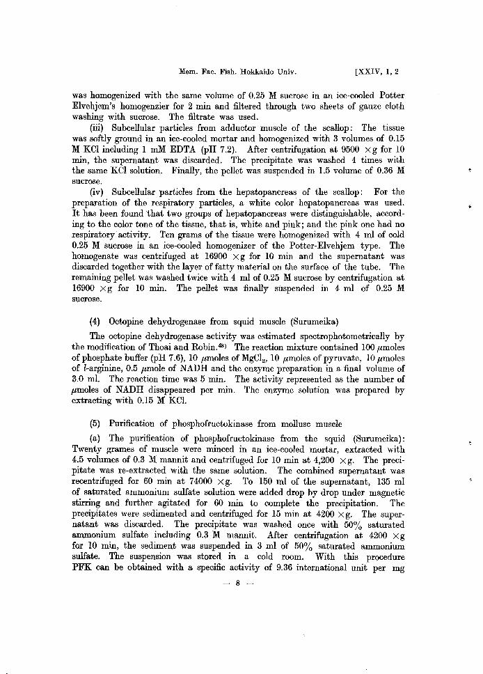
(1) The formation of the final product from fructosediphosphate
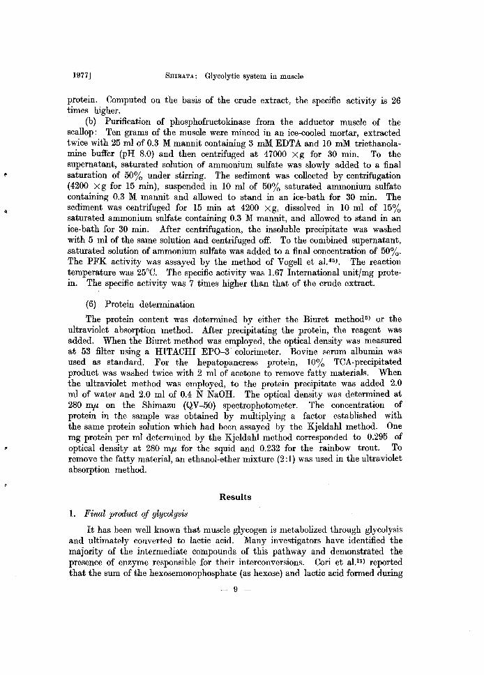
When FDP was added to the extract of a fish muscle (carp), lactic acid accumulated as end product. The result is shown in Fig. 1. In the absence of added FDP, the lactic acid formation diminished and maintained a low level during incubation time. As can be seen from A curve in Fig. 1, the addition of pyruvate to the reaction mixture stimulated the formation of lactic acid. The stimulation may depend on the acceleration of the turnover rate of oxidized and reduced coenzyme due to shortening the "transit time" which has been described by Dixon. (9) It has been recognized that all fish muscles tested hitherto have the same patterns. In contrast with the fish muscle, the extract from the squid muscle caused the accumulation of pyruvate as a final product. This result is shown in Fig. 2. FDP utilization was observed in all the experiments tested, but the lactic acid formation was not. The formation of pyruvate OCCUl"red not in the control system which was Lepage's reaction mixture,29) but in the system omitting
~4.0 E c o
~3.0 ~ u c ·-Z.O ... o
E ~ 1.0 .. "'., •
zo
A
B
c
40 60 80 120
Incubation time (min)
Fig. 1. Lactic acid formation from fructosediphosphate by the crude extract of carp muscle. Reaction mxiture was the same as that described by Lepage'·), except that 4 ,umoles of FDP and 0.6 mg of extract protein were used. Curve A, control system; Curve B, pyruvic acid and fluoride omitted; Curve C, FDP omitted. All intermediates were assayed colorimetrically.
-10-
1977] SHIBATA: Glycolytic system in muscle
~ ::J A )( 8 and C E c: C 0 .. 0 .Q ::J
~2D ... 0
... • Q.
A.B and C ___ _________ ~_CIId B
• 0 • "0 20 40 60 80 120
E :::a.. Inc ub a t ion tim e (m in)
Fig. 2. Pyruvic acid and lactic acid formation from frutosediphosphate by the crude extract of squid muscle. Experimental conditions were the same as in Fig. 1, except that 0.9 mg of extract protein was used. Curve A, control system; Curve B, pyruvic acid ommited; Curve C, pyruvic acid and fluoride omitted; open symbols, consumed FDP; fill symbols, formed pyruvic acid; broken line, formed lactic acid. All intermediates were assayed colorimetrically.
100 10
00 100
50
50
Carp Squid Scallop Hen clam
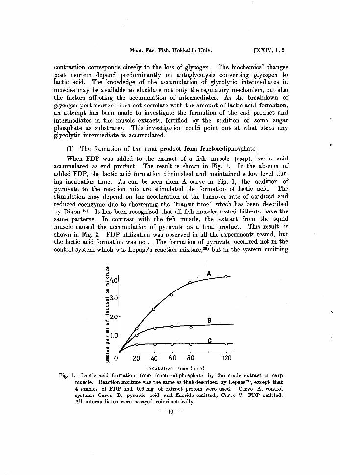
Fig. 3. Proportion of the formations of glycolytic end products from fructosediphosphate by the extracts of several marine animal muscles. left ordinate, consumed substrate as per cent of added concentration; right ordinate, formed end products as per cent of consumed substrate. open column, FDP; hatched column, pyruvic acid; solid column, lactic acid. The dotted column gives the maximum values obtained.
pyruvate and fluoride. The addition of increasing FDP resulted in a greater accumulation of pyruvate. The reaction product was identified by paper chromatography of hydrazone derivative; according to Kazuki and Kanayuki, liD)
it was pyruvic acid. In the marine molluscs tested such as the squid, the octopus, the scallop and some shells, it has been found that pyruvic acid is a final
-11-
Mem. Fac. Fish. Hokkaido Univ. [XXIV, 1,2
product of glycolysis. The pattern of formation of the final product in fish and molluscs was compared in Fig. 3. The amounts of formed pyruvate is different not only from species to species, but also according to the types of muscle. The pyruvate formation in the mantle muscle from the squid and the octopus was more active than the adductor muscle of the shell. The pyruvate formation in the adductor muscle of the shell as striated muscle was larger than that in the foot muscle as smooth muscle. In other words, its formation could correspond to the capacity of glycolysis due to the muscle function as in the case of lactic acid production. The pyruvate accumulation in molluscs must be due to a very low LDH activity as will be described in the following chapter. As mentioned above, hereafter we will deal with lactic acid in fish and pyruvic acid in molluscs as a final product. It has been assumed that FDP breaks down to a final product through the glycolytic pathway, but as shown in Fig. 3, there was no correlation between the loss of FDP and the amount of final product appearing. Lactic acid was formed by 20-30% of consumed FDP in the carp and pyruvate by 40-50% in the squid. Producing 2 moles of final product from 1 mole of FDP, at what step does the remaining part of consumed FDP stay? The reason prompted us to utilize 3-PGA as substrate in order to do a more complete study.
7.0
• ~ 6.0 -)(
E c 5.0 o -co .0 ::;4.0 c .. ~ 3.0 E
.. ~ 2.0 o E '"
1.0
o 10 20 30 40 60 Incubation tim. (min)
120
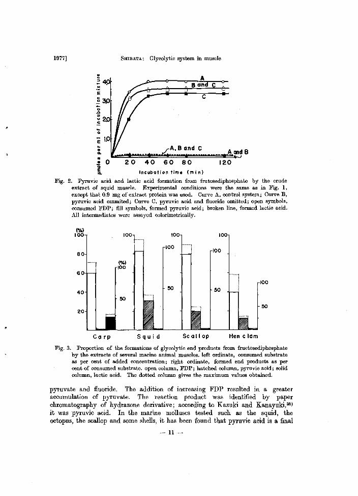
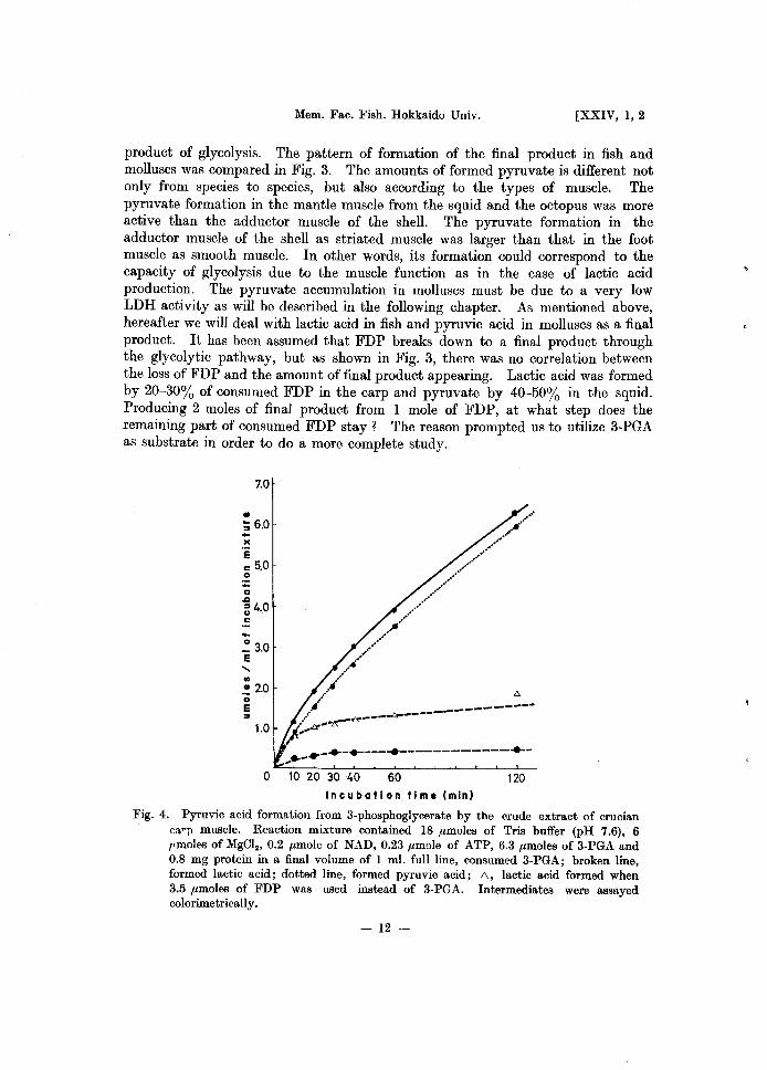
Fig. 4. Pyruvic acid formation from 3-phosphoglycerate by the crude extract of crucian ca~Tl muscle. Reaction mixture contained 18 .umoles of Tris buffer (pH 7.6), 6 !fmoles of MgCl., 0.2 .umole of NAD, 0.23 .umole of ATP, 6.3 .umoles of 3-PGA and 0.8 mg protein in a final volume of I m!. full line, consumed 3-PGA; broken line, formed lactic acid; dotted line, formed pyruvic acid; 1:0., lactic acid formed when 3.5 .umoles of FDP was used instead of 3-PGA. Intermediates were assayed colorimetrically.
- 12 -
1977] SHIBATA: Glycolytic system in muscle
(2) The formation of the final product from 3-phosphoglycerate
3-PGA is a substrate closer to the final product than FDP. 3-PGA was added to the muscle extract. The results are shown in Figs. 4 and 5. The couse
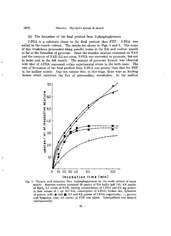
. of this breakdown proeceeded along parallel routes in the fish and mollusc muscle as far as the formation of pyruvate. Since the reaction mixture contained no NAD and the turnover of NAD did not occur, 3-PGA was converted to pyruvate, but not to lactic acid in the fish muscle. The amount of pyruvate formed was identical with that of 3-PGA consumed within experimental errors in the both cases. The rate of formation of the final product from 3-PGA was greater than that for FDP in the mollusc muscle. One can assume that, in this stage, there were no limiting factors which restricted the flux of intermediary metabolism. In the mollusc
• ~
7.0
~5.0 )(
E
~ 5.0 ... 10 .a ~4.0 c .
.... o
II)
~ 2.0 o E :J
o 10 20 30 40 50 100 Incubation time (min)
Fig. 5. Pyruvic acid formation from 3-phosphoglycerate by the crude extract of squid muscle. Reaction mxiture contained 18 /Lmoles of Tris buffer (pH 7.6), 6.6 /Lmoles of MgCl., 0.2 /Lmole of NAD, varying concentrations of 3-PGA and 0.8 mg protein in final volume of 1 ml. full line, consumption of 3-PGA; broken line, formation of pyruvic acid; • and ., 3.1 and 6.2 /Lmoles of 3-PGA respectively; 6, pyruvic acid formation when 4.6 /Lmoles of FDP was added. Intermediates was assayed colorimetrically.
- 13 -
Mem. Fac. Fish. Hokkaido Univ. [XXIV, 1,2
muscle, the reaction proceeded with endogenous cofators except NAD, but in the case of the fish, the rate was very low without addition of adenine nucleotide. The difference may depend on the content of endogenous adenine nucleotide in vivo.
(3) The formation of the final product from glucose, glycogen, and hexosephosphate
Glucose was added in the Lepage's glycolytic system,29) but we have found that there was no change in the content of glucose during the reaction in both the fish and mollusc. Hexokinase or glucokinase in the muscle was very low and could not directly phosphorylate glucose. Maclead et a151) observed a low hexokinase activity in the salmon skeletal muscle and the formation of lactic acid in the muscle homogenate that yeast hexokinase was added.
The existence of glycogen in the skeletal muscle is also important as a soruce of energy for the glycolytic system. However, no breakdown of glycogen was
o e :i.
3.0
I I
P I
I I
/1
0'/'~
.."".._----0-
o l0203040 60 80 100 Incubation time (min)
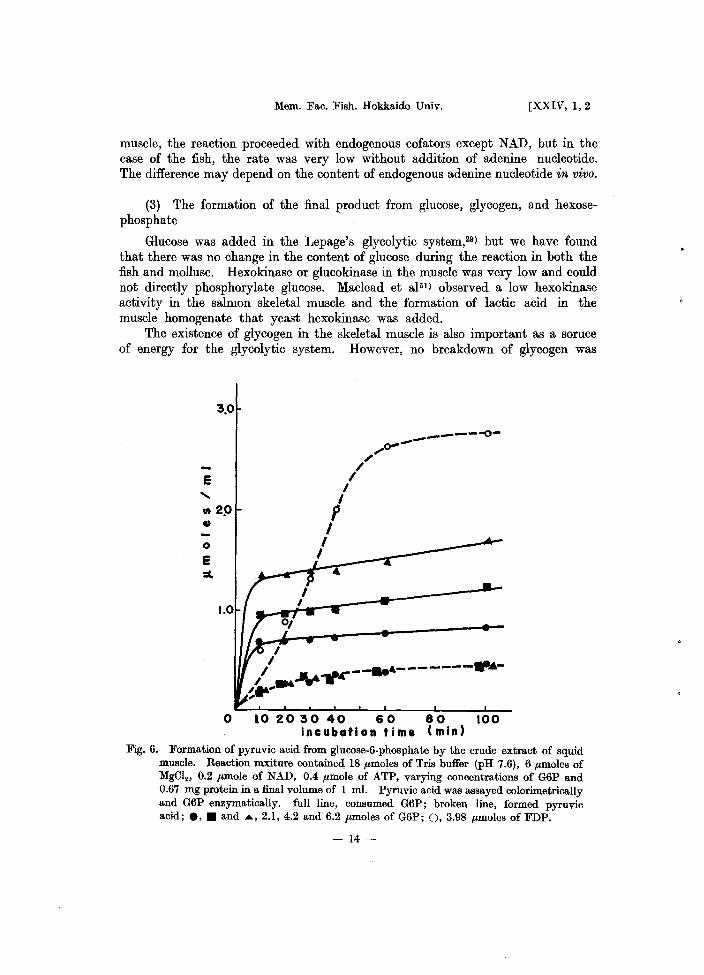
Fig. 6. Formation of pyruvic acid from glucose-6-phosphate by the crude extract of squid muscle. Reaction mxiture contained 18 ,umoles of Tris buffer (pH 7.6), 6 ,umoles of MgClz' 0.2 "mole of NAD, 0.4 ,umole of ATP, varying concentrations of G6P and 0.67 mg protein in a final volume of 1 ml. Pyruvic acid was assayed colorimetrically and G6P enzymatically. full line, consumed G6P; broken line, formed pyruvic acid; e, • and .... , 2.1, 4.2 and 6.2 ,umoles of G6P; 0, 3.98 ",moles of FDP.
- 14 -
1977] 5mBATA: Glycolytic system in muscle
observed in the muscle extract by these experiments in vitro, although the rapid breakdown of glycogen in the post mortem muscle had been observed by many investigators. The addition of AMP had no effect. Perhaps, phosphorylase catalyzing the breakdown of glycogen to hexosephosphate may have a limiting effect on glycolysis. As we found that the muscle extract had a high phosphorylase activity in the presence of AMP according to the method of Sumner and Somers41),
the phosphorylase in the extract may lack the factors required for the utilization of glycogen.
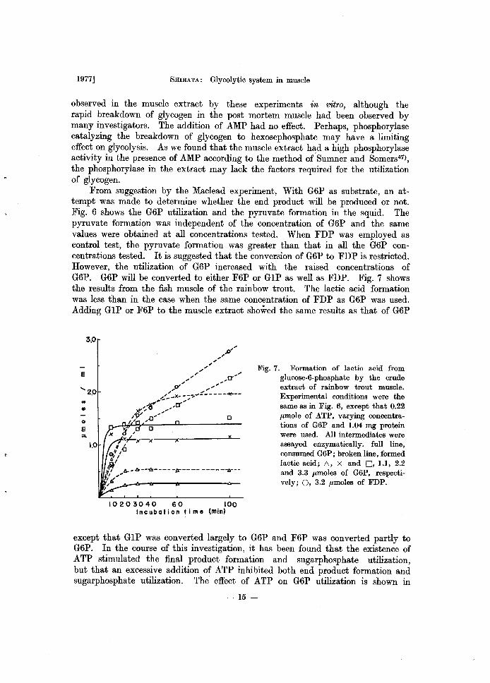
From suggestion by the Maclead experiment, With G6P as substrate, an attempt was made to determine whether the end product will be produced or not. Fig. 6 shows the G6P utilization and the pyruvate formation in the squid. The pyruvate formation was independent of the concentration of G6P and the same values were obtained at all concentrations tested. When FDP was employed as control test, the pyruvate formation was greater than that in all the G6P concentrations tested. It is suggested that the conversion of G6P to FDP is restricted. However, the utilization of G6P increased with the raised concentrations of G6P. G6P will be converted to either F6P or GIP as well as FDP. Fig. 7 shows the results from the fish muscle of the rainbow trout. The lactic acid formation was less than in the case when the same concentration of FDP as G6P was used. Adding G IP or F6P to the muscle extract showed the same results as that of G6P
E
., .. o E
3.0
10203040 60 100 Incubation time (min)
Fig. 7. Formation of lactic acid from glucose-6-phosphate by the crude extract of rainbow trout muscle. Experimental conditions were the same as in Fig. 6, except that 0.22 flmole of ATP, varying concentrations of G6P and 1.04 mg protein were used. All intermediates were assayed enzymatically. full line, consumed G6P; broken line, formed lactic acid; /:;., X and D, 1.1, 2.2 and 3.3 flmoles of G6P, respectively; 0, 3.2 flmoles of FDP_
except that GIP was converted largely to G6P and F6P was converted partly to G6P. In the course of this investigation, it has been found that the existence of ATP stimulated the final product formation and sugarphosphate utilization, but that an excessive addition of ATP inhibited both end product formation and sugarphosphate utilization. The effect of ATP on G6P utilization is shown in
- 15 -
20
E
Q) 1.0
o
E ::1..
o
Mem. Fac; Fish. Hokkaido Univ. [XXIV, 1,2
P. I ,
, , b, ...... .... ...
, ...... '!oo~ •• '
. .. . .' ....
p' .... , $-"., ...... .. " I''', '-',
..... ··0··· ......... .... ............. ..
.. 0 •• ' ',..... ......0 ..... __ _
0: ............ ---------0--P .. ------_______ ! __
12 4 6 8 10 "A
15 T P
20 mM
25
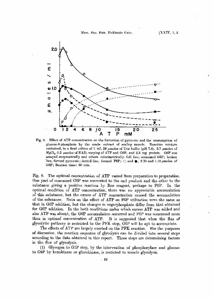
Fig. 8. Effect of ATP concentration on the formation of pyruvate and the consumption of glucose-6-phosphate by the crude extract of scallop muscle. Reaction mixture contained, in a final volime of 1 ml, 19 pmoles of Tris buffer (pH 7.6), 3.7 pmoles of MgCI2 ,0.2 pmoles of NAD, varying of ATP and G6P, and 2.3 mg protein. G6P was assayed enzymatically and others colorimetrically. full line, consumed G6P; broken line, formed pyruvate; dotted line, formed F6P; 0 and ., 2.30 and 1.15 pmoles of G6P; Reation time: 60 min.
Fig. 8. The optimal concentration of ATP varied from preparation to preparation. One part of consumed G6P was converted to the end product and the other to the substance giving a positive reaction by Roe reagent, perhaps to F6P. In the optimal condition of ATP concentration, there was no appreciable accumulation of this substance, but the excess of ATP concentration caused the accumulation of the substance. Data on the effect of ATP on F6P utilization were the same as that in G6P addition, but the changes in sugarphosphate differ from that obtained for G6P addition. In the both conditions under which excess ATP was added and also ATP was absent, the G6P accumulation occurred and F6P was consumed more than in optimal concentration of ATP. It is suggested that when the flux of glycolytic pathway is restricted to the PFK step, G6P will be apt to accumulate.
The effects of ATP are largely exerted on the PFK reaction. For the purposes of discussion, the reaction sequence of glycolysis can be divided into several steps according to the data obtained in this report. Those steps are determining factors in the flux of glycolysis.
(1) Glycogen to GIP step, by the intervention of phosphorylase and glucose to G6P by hexokinase or glucokinase, is resticted to muscle glycolysis.
- 16 -
1977] SHIBATA: Glycolytic system in muscle
(2) In the 'G1P or G6P to FDP' step, by the intervention of three enzymes, GPM, PGI and PFK, PFK is a limiting factor effected by the ATP concentration. The interconversion of hexosephosphate is affected by the PFK action.
(3) In the 'FDP to 3-PGA' steps by the intervention of five enzymes, it will be assumed that there are some limiting factors, because 3-PGA was converted to a final product without the restriction of flux. However, the conclusion concerning the site of reaction limiting glycolysis remains uncertain.
(4) This problem is limited in the case of the fish muscle. In the process 'pyruvate to lactate' by the intervention of LDH, the reaction catalyzes consistently the oxidation of reduced conenzyme. As the pyruvate accumulation increased with the concomitant of lactate accumulation in the fish muscle extract, it is assumed that the rate of formation of reduced coenzyme was greater than that of oxidized coenzyme. LDH regulated the net flux of glycolysis when the glycolytic reaction mixture containing pyruvate was used. The addition of pyruvate is necessary for maximum yields of lactate.
Pyruvate accumulation as glycolytic end product was found in the insect muscle. 52) However, this phenomenon occurred in the flight muscle, but not in the jumping muscle. It is suggested that there is a specific action in the marine mollusc muscle, as we interpret the struggle action of the insect flight muscle. Recently, Agosin and Kepette53,M) reported that in Echinococcus, final product of glycolysis would be pyruvate. Some investigators55- 64) proposed the modified glycolytic scheme for parasitesli3- 62) and some invertebrates,63,64) because of less lactate formation from glucose. We assume that there is a modified glycolytic scheme in the muscle of a marine mollusc.
2. The formation of CI.-glycerophosphate
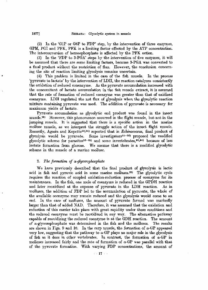
We have previously described that the final product of glycolysis is lactic acid in fish and pyruvic acid in some marine molluscs.20) The glycolytic cycle requires the reaction of coupled oxidation-reduction process of coenzyme for its maintenance. In the fish, one mole of coenzyme is reduced in the GPDH reaction and later reoxidized at the expense of pyruvate in the LDH reaction. As in molluscs, the addition of FDP led to the accumulation of pyruvate, the whole of the available coenzyme may remain reduced and the glycolysis would come to an end. In the case of molluscs, the amount of pyruvate formed was markedly larger than that of added NAD. Therefore, it was assumed that the oxidation and reduction of this carrier take place with great rapidity under these conditions and the reduced coenzyme must be reoxidized in any way. The alternative pathway capable of reoxidizing the reduced coenzyme is at the GDH reaction. The amount of CI.-glycerophosphate was determined in the fish and the molluscs. The results are shown in Figs. 9 and 10. In the carp muscle, the formation of CI.-GP appeared very low, suggesting that the pathway to CI.-GP plays no major role in the glycolysis of fish as it does in other vertebrates. In contrast, the formation of CI.-GP in molluscs increased fairly and the rate of formation of CI.-GP was parallel with that of the pyruvate formation. With varying FDP concentrations, the amount of
- 17 -
... 8. I/) \.0 • "0 E :::L.
o
Mem. Fac. Fish. Hokkaido Univ. [XXIV, 1,2
c
B
10 2030 40 60 100 Incubation time (min)
Fig. 9. Formation of a-glycerophosphate from fructosediphosphate by the crude extract of carp muscle. Reaction mixture contained, in a final volume of 1 ml, 18 pmoles of Tris buffer (pH 7.6), 6 pmoles of MgCI., 0.2 pmole of NAD, 5.9 pID.oles of FDP, and 1.4 mg protein. FDP was assayed colorimetrically and others enzymatically. Curve A, consumed FDP; Curve B, a-GP; Curve C, formed lactic acid .
• ~.O -)( E c3.0 o -u ~.o ()
c
.... ~1.0 e ... • Q,
';0 l
I 0 20 30 40 60 100 I n cu ba t ion tim. (m in)
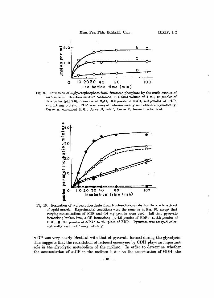
Fig. 10. Formation of a-glycerophosphate from fructosediphosphate by the crude extract of squid muscle. Experimental conditions were the same as in Fig. 10, except that varying concentrations of FDP and 0.6 mg protein were used. full line, pyruvate formation; broken line, a-GP formation; 0, 4.5 pmoles of FDP; (), 2.2 pmoles of FDP; e, 3.1 pmoles of 3-PGA in the place of FDP. Pyruvate was assayed colori metrically and a-GP enzymatically.
cx-GP was very nearly identical with that of pyruvate formed during the glycolysis. This suggests that the reoxidation of reduced coenzyme by GDH plays an important role in the glycolytic metabolism of the mollusc. In order to determine whether the accumulation of cx-GP in the mollusc is due to the specification of GDH, the
- 18 -
1977] 5mBATA: Glycolytic system in muscle
assay of the activity and the determination of Michaelis constant of the mollusc GDH was made. The level of the GDH activity in the mollusc was of the same order as that in the fish, but the LDH activity was much less than that of the fish enzyme and the lowest of all the glycolytic enzymes, as will be described below. Michaelis constant for NADH, 4.4 X 10⁻⁵ M was of the same order as that of other vertebrates enzyme. The direct cause of o:-GP accumulation in the mollusc could not be attributed to a specific action of GDH, but to a very low LDH activity. Chefurka6li) reported that insect muscles accumulate o:-GP in greater amounts than lactic acid. It may be supported that there is the existence of a different intermediary metabolism, as in the insect flight muscle, although the physiological role of o:-GP in the mollusc remains obscure.
3. Significance of pyruvic acid formation in moUuscs
By the observation that pyruvate and o:-GP are formed through the glycolytic pathway in the mollusc muscle, it was assumed that carbohydrate metabolism in the mollusc differs considerably from the commonly accepted pattern in other organisms. In general, pyruvate is aerobically oxidized to CO2 and H 20 through the TCA cycle and o:-GP is led to either fat metabolism or energy metabolism through the glycerol phosphate cycle. It was reported that the active glycerol phosphate cycle exists in the flight muscle of insects. 66-68) Moreover, It was reported that in many parasites, succinate and volatile fatty acid are the final products of carbohydrate metabolism. 69) Kubista52) reported the conversion of pyruvate into acetate in the cockroach. The accumulation of glycerol was observed in the pupae of the silkworm60) and the moth10) with concomitant of glycogen disappearance. These changes might be due to a modified glycolytic scheme in insects. We have observed that a fresh squid muscle contained 1.41 mg% pyruvate and 11 mg% lactic acid and also that both pyruvate and lactate were not significantly accumulated in post mortem muscles. Perhaps the pyruvate formed must be utilized rapidly through another pathway. In order to demonstrate the pyruvate utilizing routes, an attempt was made to study the activities of aerobic respiration and octopine synthesis.
(1) Respiratory activity
The direct utilization of pyruvate is aerobic oxidation in mitochondrial particles. A number of research workers have proposed evidence for the presence of electron transfer components in marine invertebrates,11-15) especially crustaceans 16-83) and mussels,84,85) and some cytochrome pigments86) were purified. However, little detailed is available about the preparation and nature of mitochondrial particles, containing cytochromes and respiratory enzymes, except Beechey's report 80) on the hepatopancreas of the crab. Then, we have attempted to prepare the subcellular fraction from the muscles and the hepatopancreas of two species of molluscs and to study the oxidation of the pyruvate and other members of the TCA cycle.
The oxidiative capacities of the TCA cycle intermediates in the squid muscle
- 19-
Mem. Fac. Fish. Hokkaido Univ. [XXIV, 1,2
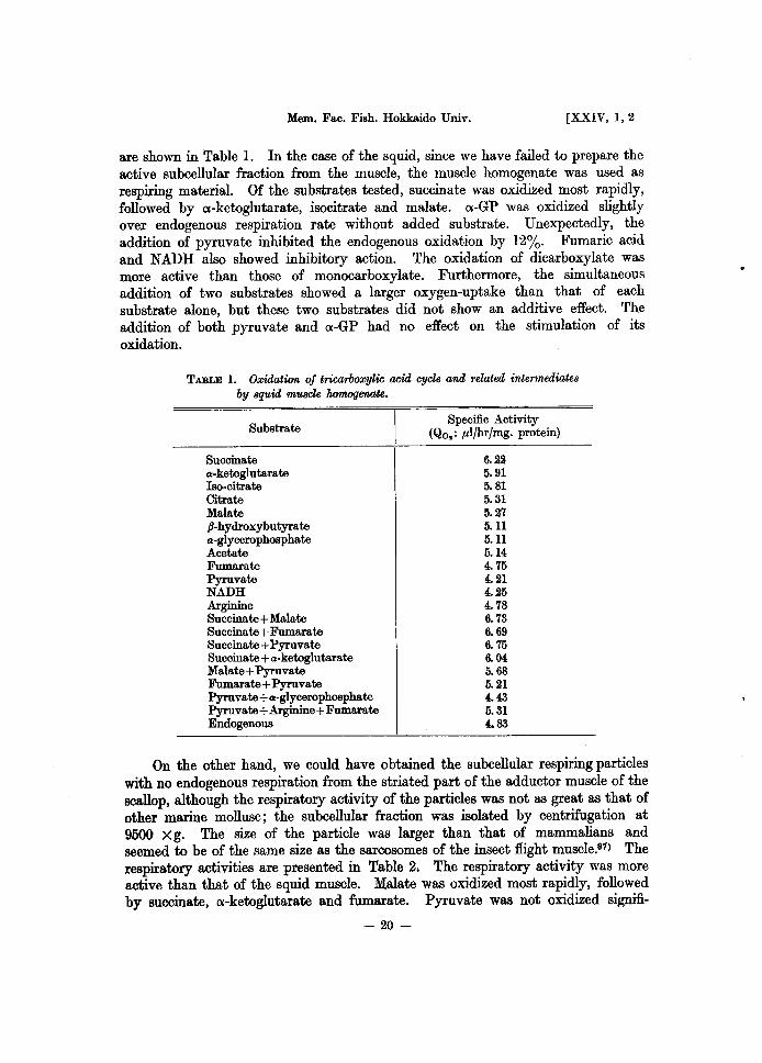
are shown in Table l. In the case of the squid, since we have failed to prepare the active subcellular fraction from the muscle, the muscle homogenate was used as respiring material. Of the substrates tested, succinate was oxidized most rapidly, followed by ex-ketoglutarate, isocitrate and malate. ex-GP was oxidized slightly over endogenous respiration rate without added substrate. Unexpectedly, the addition of pyruvate inhibited the endogenous oxidation by 12%. Fumaric acid and NADH also showed inhibitory action. The oxidation of dicarboxylate was more active than those of monocarboxylate. Furthermore, the simultaneous addition of two substrates showed a larger oxygen-uptake than that of each substrate alone, but theBe two substrates did not show an additive effect. The addition of both pyruvate and ex-GP had no effect on the stimulation of its oxidation.
TABLE 1. Oxidation of tricarboxylic acid cycle and related intermediates by squid muscle homogenate.
Substrate
Succinate a-ketoglutarate Iso-citrate Citrate Malate P-hydroxybutyrate a-glycerophosphate Acetate Fumarate Pyruvate NADH Arginine Succinate + Malate Succinate + Fumarate Succinate + Pyruvate Succinate + a-ketoglutarate Malate + Pyruvate Fumarate + Pyruvate Pyruvate + a-glycerophosphate Pyruvate + Arginine + Fumarate Endogenous
Specific Activity (Qo.: pl/hr/mg. protein)
6.22 5.91 5.81 5.31 5.27 5.11 5.11 5.14 4.75 4.21 4.25 4.78 6.73 6. 69 6.75 6. 04 5.68 5.21 4.43 5.31 4.83
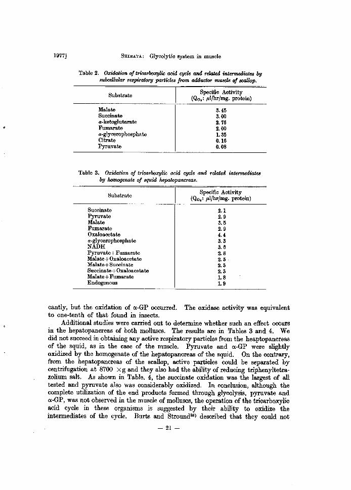
On the other hand, we could have obtained the subcellular respiring particles with no endogenous respiration from the striated part of the adductor muscle of the scallop, although the respiratory activity of the particles was not as great as that of other marine mollusc; the subcellular fraction was isolated by centrifugation at 9500 X g. The size of the particle was larger than that of mammalians and seemed to be of the same size as the sarcosomes of the insect flight muscle.S'1) The respiratory activities are presented in Table 2. The respiratory activity waS more active than that of the squid muscle. Malate was oxidized most rapidly, followed by succinate, ex-ketoglutarate and fumarate. Pyruvate was not oxidized signifi-
- 20-
1977] SHIBATA: Glycolytic system in muscle
Table 2. Oxidation of tricarboxylic acid cycle and related intermediates by 8'Ubcellular respiratory particle8 from adductor muscle of scallop.
Substrate
Malate Succinate a-ketoglutarate Fumarate a-glycerophosphate Citrate Pyruvate
Specific Activity (Qo,: pl/hr/mg. protein)
3.45 3.00 2.76 2.00 1.35 0.16 0.08
Table 3. Oxidation of tricarboxylic acid cycle and related intermediates by homogenate of 8quid hepatopancreas.
Substrate
Succinate Pyruvate Malate Fumarate Oxaloacetate a-glycerophosphate NADH Pyruvate + Fumarate Malate + Oxaloacetate Malate + Succinate Succinate + Oxaloacetate Malate + Fumarate Endogenous
Specific Activity (Qo.: plfhr/mg. protein)
2.1 2.9 3.5 2.9 4.4 3.3 3.6 2.8 2.5 2.3 2.3 1.8 1.9
cantly, but the oxidation of o:-GP occurred. The oxidase activity was equivalent to one-tenth· of that found in insects.
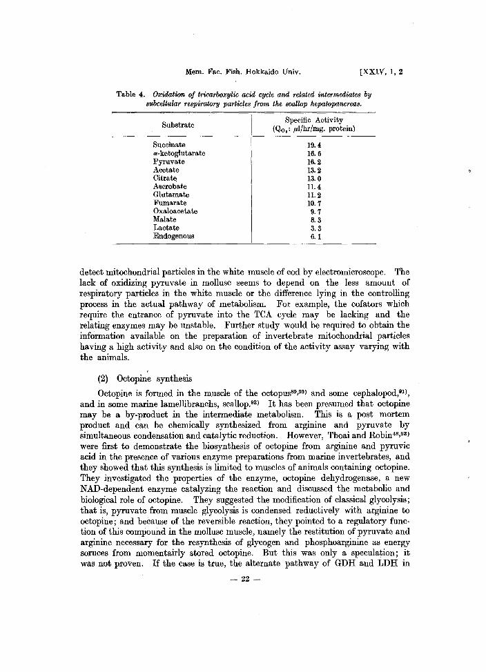
Additional studies were carried out to determine whether such an effect occurs in the hepatopancreas of both molluscs. The results are in Tables 3 and 4. We did not succeed in obtaining any active respiratory particles from the heaptopancreas of the squid, as in the case of the muscle. Pyruvate and o:-GP were slightly oxidized by the homogenate of the hepatopancreas of the squid. On the contrary, from the hepatopancreas of the scallop, active particles could be separated by centrifugation at 8700 X g and they also had the ability of reducing triphenyltetrazolium salt. As shown in Table. 4, the succinate oxidation was the largest of all tested and pyruvate also was considerably oxidized. In conclusion, although the complete utilization of the end products formed through glycolysis, pyruvate and o:-GP, was not observed in the muscle of molluscs, the operation of the tricarboxylic acid cycle in these organisms is suggested by their ability to oxidize the intermediates of the cycle. Burts and Stround88) described that they could not
- 21-
Mem. Fac. Fish. Hokkaido Univ. [XXIV, 1,2
Table 4. Oxidation of tricarboxylic acid cycle and related intermediates by subcellular respiratory particles from the scallop hepatopancreas.
Substrate
Succinate a-ketoglutarate Pyruvate Acetate Citrate Ascrobate Glutamate Fumarate Oxaloacetate Malate Lactate Endogenous
Specific Activity (Qo.: J.lIJhrJmg. protein)
19.4 16.6 16.2 13.2 13.0 11.4 11.2 10.7 9.7 8.3 3.3 6.1
detect mitochondrial particles in the white muscle of cod by electromicroscope. The lack of oxidizing pyruvate in mollusc seems to depend on the less amount of respiratory particles in the white muscle or the difference lying in the controlling process in the actual pathway of metabolism. For example, the cofators which require the entrance of pyruvate into the TeA cycle may be lacking and the relating enzymes may be unstable. Further study would be required to obtain the information available on the preparation of invertebrate mitochondrial particles having a high activity and also on the condition of the activity assay varying with the animals.
(2) Octopine synthesis
Octopine is formed in the muscle of the octopus89,90) and some cephalopod,91), and in some marine lamellibranchs, scallop.92) It has been presumed that octopine may be a by-product in the intermediate metabolism. This is a post mortem product and can be chemically synthesized from arginine and pyruvate by simultaneous condensation and catalytic reduction. However, Thoai and Robin48,93) were first to demonstrate the biosynthesis of octopine from arginine and pyruvic acid in the presence of various enzyme preparations from marine invertebrates, and they showed that this synthesis is limited to muscles of animals containing octopine. They investigated the properties of the enzyme, octopine dehydrogenase, a new NAD-dependent enzyme catalyzing the reaction and discussed the metabolic and biological role of octopine. They suggested the modification of classical glycolysis; that is, pyruvate from muscle glycolysis is condensed reductively with arginine to octopine; and because of the reversible reaction, they pointed to a regulatory function of this compound in the mollusc muscle, namely the restitution of pyruvate and arginine necessary for the resynthesis of glycogen and phosphoarginine as energy soruces from momentairly stored octopine. But this was only a speculation; it was not proven. If the case is true, the alternate pathway of GDH and LDH in
- 22-
1977] SHIBATA: Glycolytic system in muscle
2.0
,., 01.5
X
~ 1.0 (I)
O.s
" " " " 5 0 5 10 15
SxIO!i(M) 20 25
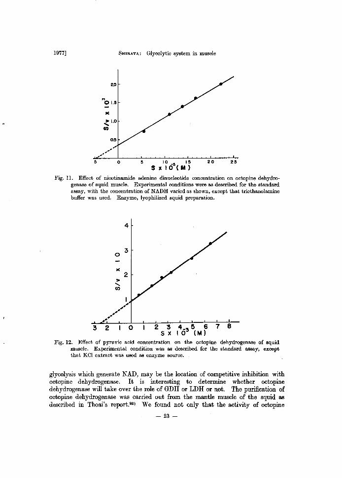
Fig. ll. Effect of nicotinamide adenine dinucleotide concentration on octopine dehydrogenase of squid muscle. Experimental conditions were as described for the standard assay, with the concentration of NADH varied as shown, except that triethanolamine buffer was used. Enzyme, lyophilized squid preparation.
3 2
4
3 o
> ...... (/)
2
o 2 3 4_ 5 6 7 8 S X I 0 3 (M)
Fig. 12. Effect of pyruvic acid concentration on the octopine dehydrogenase of squid muscle. Experimental condition was as described for the standard assay, except that KCl extract was used as enzyme source.
glycolysis which generate NAD, may be the location of competitive inhibition with octopine dehydrogenase. It is interesting to determine whether octopine dehydrogenase will take over the role of GDH or LDH or not. The purification of octopine dehydrogenase was carried out from the mantle muscle of the squid as described in Thoai's report.93) We found not only that the activity of octopine
- 23-
Mem. Fac. Fish. Hokkaido Univ. [XXIV, 1, 2
4
0 :3
x
> 2 ....... C/)
# ##
" #' ##
2 0
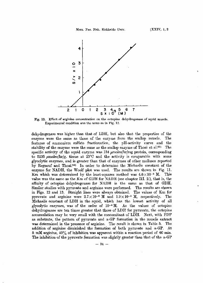
Fig. 13. Effect of arginine concentration on the octopine dehydrogenase of squid muscle. Experimental condition was the same as in Fig. 12.
dehydrogenase was higher than that of LDH, but also that the properties of the enzyme were the same as those of the enzyme from the scallop muscle. The features of ammonium sulfate fractionation, the pH -activity curve and the stability of the enzyme were the same as the scallop enzyme of Thoai et a1.93 ) The specific activity of the squid enzyme was 134 pmoles/hr/mg protein, corresponding to 5235 pmoles/hr/g. tissue at 25°C and the activity is comparable with some glycolytic enzymes, and is greater than that of enzymes of other molluscs reported by Regnouf and Thoai.94) In order to determine the Michaelis constant of the enzyme for NADH, the Woolf plot was used. The results are shown in Fig. 11. Km which was determined by the least-squares method was 4.6 X 10⁻⁵ M. This value was the same as the Km of GDH for NADH (see chapter III. 2.), that is, the affinity of octopine dehydrogenase for NADH is the same as that of GDH. Similar studies with pyruvate and arginine were performed. The results are shown in Figs. 12 and 13. Straight lines were always obtained. The values of Km for pyruvate and arginine were 2.7x1O-3 M and 1.9x1O-3 M, respectively. The Michaelis constant of LDH in the squid, which has the lowest activity of all glycolytic enzymes, was of the order of 10-4 M. As the values of octopine dehydrogenase are ten times greater that those of LDH for pyruvate, the octopine accumulation may be very small with the concomitant of LDH. Next, with FDP as substrate, the pattern of pyruvate and ex-GP formation in the muscle extract was determined in the presence of arginine. The result is shown in Table 5. The addition of arginine diminished the formation of both pyruvate and ex-GP. At 5 roM arginine, 40% of inhibition was apparent within a reaction period of 60 min. The inhibition of the pyruvate formation was slightly greater than that of the ex-GP
- 24-
1977] SHIBATA: Glycolytic system in muscle
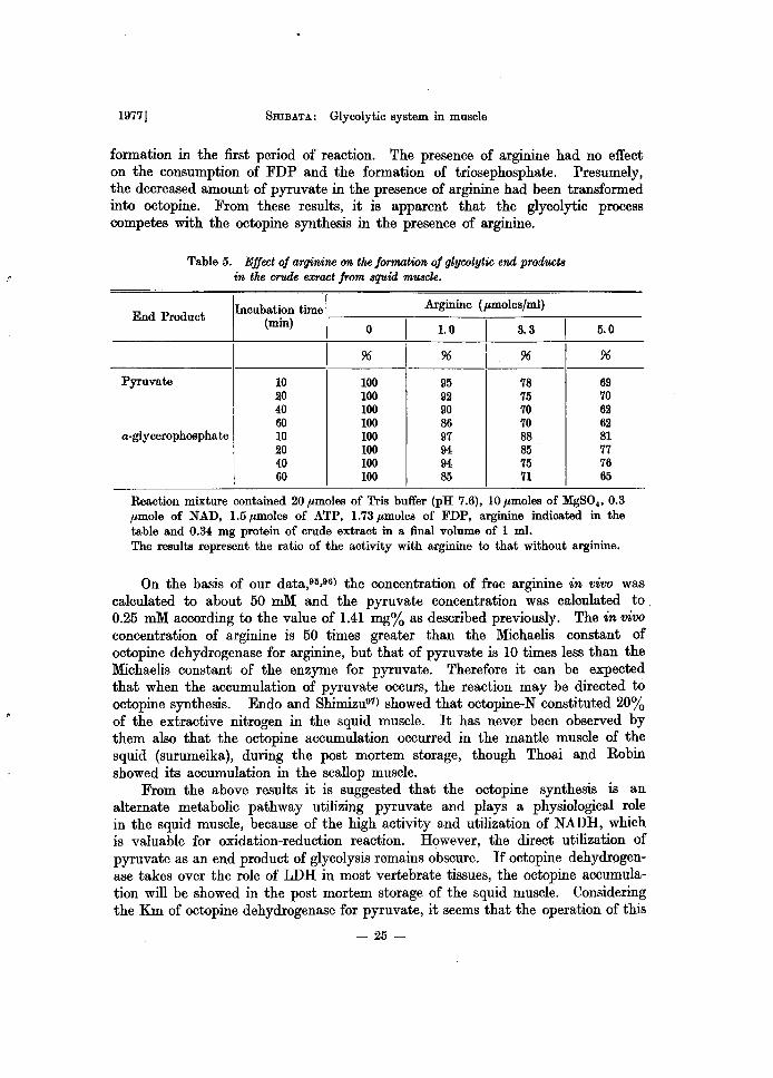
formation in the first period of reaction. The presence of arginine had no effect on the consumption of FDP and the formation of triosephosphate. Presumely, the decreased amount of pyruvate in the presence of arginine had been transformed into octopine. From these results, it is apparent that the glycolytic process competes with the octopine synthesis in the presence of arginine.
Table 5. Effect of arginine on the formation of glycolytic end products in the crude exract frMn squid muscle.
End Product
IIncubation time I Arginine (pmoles/ml)
(min) -----O---.--------~-------.---------
LO &3 ~O
I % % I % %
Pyruvate 10 100 95 78 69 20 100 92 75 70 40 100 90 70 62 60 100 86 70 62
a-glycerophosphate 10 100 97 88 81 20 100 94 85 77 40 100 94 75 76 60 100 85 71 65
Reaction mixture contained 20 pmoles of Tris buffer (pH 7.6), 10 pmoles of MgSO" 0.3 pmole of NAD, 1.5 pmoles of ATP, 1.73 pmoles of FDP, arginine indicated in the table and 0.34 mg protein of crude extract in a final volume of 1 ml. The results represent the ratio of the activity with arginine to that without arginine.
On the basis of our data,95,96) the concentration of free arginine in vivo was calculated to about 50 mM and the pyruvate concentration was calculated to. 0.25 mM according to the value of l.41 mg% as described previously. The in vivo concentration of arginine is 50 times greater than the Michaelis constant of octopine dehydrogenase for arginine, but that of pyruvate is 10 times less than the Michaelis constant of the enzyme for pyruvate. Therefore it can be expected that when the accumulation of pyruvate occurs, the reaction may be directed to octopine synthesis. Endo and Shimizu91) showed that octopine-N constituted 20% of the extractive nitrogen in the squid muscle. It has never been observed by them also that the octopine accumulation occurred in the mantle muscle of the squid (surumeika), during the post mortem storage, though Thoai and Robin showed its accumulation in the scallop muscle.
From the above results it is suggested that the octopine synthesis is an alternate metabolic pathway utilizing pyruvate and plays a physiological role in the squid muscle, because of the high activity and utilization of NADH, which is valuable for oxidation-reduction reaction. However, the direct utilization of pyruvate as an end product of glycolysis remains obscure. If octopine dehydrogenase takes over the role of LDH in most vertebrate tissues, the octopine accumulation will be showed in the post mortem storage of the squid muscle. Oonsidering the Km of octopine dehydrogenase for pyruvate, it seems that the operation of this
- 25-
Mem. Fac. Fish. Hokkaido Univ. [XXIV, 1, 2
enzyme would appeared under the condition of accumulating pyruvate.
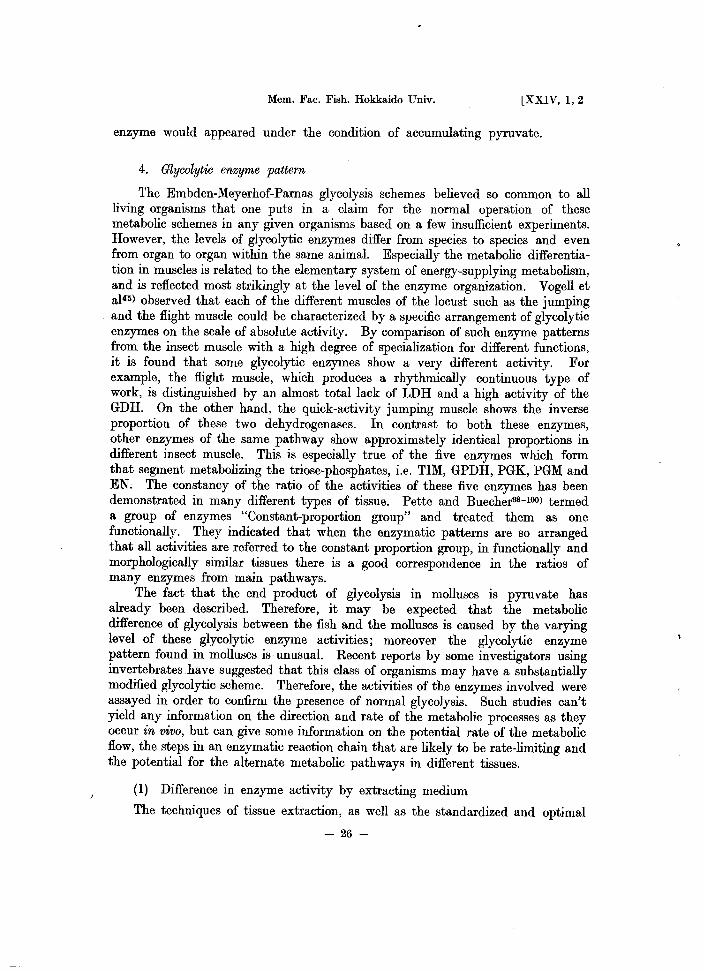
4. Glycolytic enzyme pattern
The Embden-Meyerhof-Parnas glycolysis schemes believed so common to all living organisms that one puts in a claim for the normal operation of these metabolic schemes in any given organisms based on a few insufficient experiments. However, the levels of glycolytic enzymes differ from species to species and even from organ to organ within the same animal. Especially the metabolic differentiation in muscles is related to the elementary system of energy-supplying metabolism, and is reflected most strikingly at the level of the enzyme organization. Vogell et a145} observed that each of the different muscles of the locust such as the jumping and the flight muscle could be characterized by a specific arrangement of glycolytic enzymes on the scale of absolute activity. By comparison of such enzyme patterns from the insect muscle with a high degree of specialization for different functions, it is found that some glycolytic enzymes show a very different activity. For example, the flight muscle, which produces a rhythmically continuous type of work, is distinguished by an almost total lack of LDH and a high activity of the GDH. On the other hand, the quick-activity jumping muscle shows the inverse proportion of these two dehydrogenases. In contrast to both these enzymes, other enzymes of the same pathway show approximately identical proportions in different insect muscle. This is especially true of the five enzymes which form that segment metabolizing the triose-phosphates, i.e. TIM, GPDH, PGK, PGM and EN. The constancy of the ratio of the activities of these five enzymes has been demonstrated in many different types of tissue. Pette and Buecher98- 1OO) termed a group of enzymes "Constant-proportion group" and treated them as one functionally. They indicated that when the enzymatic patterns are so arranged that all activities are referred to the constant proportion group, in functionally and morphologically similar tissues there is a good correspondence in the ratios of many enzymes from main pathways.
The fact that the end product of glycolysis in molluscs is pyruvate has already been described. Therefore, it may be expected that the metabolic difference of glycolysis between the fish and the molluscs is caused by the varying level of these glycolytic enzyme activities; moreover the glycolytic enzyme pattern found in molluscs is unusual. Recent reports by some investigators using invertebrates have suggested that this class of organisms may have a substantially modified glycolytic scheme. Therefore, the activities of the enzymes involved were assayed in order to confirm the presence of normal glycolysis. Such studies can't yield any information on the direction and rate of the metabolic processes as they occur in vivo, but can give some information on the potential rate of the metabolic flow, the steps in an enzymatic reaction chain that are likely to be rate-limiting and the potential for the alternate metabolic pathways in different tissues.
(1) Difference in enzyme activity by extracting medium
The techniques of tissue extraction, as well as the standardized and optimal
- 26-
1977] 5mBATA: Glycolytic system in muscle
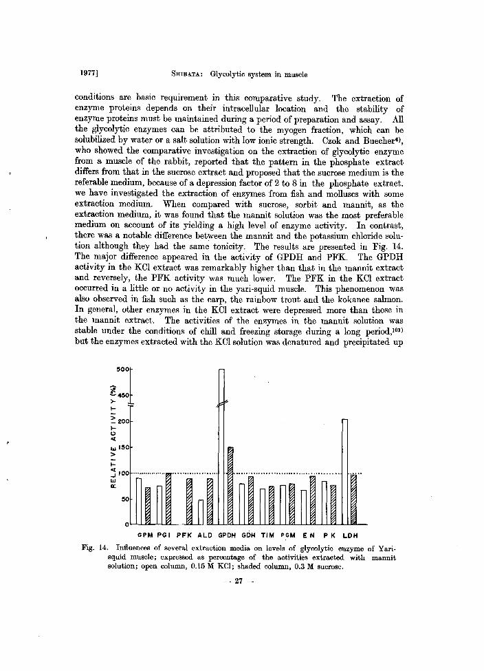
conditions are basic requirement in this comparative study. The extraction of enzyme proteins depends on their intracellular location and the stability of enzyme proteins must be maintained during a period of preparation and assay. All the glycolytic enzymes can be attributed to the myogen fraction, which can be solubilized by water or a salt solution with low ionic strength. Czok and Buecher6), who showed the comparative investigation on the extraction of glycolytic enzyme from a muscle of the rabbit, reported that the pattern in the phosphate extract differs from that in the sucrose extract and proposed that the sucrose medium is the referable medium, because of a depression factor of 2 to 8 in the phosphate extract. we have investigated the extraction of enzymes from fish and molluscs with some extraction medium. When compared with sucrose, sorbit and mannit, as the extraction medium, it was found that the mannit solution was the most preferable medium on account of its yielding a high level of enzyme activity. In contrast, there was a notable difference between the mannit and the potassium chloride solution although they had the same tonicity. The results are presented in Fig. 14. The major difference appeared in the activity of GPDH and PFK. The GPDH activity in the KOl extract was remarkably higher than that in the mannit extract and reversely, the PFK activity was much lower. The PFK in the KCI extract occurred in a little or no activity in the yari-squid muscle. This phenomenon was also observed in fish such as the carp, the rainbow trout and the kokanee salmon. In general, other enzymes in the KOl extract were depressed more than those in the mannit extract. The activities of the enzymes in the mannit solution was stable under the conditions of chill and freezing storage during a long period,101) but the enzymes extracted with the KCI solution was denatured and precipitated up
500
1 ;e ~450 I-
:: 200 l-t> C(
w 150 > I-~ 100················ ..•...........•.... w a:
GPM PG I PFK ALD GPDH GDH TIM PGM EN P K LDH
Fig. 14. Influences of several extraction media on levels of glycolytic enzyme of Yari· squid muscle; expressed as percentage of the activities extracted with mannit solution; open column, 0.15 M KCl; shaded column, 0.3 M sucrose.
- 27-
Mem. Fac. Fish. Hokkaido Univ. [XXIV, 1,2
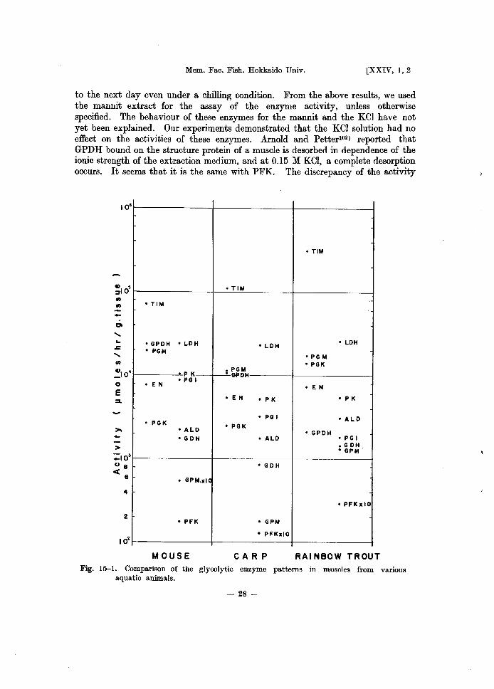
to the next day even under a chilling condition. From the above results, we used the mannit extract for the assay of the enzyme activity, unless otherwise specified. The behaviour of these enzymes for the mannit and the KCI have not yet been explained. Our experiments demonstrated that the KCI solution had no effect on the activities of these enzymes. Arnold and Petter102) reported that GPDH bound on the structure protein of a muscle is desorbed in dependence of the ionic strength of the extraction medium, and at 0.15 M KCI, a complete desorption occurs. It seems that it is the same with PFK. The discrepancy of the activity
10'
-CIt
..... ...
.s::
..... (/)
~IO' o E :s.
.. 2
• TIM
• GPDH • LDH • PGM
P K
• EN • PG I
• PGK • ALD ·GDH
• GPM,xIC
• PFK
MOUSE
• TIM
• TIM
• LDH • LDH
• PG M • PGK
• PGM • GPDH
• EN
• EN • P K • P K
• PG I • A LD • PGK
• GPDH • ALD • PG I
• G DH • GPM
• GDH
• PFKxlO
• GPM
• PFKxlO
CAR P RAI NBOW TROUT
Fig. 15-1. Comparison of the glycolytic enzyme patterns in muscles from various aquatic animals.
- 28-
.<
1977] SHIBATA: Glycolytic system in muscle
existing between both extraction mediums may be caused by the extraction of these enzymes.
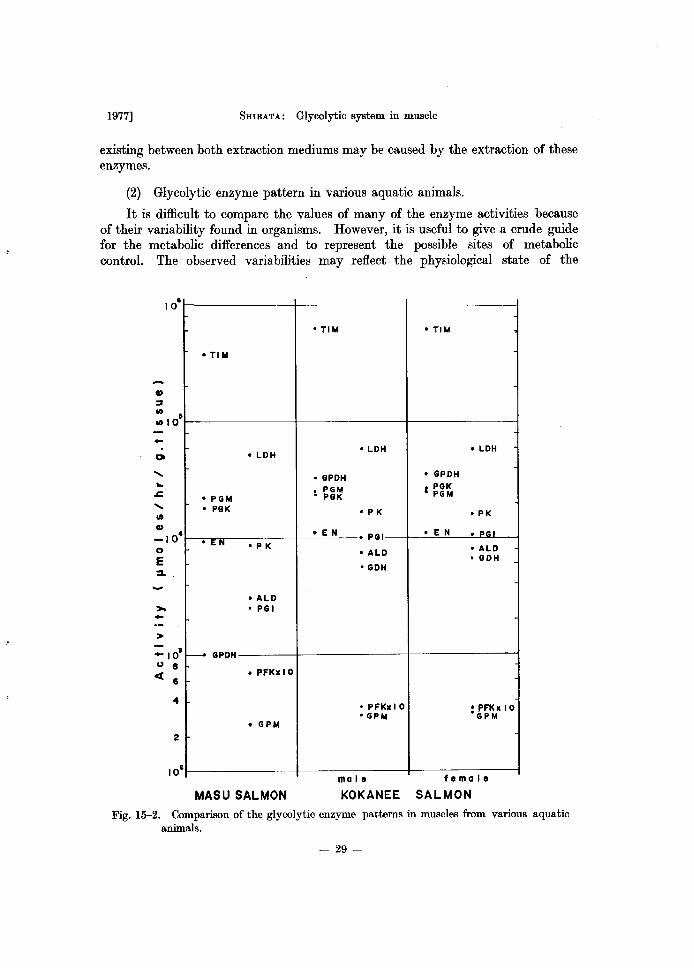
(2) Glycolytic enzyme pattern in various aquatic animals.
It is difficult to compare the values of many of the enzyme activities because of their variability found in organisms. However, it is useful to give a crude guide for the metabolic differences and to represent the possible sites of metabolic control. The observed variabilities may reflect the physiological state of the
-01
........
.c ........
:>. -
10·
fi
4
2
10 t
• TI M
• LOH
• PGM • PGK
• EN 'PK
• ALO • PG I
f------ GPOH
• PFKIIIO
• GPM
MASU SALMON
• TIM • TIM
• LOH • LOH
• GPOH • GPOH
• PGM I PGK • PGK PGM
• P K • PK
• E N • PGI • E N • PG
• ALO • ALO • GOH
• GOH
• PFl<xl 0 : PFl<x 10 • GPM GPM
male female
KOKANEE SALMON
Fig. 15-2. Comparison of the glycolytic enzyme patterns in muscles from various aquatic animals.
- 29-
Mem. Fac. Fish. Hokkaido Univ. [XXIV, 1,2
• TI M
., ~
II)
II) lOs
- 'TlM • LDH • TIM
til
...... • PGK ...
~ • PG M ...... II) • EN • PK
., • LDH _104
0 • GPDH • LDH E :so
,..;
• PGI • PGK • GPDH · i DH • PGM
• PGM • LD
~ - • PGI • PGK
• EN • EN • PK
> • PGI • GPDH
_lOS • PK u 8 ct
6
4
: G6~ • ALD • GDH
• PFKxlO • GPMxlO ::~BxIO
• GPM
• PFKxlO
2
LAMPREY REO SALMON COD Fig. 15-3. Comparison of the glycolytic enzyme patterns in muscles from various aquatic
animals.
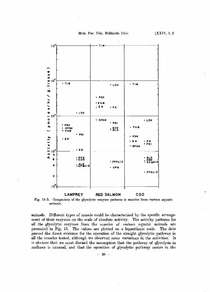
animals. Different types of muscle could be characterized by the specific arrangement of their enzymes on the scale of absolute activity. The activity patterns for all the glycolytic enzymes from the muscles of various aquatic animals are presented in Fig. 15. The values are plotted on a logarithmic scale. The date proved the direct evidence for the operation of the straight glycolytic pathway in all the muscles tested, although we observed some variations in the activities. It is obvious that we must discard the assumption that the pathway of glycolysis in molluscs is unusual, and that the operation of glycolytic pathway occurs in the
- 30-
1977] 5mBATA: Glycolytic system in muscle
· , .. • TIM
·TlM
• LDH • LDH
• PGK • PGr<
• PGM ·.E N • P K
• EN • GPDH • PGM
• P K • PGK • P GM • E N • P K
• GPDH • GDH
• ALD • PG I • GDH • ALD • ALD • PGI • PFK
• GPDH
• PGI • GDH >
• PFKxlO
• GPM 4
• PFKxlO
2 • GPM • GPM • LDH
YELLOW TAIL MACKEREL SURUME SQUID
Fig. 15-4. Comparison of the glycolytic enzyme patterns in muscles from various aquatic animals.
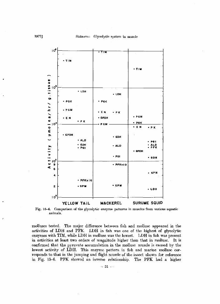
molluscs tested. The major difference between fish and mollusc appeared in the activities of LDH and PFK. LDH in fish was one of the highest of glycolytic enzymes with TIM, while LDH in mollusc was the lowest. LDH in fish was present in activities at least two orders of magnitude higher than that in mollusc. It is confirmed that the pyruvate accumulation in the mollusc muscle is caused by the lowest activity of LDH. This enzyme pattern in fish and marine mollusc corresponds to that in the jumping and flight muscle of the insect shown for reference in Fig. 15-6. PFK showed an inverse relationship. The PFK had a higher
- 31 -
..
>
"10' () 8
ct 6
4
2
Mem. Fac. Fish. Hokkaido Univ.
• TIM
• TI M
• GPOH • PGM
• PGM • EN • PGK
• E N • P K • PGI • PGK • P K
• PGI
• ALO
• ALO'GOH • PFK
• GPOH
• GOH'LOH
• PFK
• LOH
• GPM
.GP M
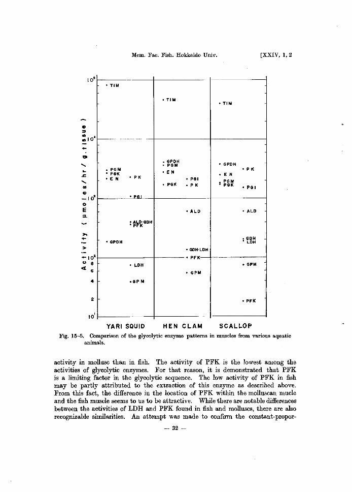
YARI SQUID HEN CLAM
[XXIV, 1,2
• TIM
• GPOH • P K
• E N
• PGM • PGK
• PG I
• ALO
• GOH • LOH
• GPM
• PFK
SCALLOP
Fig. 15-5. Comparison of the glycolytic enzyme patterns in muscles from various aquatic animals.
activity in mollusc than in fish. The activity of PFK is the lowest among the activities of glycolytic enzymes. For that reason, it is demonstrated that PFK is a limiting factor in the glycolytic sequence. The low activity of PFK in fish may be partly attributed to the extraction of this enzyme as described above. From this fact, the difference in the location of PFK within the molluscan mucle and the fish muscle seems to us to be attractive. While there are notable differences between the activities of LDH and PFK found in fish and molluscs, there are also recognizable similarities. An attempt was made to confirm the constant-propor-
- 32-
'-
1977] 5mBATA: Glycolytic system in muscle
• TIM • TIM
• TIM II)
:::J til
til 104
-• PGM
• PGM
• GPDH 'LDH-• GDH • GPDH • PGK
0
..... • PK • PGK
• EN • ALD .. • E iii .r: • ALD
..... til
• P K • P K • PFK • GDH
II)
-10· 0
r--:--:~~ • PFK
E :L : ~"K
• PGI
• ALD >--> • LDH
: 101
u 8 • GDH • LDH
cl 6
4
• PFK
2
10' • GPM flight mUlcle jumplngmulcle
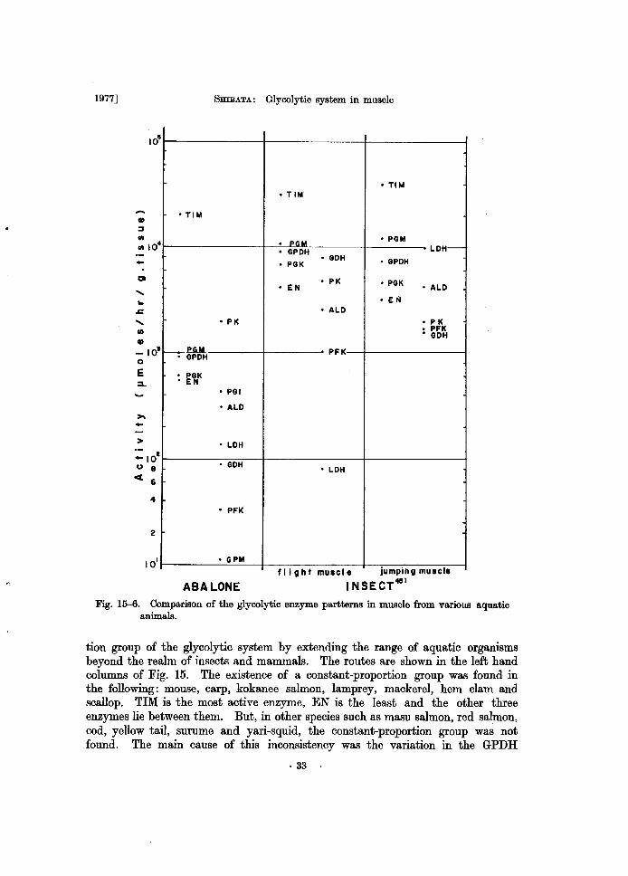
ABALONE I NSECT4II1
Fig. 15--6. Comparison of the glycolytic enzyme partterns in muscle from various aquatic animals.
tion group of the glycolytic system by extending the range of aquatic organisms beyond the realm of insects and mammals. The routes are shown in the left hand columns of Fig. 15. The existence of a constant-proportion group was found in the following: mouse, carp, kokanee salmon, lamprey, mackerel, hem clam and scallop. TIM is the most active enzyme, EN is the least and the other three enzymes lie between them. But, in other species such as masu salmon, red salmon, cod, yellow tail, surume and yari-squid, the constant-proportion group was not found. The main cause of this inconsistency was the variation in the GPDH
- 33-
Mem. Fac. Fish. Hokkaido Univ. [XXIV, 1,2
activity. In all cases, the GPDH activity decreased and was less than the other four enzymes. Although GPDH is one of the most active enzymes in the glycolytic sequence in mammals, GPDH in aquatic animals is not always so. However, the other four enzymes except GPDH appeared to correlate with each other. The other enzymes, except for these five, were arranged in the order of their magnitude in the right-hand columns of Fig. 15. The approximately identical pattern were shown in different animals. Exclusive of LDH and PFK, PK was the most active enzyme and GPM was the least. Other enzymes were inserted between them, although some fluctuations in the activity level were observed between ALD and GDH. This similarity will indicate that the organized system of enzymatic activities is a physiological structure corresponding to the function.
Comparing the level of activity between animal species, it was found that migrating fish such as salmon and mackerel had a higher activity than settled fish such as cod. The squid had the highest activity of all marine mollusc tested. The GDH of the squid was the same level as that of the jumping muscle of the insect, but was ten times less than those of the flight muscle. It was also demonstrated that a fast acting skeletal muscle has a high capacity for glycolysis.
(3) Activity level of the dark muscle
The nature of the muscle pigmentation and its physiological significance have been studied since the late nineteenth century. More recently some aspects have been discussed by Pette99 ) and Perry.I03) To some extent, the degree of pigmentation correlates with the slowness of contraction, i.e. in proportion with the time spent in the contracted state. In fish, the physiological function of the dark muscle has been discussed by many investigators. It is well known that the function of the dark muscle corresponds to that of the liver. The dark muscle in fish is easily discernible morphologically from the white muscle. The glycolytic enzyme patterns of the dark muscle were compared with that of the white. The absolute level of the activities in the dark muscle is shown in Fig. 16. The enzyme patterns were identical with that of the white muscle and the constant proportion group also clearly appeared. The ratio of activity of the dark muscle compared to that of the white muscle is shown in Fig. 17. The absolute level of most enzymes in the dark muscle was lower by half or less than those in the white muscle, except ALD and PFK. The ALD activity in the dark muscle was extremely low and PFK was raised twice as much. It is also demonstrated that the enzyme pattern of the dark muscle has a good correspondence with that of the liver but its activity level is higher than that of the liver. From the view point of the level of glycolytic activity, the dark muscle has intermediate properties between the white muscle and the liver. Fukudal04) has reported that the LDH activity was greater in the dark muscle than in the white muscle and emphasized the significance of LDH in the dark muscle. He assayed the rate of the backward reaction. Assaying the rate of the forward reaction, Kuroki,105) like ourselves, has observed that LDH in the dark muscle was less than in white muscle. If it was so, the faster rate of backward reaction would be benefit
- 34-
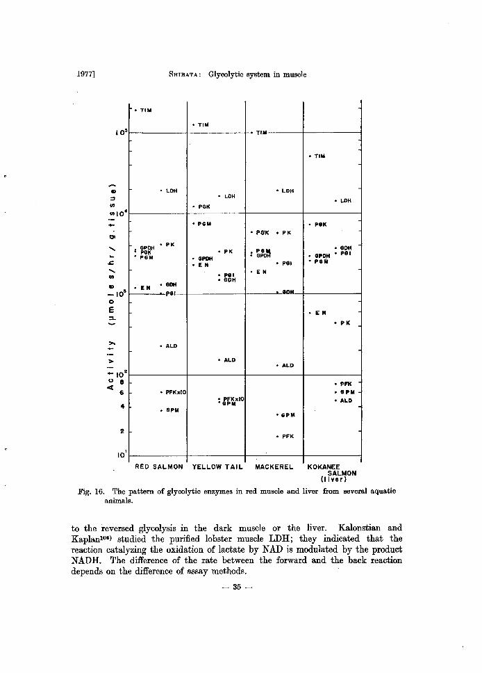
1977]
10 5
-
>
_10 2
U 8 ct
6
4
2
10 I
5mBATA: Glycolytic system in muscle
• TIM
• TIM
• TI
• LOH • LOH • LOH
• PGK
• PGM
• PG'K • PK
I ~~~ • PK
• PK • PG~ • PGM • GPOH • GP
• EN • PGI
• PGI • EN • GOH
• EN ·GDH
P81 • 80H
• ALO
• ALO • ALO
• PFKxlO
: G~~xIO • GPM
·GPM
• PFK
RED SALMON YELLOW TAIL MACKEREL
• TIM
• LDH
• P8K
·80H • GPOH • PGI • PGM
• EN
• Pit
• PFK
• GPM
• ALO
KOKANEE SAUl/ON
(t jver)
Fig. 16. The pattern of glycolytic enzymes in red muscle and liver from several aquatic animals.
to the reversed glycolysis in the dark muscle or the liver. Kalonstian and Kaplanl06) studied the purified lobster muscle LDH; they indicated that the reaction catalyzing the oxidation of lactate by NAD is modulated by the product NADH. The difference of the rate between the forward and the back reaction depends on the difference of assay methods.
- 35-
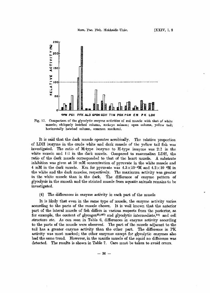
250
l >- 200 I-
> I-(,) 150 C
III >
Mem. Fac. Fish. Hokkaido Univ. [XXIV, 1,2
;: 100 -•••....•• _._ .•
C -I III
II: 50
GPN PGI PFKALDGPDHGDH TIM PGKPGN EN PK LDH
Fig. 17. Comparison of the glycolytic enzyme activities of red muscle with that of white muscle; obliquely hatched column, sockeye salmon; open column, yellow tail; horizontally hatched column, common mackerel.
It is said that the dark muscle operates aerobically. The relative proportion of LDH isozyme in the crude white and dark muscle of the yellow tail fish was investigated. The ratio of l\1-type isozyme to H-type isozyme was 2:1 in the white muscle and 1:1 in the dark muscle. Compared to mammalian LDH, the ratio of the dark muscle corresponded to that of the heart muscle. A substrate inhibition was given at 10 ruM concentration of pyruvate in the white muscle and 4 ruM in the dark muscle. Km for pyruvate was 4.3 X 10-41\1 and 4.2 X 10-41\1 in the white and the dark muscles, respectively. The maximum activity was greater in the white muscle than in the dark. The difference of enzyme pattern of glycolysis in the smooth and the striated muscle from aquatic animals remains to be investigated.
(4) The differences in enzyme activity in each part of the muscle
It is likely that even in the same type of muscle, the enzyme activity varies according to the parts of the muscle chosen. It is well known that the anterior part of the lateral muscle of fish differs in various respects from the posterior, as for example, the content of glycogen23,101) and glycolytic intermediate,23) and cell structure etc. As can seen in Table 6, differences in enzyme activity according to the parts of the muscle were observed. The part of the muscle adjacent to the tail has a greater enzyme activity than the other part. The difference in PK activity was most marked; the other enzymes except for glycolytic enzymes also had the same trend. However, in the mantle muscle of the squid no difference was detected. The results is shown in Table 7. Care must be taken to avoid errors.
- 36-
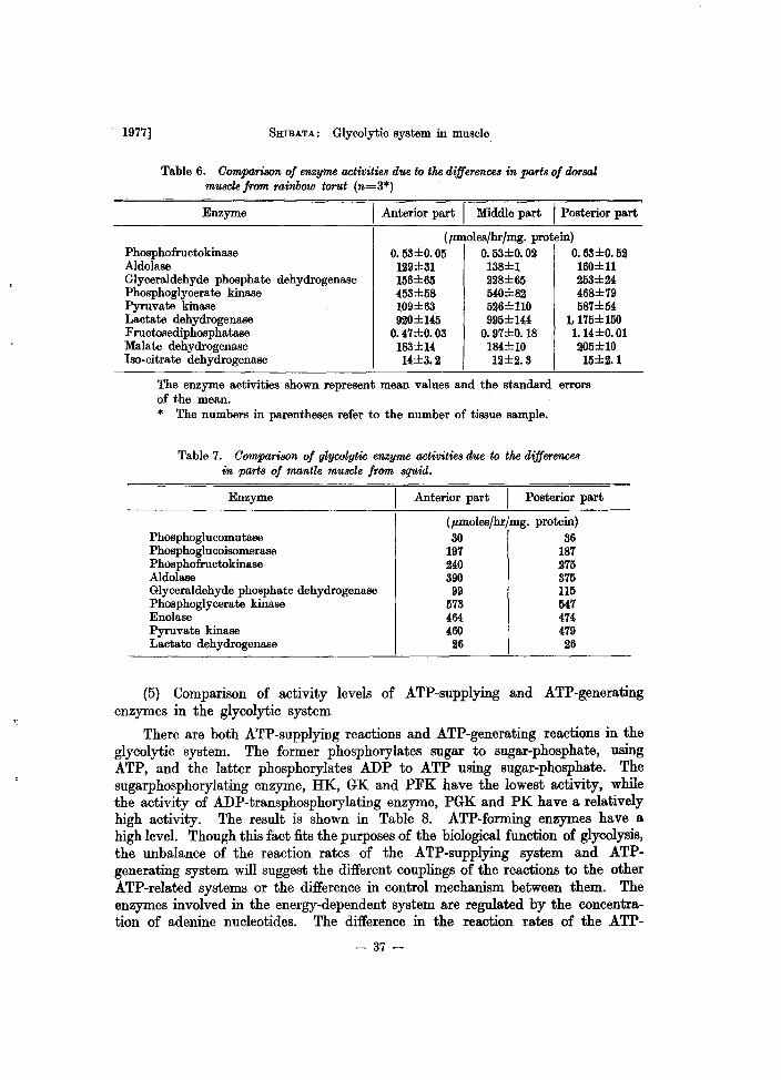
1977] 5mBATA: Glycolytic system in muscle
Table 6. Comparison of enzyme activities due to the differences in parts of dorsal muscle from rainbow tomt (71,=3*)
Enzyme I Anterior part I Middle part I Posterior part
Phosphofructokinase Aldolase Glyceraldehyde phosphate dehydrogenase Phosphoglycerate kinase Pyruvate kinase Lactate dehydrogenase Fructosediphosphatase Malate dehydrogenase Iso· citrate dehydrogenase
(pmoles/hr/mg. protein) ~~±~OO ~~±~~ ~~±~~ 129±31 138±1 160±11 U~~ ~~~ ~±M 453±58 540±82 468±79 109±~ 526±110 587±54 920±145 995±144 1,175±150
0.47±0.03 0.97±O.18 1. 14±0.01 183±14 184±10 205±10 14±&2 12±&3 U±&1
The enzyme activities shown represent mean values and the standard errors of the mean. * The numbers in parentheses refer to the number of tissue sample.
Table 7. Comparison of glycolytic enzyme activities due to the differences in parts of mantle muscle from squid.
Enzyme
Phosphoglucomutase Phosphoglucoisomerase Phosphofructokina.se Aldolase Glyceraldehyde phosphate dehydrogenase Phosphoglycerate kinase Enolase Pyruvate kinase Lactate dehydrogenase
Anterior part Posterior part
(pmoles/hr/mg. protein) 30 36
197 187 MO 275 390 375
99 115 573 547 464 474 460 479 26 26
(5) Comparison of activity levels of ATP-supplying and ATP-generating enzymes in the glycolytic system
There are both ATP-supplying reactions and ATP-generating reactions in the glycolytic system. The former phosphorylates sugar to sugar~phosphate, using ATP, and the latter phosphorylates ADP to ATP using sugar-phosphate. The sugarphosphorylating enzyme, HK, GK and PFK have the lowest activity, while the activity of ADP-transphosphorylating enzyme, PGK and PK have a relatively high activity. The result is shown in Table 8. ATP-forming enzymes have a. high level. Though this fact fits the purposes of the biological function of glycolysis, the unbalance of the reaction rates of the ATP-supplying system and ATPgenerating system will suggest the different couplings of the reactions to the other ATP-related systems or the difference in control mechanism between them. The enzymes involved in the energy-dependent system are regulated by the concentration of adenine nucleotides. The difference in the reaction rates of the ATP-
- 37-
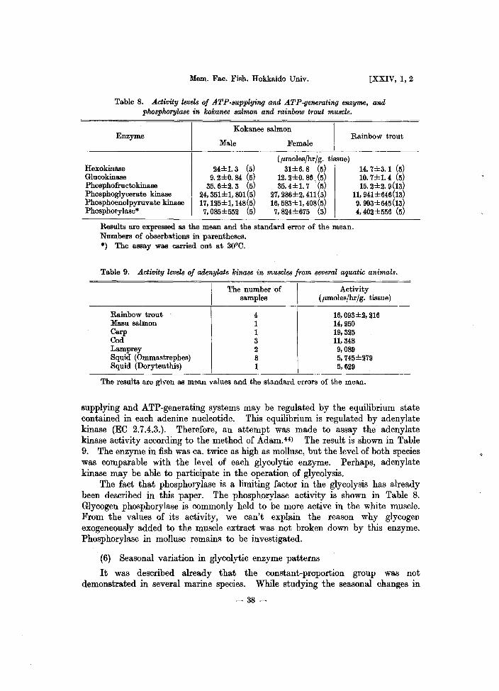
Mem. Fac. Fish. Hokkaido Univ. [XXIV, 1,2
Table 8. Activity levels of ATP-supplying and ATP-generating enzyme, and phosphorylase in kokanee salmon and rainbow trout muscle.
Enzyme
Hexokinase Glucokinase Phosphofructokinase Phosphoglycerate kinase Phosphoenolpyruvate kinase Phosphorylase*
Kokanee salmon
Male
24±1.3 (5) 9. 2±0. 84 (5)
35. 6±2. 3 (5) 24, 351±1, 801 (5) 17, 125±1, 148(5} 7,085±552 (5)
Female
(p,moles/hr/g. tissue) 31±6.8 (5)
12. 2±0. 86 (5) 35. 4±1. 7 (5)
27, 286±2, 411(5} 16, 583±1,408(5} 7,824±675 (5)
Rainbow trout
14. 7±3. 1 (5) 10. 7±1. 4 (5) 15. 2±2. 9(13}
11,941±646(13} 9.903±645(13} 4,402±556 (5)
Results are expressed as the mean and the standard error of the mean. Numbers of obserbations in parentheses. *) The assay was carried out at 30°C.
Table 9. Activity levels of adenylate kinase in muscles from several aquatic animals.
Rainbow trout Masu salmon Carp Cod Lamprey Squid (Ommastrephes) Squid (Doryteuthis)
The number of samples
4 1 1 3 2 8 1
Activity (p,moles/hr/g. tissue)
16, 093±2, 216 14,950 19,325 11,348 9,089 5,745±279 5,629
The results are given as mean values and the standard errors of the mean.
supplying and ATP-generating systems may be regulated by the equilibrium state contained in each adenine nucleotide. This equilibrium is regulated by adenylate kinase (EO 2.7.4.3.). Therefore, an attempt was made to assay the adenylate kinase activity according to the method of Adam. (4) The result is shown in Table 9. The enzyme in fish was ca. twice as high as mollusc, but the level of both species was comparable with the level of each glycolytic enzyme. Perhaps, adenylate kinase may be able to participate in the operation of glycolysis.
The fact that phosphorylase is a limiting factor in the glycolysis has already been described in this paper. The phosphorylase activity is shown in Table 8. Glycogen phosphorylase is commonly held to be more active in the white muscle. From the values of its activity, we can't exphtin the reason why glycogen exogeneously added to the muscle extract was not broken down by this enzyme. Phosphorylase in mollusc remains to be investigated.
(6) Seasonal variation in glycolytic enzyme patterns
It was described already that the constant-proportion group was not demonstrated in several marine species. While studying the seasonal changes in
- 38-
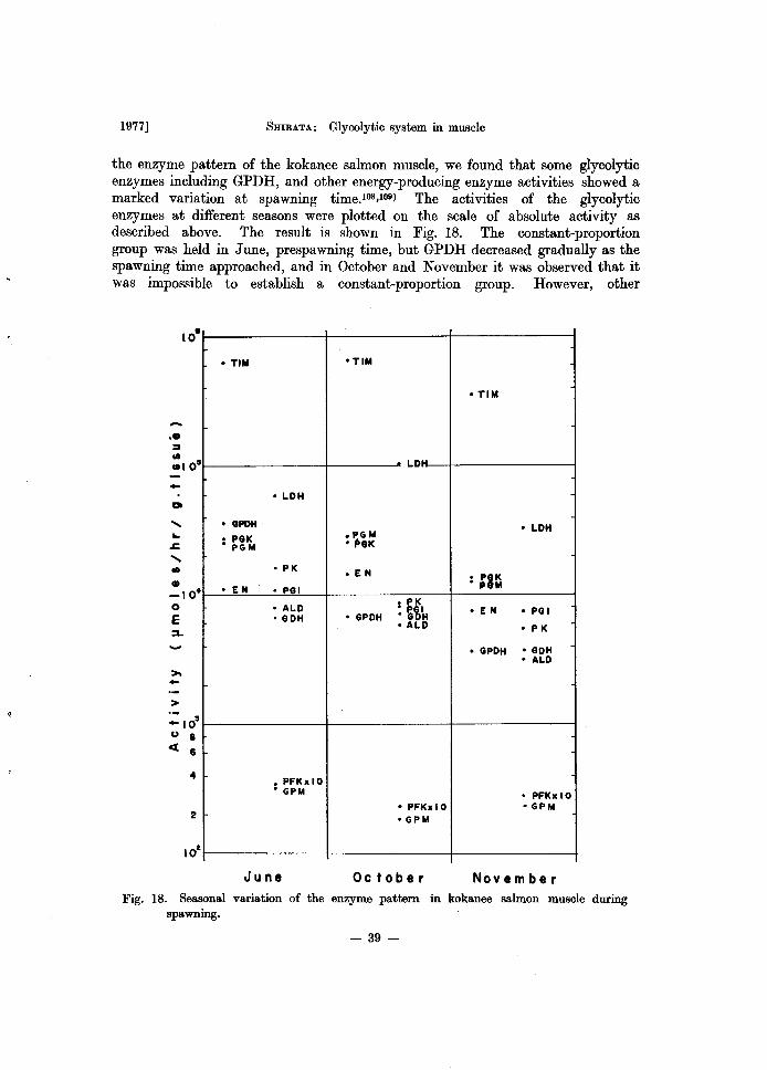
1977] SHIBATA: Glycolytic system in muscle
the enzyme pattern of the kokanee salmon muscle, we found that some glycolytic enzymes including GPDH, and other energy-producing enzyme activities showed a marked variation at spawning time.108,109) The activities of the glycolytic enzymes at different seasons were plotted on the scale of absolute activity as described above. The result is shown in Fig. 18. The constant-proportion group was held in June, prespawning time, but GPDH decreased gradually as the spawning time approached, and in October and November it was observed that it was impossible to establish a constant-proportion group. However, other
-at
....... .. ..c .......
>
:: 103
u 8 <[ 6
4
2
• TIM
• LDH
° GPOH
• PGK • PG M
• PK
• EN • PGI
• ALD • GDH
° PFKx 10 • GPM
June
·TIM
• TIM
LD
.PGM • LDH
• (>GI(
• EN • PIK ." M
• PK : ~Y,IH ° EN ° PGI
• GPDH ° ALD • P K
• GPDH • GDH ° ALD
° PFKxlO ° PFKxlO ° GPM oGPM
October November Fig. 18. Seasonal variation of the enzyme pattern in kokanee salmon muscle during
spawning.
- 39-

Mem. Fac. Fish. Hokkaido Univ. [XXIV, 1,2
triosephosphate metabolizing enzymes varied little. The possibility exists that the observed variation in GPDH activity may be under different physiological conditions. Since GPDH displays a greater variability in activity than most of the other enzymes, the variability may reflect the physiological state of the animal and represent a possible site of metabolic control. It would assumed that GPDH in the muscle of an aquatic animal has characteristic properties which differ from other animal enzymes. The physiological meaning of decreased GPDH activity remains obscure, whether this phenomenon is caused by the decline of the enzyme protein or the change in catalytic properties.
From the studies described in this chapter, although the activity levels of glycolysis varied with various species, organs, tissues and types of muscles in animals, it is confirmed that a certain congruence of the glycolytic pattern can be established, by arranging it in the order of its activities, in the muscle of all marine animals tested, and assuming that the sequence, constructed orderly, can also be treated functionally as one group. If it were possible to maintain the constancy under the various physiological conditions of animals, it would follow that the level of the enzyme proportion group can simply be recognized by the activity measurement of the key enzymes as a reference. The principle of proportionality seems to facilitate greatly the drawing of the enzyme maps for normal and abnormal states. Brosemer et al.no) reported that the constant-proportion group was supported during the development of differentiation of locust flight muscle. In our laboratory, it was found that the constant proportion group was demonstrated in the liver of the kokanee salmon during the spawning periodl11). Fitch and Chaihoffll2) showed that the alterations in the glycolytic enzyme activity might be extended as a result of the changes in carbohydrate intake. Weberll3) showed that the pattern of the key glycolytic enzymes in the liver followed a synchronous behavior under various nutritional and hormonal conditions. However, as can be seen in Fig. 18, the constancy was not demonstrated in the muscle of the salmon at the periods of near spawning and spawning. Since the observed variabilities reflect the physiological state of the animal, a large variation of GPDH may represent that the GPDH activity or protein is regulated to adapt to the circumstances, in which fish lives. GPDH in marine animal muscle may be one of regulatory enzyme in the glycolytic process, although in mammal it is known as non-regulatory enzyme. And since the experiment which was carried out at the same time showed a constant proportion group in the kokanee salmon liver, it revealed the operation of a different regulation in the muscle of the fish and in the liver. The physiological and biochemical meaning of GPDH variation in muscle is not clear, but GPDH in the muscles of marine animals are, in general, variable as can be seen in Fig. 15.
The fact that LDH had the lower activity in the mollusc muscle is an unusual case; yet it represents a characteristic of the mollusc muscle.
The difficulty of direct comparison of the activity levels exists in the extraction of the enzyme from the cell as well as a physiological state of the animal. The location of the enzyme in the cells influences its activity. Most of the glycolytic enzymes were completely extracted from the extramitochondrial
-40-
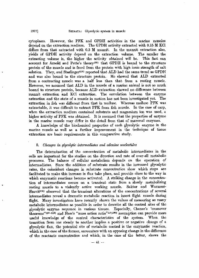
1977] SHIBATA: Glycolytic system in muscle
cytoplasm. However, the PFK and GPDH activities in the marine muscles depend on the extraction medium. The GPDH activity extracted with 0.15 M KOI differs from that extracted with 0.3 M mannit. In the mannit extraction also, yields of GPDH activity depend on the extraction volume. The smaller the extracting volume is, the higher the activity obtained will be. This fact can account for Arnold and Pette's theoryl02) that GPDH is bound to the structure protein of the muscle and is freed from the protein with high ionic strength of salt solution. They, and Starlinger114) reported that ALD had the same trend as GPDH and was also bound to the structure protein. He showed that ALD extracted from a contracting muscle was a half less than that from a resting muscle. However, we assumed that ALD in the muscle of a marine animal is not so much bound to structure protein, because ALD extraction showed no difference between mannit extraction and KCI extraction. The correlation between the enzyme extraction and the state of a muscle in motion has not been investigated yet. The extraction in fish was different from that in mollusc. Whereas mollusc PFK was extractable, it was difficult to extract PFK from fish muscle. In the case of carp, when the extraction medium contained substrate and magnesium ion was used, a higher activity of PFK was obtained. It is assumed that the properties of enzyme in the marine muscle may differ in the detail from that of mammal enzymes.
A knowledge of the biochemical properties of each glycolytic enzyme in the marine muscle as well as a further improvement in the technique of tissue extraction are basic requirements in this comparative study.
5. Changes in glycolytic intermediates and adenine nucleotiiles
The determination of the concentration of metabolic intermediates in the cells are important for the studies on the direction and rate of over-all metabolic processes. The balance of cellular metabolism depends on the operation of intermediates. Since the addition of substrate results in the increased glycolytic rates, the coincident changes in substrate concentration show which steps are facilitated to make this increase in flux take place, and provide clues to the way in which enzymatic reactions become activated. A striking change in the concentration of intermediates occurs on a transient state from a slowly metabolizing resting muscle to a violently active working muscle. Saktor and WormserShavit115) observed that the transient alterations of the concentrations of several intermediates reveal a dramatic metabolic reaction in insect flight muscle during flight. Many investigators have recently shown the values of measuring as many metabolic intermediates as possible in order to describe all the control sites of the glycolytic enzyme sequence in various tissues. Especially, Chance's 'crossover theorems"116-118) and Hess's "mass action ratio"119,120) assumption can provide more useful knowledge of the control characteristics of the system. When the transition from one steady to another implies a positive or negative change of a glycolytic flux, the potential site of metabolic control is the enzymatic reaction, which in the case of the former, encounters with an opposing change in the difference of the reactants concentration and which, in the case of the latter, shows the
- 41-
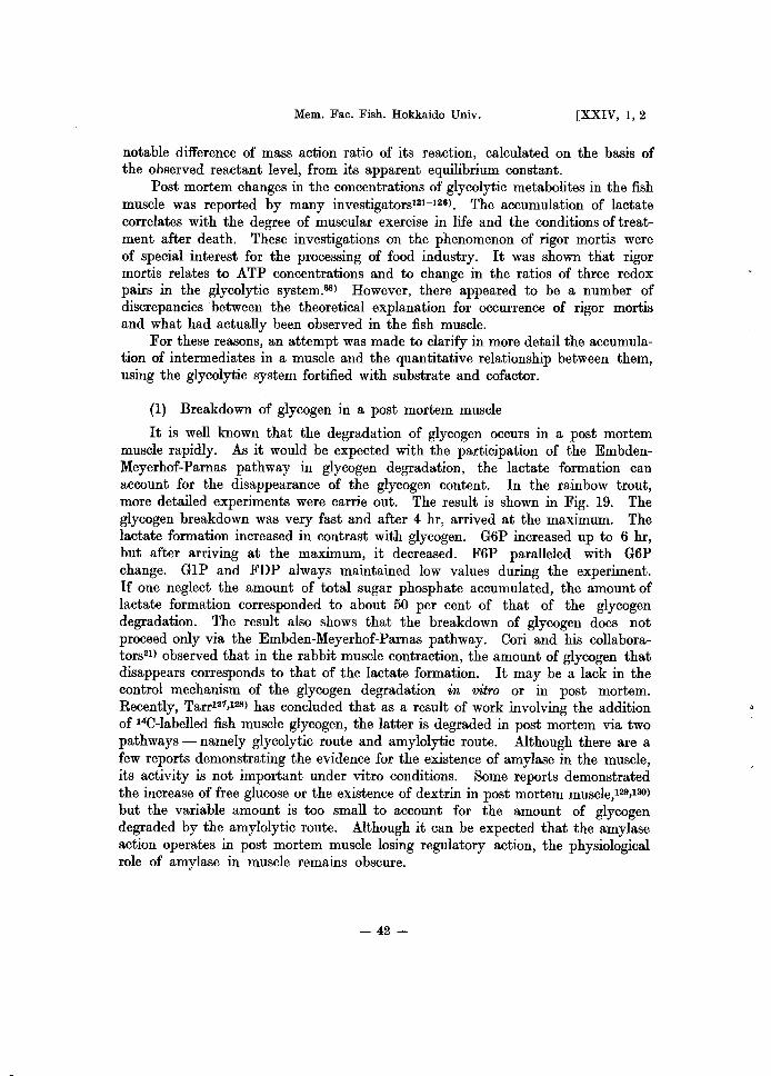
Mem. Fac. Fish. Hokkaido Univ. [XXIV, 1,2
notable difference of mass action ratio of its reaction, calculated on the basis of the observed reactant level, from its apparent equilibrium constant.
Post mortem changes in the concentrations of glycolytic metabolites in the fish muscle was reported by many investigators121-126). The accumulation of lactate correlates with the degree of muscular exercise in life and the conditions of treatment after death. These investigations on the phenomenon of rigor mortis were of special interest for the processing of food industry. It was shown that rigor mortis relates to ATP concentrations and to change in the ratios of three redox pairs in the glycolytic system.88) However, there appeared to be a number of discrepancies between the theoretical explanation for occurrence of rigor mortis and what had actually been observed in the fish muscle.
For these reasons, an attempt was made to clarify in more detail the accumulation of intermediates in a muscle and the quantitative relationship between them, using the glycolytic system fortified with substrate and cofactor.
(1) Breakdown of glycogen in a post mortem muscle
It is well known that the degradation of glycogen occurs in a post mortem muscle rapidly. As it would be expected with the participation of the EmbdenMeyerhof-Parnas pathway in glycogen degradation, the lactate formation can account for the disappearance of the glycogen content. In the rainbow trout, more detailed experiments were carrie out. The result is shown in Fig. 19. The glycogen breakdown was very fast and after 4 hr, arrived at the maximum. The lactate formation increased in contrast with glycogen. G6P increased up to 6 hr, but after arriving at the maximum, it decreased. F6P paralleled with G6P change. G1P and FDP always maintained low values during the experiment. If one neglect the amount of total sugar phosphate accumulated, the amount of lactate formation corresponded to about 50 per cent of that of the glycogen degradation. The result also shows that the breakdown of glycogen does not proceed only via the Embden-Meyerhof-Parnas pathway. Cori and his collaborators21) observed that in the rabbit muscle contraction, the amount of glycogen that disappears corresponds to that of the lactate formation. It may be a lack in the control mechanism of the glycogen degradation in vitro or in post mortem. Recently, Tarr121,128) has concluded that as a result of work involving the addition of HC-Iabelled fish muscle glycogen, the latter is degraded in post mortem via two pathways - namely glycolytic route and amylolytic route. Although there are a few reports demonstrating the evidence for the existence of amylase in the muscle, its activity is not important under vitro conditions. Some reports demonstrated the increase of free glucose or the existence of dextrin in post mortem muscle,129,130) but the variable amount is too small to account for the amount of glycogen degraded by the amylolytic route. Although it can be expected that the amylase action operates in post mortem muscle losing regulatory action, the physiological role of amylase in muscle remains obscure.
- 42 ~
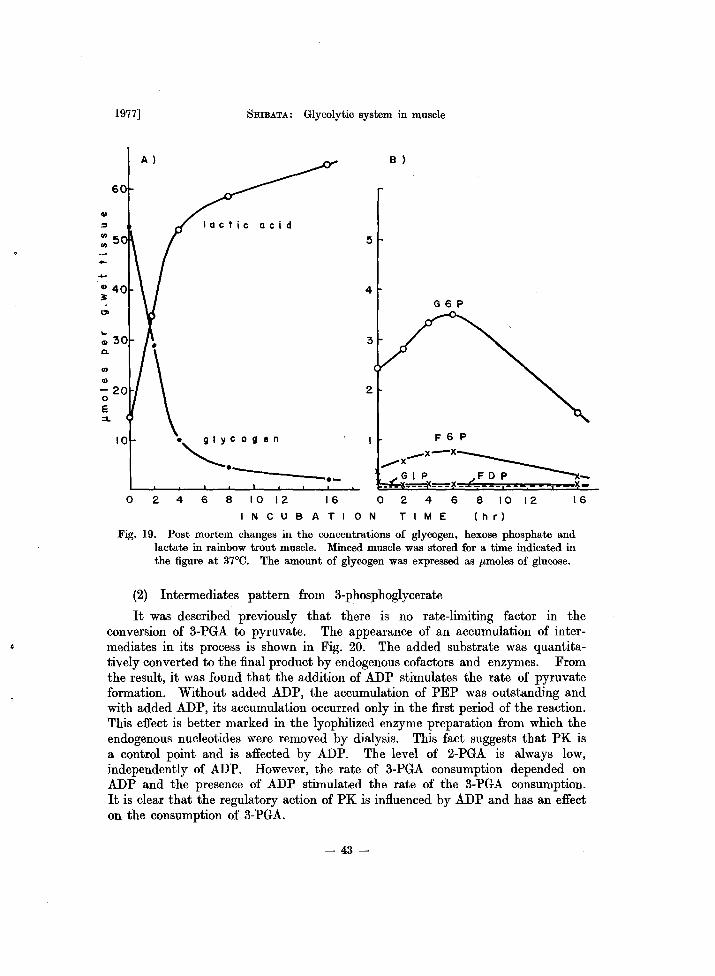
1977] 5mBATA: Glycolytic system in muscle
A )
60
lactic acid
5
4
10 • glycogen
~.---------.-o 2 4 6 8 10 12 16
NCUBATION
B )
G6P
F 6 P
_x-x~ ,.,..X GIP FOP _
___ ___ ~X _ x_
o 2 4 6 8 10 12 16
TIM E ( h r)
Fig. 19. Post mortem changes in the concentrations of glycogen, hexose phosphate and lactate in rainbow trout muscle. Minced muscle was stored for a time indicated in the figure at 37°C. The amount of glycogen was expressed as Ilmoles of glucose.
(2) Intermediates pattern from 3-phosphoglycerate
It was described previously that there is no rate-limiting factor in the conversion of 3-PGA to pyruvate. The appearance of an accumulation of intermediates in its process is shown in Fig. 20. The added substrate was quantitatively converted to the final product by endogenous cofactors and enzymes. From the result, it was found that the addition of ADP stimulates the rate of pyruvate formation. Without added ADP, the accumulation of PEP was outstanding and with added ADP, its accumulation occurred only in the first period of the reaction. This effect is better marked in the lyophilized enzyme preparation from which the endogenous nucleotides were removed by dialysis. This fact suggests that PK is a control point and is affected by ADP. The level of 2-PGA is always low, independently of ADP. However, the rate of 3-PGA consumption depended on ADP and the presence of ADP stimulated the rate of the 3-PGA consumption. It is clear that the regulatory action of PK is influenced by ADP and has an effect on the consumption of 3-PGA.
- 43-
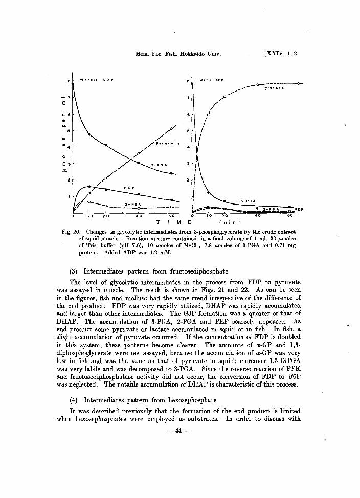
8
-7
E
... 6 QI
Q.
5
Ql4
o
E 3
::x..
2
Mem. Fac. Fish. Hokkaido Univ.
Without AOP 8
7
6
5
4
/fl'
/.~--O_-_-c. 2 - P G A ~ -----_0-______ 0--
10 20 40 60 0
With AOP
/" r!
I I
I I I
",,17'"
I , I
10 20
[XXIV, 1,2
...0-----------0-. ,..._-- pyruvate
... "
3- PG A
40 60
T M E (m in)
Fig. 20. Changes in glycolytic intermediates from 3-phosphoglycerate by the crude extract of squid muscle. Reaction mixture contained, in a final volume of 1 ml, 30 pmoles of Tris buffer (pH 7.6), 10 pmoles of MgC12 , 7.8 pmoles of 3-PGA and 0.71 mg protein. Added ADP was 4.2 mM.
(3) Intermediates pattern from fructosediphosphate
The level of glycolytic intermediates in the process from FDP to pyruvate was assayed in muscle. The result is shown in Figs. 21 and 22. As can be seen in the figures, fish and mollusc had the same trend irrespective of the difference of the end product. FDP was very rapidly utilized, DHAP was rapidly accumulated and larger than other intermediates. The G3P formation was a quarter of that of DHAP. The accumulation of 3-PGA, 2-PGA and PEP scarcely appeared. As end product some pyruvate or lactate accumulated in squid or in fish. In fish, a slight accumulation of pyruvate occurred. If the concentration of FDP is doubled in this system, these patterns become clearer. The amounts of o:-GP and 1,3-diphosphoglycerate were not assayed, because the accumulation of o:-GP was very low in fish and was the same as that of pyruvate in squid; moreover 1,3-DiPGA was very labile and was decomposed to 3-PGA. Since the reverse reaction of PFK and fructosediphosphatase activity did not occur, the conversion of FDP to F6P was neglected. The notable accumulation of DHAP is characteristic of this process.
(4) Intermediates pattern from hexosephosphate
It was described previously that the formation of the end product is limited when hexosephosphates were employed as substrates. In order to discuss with
-44-
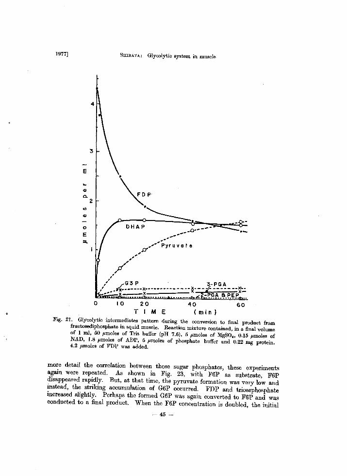
1977]
E
CI)
Q.
CI)
CI)
o E =..
4
2
, ,
SHIBATA: Glycolytic system in muscle
'",. .~
DHAP .0_.---...,-
If"'" ,
,IY
.. ,-,,- pyruvate
" G3 P 3-PGA " ~.-----x-------------- ~- ... -;/- -------f..: ', •• -- x • x ................ ¥.?:.~~A .. ~.~.~.~ .. o 10 20 40 60
TIME (min) Fig. 21. Glycolytic intermediates pattern during the conversion to final product from fructosediphosphate in squid muscle. Reaction mixture contained, in a final volume of 1 ml, 50 pmoles of Tris buffer (pH 7.6), 5 pmoles of MgSO" 0.15 pmoles of NAD, 1.8 pmoles of ADP, 5 pmoles of phosphate buffer and 0.22 mg protein. 4.2 pmoles of FDP was added.
more detail the correlation between those sugar phosphates, these experiments again were repeated. As shown in Fig. 23, with F6P as substrate, F6P disappeared rapidly. But, at that time, the pyruvate formation was very low and instead, the striking accumulation of G6P occurred. FDP and triosephosphate increased slightly. Perhaps the formed G6P was again converted to F6P and was conducted to a final product. When the F6P concentration is doubled, the initial - 45-
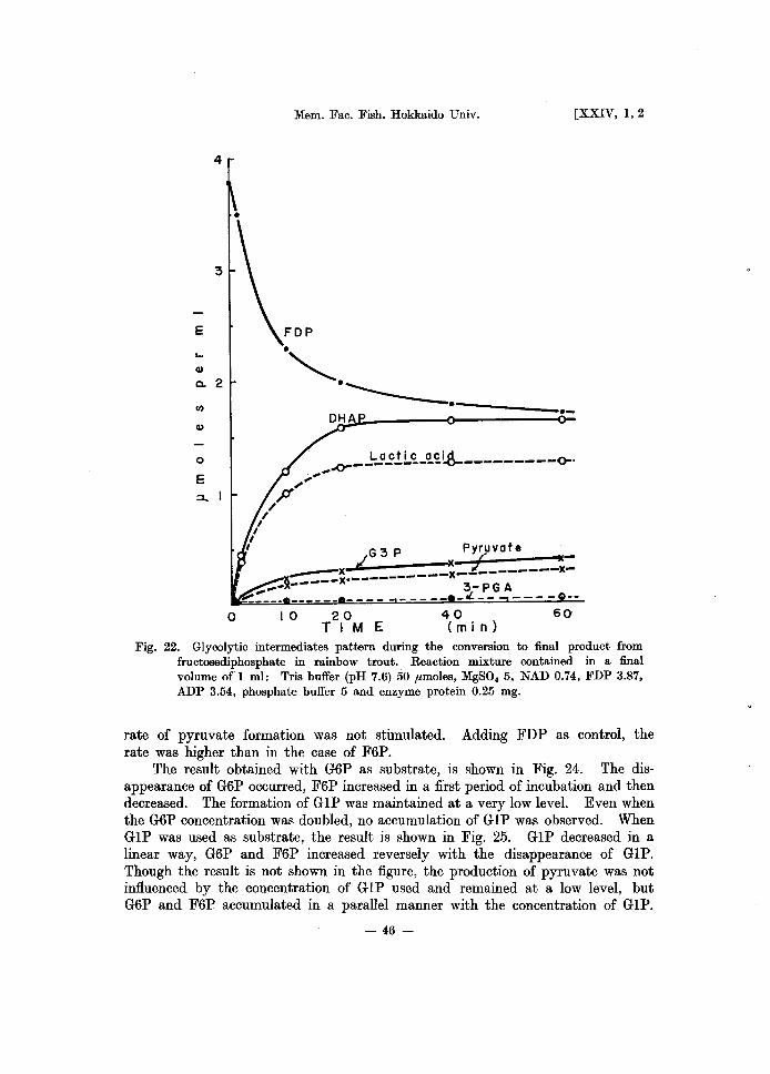
4
E
... Q)
Co 2
I/)
Q)
o
E
o
Mem. Fac. Fish. Hokkaido Univ. [XXIV, 1,2
FOP
.", . ------.---.-
G .,. P Pyruvate i v x ~ ~----:x . __________ x_L-------x----- x 3-PGA
10 20 TIM E
_ _ _ _ _ _ tL. _ _ - - - - --
40 60 (m j n)
Fig. 22. Glycolytic intermediates pattern during the conversion to final product from fructosediphosphate in rainbow trout. Reaction mixture contained in a final volume of 1 ml: Tris buffer (pH 7.6) 50 ,umoles, MgSO. 5, NAD 0.74, FDP 3.87, ADP 3.54, phosphate buffer 5 and enzyme protein 0.25 mg.
rate of pyruvate formation was not stimulated. Adding FDP as control, the rate was higher than in the case of F6P.
The result obtained with G6P as substrate, is shown in Fig. 24. The disappearance of G6P occurred, F6P increased in a first period of incubation and then decreased. The formation of G IP was maintained at a very low level. Even when the G6P concentration was doubled, no accumulation of GIP was observed. When GIP was used as substrate, the result is shown in Fig. 25. GIP decreased in a linear way, G6P and F6P increased reversely with the disappearance of GIP. Though the result is not shown in the figure, the production of pyruvate was not influenced by the concentration of G IP used and remained at a low level, but G6P and F6P accumulated in a parallel manner with the concentration of GIP.
- 46-
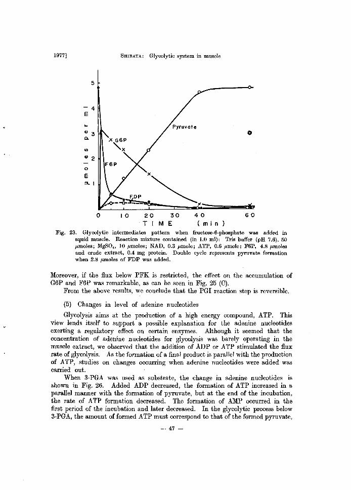
1977]
5
4 E
... Q) 3 Q.
I/)
Q) 2
0
E ::1..1
FEiP
o
5mBATA: Glycolytic system in muscle
10 20 30 TIM E
40 ( min)
60
Fig. 23. Glycolytic intermediates pattern when fructose-6-phosphate was added in squid muscle. Reaction mixture contained (in 1.0 mIl: Tris buffer (pH 7.6), 50 flmoles; MgSO., 10 flmoles; NAD, 0.3 flmole; ATP, 0.6 flmole; F6P, 4.8 flmoles and crude extract, 0.4 mg protein. Double cycle represents pyruvate formation when 2.8 flmoles of FDP was added.
Moreover, if the flux below PFK is restricted, the effect on the accumulation of G6P and F6P was remarkable, as can be seen in Fig. 25 (0).
From the above results, we conclude that the PGI reaction step is reversible.
(5) Changes in level of adenine nucleotides
Glycolysis aims at the production of a high energy compound, ATP. This view lends itself to support a possible explanation for the adenine nucleotides exerting a regulatory effect on certain enzymes. Although it seemed that the concentration of adenine nucleotides for glycolysis was barely operating in the muscle extract, we observed that the addition of ADP or ATP stimulated the flux rate of glycolysis. As the formation of a final product is parallel with the production of ATP, studies on changes occurring when adenine nucleotides were added was carried out.
When 3-PGA was used as substrate, the change in adenine nucleotides is shown in Fig. 26. Added ADP decreased, the formation of ATP increased in a parallel manner with the formation of pyruvate, but at the end of the incubation, the rate of ATP formation decreased. The formation of AMP occurred in the first period of the incubation and later decreased. In the glycolytic process below 3-PGA, the amount offormed ATP must correspond to that of the formed pyruvate,
- 47-
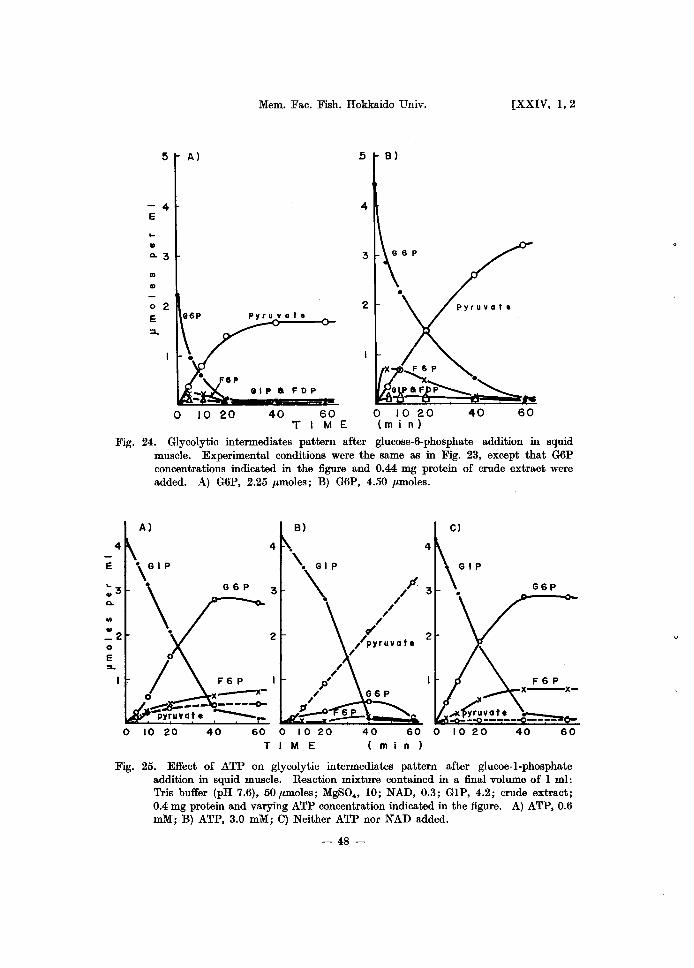
E
0. .,
0
E
5 A)
4 E
II
0.3
... .. o 2 E e6P
::l...
o 10 20
Mem. Fac. Fish. Hokkaido Univ.
pyruvate
FOP
40 60 TIM E
Jj
4
3
B)
o 10 20 (m in)
[XXIV, 1,2
Fig. 24. Glycolytic intermediates pattern after glucose-6-phosphate addition in squid muscle. Experimental conditions were the same as in Fig. 23, except that G6P concentrations indicated in the figure and 0.44 mg protein of crude extract were added. A) G6P, 2.25 ,umoles; B) G6P, 4.50 ,umoles.
A) B) C)
4 4
\. GI P
4
\GIP
G6P 3 \ /" 3 G6P
// /
2 v' //pyruvate
2
/
~ ~F6P x---x-
.At~vate _ -- ----- ---=-0 10 20 40 60 0 10 20 40 60 0 10 20 40 60
T 1M E ( m n )
Fig. 25. Effect of ATP on glycolytic intermediates pattern after glucoe-l-phosphate addition in squid muscle. Reaction mixture contained in a final volume of 1 ml: Tris buffer (pH 7.6), 50 ,umoles; MgSO., 10; NAD, 0.3; GIP, 4.2; crude extract; 0.4 mg protein and varying ATP concentration indicated in the figure. A) ATP, 0.6 mM; B) ATP, 3.0 roM; C) Neither ATP nor NAD added.
- 48-
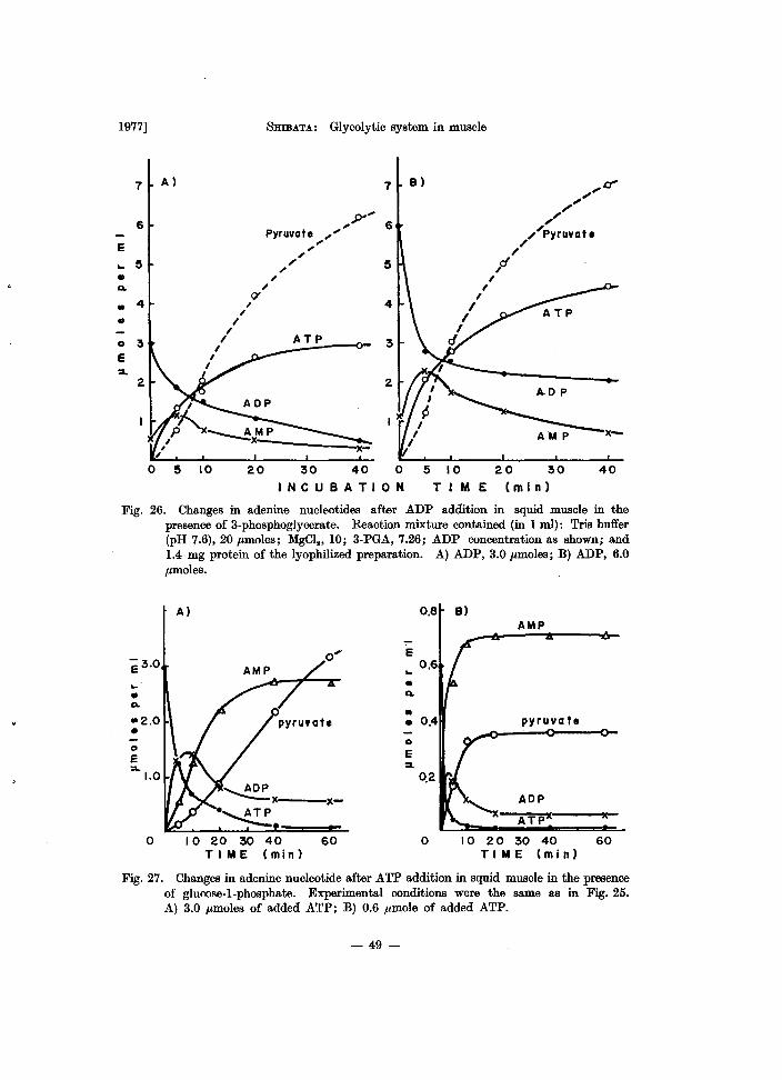
1977]
7 A)
6
E .. ~
• Q.
4 .. , • /
/ 0 3 /
I E I
:I.. I
2
o 10
5mBATA: Glycolytic system in muscle
7 B)
,P'''' ...... 6 Pyruvate , , , , ... ~ , " " 9' ,
20
4
ATP 3
2
30 40 0
INCUBATION
5 10 TIM E
"cJ' ,," ,," ....... :yruvate ... ...
p
20 30 40 (m in)
Fig. 26. Changes in adenine nucleotides after ADP addition in squid muscle in the presence of 3-phosphoglycerate. Reaction mixture contained (in 1 ml): Tris buffer (pH 7.6), 20 ,amoles; MgCl., 10; 3-PGA, 7.26; ADP concentration as shown; and 1.4 mg protein of the lyophilized preparation. A) ADP, 3.0 ,amoles; B) ADP, 6.0 ,amoles.
A) 0,8 B) AMP
0 ... · .. .. • • Q.
Q. .. pyruva te
0 0
E e ~
0,2
x- ADP ATPl( Ie
0 10 20 30 40 60 0 10 20 30 40 60 TIME (mi n) TI ME (m in)
Fig, 27, Changes in adenine nucleotide after ATP addition in squid muscle in the presence of glucose-I-phosphate. Experimental conditions were the same as in Fig. 25. A) 3.0 ,amoles of added ATP; B) 0.6 ,amole of added ATP.
- 49-
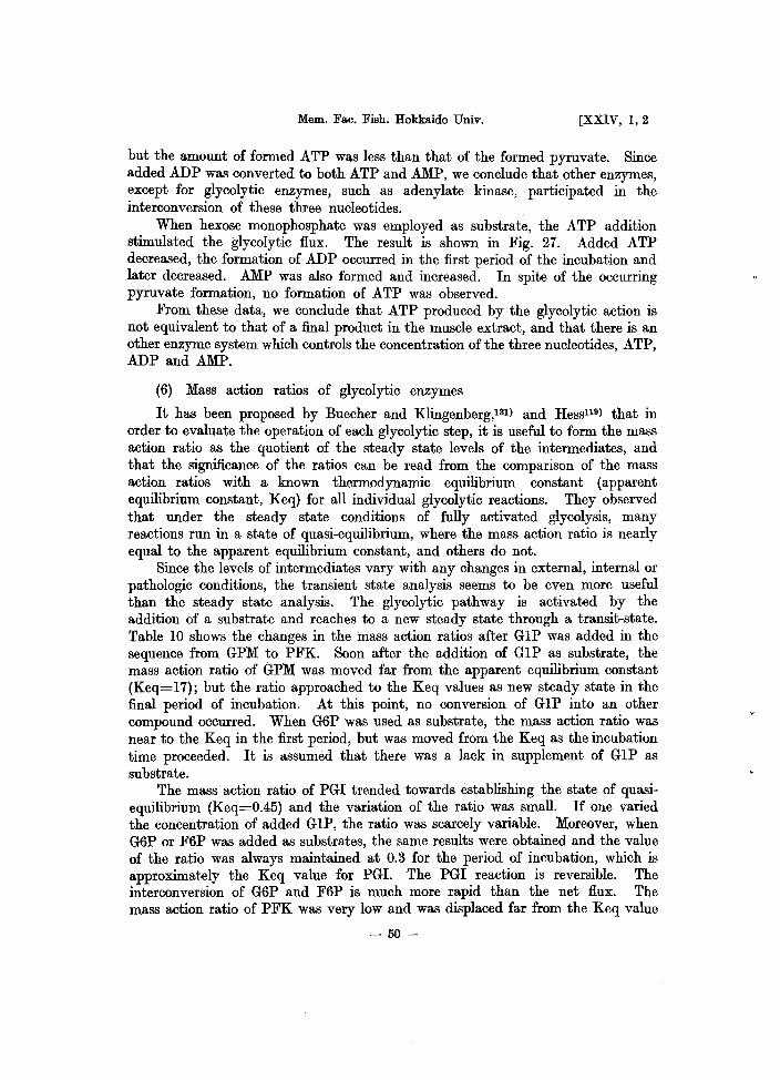
Mem. Fac. Fish. Hokkaido Univ. [XXIV, 1,2
but the amount of formed ATP was less than that of the formed pyruvate. Since added ADP was converted to both ATP and AMP, we conclude that other enzymes, except for glycolytic enzymes, such as adenylate kinase, participated in the interconversion of these three nucleotides.
When hexose monophosphate was employed as substrate, the ATP addition stimulated the glycolytic flux. The result is shown in Fig. 27. Added ATP decreased, the formation of ADP occurred in the first period of the incubation and later decreased. AMP was also formed and increased. In spite of the occurring pyruvate formation, no formation of ATP was observed.
From these data, we conclude that ATP produced by the glycolytic action is not equivalent to that of a final product in the muscle extract, and that there is an other enzyme system which controls the concentration of the three nucleotides, ATP, ADP and AMP.
(6) Mass action ratios of glycolytic enzymes
It has been proposed by Buecher and Klingenberg,131} and Hess1l9} that in order to evaluate the operation of each glycolytic step, it is useful to form the mass action ratio as the quotient of the steady state levels of the intermediates, and that the significance of the ratios can be read from the comparison of the mass action ratios with a known thermodynamic equilibrium constant (apparent equilibrium constant, Keq) for all individual glycolytic reactions. They observed that under the steady state conditions of fully activated glycolysis, many reactions run in a state of quasi-equilibrium, where the mass action ratio is nearly equal to the apparent equilibrium constant, and others do not.
Since the levels of intermediates vary with any changes in external, internal or pathologic conditions, the transient state analysis seems to be even more useful than the steady state analysis. The glycolytic pathway is activated by the addition of a substrate and reaches to a new steady state through a transit-state. Table 10 shows the changes in the mass action ratios after G1P was added in the sequence from GPM to PFK. Soon after the addition of G1P as substrate, the mass action ratio of GPM was moved far from the apparent equilibrium constant (Keq=17); but the ratio approached to the Keq values as new steady state in the final period of incubation. At this point, no conversion of G 1P into an other compound occurred. When G6P was used as substrate, the mass action ratio was near to the Keq in the first period, but was moved from the Keq as the incubation time proceeded. It is assumed that there was a lack in supplement of G 1P as substrate.
The mass action ratio of PGI trended towards establishing the state of quasiequilibrium (Keq=0.45) and the variation of the ratio was small. If one varied the concentration of added G1P, the ratio was scarcely variable. Moreover, when G6P or F6P was added as substrates, the same results were obtained and the value of the ratio was always maintained at 0.3 for the period of incubation, which is approximately the Keq value for PGI. The PGI reaction is reversible. The interconversion of G6P and F6P is much more rapid than the net flux. The mass action ratio of PFK was very low and was displaced far from the Keq value
- 50-
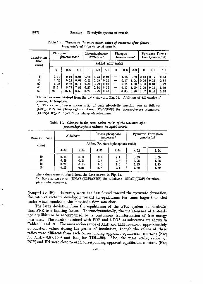
1977] SHIBATA: Glycolytic system in muscle
Table 10. Ohanges in the mass action ratios of reactants after glucose, I-phosphate addition in squid muscle.
Phospho- I Ph.osphoglucose I Phospho-
I Pyruvate Forma-
Incubation glucomutase* lsomerase* fructokinase* tion (pmoles/ml) time
Added ATP (mM) (min)
0 1 0.6 1 3.01 0 1 0.61 3.01 0 1 0.61 3.01 0 1 0.61 3.0
5 0.14 0.07 0.04 0.26 0.23 0.21 - 4.23 0.69 0.06 0.17 0.13 10 0.32 0.19 0.04 0.31 0.59 0.15 - 0.17 1.04 0.09 0.34 0.37 20 1.08 0.72 0.11 0.30 0.29 0.21 - 0.13 1.00 0.06 0.34 0.92 40 11.3 8.73 7.92 0.27 0.14 0.26 - 0.10 1.20 0.09 0.37 2.19 60 22 16.6 6.50 0.27 0.29 0.23 - 0.08 0.96 0.07 0.43 3.18
The values were obtained from the data shown in Fig. 25. Addition of 4.2 pmoles of glucose, I-phosphate. *) The value of mass action ratio of each glycolytic reaction was as follows: (G6P)/(GlP) for phosphoglucomutase; (F6P)/(G6P) for phospoglucose isomerase; (FDP)(ADP)/(F6P)(ATP) for phosphofructokinase.
Table ll. Ohanges in the mass action ratios of the reactants after fructosediphosphate addition in squid muscle.
Aldolase* I Triose phosphate
I Pyruvate Formation
Reaction Time isomerase* pmoles/ml .
(min) Added Fructosediphosphate (mM)
4.32 1 8.64 1 4.32 1 8.64 1 4.32 1 8.64
10 0.14 0.11 8.6 9.1 0.63 0.60 20 0.19 0.15 7.5 7.5 1.05 1.08 40 0.21 0.18 6.0 7.3 1.43 1.43 60 0.12 0.26 14.5 7.1 1.62 1.59
The values were obtained from the data shown in Fig, 21. *) Mass action ratio: (DHAP)(G3P)/(FDP) for aldolase; (DHAP)/(G3P) for triose phosphate isomerase.
(Keq=1.2X 10³). However, when the flux flowed toward the pyruvate formation, the ratio of ractants developed toward an equilibrium ten times larger than that under which condition the metabolic flow was slow.
The large deviation from the equilibrium of the PFK system demonstrates that PFK is a limiting factor. Thermodynamically, the maintenance of a steady non-equilbirium is accompanied by a continuous transformation of free energy into heat. The results obtained with FDP and 3-PGA as substrates are shown in Tables 11 and 12. The mass action ratios of ALD and TIM remained approximately at constant values during the period of incubation, though the values of these ratios were different from each corresponding apparent equilibrium constant (Keq for ALD=6.8 X 10⁻⁵ and Keq for TIM=22). Also, the mass action ratios of PGM and EN were close to each corresponding apparent equilibrium constant (Keq
- 51-
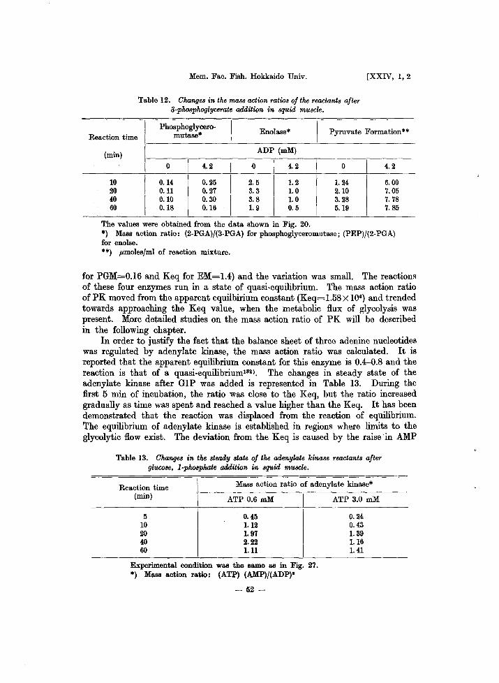
Mem. Fac. Fish. Hokkaido Univ. [XXIV, 1,2
Table 12. Changes in the mass action ratios of the reactants after 3-phosphoglycerate addition in squid muscle.
Phosphoglycero-
I Enolase* I Pyruvate Formation** Reaction time mutase*
(min) ADP (mM)
0 I 4.2 I 0 I 4.2 I 0 I 4.2
10 0.14 0.25 2.5 1.2 1.24 6.00 20 0.11 0.27 3.3 1.0 2.10 7.05 40 0.10 0.30 3.8 1.0 3.28 7.78 60 0.18 0.16 1.9 0.5 5.19 7.85
The values were obtained from the data shown in Fig. 20. *) Mass action ratio: (2-PGA)/(3-PGA) for phosphoglyceromutase; (PEP)/(2-PGA) for enolse. **) ",moles/ml of reaction mixture.
for PGM=0.16 and Keq for EM=1.4) and the variation was small. The reactions of these four enzymes run in a state of quasi-equilibrium. The mass action ratio of PK moved from the apparent equilbirium constant (Keq=1.58X 1(4) and trended towards approaching the Keq value, when the metabolic flux of glycolysis was present. More detailed studies on the mass action ratio of PK will be described in the following chapter.
In order to justify the fact that the balance sheet of three adenine nucleotides was regulated by adenylate kinase, the mass action ratio was calculated. It is reported that the apparent equilibrium constant for this enzyme is 0.4-0.8 and the reaction is that of a quasi-equilibrium132). The changes in steady state of the adenylate kinase after G IP was added is represented in Table 13. During the first 5 min of incubation, the ratio was close to the Keq, but the ratio increased gradually as time was spent and reached a value higher than the Keq. It has been demonstrated that the reaction was displaced from the reaction of equilibrium. The equilibrium of adenylate kinase is established in regions where limits to the glycolytic flow exist. The deviation from the Keq is caused by the raise in AMP
Table 13. Changes in the steady state of the adenylate kinase reactants after glucose, I-phosphate addition in squid muscle.
Reaction time (min)
Mass action ratio of adenylate kinase*
5 10 20 40 60
ATP 0.6 mM
0.45 1.12 1.97 2.22 1.11
Experimental condition was the same as in Fig. 27. *) Mass a.ction ratio: (ATP) (AMP)/(ADP)'
- 52-
ATP 3.0 mM
0.24 0.43 1.39 1.16 1.41
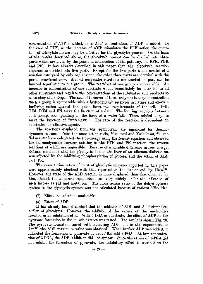
1977] 5mBATA: Glycolytic system in muscle
concentration, if ATP is added, or in ATP concentration, if ADP is added. In the case of PFK, as the increase of AMP stimulates the PFK action, the operation of adenylate kinase may be effective for the glycolytic process. On the basis of the results described above, the glycolytic process can be divided into three parts which are given by the points of intersection of the pathway, i.e. PFK, PGK and PK. It has already described in this paper that the glycolytic reaction sequence is divided into five parts. Except for the two parts which consist of a reaction catalyzed by only one enzyme, the other three parts are identical with the parts considered now. Several enzymatic reactions constructed in part can be lumped together into one group. The reactions of one group are reversible. An increase in concentration of one substrate would immediately be extended to all other substrates and regulate the concentrations of the substrates and products so as to obey their Keqs. The rate of turnover of these enzymes is enzyme-controlled. Such a group is comparable with a hydrodynamic reservoir in nature and exerts a buffering action against the quick functional requirements of the cell. PGI, TIM, PGM and EN serve the function of a dam. The limiting reactions between such groups are operating in the form of a water-fall. These related enzymes serve the function of "water-gate". The rate of the reaction is dependent on substrates or effective agents.
The reactions displaced from the equilibrium are significant for thermodynamic reasons. From the mass action ratio, Mizukami and Yoshikawa,134) and Sakurai135) have calculated the free-energy using the Nernst equation and observed the thermodynamic barriers existing in the PFK and PK reaction, the reverse reactions of which are impossible. Because of a notable difference in free energy, Sakurai concluded that the glycolytic flux in the liver of an alloxane-diabetic rat was affected by the inhibiting phosphorylation of glucose, and the action of ALD and PK.
The mass action ratios of most of glycolytic enzymes reported in this paper were approximately identical with that reported in the tumor cell by Hess.n9)
However, the state of the ALD reaction is more displaced than that obtained by him, though the apparent equilibrium can vary widely under the influence of such factors as pH and metal ion. The mass action ratio of the dehydrogenase system in the glycolytic system was not calculated because of various difficulties.
(7) Effect of adenine nucleotides
(a) Effect of ADP It has already been described that the addition of ADP and ATP stimulates
a flux of glycolysis. However, the addition of the excess of the nucleotides resulted in an inhibition of it. With 3-PGA as substrate, the effect of ADP on the pyruvate formation in the muscle extract was tested. The result is shown Fig. 28. The pyruvate formation raised with increasing ADP, but in this experiment, at 7 mM, the ADP maximum value was obtained. When further ADP was added, it inhibited the formation of pyruvate at above 6.4 ruM 3-PGA. At low concentration of 3-PGA, the ADP inhibition did not appear. Since the excess of 3-PGA did not inhibit the formation of pyruvate, the inhibitory effect is ascribed to the
- 53-
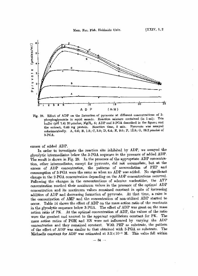
Mem. Fac. Fish. Hokkaido Univ. [XXIV, 1,2
6
E ..... I/)
5 Q)
"0 E =4 Q) - 3 0
> =' 2 ... >-a.
---- ----- -----'. -----------.-A ____ -,------~~---_. • ._----- -----------------_0-
012345 10 15 20 25
A 0 p em M)
Fig. 28. Effect of ADP on the formation of pyruvate at different concentrations of 3-phosphoglycerate in squid muscle. Reaction mixture contained (in 1 ml): Tris buffer (pH 7.4) 20 Jlmoles; MgCI., 5; ADP and 3-PGA described in the figure; and the extract, 0.46 mg protein. Reaction time, 5 min. Pyruvate was assayed colorimetrically. A, 0.6; B, 1.8; C, 3.8; D, 6.4; E, 9.6; F, 12.8; G, 19.2 Jlmoles of 3-PGA.
excess of added ADP. In order to investigate the reaction site inhibited by ADP, we assayed the
glycolytic intermediates below the 3-PGA sequence in the presence of added ADP. The result is shown in Fig. 29. In the presence of the appropriate ADP concentration, other intermediates, except for pyruvate, did not accumulate, but at the excess of ADP concentration, the patterns of accumulation of PEP and consumption of 3-PGA were the same as when no ADP was added. No significant change in the 2-PGA concentration depending on the ADP concentrations occurred. Following the changes in the concentrations of adenine nucleotides, the ATP concentration reached their maximum values in the presence of the optimal ADP concentration and its maximum values remained constant in spite of increasing addition of ADP and decreasing formation of pyruvate. At that time, a raise in the concentration of AMP and the concentration of non-utilized ADP started to occur. Table 14 shows the effect of ADP on the mass action ratio of the reactants in the glycolytic sequence below 3-PGA. The effect of ADP was great on the mass action ratio of PK. At the optimal concentration of ADP, the values of the ratio were the greatest and nearest to the apparent equilibrium constant for PK. The mass action ratios of PGM and EN were not influenced by varying the ADP concentration and they remained constant. With PEP as substrate, the pattern of the effect of ADP was similar to that obtained with 3-PGA as substrate. The Michaelis constant for ADP was estimated at 3.2 X 10⁻⁴ M. This value fell within
-54-
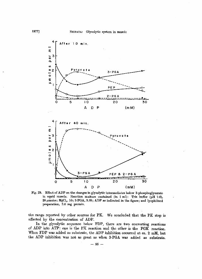
1977]
4
E
.... 3 CI)
Q.
o E ::1..(
, ,
SHIBATA: Glycolytic system in muscle
After (0 min.
~---- - ....... r- __ ~-- -t--2- PG A
---·--------r-------,-------~1-o
~:r Q. •
Vl
Cl)2
o E
o
5 (0
A
20 30
D P (m M)
After 40 min.
_----0--- -_ ~' ----" , ~-
" ..... , P Y r u vat e I .. , , ,
I '. , , , , ,
PEP a 2-PG A ... ----- -----
5 10 20 30
A D P (mM)
Fig. 29. Effect of ADP on the changes in glycolytic intermediates below 3-phosphoglycerate in squid muscle. Reaction mxiture contained (in 1 ml): Tris buffer (pH 7.6), 20 pmoles; MgC12' 10; 3-PGA, 3.59; ADP as indicated in the figure; and lyophilized preparation, 3.0 mg protein.
the range reported by other sources for PK. We concluded that the PK step is affected by the concentration of ADP.
In the glycolytic sequence below FDP, there are two converting reactions of ADP into ATP: one is the PK reaction and tJ:te other is the PGK reaction. When FDP was added as substrate, the ADP inhibition occurred at ca. 2 mM, but the ADP inhibition was not as great as when 3-PGA was added as substrate.
- 55-
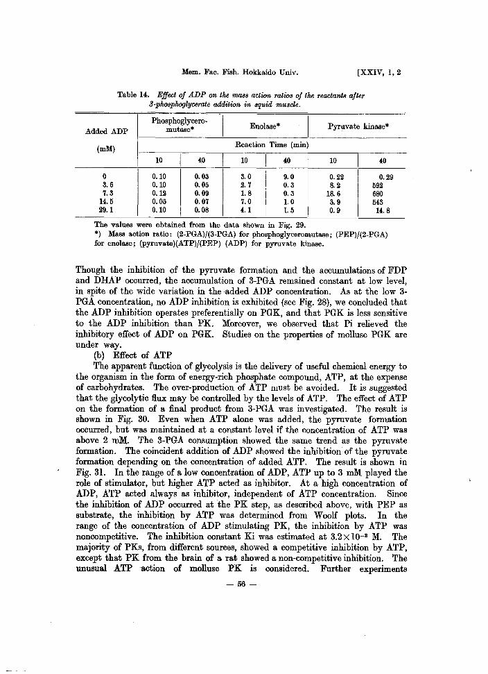
Mem. Fac. Fish. Hokkaido Univ. [XXIV, 1,2
Table 14. Effect of ADP on the 'f/U1,88 action ratio8 of the reactant8 after a.phosphoglycerate addition in 8quid mU8cle.
Phosphoglycero.
I Enolase*
I Pyruvate kinase*
Added ADP mutase*
(mM) Reaction Time (min)
10 I 40 I 10 I 40 I 10 I 40
0 0.10 0.03 3.0 9.0 0.22 0.29 3.6 0.10 0.05 2.7 0.3 8.2 592 7.3 0.12 0.09 1.8 0.3 18.6 680
14.5 0.05 0.07 7.0 1.0 3.9 543 29.1 0.10 0.08 4.1 1.5 0.9 14.8
The values were obtained from the data shown in Fig. 29. *) Mass action ratio: (2.PGA)/{3·PGA) for phosphoglyceromutase; (PEP)/{2.PGA) for enolase; (pyruvate)(ATP)/{PEP) (ADP) for pyruvate kinase.
Though the inhibition of the pyruvate formation and the accumulations of FDP and DHAP occurred, the accumulation of 3-PGA remained constant at low level, in spite of the wide variation in the added ADP concentration. As at the low 3-PGA concentration, no ADP inhibition is exhibited (see Fig. 28), we concluded that the ADP inhibition operates preferentially on PGK, and that PGK is less sensitive to the ADP inhibition than PK. Moreover, we observed that Pi relieved the inhibitory effect of ADP on PGK. Studies on the properties of mollusc PGK are under way.
(b) Effect of ATP The apparent function of glycolysis is the delivery of useful chemical energy to
the organism in the form of energy-rich phosphate compound, ATP, at the expense of carbohydrates. The over-production of ATP must be avoided. It is suggested that the glycolytic flux may be controlled by the levels of ATP. The effect of ATP on the formation of a final product from 3-PGA was investigated. The result is shown in Fig. 30. Even when ATP alone was added, the pyruvate formation occurred, but was maintained at a constant level if the concentration of ATP was above 2 roM. The 3-PGA consumption showed the same trend as the pyruvate formation. The coincident addition of ADP showed the inhibition of the pyruvate formation depending on the concentration of added ATP. The result is shown in Fig. 31. In the range of a low concentration of ADP, ATP up to 3 mM played the role of stimulator, but higher ATP acted as inhibitor. At a high concentration of ADP, ATP acted always as inhibitor, independent of ATP concentration. Since the inhibition of ADP occurred at the PK step, as described above, with PEP as substrate, the inhibition by ATP was determined from Woolf plots. In the range of the concentration of ADP stimulating PK, the inhibition by ATP was noncompetitive. The inhibition constant Ki was estimated at 3.2 X 10⁻³ M. The majority of PKs, from different sources, showed a competitive inhibition by ATP, except that PK from the brain of a rat showed a non-competitive inhibition. The unusual ATP action of mollusc PK is considered. Further experiments
-56-
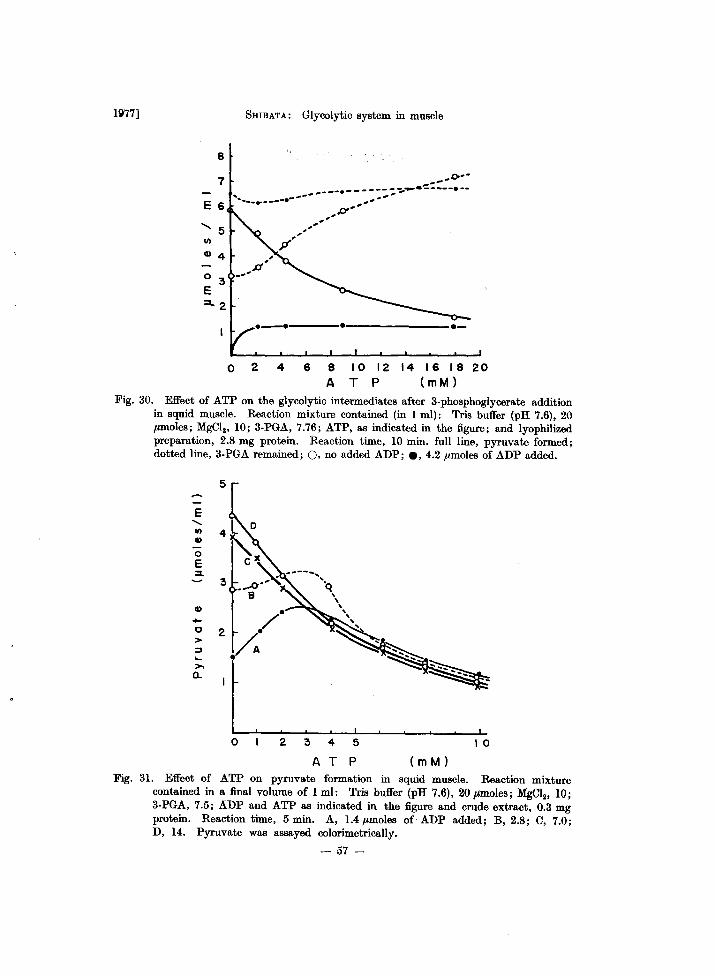
1977]
8
7
E 6
'5 II)
Q) 4
o 3 E
SHIBATA: Glycolytic system in muscle
-- 0----__ • ______ _ ~ ..... c _____ • __
''''' .. _-.--_ ... - ... ---- ,_ ... ,
-' ,-
,p'"
p-'-
.---4 e ______________ __
o 2 4 6 8 10 12 14 16 18 20 A T P (mM)
Fig. 30. Effect of ATP on the glycolytic intermediates after 3-phosphoglycerate addition in squid muscle. Reaction mixture contained (in 1 ml): Tris buffer (pH 7.6), 20 ,umoles; MgCI2, 10; 3-PGA, 7.76; ATP, as indicated in the figure; and lyophilized preparation, 2.8 mg protein. Reaction time, 10 min. full line, pyruvate formed; dotted line, 3-PGA remained; 0, no added ADP; e, 4.2 ,umoles of ADP added.
5
E "-II)
Q)
0 E :I.
3
Q) • ~
2 / 0 > V. ::J ... >-
t 0...
0 2 3 4 5 10
A T P (m M)
Fig. 31. Effect of ATP on pyruvate formation in squid muscle. Reaction mixture contained in a final volume of 1 ml: Tris buffer (pH 7.6), 20 ",moles; MgCl20 10; 3-PGA, 7.5; ADP and ATP as indicated in the figure and crude extract, 0.3 mg protein. Reaction time, 5 min. A, 1.4,umoles of ADP added; B, 2.8; C, 7.0; D, 14. Pyruvate was assayed colorimetrically.
- 57-
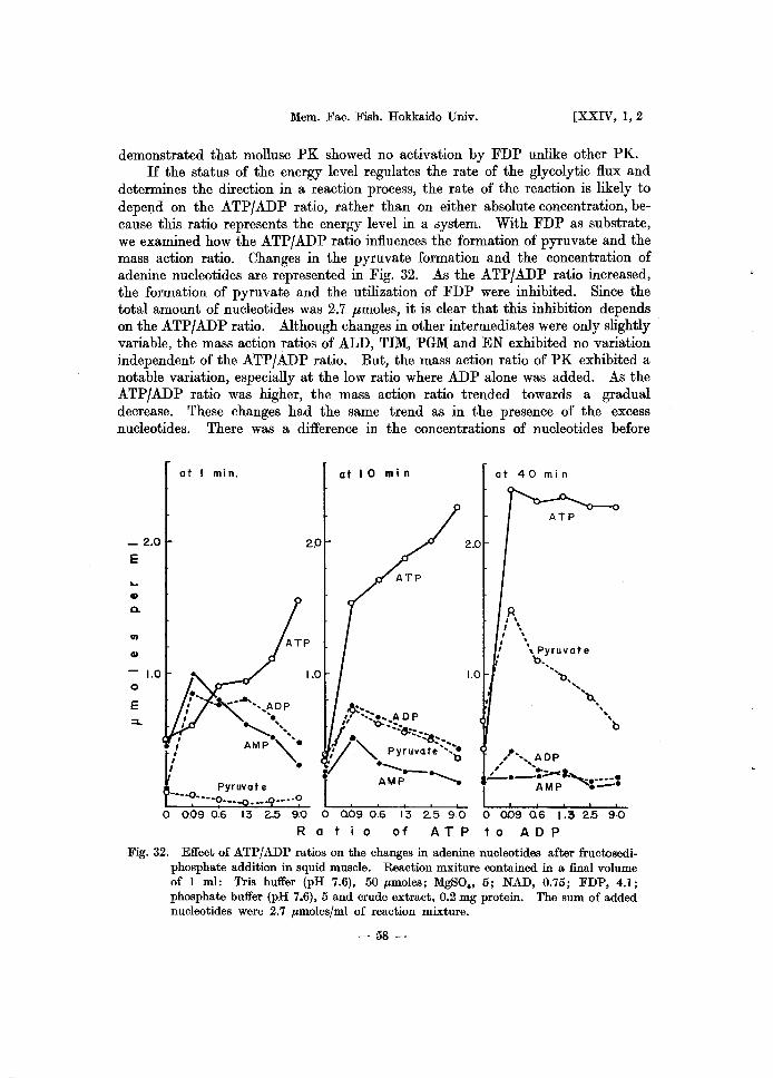
Mem. Fac. Fish. Hokkaido Univ. [XXIV, 1,2
demonstrated that mollusc PK showed no activation by FDP unlike other PK. If the status of the energy level regulates the rate of the glycolytic flux and
determines the direction in a reaction process, the rate of the reaction is likely to depend on the ATP/ADP ratio, rather than on either absolute concentration, because this ratio represents the energy level in a system. With FDP as substrate, we examined how the ATPjADP ratio influences the formation of pyruvate and the mass action ratio. Changes in the pyruvate formation and the concentration of adenine nucleotides are represented in Fig. 32. As the ATPjADP ratio increased, the formation of pyruvate and the utilization of FDP were inhibited. Since the total amount of nucleotides was 2.7 ,umoles, it is clear that this inhibition depends on the ATP/ADP ratio. Although changes in other intermediates were only slightly variable, the mass action ratios of ALD, TIM, PGM and EN exhibited no variation independent of the ATP/ADP ratio. But, the mass action ratio of PK exhibited a notable variation, especially at the low ratio where ADP alone was added. As the ATP/ADP ratio was higher, the mass action ratio trended towards a gradual decrease. These changes had the same trend as in the presence of the excess nucleotides. There was a difference in the concentrations of nucleotides before
_ 2.0
E
• 0.
en
o
E
at I min.
2.0
___ Pyruva t e -0----0 ____ _ ___ 0----0
o 0.09 0.6 1.3 2.5 9.0
R
at 10m in
2.0
1.0
at40min
ATP
R , \
I '. I \
I \
I \pyruvate , b.,
...... b ..
,I •••• ADP , '.
... ' .... ~ .. , , 'b
" .. ;-1.: ~.----. ~.--.-.
I , ~MP, ~-~ o 0.09 0.6 1.3 25 9·0
to AD P
Fig. 32. Effect of ATP/ADP ratios on the changes in adenine nucleotides after fructosediphosphate addition in squid muscle. Reaction mxiture contained in a final volume of 1 ml: Tria buffer (pH 7.6), 50 ,umoles; MgSO., 5; NAD, 0.75; FDP, 4.1; phosphate buffer (pH 7.6), 5 and crude extract, 0.2 mg protein. The sum of added nucleotides were 2.7 ,umoles/ml of reaction mixture.
- 58-
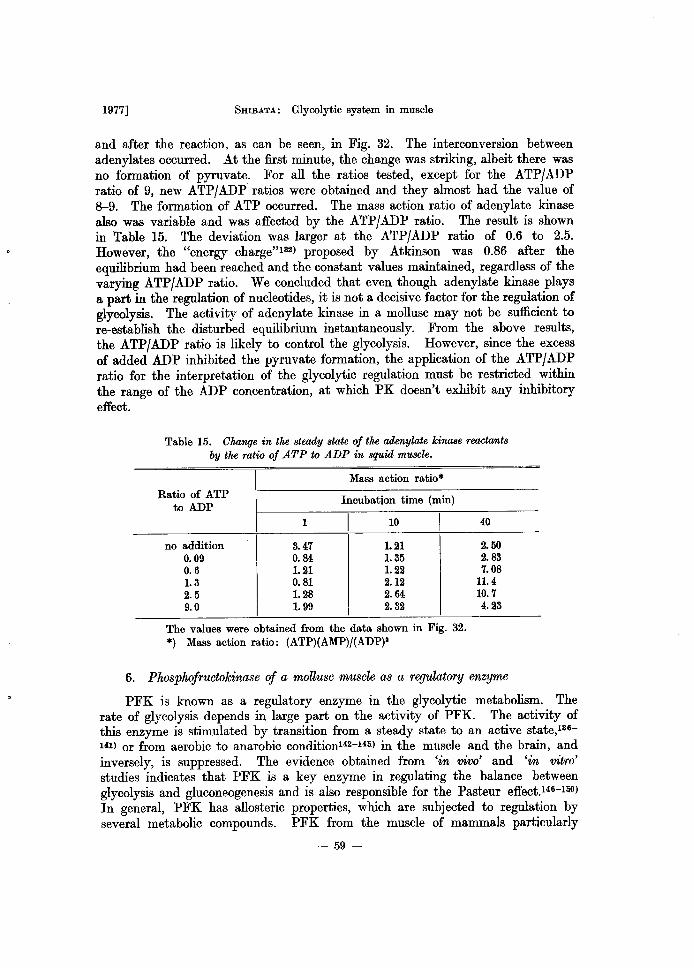
1977] SHIBATA: Glycolytic system in muscle
and after the reaction, as can be seen, in Fig. 32. The interconversion between adenylates occurred. At the first minute, the change was striking, albeit there was no formation of pyruvate. For all the ratios tested, except for the ATP/ADP ratio of 9, new ATP/ADP ratios were obtained and they almost had the value of 8-9. The formation of ATP occurred. The mass action ratio of adenylate kinase also was variable and was affected by the ATP/ADP ratio. The result is shown in Table 15. The deviation was larger at the ATP/ADP ratio of 0.6 to 2.5. However, the "energy charge" 132) proposed by Atkinson was 0.86 after the equilibrium had been reached and the constant values maintained, regardless of the varying ATP/ADP ratio. We concluded that even though adenylate kinase plays a part in the regulation of nucleotides, it is not a decisive factor for the regulation of glycolysis. The activity of adenylate kinase in a mollusc may not be sufficient to re-establish the disturbed equilibrium instantaneously. From the above results, the ATP/ADP ratio is likely to control the glycolysis. However, since the excess of added ADP inhibited the pyruvate formation, the application of the ATP/ADP ratio for the interpretation of the glycolytic regulation must be restricted within the range of the ADP concentration, at which PK doesn't exhibit any inhibitory effect.
Table 15. Clwnge in the steady state of the adenylate kinase reactants by the ratio of ATP to ADP in squid muscle.
Mass action ratio* Ratio of ATP
Incubation time (min) to ADP
1 I 10 I no addition 3.47 1.21
0.09 0.84 1.35 0.6 1.21 1.22 1.3 0.81 2.12 2.5 1. 28 2.64 9.0 1.99 2.32
The values were obtained from the data shown in Fig. 32. *) Mass action ratio: (ATP)(AMP)/(ADP)a
40
2.50 2.83 7.08
11.4 10.7 4.23
6. Phosphofructokinase of a mollusc muscle as a regulatory enzyme
PFK is known as a regulatory enzyme in the glycolytic metabolism. The rate of glycolysis depends in large part on the activity of PFK. The activity of this enzyme is stimulated by transition from a steady state to an active state,136-1(1) or from aerobic to anarobic conditionI42-I4li) in the muscle and the brain, and inversely, is suppressed. The evidence obtained from 'in vivo' and 'in vitro' studies indicates that PFK is a key enzyme in regulating the balance between glycolysis and gluconeogenesis and is also responsible for the Pasteur effect.146-1liO) In general, PFK has allosteric properties, which are subjected to regulation by several metabolic compounds. PFK from the muscle of mammals particularly
- 59-
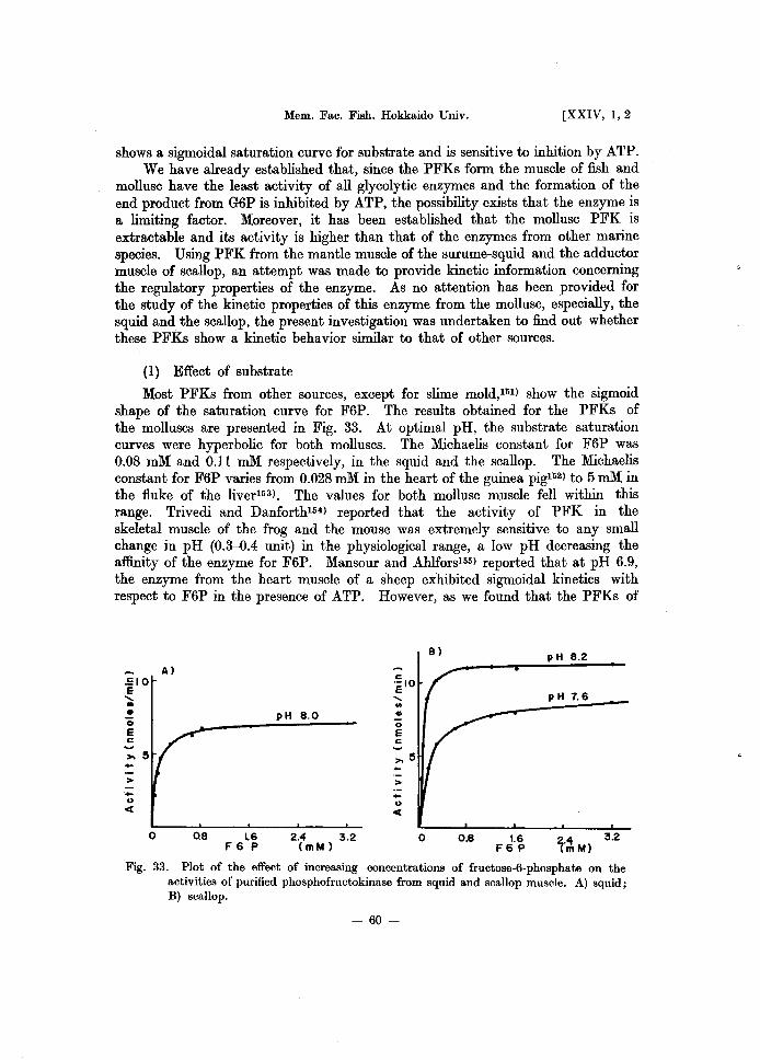
Mem. Fac. Fish. Hokkaido Univ. [XXIV, 1,2
shows a sigmoidal saturation curve for substrate and is sensitive to inhition by ATP. We have already established that, since the PFKs form the muscle of fish and
mollusc have the least activity of all glycolytic enzymes and the formation of the end product from G6P is inhibited by ATP, the possibility exists that the enzyme is a limiting factor. Moreover, it has been established that the mollusc PFK is extractable and its activity is higher than that of the enzymes from other marine species. Using PFK from the mantle muscle of the surume-squid and the adductor muscle of scallop, an attempt was made to provide kinetic information concerning the regulatory properties of the enzyme. As no attention has been provided for the study of the kinetic properties of this enzyme from the mollusc, especially, the squid and the scallop, the present investigation was undertaken to find out whether these PFKs show a kinetic behavior similar to that of other sources.
(1) Effect of substrate
Most PFKs from other sources, except for slime mold,llil) show the sigmoid shape of the saturation curve for F6P. The results obtained for the PFKs of the molluscs are presented in Fig. 33. At optimal pH, the substrate saturation curves were hyperbolic for both molluscs. The Michaelis constant for F6P was 0.08 mM and 0.11 mM respectively, in the squid and the scallop. The Michaelis constant for F6P varies from 0.028 mM in the heart of the guinea pig152) to 5 mM in the fluke of the liver153). The values for both mollusc muscle fell within this range. Trivedi and Danforth154) reported that the activity of PFK in the skeletal muscle of the frog and the mouse was extremely sensitive to any small change in pH (0.3-0.4 unit) in the physiological range, a low pH decreasing the affinity of the enzyme for F6P. Mansour and Ahlfors155) reported that at pH 6.9, the enzyme from the heart muscle of a sheep exhibited sigmoidal kinetics with respect to F6P in the presence of ATP. However, as we found that the PFKs of
':10 E
...... • • o E c
>
o
Al
pH 8.0
0.8 1.6 2.4 3.2 F 6 P (mM)
c elO
...... ."
• o E c
> -<>
'" o
Bl pH 8.2
pH 7.6
0.8 1.6 F6 P
2.4 3.2 (mM)
Fig. 33. Plot of the effect of increasing concentrations of fructose-6-phosphate on the activities of purified phosphofructokinase from squid and scallop muscle. A) squid; B) scallop.
- 60-
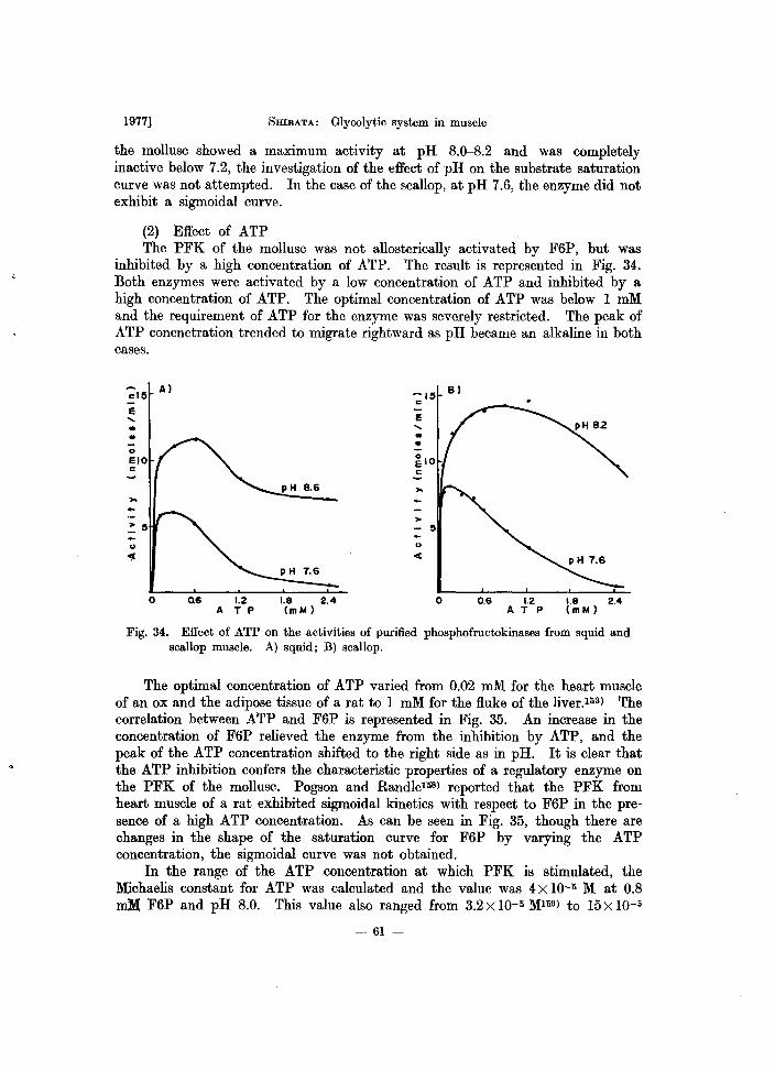
1977] SHIBATA: Glycolytic system in muscle
the mollusc showed a maximum activity at pH 8.0-8.2 and was completely inactive below 7.2, the investigation of the effect of pH on the substrate saturation curve was not attempted. In the case of the scallop, at pH 7.6, the enzyme did not exhibit a sigmoidal curve.
(2) Effect of ATP The PFK of the mollusc was not allosterically activated by F6P, but was
inhibited by a high concentration of ATP. The result is represented in Fig. 34. Both enzymes were activated by a low concentration of ATP and inhibited by a high concentration of ATP. The optimal concentration of ATP was below 1 mM and the requirement of ATP for the enzyme was severely restricted. The peak of ATP concnetration trended to migrate rightward as pH became an alkaline in both cases.
cl5 A)
E ..... • ..
>-
u .".
0 Q6 1.2 A T P
B)
-E ..... • •
oS pH 8.6 >-
>
u ct
pH 7.6
1.8 2.4 o 0.6 1.2 1.8 2.4 (mM) ATP (mM)
Fig. 34. Effect of ATP on the activities of purified phosphofructokinases from squid and scallop muscle. A) squid; B) scallop.
The optimal concentration of ATP varied from 0.02 mM for the heart muscle of an ox and the adipose tissue of a rat to 1 mM for the fluke of the liver.Ili3) The correlation between ATP and F6P is represented in Fig. 35. An increase in the concentration of F6P relieved the enzyme from the inhibition by ATP, and the peak of the ATP concentration shifted to the right side as in pH. It is clear that the ATP inhibition confers the characteristic properties of a regulatory enzyme on the PFK of the mollusc. Pogson and RandleIliS) reported that the PFK from heart muscle of a rat exhibited sigmoidal kinetics with respect to F6P in the presence of a high ATP concentration. As can be seen in Fig. 35, though there are changes in the shape of the saturation curve for F6P by varying the ATP concentration, the sigmoidal curve was not obtained.
In the range of the ATP concentration at which PFK is stimulated, the Michaelis constant for ATP was calculated and the value was 4 X 10-li M at 0.8 mM F6P and pH 8.0. This value also ranged from 3.2 X 10⁻⁵ Mlli9) to 15 X 10-5
- 61 -
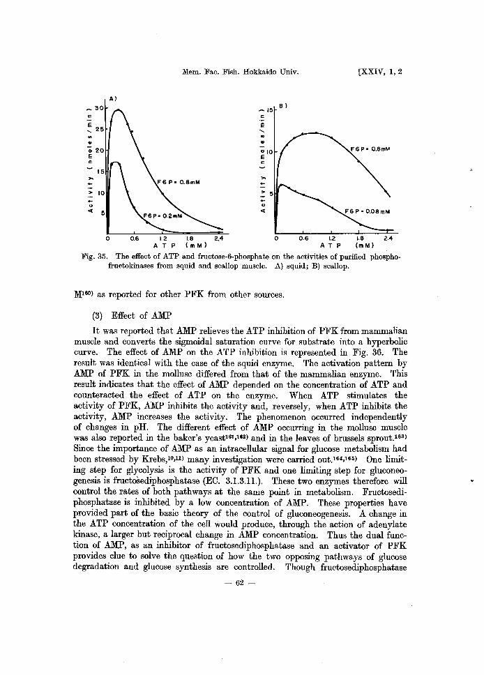
~ 30 c .-~ 25 .. .. '020 E c
15 ,...
> 10 -<> « 5
A)
Mem. Fac. Fish. Hokkaido Univ.
~ 15 B) c
E ..... .. u
o
[XXIV, 1,2
0.6 1.2 1.8 2.4 AT P (mM)
Fig. 35. The effect of ATP and fructose-6-phosphate on the activities of purified phosphofructokinases from squid and scallop muscle. A) squid; B) scallop.
1\1160) as reported for other PFK from other sources.
(3) Effect of AMP
It was reported that AMP relieves the ATP inhibition of PFK from mammalian muscle and converts the sigmoidal saturation curve for substrate into a hyperbolic curve. The effect of AMP on the ATP inhibition is represented in Fig. 36. The result was identical with the case of the squid enzyme. The activation pattern by AMP of PFK in the mollusc differed from that of the mammalian enzyme. This result indicates that the effect of AMP depended on the concentration of ATP and counteracted the effect of ATP on the enzyme. When ATP stimulates the activity of PFK, AMP inhibits the activity and, reversely, when ATP inhibits the activity, AMP increases the activity. The phenomenon occurred independently of changes in pH. The different effect of AMP occurring in the mollusc muscle was also reported in the baker's yeastI61,162) and in the leaves of brussels sprout.163)
Since the importance of AMP as an intracellular signal for glucose metabolism had been stressed by Krebs,1o,ll) many investigation were carried out.164,165) One limiting step for glycolysis is the activity of PFK and one limiting step for gluconeogenesis is fructosediphosphatase (EO. 3.1.3.11.). These two enzymes therefore will control the rates of both pathways at the same point in metabolism. Fructosediphosphatase is inhibited by a low concentration of AMP. These properties have provided part of the basic theory of the control of gluconeogenesis. A change in the ATP concentration of the cell would produce, through the action of adenylate kinase, a larger but reciprocal change in AMP concentration. Thus the dual function of AMP, as an inhibitor of fructosediphosphatase and an activator of PFK provides clue to solve the question of how the two opposing pathways of glucose degradation and glucose synthesis are controlled. Though fructosediphosphatase
- 62-

1977] SHIBATA: Glycolytic system in muscle
15 A) -- 15 B) c c .-E "-.., .,
E "-.., • ATP • 0.6 mM
elO c
~IO c
ATP .2.4 mM
>->-.. > 5 > 5 .. .. u u
c:( c:(
o 1.2 2.4 3.6 o 0.8 1.6 2.4 3.2 ATP (mM) AMP (mM)
Fig. 36. Effect of ATP on the fructose-6-phosphate saturation curve for purified phosphofructokinase from scallop muscle. The activity was assayed at pH 7.6.
from the mollusc muscle has not been investigated yet, it is supposed that the response of these two enzymes to changes in concentration of AMP may differ. Ramaiah et al.l6l} reported that the rate of the PFK reaction is seen to depend entirely on the AMP to ATP ratio, rather than on either absolute concentration. However, PFK in the mollusc showed no effect of AMP at the concentration of the above 0.8 mM, as can be seen in Fig. 36, B.
(4) Effect of ADP
ADP is the product of the PFK reaction. It is reported that the activity of PFK from the heart musclel52} and other sourcesI65,166) was stimulated by ADP, and that the ATP inhibition of the enzyme was relieved by it.
PFK from the scallop muscle, in contrast, was inhibited by ADP. The result is represented in Fig. 37. However, the effect differed depending on pH. At pH 8.2, ADP was inhibitory and at pH 7.6, the low concentration of ADP stimulated the activity and the high concentration of ADP decreased it. Thus ADP acts as a stimulator as well as an inhibitor depending upon the concentration. Since its effect is complex like the effect of AMP, the nature of the inhibition could not be determined accurately. With respect to the ADP inhibitor, the properties of PFK from the mollusc were similar to those of plantI63 ) and slime mold.151)
(5) Effect of other compounds
One of the characteristic properties of PFK is the inhibitory effect of citrate. The inhibition by citrate was increased as the ATP concentration was raised. Williamsonl61) reported that in the perfused heart, the high level of citrate inhibited glycolysis and he discussed the correlation between the PFK inhibition
- 63-
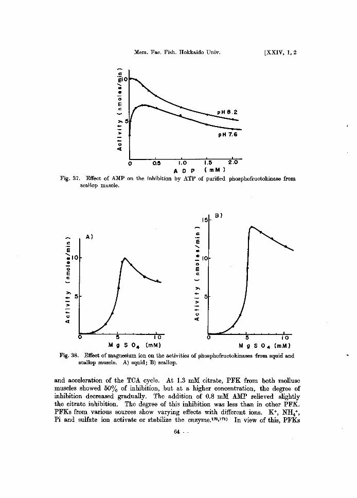
Mem. Fac. Fish. Hokkaido Univ . [XXIV, 1,2
..... II>
• 0
E c:
-> pH 7.6 -u C(
0 o.~ 1.0 I.~ 2.0
A 0 P ( m M )
Fig. 37. Effect of AMP on the inhibition by ATP of purified phosphofructokinase from scallop muscle.
c
E ";;10 • o E c
>. _ 5
>
u C(
A)
o ~ 10 M g S O. (mM)
15 8)
c
E ..... .., ~IO 0
E c:
>. - 5 > -u C(
0
Fig. 38. Effect of magnesium ion on the activities of phosphofructokinases from squid and scallop muscle. A) squid; B) scallop.
and acceleration of the TeA cycle. At 1.3 mM citrate, PFK from both mollusc muscles showed 50% of inhibition, but at a higher concentration, the degree of inhibition decreased gradually. The addition of 0.8 roM AMP relieved slightly the citrate inhibition. The degree of this inhibition was less than in other PFK. PFKs from various sources show varying effects with different ions. K+, NH4+,
Pi and sulfate ion activate or stabilize the enzyme.11O,l11) In view of this, PFKs
-64-
1977] 5mBATA: Glycolytic system in muscle
from the mollusc muscle showed a marked contrast to the mammalian enzyme. 3 mM Pi and 100 mM KCI inhibited the enzyme 15% and 35%, respectively. As PFK form the mammalian muscle shows an activation or a reversion of the ATP inhibition by Pi, there were notable differences between mammalian and mollusc muscles. The effect of Mg2+ on the activity of PFK showed a striking cooperativity. The result is represented in Fig. 38. PFK of the mollusc showed an absolute dependence on Mg2+ and a sigmoidal saturation curve for Mg2+. However, the PFK activity reached a maximum at 5.5 mM Mg2+ and was inhibited 25% and 15% at 10 mM Mg2+, for the squid and the scallop, respectively. Moreover, the cyclic AMP had no effect on the squid enzyme up to 6 X 10⁻³ M. Reduced glutathione activated the enzyme 25%.
So far, the kinetic properties of PFK from the squid and the scallop muscle were described. As the degree of complexity depends on the source of the enzyme, it is difficult to compare directly the kinetic properties of these enzymes. However, the ATP inhibition, the relief of the ATP inhibition by AMP and F6P, and the citrate inhibition of the mollusc PFK are similar to those of the enzyme in the mammalian muscle, but significant differences between the mollusc and the mammalian muscles are the effect of ADP, AMP and Pi, and the shape of the saturation curve with respect to F6P. The kinetic properties of mollusc PFKs are rather close to those of the plant, yeast and bacterial enzyme. We conclude that the mollusc PFKs exhibit complex allosteric properties, but exhibit a regulatory mechanism simpler than that of the mammalian muscle enzyme.
In view of the considerable variations among PFKs and because of our current interest in the regulation of the PFK activity from the muscles of aquatic animals, it may appear desirable to solve the following problem. First, as described previously, there is a difference in the possibility of extraction between the muscle of the mollusc and that of the fish. PFK was extracted from a carp with tris buffer containing Mg2+, ATP and mercaptoethanol from residues which was washed twice by 0.3 M mannit. It was assumed that 46 to 48% of the total activity is linked to the structure protein or to any particles in the cell. However, the activity of this PFK fraction was very labile and was lost at a 4°C temperature during the night. It is not clear whether PFK from the mollusc enzyme is linked to the muscular protein or to any particles or not. Secondly, we observed that the ability of relieving the citrate inhibition by AMP in the squid PFK decreased during 2 months of storage. As desensitization by aging may be due to a change in the binding sites of a regulatory substance, it is necessary to investigate any changes in the structure of the enzyme protein. Thirdly, the enzyme exists in multiple forms. The dissociation or association depending on the interaction of several subunits is convenient for interpreting the mechanism regulated by substrates and metabolites. The difference in the possibility of extraction, soluble and insoluble form, and the difference in sensitivity to regulatory factors may be due to the difference in the degree of subunit-association.
- 65-
Mem. Fac. Fish. Hokkaido Univ. [XXIV, 1,2
Discussion
The model sysyem fortified by adding glycolytic intermediates to the muscle extract can well account for the accumulation of glycolytic intermediates in the 'in vivo' muscle and the interaction between the glycolytic enzymes. The final product and a few intermediates trend towards accumulating in the muscle. Except for the final product, G6P shows the largest accumulation. The conversion of GIP into G6P is intervened spontaneously by GPM and the conversion of F6P into G6P occurs when the rate of the glycolytic flux is restricted to the PFK steps. Apparently, the G6P accumulation appears to be due to the inhibition of PGI, but this accumulation is due to the intense activity and reversibility of PGI. The hypothesis that the enzyme, using an accumulated intermediate as substrate, is a limiting factor, is not always valid. Even if the degradation of glycogen by phosphorylase is stimulated, it results in the accumulation of G6P because the rate of the glycolytic flux is restricted to the PFK step. Although it has reported that FDP accumulates in the intact yeast cellI) and in the acetone-dried muscle of the dogl12), the accumulation of the glycolytic intermediates below FDP is very low in the muscle of fish and mollusc. The FDP accumulation may be occur according to the lack of NAD or ADP' if PFK normally functions. If FDP accumulates, accumulation of DHAP occurs. 3-PGA accumulation is low and if 3-PGA accumulation occurs, it may be due to the inhibition of PK. ADP stimulates the rate of glycolysis, an excess of ADP inhibits it. Theoretically, the amount of final product corresponds stoichiometrically to the amount of formed ATP. In the model system, as adenylate kinase plays a role in the regulation of adenylate, ATP formed during the glycolytic process does not correspond to the amount of final product and is always less. The pattern of the accumulation of glycolytic intermediates 'in vitro' is identical with that of 'in vivo' or post mortem muscle reported by many investigators.
On the other hand, the changes in adenylates show the difference between the 'in vitro' observation and the 'in vivo' situation. The changes in adenylates 'in vivo' is influenced by muscular works, contraction-relaxation in the living muscle, and rigor and thaw-rigor in the post mortem muscle. There is a close relationship between muscular contraction and glycolysis. It is commonly believed that ATP formed from glycogen by glycolysis is utilized in muscular work. The stiffening effect accompanying rigor mortis is basically similar to a contraction of the skeletal muscles. However, the quantitative correlation between a raise and fall in ATP is not yet determined during muscular work. Although great changes in adenylates in the post mortem muscle were observed by many investigators, the changes was underwent a predominantly unidirectional degrading process. As long as a glycogen reserve exists, glycolysis continues forming ATP. The ATP could be split by myofibrillar adenosine triphosphatase and the breakdown of ATP is supressed by adenylate kinase. It is not clear in what way ATP formed by post mortem glycolysis participates in the degrading process of adenine nucleotides. The changes in adenylates in the model system including no myofibrillar adenosine triphosphatase do not faithfully reflect the actual phenomena in whole cells.
- 66-
1977] SHIBATA.: Glycolytic system in musc e
Furthermore, if the location of ATP in the different compartments of a cell can be adduced by considering the regulatory mechanism of ATP utilization, it can probably account for the notable differences in the variation of adenylates between in vivo and in vitro. The differences between the in vivo and the in vitro reaction are those in the state of equilibrium. The in vivo reaction exhibits the steady state of equilibrium, while the in vitro reaction exhibits the thermodynamic equilibrium. The fact that the glycolytic function in vivo was identical with that of in vitro suggests that the glycolytic reaction is a unidirectional degrading reaction and functions as a well integrated unit.
The discrepancy between the glycolytic function in the ante and post mortem muscle is the breakdown of glycogen. Glycogenolysis in the post mortem muscle is accopanied by an amylolytic action. Glycogenolysis in the ante mortem muscle proceeds nearly through the glycolytic pathway. The phosphorylase intervenes in this process. This discrepancy may be due to the difference in the phosphorylase action. There are two possible explanations for this assumption; that is: (1) inactivation of phosphorylase in the post mortem muscle and (2) the lack of regulatory function activating the enzyme. As described in chapter III, 4, we found that the potential activity of phosphorylase is as high as in other glycolytic enzymes, but there is no utilization of glycogen in the model system. In' fact, phosphorylase has been shown to occur in muscle in two such forms, referred to as phosphorylase a (active form) and phosphorylase b (inactive form), respectively. In the resting muscle, the b form is predominant and is changed into an active form during contraction. There are two regulatory enzyme systems present in muscle; one which converts the inactive form of phosphorylase to the active form and another which catalyzes the reverse reaction. The regulatory mechanism that activates the inactive form, is inactivated or lost by the changes in the post mortem muscle; for example, change in pH, Ca2+ effect or a cellular level of organization. It is assumed that because of the weakness in the muscular structure of fish, any damage to the tissue structure in post mortem causes the inactivation of phosphorylase with a concomitant loss of regulatory mechanism.
It has already been described in this report that mollusc PFK has no activity when pH is below 7.0. Therefore, it is interesting to investigate the changes in pH in the post mortem muscle of the squid, which exhibits no accumulation of lactate or pyruvate. That fish muscles show a higher ultimate pH value in post morten may be due to less pH-stability of the fish PFK activity. The cause of the cessation of glycolysis is the lack of FDP in the post mortem muscle. The ultimate pH value does not determine the rate nor the extent of post mortem glycolysis.
The rate of glycolysis in the post mortem muscle varies considerably with time and the temperature of storage, and the rate is frequently faster at a low temperature. Post mortem changes in fish muscle vary with the fish species, the method of killing, the amount of struggling and exercise prior to death as well as the storage temperature after death. The variation of the rate of turnover of glycolytic intermediates and associated compounds gives much scattered information. It is difficult to assess the practical implication of these findings. We investigated the stability of the glycolytic enzymes in muscle under some
- 67-
Mem. Fac. Fish. Hokkaido Univ. [XXIV, 1,2
conditions of storage after death, and found that other glycolytic enzymes, except for PFK, ALD, GPDH and LDH, are stable in both ice and frozen storage.IOI )
At the same time, we reported that the proportion of the decrease in activity of glycolytic enzymes in minced muscle is larger than that in unminced muscle and we assumed that this fact can be interpreted as the result of damage caused to the tissue. Therefore, it is presumed that the contact between substrate and enzyme is important for the post mortem muscle and that it influences the rate of glycolysis. As fish muscle has a higher water content and a weaker muscular structure than mammalian muscle, it is supposed that post mortem changes in fish muscle facilitate the diffusion of substrate or enzyme. In fact, rigor mortis in fish generally has a shorter duration than in mammals. The difference in the rate of changes in glycolytic intermediates depends on the mobility of the substrate in the post mortem muscle, which is decisive in propagating the enzyme reaction.
Furthermore, the probelm must be solved in order to clarify the changes in the post mortem muscle with regard to the interaction of one metabolic system with other metabolic systems and how the regulatory function intervenes between these systems; for example, glycolysis and respiration, and glycolysis and muscular contraction. After the tissues are no longer provided with oxygen, glycolysis then appears in the muscle tissue. Although the phenomenon of rigor mortis is not completely identical with that of the contraction of the living skeletal muscle, the determination of changes in ATP concentration and the activity of myofibrillar adenosine triphosphatase may provide a clue to resolve the problem concerning rigor mortis. When a muscle contracts, glycolysis is accelerated by the activation of phosphorylase and PFK. Both enzymes are stimulated by a common cofator cyclic AMP. Although no stimulation by cyclic AMP of mollusc PFK occurred, unlike the mammalian enzyme, the possibility exists that phosphorylase kinase, catalyzing the conversion of phosphorylase b into the a form, may be activated by cyclic AMP even in the mollusc muscle.
On the other hand, phosphorylase kinase is activated by Ca2+ ion and the contraction is characterized by a release of Ca2+ ion in the sarcoplasm, utilizing ATP rapidly. One of essential features in the post mortem changes is the gradual release of Ca2+ ions. Calcium ion is a common stimulator for glycolysis and muscular contraction. Further research may involve studies of the interaction with respect to these substances.
In molluscs such as the squid and scallop, there is the accumulation of pyruvate and (X-GP as end products of glycolysis. However, these end products did not accumulate 'in vivo' and in post mortem squid muscle. An attempt was made to determine the activities of metabolic systems utilizing these substances, but the results obtained are not sufficient to extrapolate the 'in vivo' situation. Recent reports suggest that glycolysis of mollusc muscle is unusual. Hochachka and Mustafal76 ) reported that the principal end products of glycolysis in mussel are succinate and alanine. In scallop species,177) glycolysis leads to the formation of octopine, the role of LDH in these species being supplanted by octopine dehydrogenase in the metabolism of pyruvate. As the normal Embden-Meyerhof pathway exists in these marine mollusc muscles, pyruvate perhaps becomes a starting
- 68-
1977] SHIBATA: Glycolytic system in muscle
material. We also reported that the malic enzyme (EO. 1.1.1.40) in the squid muscle has the highest activity of all the marine invertebrate muscles tested.l18- 180)
The malic enzyme function must also take into consideration the pathway of pyruvate utilization.
We have already described that mollusc PFKs exhibit a regulatory mechanism which is simpler than in the mammalian muscle enzyme and we observed that PK from the rainbow trout and the mollusc muscle also exhibit a simpler regulatory mechanism. The reason may be attributed to the characteristic properties of the enzymes from poikilotherms. They change the regulatory mechanism depending on the temperature conditions under which they live. PK from the Alaskan king-crab181) and the rainbow troutI82) show the changes in the kinetic properties depending on the temperature. With regard to poikilotherms such as fish, since there is a close connection between their distribution and the temperature of the water, temperature adaptation involves changes in both the amount of intracellular enzymes and their catalytic properties. Therefore, it frequently happens that the apparent catalytic activity is higher ail a low temperature in the metabolism of pikilotherms. The enzymes from poikilotherms are susceptible of regulatory action depending on temperature. In this repect, it is assumed that a fish enzyme has specific properties differing from that of a mammalian enzyme. From the data obtained so far, a fish enzyme is in general less heat-stable than a mammalian enzyme. It was reported that the amino acid composition of salmon aldolase is different from that of of a mammalian enzyme.183) The amino acid composition may be attributed to the difference in the stability and regulatory properties of fish enzyme. As our knowledge of fish enzyme has lagged behind t,hat of its mammalian counterpart, the changes in catalytic and regulatory properties depending on temperature must be reinvestigated. To determine the activity carried out at different temperature, we must note that fish are adaptable to their environmental temperature. There are many questions about environmental adaptation which remain to be investigated with regard to studies on enzyme structure. For example, in some cases the aggregation of enzyme subunits is strongly temperature dependent.
Little biochemical information exists on the enzymes from aquatic animal muscle. We are apt to extrapolate implicitly the observations found in mammals and apply them to the situation of fish. Further additional studies on glycolysis is required. We are intending to investigate the interesting changes in the glycolytic activity and the induction or repression of glycolytic enzymes during feeding, starvation, refeeding and administration of hormones. These results will provide clues for its practical application i.e. the method of rearing fish.
Summary
1. Finding out that the final product of glycolysis in fish differs from that in some marine molluscs, led us to expect different patterns of metabolism in fish and in mollusc, as for example, a different level of glycolytic enzyme activity, the modification of the glycolytic pathway, and the existence of other pathways to TOA
- 69-
Mem. Fac. Fish. Hokkaido Univ. [XXIV, 1,2
cycle. Moreover, because in post mortem fish muscle, the glycogen utilization by glycolysis did not result in a corresponding stoichiometric increase in lactic acid, it seemed likely that there are some rate limiting steps which prevent the lactate formation in post mortem. In order to clarify the definite correlation between the rate of glycolytic flux in ante mortem and post mortem muscle, an attempt was made to investigate the effect of adding intermediates on the glycolytic flux, the consideration of kinetic properties of the glycolytic intermediates and the properties of a regulatory enzyme.
2. When sugar phosphate was added to the muscle extract, while the formation of lactate occurred in fish, the formation of pyruvate occurred as final product of glycolysis in mollusc. The reaction sequence of glycolysis can be divided into five parts according to the apparent utilization of three sugar phosphates. The points of intersection of the pathway correspond to the regulatory enzymes.
3. In mollusc muscle, ex-glycerphosphate accumulated concomitantly with the raise in pyruvate concentration as final product of glycolysis. The physiological significance of the accumulation of pyruvate and ex-glycerophosphate in mollusc was studied with regard to the respiratory activity and octopine synthesis. The possibility that pyruvate and ex-glycerophosphate are aerobically oxidized in molluscs is demonstrated here although the utilization of pyruvate by respiration is very low in muscle. It is assumed that octopine synthesis occurs only when pyruvate is accumulated.
4. The activity patterns of glycolytic enzymes in muscle of aquatic animals is presented. The notable difference between mollusc and fish arises from the activity of lactate dehydrogenase. Lactate dehydrogenase in mollusc has one of the lowest activities of glycolytic enzymes, while lactate dehydrogenase in fish has one of the highest. However, the activity patterns of other glycolytic enzymes, except for lactate dehydrogenase, show the same trend for both fish and mollusc, in spite of the difference in the absolute activities of those enzymes. And the "constant proportion group" of the glycolytic enzyme system described by Buecher and Pette was observed in some aquatic animals, but not in others. The cause of this flucutation is due to the fall in the activity of glyceraldehyde 3-phosphate dehydrogenase. The failure to establish a constant proportion group also occurred in kokanee salmon muscle during spawning. The difference in the activities of glycolytic enzymes between white and dark muscle, and also between one part of a muscle and the other was observed. The comparison of the activity of the ATP-supplying system to that of the ATP-generating system, and the study of adenylate kinase in fish and mollusc were carried out.
5. The use of the glycolytic system fortified with substrates and cofactors revealed the pattern of accumulation of intermediates in the muscle. The quantitative relationships between them has been definitely shown by stUdying the kinetic properties of the glycolytic intermediates. The amount of degraded glycogen by means of the glycolytic route in vitro was estimated at 50 per cent. Glucose-6-phosphate and dihydroxyacetone phosphate have the tendency to accumulate in muscle, because phosphoglucose isomerase and triose phosphate isomerase reactions are reversible. ATP formed by the glycolytic action exhibited no stoichiometric
-70 -
1977] SHIBATA: Glycolytic system in muscle
corresponding to pyruvate formed at the same time. It is suggested that adenylate kinase takes part in this reaction. From changes in the mass action ratio, it is also indicated that phosphofructokinase and pyruvate kinase are regulatory enzymes in glycolysis.
6. The effect of adenine nucleotides, which regulate the rate of glycolysis was investigated. ADP plays a role as stimulator at a low concentration and as inhibtor when there is an excess of concentration. The mass action ratio of pyruvate kinase was markedly influenced by the ADP concentration. ATP also exhibited both stimulatory and inhibitory actions like ADP. It is assumed that the ATPjADP ratio regulates the rate of glycolysis within the range of physiological in vivo concentration of adenylates. Adenylate kinase played a part in the regulation of adenylates, but it is not a decisive factor for the operation of glycolysis.
7. By using partially purified phosphofructokinase from the muscle of squid and scallop, an attempt was made to provide kinetic information concerning the regulatory properties of the enzyme. The enzymes exhibited the hyperbolic shape of a substrate saturation curve. The ATP inhibition, relief of ATP by AMP and fructose-6-phosphate, and the citrate inhibition of the mollusc enzyme were similar to those of mammalian enzymes, but the mollusc enzyme exhibited a regulatory mechanism simpler than that of the mammalian enzyme.
8. The correlation between the glycolytic rates and the regulatory factors together with the real difficulties which have been encountered in extrapolating in vitro observations to the in vivo situation were discussed.
Acknowledgements
The author wishes to express his sincere thanks to Professor T. Shimomura, Professor M. Izawa and Assistant Professor T. Nakamura, Faculty of Agriculture, Hokkaido University, Sapporo, for their valuable criticisms of this study. He is greatly indebted to Professor Emeritus K. Yoshimura and Professor Emeritus T. Saito, Faculty of Fisheries, Hokkaido University, Hakodate, for their invaluable advice and their constant encouragements during the investigation. He also wishes to thank the staff, at the Chitose and Mori Branches of Hokkaido Salmon Hatchery for their help.
References
(1) Dickens, F. (1951). Anaerobic glycolysis, respiration, and the pasteur effect. p. 625-683. In Sumner, J.B. and Myrbaeck, K. (ed.), The Enzymes. 1, 1440p. Academic Press, New York.
(2) Nord, F.F. and Weiss, B. (1951). Yeast and Mold fermentations. p.684-790. Ibid, 2, 144Op. Academic Press, New York.
(3) Stumpf, P.K. (1954). Glycolysis. p. 67-108. In Greenberg, D.M., Ohemical Pathways of Metaboli8m. 1, 460p. Academic Press, New York.
(4) Bueding, E. and Farber, E. (1960). Comparative biochemistry of glycolysis. p. 411-439. In Florkin, M. and Mason, H.S., Oomparative Biochemi&try. 1, 59Op. Academic Press, New York.
-71-
Mem. Fac. Fish. Hokkaido Univ. [XXIV, 1,2
(5) Beisenherz, G. et al. (1953). Diphosphofructose-Aldolase, Phosphoglycer-aldehydeDehydrogenase, Milchsaeure-Dehydrogenase, Glycerophosphate-Dehydrogenase und Pyruvat-Kinase aus Kaninchenmuskulatur in einem Arbeitsgang. Z. Naturforsch. 8b, 555-577.
(6) Czok, R. and Buecher, T. (1960). Crystallized enzymes from the myogen of rabbit skeletal muscle. Adv. in Protein Ohem., 15, 315-415.
(7) Ling, K-H., Marcus, F. and Lardy, H.A. (1965). Purification and some properties of rabbit skeletal muscle phosphofructokinase. J. Biol. Ohem., 240, 1893-1899.
(8) Mansour, T.E., Wakid, N. and Sprouse, H. (1966). Studies on heart phosphofructokinase, purification, crystallization, and properties of sheep heart phosphofructokinase. Ibid, 241, 1512--1521.
(9) Parmeggaiani, A. et al. (1966). Crystallization and propsrties of rabbit skeletal muscle phosphofructokinase. Ibid, 241, 4625-4637.
(10) Krebs, H.A. (1963). Renal gluconeogenesis. Adv. in Enzyme Regulation, 1, 385-400. (ll) Krebs, H.A. (1964). Gluconeogenesis. Proc. Roy. Soc. Ser. B., 159, 545-564. (12) Chance, B., Estabrook, R.W. and Ghosh, A. (1960). Damped sinusoidal oscillations of
cytoplasmic reduced pyridine nucleotide in yeast cells. Proc. Natl. Acad. Sci. 51, 1244-1251.
(13) Ghosh, A. and Chance, B. (1964). Oscillations of glycolytic intermediates in yeast cells. Biochem. Biophys. Res. Oommun. 16, 174-181.
(14) Betz, A. and Chance, B. (1965). Phase relationship of glycolytic intermediates in yeast cells with oscillatory metabolic control. Arch. Biochem. Biophys. 109, 585-594.
(15) Betz, A. (1966). Metabolic flux in yeast cells with oscillatory controlled glycolysis. Physiol. Plant. 19, 1049-1054.
(16) Chance, B., Schoener, B. and Elsaesser, S. (1965). Metabolic control phenomena involved in damped sinusoidal oscillations of reduced diphosphopyridine nucleotide in a cell-free extract of Saccharomyces carlsbergensis. J. Biol. Ohem. 240, 3170--3181.
(17) Yamada, K., Niima, S. and Suzuki, T. (1953). Biochemical change of meat in newly caught fish. I. On the post mortem change of sekeletal muscle in bottom fish. Bull. Jap. Soc. Sci. Fish. 14, 23-26; II. On the post mortem change in pelagic migrating fish. Ibid, 14, 41-47.
(18) Amano, K., Bito, M. and Kawabata, T. (1953). Handling effect upon biochemical change in the fish muscle immediately after catch-I. Difference of glycolysis in the frigate mackerel killed by various methods. Bull. Jap. Soc. Sci. Fish. 19,487-498.
(19) Fujimaki, M. and Kojo, K. (1953). Handling effect upon biochemical change in the fish muscle immediately after catch. -II. Changes of acid-soluble phosphorus compounds of frigate mackerel muscle. Ibid, 19,499-504.
(20) Shibata, T. and Yoshimura, K. (1960). Enzymatic studies on the muscle of aquatic animals-II. Aerobic glycolysis of fish and mollusc muscle. Ibid, 26, 294-299.
(21) Cori, C.F. (1956). Regulation of enzyme activity in muscle during work. p. 573-583. In Gaebler, O.H. (ed.), Enzymes, Units of Biological Structure and Function. 624p. Academic Press. New York.
(22) Yoshimura, K., Kubo, S. and Ito, Y. (1953). On the biochemical changes of fish muscle after death (1). Bull. Fac. Fish. Hokkaido Univ. 4, 157-162.
(23) Nagayama, F. (1961). Enzymatic studies on the glycolysis in fish muscle·3. Contents of glycolytic intermediates. Bull. Jap. Soc. Sci. Fish. 27, 1014-lO17.
(24) Fraser, D.I., Weinstein, H.M. and Dyer, W.J. (1965). Post-mortem glycolytic and associated changes in the muscle of trap- and trawl-caught cod. J _ Fish. Res. Bd. Oanada, 22, 83-100.
(25) McCready, R.M. and Hassid, W.Z. (1957). Preparation of a.D-glucose-l-phosphate by means of potato phosphorylase. Methods in Enzymology. 3, 137-143.
- 72-
1977] SHIBATA: Glycolytic system in muscle
(26) Leloir, L.F. and Paladini, A.C. (1957). Isolation of glucose-l,6-diphosphate. Ibid, 3, 143-147.
(27) Warburg, O. and Christian, W. (1941). Isolierung und Kristallisation des Gaerungsferments Enolase. Biochem. Z. 310, 382-421.
(28) Clark, V.M. and Kirby, A.J. (1963). An improved synthesis of phosphoenolpyruvate. Biochim. Biophys. Acta. 78, 732.
(29) Umbreit, W.W. et al. (1951). The homogenate technique. p. 143-144. In Umbreit, W.W., Manometric Techniques and Tissue Metabolism. 227p. Burgess Publishing Co. Minneapolis.
(30) Fredemann, L.E. and Haugen, G.E. (1943). Pyruvic acid. II. The determination of keto acids in blood and urine. J. BioI. chem. 147, 415-442.
(31) Barker, S.B. and Summerson, W.H. (1941). The colorimetric determination of lactic acid in biological material. Ibid, 138, 535--554.
(32) Bartlett, G.H. (1959). Colorimetric assay methods for free and phosphorylated glyceric acid. Ibid, 234, 469-471.
(33) AsweU, G. (1957). Colorimetric analysis of sugars. Methods in Enzymology. 3, 73-105. (34) Hassid, W.Z. and Abraham, S. (1957). Chemical procedures for analysis of polysac
charides. Ibid. 3, 34--50. (35) Bergymer, H.U. and Klotzsch, H. (1963). D-glucose-l-phosphate. p. 131-133. In
Bergmeyer, H.U., Methods of Enzymatic Analysis. 1064p. Academic Press. New York. (36) Hohorst, H.J. (1963). D-glucose-6-phosphate and D-fructose-6-phosphate. Ibid.
134--138. (37) Buecher, T. and Hohorst, H.J. (1963). Dihydroxyacetone phosphate, fructose-l,6-
diphosphate and D-glyceraldehyde-3-phosphate. Ibid. 246--252. (38) Czok, R. and Eckert, L. (1963). D-3-phosphoglycerate, D-2-phosphoglycerate,
phosphoenolpyruvate. Ibid. 229-233. (39) Hohorst, H.J. (1963). L-( + )-Lactate. Ibid. 266--270. (40) Adam, H. (1963). Adenosine-5-tiphosphate. Ibid. 539--543. (41) Adam, H. (1963). Adenosine-5-diphosphate and adenosine-5-monophosphate. Ibid.
573-577. (42) Bublitz, C., and Kenedy, E.P. (1954). Synthesis of phosphatides in isolated mito
chondria. III. The enzymatic phosphorglation of glycerol. J. BioI. chem. 211, 951-961.
(43) Delbrueck, A., Zebe, E. and Buecher, T. (1959). Ueber Verteilungsmuster von Enzymen des Energie Iiefernden Stoffwechsels im Flugmuskel und Fettkoerper von Locusta migratoria und ihre cytologische Zuordnung. Biochem. Z. 331, 273-296.
(44) Adam, H. (1961). Nucleotidspezifitaet glykolytischen Kinasen; enzymatische liestimmung des Adenylsaeuresystems. Ibid, 335, 25--36.
(45) VogeU, W. et al. (1961). Ueber strukturelle und enzymatische Muster in Muskeln von Locusta migratoria. Ibid, 332, 81-117.
(46) Sutherland, E.W. et al. (1949). The mechanism of the phosphoglucomutase reaction. J. BioI. chem. 180, 1285-1295.
(47) Sumner, J.B. and Somers, G.F. (1959). Measurement of phosphorylase activity. p. 1030--1031. In Sumner, J.B. and Myrbaeck, K. (ed.) The Enzymes. 1, 1361p. Academic Press. New York.
(48) Thoai, N.V. and Robin, Y. (1959). Metabolism of guanidylated derivatives. VIII. Biosynthesis of octopine and repartition of the enzyme operating it in invertebrates. Biochim. Biophys. Acta. 35, 446-453. (in French)
(49) Dixon, M. (1951). Multi-enzyme system. p. 15. Cambridge Univ. Press. London. (50) Kazuki, H. and Kanayuki, H. (1958). Keto-acid. p. 95-104. Kagaku no ryoeki.
Zokango. 33. 206p. (in Japanese) Nankodo, Tokyo. (51) Macleod, R.T., Jonas, R.E.E. and Roberts, E. (1963). Glycolytic enzymes in the
- 73-
Mem. Fac. Fish. Hokkaido Univ. [XXIV, 1,2
tissues of salmonid fish (Salmo gairdnerii gairdnerii). Can. J. Biochem. and Physiol. 41, 1971-1981.
(52) Kubista, V. (1958). Anaerobe Glykolyse in den Insektenmuskeln. Biochem. Z. 330, 315-323.
(53) Agosin, M. and Repetto, Y. (1963). Studies on the metabolism of Echinococcus granulosus. -VII. Reactions of the tricarboxylic acid cycle in E. granulosus scolice. Camp. Biochem. Physiol. 8, 245-261.
(54) Agosin, M. and Repetto, Y. (1965). Studies on the metabolism of Echinococcus granulosus-VIII. The pathway to succinate in E. granulosus scolices. Ibid. 14, 299-309.
(55) Mansour, T.E. (1959). Studies on the carbohydrate metabolism of the liver fluke Fasciola heaptica. Biochim. Biophys. Acta. 34, 456-464.
(56) Ward, C.W. and Schofield, P.J. (1967). Glycolysis in Haemonchus contortus larvae and rat liver. Camp. Biochem. Physiol. 22, 33-52.
(57) Prichard, R.K. and Schofield, P.J. (1968). The glycolytic pathway in adult liver fluke, Fasciola hepatica. Ibid. 24, 697-710.
(58) Reeves, R.E. (1968). A new enzyme with the glycolytic function of pyruvate kinase. J. BioI. Chem., 243, 3203-3204.
(59) Scheibel, L.W. and Saz, H.J. (1966). The pathway for anaerobic carbohydrate dissimilation on Hymenolepis diminuta. Camp. Biochem. Physiol. 18, 151-162.
(60) Ward, C.W., Schofield, P.J. and Johnstone, I.L. (1968). Pyruvate kinase in Haemonchus oontortus larvae. Ibid, 24, 643-647.
(61) Bueding, E. and Saz, H.T. (1968). Pyruvate kinase and phosphoenolpyruvate carboxykinase activities of Ascaris muscle, Hymenolepis diminuta and Schistosoma mansoni. Ibid. 24, 511-518.
(62) Saz, H.J. and Virdrine, A. (1959). The mechanism of formation of succinate and propionate by Ascaris Iumbricoides muscle. J. BioI. Chem. 234, 2001-2005.
(63) Simpson, J.W. and Awapara, J. (1966). The pathway of glucose degradation in some invertebrates. Camp. Biochem. Physiol. 18, 537-548 ..
(64) Bennett, R.Jr. and Nakada, H.I. (1968). Comparative carbohydrate metabolism of marine molluscs-I. The intermediary of metabolism of Mytilus californianus and Haliotus rufenscens. Ibid. 24, 787-797.
(65) Chefurka, W. (1958). On the importance of glycero-phosphate dehydrogenase in glycolysing insect muscle. Biochim. Biophys. Acta. 28, 660-661.
(66) Zebe, E., Delbrueck, A. and Buecher, T. (1959). Ueber den Glycerin-l-P-cyclus im Flugmuskel von Locusta migratoria. Biochem. Z. 331, 254-272.
(67) Klingenberg, M. and Buecher, T. (1961). Glycerin-I-P und Flugmuskel-Mitochondrien. Ibid,334, 1-17.
(68) Estabrook, R.W. and Sacktor, B. (1958). a-Glycerophosphate oxidase of flight muscle mitochondria. J. BioI. Chem. 233, 1014-1019.
(69) Chino, H. (1957). Conversion of glycogen to sorbitol and glycerol in diapause egg of the Bombyx silkmworm. Nature, 180, 60tHl07.
(70) Wyatt, G.R. and Meyer, W.L. (1959). The chemistry of insect hemolymph. III. Glycerol. J. Gen. Physiol. 42, 1005-1011.
(71) Kawai, K. (1957). The metabolism of the pearl oyster, Pincto:ia martensii. III. On the tissue oxidation. Bull. Jap. Soc. Sci. Fish. 22, 62tHl30.
(72) Kawai, K. (1958). On the terminal oxidase system in marine lamellibranch tissues. Ibid, 23, 738-742.
(73) Ghiretti, F., Ghiretti, M.A. and Tosi, L. (1959). Pathways of terminal respiration in marine invertebrates. II. The cytochrome system of Aplysia. J. Gen. Physiol. 42, 1185-1205.
(74) Higashi, S. (1961). Physiological chemistry of the fresh-water bivalves-III. On
-74 -
1977] SHIBATA: Glycolytic system in muscle
ther terminal oxidase system of fresh-water cockle, Corbicula sandai. Bull. Jap. Soc. Sci. Fish. 27, 282-286.
(75) Mengebier, W.L. and Wood, L. (1967). Respiratory enzymes in mantle homogenates of Crassostroo, virginica and the effects of M inchinia nelsoni and Cochlodinium helicoides on activity levels. Compo Biochem. Phy'siol. 21, 611-617.
(76) Beechy, R.B. (1961). The difference spectrum of respiring particles isolated from the hepatopancreas of Carcinus maenas. Biochem. J. 78, 3p.
(77) Beechy, R.B. (1961). Respiratory chain phosphorylation in sub-cellular particles isolated from the hepatopancreas of Carcinus maenas. Nature, 192, 975-976.
(78) Beechy, R.B. (1961). Some spectrophotometric studies on respiring particles isolated from the hepatopancreas of Carcinus maenas. Comp. Biochem. Physiol. 3, 161-174.
(79) Burrin, D.H. and Beechy, R.B. (1963). Cytochrome oxidase and cytochromes a and as in crab mitochondria. Biochem. J. 87, 48-53.
(80) Beechy, R.B., Burrin, D.H. and Baxter, M.1. (1963). Mitochondria from the crab Carcinus maenas. Nature, 198, 1277-1279.
(81) Burrin, D.H. and Beechy, R.B. (1964). The spectrophotometric dissection of the electron transfer system of mitochondria isolated from the hepatopancreas of Carcinu8 maenas. Compo Biochem. Physiol. 12, 245-258.
(82) Mundy, K.A. and Topson, B.D. (1962). The preparation and properties of subcellular respiring particles (mitochondria) from the hepatopancreas of Carcinus maenas. Ibid. 5, 95-112.
(83) Haggins, A.H. (1966). Intermediary metabolism in Carcinus maenas. Ibid. 18, 283-290.
(84) Kawai, K. (1961). Comparative biochemical studies on cytochromes and related substances of invertebrates. I. Cytochrome components and electron transfer in fresh-water mussels. J. Biochem. 49, 427--435.
(85) Ryan, C.A. and King, T.E. (1962). Succinate dehydrogenase from the bay mussel, Mytilus edulis. Biochim. Biophs. Acta. 62, 269-278.
(86) Yamanaka, K., Mizushima, H. and Okunuki, K. (1964). Cytochrome c's purified from marine invertebrates. Ibid, 81, 223-228.
(87) Lewis, S.E. and Slater, E.C. (1954). Oxidative phosphorylation in insect sarcosomes. Biochem. J. 58, 207-217.
(88) Burt, J.R. and Stround, G.D. (1966). The metabolism of sugar phosphates in cod muscle. Bull. Jap. Soc. Sci Fish. 32, 204-212.
(89) Ackermann, D. and Mohr, M. (1937). The occurrence of octopine, agmatine and arginine in the octopod, Eledone moschata. Z. physiol. Chem. 250, 244-252.
(90) Akashi, S. (1944). Structure of octopine p. 3-28. Kagakujikengaku. (part 2) 13, 619 p. (in Japanese) Kawade-Shobo, Tokyo.
(91) Kojima, Y. and Kusakabe, H. (1955). Isolation of natural substances by ion exchange resins-ill. A new simple method for the isolation of octopine and taurine from squid muscle. J. Sci. Res. Inst. 49, 132-136.
(92) Moore, E. and Wilson, D.W. (1937). Nitrogenous extractives of scallop muscle. I. The isolation and a study of the structure of octopine. J. BioI. Chem. 119, 573-584.
(93) Thoai, N.V. et al. (1969). Octopine dehydrogenase. Purification and catalytic properties. Biochim. Biophys. Acta. 191, 46--57.
(94) Regnouf, R. and Thoai, N.V. (1970). Octopine and lactate dehydrogenases in mollusc muscle. Compo Biochem. Physiol. 32, 411--416.
(95) Yoshimura, K. and Shibata, T. (1953). Biochemical studies on "Surume-ika" (Ommastrephes sloani pacijicus). 2. The content of basic amino acid. Bull. Fac. Fish. Hokkaido Univ. 3, 211-215.
(96) Yoshimura, K. and Shibata, T. (1953). Biochemical studies on "Surume-ika" (Ommastrephes sloani pacijicus). 3. On basic substances in muscle of squid. Ibid,
- 75-
Mem. Fac. Fish. Hokkaido Univ. [XXIV, 1,2
4, 54-59. (97) Endo, K. and Shimizu, W. (1963). Studies on muscle of aquatic animl.ls-XXXVIII.
Octopine in squid muscle. Bull. Jap. Soc. Sci. Fish. 29, 362-365. (98) Pette, D., Luh, W. and Buecher, T. (1962). A constant-proportion group in the
enzyme activity pattern of the Embtn-Meyerhof chain. Biochem. Biophys. Re8. Commun. 7, 419--424.
(99) Pette, D. and Buecher, T. (1963). Proportionskonstante Gruppen in Beziehung zur Differenzierung der Enzymeaktivitaetmuster von Skelett.Muskeln des Kaninchens. Z. phY8iol. Chem. 331, 180-195.
(100) Pette, D. (1966). Mitochondrial enzyme activities. p. 28-50. In Tager, J.M. et al. (ed.), Regulation of Metaholic Proces8e8 in Mitochondria. 582p. Elsevier Publishing Co., Amsterdam.
(101) Shibata, T., Nakai, T. and Saito, T. (1969). Enzymatic studies on the muscles of aquatic animals-VIII. Changes in the activities of glycolytic and some energymetabolizing enzymes during chill and frozen storage. Bull. Fac. Fish. Hokkaido Univ. 20, 217-226.
(102) Arnold, H. and Pette, D. (1968). Binding of glycolytic enzymes to structure protein of the muscle. European. J. Biochem. 6, 163-171.
(103) Perry, S.V. (1960). Muscular contraction. p. 245-340. In Florkin, M. and Mason, H.S., Comparative Biochemi8try. 2, 245p. Academic Press. New York.
(104) Fukuda, H. (1966). Symposium on phosphorus metabolism in the muscles of aquatic animals. Bull. Jap. Soc. Sci. Fi8h. 32, 193-194.
(105) Kuroki, A. (1969). Studies on lactate dehydrogenase from tissues of fishes. Bull. Fac. Agr. Univ. Miyazaki. 16, 1-60.
(106) Kaloustian, H.D. et aI. (1969). Lactate dehydrogenase of lobster (Homarus americanus) tail muscle. 1. Physical and chemical properties. J. Biol. Chem. 244, 2891-2901.
(107) Fraser, D.I. et aI. (1966). Glycolytic metabolites and their distribution at death in the white and red muscle of cod following various degrees of antemortem muscular activity. Can. J. Biochem. 44, 1015-1033.
(108) Nakai, T., Shibata, T. and Saito, T. (1970). Seasonal variation in the metabolic activities of tissue constituents of some fishes-IV. Changes in the activity levels of the muscle glycolytic enzymes by spawning of kokanee salmon. Bull. Fac. Fish Hokkaido Univ. 21, 240-245.
(109) Nakai, T., Shibata, T. and Saito, T. (1970). Seasonal variations in the metabolic activities of tissue constituents of some fishes-V. Changes in the activity levels of some energy-metabolizing enzymes by spawning of kokanee salmon, Oncorhynchus nerka f. kenelyi. Ibid. 21, 246-251.
(110) Brosemer, R.W., Vogell, W. and Buecher, T. (1963). Morphologische und enzymatische Muster bei der Entwicklung in direkter Flugmuskeln von Locu8ta migratoria. Biochem. Z. 338, 854-910.
(Ill) Nakai, T., Shibata, T. and Saito, T. (1970). Seasonal variations in the metabolic activities of tissue constituents of some fishes·III. On the activity of energymetabolizing enzymes in the muscle and liver of kokanee salmon. Bull. Fac. Fish. Hokkaido Univ. 21, 234-239.
(112) Fitch, W.H. and Chaikoff, I.L. (1960). Extent and patterns of adaptation of enzyme activities in livers normal rat fed diets high in glucose and fructose. J. Biol. Chem. 235, 554-557.
(113) Weber, G. et al. (1966). Synchronous behavior pattern of key glycolytic enzymes: glucokinase, phosphofructokinase, and pyruvate kinase. Adv. in Enzyme Regulation, 4, 59-85.
(114) Starling, H. (1967). Ueber die Binding der Muskelaldolase an grobdisperse Partikeln in Homogenaten erregter Muskeln. Z. phY8iol. Chem. 348, 864-870.
- 76-
1977] SHIBATA: Glycolytic system in muscle
(ll5) Sacktor, B. and Wormer-Shavit, E. (1966). Regulation of metabolism in working muscle in vivo. I. Concentrations of some glycolytic, tricarboxylic acid cycle, and amino acid intermediates in insect flight muscle during flight. J. Bioi. Chem. 241, 624--631; II. Concentrations of adenine nucleotides, arginine phosphate, and inorganic phosphate in insect flight muscle during flight. Ibid, 241, 632-638.
(ll6) Chance, B. and Williams, G.R. (1955). Respiratory enzymes in oxidative phosphorylation. III. The steady state. J. Bioi. Chem. 217, 409-427.
(1l7) Chance, B. et al. (1958). Localization of interaction sites in muiticomponent transfer systems: Theorems derived from analogues. Nature, 182, II 90---11 93.
(lI8) Chance, B. and Hess, B. (1959). Metabolic control mechanisms. II. Crossover phenomena in mitochondria of ascites tumor cells. J. Bioi. Chem. 234, 2413-2415.
(ll9) Hess, B. (1963). Control of metabolic rates. p. 333-350. In Wright, B. (ed.), Control Mechanisms in Respiration and Fermentation. 357p. Ronald Press. New York.
(120) Hess, B. and Brand, K. (1965). Enzyme action in living cells. Clinical chem. 11, 223-238.
(121) Tarr, H.L.A. (1950). The acid-soluble phosphorus compounds of fish skeletal muscle. J. Fish. Res. Bd. Canada, 7, 608-612.
(122) Burt, J.R. and Jones, N.R. (1961). Changes in sugar phosphates of chilled codling (Gadus callarias) muscle. J. Sci. Food Agr. 12, 344-348.
(123) Migita, M. (1963). Postmortem change of muscle-I. Development of the study (review). Bull. Jap. Soc. Sci. Fish. 27, 934-945.
(124) Migita, M. (1964). Postmortem change of muscles-II. Studies on fish muscle (review). Ibid, 28, 456-470.
(125) Tomlison, N., Jonas, R.E.E. and Geiger, S.E. (1963). Glycolysis in lingcod muscle during frozen storage. J. Fish. Res. Bd. Canada, 20, 1145-lI52.
(126) Jones, N.R. and Marray, J. (1966). Nicotinamide adenine dinucleotide (NAD) and reduced NAD in living and chill-stored dying muscle of cod, Gaius callarias. Bull. Jap. Soc. Sci. Fish. 32, 197-203.
(127) Tarr, H.L.A. (1966). Phosphorus compounds in fish. Ibii,32, 213-223. (128) Tarr, H.L.A. (1968). Postmortem degradation of glycogen and starch in fish muscle.
J. Fish. Res. Bd. Canada, 25, 1539-1554. (129) Jones, N.R. (1958). Free sugars in chill-stored trawled codling (Gadus callarias)
muscle. J. Sci. Food Agr. 9, 672-677.
(130) Nagayama, F. (1966). Mechanisms of breakdown and synthesis of glycogen in the tissue of marine animals. Bull. Jap. Soc. Sci, Fish. 32, 188-192.
(131) Buecher, T. and Klingenberg, M. (1958). Wege des Wasserstoffs in der lebendigen Organisation. Angew. Chem. 70, 552-570.
(132) Atkinson, D.E. (1968). The energy change of the adenyl ate pool as a regulatory parameter. Interaction with feed modifiers. Biochemistry, 7, 4030-4034.
(133) Minakami, S. and Yoshikawa, H. (1966). Erythrocytes glycolysis II. Free energy changes and rate limiting steps in erythrocyte glycolysis. J. Biochem. 59, 139-144.
(134) Sakurai, T. and Hosoya, N. (1966). Metabolic regulation in diabetes mellitus. Metabolism and disease. 3, 234-241.
(135) Lowry, O.H. and Passonneau, J.V. (1964). The relationships between substrates and enzymes of glycolysis in brain. J. Bioi. Chem. 239, 31-42.
(136) Danforth, W.H. (1965). Activation of glycolytic pathway in muscle. p. 281-297. In Chance, B. et al. (ed.), Control of Energy Metabolism. 441p. Academic Press, New York.
(137) Buecher, T. (1959). Zellulaere Koordination energie-transformierender Stoffwechselketten. Angew. Chem. 71, 744.
-77
Mem. Fac. Fish. Hokkaido Univ. [XXIV, 1,2
(138) Oezand, P. and Narahara, H.T. (1964). Regulation of glycolysis in muscle. III. Influence of insulin, epinephrine, and contraction on phosphofrutokinase activity in frog skeletal muscle. J. Biol. Chem. 239, 3146-3152.
(139) Helmreich, E. and Cori, C.F. (1965). Regulation of glycolysis in muscle. Adv. in Enzyme Regulation, 3, 91-107.
(140) Williamson, J.R. et al. (1967). Glycolytic control mechanisms. IV. Kinetics of glycolytic intermediate changes during electrical discharge and recovery in the main organ of Electrophorus electricus. J. BioI. Ohem. 242, 5112-5118.
(141) Williamson, J.R. et al. (1967). Glycolytic control mechanism. V. Kinetics of high energy phosphate intermediate changes during electrical discharge and recovery in the main organ of Electrophorus electricus. Ibid, 242, 5119-5124.
(142) Lowry, O.H. et al. (1964). Effect of ischemia on known substrates and cofactors of the glycolytic pathway in brain. Ibid, 239, 18-30.
(143) Williamson, J.R. (1966). Glycolytic control mechanisms. II. Kinetics of intermediate change the aerobic-anoxic transition in perfused rat heart. Ibid, 241, 5026-5036.
(144) Sacktor, B., Wilson, J.E. and Tioker, C.F. (1966). Regulation of glycolysis in brain, in situ, during convulsions. Ibid, 241, 5071-5075.
(145) Rolleston, F.S. and Newsholme, E.A. (1967). Control of glycolysis in cerebral cortex slices. Biochem. J. 104, 524-533.
(146) Passonneau, J.V. and Lowry, O.H. (1962). Phosphofructokinase and the Pasteur effect. Biochem. Biophys. Res. Oommun. 7, 10--15.
(147) Lynen, F. et al. (1959). Phosphate turnover and pasteur effect. p. 256-276. In Wolstenholme, G.E.W. and O'Conner, C.M. (ed.), Oiba Foundation Symposium on the Regulation of Oell Metabolism. 369p. J & A. Churchill, London.
(148) Wu, R. (1965). Rate-limiting factors in glycolysis and inorganic orthophosphate transport in rat liver and kidney slices. J. BioI. Chem. 240, 2370--2381.
(149) Uyeda, K. and Racker, E. (1965). Coordinated stimulation of hexokinase and phosphofructokinase by phosphate in a reconstituted system of glycolysis. p. 127-137. In Chance, B. et al. (ed.), Oontrol of Energy Metabolism. 441p. Academic Press, New York.
(150) Spiess, J. and Meyer, E. (1968). Zum Mechanismus der pasteurschen Reaktion. Ueber die aerobe Hemmung der Hexokinase- und Phosphofructokinase-Reaktion. Z. physiol. Ohem. 349,1261-1262.
(151) Baumann, P. and Wright, B.E. (1968). The phosphofructokinase of Dictostelium discoideum. Biochemistry, 7, 3653-3661.
(152) Mansour, T.E. (1963). Studies on heart phosphofructokinase: purification, inhibition, and activation. J. BioI. Ohem. 238, 2285-2292.
(153) Mansour, T.E. and Mansour, J.W. (1962). Effect of serotonin (5-hydroxytryptamine) and adenosine 3',5'-phosphate on phosphofructokinase from the liver fluke Fasciola hepatica. Ibid, 237, 629-634.
(154) Trivedi, B. and Danforth, W.H. (1966). Effect of pH on the kinetics of frog muscle phosphofructokinase. Ibid, 241, 4110--4113.
(155) Mansour, T.E. and Ahlfors, C.E. (1968). Studies on heart phosphofructokinase. Some kinetic and physical properties of the crystalline enzyme. Ibid, 243, 2523-2533.
(156) Frenkel, R. (1968). Control of reduced diphosphopyridine nucleotide oscillations in beef heart extracts. III. Purification and kinetics of beef heart phosphofurctokinase. Arch. Biochem. Biophys. 125, 166-174.
(157) Denton, R.M. and Randle, P.J. (1966). Citrate and the regulation of adipose tissue phosphofructokinase. Biochem. J. 100, 420--423.
(158) Pogson, C.1. and Randle, P.J. (1966). The control of rat heart phosphofructokinase
- 78-
1977] SHIBATA: Glycolytic system in muscle
by citrate and other regulators. Ibid, 100, 683--693. (159) Freyer, R, Hofmann, E. and Frank, K. (1965). Eigenschaften der Phospho.
fructokinase von Ascites·Tumorzellen. Biochem. Z. 343, 335-345. (160) Layzer, RB., Rowland, L.P. and Bank, W.J. (1969). Physical and kinetic properties
of human phosphofructokinase from skeletal muscle and erythrocytes. J. Biol. Ohem. 244, 3823-3831.
(161) Ramaiah, J.A., Hathaway, J.A. and Atkinson, D.E. (1964). Adenylate as a metabolic regulator. Effect of yeast phosphofructokinase kinetics. Ibid, 239, 3619-3622.
(162) Vinuela, E., Salas, M.L. and Sols, A. (1963). End·product inhibition of yeast phosphofrutokinase by adenosine triphosphate (ATP). Biochem. Biophys. Res. Oommun. 12, 140-145.
(163) Dennis, D.T. and Coultatw, T.P. (1967). The regulatory properties of a plant phosphofructokinase during leaf development. Biochim. Biophys. Acta. 146. 129-137.
(164) Taketa, K. and Pogell, RM. (1965). Allosteric inhibition of rat liver fructose 1,6· diphosphatase by adenosine 5' .monophosphate. J. Bioi. Ohem. 240. 651-662.
(165) Underwood, A.H. and Newsholme. E.A. (1965). Some properties of fructose 1,6· diphosphatase of rat liver and their relation to the control of gluconeogenesis. Bichem. J. 95, 767-774.
(166) Atkinson, D.E. and Walton, G.M. (1965). Kinetics of regulatory enzymes. Escherichia coli phosphofructokinase. J. Bioi. Ohem. 240, 757-763.
(167) Williamson, J. (1967). Glycolytic control mechanisms. III. Effect of iodoacetatamide and fiuoroacetate on glucose metabolism in the perfused rat heart. Ibid, 242, 4476-4485.
(168) Uyeda, K. and Racker, E. (1965). Regulatory mechanisms in carbohydrate metabolism. VII. Hexokinase and phosphofructokinase. Ibid, 240, 4682--4688.
(169) Underwood, A.H. and Newsholme, E.A. (1965). Properties of phosphofructokinase from rat liver and their relation to the control of glycolysis and gluconeogenesis. Biochem. J. 95, 868-875
(170) Blangy, D., Buc, H. and Monod, J. (1968). Kinetics of the allosteric intractions of phosphofructokinase from Escherichia coli. J. Mol. Bioi. 31, 13-35.
(171) Uyeda, K. and Kurooka, S. (1970). Crystallization and properties of phosphofruc. tokinase from Clostridium pasteurianum. J. Bioi. Ohem. 245, 3315-3324.
(172) Fawaz, E.N., Manoukian, E. and Fawaz, G. (1963). Glycolysis in dog skeletal muscle. Biochem. Z. 337, 195-201.
(173) Cain, D.F. and Davies, RF. (1962). Breakdown of adenosine triphosphate (ATP) during a single contraction of working muscle. Biochem. Biophys. Res. Oommun. 8, 361-366.
(174) Kubokawa, Y. (1960). Studies on the fractions of adenosine triphosphate in muscles. I. The methods for fractional extraction of adenosine triphosphate8 in frog leg muscles. Seikagaku 32, 63--67; II. Changes of adenosine triphosphate fraction under the various physiological conditions. Ibid, 32, 163-172.
(175) Steinmetz, M.A. and Deal, W.C. (1966). Metabolic control and structure of glycolytic enzymes. III. Dissociation and subunit structure of rabbit muscle pyruvate kinase. Biochemistry, 5, 1339-1405.
(176) Hochachka, P.W., and Mustafa, T. (1972). Invertebrate facultative anaerobiosis. Science, 178, 1056-1060.
(177) Hiltz, D.F. and Dyer, W.J. (1971). Octopine in postmortem adductor muscle of the sea scallop. J. Fish. Res. Bd. Oanada, 28, 869-874.
(178) Shibata, T., Kitahara, T. and Yoshimura, K. (1965). Enzymatic studies on the mU8cle of aquatic animals·5. Purification and properties of malic enzyme from molluscan muscle. Bull. Fac. Fish, Hokkaido Univ. 16, 171-182.
- 79-
Mem. Fac. Fish. Hokkaido Univ. [XXIV, 1,2
(179) Shibata, T., Kitahara, T. and Yoshimura, K. (1966). Enzymatic studies on the muscle of aquatic animals·6. Effect of various carboxylic acids and inhibitors on squid malic enzyme. Ibid, 17, 64--69 ..
(180) Kitahara, T. and Shibata, T. (1967). Enzymatic studies on the muscle of aquatio anim'lls-7. Further purification of malic enzyme from squid muscle. Ibid, 18, 95-101.
(181) Somero, G.N. (1969). Pyruvate kinase variants of the alaskan king· crab. Evidence for temperature-dependent interconversion between two forms having distinct and adaptive kinetic properties. Biochem. J. 114, 237-241.
(182) Somero, G.N. and Hochachka, P.W. (1968). The effect of temperature on catalytic and regulatory functions of pyruvate kinases of the rainbow trout and the antarctic fish Trematomu8 bernacchii. Ibid, 110, 395-399.
(183) Nagahisa, E. and Tsuchiya, Y. (1971). Studies on the rainbow trout aldolase. II. Physico-chemical properties and amino acid compo~ition of rainbow trout muscle aldolase. Tohoku J. Agr. Res. 22, 46-55.
- 80-