enzyme-linked electrochemical dna ligation assay using magnetic beads
TRANSCRIPT

PAPER IN FOREFRONT
Enzyme-linked electrochemical DNA ligation assay usingmagnetic beads
Eva Stejskalová & Petra Horáková & Jan Vacek &
Richard P. Bowater & Miroslav Fojta
Received: 9 January 2014 /Revised: 23 March 2014 /Accepted: 3 April 2014 /Published online: 13 May 2014# Springer-Verlag Berlin Heidelberg 2014
Abstract DNA ligases are essential enzymes in all cells andhave been proposed as targets for novel antibiotics. EfficientDNA ligase activity assays are thus required for applicationsin biomedical research. Here we present an enzyme-linkedelectrochemical assay based on two terminally tagged probesforming a nicked junction upon hybridization with a templateDNA. Nicked DNA bearing a 5' biotin tag is immobilized onthe surface of streptavidin-coated magnetic beads, and ligatedproduct is detected via a 3' digoxigenin tag recognized bymonoclonal antibody-alkaline phosphatase conjugate. Enzy-matic conversion of napht-1-yl phosphate to napht-1-ol en-ables sensitive detection of the voltammetric signal on apyrolytic graphite electrode. The technique was tested underoptimal conditions and various situations limiting or preclud-ing the ligation reaction (such as DNA substrates lacking 5′-phosphate or containing a base mismatch at the nick junction,or application of incompatible cofactor), and utilized for the
analysis of the nick-joining activity of a range of recombinantEscherichia coli DNA ligase constructs. The novel techniqueprovides a fast, versatile, specific, and sensitive electrochem-ical assay of DNA ligase activity.
Keywords Electrochemistry . Enzyme labeling . DNAligase . NickedDNA .Magnetic beads . Immobilized assays
Introduction
DNA ligases are ubiquitous enzymes involved in the replica-tion, recombination, and repair of DNA [1–3]. These impor-tant biomolecules catalyze sealing of a nicked junction be-tween two DNA strands hybridized to a complementary DNAtemplate by creating a phosphodiester bond between the op-posing 3' hydroxyl group and 5' phosphate moiety at the site ofthe nick. DNA ligases have a modular architecture composedof a central nucleotidyltransferase domain surrounded by thesubstrate-binding and auxiliary domains [1, 2]. Based on theirdependence of the cofactor that provides an adenylate group,DNA ligases can be classified into two families: NAD-dependent ligases are only found in bacteria, whereas ATP-dependent ligases occur in viruses, archaea, and eukaryoticorganisms as well as some bacteria [3, 4]. Investigation of theNAD-dependent ligases could have a considerable clinicalimpact as these enzymes are absent in humans and, thus, arepotential targets for broad-spectrum antibiotics. Therefore,their characterization plays a significant role in new antibac-terial drug design and development [4–6], and several poten-tial inhibitors have recently been identified [7–11].
Enzymatic ligation of DNA is a versatile technique broadlyused for biochemical and molecular biological analyses. Gelelectrophoresis is a convenient tool widely used for numerousbiochemical assays, including the characterization of DNAligases [12]. However, gel electrophoresis is a rather time-
E. Stejskalová : P. Horáková :M. Fojta (*)Institute of Biophysics, v.v.i., Academy of Sciences of the CzechRepublic, Královopolská 135, 612 65 Brno, Czech Republice-mail: [email protected]
J. VacekDepartment of Medical Chemistry and Biochemistry, Faculty ofMedicine and Dentistry, Palacký University, Hněvotínská 3,775 15 Olomouc, Czech Republic
R. P. BowaterSchool of Biological Sciences, University of East Anglia,Norwich NR4 7TJ, UK
M. FojtaCentral European Institute of Technology, Masaryk University,Kamenice 753/5, 625 00 Brno, Czech Republic
Present Address:E. StejskalováInstitute of Molecular Genetics, v.v.i., Academy of Sciences of theCzech Republic, Vídeňská 1083, 142 20 Prague, Czech Republic
Anal Bioanal Chem (2014) 406:4129–4136DOI 10.1007/s00216-014-7811-y

consuming and laborious technique and, therefore, severalalternative assays of DNA ligase activity were recently devel-oped (e.g., SNPs analysis via ligation using fluorescentlylabeled probes [13, 14], a molecular beacon-based approach[15], nicked hairpin-based fluorescence technique [16] orsurface plasmon resonance-based approach observing thestructural change of stem-loop DNA probe [17]). In additionto these optical techniques, several electrochemical nucleicacid ligase assays have been proposed. The latter includeapplication of electrode surface-confined nicked hairpin tech-nique [16], molecular beacon-based approaches [18, 19], ananopart ic le-based piezoelectr ic sensor [20], orbiometallization-based technique [21]. Recently, we proposed[22] a label free electrochemical DNA ligation assay usingmercury-based electrodes and nicked plasmid DNA sub-strates. In this work, we developed another alternative basedon the digoxigenin-labeled probe detected via a “double-sur-face” [23] enzyme-linked electrochemical assay. Comparedwith the previously reported technique [22], the one presentedhere is inherently more versatile because of an easy design ofvarious synthetic oligonucleotide substrates in which the nickjunctions can be situated in diverse sequence contexts and/orstructure perturbations (preparation of such palette of nickedplasmid DNAs is much more complicated). Using magneticbeads as a carrier for the ligase substrates and alkaline phos-phatase for the detection of ligation products, we present afast, sensitive, and inherently versatile assay of DNA ligaseactivity.
Experimental
Materials
Synthetic oligodeoxynucleotides (ODNs; Table 1) were pur-chased from VBC Biotech - Vienna, Austria. T4 DNA ligase(T4lig) was obtained from TaKaRa - Shiga, Japan, T4 poly-nucleotide kinase from New England Biolabs - Ipswich, UK,mouse monoclonal anti-digoxin antibody-alkaline phospha-tase conjugate and napht-1-yl phosphate disodium salt werepurchased from Sigma - Saint Louis, MO, USA, and γ-32P-ATP from MP Biomedicals), Santa Ana, CA, USA. Overex-pression and purification of the E. coli DNA ligase recombi-nant proteins (full length LigA, fragments ΔBRCT, BRCT,and C-term) was conducted as described previously [12].Other chemicals were of analytical grade.
DNA ligation test by denaturing polyacrylamide gelelectrophoresis
γ-32P-labeled ODNs were prepared according to standardtechniques [24]. Sixteen pmol of the template ODN (usuallynot labeled with 32P) and equimolar amounts of each of the
two probes, one of them 32P-labeled (5′-half or 3′-half-OH-dig; in the latter the 5′-OH group was phosphorylated upon32P labeling) were pre-annealed by heating to 95 °C in anEppendorf thermomixer and allowed to cool slowly to roomtemperature in the thermoblock. Then the substrate was mixedwith 650 units of T4lig and incubated for 1 h at 20 °C inbuffered solution (66 mM Tris-HCl, pH 7.6, 6.6 mM MgCl2,10 mM DTT, 0.1 mM ATP). The products of the ligationreaction were mixed with loading buffer (80 % formamide,10 mM EDTA, 1 mg ml–1 xylene cyanol, 1 mg ml–1
bromphenol blue) and electrophoresed in 15 % denaturingpolyacrylamide gel (PAGE) containing 1× TBE buffer (pH=8) [24] and 7 M urea at 25 W for 50 min. Gels were dried andradioactivity was visualized using a Phosphorimager Storm.
Ligation assay with enzyme-linked electrochemical detectionon magnetic beads surface
Preparation and immobilization of oligonucleotide substrates
If not stated otherwise, nicked duplex oligodeoxynucleotide(ODN) substrates composed of an unlabeled template (temp)strand and two half-strands (5′-half-bio and 3′-half-P-dig) thatwere complementary to adjacent segments of the template (seeTable 1 for sequences and Fig. 1 for scheme) wereimmobilized onto magnetic Dynabeads M-280 streptavidin(Life Technologies- Waltham, MA, USA) via the biotinylated5′-half-bio strand. We used 20 μL aliquots of the supplier’sbead suspension per sample. The beads were washed twice by
Table 1 Synthetic ODNs used in this work
ODN acronym Nucleotide sequence
Temp 5'-GAG CTC GAATTA GTC AGC TGAGGATGC AAATTC ACT GGC C-3′
Full-ctrla 5′-bio-GGC CAG TGA ATT TGC ATCCTC AGC TGA CTA ATT CGA GCTC-dig-3′
5′-half 5′-GGC CAG TGA ATT TGC ATC C-3′
5′-half-bio 5′-bio-GGC CAG TGA ATT TGC ATC C-3′
5′-half-mut-biob 5′-bio-GGC CAG TGA ATT TGC ATC T-3′
3′-half-OH-digc 5′-OH-TCA GCT GAC TAATTC GAGCTC-dig-3′
3′-half-P-digd 5′-p-TCA GCT GAC TAATTC GAGCTC-dig-3′
bio: 5′-biotin adaptor for capture at BStv; dig: 3′-digoxigenin tag forattachment of the anti-digoxin antibody-ALP conjugatea Identical with the product of 5’-half-bio+3’-half-P-dig ligationb 5′-Half strand with C→ T mutation at 3′-end to form T•G mismatch atthe junctionc Unligatable 3’-half strand (with 5′-OH)d Ligatable 3′-half strand (with 5′-phosphate)
4130 E. Stejskalová et al.

50 μL of 0.3 M NaCl, 10 mM Tris-HCl, pH 7.5 (furtherreferred to as buffer H). Immobilization was performed simul-taneously with the substrate strands hybridization during 30-min shaking of the beads in 20 μL of solution containing (ifnot stated otherwise) 50 pmol of the substrate in the buffer H.Unbound ODNs were removed by triplicate washing of thebeads in the buffer H.
Ligation reactions
Incubation of immobilized substrates was performed using350 units of T4lig in 20 μL of buffered solution (66 mMTris-HCl, pH 7.6, 6.6 mMMgCl2, 10mMDTT, 0.1 mMATP)during mild shaking at 20 °C for 30 min. Enzyme was re-moved by triplicate washing with buffer H and DNA duplexwas denatured by incubation in 50 μL of 0.2 M NaOHsolution with shaking for 10 min. Triplicate washing with0.2 M NaOH solution was performed to remove denaturedtemplate strands quantitatively (see the Results and discussion
section for explanation), followed by triplicate washing withbuffer H. Ligation with the recombinant E. coli LigA proteinswas performed as above but with 0.1 mM NAD+ used ascofactor (instead of ATP). Experiments were typically per-formed as triplicates, and the relative standard deviation didnot exceed 15 %.
Enzyme-linked electrochemical detection
The surface of the beads was blocked by incubation in 50 μLof milk solution (2.5 g of powdered milk dissolved in 50 mLof PBS - 0.28 M NaCl, 5.5 mM KCl, 24 mM NaHPO4,3.5 mM KH2PO4, pH 7.4) for 15 min to prevent nonspecificadsorption of the antibody-enzyme conjugate. Then 50 μL ofanti-digoxin mouse monoclonal antibody-ALP conjugate so-lution (1000× diluted in milk-PBS) was added to the beads,and samples were shaken for 30 min. Free antibody conjugatewas removed by triplicate washing with PBS containing0.05 % Tween 20 followed by triplicate washing with bufferH. The beads were then incubated with 50 μL of 5 mMsodium napht-1-yl phosphate solution in 0.5 M K2CO3 and0.5 M NaHCO3, pH 9.5, with shaking for 30 min. Afterincubation, the supernatant containing enzymatically pro-duced napht-1-ol was added to 1 mL of background electro-lyte (0.5 M K2CO3 and 0.5 M NaHCO3, pH 9.5). Finally, thesamples were analyzed electrochemically, as described below.All incubation steps were conducted at 20 °C.
Voltammetric measurements
CHI440 Electrochemical Workstation (CH Instruments, Inc.,-Austin, TX, USA) was used for all measurements in connec-tion with a three-electrode system (basal plane pyrolyticgraphite electrode (PGE) as a working electrode, Ag/AgCl/3 M KCl as a reference, and platinum wire as a counterelectrode). An electroactive indicator, napht-1-ol, was detect-ed via its electrochemical oxidation by linear sweep voltamm-etry (LSV) in 0.5 M K2CO3 and 0.5 MNaHCO3, pH 9.5, withinitial potential –0.5 V, end potential 0.9 V, scan rate 1 V s–1,potential step 5 mV. With the exception of Fig. 3A, peakheights are plotted in the graphs as current densities (inμA.mm2) to eliminate effects of different surface areas ofelectrodes used in this work.
Results and discussion
Our recent work on the label-free electrochemical monitoringof ligase activity [22] presented a simple technique utilizingvariations in the electrochemical behavior of DNA at themercury electrode upon formation/sealing of the free ends ofDNA. A circular plasmid with a single-stranded nick was usedas the substrate in that assay. In this work, we used specifically
5’
DIG
bio
BStv
3’
temp
P
1. T4 DNA ligase2. denaturation3. anti-DIG-ALP conjugate
3’5’
bio
BStv ALP
P
carbon electrode
N
DIG
5’-half-bio3’-half-P-dig
5‘
3‘
Fig. 1 Ligation assay with enzyme-linked electrochemical detectionusing magnetic beads. A nicked duplex ODN substrate is captured onthe streptavidin-coated magnetic beads (BStv). The single strand break isligated, followed by removal of the DNA temp strand by alkaline dena-turation. Then the monoclonal anti-DIG antibody conjugate with alkalinephosphatase (ALP) is bound to the DIG labels of the ligation product.After washing, the substrate for ALP (napht-1-yl phosphate) is added andenzymatically converted to napht-1-ol, an electroactive indicator, whichis subsequently detected by linear sweep voltammetry at a pyrolyticgraphite electrode, giving an analytical signal (peak N)
Enzyme-linked electrochemical DNA ligation assay 4131

designed synthetic ODN substrates and a magnetic beads-based protocol to develop a simple and versatile enzyme-linked electrochemical DNA ligation assay.
The principle of the technique developed in this study isdepicted in Fig. 1. Duplex ODN substrate containing asingle nick is composed of a full-length template (temp,see Table 1 for nucleotide sequence) and two half-strands,5′-half-bio and 3′-half-P-dig, bearing biotin or digoxigenin(DIG) affinity tags at their 5′- or 3′-termini, respectively(Table 1, Fig. 1). The 5'- phosphate group present in the 3′-half-P-dig is essential for the ligation event taking place[1–3]. After the hybridization to the temp ODN the halfstrands are juxtaposed and can be joined by a DNA ligase.The product formed upon ligation bears 5′-biotin and 3′-DIGtags at the ends of a continuous single strand. Thus, whenthe product is linked via the biotin adaptor to streptavidin-coated magnetic beads (BStv), the DIG label stays attachedto the beads after the duplex denaturation (Fig. 1). This is notthe case in the absence of ligation because the DIG-labeled3′-half strand is removed from the bead surface during thedenaturation step. The ligation product can be detected byenzyme-linked electrochemical assay using an anti-DIG anti-body conjugate possessing an enzyme activity (here, alkalinephosphatase, ALP, was used). Conversion of an electrochem-ically inactive substrate (naphth-1-yl phosphate) enables de-tection of the DIG label with high specificity and sensitivitybecause of the biocatalytic signal amplification by ALP. Theproduct of the ALP-catalyzed reaction, naphth-1-ol, can bedetected voltammetrically on the PGE because of the produc-tion of peak N. The height of peak N is proportional to thenumber of napht-1-ol molecules released, which in turn de-pends on the number of DIG labels remaining at the beadsbecause of the ODN ligation.
Importantly, the protocol described above is feasible in twoways. Ligation of the nick in the substrate duplex can either beperformed in solution, followed by pull-down of the ligatedproduct at the BStv, or the ligation reaction can be conductedat the BStv surface on the immobilized substrate duplex (asdepicted in Fig. 1). Both arrangements were tested in theexperiments described below.
Ligation of the 5′-half and 3′-half-P-dig half strandson the complementary template by T4lig was firstchecked by denaturing polyacrylamide gel electrophore-sis (PAGE, Fig. 2). The template ODN (temp) washybridized with equimolar amounts of each of the halfstrands (3′-half-OH-dig ODN was used for this purposeafter its 5′-terminal phosphorylation using polynucleo-tide kinase and γ-32P-ATP to introduce radioactive labeland simultaneously the essential 5′-phosphate). Ligationwas performed in buffered solution at 20 °C for 1 h andanalyzed by denaturing PAGE (see autoradiogram inFig. 2). Each of the individual ODNs was also labeledwith 32P and loaded onto the gel. Since the mobility of
the ODNs depends on their size, the fastest migratingbands correspond to each of the half strands (in lanes 2and 4). The slowest migrating band (lane 3) correspondsto the full-length ligation product, confirming successfulligation of the half strands. When the 5′-half ODN (butnot the 3′-half-OH-dig) was 32P-labeled, no ligation wasobserved in agreement with expectations in the absenceof the 5′-terminal phosphate at the nick junction (Fig. 2,lane 2). In the absence of the ligase (lane 4), a bandcorresponding to the 3′-half-P-dig ODN was observedinstead of the ligation product. The shift in gel mobilityof the ligation product in comparison with a controlODN possessing the same number of nucleotides(radiolabeled temp, lane 1) was most likely caused bythe DIG label at its 3′-end.
Next, we focused on optimizing the enzyme-linkedelectrochemical detection of the ligation products. Func-tionality of the detection system was first tested usingthe positive control full-ctrl ODN (Table 1), which isidentical to the expected product of 5′-half-bio and 3′-half-P-dig ligation, bearing biotin and DIG tags at its 5′-and 3′-ends, respectively. After capture of the full-ctrlODN at the BStv and performing the procedure de-scribed above, a well-developed peak N was observed(Fig. 3A, curve 2). The height of peak N depended onthe amount of the full-ctrl ODN in solution from whichit was immobilized at the beads and was zero in ab-sence of the ODN (Fig. 3B), confirming that the ana-lytical signal responded to the surface concentration ofthe DIG label detected by the electrochemical enzymelinked immunoassay. Control reactions demonstratedthat negligible signals were measured when the sameprocedure was performed with 5′-half-bio (binding toBStv but not possessing the DIG tag) or the 3′-half-P-
1 2 3 4
temp
ligation product
5‘-half
3‘-half-P-dig
Fig. 2 Denaturing PAGE analysis of DNA ligation. Lane 1: 32P-tempODN strand, lane 2: temp+3′-half-OH-dig+32P-5′-half+T4lig (no ligationdue to absence of phosphate at 5′ end of the junction), lane 3: temp+32P-3′-half-P-dig+5′-half+T4lig (positive ligation), lane 4: as in lane 3 butwithout the ligase. Samples of 16 pmol ODNs were incubated with 650units of T4lig for 1 h at 20 °C, and analyzed by 15% PAGE in TBE buffer(pH=8) and 7 M urea. Gels were dried and radioactivity was visualizedusing a Phosphorimager Storm
4132 E. Stejskalová et al.

dig (possessing the DIG tag but not binding to theBStv) (data not shown). As expected, very low signalswere detected for the nicked duplex composed of temp,5′-half-bio, and 3′-half-P-dig strands when no ligase waspresent in the reaction mixture (Fig. 3A, curve 4) or inthe case of using unligatable 3′-half-OH-dig half strand(even when the substrate-covered beads were incubatedwith T4lig, Fig. 3A, curve 3). Importantly, when beadswith the nicked duplex substrate composed of temp, 5′-half-bio, and 3′-half-P-dig strands were incubated withT4lig and its optimal reaction medium (including ATP),a well-developed peak N was detected (Fig. 3A, curve1). Thus, the assay was able to detect successful liga-tion of the 5′-half-bio and 3′-half-P-dig half strandsbecause of the formation of the continuous single strandanchored to the BStv surface by the biotin adaptor andpresenting the DIG label on its opposite end (Fig. 1).For the determination of the optimal concentration ofthe probes for the assay the control ODN probe (full-ctrl) was used. A well-developed peak N was observedat a concentration of DIG label of approximately20 pmol per sample and higher (Fig. 3B). Based onthis observation, we used 50 pmol of the nicked duplex
ODN (temp+3′-half-P-dig+5′-half-bio) per sample assufficient DNA substrate amount to generate a strong,reproducible analytical signal.
Similar results were obtained for both experimentalset-ups [i.e., ligation in solution prior to capture of thesubstrates/products at the beads and ligation at the beads(Fig. 3C)]. Interestingly, ligation conducted directly onthe surface of the beads provided apparently betterdiscrimination between positive and negative responses(lower background/false-positive signals). This improvedsignal may be connected with applying more washingsteps, resulting in more efficient removal of nonspecif-ically adsorbed ODNs, though additional experimentsunder a much greater variety of conditions are requiredto confirm this point. It should be emphasized thateffective denaturation of the substrate duplex and re-moval of the temp strand after the ligation step provedto be crucial for the positive/negative signal discrimina-tion. In case of incomplete denaturation and/or washing-off of the template and unligated DIG-labeled halfstrands, false positives were observed in the controlexperiments. For example, thermal denaturation orwashing with neutral media produced high background
ba
c
curr
ent( pe
ak h
eigh
t (-2
)pe
ak h
eigh
t (-2
)
amount of full-ctrl ODN (pmol)
N
potential (V)
µ A m
mµA
mm
µA)
Fig. 3 (A) Typical linear sweep voltammograms resulting from theenzyme-linked DNA ligation assay at BStv. Curve 1 (red): ligation underoptimum conditions (temp+3′-half-P-dig+5′-half-bio+T4lig); curve 2(black): positive control (full-ctrl ODN immobilized at BStv); curve 3(green): unligatable break control (temp+3′-half-OH-dig+32P-5′-half-bio+T4lig, i.e., no 5' phosphate at the junction); curve 4 (blue): negativecontrol (as curve 1 but without ligase); curve 5 (gray): backgroundelectrolyte (0.5 M K2CO3 and 0.5 M NaHCO3, pH 9.5). 50 pmol ofODNs and 350 units of T4lig per sample were used. Electrochemicalmeasurement: LSV, Ei=–0.5 V, Efin=+0.9 V, scan rate 1 Vs–1, potentialstep 5 mV. (B) Dependence of the height of peak N on molar amount of
ligation product present in a sample. A 40-mer positive control full-ctrlODN (bearing 3'-DIG and 5'-biotin tags) was immobilized on the BStvsurface from aliquots containing the stated amount of the ODN, followedby the enzyme-linked electrochemical assay. (C) Comparison of re-sponses resulting from ligation in solution and at the BStv surface. Lightcolumns correspond to ligation performed according to scheme in Fig. 1(as in (A) here), dark columns to ligation in solution (as in Fig. 2 but withnonradioactive samples and using 5'-half-bio) followed by pull-down ofthe ligation products at the BStv and the enzyme linked electrochemicalassay. Sample numbering as in (A)
Enzyme-linked electrochemical DNA ligation assay 4133

levels of signal, probably due to reformation of certainamount of the nicked duplexes of residual temp and 3′-half-dig strands and thus maintaining the DIG label atthe beads (data not shown). Thus, alkaline denaturationby 0.2 M sodium hydroxide solution followed by sub-sequent triplicate washing by the same solution wasestablished as the most efficient way to remove non-ligated ODNs. Owing to its better performance, forsubsequent experiments we selected the procedure in-volving ligation of substrates immobilized at the beadssurface.
We further optimized the experimental conditions to obtainthe best discrimination between ligated and unligated samples.Wemeasured enzyme concentration and time dependences forthe ligation reaction (Fig. 4A, B). Peak N intensity increasedwith the T4lig concentration up to approximately 300 unitsper sample (50 pmol ligation sites), and after this value the
maximum peak height was reached (indicating that all nicksligatable under the given conditions were sealed). We, there-fore, established using 350 units of the T4lig as sufficient toprevent limitation of the ligation rate by the ligase concentra-tion in further experiments. As for the duration of the ligation,the maximum height of the peak N was observed after 20 minof the enzymatic reaction. Thus, incubations with the enzymeof 30 min in length were considered to be sufficient for thequantitative ligation of the samples under optimumconditions.
The next experiments assessed the effects of the nickjunction structure and cofactor type. In general, DNA ligasesrecognize base pair complementarity at the ligation site. In thepresence of a base pair mismatch, the reaction is less efficient,but in the case of the T4 DNA ligase, not fully abrogated [25,26]. In our assay, this was reflected in a significantly lowerpeak N value when the nick junction contained a single basemismatch at the 3' end (using 5'-half-mut-bio with C → Tsubstitution, see Table 1) (Fig. 4C). Further, T4lig is a memberof the family of ATP-dependent ligases and ATP is thereforerequired as the donor of the adenylate group [1–3]. In agree-ment with these observations, if NAD+ was provided in thecontrol experiments instead of ATP, no peak N was observedbecause of the absence of DNA ligation under these condi-tions (Fig. 4B).
Finally, we applied the presented assay to probe DNAligase activity in recombinant E. coli DNA ligase (LigA)constructs. Bacterial LigA has a modular architecture withseveral well-distinguished domains [1–4]. The BRCT domainat the LigA C terminus assists DNA binding by the enzyme,but it is not essential for the DNA nick-joining activity [12].
a
b
peak
hei
ght (
-2)
peak
hei
ght (
A m
m-2
)
peak
heig
ht(
A m
m-2
)
time (min)
units of T4 DNA ligase
A m
m
Fig. 4 (A) Dependence of the peak N height on the amount of T4ligpresent in a sample. Fifty pmol of the nicked duplex ODN substrate(temp+3'-half-P-dig+5'-half-bio) was used. Ligation was performed atthe BStv surface with the stated amount of T4lig during shaking for30 min at 20 °C, and the enzyme linked electrochemical detection ofthe ligated product was performed as described. Inset: dependence of thepeak N height on the time of ligation. T4lig amount was 350 units persample and other conditions as in (A). (B) Effects of substrate structureand cofactor type. Reactions were performed with 50 pmol ODN sub-strates, 350 units of T4lig, and other conditions as in (A). Substrates wereas follows: Opt – optimum substrate (temp+3'-half-P-dig+5'-half-bio),cofactor ATP; mut – mismatch at 3'-position of the nick junction(temp+3'-half-P-dig+5'-half-mut-bio), cofactor ATP; NAD – optimumsubstrate but cofactor ATP replaced by NAD+; no 5'P – unligatablejunction lacking 5'-phosphate (temp+3'-half-OH-dig+5'-half-bio), cofac-tor ATP
269
BRCTBRCT
1 593 671
C-term
peak
hei
ght (
A m
m-2
)
Fig. 5 Analysis of DNA ligase activity in recombinant constructs ofE. coli LigA. The ligation assay was performed according to the schemein Fig. 1 with 50 pmol of the optimum substrate. T4 – control ligationreaction with 350 units of T4lig, cofactor ATP; other samples – 25 pmolof the given protein, cofactor NAD+; no enz – negative control withoutligase. All other reaction conditions were as in Fig. 3C. Inset, simplifiedmap of the LigA molecule showing the fragments ΔBRCT, C-term, andBRCT
4134 E. Stejskalová et al.
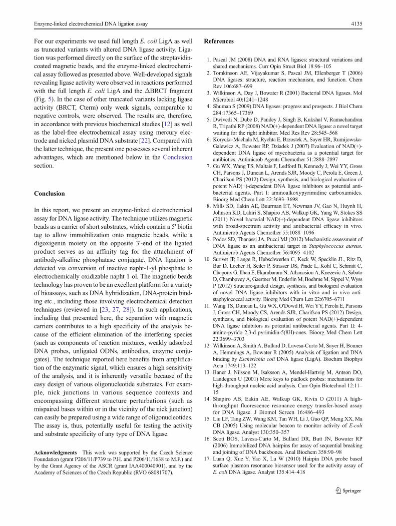
For our experiments we used full length E. coli LigA as wellas truncated variants with altered DNA ligase activity. Liga-tion was performed directly on the surface of the streptavidin-coated magnetic beads, and the enzyme-linked electrochemi-cal assay followed as presented above.Well-developed signalsrevealing ligase activity were observed in reactions performedwith the full length E. coli LigA and the ΔBRCT fragment(Fig. 5). In the case of other truncated variants lacking ligaseactivity (BRCT, Cterm) only weak signals, comparable tonegative controls, were observed. The results are, therefore,in accordance with previous biochemical studies [12] as wellas the label-free electrochemical assay using mercury elec-trode and nicked plasmid DNA substrate [22]. Compared withthe latter technique, the present one possesses several inherentadvantages, which are mentioned below in the Conclusionsection.
Conclusion
In this report, we present an enzyme-linked electrochemicalassay for DNA ligase activity. The technique utilizes magneticbeads as a carrier of short substrates, which contain a 5' biotintag to allow immobilization onto magnetic beads, while adigoxigenin moiety on the opposite 3'-end of the ligatedproduct serves as an affinity tag for the attachment ofantibody-alkaline phosphatase conjugate. DNA ligation isdetected via conversion of inactive napht-1-yl phosphate toelectrochemically oxidizable napht-1-ol. The magnetic beadstechnology has proven to be an excellent platform for a varietyof bioassays, such as DNA hybridization, DNA-protein bind-ing etc., including those involving electrochemical detectiontechniques (reviewed in [23, 27, 28]). In such applications,including that presented here, the separation with magneticcarriers contributes to a high specificity of the analysis be-cause of the efficient elimination of the interfering species(such as components of reaction mixtures, weakly adsorbedDNA probes, unligated ODNs, antibodies, enzyme conju-gates). The technique reported here benefits from amplifica-tion of the enzymatic signal, which ensures a high sensitivityof the analysis, and it is inherently versatile because of theeasy design of various oligonucleotide substrates. For exam-ple, nick junctions in various sequence contexts andencompassing different structure perturbations (such asmispaired bases within or in the vicinity of the nick junction)can easily be prepared using a wide range of oligonucleotides.The assay is, thus, potentially useful for testing the activityand substrate specificity of any type of DNA ligase.
Acknowledgments This work was supported by the Czech ScienceFoundation (grant P206/11/P739 to P.H. and P206/11/1638 to M.F.) andby the Grant Agency of the ASCR (grant IAA400040901), and by theAcademy of Sciences of the Czech Republic (RVO 68081707).
References
1. Pascal JM (2008) DNA and RNA ligases: structural variations andshared mechanisms. Curr Opin Struct Biol 18:96–105
2. Tomkinson AE, Vijayakumar S, Pascal JM, Ellenberger T (2006)DNA ligases: structure, reaction mechanism, and function. ChemRev 106:687–699
3. Wilkinson A, Day J, Bowater R (2001) Bacterial DNA ligases. MolMicrobiol 40:1241–1248
4. Shuman S (2009) DNA ligases: progress and prospects. J Biol Chem284:17365–17369
5. Dwivedi N, Dube D, Pandey J, Singh B, Kukshal V, RamachandranR, Tripathi RP (2008) NAD(+)-dependent DNA ligase: a novel targetwaiting for the right inhibitor. Med Res Rev 28:545–568
6. Korycka-Machala M, Rychta E, Brzostek A, Sayer HR, Rumijowska-Galewicz A, Bowater RP, Dziadek J (2007) Evaluation of NAD(+)-dependent DNA ligase of mycobacteria as a potential target forantibiotics. Antimicrob Agents Chemother 51:2888–2897
7. GuWX,Wang TS, Maltais F, Ledford B, Kennedy J, Wei YY, GrossCH, Parsons J, Duncan L, Arends SJR, Moody C, Perola E, Green J,Charifson PS (2012) Design, synthesis, and biological evaluation ofpotent NAD(+)-dependent DNA ligase inhibitors as potential anti-bacterial agents. Part I: aminoalkoxypyrimidine carboxamides.Bioorg Med Chem Lett 22:3693–3698
8. Mills SD, Eakin AE, Buurman ET, Newman JV, Gao N, Huynh H,Johnson KD, Lahiri S, Shapiro AB, Walkup GK, YangW, Stokes SS(2011) Novel bacterial NAD(+)-dependent DNA ligase inhibitorswith broad-spectrum activity and antibacterial efficacy in vivo.Antimicrob Agents Chemother 55:1088–1096
9. Podos SD, Thanassi JA, Pucci MJ (2012) Mechanistic assessment ofDNA ligase as an antibacterial target in Staphylococcus aureus.Antimicrob Agents Chemother 56:4095–4102
10. Surivet JP, Lange R, Hubschwerlen C, Keck W, Specklin JL, Ritz D,Bur D, Locher H, Seiler P, Strasser DS, Prade L, Kohl C, Schmitt C,ChapouxG, Ilhan E, EkambaramN,AthanasiouA, KnezevicA, SabatoD, ChamboveyA,GaertnerM, EnderlinM,BoehmeM, Sippel V,WyssP (2012) Structure-guided design, synthesis, and biological evaluationof novel DNA ligase inhibitors with in vitro and in vivo anti-staphylococcal activity. Bioorg Med Chem Lett 22:6705–6711
11. Wang TS, Duncan L, GuWX, O'DowdH,Wei YY, Perola E, ParsonsJ, Gross CH, Moody CS, Arends SJR, Charifson PS (2012) Design,synthesis, and biological evaluation of potent NAD(+)-dependentDNA ligase inhibitors as potential antibacterial agents. Part II: 4-amino-pyrido 2,3-d pyrimidin-5(8H)-ones. Bioorg Med Chem Lett22:3699–3703
12. Wilkinson A, Smith A, Bullard D, Lavesa-Curto M, Sayer H, BonnerA, Hemmings A, Bowater R (2005) Analysis of ligation and DNAbinding by Escherichia coli DNA ligase (LigA). Biochim BiophysActa 1749:113–122
13. Baner J, Nilsson M, Isaksson A, Mendel-Hartvig M, Antson DO,Landegren U (2001) More keys to padlock probes: mechanisms forhigh-throughput nucleic acid analysis. Curr Opin Biotechnol 12:11–15
14. Shapiro AB, Eakin AE, Walkup GK, Rivin O (2011) A high-throughput fluorescence resonance energy transfer-based assayfor DNA ligase. J Biomol Screen 16:486–493
15. Liu LF, Tang ZW,Wang KM, TanWH, Li J, Guo QP, Meng XX, MaCB (2005) Using molecular beacon to monitor activity of E-coliDNA ligase. Analyst 130:350–357
16. Scott BOS, Lavesa-Curto M, Bullard DR, Butt JN, Bowater RP(2006) Immobilized DNA hairpins for assay of sequential breakingand joining of DNA backbones. Anal Biochem 358:90–98
17. Luan Q, Xue Y, Yao X, Lu W (2010) Hairpin DNA probe basedsurface plasmon resonance biosensor used for the activity assay ofE. coli DNA ligase. Analyst 135:414–418
Enzyme-linked electrochemical DNA ligation assay 4135

18. He XX, Ni XQ, Wang YH, Wang KM, Jian LX (2011)Electrochemical detection of nicotinamide adenine dinucleotidebased on molecular beacon-like DNA and E. coli DNA ligase.Talanta 83:937–942
19. Wu ZS, Jiang JH, Shen GL, Yu RQ (2007) Highly sensitive DNAdetection and point mutation identification: an electrochemical ap-proach based on the combined use of ligase and reverse molecularbeacon. Hum Mutat 28:630–637
20. Pang LL, Li JS, Jiang JH, Le Y, Shen GL, Yu RQ (2007) A noveldetection method for DNA point mutation using QCM based onFe3O4/Au core/shell nanoparticle and DNA ligase reaction.Sensors Actuators B Chem 127:311–316
21. Zhang P, Chu X, Xu XM, Shen GL, Yu RQ (2008) Electrochemicaldetection of point mutation based on surface ligation reaction andbiometallization. Biosens Bioelectron 23:1435–1441
22. Vacek J, Cahova K, Palecek E, Bullard DR, Lavesa-Curto M,Bowater RP, Fojta M (2008) Label-free electrochemical monitoringof DNA ligase activity. Anal Chem 80:7609–7613
23. Palecek E, Fojta M (2007) Magnetic beads as versatile tools forelectrochemical DNA and protein biosensing. Talanta 74:276–290
24. Sambrook J, Russell DW (2001) Molecular cloning—a laboratorymanual, 3rd ed. Cold Spring Harbor Laboratory Press, NY
25. Goffin C, Bailly V, Verly WG (1987) Nicks 3' or 5' to AP sites or tomispaired bases, and one-nucleotide gaps can be sealed by T4 DNA-ligase. Nucleic Acids Res 15:8755–8771
26. Tsiapali CM, Narang SA (1970) On fidelity of phage T4-inducedpolynucleotide ligase in joining of chemically synthesized deoxyri-bonucleotides. Biochem Biophys Res Commun 39:631–636
27. Kuramitz H (2009) Magnetic microbead-based electrochemical im-munoassays. Anal Bioanal Chem 394:61–69
28. Palecek E, Bartosik M (2012) Electrochemistry of nucleic acids.Chem Rev 112:3427–3481
Eva Stejskalova successfullycompleted her Master’s projecton electrochemical DNA bioas-says at the Institute of Biophysics,ASCR, Brno, Czech Republic,and is currently working as aPhD student at the Institute ofMolecular Genetics, Prague,Czech Republic, where she workson the biology of pre-mRNAsplicing.
Petra Horakova obtained herPhD at the University of Pardubi-ce, Czech Republic and workedas a postdoc at the Departmentof Biophysical Chemistry andMolecular Oncology at the Insti-tute of Biophysics, ASCR, BrnoCzech Republic, before going onmaternity leave. Her work is fo-cused on electrochemical analysisof DNA and its application in mo-lecular diagnostics.
Jan Vacek is an associate profes-sor at the Faculty ofMedicine andDentistry, Palacky University inOlomouc, Czech Republic. He re-ceived his PhD in Molecular andCell Biology from Masaryk Uni-versity in Brno, Czech Republic.His research interests include thestudy of DNA, protein, and theirinteractions with natural com-pounds and drugs using electro-chemistry, liquid separation ap-proaches, and mass spectrometry.
Richard Bowater is a senior lec-turer at the School of BiologicalSciences, University of East An-glia in Norwich, United King-dom. His research career has in-cluded a range of studies that haveused classical biochemical tech-niques, biophysical methods ofanalysis, and molecular biologyprocedures. His current researchcharacterizes DNA repair pro-cesses from a variety of organ-isms, feeding into wider studiesof biochemical mechanisms thatinfluence genome stability.
Miroslav Fojta is head of the De-partment of Biophysical Chemistryand Molecular Oncology at the In-stitute of Biophysics, ASCR, in Br-no, Czech Republic. He is also asso-ciate professor of molecular biologyat the Masaryk University in Brnoand a research group leader at theCentral European Institute of Tech-nology in Brno. His main researchinterests include electrochemistry ofnucleic acids, chemical modificationof biopolymers, and its applicationin analysis of nucleotide sequences,
DNA damage, and protein–DNA interactions.
4136 E. Stejskalová et al.