enzyme immunoassay for detection of rubella specific igm antibody
TRANSCRIPT
394
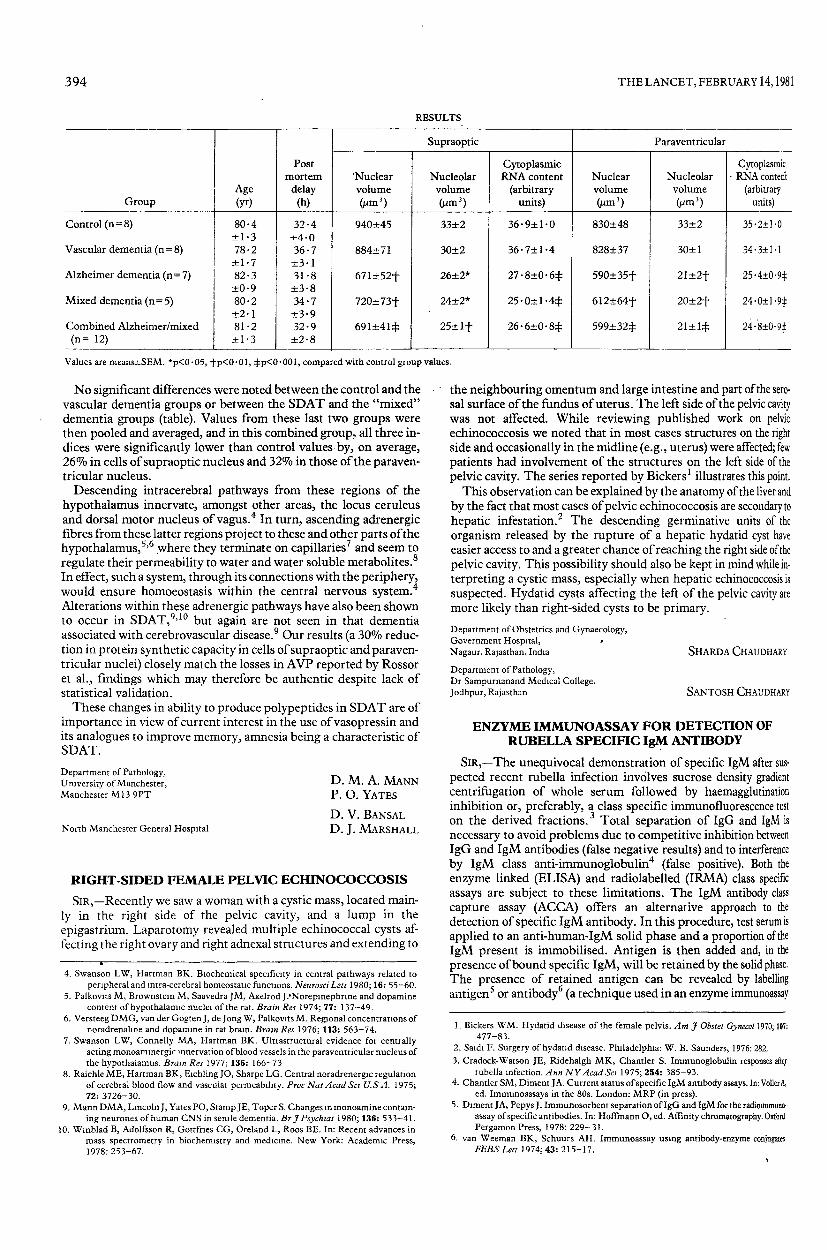
RESULTS
Values are means±SEM. *p<0-05, -)hp<0’01, :j:p<0’00i, compared with control group values.
No significant differences were noted between the control and thevascular dementia groups or between the SDAT and the "mixed"dementia groups (table). Values from these last two groups werethen pooled and averaged, and in this combined group, all three in-dices were significantly lower than control values-by, on average,26% in cells of supraoptic nucleus and 32% in those of the paraven-tricular nucleus.
Descending intracerebral pathways from these regions of thehypothalamus innervate, amongst other areas, the locus ceruleusand dorsal motor nucleus of vagus. 4In turn, ascending adrenergicfibres from these latter regions project to these and other parts of thehypothalamus,5,6 .where they terminate on capillaries 7and seem toregulate their permeability to water and water soluble metabolites.
8
In effect, such a system, through its connections with the periphery,would ensure homoeostasis within the central nervous system.4 4Alterations within these adrenergic pathways have also been shownto occur in SDAT,9,10 but again are not seen in that dementiaassociated with cerebrovascular disease.9 Our results (a 30% reduc-tion in protein synthetic capacity in cells of supraoptic and paraven-tricular nuclei) closely match the losses in AVP reported by Rossoret al., findings which may therefore be authentic despite lack ofstatistical validation.These changes in ability to produce polypeptides in SDAT are of
importance in view of current interest in the use of vasopressin andits analogues to improve memory, amnesia being a characteristic ofSDAT.
Department of Pathology,University of Manchester,Manchester M13 9PT
North Manchester General Hospital
D. M. A. MANNP. O. YATES
D. V. BANSAL
D. J. MARSHALL
RIGHT-SIDED FEMALE PELVIC ECHINOCOCCOSIS
SIR,-Recently we saw a woman with a cystic mass, located main-ly in the right side of the pelvic cavity, and a lump in the
epigastrium. Laparotomy revealed multiple echinococcal cysts af-fecting the right ovary and right adnexal structures and extending to
4. Swanson LW, Hartman BK. Biochemical specificity in central pathways related toperipheral and intra-cerebral homeostatic functions. Neurosci Lett 1980; 16: 55-60.
5. Palkovits M, Brownstein M, Saavedra JM, Axelrod J. Norepinephrine and dopaminecontent of hypothalamic nuclei of the rat. Brain Res 1974; 77: 137-49.
6. Versteeg DMG, van der Gogten J, de Jong W, Palkovits M. Regional concentrations ofnoradrenaline and dopamine in rat brain. Brain Res 1976; 113: 563-74.
7. Swanson LW, Connelly MA, Hartman BK. Ultrastructural evidence for centrallyacting monoaminergic innervation of blood vessels in the paraventricular nucleus ofthe hypothalamus. Brain Res 1977; 136: 166-73
8. Raichle ME, Hartman BK, Eichling JO, Sharpe LG. Central noradrenergic regulationof cerebral blood flow and vascular permeability. Proc Nat Acad Sci U.S A. 1975;72: 3726-30.
9. Mann DMA, Lincoln J, Yates PO, Stamp JE, Toper S. Changes in monoamine contain-ing neurones of human CNS in senile dementia. Br J Psychiat 1980; 136: 533-41.
10. Winblad B, Adolfsson R, Gottfries CG, Oreland L, Roos BE. In: Recent advances inmass spectrometry in biochemistry and medicine. New York: Academic Press,1978: 253-67.
the neighbouring omentum and large intestine and part of the sero-sal surface of the fundus of uterus. The left side of the pelvic cavitywas not affected. While reviewing published work on pelvicechinococcosis we noted that in most cases structures on the rightside and occasionally in the midline (e.g., uterus) were affected; fewpatients had involvement of the structures on the left side of thepelvic cavity. The series reported by Bickers 1 illustrates this point.This observation can be explained by the anatomy of the liver and
by the fact that most cases of pelvic echinococcosis are secondary tohepatic infestation.z The descending germinative units of the
organism released by the rupture of a hepatic hydatid cyst haveeasier access to and a greater chance of reaching the right side of thepelvic cavity. This possibility should also be kept in mind while in-terpreting a cystic mass, especially when hepatic echinococcosis issuspected. Hydatid cysts affecting the left of the pelvic cavity aremore likely than right-sided cysts to be primary.Department of Obstetrics and Gynaecology,Government Hospital,Nagaur, Rajasthan, India
Department of Pathology,Dr Sampurnanand Medical College,Jodhpur, Rajasthan
SHARDA CHAUDHARY
SANTOSH CHAUDHARY
ENZYME IMMUNOASSAY FOR DETECTION OFRUBELLA SPECIFIC IgM ANTIBODY
SiR,-The unequivocal demonstration of specific IgM after sus-pected recent rubella infection involves sucrose density gradientcentrifugation of whole serum followed by haemagglutinationinhibition or, preferably, a class specific immunofluorescence teston the derived fractions. Total separation of IgG and IgM isnecessary to avoid problems due to competitive inhibition betweenIgG and IgM antibodies (false negative results) and to interferenceby IgM class anti-immunoglobulin (false positive). Both the
enzyme linked (ELISA) and radiolabelled (IRMA) class specificassays are subject to these limitations. The IgM antibody classcapture assay (ACCA) offers an alternative approach to thedetection of specific IgM antibody. In this procedure, test serum isapplied to an anti-human-IgM solid phase and a proportion of theIgM present is immobilised. Antigen is then added and, in thepresence of bound specific IgM, will be retained by the solid phase.The presence of retained antigen can be revealed by labellingantigen or antibody6 (a technique used in an enzyme immunoassay
1. Bickers WM. Hydatid disease of the female pelvis. Am J Obstet Gynecol 1970; 107:477-83.
2. Saidi F. Surgery of hydatid disease. Philadelphia: W. B. Saunders, 1976: 282.3. Cradock-Watson JE, Ridehalgh MK, Chantler S. Immunoglobulin responses after
rubella infection. Ann NY Acad Sci 1975; 254: 385-93.4. Chantler SM, Diment JA. Current status of specific IgM antibody assays. In: Voller A,
ed. Immunoassays in the 80s. London: MRP (in press).5. Diment JA, Pepys J. Immunosorbent separation of IgG and IgM for the radioimmuno-
assay of specific antibodies. In: Hoffmann O, ed. Affinity chromatography. OxfordPergamon Press, 1978: 229-31.
6. van Weeman BK, Schuurs AH. Immunoassay using antibody-enzyme conjugatesFEBS Lett 1974; 43: 215-17.

395
Fig. I-Rubella specific IgM response detected by an enzyme IgMcapture assay in sera from individuals after recent and pastinfection and from controls.
Controls include a group of rheumatoid factor (RF) positive individuals,shown separately.
1=rubella specific IgM confirmed.Absorbance values=OD492 test minus OD492 standard seronegative serum.
for detecting IgM antibodies to hepatitis A).5 We have investigatedan enzyme ACCA for the detection of rubella specific IGM.Polystyrene ’Removastrip’ wells (Dynatech) were coated with an
immunoglobulin preparation of sheep anti-human-IgM (50 g/ml) in 0-05molll "tris" buffer pH 7-5 5 and then incubated with test samples of humanserum diluted 1/200 in 2% bovine serum albumin/saline for 3 h at 37°C. Afterthe wells had been washed, excess rubella haemagglutinin antigen was addedand allowed to stand at 37°C for 4 h. After a further wash, the working dilutionof horseradish peroxidase labelled anti-rubella conjugate prepared byperiodate oxidation and diluted in 20% sheep serum/saline was added to thewells and incubated for 2 h at 37°C. The amount of conjugate bound wasdetermined by adding o-phenylenediamine/urea peroxide and measuring theabsorbance at 492 nm.
The results obtained with 28 samples taken from twenty-fiveadult females at various intervals following rubella infection, 15samples from individuals with high rubella IgG alone, 15 samplesfrom individuals with rubella IgG and rheumatoid factor, and 11seronegative individuals are shown in fig. 1. Absorbance valuesabove those obtained with the control groups were observed in thesamples taken after acute infection and as early as one day after thereported onset of a rash. Rubella specific IgM was confirmed by anindependent test (closed circle) in all these sera: The meanabsorbance value for the control rheumatoid factor positive groupwas marginally greater than that observed with the seronegativesamples. A limited number of post-vaccination samples have alsobeen tested; a proportion were positive in our assay but the levels ofIgM present were considerably less than those found followingnatural infection. Our failure to detect specific IgM in all post-vaccination sera may be due to the limiting test sensitivity or mayreflect a failure to obtain samples during the period of peakresponse.The sensitivity of our capture assay has been compared with that
obtained in indirect immunofluorescence tests performed on frac-tionated sera and the results (fig. 2) indicate comparable sensitivity
7. Duermeyer W, Wielaard F, van der Veen J. A new principle for the detection of specificIgM antibodies applied to an ELISA for hepatitis A. J Med Virol 1979; 4: 25-32.
I,"’T I,IV I,U"’T If"’’"’’’ " .,,"’--.-
Immunofluorescence Titre
Fig. 2-Comparison of rubella specific IgM measured by enzymecapture assay and indirect immunofluorescence on fractionatedsera.
with a correlation coefficient of 0-88. Further investigations are inprogress to examine the limit of sensitivity of our assay.We thank Dr J. Best and Dr J. Cradock-Watson for the supply of test samples
and access to clinical data.
Wellcome Research Laboratories,Beckenham, Kent BR3 3BS
JOHN A. DIMENTSHIREEN M. CHANTLER
PLASMA-TRIFLUOPERAZINE CONCENTRATIONSDURING HIGH DOSE THERAPY
SIR,-Trifluoperazine has been used in psychiatry since 1958, butthere is no published information on its plasma concentrations. 1,2,Existing neuroleptic drug assays are unsuitable for this
compound.3,4 A high pressure liquid chromatographic (HPLC)method for trifluoperazine has been developed by one ofus (S.H.C.)and we now report detection and measurement of trifluoperazine inthe plasma of a single patient receiving high doses.The patient, an 18-year-old man, entered hospital during a schizo-
phrenic episode within a recurrent illness. In an attempt to deter-mine suitable treatment, he was first treated with trifluoperazineliquid concentrate at a dose of 15 mg x 3 daily. This was later changedto 30 mg and then to 80 mg, both at bedtime. No trifluoperazine wasdetected in his plasma at the two lower doses, but the concentrationsgiven in the table were recorded after an 80 mg test dose, given onemorning as a dose additional to the normal daily 80 mg regimen.Blood was collected into heparinised tubes and the trifluoperazine was ex-
tracted into hexane from alkalinised plasma. The concentrated extractswere examined by HPLC using a ’Micropak-CN’ column (Varian), a mobilephase of methanol (90%)/5 mmolll ammonium acetate (10%) at 2 - 5 5 ml/mm,and ultraviolet (254 nm) detection. Standards (spiked plasma) and patientsamples were handled similarly. The calibration equation was of the formy=a+bx (r2) where y is instrument response in absorbance units, a is the in-tercept on the y axis, b is the slope, x is the plasma concentration of triflu-operazine in ng/ml, and r’ is the square of the correlation coefficient:
y=(-l-071xlo"’)+(2-46xlo’
1. van Praag HM. Psychotropic drugs: a guide for the practitioner. London: Macmillan,1978.
2. Cooper TB. Plasma level monitoring of antipsychotic drugs. Clin Pharmacokin 1978;3: 14-38.
3. Curry SH, Marshall JHL. Plasma levels of chlorpromazine and some of its relativelynon-polar metabolites in psychiatric patients Life Sci 1968, 7: 9
4 Curry SH Determination of nanogram quantities of chlorpromazine and some of itsmetabolites in plasma using gas-liquid chromotography with an electron-capturedetector Analyt Chem 1968, 40: 1251-55
5. Butterfield AG, Sears RW. High-performance liquid chromatographic determinationof perphenazine and amitriptyline hydrochloride in two-component tablet formula-tions. J Pharm Sci 1977; 66: 1117-19.