enzyme-immobilized hydrogels to create hypoxia for in
TRANSCRIPT
Enzyme-immobilized hydrogels to create hypoxia for in vitro cancer cell culture
Camron S. Dawes,1 Heiko Konig,2,3 and Chien-Chi Lin1,3,*
1 Department of Biomedical Engineering, Indiana University-Purdue University Indianapolis, Indianapolis, IN, 46202, USA 2 Department of Medicine, Division of Hematology/Oncology, Indiana University School of Medicine, Indianapolis, IN, 46202, USA 3 Indiana University Melvin & Bren Simon Cancer Center, Indianapolis, IN, 46202, USA
*To whom correspondence should be made:
Prof. Chien-Chi Lin Associate Professor of Biomedical Engineering Indiana University-Purdue University Indianapolis 723 W. Michigan St. SL220K Indianapolis, IN 46202 Phone: (317)274-0760 Email: [email protected]
_________________________________________________________________________________ This is the author's manuscript of the article published in final edited form as:
Dawes, C. S., Konig, H., & Lin, C.-C. (2017). Enzyme-immobilized hydrogels to create hypoxia for in vitro cancer cell culture. Journal of Biotechnology, 248, 25–34. https://doi.org/10.1016/j.jbiotec.2017.03.007
2
Abstract
Hypoxia is a critical condition governing many aspects of cellular fate processes. The
most common practice in hypoxic cell culture is to maintain cells in an incubator with controlled
gas inlet (i.e., hypoxic chamber). Here, we describe the design and characterization of enzyme-
immobilized hydrogels to create solution hypoxia under ambient conditions for in vitro cancer
cell culture. Specifically, glucose oxidase (GOX) was acrylated and co-polymerized with
poly(ethylene glycol)-diacrylate (PEGDA) through photopolymerization to form GOX-
immobilized PEG-based hydrogels. We first evaluated the effect of soluble GOX on inducing
solution hypoxia (O2 < 5%) and found that both unmodified and acrylated GOX could sustain
hypoxia for at least 24 hours even under ambient air condition with constant oxygen diffusion
from the air-liquid interface. However, soluble GOX gradually lost its ability to sustain hypoxia
after 24 hours due to the loss of enzyme activity over time. On the other hand, GOX-
immobilized hydrogels were able to create hypoxia within the hydrogel for at least 120 hours,
potentially due to enhanced protein stabilization by enzyme ‘PEGylation’ and immobilization. As
a proof-of-concept, this GOX-immobilized hydrogel system was used to create hypoxia for in
vitro culture of Molm14 (acute myeloid leukemia (AML) cell line) and Huh7 (hepatocellular
carcinoma (HCC) cell line). Cells cultured in the presence of GOX-immobilized hydrogels
remained viable for at least 24 hours. The expression of hypoxia associated genes, including
carbonic anhydrase 9 (CA9) and lysyl oxidase (LOX), were significantly upregulated in cells
cultured with GOX-immobilized hydrogels. These results have demonstrated the potential of
using enzyme-immobilized hydrogels to create hypoxic environment for in vitro cancer cell
culture.
Keywords: Hypoxia; cancer; hydrogel; enzyme immobilization; glucose oxidase.
3
1. Introduction
Hypoxia, the lack of adequate oxygen (O2) supply in cells and tissues, is a physiological
condition of many healthy and diseased tissues in the body. For example, O2 concentration is
around 20% in the lungs; ~13% in the alveoli; ~5% in the circulation system and the bone
marrow; and below 5% in multicellular tissues (De Miguel et al., 2015; Simon and Keith, 2008).
Hypoxia is implicated in both normal physiological events and pathological conditions, including
ischemia, tumors, and inflamed tissues. As such, O2 concentration should be considered as a
critical experimental condition when performing in vitro cell studies (Hockel and Vaupel, 2001;
Liu and Simon, 2004; Semenza, 2000; Simon and Keith, 2008). Hypoxia stabilizes the
expression of hypoxia inducible factors (HIFs) (Semenza, 2000), which are heterodimeric
transcription factors that regulate many downstream genes and cell fate processes (Liu and
Simon, 2004; Semenza, 2000), including proliferation, metabolism, apoptosis, stress response,
angiogenesis, and migration. Hypoxia is also a key factor regulating tumor growth and drug
resistance (De Miguel et al., 2015; Giaccia et al., 2003; Hockel and Vaupel, 2001; Liu and
Simon, 2004).
The gold standard to induce hypoxia ([O2] < 5%) for in vitro cell culture is through using a
cell culture chamber with controlled gas supplies (i.e., hypoxic chamber). However, the time
needed to reach equilibrium of O2 partial pressure between the chamber atmosphere and the
culture medium could take several hours (Allen et al., 2001). Another challenge of using a
hypoxic chamber is that O2 diffusion from the air to the cell culture media occurs rapidly once
the culture plates are removed from the hypoxic chamber. Unfortunately, studies have shown
that even brief exposure of some cells to ambient air would cause drastic changes in certain
hypoxia-related gene expression (Broxmeyer et al., 2015). For this reason, a glovebox is
required if one wishes to maintain hypoxia throughout the experiment. The high front-end cost
and dedicated space required for a hypoxic chamber system also limit its implementation to
selected laboratories. In addition, it is challenging to perform real-time imaging or other

4
instrument-based live cell assays under hypoxia even with the use of a glovebox. Furthermore,
one hypoxic chamber system can only provide one fixed O2 tension for one experiment, which
significantly retards the progress of scientific discovery related to varied O2 tensions (e.g.,
hypoxia gradient, multiplex hypoxic drug testing, etc.).
Another method to induce hypoxia for cell culture is through introducing pre-equilibrated
media with lower O2 tension into the cell culture vessels, such as bioreactors or microfluidic
devices. Bioreactors are the standard operation for scale-up production of cells or biological
products but not ideal for mechanistic studies of hypoxia-induced cellular response. On the
other hand, a microfluidic culture system permits real-time imaging of hypoxic cell culture and
allows creation of complex hypoxia patterns within the confined microenvironment. For example,
Peng et al. used chemical scavengers to reduce aqueous O2 content within a patterned array of
cells in wells of a microfluidic device with geometry matching that of a 96-well plate (Peng et al.,
2013). This method is beneficial in that multiple O2 profiles can be developed rapidly through
pre-equilibrated media in different wells of a single device. However, setting up microfluidic cell
culture requires special instruments and project-specific microfluidic design. The applicability of
such system in higher or enhanced-throughput analysis is also limited (e.g., drug screening and
testing under various hypoxic conditions). It is also not an easy task to integrate microfluidic
system with three-dimensional (3D) cell culture.
Hypoxic response in the cells can also be simulated using chemicals that upregulate or
stabilize the expression of HIF1α. Cobalt chloride (CoCl2) or desferrioxamine are two examples
of such chemicals (An et al., 1998). Although this is a relatively simple strategy to mimic hypoxic
response in the cells, the use of chemical only regulates cellular and molecular responses
directly downstream of HIF. Furthermore, these chemicals could affect cell survival, metabolism,
and morphology differently compared to real O2 deprivation (Han et al., 2006).
Recently, O2-consuming enzymatic reactions are being developed as an alternative
route to the aforementioned methods. The most notable example is the use of glucose oxidase
5
(GOX) and catalase (CAT) (Gibson et al., 1964; Kirkman and Gaetani, 2007). GOX oxidizes β-
D-glucose while consuming O2 to produce gluconic acid and hydrogen peroxide (H2O2). CAT is
commonly added to reduce the cytotoxic H2O2 to one mole of water and a half mole of O2
(Figure 1A). This system has been used to induce hypoxia in solutions and in microfluidic
devices (Askoxylakis et al., 2011; Baumann et al., 2008; Huang et al., 2013; Li et al., 2016;
Millonig et al., 2009; Mueller et al., 2009; Rajan et al., 2013; Sobotta et al., 2013; Zitta et al.,
2012). The use of GOX/CAT is beneficial in that the system provides a rapid onset of hypoxia
(usually within a few minutes) (Askoxylakis et al., 2011; Baumann et al., 2008; Huang et al.,
2013; Li et al., 2016; Millonig et al., 2009; Mueller et al., 2009; Rajan et al., 2013; Sobotta et al.,
2013; Zitta et al., 2012). One drawback to any GOX system, however, is the production of
hydrogen peroxide, a reactive oxygen species (ROS) (Fruehauf and Meyskens, 2007) whose
accumulation would not only cause undesired cellular response but also inactivate both GOX
and CAT (Hielscher and Gerecht, 2015; Pal et al., 2000; Trachootham et al., 2009; Tse and
Gough, 1987). Thus far, the applications of GOX/CAT system have been focused on glucose
sensing and pH-induced responses (Wu et al., 2011). For example, Choi et al. prepared GOX-
immobilized poly(ethylene glycol) (PEG) hydrogels and studied the effect of gel compositions on
immobilized enzyme activity. Although the production of H2O2 was quantified to evaluate
kinetics of the immobilized enzyme kinetics, O2 contents were not monitored (Choi et al., 2008).
Some recent work has started to explore the ability of GOX/CAT reactions to induce hypoxia for
in vitro cell culture (Askoxylakis et al., 2011; Baumann et al., 2008; Huang et al., 2013; Li et al.,
2016; Millonig et al., 2009; Mueller et al., 2009; Rajan et al., 2013; Sobotta et al., 2013). The
GOX/CAT system has also been adapted to 3D printed inserts (Li et al., 2016) where GOX and
CAT were coated on printed disks and the degrees of solution hypoxia were controlled by the
distance between the enzyme-immobilized disks and the solution in the culture plate. In that
design, hypoxia conditions (between 0 and ~12% O2) were maintained for up to 5 hours and the
system was used to induce hypoxic response in peritoneal macrophages.
6
Other enzymes (e.g., laccase) have also been used to create hypoxia (Blatchley et al.,
2015; Park et al., 2014; Park and Gerecht, 2014). In the laccase system, a fixed amount of
substrate (i.e., ferulic acid, FA) was immobilized to a polymer backbone. The FA-immobilized
polymer was then crosslinked by laccase-mediated enzymatic reaction, which also consumes
O2. Recently, Lewis et al. extended the timespan of laccase-induced hypoxia by limiting the
diffusion of O2 into the FA-crosslinked hydrogel. Together with the enzymatic O2 depletion, the
system was successfully used to study the impact of hypoxia on sarcoma cell invasion and
migration (Lewis et al., 2016).
Here, we present an immobilized enzyme strategy for inducing hypoxia within and
surrounding the PEG-based hydrogel for in vitro cancer cell culture. Immobilization of oxygen-
consuming GOX within covalently crosslinked hydrogels provide an easy method to control
solution oxygen tension without using external devices. Furthermore, GOX-immobilized
hydrogels can be readily added to or removed from cell culture without disturbing cells. The
crosslinked PEG hydrogel network also provides opportunities for immobilizing multiple
proteins/enzymes or other functional molecules for other biomedical applications. In this
contribution, we systematically studied the ability of GOX, PEG-acrylate modified GOX (i.e.,
GOXPEGA), and GOX-immobilized hydrogels to induce solution hypoxia. Furthermore, we utilized
GOX-immobilized hydrogels to induce hypoxia for in vitro culture of anchorage-independent
acute myeloid leukemia cells Molm14 and anchorage-dependent hepatocellular carcinoma cells
Huh7. We evaluated the effects of GOX-immobilized hydrogels on cancer cell fate, including
viability, proliferation, and hypoxia-associated gene expression.
2. Materials & methods
Linear PEG (Mn = 2 kDa) was purchased from Sigma-Aldrich. Glucose oxidase (0243-
500KU) and catalase (LS001847) were purchased from Amresco and Worthington Biochemical,
respectfully. Acrylate-PEG-succinimidyl valerate (Acryl-PEG-SVA, MW 3,400 Da) was obtained
7
from Laysan Bio Inc. Zeba Spin Desalting Columns (7K MWCO), 2,4,6-trinitrobenzene sulfonic
acid (TNBSA), and β-D-glucose were purchased from Thermo Scientific. Penicillin-streptomycin,
antibiotic-antimycotics, fetal bovine serum (FBS), Roswell Park Memorial Institute media
(RPMI), and Dulbecco’s modified Eagle’s medium (DMEM) were acquired from Life
Technologies. HEPES and Dulbecco’s phosphate-buffered saline (DPBS) were purchased from
Lonza. Membrane culture plate inserts (PIXP-012-50) were purchased from EMD Millipore.
Tryphan blue and AlamarBlue® reagents were purchased from Mediatech and Fisher Scientific,
respectfully.
2.1 Macromer synthesis and characterization
PEG-diacrylate (PEGDA) was synthesized according to an established protocol (Hao
and Lin, 2014) and characterized with 1H-NMR (Bruker 500). The degree of PEGDA
functionalization was around 89% (Figure S1). Photoinitiator lithium aryl phosphonate (LAP)
was synthesized as described elsewhere (Fairbanks et al., 2009).
To facilitate enzyme immobilization within hydrogels, glucose oxidase was acrylated
using Acryl-PEG-SVA (Choi et al., 2008). Briefly, the enzyme was first dissolved at 20 mg/mL in
PBS supplemented with 2 mM EDTA (pH 8.5) and 50 mM sodium carbonate. Acryl-PEG-SVA
was added at 200x molar excess to enzyme concentration and the reaction was allowed to
proceed at room temperature for 2 hours with stirring. During the reaction, primary amines on
the surface of the enzyme reacted with SVA groups to afford PEG-acrylate (PEGA)-modified
GOX (GOXPEGA). Unreacted macromers were removed using size exclusion chromatography
columns (Zeba Spin Desalting column). Un-modified GOX at the same concentration was also
passed through the columns and used as controls to account for any loss/entrapment of
enzyme within the columns. Following synthesis, both GOX and GOXPEGA were assayed using
TNBSA assay to determine the degree of PEGA functionalization. For each assay, enzyme
samples were diluted to 30-35 µg/mL. A series of lysine hydrochloride solutions (0-10 µg/mL,
8
200 µL/well) were used as standards. 100 µL of 0.01% TNBSA reagent was added into wells of
a 96-well plate, which was sealed and incubated at 37°C for 2 hours, followed by cooling for 5
minutes. Absorbance at 335 nm was measured using a microplate reader (SynergyHT BioTek).
The degree of PEGA functionalization on GOX was determined as the concentration of
remaining amine groups on GOXPEGA over that of the un-modified GOX.
2.2 Characterization of enzymatic activity of GOXPEGA
To examine the enzyme activity, O2 consumption in the presence of the enzyme and
glucose was quantified. The changes in O2 content over time in the presence of GOX or
GOXPEGA (Vo = ∆[O2] / ∆Time) was defined as the reaction velocity. Enzyme was dissolved PBS
(pH 7.4) at 0.13 µM in a 2 mL microtube with constant stirring at 25°C. The oxygen consumption
reactions were carried out under ambient air with constant oxygen diffusion from the air to mimic
actual cell culture condition. Stock β-D-glucose solution was injected at the start of every
measurement to give starting concentrations of 0.30 to 25 mM [S]I. Dissolved O2 concentration
was monitored for 3 minutes using an O2 probe and meter (Microx4, PreSens). O2 contents
were plotted as a function of time and the initial linear portion of the curve was used for Vo
calculation (change in substrate concentration over time). Non-linear regression analysis and
curve fitting was applied to paired Vo and [S]I using the equation Vo =Vmax∙[S]/(Km + [S]). In the
equation, Vmax is the theoretical maximum enzyme reaction velocity and Km is the Michaelis-
Menten constant, the equilibrium dissociation constant (i.e., affinity) for the enzyme and the
substrate.
2.3 Synthesis and characterization of enzyme-immobilized hydrogels
All macromer solutions were sterilized by passing through 0.22 µm syringe filters.
PEGDA hydrogels (15 wt%) were polymerized aseptically through radical mediated
photopolymerization in the absence or presence of GOXPEGA monomer (6 mg/mL), and LAP (1
mM) as the photoinitiator. 60 µL gels were injected between two glass slides separated by

9
Teflon spacers (2 mm) and gelation was initiated with a UV lamp (365 nm, 5 mW/cm2, 2 min
exposure). Following photopolymerization, hydrogels (~ 3.1 mm dia. x 2 mm thickness) were
incubated in DPBS for 24 hours at 37°C.
O2 concentration in solution was measured with a dipping-type O2 sensor (Microx4,
PreSens). For solution based measurements, the probe was extended to ~2 mm above the
bottom of the 24 well plate or 1 mm above the gel (~2 mm from the liquid-air interface). To
measure the H2O2 produced during the reactions, 10 µL aliquots of the solutions were collected
and quantified with a Quantichrom Peroxide Assay Kit following the manufacturer’s protocol
(BioAssay Systems).
2.4 Cell culture and viability assays
A suspension cell type, human acute myeloid leukemia (AML) cells Molm14, was
purchased from Leibniz Institute, German Collection of Microorganisms and Cell Cultures. Cells
were maintained in RPMI media supplemented with 10% fetal bovine serum (FBS) and 1%
penicillin-streptomycin 25 mM HEPES, and 25 mM β-D-Glucose. 400,000 cells/mL of Molm14
cells were seeded per well in non-treated 24 well plates. GOXPEGA gels (15 wt% PEGDA, 6
mg/mL GOXPEGA, 60 µL per gel) were added to half of the wells (one gel per well) containing
0.54 mg/mL catalase. Remaining wells were placed with gels without immobilized enzyme. In
vitro O2 concentration was measured ~1 mm above the hydrogel with a dipping-type O2 sensor
(PreSens). Adherent cell type human hepatocellular carcinoma cells (Huh7) were grown in high
glucose DMEM supplemented with 10% FBS, 1% antibiotic antimycotics, and 25 mM HEPES.
Cells were seeded on treated 24 well plates with 1 mL per well of cell suspension (60,000
cells/mL) and allowed to grow/spread for 48 hours prior to the onset of the experiments, at
which time (labeled as 0 hours) culture media was refreshed in all wells. At the onset of the
experiment, membrane inserts containing GOXPEGA gels were placed in the wells and the

10
medium was supplemented with 0.54 mg/mL CAT. Half of the wells only had media refreshed
and were used as control groups for the experiment (no enzyme added).
Molm14 cell viability and density were characterized by tryphan blue staining and
counting with a hemocytometer. AlamarBlue® reagent (10x dilution in media) was used for
assaying metabolic activity of Huh7 cells. After a 90 min incubation, 200 µL from each well was
transferred to a clear 96-well microplate and read for fluorescence (excitation/emission: 560/590
nm).
2.5 RNA isolation and real time PCR
RNA isolation was carried out using NucleoSpin RNA II kit (Clontech). Briefly, 600 µL of
lysis buffer was added to each well containing cells. Cell lysates were snap frozen and stored in
-80°C until assay. After thawing the lysates, 600 µL of 70% RNase free ethanol was added,
pipetted vigorously, and then run through NucleoSpin RNA columns. After desalting/purification
steps, RNA was eluted with DNase/RNase-free H2O and quantified by spectroscopy (NanoDrop
2000, Thermo Scientific). Isolated RNA was stored at -80°C.
Complementary DNA was generated from the isolated total RNA by using PrimeScript
RT reagent kit (Clontech, TaKaRa). Gene expression was analyzed by real time quantitative
PCR using SYBR Premix Ex Taq II Kit (Clontech, TaKaRa). The kit components, cDNA, and
primers were mixed in a PCR plate and analyzed on a 7500 Fast Real-Time PCR machine
(Applied Biosystems). Thermocycling parameters were one cycle at 95°C for 30s, followed by
95°C for 3s, 60°C for 30s, and repeat for 45 cycles. Gene expression results were analyzed
using 2-∆∆CT methodology. For each experimental condition, cycle count was first standardized to
ribosomal 18S housekeeping gene (∆CT level) and then normalized with respect to the media
control group for that specific time point (∆∆CT level; media control values were set as one-
fold). Table S1 lists all primer sequences used for real-time PCR.
2.6 Statistics
11
GraphPad Prism 5 was used for all curve fitting and statistical analyses. Significance
comparison between experimental groups was performed using Two-Way ANOVA with
Bonferroni post testing. Michaelis-Menten parameters for GOX and GOXPEGA were generated
with the non-linear regression suite in GraphPad Prism 5 software. All experiments were
conducted a minimum of three times with data presentation as the mean ± standard error of the
mean (SEM). One, two, or three asterisks represent p < 0.05, 0.01, or 0.001, respectively.
3. Results
3.1 Enzyme-induced hypoxia
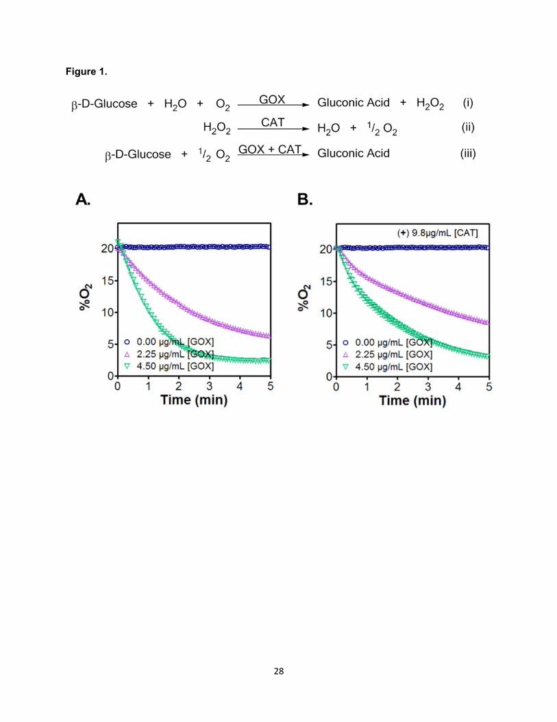
GOX/CAT systems have been used to induce pH changes in aqueous environment due
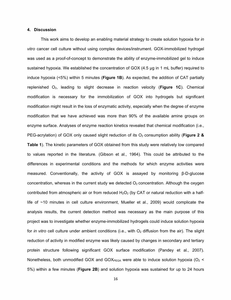
to the production of gluconic acid (Figure 1A). To gain insights into the capability of this
enzymatic system on inducing hypoxia in solution, we quantified the reduction of O2 in aqueous
buffers supplemented with different concentrations of GOX in the absence (Figure 1B) or
presence (Figure 1C) of CAT. The O2 tensions of the buffers were monitored using an O2
sensor and recorded as a function of time. As expected, higher concentrations of GOX were
able to deplete O2 faster. As shown in Figure 1B, O2 tension was reduced within 5 minutes to
~6% or to ~2.5% when 2.25 μg/mL or 4.5 μg/mL of GOX was added, respectively. With the
inclusion of 9.8 μg/mL of CAT in the solution, the rate of O2 consumption was slowed down to
~9% and to 3.2% within 5 minutes (Figure 1C). The decrease in O2 consumption is not
surprising as the addition of CAT produces one-half mole of O2 per mole of H2O2 consumed.
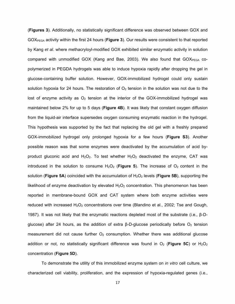
In order to fabricate enzyme-immobilized hydrogels capable of inducing hypoxia, the
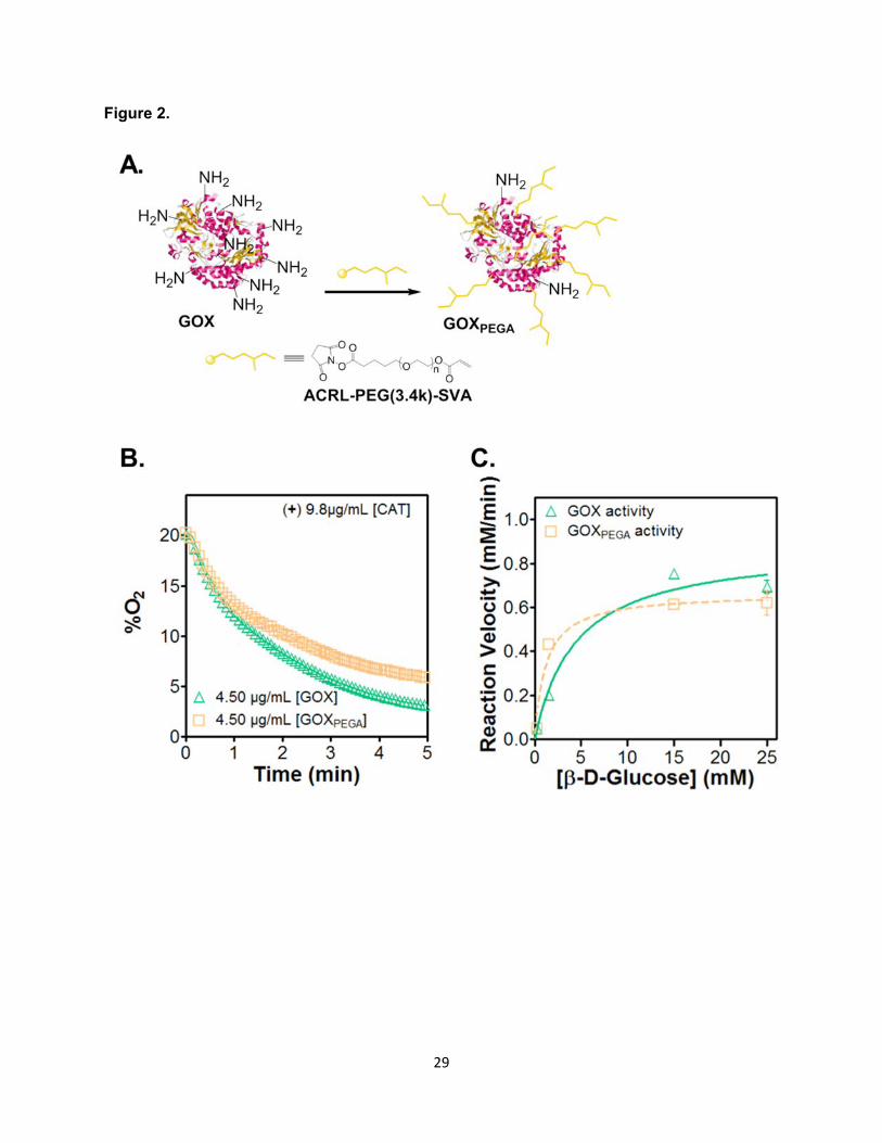
primary amine groups on GOX were functionalized with 200-fold molar excess of Acryl-PEG-
SVA (Figure 2A) (Choi et al., 2008). TNBSA assay results showed an average of 93 ± 1.7%
(Mean ± SEM, N = 5) of the primary amines on enzyme surface were functionalized with Acryl-
PEG. The acrylate moieties on the surface of Acryl-PEG-GOX (i.e., GOXPEGA) permit its
homopolymerization with PEGDA to afford enzyme-immobilized hydrogels. The modification,
12
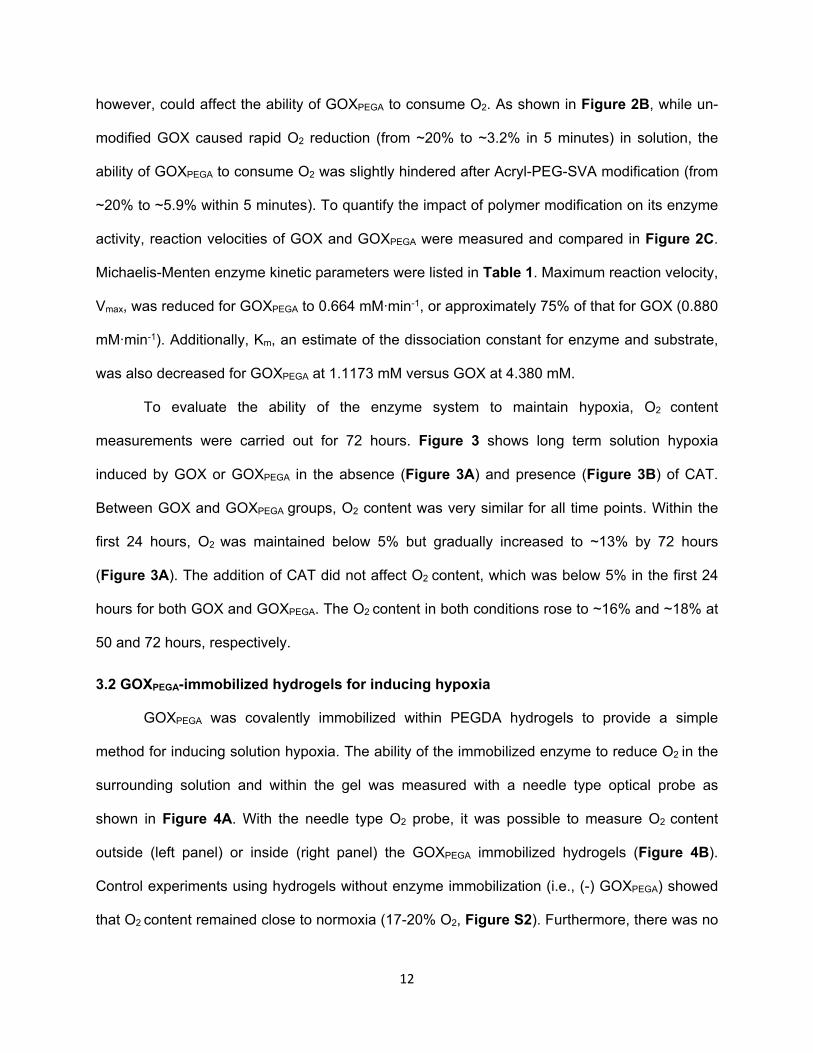
however, could affect the ability of GOXPEGA to consume O2. As shown in Figure 2B, while un-
modified GOX caused rapid O2 reduction (from ~20% to ~3.2% in 5 minutes) in solution, the
ability of GOXPEGA to consume O2 was slightly hindered after Acryl-PEG-SVA modification (from
~20% to ~5.9% within 5 minutes). To quantify the impact of polymer modification on its enzyme
activity, reaction velocities of GOX and GOXPEGA were measured and compared in Figure 2C.
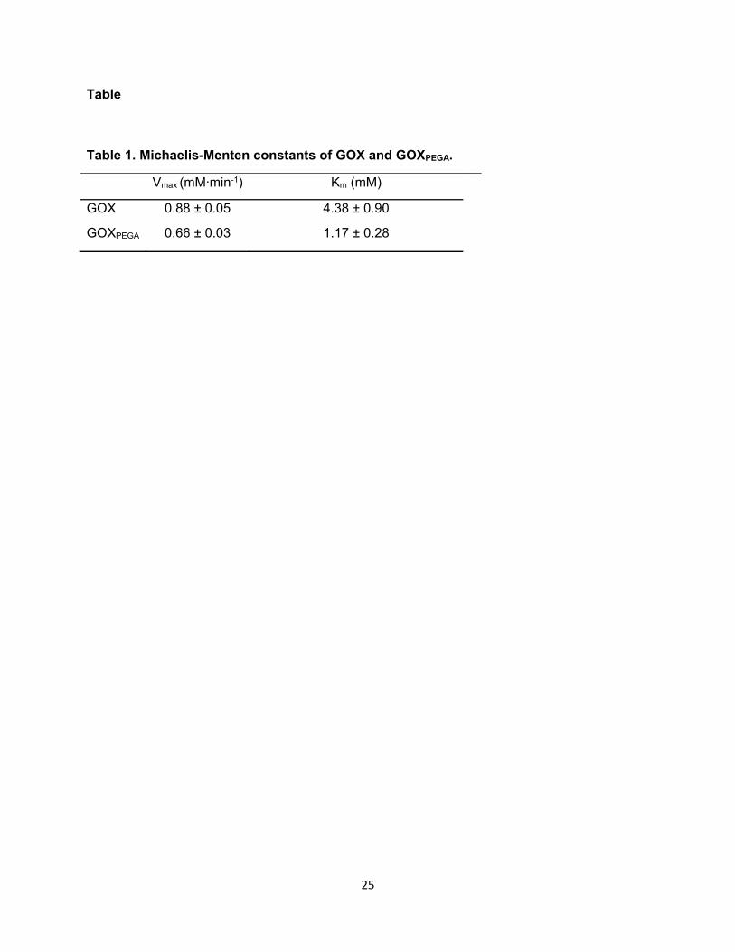
Michaelis-Menten enzyme kinetic parameters were listed in Table 1. Maximum reaction velocity,
Vmax, was reduced for GOXPEGA to 0.664 mM∙min-1, or approximately 75% of that for GOX (0.880
mM∙min-1). Additionally, Km, an estimate of the dissociation constant for enzyme and substrate,
was also decreased for GOXPEGA at 1.1173 mM versus GOX at 4.380 mM.
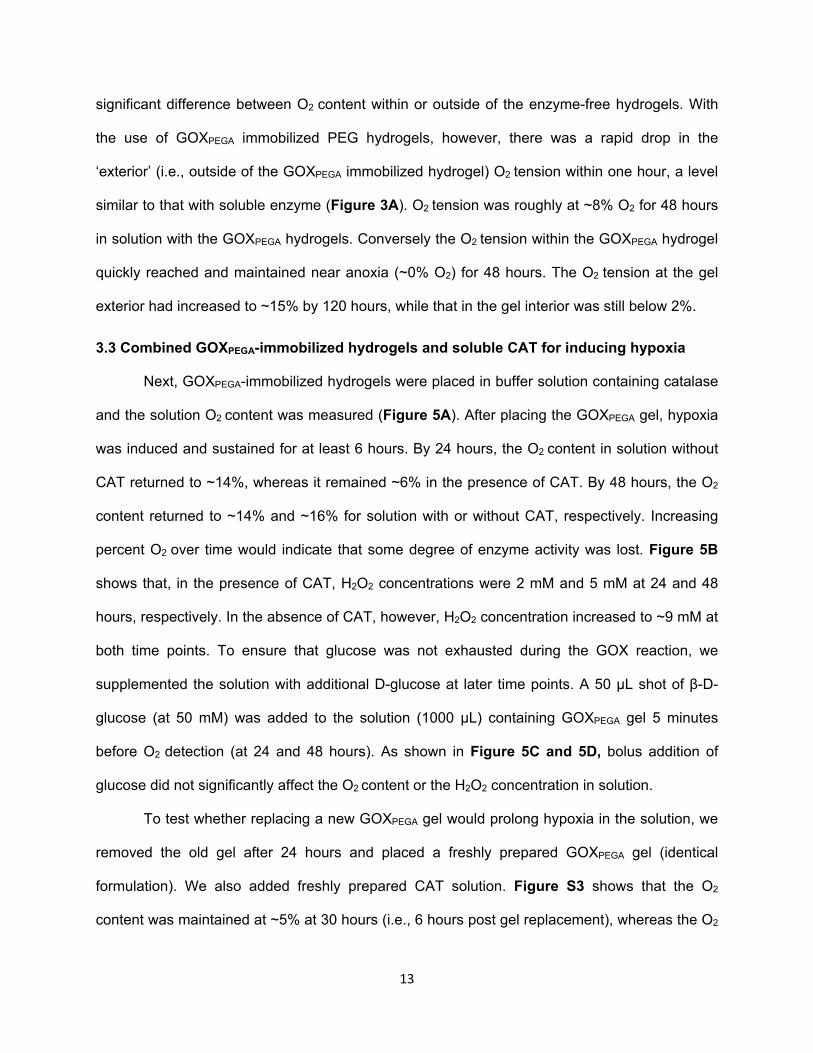
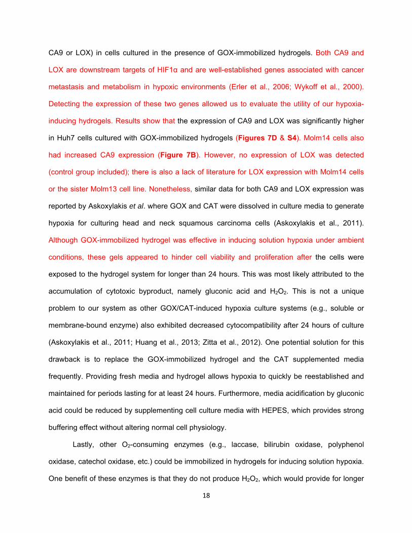
To evaluate the ability of the enzyme system to maintain hypoxia, O2 content
measurements were carried out for 72 hours. Figure 3 shows long term solution hypoxia
induced by GOX or GOXPEGA in the absence (Figure 3A) and presence (Figure 3B) of CAT.
Between GOX and GOXPEGA groups, O2 content was very similar for all time points. Within the
first 24 hours, O2 was maintained below 5% but gradually increased to ~13% by 72 hours
(Figure 3A). The addition of CAT did not affect O2 content, which was below 5% in the first 24
hours for both GOX and GOXPEGA. The O2 content in both conditions rose to ~16% and ~18% at
50 and 72 hours, respectively.
3.2 GOXPEGA-immobilized hydrogels for inducing hypoxia
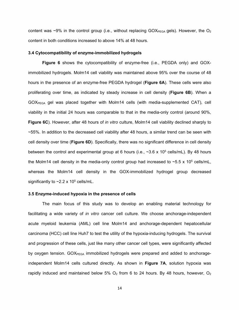
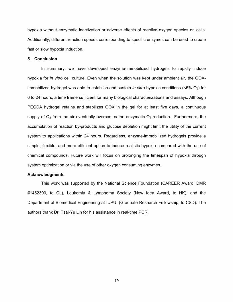
GOXPEGA was covalently immobilized within PEGDA hydrogels to provide a simple
method for inducing solution hypoxia. The ability of the immobilized enzyme to reduce O2 in the
surrounding solution and within the gel was measured with a needle type optical probe as
shown in Figure 4A. With the needle type O2 probe, it was possible to measure O2 content
outside (left panel) or inside (right panel) the GOXPEGA immobilized hydrogels (Figure 4B).
Control experiments using hydrogels without enzyme immobilization (i.e., (-) GOXPEGA) showed
that O2 content remained close to normoxia (17-20% O2, Figure S2). Furthermore, there was no
13
significant difference between O2 content within or outside of the enzyme-free hydrogels. With
the use of GOXPEGA immobilized PEG hydrogels, however, there was a rapid drop in the
‘exterior’ (i.e., outside of the GOXPEGA immobilized hydrogel) O2 tension within one hour, a level
similar to that with soluble enzyme (Figure 3A). O2 tension was roughly at ~8% O2 for 48 hours
in solution with the GOXPEGA hydrogels. Conversely the O2 tension within the GOXPEGA hydrogel
quickly reached and maintained near anoxia (~0% O2) for 48 hours. The O2 tension at the gel
exterior had increased to ~15% by 120 hours, while that in the gel interior was still below 2%.
3.3 Combined GOXPEGA-immobilized hydrogels and soluble CAT for inducing hypoxia
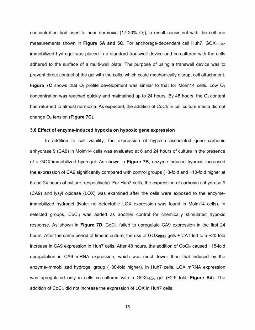
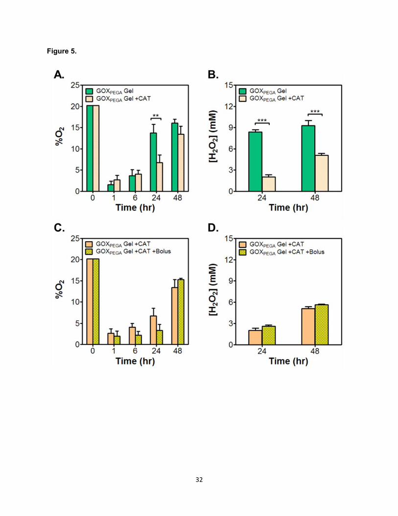
Next, GOXPEGA-immobilized hydrogels were placed in buffer solution containing catalase
and the solution O2 content was measured (Figure 5A). After placing the GOXPEGA gel, hypoxia
was induced and sustained for at least 6 hours. By 24 hours, the O2 content in solution without
CAT returned to ~14%, whereas it remained ~6% in the presence of CAT. By 48 hours, the O2
content returned to ~14% and ~16% for solution with or without CAT, respectively. Increasing
percent O2 over time would indicate that some degree of enzyme activity was lost. Figure 5B
shows that, in the presence of CAT, H2O2 concentrations were 2 mM and 5 mM at 24 and 48
hours, respectively. In the absence of CAT, however, H2O2 concentration increased to ~9 mM at
both time points. To ensure that glucose was not exhausted during the GOX reaction, we
supplemented the solution with additional D-glucose at later time points. A 50 μL shot of β-D-
glucose (at 50 mM) was added to the solution (1000 μL) containing GOXPEGA gel 5 minutes
before O2 detection (at 24 and 48 hours). As shown in Figure 5C and 5D, bolus addition of
glucose did not significantly affect the O2 content or the H2O2 concentration in solution.
To test whether replacing a new GOXPEGA gel would prolong hypoxia in the solution, we
removed the old gel after 24 hours and placed a freshly prepared GOXPEGA gel (identical
formulation). We also added freshly prepared CAT solution. Figure S3 shows that the O2
content was maintained at ~5% at 30 hours (i.e., 6 hours post gel replacement), whereas the O2
14
content was ~9% in the control group (i.e., without replacing GOXPEGA gels). However, the O2
content in both conditions increased to above 14% at 48 hours.
3.4 Cytocompatibility of enzyme-immobilized hydrogels
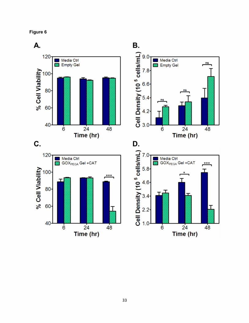
Figure 6 shows the cytocompatibility of enzyme-free (i.e., PEGDA only) and GOX-
immobilized hydrogels. Molm14 cell viability was maintained above 95% over the course of 48
hours in the presence of an enzyme-free PEGDA hydrogel (Figure 6A). These cells were also
proliferating over time, as indicated by steady increase in cell density (Figure 6B). When a
GOXPEGA gel was placed together with Molm14 cells (with media-supplemented CAT), cell
viability in the initial 24 hours was comparable to that in the media-only control (around 90%,
Figure 6C). However, after 48 hours of in vitro culture, Molm14 cell viability declined sharply to
~55%. In addition to the decreased cell viability after 48 hours, a similar trend can be seen with
cell density over time (Figure 6D). Specifically, there was no significant difference in cell density
between the control and experimental group at 6 hours (i.e., ~3.6 x 10⁵ cells/mL). By 48 hours
the Molm14 cell density in the media-only control group had increased to ~5.5 x 10⁵ cells/mL,
whereas the Molm14 cell density in the GOX-immobilized hydrogel group decreased
significantly to ~2.2 x 10⁵ cells/mL.
3.5 Enzyme-induced hypoxia in the presence of cells
The main focus of this study was to develop an enabling material technology for
facilitating a wide variety of in vitro cancer cell culture. We choose anchorage-independent
acute myeloid leukemia (AML) cell line Molm14 and anchorage-dependent hepatocellular
carcinoma (HCC) cell line Huh7 to test the utility of the hypoxia-inducing hydrogels. The survival
and progression of these cells, just like many other cancer cell types, were significantly affected
by oxygen tension. GOXPEGA immobilized hydrogels were prepared and added to anchorage-
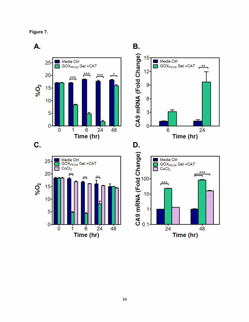
independent Molm14 cells cultured directly. As shown in Figure 7A, solution hypoxia was
rapidly induced and maintained below 5% O2 from 6 to 24 hours. By 48 hours, however, O2
15
concentration had risen to near normoxia (17-20% O2), a result consistent with the cell-free
measurements shown in Figure 5A and 5C. For anchorage-dependent cell Huh7, GOXPEGA-
immobilized hydrogel was placed in a standard transwell device and co-cultured with the cells
adhered to the surface of a multi-well plate. The purpose of using a transwell device was to
prevent direct contact of the gel with the cells, which could mechanically disrupt cell attachment.
Figure 7C shows that O2 profile development was similar to that for Molm14 cells. Low O2
concentration was reached quickly and maintained up to 24 hours. By 48 hours, the O2 content
had returned to almost normoxia. As expected, the addition of CoCl2 in cell culture media did not
change O2 tension (Figure 7C).
3.6 Effect of enzyme-induced hypoxia on hypoxic gene expression
In addition to cell viability, the expression of hypoxia associated gene carbonic
anhydrase 9 (CA9) in Molm14 cells was evaluated at 6 and 24 hours of culture in the presence
of a GOX-immobilized hydrogel. As shown in Figure 7B, enzyme-induced hypoxia increased
the expression of CA9 significantly compared with control groups (~3-fold and ~10-fold higher at
6 and 24 hours of culture, respectively). For Huh7 cells, the expression of carbonic anhydrase 9
(CA9) and lysyl oxidase (LOX) was examined after the cells were exposed to the enzyme-
immobilized hydrogel (Note: no detectable LOX expression was found in Molm14 cells). In
selected groups, CoCl2 was added as another control for chemically stimulated hypoxic
response. As shown in Figure 7D, CoCl2 failed to upregulate CA9 expression in the first 24
hours. After the same period of time in culture, the use of GOXPEGA gels + CAT led to a ~20-fold
increase in CA9 expression in Huh7 cells. After 48 hours, the addition of CoCl2 caused ~15-fold
upregulation in CA9 mRNA expression, which was much lower than that induced by the
enzyme-immobilized hydrogel group (~80-fold higher). In Huh7 cells, LOX mRNA expression
was upregulated only in cells co-cultured with a GOXPEGA gel (~2.5 fold, Figure S4). The
addition of CoCl2 did not increase the expression of LOX in Huh7 cells.
16
4. Discussion
This work aims to develop an enabling material strategy to create solution hypoxia for in
vitro cancer cell culture without using complex devices/instrument. GOX-immobilized hydrogel
was used as a proof-of-concept to demonstrate the ability of enzyme-immobilized gel to induce
sustained hypoxia. We established the concentration of GOX (4.5 µg in 1 mL buffer) required to
induce hypoxia (<5%) within 5 minutes (Figure 1B). As expected, the addition of CAT partially
replenished O2, leading to slight decrease in reaction velocity (Figure 1C). Chemical
modification is necessary for the immobilization of GOX into hydrogels but significant
modification might result in the loss of enzymatic activity, especially when the degree of enzyme
modification that we have achieved was more than 90% of the available amine groups on
enzyme surface. Analyses of enzyme reaction kinetics revealed that chemical modification (i.e.,
PEG-acrylation) of GOX only caused slight reduction of its O2 consumption ability (Figure 2 &
Table 1). The kinetic parameters of GOX obtained from this study were relatively low compared
to values reported in the literature. (Gibson et al., 1964). This could be attributed to the
differences in experimental conditions and the methods for which enzyme activities were
measured. Conventionally, the activity of GOX is assayed by monitoring β-D-glucose
concentration, whereas in the current study we detected O2 concentration. Although the oxygen
contributed from atmospheric air or from reduced H2O2 (by CAT or natural reduction with a half-
life of ~10 minutes in cell culture environment, Mueller et al., 2009) would complicate the
analysis results, the current detection method was necessary as the main purpose of this
project was to investigate whether enzyme-immobilized hydrogels could induce solution hypoxia
for in vitro cell culture under ambient conditions (i.e., with O2 diffusion from the air). The slight
reduction of activity in modified enzyme was likely caused by changes in secondary and tertiary
protein structure following significant GOX surface modification (Pandey et al., 2007).
Nonetheless, both unmodified GOX and GOXPEGA were able to induce solution hypoxia (O2 <
5%) within a few minutes (Figure 2B) and solution hypoxia was sustained for up to 24 hours
17
(Figures 3). Additionally, no statistically significant difference was observed between GOX and
GOXPEGA activity within the first 24 hours (Figure 3). Our results were consistent to that reported
by Kang et al. where methacryloyl-modified GOX exhibited similar enzymatic activity in solution
compared with unmodified GOX (Kang and Bae, 2003). We also found that GOXPEGA co-
polymerized in PEGDA hydrogels was able to induce hypoxia rapidly after dropping the gel in
glucose-containing buffer solution. However, GOX-immobilized hydrogel could only sustain
solution hypoxia for 24 hours. The restoration of O2 tension in the solution was not due to the
lost of enzyme activity as O2 tension at the interior of the GOX-immobilized hydrogel was
maintained below 2% for up to 5 days (Figure 4B). It was likely that constant oxygen diffusion
from the liquid-air interface supersedes oxygen consuming enzymatic reaction in the hydrogel.
This hypothesis was supported by the fact that replacing the old gel with a freshly prepared
GOX-immobilized hydrogel only prolonged hypoxia for a few hours (Figure S3). Another
possible reason was that some enzymes were deactivated by the accumulation of acid by-
product gluconic acid and H2O2. To test whether H2O2 deactivated the enzyme, CAT was
introduced in the solution to consume H2O2 (Figure 5). The increase of O2 content in the
solution (Figure 5A) coincided with the accumulation of H2O2 levels (Figure 5B), supporting the
likelihood of enzyme deactivation by elevated H2O2 concentration. This phenomenon has been
reported in membrane-bound GOX and CAT system where both enzyme activities were
reduced with increased H2O2 concentrations over time (Blandino et al., 2002; Tse and Gough,
1987). It was not likely that the enzymatic reactions depleted most of the substrate (i.e., β-D-
glucose) after 24 hours, as the addition of extra β-D-glucose periodically before O2 tension
measurement did not cause further O2 consumption. Whether there was additional glucose
addition or not, no statistically significant difference was found in O2 (Figure 5C) or H2O2
concentration (Figure 5D).
To demonstrate the utility of this immobilized enzyme system on in vitro cell culture, we
characterized cell viability, proliferation, and the expression of hypoxia-regulated genes (i.e.,
18
CA9 or LOX) in cells cultured in the presence of GOX-immobilized hydrogels. Both CA9 and
LOX are downstream targets of HIF1α and are well-established genes associated with cancer
metastasis and metabolism in hypoxic environments (Erler et al., 2006; Wykoff et al., 2000).
Detecting the expression of these two genes allowed us to evaluate the utility of our hypoxia-
inducing hydrogels. Results show that the expression of CA9 and LOX was significantly higher
in Huh7 cells cultured with GOX-immobilized hydrogels (Figures 7D & S4). Molm14 cells also
had increased CA9 expression (Figure 7B). However, no expression of LOX was detected
(control group included); there is also a lack of literature for LOX expression with Molm14 cells
or the sister Molm13 cell line. Nonetheless, similar data for both CA9 and LOX expression was
reported by Askoxylakis et al. where GOX and CAT were dissolved in culture media to generate
hypoxia for culturing head and neck squamous carcinoma cells (Askoxylakis et al., 2011).
Although GOX-immobilized hydrogel was effective in inducing solution hypoxia under ambient
conditions, these gels appeared to hinder cell viability and proliferation after the cells were
exposed to the hydrogel system for longer than 24 hours. This was most likely attributed to the
accumulation of cytotoxic byproduct, namely gluconic acid and H2O2. This is not a unique
problem to our system as other GOX/CAT-induced hypoxia culture systems (e.g., soluble or
membrane-bound enzyme) also exhibited decreased cytocompatibility after 24 hours of culture
(Askoxylakis et al., 2011; Huang et al., 2013; Zitta et al., 2012). One potential solution for this
drawback is to replace the GOX-immobilized hydrogel and the CAT supplemented media
frequently. Providing fresh media and hydrogel allows hypoxia to quickly be reestablished and
maintained for periods lasting for at least 24 hours. Furthermore, media acidification by gluconic
acid could be reduced by supplementing cell culture media with HEPES, which provides strong
buffering effect without altering normal cell physiology.
Lastly, other O2-consuming enzymes (e.g., laccase, bilirubin oxidase, polyphenol
oxidase, catechol oxidase, etc.) could be immobilized in hydrogels for inducing solution hypoxia.
One benefit of these enzymes is that they do not produce H2O2, which would provide for longer
19
hypoxia without enzymatic inactivation or adverse effects of reactive oxygen species on cells.
Additionally, different reaction speeds corresponding to specific enzymes can be used to create
fast or slow hypoxia induction.
5. Conclusion
In summary, we have developed enzyme-immobilized hydrogels to rapidly induce
hypoxia for in vitro cell culture. Even when the solution was kept under ambient air, the GOX-
immobilized hydrogel was able to establish and sustain in vitro hypoxic conditions (<5% O2) for
6 to 24 hours, a time frame sufficient for many biological characterizations and assays. Although
PEGDA hydrogel retains and stabilizes GOX in the gel for at least five days, a continuous
supply of O2 from the air eventually overcomes the enzymatic O2 reduction. Furthermore, the
accumulation of reaction by-products and glucose depletion might limit the utility of the current
system to applications within 24 hours. Regardless, enzyme-immobilized hydrogels provide a
simple, flexible, and more efficient option to induce realistic hypoxia compared with the use of
chemical compounds. Future work will focus on prolonging the timespan of hypoxia through
system optimization or via the use of other oxygen consuming enzymes.
Acknowledgments
This work was supported by the National Science Foundation (CAREER Award, DMR
#1452390, to CL), Leukemia & Lymphoma Society (New Idea Award, to HK), and the
Department of Biomedical Engineering at IUPUI (Graduate Research Fellowship, to CSD). The
authors thank Dr. Tsai-Yu Lin for his assistance in real-time PCR.
20
References
Allen, C.B., Schneider, B.K., White, C.W., (2001) Limitations to oxygen diffusion and
equilibration in in vitro cell exposure systems in hyperoxia and hypoxia. American journal
of physiology. Lung cellular and molecular physiology 281, L1021-1027.
An, W.G., Kanekal, M., Simon, M.C., Maltepe, E., Blagosklonny, M.V., Neckers, L.M., (1998)
Stabilization of wild-type p53 by hypoxia-inducible factor 1alpha. Nature 392, 405-408.
Askoxylakis, V., Millonig, G., Wirkner, U., Schwager, C., Rana, S., Altmann, A., Haberkorn, U.,
Debus, J., Mueller, S., Huber, P.E., (2011) Investigation of tumor hypoxia using a two-
enzyme system for in vitro generation of oxygen deficiency. Radiation oncology 6, 35.
Baumann, R.P., Penketh, P.G., Seow, H.A., Shyam, K., Sartorelli, A.C., (2008) Generation of
oxygen deficiency in cell culture using a two-enzyme system to evaluate agents
targeting hypoxic tumor cells. Radiation research 170, 651-660.
Blandino, A., Macıas, M., Cantero, D., (2002) Modelling and simulation of a bienzymatic
reaction system co-immobilised within hydrogel-membrane liquid-core capsules.
Enzyme and microbial technology 31, 556-565.
Blatchley, M., Park, K.M., Gerecht, S., (2015) Designer Hydrogels for Precision Control of
Oxygen Tension and Mechanical Properties. Journal of materials chemistry. B, Materials
for biology and medicine 3, 7939-7949.
Broxmeyer, H.E., O'Leary, H.A., Huang, X., Mantel, C., (2015) The importance of hypoxia and
extra physiologic oxygen shock/stress for collection and processing of stem and
progenitor cells to understand true physiology/pathology of these cells ex vivo. Current
opinion in hematology 22, 273-278.
Choi, D., Lee, W., Park, J., Koh, W., (2008) Preparation of poly(ethylene glycol) hydrogels with
different network structures for the application of enzyme immobilization. Bio-medical
materials and engineering 18, 345-356.
21
De Miguel, M.P., Alcaina, Y., de la Maza, D.S., Lopez-Iglesias, P., (2015) Cell metabolism
under microenvironmental low oxygen tension levels in stemness, proliferation and
pluripotency. Current molecular medicine 15, 343-359.
Erler, J.T., Bennewith, K.L., Nicolau, M., Dornhofer, N., Kong, C., Le, Q.-T., Chi, J.-T.A., Jeffrey,
S.S., Giaccia, A.J., (2006) Lysyl oxidase is essential for hypoxia-induced metastasis.
Nature 440, 1222-1226.
Fairbanks, B.D., Schwartz, M.P., Bowman, C.N., Anseth, K.S., (2009) Photoinitiated
polymerization of PEG-diacrylate with lithium phenyl-2,4,6-trimethylbenzoylphosphinate:
polymerization rate and cytocompatibility. Biomaterials 30, 6702-6707.
Fruehauf, J.P., Meyskens, F.L., Jr., (2007) Reactive oxygen species: a breath of life or death?
Clinical cancer research : an official journal of the American Association for Cancer
Research 13, 789-794.
Giaccia, A., Siim, B.G., Johnson, R.S., (2003) HIF-1 as a target for drug development. Nature
reviews. Drug discovery 2, 803-811.
Gibson, Q.H., Swoboda, B.E., Massey, V., (1964) Kinetics and Mechanism of Action of Glucose
Oxidase. The Journal of biological chemistry 239, 3927-3934.
Han, Y.H., Xia, L., Song, L.P., Zheng, Y., Chen, W.L., Zhang, L., Huang, Y., Chen, G.Q., Wang,
L.S., (2006) Comparative proteomic analysis of hypoxia-treated and untreated human
leukemic U937 cells. Proteomics 6, 3262-3274.
Hao, Y., Lin, C.C., (2014) Degradable thiol-acrylate hydrogels as tunable matrices for three-
dimensional hepatic culture. Journal of biomedical materials research. Part A 102, 3813-
3827.
Hielscher, A., Gerecht, S., (2015) Hypoxia and free radicals: role in tumor progression and the
use of engineering-based platforms to address these relationships. Free radical biology
& medicine 79, 281-291.
22
Hockel, M., Vaupel, P., (2001) Tumor hypoxia: definitions and current clinical, biologic, and
molecular aspects. Journal of the National Cancer Institute 93, 266-276.
Huang, Y., Zitta, K., Bein, B., Steinfath, M., Albrecht, M., (2013) An insert-based enzymatic cell
culture system to rapidly and reversibly induce hypoxia: investigations of hypoxia-
induced cell damage, protein expression and phosphorylation in neuronal IMR-32 cells.
Disease models & mechanisms 6, 1507-1514.
Kang, S.I., Bae, Y.H., (2003) A sulfonamide based glucose-responsive hydrogel with covalently
immobilized glucose oxidase and catalase. Journal of Controlled Release 86, 115-121.
Kirkman, H.N., Gaetani, G.F., (2007) Mammalian catalase: a venerable enzyme with new
mysteries. Trends in biochemical sciences 32, 44-50.
Lewis, D.M., Park, K.M., Tang, V., Xu, Y., Pak, K., Eisinger-Mathason, T.S., Simon, M.C.,
Gerecht, S., (2016) Intratumoral oxygen gradients mediate sarcoma cell invasion.
Proceedings of the National Academy of Sciences of the United States of America 113,
9292-9297.
Li, C., Chaung, W., Mozayan, C., Chabra, R., Wang, P., Narayan, R.K., (2016) A New Approach
for On-Demand Generation of Various Oxygen Tensions for In Vitro Hypoxia Models.
PloS one 11, e0155921.
Liu, L., Simon, M.C., (2004) Regulation of transcription and translation by hypoxia. Cancer
biology & therapy 3, 492-497.
Millonig, G., Hegedusch, S., Becker, L., Seitz, H.K., Schuppan, D., Mueller, S., (2009) Hypoxia-
inducible factor 1 alpha under rapid enzymatic hypoxia: cells sense decrements of
oxygen but not hypoxia per se. Free radical biology & medicine 46, 182-191.
Mueller, S., Millonig, G., Waite, G.N., (2009) The GOX/CAT system: a novel enzymatic method
to independently control hydrogen peroxide and hypoxia in cell culture. Advances in
medical sciences 54, 121-135.

23
Pal, P., Datta, S., Bhattacharya, P., (2000) Studies on the modeling and simulation of a
sequential bienzymatic reaction system immobilized in emulsion liquid membrane.
Biochemical engineering journal 5, 89-100.
Pandey, P., Singh, S.P., Arya, S.K., Gupta, V., Datta, M., Singh, S., Malhotra, B.D., (2007)
Application of thiolated gold nanoparticles for the enhancement of glucose oxidase
activity. Langmuir : the ACS journal of surfaces and colloids 23, 3333-3337.
Park, K.M., Blatchley, M.R., Gerecht, S., (2014) The design of dextran-based hypoxia-inducible
hydrogels via in situ oxygen-consuming reaction. Macromolecular rapid communications
35, 1968-1975.
Park, K.M., Gerecht, S., (2014) Hypoxia-inducible hydrogels. Nature communications 5, 4075.
Peng, C.C., Liao, W.H., Chen, Y.H., Wu, C.Y., Tung, Y.C., (2013) A microfluidic cell culture
array with various oxygen tensions. Lab on a chip 13, 3239-3245.
Rajan, N., Narayan, A., Wu, Z., Wu, P., Ahn, C.H., Narayan, R.K., Li, C., (2013) A novel oxygen
tension programmable microfluidic system (oPROMs) for in vitro cell biology studies.
2013 Transducers & Eurosensors XXVII: The 17th International Conference on Solid-
State Sensors, Actuators and Microsystems (TRANSDUCERS & EUROSENSORS
XXVII), pp. 412-415.
Semenza, G.L., (2000) HIF-1: mediator of physiological and pathophysiological responses to
hypoxia. Journal of applied physiology 88, 1474-1480.
Simon, M.C., Keith, B., (2008) The role of oxygen availability in embryonic development and
stem cell function. Nature reviews. Molecular cell biology 9, 285-296.
Sobotta, M.C., Barata, A.G., Schmidt, U., Mueller, S., Millonig, G., Dick, T.P., (2013) Exposing
cells to H2O2: a quantitative comparison between continuous low-dose and one-time
high-dose treatments. Free radical biology & medicine 60, 325-335.
24
Trachootham, D., Alexandre, J., Huang, P., (2009) Targeting cancer cells by ROS-mediated
mechanisms: a radical therapeutic approach? Nature reviews. Drug discovery 8, 579-
591.
Tse, P.H., Gough, D.A., (1987) Time-dependent inactivation of immobilized glucose oxidase
and catalase. Biotechnology and bioengineering 29, 705-713.
Wu, Q., Wang, L., Yu, H., Wang, J., Chen, Z., (2011) Organization of glucose-responsive
systems and their properties. Chemical reviews 111, 7855-7875.
Wykoff, C.C., Beasley, N.J.P., Watson, P.H., Turner, K.J., Pastorek, J., Sibtain, A., Wilson,
G.D., Turley, H., Talks, K.L., Maxwell, P.H., Pugh, C.W., Ratcliffe, P.J., Harris, A.L.,
(2000) Hypoxia-inducible Expression of Tumor-associated Carbonic Anhydrases.
Cancer Research 60, 7075-7083.
Zitta, K., Meybohm, P., Bein, B., Huang, Y., Heinrich, C., Scholz, J., Steinfath, M., Albrecht, M.,
(2012) Salicylic acid induces apoptosis in colon carcinoma cells grown in-vitro: influence
of oxygen and salicylic acid concentration. Experimental cell research 318, 828-834.
25
Table
Table 1. Michaelis-Menten constants of GOX and GOXPEGA.
Vmax (mM∙min-1) Km (mM)
GOX 0.88 ± 0.05 4.38 ± 0.90
GOXPEGA 0.66 ± 0.03 1.17 ± 0.28
26
Figure Captions
Figure 1. Enzyme reaction mechanisms and soluble GOX induced solution hypoxia. (A)
Reaction equilibrium of GOX (i), CAT (ii), and combined reaction (iii). (B, C) GOX-induced O2
consumption in the absence (B) or presence of 9.8 µg/mL CAT (C). All reactions were carried
out in pH 7.4 PBS with constant stirring, at room temperature, and with 25 mM β-D-Glucose.
(Mean ± SEM, n ≥ 3).
Figure 2. Effect of GOX modification on oxygen consumption. (A) Reaction scheme of GOX
modification using Acryl-PEG-SVA. Protein structure for GOX was obtained from the RCSB
Protein Data Bank (PDB-ID, 3QVP). (B) O2 consumption profile using soluble GOX or GOXPEGA,
9.8 µg/mL CAT, and 25mM β-D-Glucose. (C) Reaction velocity of O2 consumption by GOX or
GOXPEGA as a function of substrate β-D-glucose concentration. Values were generated from
using 0.260 µM GOX or GOXPEGA with 0.30 to 25 mM of β-D-glucose. All reactions were carried
out in pH 7.4 PBS with constant stirring at 25°C. (Mean ± SEM, n ≥ 3).
Figure 3. Effect of GOX modification on oxygen consumption over extended time. (A, B)
O2 consumption profiles of unmodified and acrylated GOX in the absence (B) or presence of
soluble CAT (450 µg/mL). All reactions were carried out in pH 7.4 PBS, at 25°C, with 25 mM β-
D-Glucose. (**p < 0.01. ***p < 0.001. Mean ± SEM, n ≥ 3).
Figure 4. Effect of hydrogel-immobilized GOXPEGA on oxygen consumption. (A) Schematic
of O2 measurement within and outside of a PEGDA hydrogel. The sensor probe was fully
extended from the needle for measuring O2 tension exterior to the hydrogel (left). To measure
O2 content at the interior of the hydrogel (right), the optic fiber was recessed within its needle
housing to prevent damage of the gel matrix to the probe. After penetration the fiber was
27
extended to the tip of the needle cannula so that it was exposed to the interior of the hydrogel.
(B) O2 consumption at the interior or exterior of GOX-immobilized hydrogels (120 µL of 8 wt%
PEGDA gel with 4 mg/mL GOXPEGA). (***p < 0.001. Mean ± SEM, n ≥ 3).
Figure 5. Effect of supplements on oxygen consumption and H2O2 production by GOXPEGA
gels. (A, B) Effect of soluble CAT addition on O2 tension (A) and H2O2 accumulation (B). (C, D)
Effect of additional glucose on O2 tension (C) and H2O2 accumulation (D) in the presence of
soluble CAT. Additional bolus injections of glucose (50 µL of 500 mM) was delivered 5 minutes
before measuring O2 at 24 and 48 hour time points. Hydrogels (60 µL) were formed by 15 wt%
PEGDA co-polymerized with 6 mg/mL GOXPEGA. All reactions were carried out in pH 7.4 PBS at
37°C. (**p < 0.01. ***p < 0.001. Mean ± SEM, n ≥ 3).
Figure 6. Cytocompatibility of PEGDA hydrogels with or without immobilized GOXPEGA.
Molm14 cell viability (A, C) and density (B, D) when cultured in the absence (i.e., Empty Gel; A
& B) or presence (C, D) of immobilized GOXPEGA Gel + CAT (6 mg/mL). Hydrogels were formed
by 15 wt% PEGDA. CAT in media: 0.54 mg/mL (*p < 0.05. ***p < 0.001. Mean ± SEM, n ≥ 3).
Figure 7. Effect of enzyme-induced hypoxia on cell fate in vitro. O2 profile (A, C) and CA9
mRNA expression (B, D) in Molm14 (A, B) or Huh7 cells (C, D) cultured in the presence of a
GOXPEGA (6 mg/mL) immobilized 15 wt% PEGDA hydrogel. CAT in media: 0.54 mg/mL. CoCl2
(150 µM) was added separately as an additional control group (*p < 0.05, **p < 0.01, ***p <
0.001. Mean ± SEM, n ≥ 3).
28
Figure 1.
29
Figure 2.

30
Figure 3.

31
Figure 4.
32
Figure 5.
33
Figure 6
34
Figure 7.