enhancement of the gut barrier integrity by a microbial ... · microbial metabolite through the...
TRANSCRIPT

ARTICLE
Enhancement of the gut barrier integrity by amicrobial metabolite through the Nrf2 pathwayRajbir Singh1, Sandeep Chandrashekharappa2, Sobha R. Bodduluri1, Becca V. Baby1, Bindu Hegde1,
Niranjan G. Kotla2, Ankita A. Hiwale2, Taslimarif Saiyed3, Paresh Patel3, Matam Vijay-Kumar4,
Morgan G.I. Langille5, Gavin M. Douglas5, Xi Cheng 4, Eric C. Rouchka 6, Sabine J. Waigel7,
Gerald W. Dryden7, Houda Alatassi8, Huang-Ge Zhang1, Bodduluri Haribabu1,
Praveen K. Vemula 2 & Venkatakrishna R. Jala1
The importance of gut microbiota in human health and pathophysiology is undisputable.
Despite the abundance of metagenomics data, the functional dynamics of gut microbiota in
human health and disease remain elusive. Urolithin A (UroA), a major microbial metabolite
derived from polyphenolics of berries and pomegranate fruits displays anti-inflammatory,
anti-oxidative, and anti-ageing activities. Here, we show that UroA and its potent synthetic
analogue (UAS03) significantly enhance gut barrier function and inhibit unwarranted
inflammation. We demonstrate that UroA and UAS03 exert their barrier functions through
activation of aryl hydrocarbon receptor (AhR)- nuclear factor erythroid 2–related factor 2
(Nrf2)-dependent pathways to upregulate epithelial tight junction proteins. Importantly,
treatment with these compounds attenuated colitis in pre-clinical models by remedying
barrier dysfunction in addition to anti-inflammatory activities. Cumulatively, the results
highlight how microbial metabolites provide two-pronged beneficial activities at gut epithe-
lium by enhancing barrier functions and reducing inflammation to protect from colonic
diseases.
https://doi.org/10.1038/s41467-018-07859-7 OPEN
1 Department of Microbiology and Immunology, James Graham Brown Cancer Center, University of Louisville, Louisville, KY 40202, USA. 2 Institute for StemCell Biology and Regenerative Medicine (inStem), GKVK campus, Bangalore, Karnataka 560065, India. 3 Centre for Cellular and Molecular Platforms(C-CAMP), GKVK campus, Bangalore, Karnataka 560065, India. 4 Department of Physiology and Pharmacology, University of Toledo College of Medicineand Life Sciences, Toledo, OH 43614, USA. 5Department of Pharmacology, Dalhousie University, Halifax, B3H 4R2 Nova Scotia, Canada. 6 ComputerEngineering and Computer Science, Kentucky Biomedical Research Infrastructure Network, University of Louisville, Louisville, KY 40202, USA. 7Departmentof Medicine, University of Louisville, Louisville, KY 40202, USA. 8Department of Pathology, University of Louisville, Louisville, KY 40202, USA.Correspondence and requests for materials should be addressed to P.K.V. (email: [email protected]) or to V.R.J. (email: [email protected])
NATURE COMMUNICATIONS | (2019) 10:89 | https://doi.org/10.1038/s41467-018-07859-7 | www.nature.com/naturecommunications 1
1234
5678
90():,;

Inflammatory bowel diseases (IBD) consisting of Crohn’s andulcerative colitis are resultant of dysregulation of the immunesystem leading to intestinal inflammation and microbial dys-
biosis. Numerous studies in recent years highlighted the pivotalrole of gut microbiota and their metabolites in host physiologicalprocesses including immune, metabolic, neurological, and nutri-tional homeostasis1–4. Thus, alterations in gut microbiota havebeen associated with adverse outcomes in cancer, IBD, neurolo-gical disorders, obesity, and diabetes1,5–7. Microbiota and theirmetabolites are in close proximity to the gut epithelium thatconstitutes a single cell-layer separating host components fromthe external environment. Gut barrier integrity is maintained bythe tight junction proteins such as claudins (Cldn), Zona occludin-1 (ZO1), and occludin (Ocln) that are critical for epithelial cellbarrier functions8,9. Previously, it has been reported that levels oftight junction proteins are significantly down regulated underIBD conditions leading to increased gut permeability to microbialligands and noxious metabolites resulting in systemic inflam-matory responses8,10. Despite the availability of large metage-nomics data, the functional dynamics of microbiota and theirmetabolites in IBDs are unknown. Therefore, we tested thehypothesis that certain microbial metabolites will prevent gutpermeability by enhancing barrier functions in addition toblocking inflammation. Treatment with such microbial metabo-lites will offer better therapeutic options for IBDs.
Consumption of diets containing berries and pomegranateshave demonstrated significant beneficial effects on humanhealth11–14. Especially, pomegranate extract or juice containinghigh levels of polyphenolic compounds such as ellagitannins(ETs) and ellagic acid (EA) have been suggested to preventhypertension15 and protect against myocardial ischemia andreperfusion injury16. They have been recognized as potential non-toxic chemo-preventive compounds against chronic diseases suchas cancer, diabetes, cardiovascular and neurodegenerative dis-orders17. It has been suggested that further downstream meta-bolites of EA known as ‘urolithins’ generated by gut microbiotarender potential health benefits, in vivo18,19. Among urolithins,Urolithin A (UroA) displayed potent anti-inflammatory, anti-oxidative and anti-ageing properties compared to other metabo-lites20–23. Due to life style variations and antibiotic/drug usage,presence of bacteria that metabolize dietary EA to urolithins havebeen variable among human populations. Thus, not only theconsumption of diets enriched in polyphenols is required but alsothe presence of microbes that convert them into beneficialmetabolites is critical for manifestation of their health effects. Atthis time, the targets or pathways through which such microbialmetabolites regulate physiological processes are largely unknown.
In this study, we examined the activities of UroA and a potentsynthetic structural analogue UAS03 and identified that inaddition to the anti-inflammatory activities, these compoundsstrongly enhanced gut barrier function. We demonstrate thatboth UroA and UAS03 enhance barrier function by inducingtight junction proteins through activating aryl hydrocarbonreceptor (AhR)-nuclear factor erythroid 2–related factor 2 (Nrf2)-dependent pathways. Further, oral treatment with UroA/UAS03 significantly mitigated systemic inflammation and colitissuggesting potential therapeutic applications for the treatment ofIBD.
ResultsSynthesis and anti-inflammatory activities of UroA andUAS03. UroA (3,8-dihydroxy-6H-dibenzo[b,d]pyran-6-one) hasa lactone (cyclic ester bond) that connects two mono-hydroxylphenyl rings leading to a planar structure (Fig. 1a). Gastric pH ordigestive enzymes can hydrolyze the lactone ring, which opens
the ring resulting in the loss of the planar structure and poten-tially its activities. To generate more stable and potent com-pounds, we synthesized non-hydrolyzable cyclic ether derivative,UAS03 (6H-benzo[c]chromene-3,8-diol) (Fig. 1a). The stability ofboth compounds was examined under conditions of gastric pHand digestive enzymes. The results showed that UAS03 indeed isstable at gastric pH and also in the presence of gastric enzymese.g., esterases and proteases (Fig. 1a). Both UroA and UAS03-significantly decreased LPS induced IL-6 and TNF-α in mousebone marrow derived macrophages (BMDMs) withUAS03 showing anti-inflammatory activities even at nano molarconcentrations (Fig. 1b). Next, anti-inflammatory activities ofUroA and UAS03 were examined in vivo in a LPS-inducedperitonitis mouse model. UroA or UAS03 treatment significantlyreduced the LPS-induced increase in serum IL-6 and TNF-αlevels (Fig. 1c). These results suggest that UAS03 is a potentstructural analogue of UroA with increased anti-inflammatoryactivities.
UroA/UAS03 induce tight junction proteins. Since, microbialmetabolites are in close proximity to gut epithelium; we surmisethat metabolites could have a direct impact on epithelial cellfunction. To examine such effects, we performed RNA-Seq ana-lysis of epithelial cell line (HT29) exposed to UroA. The analysiswas performed as described in methods and to determine sig-nificance of differential gene expression, cuffdiff2 algorithm wasused. Based on an uncorrected p-value cutoff of 0.05, 1960 geneswere determined to be significantly differentially expressed as aresult of UroA treatment in HT29 cells. Further restricting thislist, 437 genes were found to be differentially expressed at FDRcorrected q value <0.05 in UroA treated HT29 cells (Supple-mentary Fig. 1 and Supplementary Data 1). The pathway analysisusing this restricted gene list was performed using IngenuityPathway Analysis (IPA) software (Supplementary Fig. 1). TheEukaryotic Initiation Factor 2 (eIF2), mammalian target ofrapamycin (mTOR) and mitochondrial dysfunction pathwayswere emerged as top 3 pathways. The impact of UroA on mito-chondrial dysfunction (pathways of mitophagy) have beendescribed in previously by Ryu D et al.22. They demonstrated thatUroA induced mitophagy and prolonged the lifespan of C. elegansand increased muscle function in rodents. The impact of UroA onmTOR and eIF2 pathways need to be established in the context ofcolon epithelial functions. The RNA-Seq analysis showed thatcytochrome P450 1A1 (Cyp1A1) is among the top 3 UroAupregulated genes (Supplementary Data 1). The pathways ana-lysis further indicated that the Nrf2 and AhR signaling pathwaysare in top 25 (Supplementary Fig. 1). We surmise that regulationof barrier function is of critical importance in mitigating IBDs.Therefore, we examined the expression of the tight junctionproteins in RNA-Seq data and found that claudin 4 (Cldn4) isupregulated in UroA treated cells (Supplementary Fig. 2)24. Inaddition to Cldn4 and Cyp1A1, UroA also significantly increasedthe expression of heme oxygenase 1 (HMOX1 or HO1) (Supple-mentary Fig. 2). HO1 is well known Nrf2-dependent gene, whichexerts wide variety of beneficial activities including removal oftoxic heme, protection against oxidative stress, regulation ofapoptosis, and inflammation25. Based on these observations, wehypothesized that UroA and UAS03 will induce tight junctionproteins and enhance barrier function through AhR and Nrf2pathways
Ingenuity Pathway Analysis (IPA) revealed significant enrich-ment of Nrf2 and AhR signaling pathways (SupplementaryFig. 1), supporting a role for these pathways in UroA signaling. Apotential therapeutic avenue in IBD is the ability to increasebarrier function. It was therefore of interest that we observed a
ARTICLE NATURE COMMUNICATIONS | https://doi.org/10.1038/s41467-018-07859-7
2 NATURE COMMUNICATIONS | (2019) 10:89 | https://doi.org/10.1038/s41467-018-07859-7 | www.nature.com/naturecommunications

significant increase in expression of the tight junction proteinCldn4 in UroA treated cells. Although not statistically significantin our RNA-seq dataset, we further observed an increase inexpression of additional tight junction proteins ZO-1 and Ocln1using real-time PCR (Fig. 1d). The increased levels of theseproteins by UroA or UAS03 was confirmed by western blots(Fig. 1e) and Cldn4 by confocal imaging (Fig. 1f) in both HT29
and another colon epithelial cell line, Caco2. Further, we observedelevated expression of Cldn4 in the colons of mice treated withUroA/UAS03 (Supplementary Fig. 3a). The functional conse-quence of increased tight junction proteins was examined usingin vitro FITC-dextran permeability assay in transwell plates. Asshown in Fig. 1g, pretreatment of Caco2 or HT29 cells withUAS03 or UroA significantly inhibited LPS induced leakage of
UroAa
d e
f g
b cUAS03BMDM Serum
IL-6
***
** ** *
***** ** **
** ******* **
**
***
*** ***
*** ***
*****
****
*
******** ******
**********
************
IL-6TNF-αα TNF-α
1002
1.5
8 2.0
1.5
1.0
0.5
0.0
6
4
2
0
Veh
Veh
Uro
A
LPS
Cldn4β-actin
Oclnβ-actin
ZO-1β-actin
LPS
UA
S03
1.5
1.0
0.5
0.0
0.8
0.4
0.0
0.8
0.4
0.0
Veh
Veh
Uro
AV
ehU
roA
UA
S03
Veh
Uro
AU
AS
03
Veh
Uro
AU
AS
03
Veh
Uro
A
UA
S03
Veh
Uro
A
UA
S03
UA
S03
1.0
0.5
0.0
1
00 0.1
UroA + LPSUAS03 + LPS UroA + LPS
UAS03 + LPS
[Compound] μM1 10 50 0 0.1
[Compound] μM1 10 50
50
0
4
3 4
3
2
1
0
2
1
0
2
0
Veh
Uro
A
UA
S03
Veh
Uro
A
UA
S03
Veh
Uro
A
UA
S03
Veh UroA UAS03
Veh
HT
29C
aco
2
UroA UAS03
Gre
en R
FU
FIT
C-D
ex (
μg/m
L)
FIT
C-D
ex (
μg/m
L)
Caco2
400 3000 8 4
2
0
4
0
1500
0
200
0
Caco2
– + + ++
+–––
– – –
– + + ++
+–––
– – –
HT29 HT29
Veh UroA UAS03
Veh UroA UAS03
LPSUroAUAS03
LPSUroAUAS03
Time (h) 0 12 0 12UroA
Cldn4 Ocln ZO1
UAS03
Sta
bili
ty (
%)
Fo
ld c
han
ge
IL-6
(n
g/m
L)
IL-6
(n
g/m
L)
TN
F-α
(n
g/m
L)
Cld
n4/
β-ac
tin
Ocl
n/β
-act
inZ
O-1
/β-a
ctin
TN
F-α
(n
g/m
L)
O
OH
HOO
O
OH
HO
Fig. 1 UAS03 is a potent anti-inflammatory structural analogue of UroA and induces tight junction proteins. a Chemical structures of UroA, UAS03. UroA/UAS03 stability was examined in the presence of gastric pH 2.0 and digestive enzymes. UroA and UAS03 (0.2 mg/ml) were incubated with digestiveenzymes (esterases and proteases, 100 U/ml) for 12 h at 37 °C and compound levels were quantified. b BMDMs were stimulated with LPS (50 ng/ml)without or with UroA (blue line)/UAS03 (purple line) (0.1, 1, 10, 25, and 50 µM) for 6 h. IL-6 and TNF-α levels in supernatants were measured. c C57BL/6mice (n= 3–4) were pretreated with UroA (20mg/kg) and UAS03 (20mg/kg). After 4 h, LPS (2 mg/kg) was injected intraperitoneally. Post 4 h of LPSadministration, serum levels of IL-6 and TNF-α was measured. d–f HT29 or Caco2 cells were treated with vehicle (DMSO-0.01%) or UroA/UAS03 (50μM) for 24 h. d The fold changes in mRNA levels of claudin 4 (Cldn4), occludin (Ocln), and Zona occludens 1 (ZO1) in HT29 cells were determined by RTPCR method. e UroA/UAS03 induced protein expression of Cldn4, Ocln, and ZO1 in HT29 cells were determined by immunoblots and quantified by ImageJ software. f Caco2 or HT29 cells were grown on coverslip bottom FluroDish and treated with Vehicle, UroA/UAS03 for 24 h. The cells were stained withanti-Cldn4 followed by secondary antibody tagged with Alexa-488. Nucleus was stained using DAPI. The confocal images were captured. The greenintensity (n= 15–20 cell membrane regions) was measured. Scale bars for Caco2 and HT29 cells indicate 50 and 25 μm respectively. gMonolayer HT29 orCaco2 cells on transmembranes were treated with vehicle or UroA/UAS03 (50 μM) for 24 h followed by treatment with LPS (50 ng/ml) for 2 h. FITC-dextran was added to these cells (top of the membrane) and incubated for 2 h and FITC-dextran levels in bottom chamber well was measured. Results arerepresentative of three independent experiments with triplicates for each concentration. *p < 0.05, **p < 0.01, ***p < 0.001, unpaired t-test between Veh,UroA, or UAS03. Error bars, ±SEM. Source Data are provided as a Source Data File
NATURE COMMUNICATIONS | https://doi.org/10.1038/s41467-018-07859-7 ARTICLE
NATURE COMMUNICATIONS | (2019) 10:89 | https://doi.org/10.1038/s41467-018-07859-7 | www.nature.com/naturecommunications 3

FITC-dextran into bottom chambers. Overall, these resultssuggest that treatment with UroA/UAS03 increased the expres-sion of tight junction proteins potentially enhancing the gutbarrier integrity.
AhR mediates the activities of UroA/UAS03. RNA-Seq data andreal-time PCR data suggested that UroA significantly upregulatedCyp1A1 (Supplementary Data 1, Supplementary Fig. 2, Fig. 2a, b).The P450-Glo Cyp1A1 assay (Fig. 2c) as well as 7-ethoxyresor-ufin-O-deethylase (EROD) assay (Supplementary Fig. 4) wereperformed to determine, whether the Cyp1A1 enzyme activitywas similarly affected. UroA/UAS03 significantly inducedCyp1A1 activity in colon epithelial cells (Fig. 2c and Supple-mentary Fig. 4). Since Cyp1A1 is a well-known downstreamtarget of AhR signaling26, we examined whether UroA/UAS03mediate their actions through AhR. In these assays, we utilizedwell established potent AhR ligands [2,3,7,8-tetrachlorodibenzo-p-dioxin (TCDD) or 6-Formylindolo[3,2-b]carbazole (FICZ) andlow affinity AhR ligand (beta-naphthoflavone (BNF)] to comparethe Cyp1A1 activities with UroA/UAS03. UroA/UAS03 activatedCyp1A1 similar to low affinity AhR ligand BNF at 50 µM. Asexpected, the high affinity ligands such as FICZ and TCDDshowed increased Cyp1A1 activity even at nano molar con-centrations compared to UroA/UAS03/BNF (Fig. 2c and Sup-plementary Fig. 4). More importantly, we tested whether UroA/UAS03 induce the Cyp1A1 activities in vivo using wild type andAhR−/− mice. As shown in Fig. 2d, UroA/UAS03 significantlyactivated Cyp1A1 activity in colon and liver of wild type but notin AhR−/− mice. Moreover, UroA/UAS03 treated wild type miceshowed relatively more Cyp1A1 activity in colon tissues com-pared to BNF and FICZ treated mice (Supplementary Fig. 5a, b).Interestingly, FICZ and BNF that are delivered through intraperitoneum (i.p) showed more Cyp1A1 activity in liver comparedto UroA/UAS03 that are delivered through oral route (Supple-mentary Fig. 5a, b). It could be attributed to first pass effect. Todirectly compare the administration route, we delivered UroA orUAS03 or FICZ through i.p. and determined Cyp1A1 enzymeactivities. As expected, high affinity AhR ligand, FICZ, inducedCyp1A1 activity ~30 fold in liver compared to 5–6 fold by UroA/UAS03 (Supplementary Fig. 5c). In colons, FICZ only increa-sed Cyp1A1 activity up to ~5 fold, whereas UroA/USA03increased by only ~3 fold. In summary, these results suggestthat UroA/UAS03 upregulate expression of Cyp1A1 andenhances the enzyme activity through AhR albeit at low levelsin vivo.
The direct activation of AhR by UroA/UAS03 was examined inHT29 cells by XRE-luciferase reporter assay as well as nucleartranslocation of AhR. The data showed that UroA/UAS03treatment resulted in 2–4 fold induction of luciferase activity(Fig. 2e) compared to the high affinity ligand MeBio that causedhigher levels (~15 fold) of AhR activation. Both UroA and UAS03induced the nuclear translocation of AhR (Fig. 2f, g). AhR wasupregulated in mice treated with UroA or UAS03 (SupplementaryFig. 3b). Next, we asked whether AhR or Cyp1A1 are required forUroA/UAS03 mediated upregulation of tight junction protein,Cldn4. For this purpose, AhR or Cyp1A1 expression wassuppressed using siRNA knockdown and Cldn4 expression wasexamined. As shown in Fig. 2h, i and Supplementary Fig. 6,UroA/UAS03 failed to induce Cldn4 both in AhR or Cyp1A1knockdown cells. In addition, we also deleted Cyp1A1 in HT29cells using CRISPR/Cas9 methods and examined UroA/UAS03mediated activities. Deletion of Cyp1A1 did not show effect onbasal levels of Cldn4 compared to parental HT29 cells(Supplementary Fig. 7a). As shown in Supplementary Fig. 7b–d,UroA/UAS03 failed to upregulate Cldn4 or NQO1 in Cyp1A1
deleted cells. These results suggest that UroA/US03 induce theexpression of tight junction proteins through activation of AhR-Cyp1A1-dependent pathway.
UroA/UAS03 enhance gut barrier function through Nrf2. SinceAhR is required for UroA mediated activities, we analyzedexisting AhR-ligand Chip analysis using ChIP-Atlas (http://chip-atlas.org/target_genes) that were performed on breast cancer cellline MCF-7 (http://dbarchive.biosciencedbc.jp/kyushu-u/hg19/target/AHR.1.html). The analysis suggested that Nrf2 is a targetof AhR signaling cascade (Supplementary Fig. 8a). Similarly, AhRalso has influence on tight junction proteins such as Ocln, TJP3,Cldn2, 3 and 5 (Supplementary Fig. 8b). Furthermore, thepathway analysis of our RNA seq data (Ingenuity) also revealedthat AhR and Nrf2 pathways are listed in top 25 (SupplementaryFig. 1). Previously, it was shown that TCDD mediates some of itsactivities through Nrf2 pathways27,28. Therefore, we hypothesizedthat UroA/UAS03 induce tight junction proteins through acti-vating AhR-Nrf2 dependent pathways. We tested this hypothesisin colon epithelial cells as well as in mice deficient in AhR andNrf2. Treatment with UroA/UAS03 significantly upregulatedNrf2 both at mRNA and protein levels (Supplementary Fig. 9aand Fig. 3a) and induced its nuclear translocation in HT29 cells(Fig. 3b, c). Nrf2-promoter activities were validated utilizingARE-luciferase assays, where UroA/UAS03 significantlyenhanced luminescence upon treatment (Supplementary Fig. 9b)similar to known Nrf2 activator sulforaphane (SFN) albeit atlower levels. Nrf2 and its target gene HO1 are upregulated in thecolons of wild type mice treated with UroA/ UAS03 (Supple-mentary Fig. 3) as well as in HT29 cells (Supplementary Fig. 9c).To examine the precise function and interdependency of AhR-Nrf2 pathways in UroA/UAS03 induced Cldn4 upregulation, weutilized colon explants from C57BL/6 (wild type, WT), AhR−/−
and Nrf2−/− mice. NAD(P)H:quinone oxidoreductase (NQO1)encodes cytoplasmic 2-electron reductase and the induction isshared by both AhR and Nrf2 pathways27. We examined whetherUroA/UAS03 upregulate expression of NQO1 in colon explants ofthese mice. Treatment with UroA/UAS03 induced the expressionof both Nrf2, NQO1, and Cldn4 in WT colon explants (Fig. 3d, eand Supplementary Fig. 10). But these compounds failed to induceCldn4 and NQO1 in both Nrf2−/− and AhR−/− colon explants aswell as Nrf2 in AhR−/− mice colon explants (Fig. 3d, e, Supple-mentary Fig. 10) suggesting requirement of AhR and Nrf2expression for UroA/UAS03 mediated activities. The basal levelcomparison of expression of Cldn4 and NQO1 in WT, AhR−/−,and Nrf2−/− mice colon explants suggests that lack of AhRand Nrf2 reduced the expression of NQO1 and Cldn4 (Supple-mentary Fig. 10a). The data suggest that expression of Clnd4 isreduced in AhR−/− and Nrf2−/− but not in Cyp1A1 knock downcells.
To define the in vivo requirement of AhR and Nrf2 forUroA/UAS03 mediated upregulation of tight junction proteins,we utilized WT, Nrf2−/−, and AhR−/− mice. Examination ofbasal level expression of Cldn4, NQO1 in the colon tissues ofthese mice suggests that lack of AhR or Nrf2 have reducedsignificantly NQO1 levels, but did not show statisticalsignificance for reduction of Cldn4 albeit there was a trendtowards reduced expression. (Supplementary Fig. 11c). Themice were treated daily with UroA/UAS03 (orally, 20 mg/kgbodyweight) for 7 days and barrier functions were analyzed.Treatment with UroA/UAS03 significantly upregulated Nrf2and tight junction proteins (Cldn4, NQO1, Ocln, ZO1,and TJP3) in WT mice (Fig. 3f and Supplementary Fig. 11).In contrast, UroA/UAS03 failed to induce these proteins inNrf2−/− and AhR−/− mice (Fig. 3f and Supplementary Fig. 11).
ARTICLE NATURE COMMUNICATIONS | https://doi.org/10.1038/s41467-018-07859-7
4 NATURE COMMUNICATIONS | (2019) 10:89 | https://doi.org/10.1038/s41467-018-07859-7 | www.nature.com/naturecommunications

8
1.510
8
6
4
2
0
15
2.0
1.5
1.0
0.5
0.0
10
5
0
1.0
0.5
0.0
**
*** ***
***
***
**
******
***
***
***
***
*
**
**
***
****
*****
**
***
Cyp1A1
CYP1A1
ββ-actin
μM
β-actin
β-actinβ-actin
Cyp1A1
a b c
d e
f g
h i
EROD assayColon
Cytosolic
Nuclear
Veh UroA UAS03 Veh UroA UAS03
VehUroAUAS03
Veh UroA UAS03VehUroAUAS03
nsns
Veh
Uro
A
UA
S03
Veh
Uro
A
UA
S03
AhR
Lamin B
CYP1A1
Cldn4
ns ns
ns
ns
nsns
ns
WT
Cytosol Nuclear
AhR–/– WT AhR–/–
ns
Liver
P450-Glo Cyp1A1 assay
AhR-Reporter assay
Veh
DAPI AhR Merge
UroA UAS034
50 804
2.0
1.5
1.0
0.5
0.0
2.0
1.5
1.0
0.5
0.0
3
2
1
0
60
40
20
0
40
30
20
10
3000
1.5
1.0
0.5
0.0
Veh
Uro
A
UA
S03
Veh
Uro
A
UA
S03
Veh
Uro
A
UA
S03
Veh
Uro
A
UA
S03
2000
1000
AhR
Cldn4
0
0
Fo
ld c
han
ge
Cyp
1A1
acti
vity
(nM
ol/m
in/m
g)
Fo
ld c
han
ge
Fo
ld c
han
ge
Fo
ld c
han
ge
Ah
R/L
amin
B
CY
P1A
1/β-
acti
n
0
Veh
Uro
A
UA
S03
Veh
Red
RF
U
Ah
R/β
-act
in
Cld
n4/
β-ac
tin
Cld
n4/
β-ac
tin
Uro
AU
AS
03
Veh
Uro
A
UroA
UroA
Cytosol NuclearV
eh
Veh 1 150 50 50 MeBio1
UAS03
UAS03 EA
FICZ
UA
S03
Veh
Sc siRNA AhR SiRNA Sc SiRNASc siRNA Sc SiRNA
Ahr SiRNACyp1A1 siRNA CYP1A1 SiRNA
UroA UAS03
Fig. 2 UroA/UAS03 enhance tight junction proteins in AhR-dependent manner. a HT29 cells were treated with vehicle (DMSO-0.01%)/UroA/UAS03 (50μM) for 24 h. mRNA levels of Cytochrome P450 1A1 (Cyp1A1) was measured by RT PCR. b Cyp1A1 protein levels were measured using immunoblots andquantified band intensities by Image J software. c Cyp1A1 enzyme activity was measured by P450-Glo Cyp1A1 assay. HT29 cells were treated with UroA orUAS03 (0.1, 1, 10, 25, 50 µM) or FICZ (0.1, 1, 10, 25, 50 nM) for 24 h and enzyme Cyp1A1 activity was measured. d C57BL/6 and AhR−/− (n= 3) micewere treated orally with Vehicle (0.25% CMC), UroA or UAS03 (20mg/kg) for 1 week and Cyp1A1 activity was measured in colons and livers byethoxyresorufin-O-deethylase (EROD) assay. e The cells expressing AhR-reporter (luciferase) were treated with Veh or UroA/UAS03 or ellagic acid (EA)or MeBio (AhR high affinity ligand) for 6 h and fold change of luminescence over vehicle treatment was measured. f Immunofluorescence confocal imagesof HT29 cells treated with vehicle/UroA/UAS03 (50 μM) for 6 h. The cells were stained with anti-AhR antibody (red) and DAPI (blue). Relativefluorescence (n= ~20 cells) in the cytosol and nucleus was measured. The scale bar indicates 10 μm. g AhR levels in cytosol and nuclear fractions of HT29cells treated for 2 h with Veh or UroA/UAS03 (50 μM). h AhR or i Cyp1A1 was knocked down using siRNA in HT29 cells and the cells were treated withvehicle/UroA/UAS03 (50 μM) for 24 h and immnunoblots were performed to detect expression of AhR, Cyp1A1, and Cldn4. Scrambled (Sc) siRNAtransfections were used as controls. Immunoblots were quantified using Image J software. The data is representative of two independent repeats withtriplicate wells for each treatment. Statistics performed using unpaired t-test using Graphpad Prism software. All in vitro studies were performed intriplicates. Error bars, ±SEM; ***p < 0.001; **p < 0.01; **p < 0.05. Source Data are provided as a Source Data File
NATURE COMMUNICATIONS | https://doi.org/10.1038/s41467-018-07859-7 ARTICLE
NATURE COMMUNICATIONS | (2019) 10:89 | https://doi.org/10.1038/s41467-018-07859-7 | www.nature.com/naturecommunications 5

UroA/UAS03 induced NQO1 expression was also confirmedin HT29 cells (Supplementary Fig. 9d). Overall theseresults suggest that both AhR and Nrf2 are required forUroA/UAS03 mediated upregulation of tight junction proteinsand NQO1.
Treatment with UroA/UAS03 mitigates colitis. The physiolo-gical relevance of UroA/UAS03 regulated barrier function wasexamined in the 2,4,6-Trinitrobenzenesulfonic acid (TNBS)-induced colitis model29. Oral treatment with UroA/UAS03 (20mg/kg at 12 h intervals) significantly protected from TNBS-
HT29- Nrf2a b
c d
e
f
Colon explants mRNA
Cldn4 Nrf2 HO1
Cytosol
WT Nrf2–/–
Nrf2–/–
AhR–/–
AhR–/–
NuclearCytosol Nuclear
Veh
Nrf2 DAPI Merge
UroA UAS03
Veh
Gre
en R
FU
Fo
ld c
han
ge
Fo
ld c
han
ge
Fo
ld c
han
ge
Uro
AU
AS
03Nrf2
Veh
Uro
A
UA
S03
VehUroAUAS03
VehUroAUAS03
VehUroAUAS03
Veh
UroA
UAS03
Veh
UroA
UAS03
Vehicle
UroA
UAS03
Veh
Uro
A
UA
S03
*
***
** *** *******
* ***
*
***
**
**
**
4
1.5
1.0
0.5
0.0
1.5
1.0
0.5
0.0
2
1
0
2
1
0
2
0
3000
2
8
6
4
2
0
4
3
2
1
0
2
1
0
2
1
0
1
0
2000
1000
0
Veh
Uro
AU
AS
03
Veh
Uro
A
UA
S03
Veh
Uro
A
UA
S03
Veh
Uro
A
UA
S03
Veh
Uro
AU
AS
03
Nrf
2/β β-
acti
n
Nrf
2/β-
acti
n
Nrf
2/L
amin
B
Nrf2
Nrf2
Lamin B
Cldn4
Fo
ld c
han
ge
(vs
WT
veh
)F
old
ch
ang
e(v
s W
T v
eh)
Fo
ld c
han
ge
(vs
WT
veh
)
Fo
ld c
han
ge
(vs
WT
veh
)
Cldn4
WT
Nrf2–/– AhR–/–WT
Nrf2–/– AhR–/–WT
Veh
Uro
A
UA
S03
Veh
Uro
A
UA
S03
Veh
Uro
A
UA
S03
Nrf2–/– AhR–/–WT
Cldn4 NQO1
Nrf2–/– AhR–/–WT Nrf2–/– AhR–/–WT
Nrf2–/– AhR–/–WT Nrf2–/– AhR–/–WT
NQO1
NQO1
β-actin
β-actin
β-actin
β-actin
Cldn4
NQO1
β-actin
β-actin
Fig. 3 Nrf2 is required for UroA/UAS03 mediated upregulation of tight junction proteins. a Nrf2 levels were determined by immunoblots in HT29 cellstreated with vehicle/UroA/UAS03 (50 μM) for 24 h. b Nrf2 expression in cytosolic and nuclear fractions of HT29 cells treated with Veh/UroA/UAS03(50 μM) for 6 h. c Immunofluorescence confocal images of HT29 cells treated with vehicle/UroA/UAS03 (50 μM) for 6 h. The cells were stained withanti-Nrf2 antibody and DAPI. Relative green fluorescence (n= ~20 cells) intensity was measured. Scale bars indicate 25 μm. d Expression of Cldn4 andNQO1 in colon explants from WT, Nrf2−/−, and AhR−/− mice treated with vehicle/UroA/UAS03 (50 μM) for 24 h. Immunoblots were quantified usingImage J software. emRNA levels of Cldn4, Nrf2, and HO1 from colon explant cultures was measured by real-time PCR using SyBr green method. f C57BL/6,Nrf2−/−, and AhR−/− mice (n= 3) treated orally daily with veh or UroA/UAS03 (20mg/kg) for 1 week. Cldn4 and NQO1 protein levels in colons weremeasured by immunoblots and quantified by Image J software. All in vitro studies were performed in triplicates. The immunoblots of colon explants andcolon tissues were quantified from at least 6 independent runs. The levels of proteins were normalized to β-actin and Wild type vehicle treatment was setto 1 and calculated the fold changes. Statistics performed using unpaired t-test using Graphpad Prism software. Error bars, ±SEM; *p < 0.05; **p < 0.01; ***p< 0.001. Source Data are provided as a Source Data File
ARTICLE NATURE COMMUNICATIONS | https://doi.org/10.1038/s41467-018-07859-7
6 NATURE COMMUNICATIONS | (2019) 10:89 | https://doi.org/10.1038/s41467-018-07859-7 | www.nature.com/naturecommunications

induced body weight loss (Fig. 4a), reduced disease activity index(DAI) score (Fig. 4b) and intestinal permeability (Fig. 4c). UroA/UAS03 treatment significantly protected from TNBS-inducedcolon shortening (Fig. 4d, e) and reduced weight to length ratio(Fig. 4f) suggesting decreased colonic inflammation. UroA/UAS03 treatment also reduced neutrophil infiltration as evidentfrom myeloperoxidase (MPO) activity (Fig. 4g) as well as serum
inflammatory markers such as IL-6, TNF-α, CXCL1, and IL-1β(Fig. 4h) that are hallmarks of ulcerative colitis. Consistent withthese findings, H&E analysis of colon sections showed sig-nificantly less tissue damage and inflammation scores (Fig. 4i).Furthermore, UroA/UAS03 also protected from TNBS-induceddownregulation of Cldn4 in the colons of these mice (Fig. 4j). Wefurther examined the effects of dose and frequency of UroA/
TreatmentsTNBS
a b c d
e f g
h
i j
No TNBS Vehicle UroA UAS03
Hour
1108 8 10
5
0
4
0
0.08 6
3
0
0.04
0.00
4
0
95
80
65
3000 1200
6
4
2
0
Veh
Ctr
l
Veh
Uro
A
UA
S03
Uro
AU
AS
03
Veh
Uro
AU
AS
03
Ctr
l
TNBS TNBS
600 700
350
0
Cldn4
ββ-actin
300
0
600
0
1500
0
No TNBS-Ctrl
UroA + TNBS UAS03 + TNBS
Veh + TNBSTNBS
1.5
1.0
0.5
0.0
0 24
No TNBSVeh + TNBS
No TNBS
Veh + TNBS
Veh + TNBS
UroA + TNBSUAS03 + TNBS
UroA + TNBS
UAS03 + TNBS
UroA + TNBSUAS03 + TNBS
Hours
Bo
dy
wei
gh
t (%
)IL
-6 (
pg
/mL
)
TN
F-α
(p
g/m
L)
Infl
amm
atio
nsc
ore
CX
CL
1 (p
g/m
L)
IL-1
β (p
g/m
L)
Cld
n4/
β-ac
tin
Co
lon
wei
gh
t/le
ng
th(g
/cm
)
MP
O a
ctiv
ity
(U/m
g)
DA
I sco
re
FIT
C-D
ex (
μg/m
L)
Co
lon
len
gth
(cm
)
48 72 24Hours
48 72
0 24
** **
***
*****
*** ****** ********
******
***
****
***
***
** ****** ******* **
*** *
***
***
****
*
48 72
TNBS
Fig. 4 UroA/UAS03 treatment attenuates TNBS-induced colitis in mice. Colitis was induced by intrarectal administration of TNBS (2.5 mg/mouse) inC57BL/6 (8 week age old, n= 5/group) mice. Mice were orally treated with vehicle or UroA (20mg/kg) or UAS03 (20mg/kg body weight) every 12 hpost-TNBS instillation for 60 h and the experiment terminated at 72 h. Representative data from one of three independent experiments is shown. a Percentbody weight loss (No TNBS- Solid black line; Veh+ TNBS- Solid red line; UroA+ TNBS- Solid blue line; UAS03+ TNBS- Solid purple line). b diseaseactivity index, c intestinal permeability, d colon lengths were measured. e Gross morphological changes of colon, f ratio of colon weight/length, g colonicmyeloperoxidase (MPO) levels, h serum IL-6, TNF-α, CXCL1, and IL-1β levels, i microphotographs of hematoxylin and eosin (H&E) stained sections ofcolons and inflammation scores are shown. Scale bar indicates 300 μm. j Cldn4 expression in the colons of these mice (n= 3) was measured byimmunoblots and quantified. Statistical analysis was performed (unpaired t-test) using Graphpad Prism software. Error bars, ±SEM ***p < 0.001; **p < 0.01*p < 0.05. Source Data are provided as a Source Data File
NATURE COMMUNICATIONS | https://doi.org/10.1038/s41467-018-07859-7 ARTICLE
NATURE COMMUNICATIONS | (2019) 10:89 | https://doi.org/10.1038/s41467-018-07859-7 | www.nature.com/naturecommunications 7

UAS03 treatments as well as their preventive efficacy in miti-gating colitis. As shown in Supplementary Fig. 12, UroA/UAS03mitigated TNBS-induced colitis with a single treatment at 4 or 20mg/kg body weight. The comparisons bodyweights at each timepoints suggest that TNBS treatment in all the groups led todecrease in body weight and treatment seems to decrease the lossof body weight, but did not reach significance (SupplementaryFig. 13). However, treatments significantly showed impact onother parameters such as protecting from shortening of colons,blocking inflammatory mediators. Supplementing wild type micewith UroA or UAS03 did not exhibit any signs of toxicity asevident from no observed changes in their body weights, CBCcounts as well as serum ALT and AST levels (SupplementaryFig. 14).
Since UroA/UAS03 exhibited strong barrier protective activ-ities by upregulating tight junction proteins, we investigatedwhether regular exposure to these metabolites would havesustained beneficial effects in preventing colitis. The prophylacticactivity profile of UroA/UAS03 was examined in the TNBS-induced colitis model. WT mice were orally fed daily with vehicleor UroA/UAS03 for 1 week followed by TNBS administration toinduce colitis. These mice did not receive any further UroA/
UAS03. The treatment regimen and percent bodyweights areshown in Fig. 5a and Supplementary Fig. 15. The pre-treated micewere significantly protected from TNBS-induced colon short-ening and colonic inflammation (colon length/weight) similar toa therapeutic regimen (Fig. 5b–d). Pre-treatment also significantlyenhanced barrier function and decreased TNBS-induced inflam-mation (Fig. 5e, f). These results suggest that UroA/UAS03mediated enhanced gut barrier function will likely have long-termbeneficial effects in preventing colitis. In therapeutic regimen,mice were treated with UroA or UAS03 24 h post-TNBS, wheremice develop severe colitis. In this setting, treatment with UroA/UAS03 also significantly reversed the colitis phenotype byreducing shortening of colons, gut permeability and inflammationcompared to vehicle treatment.
The therapeutic applications of UroA/UAS03 were alsoexamined in the dextran sodium sulfate (DSS)-induced colitismodel. DSS chemically disrupts the epithelial cell barrier andleads to increased penetration of bacteria resulting in inflamma-tion and colonic tissue damage. As shown in SupplementaryFig. 16, the mice treated with UroA/UAS03 were significantlyprotected from 3% DSS induced acute colitis. UroA/UAS03treatment mice displayed overall decreased DAI scores during the
Pre- TNBS Treatment
TNBS
Post- TNBS Treatment
UroAUroAVehicleNo TNBS UAS03UAS03
Pre-TNBS
a
d
4
3
2
1
0FIT
C-D
ex (
μμg/m
L)
Pre- Post- TNBS
************ ***
f
2000
1000
0
IL-6
(p
g/m
L)
Pre- Post- TNBS
800
TN
F-α
(p
g/m
L)
0
400
Pre- Post- TNBS
************ **
******
****** **
g
0.08
Co
lon
len
gth
/wei
gh
t(g
m/c
m) 0.06
0.04
0.02
0.00
Pre- Post- TNBS
No TNBS******
********
Veh
UroA
UAS03
e
Treatment
Day 1 2 3 4 5 6 7 8 9 11
TNBS
TNBS
Post-TNBS
Treatment
Day 1 2 3 4 5 6 7 8 9 11
TNBS
TNBS
b
100
90
80
700 20 40
Time (hrs)60 80
110
No TNBS UroAPre-TNBS
Post-TNBS
cUAS03
UroAUAS03
Vehicle
% B
od
y w
eig
ht
Fig. 5 UroA/UAS03 prevent TNBS-induced colitis and sustain beneficial barrier activities. a Pre-TNBS treatment. Male C57BL/6 mice (n= 5 per group at7–8 week old age) were given orally vehicle (Veh; 0.25% sodium carboxymethylcellulose) or UroA or UAS03 (20mg/kg/bodyweight) daily for one weekfollowed by rectal administration of TNBS to induce colitis. These mice did not receive any treatment post-TNBS administration. Mice were euthanized 72 hpost-TNBS administration and characterized. b Post-TNBS treatment. Another set group of C57BL/6 mice (n= 5 per group at 7–8 week old age) receivedVeh or UroA or UAS03 (20mg/kg) 24, 48, and 72 h post-TNBS. c Percent body weight loss was recorded after TNBS-administration. (No TNBS- Solidblack line; Veh+ TNBS- Solid red line; Pre-TNBS+UroA- Solid blue line; Pre-TNBS+UAS03- solid purple line; Post-TNBS+UroA- dashed blue line; Post-TNBS+UAS03- dashed purple line). d Representative colon images of control (no TNBS) along with vehicle/UroA/UAS03 treated mice from pre- andpost-treatment groups. e Ratio of colon weight/length, f intestinal permeability was evaluated using FITC-dextran leakage assay. g Serum levels of IL-6 andTNF-α were measured using standard ELISA methods. Statistical analysis was performed (unpaired t-test) using Graphpad Prism software. Error bars,±SEM ***p < 0.001. Source Data are provided as a Source Data File
ARTICLE NATURE COMMUNICATIONS | https://doi.org/10.1038/s41467-018-07859-7
8 NATURE COMMUNICATIONS | (2019) 10:89 | https://doi.org/10.1038/s41467-018-07859-7 | www.nature.com/naturecommunications

disease progression. Importantly, UroA/UAS03 treatments pro-tected from shortening of colons, decreased gut permeability andreduced inflammation compared to vehicle treatment (Supple-mentary Fig. 16) at the end of experiment on day 15. Further, thetherapeutic efficacies of UroA/UAS03 were also examined in achronic DSS model, where mice were given 4 cycles of 2% DSS indrinking water for 7 days with an interval of 14 days in each cycleon regular water (Fig. 6a). Treatment with UroA/UAS03 signifi-cantly protected from DSS-induced colitis as evident fromdecreased gut permeability (Fig. 6b), reduced shortening ofcolons (Fig. 6c, d), increased colon weight/length ratio (Fig. 6e),reduced inflammation (serum IL-6, IL-1β, TNF-α as well ascolonic tissue MPO levels) (Fig. 6f, g). Analysis of tight junctionproteins in these mice also suggest that treatment with UroA/UAS03 enhanced the expression of Cldn4 (Fig. 6h). These resultshighlight the model independent beneficial activities of UroA/UAS03 in preserving the barrier integrity and mitigating colonicinflammation.
UAS03/UroA mediated protection against colitis requiresAhR-Nrf2 pathways. The studies described above indicated theimportance of AhR-Nrf2 pathway in UroA/UAS03 enhancedbarrier function. To examine the relevance of these pathways incolitis, we tested the in vivo requirement for Nrf2 (Fig. 7) andAhR (Fig. 8). Treatment of Nrf2−/− mice with UroA/UAS03failed to restore body weight loss caused by TNBS-induced colitis(Fig. 7a, b and Supplementary Fig. 17) or protect from shortening
of colons (Fig. 7c). UroA/UAS03 treatment did not enhancebarrier function in Nrf2−/− mice as evident from similar FITC-dextran leakage in UroA/UAS03 treated mice compared tovehicle treatment (Fig. 7d). These results demonstrated thatUroA/UAS03 enhanced gut barrier integrity requires theexpression of Nrf2. Interestingly, UroA/UAS03 partially reducedserum inflammatory mediators such as IL-6 and TNF-α levels inNrf2−/− mice (Fig. 7e), suggesting that UroA/UAS03 couldmediate some of the anti-inflammatory activities in Nrf2-independent manner. To define the role of AhR in UroA/UAS03 mediated protective activities, the TNBS-induced colitismodel was executed in AhR−/− mice along with wild type mice(Fig. 8a). As expected AhR−/− mice were more susceptible toTNBS-induced colitis model as evident from rapid loss of bodyweight (Fig. 8b and Supplementary Fig. 18). Therefore, we ter-minated the experiment at post 60 h TNBS administration(Fig. 8a). Treatment with UroA/UAS03 failed to protect fromshortening of colon lengths in AhR−/− mice compared to wildtype mice (Fig. 8c, d). Additionally, UroA/UAS03 failed to correctthe barrier dysfunction in AhR−/− mice as evident from in vivopermeability assays (Fig. 8e). Analysis of serum inflammatorymediators suggest that UroA/UAS03 failed to reduce IL-6 andslightly reduced the TNF-α in AhR−/− mice, whereas UroA/UAS03 treatments significantly reduced IL-6 and TNF-α in wild-type mice as observed above (Fig. 8f). Based on these results wepropose that UroA/UAS03 exert protective barrier functionalactivities through AhR-Nrf2-dependent pathways by inducingtight junction proteins (Fig. 8g).
Treatmenta
b c d e
hgf
Day
FIT
C-D
ex (
ng
/mL
)IL
-6 (
pg
/mL
)
MP
O a
ctiv
ity
(U/m
g t
issu
e)
Co
lon
len
gth
(cm
)
Co
lon
wei
gh
t/le
ng
th(g
/cm
)
0
3000 10 0.05
0.04
0.03
0.02
0.01
0.00
86420
2000
1500
1000
500
0
IL-1
ββ (p
g/m
L)
TN
F-α
(p
g/m
L)1500
1000
500
0
1500 3
2
1
0
1000
500
0
1000
DSS
None Veh UroA UAS03
0
2%DSS
***
************
*********
********
*********
*** ****** ***** **
*
*
******
Cldn4
β-actin
2%DSS 2%DSS 2%DSS7 14 21 28 35 42 49 56 63 70 77 89
DSS
No
ne
Veh
icle
Uro
AU
AS
03
DSS
No
ne
Veh
icle
Uro
AU
AS
03
DSS
No
ne
Veh
icle
Uro
AU
AS
03
DSS
No
ne
Veh
icle
Uro
AU
AS
03
DSS
No
ne
Veh
icle
Uro
AU
AS
03
DSS
No
ne
Veh
icle
Uro
AU
AS
03
DSS
No
DS
S
Veh
Uro
A
UA
S03
DSS
No
DS
SV
ehic
leU
roA
UA
S03
Fig. 6 Treatment with UroA/UAS03 mitigate DSS-induced chronic colitis. a C57BL/6 mice (7–8 week age old) were treated with four cycles of DSS (2%)with 7 days/cycle with an interval of 14 days with regular water. Control group of mice (n= 5) received the regular water without DSS. UroA/UAS03 (20mg/kg/day/body weight) that was resuspended in 0.25% sodium carboxymethylcellulose (CMC) solution (n= 9) or vehicle (CMC) (n= 9) wasadministered on 4th and 6th day of each DSS cycle and one treatment while on regular water. n= 5/control; n= 9/veh and UroA; n= 8/UAS03 group)Mice were euthanized at day 89 and the colitis phenotype was characterized. b Intestinal permeability using FITC-dextran was evaluated. c Representativecolon images d colon lengths, e ratios of colon weight/length are shown. f Serum levels of IL-6, IL-1β, and TNF-α were measured using ELISA methods. gMPO levels were determined in colon tissues. h Cldn4 expression in the colons of these mice (n= 3) was measured by immunoblots. Statistics performedusing unpaired t-test using Graphpad Prism software. Error bars, ±SEM ***p < 0.001; **p < 0.01; *p < 0.05. Source Data are provided as a Source Data File
NATURE COMMUNICATIONS | https://doi.org/10.1038/s41467-018-07859-7 ARTICLE
NATURE COMMUNICATIONS | (2019) 10:89 | https://doi.org/10.1038/s41467-018-07859-7 | www.nature.com/naturecommunications 9

Since the macrophages are critical mediators of colonicinflammation in IBDs30,31, we determined if UroA/UAS03mediated anti-inflammatory activities require the AhR-Nrf2pathways in macrophages. First, we examined whether UroA/UAS03 activates Nrf2-dependent pathways in macrophages. Theresults showed that treatment with UroA/UAS03 significantlyupregulated Nrf2 expression and induced its nuclear transloca-tion, as well as upregulation of Nrf2-target genes such as HO1expression in macrophages (Supplementary Fig. 19a–e). Further,analysis of UroA/UAS03 mediated down regulation of LPS-induced IL-6 production in macrophages from WT, Nrf2−/− andAhR−/− mice showed that LPS-induces much higher levels of IL-6 in Nrf2−/− and AhR−/− macrophages relative to WT (Fig. 8h).UroA/UAS03 also reduced the NF-κB activation in an AhR-dependent manner in macrophages (Supplementary Fig. 19f).AhR−/− BMDM are hyper responsive to LPS stimulation asevident from increased NF-κB activation as well as increasedlevels of IL-6 compared to wild type (Fig. 8h and SupplementaryFig. 19g). Despite significant lowering of IL-6 levels by UroA/UAS03 in Nrf2−/− macrophages, these reduced levels are stillhigher compared to LPS-induced IL-6 in WT macrophages.When compared, the fold reduction upon treatments (Supple-mentary Fig. 20), UroA/UAS03 reduced IL-6 in Nrf2−/− similarto WT indicating Nrf2-independent anti-inflammatory activitiesboth in vivo (TNBS model) and in vitro BMDM (LPS-induced IL-6). In contrast, UroA/UAS03 did not block LPS-induced IL-6production in AhR−/− macrophages up to 30 μM as well as inAhR−/− mice in TNBS-induced colitis model suggesting that
UroA/UAS03 mediate anti-inflammatory activities through AhR-dependent manner. AhR−/− BMDM slight decrease in IL-6 levelsat 50 μM dose may suggest some of unknown AhR-independentanti-inflammatory activities. The results presented here highlightthat single microbial metabolite regulates the barrier function inepithelial cells via the activating AhR-Nrf2 signaling pathwaysand also anti-inflammatory activities in AhR dependentpathways.
DiscussionIn this study, we identified that microbial metabolite UroA, and itsanalogue UAS03, increases overall gut health by enhancing barrierfunction in addition to their anti-inflammatory activities. UroA/UAS03 activate the phase I (AhR-Cyp1A1) and phase II (Nrf2-anti-oxidative pathways) metabolic pathways to enhance expressionof tight junction proteins and inhibit inflammation. We furtherdemonstrate that treatment with these compounds significantlymitigated colitis both in preventive and therapeutic settings.
A key physiological function of gut microbiota is to catabolizedietary components into absorbable metabolites. Despite theidentification of numerous microbial metabolites, the moleculartargets and mechanisms of action for many metabolites isunknown. Urolithins, derived from dietary polyphenols such asETs, EA by microbiota are linked to the beneficial effects asso-ciated with high consumption of fruits and vegetables inhumans11,18,19. It was reported that the Bifidobacterium pseudo-catenulatum INIA P815 strain was able to metabolize EA to
TreatmentsTNBS
TNBS
NO
TN
BS
Veh
Uro
A
UA
S03
NO
TN
BS
Veh
Uro
A
UA
S03
TNBS
Hr
b
d e f
c
a
0
No TNBSUroA+TNBS
WT
WT
Nrf2–/–
WT Nrf2–/– WT Nrf2–/– WT Nrf2–/– WT Nrf2–/–
Nrf2–/–
110
Bo
dy
wei
gh
t (%
)
100
90
80
UAS03+TNBSVeh+TNBS
No TNBS UroA + TNBS UAS03 + TNBSVeh + TNBS
12 24 36 48 60 72
0
Co
lon
len
gth
(cm
)
0
5
10*** ***
*** *******
*** ****
*******
*** ***
****
****
***
FIT
C-D
ex (
μg/m
L)
IL6
(pg
/mL
)
0
5
10 2000
1000
0
TN
F-α
(p
g/m
L) 2000
1000
0
24 48Hrs
72
110
Bo
dy
wei
gh
t (%
)
100
90
80
0 24 48Hrs
72
Fig. 7 UroA/UAS03 utilize Nrf2 pathways to mitigate colitis. a–e Colitis was induced using TNBS in C57BL/6 (WT) and Nrf2−/− mice (n= 4–5/group7–8 week old age). Mice were treated with Veh or UroA/UAS03 (20mg/kg bodyweight) every 12 h post TNBS administration ending at 72 h.Representative data from two independent experiments is shown. a TNBS-induced colitis experimental design and treatment regimen. b Percent bodyweight loss (No TNBS- Solid black line; Veh+ TNBS- Solid red line; UroA+ TNBS- Solid blue line; UAS03+ TNBS- Solid purple line), c representative colonimages, d colon lengths, e gut permeability, f serum levels of IL-6 and TNF-α were determined. Statistical analysis was performed (unpaired t-test) usingGraphpad Prism software. Error bars, ±SEM ***p < 0.001; **p < 0.01 *p < 0.05. Source Data are provided as a Source Data File
ARTICLE NATURE COMMUNICATIONS | https://doi.org/10.1038/s41467-018-07859-7
10 NATURE COMMUNICATIONS | (2019) 10:89 | https://doi.org/10.1038/s41467-018-07859-7 | www.nature.com/naturecommunications

produce UroA and UroB32. Large inter-individual variability inUroA levels18 suggests that bacteria responsible for UroA pro-duction may also be highly variable in humans. Urolithin levelscan reach up to micro molar concentrations in human serumdepending on their microbiota composition18. The premise ofthis study is that direct supplementation of UroA will overcomethe intrinsic variation in microbiota among populations and offerhealth benefits. In this regard, we also successfully developedUAS03, a more stable and potent structural analogue of UroA
that displayed increased gut barrier protection and anti-inflammatory activities.
Previous studies demonstrated inhibitory activities of uro-lithins in inflammation, proliferation, and aging in variousmodels20,22,33. However, the molecular targets or mechanisms ofaction of these metabolites on pathophysiological processes areunknown. Our approach of searching for an epithelial cell func-tion for these metabolites by RNA-Seq analysis revealed severalimportant clues for their function and potential mechanisms.
TNBS
Hour 0 12 24 36 48 60
Treatment Euthanizea
WT AhR–/–
Veh + TNBS
UAS03 + TNBS
No TNBS
UroA + TNBS
100
90
80
0 24 48Hrs
72
110
Bo
dy
wei
gh
t (%
)
100
90
80
0 24 48Hrs
72
110
Bo
dy
wei
gh
t (%
)
b
3
2
1
0
ns
***
******
AhR–/–WT
FIT
C-D
ex (
μμg/m
L)
10
5
0
ns****** *
AhR–/–WT
Co
lon
len
gth
(cm
)
d
Enhanced gut barrierfunction
Intermediate metabolites Claudin4 Occludin
Nrf2CYP1A1
AhR
AhR
L
L
L
LL
L
Keap1
XAP2
XRE Nucleus
CYP1A1Nrf2
ARE
HO1
Cldn4
Ocln
Phase lenzymes
Phase llenzymes
XAP2
ARNT
ARNT
AhR
AhR
XAP2
HS
P90
HS
P90
HS
P90
HS
P90
HS
P90
HS
P90
Cytoplasm
UroAUAS03 HO
HO
OH
OH
O
OO
g
e
TNBS
WT AhR–/–
TNBS
No
TN
BS
No
TN
BS
Veh
Uro
A
UA
S03
Veh
Uro
A
UA
S03
c
AhR–/–
4000 ****
******
3000
2000
1000
0WT
TN
F-α
(p
g/m
L)
AhR–/–
4000ns
***
******
**
3000
2000
1000
0WT
IL-6
(p
g/m
L)
AhR–/–Nrf2–/–
****** **
****
*********
*****
**
***
WT
4000
3000
2000
1000
Veh
LP
S
Veh
LP
S
LPS LPS LPS
0
IL-6
(p
g/m
L)
UroA UroA UAS03 Veh
LP
S
UroA UAS03UAS03
h
fVeh + TNBS UAS03 + TNBSNo TNBS UroA + TNBS
Fig. 8 UroA/UAS03 exert beneficial activities through AhR-dependent pathways. a–e Colitis was induced using TNBS in C57BL/6 (WT) and AhR−/− mice(n= 4/group 7–8 week old age). Mice were treated with Veh or UroA/UAS03 (20mg/kg bodyweight) every 12 h post TNBS administration and micewere euthanized at post 60 h TNBS administration. a TNBS-induced colitis experimental design and treatment regimen. b Percent body weight loss (NoTNBS- Solid black line; Veh+ TNBS- Solid red line; UroA+ TNBS- Solid blue line; UAS03+ TNBS- Solid purple line), c representative colon images, d colonlengths, e gut permeability, f serum levels of IL-6 and TNF-α were determined. Statistical analysis was performed (unpaired t-test) using Graphpad Prismsoftware. Error bars, ± SEM ***p < 0.001; **p < 0.01; *p < 0.05. g AhR-Nrf2 dependent tight junction protein regulation by UroA/UAS03. UroA/UAS03 (L:ligands) bind to AhR and activate its nuclear translocation to induce expression of Cyp1A1 and Nrf2. Further, UroA/UAS03 causes Nrf2-dependentupregulation of tight junction proteins and enhanced barrier function. h LPS (50 ng/ml)-induced IL-6 levels were measured in the presence of Vehicle orUroA or UAS03 (0.1, 1, 10, 20, 30, and 50 μM) in bone marrow derived macrophages (BMDM) from wild type (WT), Nrf2−/− and AhR−/− mice. The datais representative of two independent experiments with triplicates. Statistical analysis was performed (unpaired t-test) using Graphpad Prism software.Error bars, ±SEM ***p < 0.001; **p < 0.01 *p < 0.05. Source Data are provided as a Source Data File
NATURE COMMUNICATIONS | https://doi.org/10.1038/s41467-018-07859-7 ARTICLE
NATURE COMMUNICATIONS | (2019) 10:89 | https://doi.org/10.1038/s41467-018-07859-7 | www.nature.com/naturecommunications 11
UroA/UAS03 mediated up regulation of tight junction proteins(e.g., Cldn4, Ocln, and ZO1) and protection from LPS inducedleakage in epithelial monolayers showed that these metabolitesclearly play an important role in the regulation of barrier func-tion. Tight junctions consist of both transmembrane proteins(e.g., occludin, claudins, junctional adhesion molecules, and tri-cellulin) as well as peripheral membrane proteins (e.g., ZO-1 andcingulin) to regulate paracellular permeability and maintain gutbarrier function. The disruption of tight junctions leads to barrierdysfunction and is implicated in IBDs and other disorders34. Inparticular, gut barrier dysfunction leads to bacterial invasion andexcessive inflammation35,36. The inflammatory cytokines andgrowth factors such as TNF-α, IFNγ, IL-1β, TGF-α, and plateletderived growth factors as well as bacterial endotoxins (LPS) areknown to increase permeability by disrupting tight junctions37.Thus, barrier dysfunction and inflammation form a self-perpetuating loop in IBDs and blocking one of these is ofteninsufficient for mitigating the disease process.
Our RNA-seq studies and expression analysis showed that inaddition to upregulation of Cldn4, UroA also induced theexpression of Cyp1A1 and HO1 in colon epithelial cells. SinceCyp1A1 and HO1 represent the activation of phase I and phase IIdrug metabolic pathways, these results suggested the potentialinvolvement of AhR and Nrf-2 in mediating UroA/UAS03functions. AhR is a nuclear transcription factor that responds toboth xenobiotic and endogenous ligands leading to cell-specificgene regulation and cellular functions. AhR activation isresponsible for the induction of multiple Phase I and Phase IIxenobiotic chemical metabolizing enzymes such as Cyp1A138.AhR can be activated by many chemicals including environ-mental polycyclic aromatic hydrocarbons, coal tar, phytochem-icals, and products from commensal bacteria and tryptophanmetabolism39. Historically, human exposure to high affinity AhRligand, TCDD (which is not metabolized by Cyp1A1) displayedsevere adverse events such as appearance of cysts, eruptions,pustules, and erythema as well as life threatening manifestationsincluding liver, renal failures, myocardial degeneration39. How-ever, AhR activation by FICZ ligand (amenable to Cyp1A1 drugmetabolism) has been implicated in controlling the immunesystem and protection from colitis40,41. Our studies revealed thatUroA/UAS03 treatments induced the expression and nucleartranslocation of AhR and enhanced transcription of XRE-targetgenes as well as induced Cyp1A1 enzyme activities withoutexhibiting toxicity.
Interestingly, UroA/UAS03 failed to induce Cldn4/NQO1 inCyp1A1 knockdown cells. Previously, it was reported that over-expression of Cyp1A1 in mice resulted in depletion of naturalAhR ligands and deletion of Cyp1-enzymes in mice (Cyp1A1,Cyp1A2, Cyp1B1 triple knockout mice) led to increase in avail-ability of AhR ligands and increased their activities42. Similarly,we also anticipated that deletion of drug metabolizing anddetoxifying enzyme, Cyp1A1, would enhance the availability ofUroA/UAS03 and enhance their activities. However, we did notobserve increased activities upon deletion of Cyp1A1 in colonepithelial cells. We still do not understand the completemechanisms for these observations. It may be necessary to deleteall Cyp enzymes to avoid compensation mechanisms to detectincreased activities. It also is possible that UroA/UAS03 undergoCyp1A1 drug metabolism and generates unknown active phase Imetabolites, which could activate Nrf2 pathways. It was reportedby Gimenez-Bastida et al.43 that UroA-glucuronide (UroA-Gluc)forms displayed beneficial activities, where treatment with UroA-Gluc ameliorated the TNF-α induced inflammation and asso-ciated molecular markers in human aortic endothelial cells. UroAis known to circulate as glucuronide and sulfate conjugates as wellas parent form (UroA) in plasma20. Therefore, it is possible that
products from UroA drug metabolism Phase I and II inter-mediates could also exert certain beneficial activities. Alter-natively, UroA/UAS03 could induce basal ROS that is dependenton expression of Cyp1A1 and leading to activation of Nrf2-pathways. Additional studies are required to support these pos-sibilities and to define precise role of Cyp1A1 in UroA/UAS03mediated activities using Cyp1-enzyme whole body and intestinalepithelial cells (villin) specific knock out mice.
UroA/USA03 failed to exert their activities in cells lacking AhRor in AhR−/− colon explants as well as in AhR−/− mice sug-gesting a critical role for the AhR pathway in mediating UroA/UAS03 activities. While the regulation of immune cell function byAhR has been previously demonstrated41,44, our current studieshighlight the importance of this pathway in epithelial cells toregulate tight junction proteins and barrier function.
Previous studies suggested that interdependency of AhR andNrf2 pathways28,45,46. Nrf2, a basic region-leucine zipper tran-scription factor, protects cells and tissues from oxidative stress byinducing the expression of antioxidant and phase II-enzymessuch as glutathione S-transferase and NQO147 as well as con-trolling LPS-induced inflammation48. Our studies both in vitroand in vivo suggest that UroA/UAS03 significantly induced theexpression of Nrf2 as well as its target genes such as HO1 andNQO1 in colon epithelium. Furthermore, our results also showedthat AhR-Cyp1A1-Nrf2 pathways are essential for UroA/UAS03mediated upregulation of tight junction proteins (Figs. 2 and 3).
Our extensive studies in colitis models revealed that treatmentwith UroA/UAS03 significantly enhanced tight junction proteins,decreased gut permeability, and reduced local and systemicinflammation leading to attenuation of colitis (Figs. 4–8). Even asingle dose of UroA/UAS03 exhibited therapeutic efficaciesagainst TNBS-induced colitis. Importantly, prophylactic benefitsof UroA/UAS03 on gut barrier function and prevention of colitisdevelopment was observed (Fig. 5). The mice pre-treated withUroA/UAS03 prior to TNBS administration significantly reducedgut permeability (Fig. 5e), which is consistent with increasedexpression of tight junction proteins. Despite not receiving fur-ther treatments post-TNBS administration, these mice wereprotected from disease development suggesting prophylacticeffects of these compounds through enhanced barrier function.Moreover, UroA/UAS03 supplementing daily for 7 days inducedexpression of AhR, Nrf2, and Cldn4 in the colons of wild-typemice without observable toxicity (Fig. 3f, Supplementary Figs. 3,11 and 14) suggesting potential translational applications forthese compounds. Further, treatment with UroA/UAS03 alsomitigated both chronic and acute DSS-induced colitis indicatingmodel independent beneficial activities of these metabolites Fig. 6and Supplementary Fig. 16). Previously, it was shown that micelacking AhR or Nrf2 are more susceptible to colitis compared towild type40,49. In contrast to the toxicity associated with the highaffinity AhR ligands such as TCDD, UroA/UAS03 are low affinitynon-toxic AhR ligand (partial agonist) like BNF that suppressedthe pathogenesis of DSS-induced colitis40.
It was suggested that Nrf2 protects from colitis through reg-ulation of pro-inflammatory cytokines and induction of phase IIdetoxifying enzymes. Kobayashi et al.50 demonstrated thatNrf2 suppresses inflammation through redox control, by oppos-ing the transcriptional upregulation of proinflammatory cytokinegenes and identified Nrf2 as the upstream regulator of cytokineproduction. Previously, it was also demonstrated that ablation ofNrf2 leads to enhancement of NF-κB activation resulting inincreased inflammatory cytokines production51 and severe coli-tis49. We also observed increased basal level of inflammatorymediators in Nrf2−/− mice compared to wild-type mice as well asin Nrf2−/− BMDM. Further, addition of LPS significantly upre-gulated IL-6 in Nrf2−/− BMDM compared to wild-type BMDM
ARTICLE NATURE COMMUNICATIONS | https://doi.org/10.1038/s41467-018-07859-7
12 NATURE COMMUNICATIONS | (2019) 10:89 | https://doi.org/10.1038/s41467-018-07859-7 | www.nature.com/naturecommunications

as well as in TNBS-induced colitis model. UroA/UAS03 failed torepair TNBS-induced barrier dysfunction and colitis in Nrf2−/−
mice (Fig. 7).The role of AhR in UroA/UAS03 mediated upregulation of
tight junction proteins was demonstrated using AhR siRNA,colon explants from AhR−/− mice as well as in vivo treatments inAhR−/− mice. Additionally, UroA/UAS03 failed to mitigateTNBS-induced colitis in mice lacking AhR (Fig. 8). Interestingly,UAS03 seems to have some protective role against rapid bodyweight loss in AhR−/− mice that are treated with TNBS. It is notclear at this time, why UAS03 exhibits these beneficial effects.However, it did not protect against other parameters such asshortening colon lengths, increased permeability, and increasedinflammatory mediators. We acknowledge the inherent problemsof AhR−/− mice. AhR−/− mice inherently suffer from variety oforgan disorders including a decline in the efficacy of theirimmune system and high sensitivity to inflammatory stimuli52.Previously, it was demonstrated that activation of AhR protectsagainst colitis41,53 and AhR−/− mice develop severe colitiscompared to wild type mice and display increased inflammatorymediators54. Similarly, our studies also showed that increasedsusceptibility to TNBS-induced colitis in AhR−/− mice comparedto wild type. AhR−/− mice exhibited rapid body weight lossleading to termination of the experiment at 60 h. Generally, theendogenous ligands of AhR regulate multiple functions in thebody via AhR and maintains the homeostasis in wild type mice.In AhR−/− mice, the endogenous ligands cannot act as bior-egulatory molecules due to lack of AhR potentially leading tosevere colitis phenotype compared to wild type mice. It istherefore possible that failure of UroA/UAS03 mediated protec-tive activities against colitis in AhR−/− mice may not provideconclusive evidence for AhR role. Further studies are warrantedusing Villin Cre AhR floxed mice, to tease out involvement of AhRin UroA/UAS03 mediated protective activities in colitis models.Despite plethora effects in AhR−/− mice, studies involving colonepithelial cells, siRNA knockdown, AhR−/− colon explant studiesas well as BMDM studies reinforces the involvement of AhRpathways in mediating UroA/UAS03 barrier and anti-inflammatory activities. Recent study from Dr. Brigitta Stock-inger’s group55 also suggests AhR protects from inflammatorydamage by maintaining intestinal stem cell homeostasis andbarrier integrity supporting our observations that activation ofAhR enhances the barrier integrity. In this paper, they demon-strated that AhR promotes barrier function through directactivity on intestinal epithelial cells (IEC) by using mice that lackAhR in IEC (VillinCreAhrfl/fl). These mice exhibit decreasedexpression of Muc2 and increased levels of IL-6 suggesting thatAhR role in barrier integrity and inflammation55.
The current studies highlight the critical requirement for AhR-Nrf2 in protecting from barrier dysfunction. It is possible thatUroA/UAS03 are exerting colitis protective activities by twopronged mechanisms of action. These compounds directly act onimmune cells (e.g., macrophages) to prevent LPS/bacterialinduced inflammation as well as exhibit anti-oxidative activitiesthrough AhR-Nrf2 pathways. Most importantly, these metabo-lites have direct impact on gut epithelium and gut barrier func-tion by upregulating tight junction proteins. Enhanced barrierfunction reduces the bacterial leakage in the gut leading to sig-nificant reduction in systemic inflammation. To delineate theeffects on immune cells versus epithelial cells, further in depthstudies involving cell specific deletion of AhR and Nrf2 intransgenic mice using Cre/lox methodologies are required. RNA-Seq pathway analysis as well as studies by Ryu et al.22 suggest thatUroA plays an important role in regulating mitochondria func-tions through inducing mitophagy. Several evidences suggest thatmitochondrial dysfunction is a major contributor in the
pathophysiology of IBD56. It was shown that isolated enterocytesfrom IBD patients have swollen mitochondria with irregularcristae57. The intestinal epithelial cells isolated from experimentalcolitis mice models also exhibited abnormal mitochondrialstructures58. Therefore, we speculate that in addition to anti-inflammatory and barrier protective activities, UroA/UAS03 maypotentially reduce IBD through regulating mitochondrialdysfunction.
Evolutionarily, human-microbiota developed indigenousmechanisms to protect from external challenges. It is possible thatexcess use of antibiotics and modern dietary trends led tomicrobial dysbiosis resulting in the elimination of some bacterialpopulations that are capable of producing beneficial metabolites.More rigorous and systematic studies are required to assess thebeneficial advantages of direct consumption of metabolites inhumans both in healthy and disease conditions, whether sup-plementation of metabolites could overcome the dysbiosis. Thecurrent study summarizes one such metabolite, UroA and itsanalogue, UAS03 with activities in mitigating IBDs by enhancinggut barrier function and reducing inflammation. Existing IBDtreatments include utilizing anti-TNF-α antibodies to reduceinflammation; here we suggest that enhancing gut barrier func-tions in addition to inhibiting inflammation might provide bettertherapeutic options for control of IBDs. Overall, UroA/UAS03will not only be efficacious in IBD-related diseases but may alsohave significant translational implications in other disordersinvolving barrier dysfunction and inflammation such as alcoholliver diseases, neurological disorders, and colon cancers.
MethodsMaterials. General laboratory chemicals and reagent solutions were purchasedfrom Sigma-Aldrich (St. Louis, MO). ELISA kits for IL-6 and TNF-α were pur-chased from Bio-legend. ELISA kit for CXCL1 was purchased from R&D systems.All antibodies were purchased from Santacruz unless otherwise specified. LPS waspurchased from Sigma Aldrich. Colitis grade DSS (36,000–50,000M.W) was pur-chased from MP Bio. UroA was custom synthesized as previously described23.
Mice. C57BL/6 mice were either bred in our animal facility or purchased fromJackson Laboratories. Breeding pairs of Nrf2−/− mice (B6.129x1-Nfe2/2tm1Ywk/J,stock # 0170009) were purchased from Jackson Laboratories and bred at U of Lanimal facility to generate experimental animals. AhR−/− mice (Model# 9166)were purchased from Taconic Laboratories. We utilized the mice at the ages ofbetween 7–9 weeks age old for colitis experiments. Mice were kept in specificpathogen-free (SPF) barrier conditions with temperature-controlled room withalternate 12 h cycles of dark and light. Animals were allowed free excess to feed andwater ad libitum. All studies were performed under approved protocols fromInstitutional Animal Care and Use Committee (IACUC), University of Louisville,Louisville, KY, USA. Source Data for all the bar graphs are provided as a SourceData File.
Synthetic procedure for synthesis of UAS03. Chemically UroA (3,8-dihydroxy-6H-dibenzo[b,d]pyran-6-one) structure has a bridge ester, lactone, and twohydroxyl on two phenyl rings. UroA has a lactone (cyclic ester) bond that connectstwo phenyl rings and leads to the planar structure. Gastric pH or digestive enzymescan hydrolyze the lactone bond leading to opening of the ring. This will result inlosing the planar structure, becomes propeller structure, and potentially loses itsactivities. To generate more stable and potent compounds, we have synthesizednon-hydrolyzable cyclic ether derivative, UAS03 by the following procedure(Supplementary Fig. 21).
Sodium borohydride (0.165 g, 4.38 mmol) was added to dry THF (10 ml), andthe mixture was cooled 10 °C before borontrifluoride etherate (0.80 g, 5.7 mmol)was added drop wise over a period of 1 h. Then 3,8-dihydroxy-6H-benzo[c]chromen-6-one (Uro-A) (0.5 g, 2.19 mmol) in THF (5 ml) was added over a periodof 10 min. The mixture was allowed to stir for 5 h at 50 °C. The completion ofreaction was monitored by thin layer chromatography (TLC). The reaction wasquenched with methanol. 3 N aqueous HCl solution (10 ml) was added, and themixture was gently heated to 50 °C for 30 min. The reaction mixture was adjustedto neutral with 10% NaOH solution, and the volatiles were evaporated underreduced pressure. The crude product was purified by column chromatographyusing 50% ethylacetae in Hexane with 60–120 mesh silica gel to get pure 6H-benzo[c]chromene-3,8-diol product.
MS (M+1)= 215.2. 1H-NMR (DMSO-d6): δ: 9.49 (2H, s), 7.51–7.50 (1H, d, J= 6.6 Hz), 7.48–7.47 (1H, d, J= 6.6 Hz), 6.75–6.73 (1H, m), 6.61 (1H, s), 6.48–6.46
NATURE COMMUNICATIONS | https://doi.org/10.1038/s41467-018-07859-7 ARTICLE
NATURE COMMUNICATIONS | (2019) 10:89 | https://doi.org/10.1038/s41467-018-07859-7 | www.nature.com/naturecommunications 13

(1H, m), 6.32 (1H, s), 4.96 (2H, s). 13C-NMR (DMSO-d6): δ: 158.10, 156.71, 154.93,131.88, 123.86, 122.79, 121.66, 115.72, 114.89, 111.84, 110.07,103.95, 68.18.
Cell cultures. Human colon epithelil carcinoma cell lines, HT29 (ATCC # HTB-38TM) and Caco2 cells (ATCC # HTB-37TM) were maintained in DMEM-highglucose and EMEM-high glucose (Cornings; 10-009CV) respectively, supple-mented with 10% fetal bovine serum, 1X penicillin-streptomycin solution (100 U/ml penicillin, and 100 µg/ml streptomycin; Sigma Aldrich) in a humidified atmo-sphere (5% CO2, 95% air, 37 °C). Mouse bone marrow derived macrophages(BMDMs) were isolated and cultured using the following procedure59. Briefly, micewere killed by CO2 anesthesia, rinsed in 70% ethanol and bone marrow was iso-lated from tibias and femurs. Bone marrow cells were plated (2 × 10⁶ cell/ml) inDMEM-high glucose (HyClone) supplemented with 10% FBS, 1% glutamine, 1Xpenicillin-streptomycin solution and 50 ng/mL mouse M-CSF (R&D Systems Inc.,Minneapolis, MN) for 7 days for differentiation.
Measurements of IL-6 and TNF-α levels in BMDM. BMDM were plated in 96(10,000 cells/well) and 12 wells (0.1 × 10⁶ cells/well) plate for ELISA and RNAisolation. To evaluate the anti-inflammatory properties, BMDMs were stimulatedwith E. coli-derived lipopolysaccharides (LPS; O55:B5; Sigma) at 50 ng/mL con-centration for six hours alone or in combination with UroA or UAS03 at indicatedconcentrations (0.01, 0.1, 1, 10, 25, and 50 μM) in quadruplicates. For cytokineproduction via ELISA, the supernatant was collected and centrifuged at 10,000 × gfor 10 min at 4 °C to pellet down any cell and cytokines were quantified using IL-6and TNF-α specific ELISA kit (Biolegend) following manufacturer’s instruction.
LPS-induced peritonitis. Male mice (C57BL/6J; 6–8 weeks old) were randomlydivided in 3 groups viz. vehicle (0.25% sodium carboxymethylcellulose (CMC)),UroA and UAS03. UroA and UAS03 groups received oral gavage of respectivecompounds (20 mg/kg in 100 μl of volume) at 0, 6, 12, 18, and 24 h. Vehicle groupreceived same volume of CMC at same time. After 24 h, mice were injected intra-peritoneally with LPS (2 mg/kg; Sigma-Aldrich). Post 4 h LPS challenge, mice werekilled and blood was collected. The serum was prepared using BD Microtainerseparator tubes. The serum samples were analyzed for IL-6 and TNF-α usingrespective ELISA assay kit (Biolegend).
Real-time PCR. Total RNA was isolated from cells/tissue using Maxwell® 16 LEVsimplyRNA tissue kit (Promega) and reverse transcribed with TaqMan™ Reversetranscription Kit (Applied Biosystems, CA, USA). The transcribed cDNA (afterdilution) was mixed with 100 nM gene specific primers (Real time primers LLC)and 1X SYBR green reaction mix (Power SYBR® Green PCR Master Mix; AppliedBiosystems, CA, USA). Changes in gene expression was analyzed using CFX96TM
Real-Time System (Bio Rad) and fold change in expression was calculated using2-ΔΔCT method using GAPDH/β-actin as house keeping gene and normalized withuntreated control.
In vitro permeability study. For in vitro cellular permeability studies, Caco2 cellsor HT29 cells (2 × 10⁴ cells/cm2) were seeded in 24-well Transwell® plates(Cornings; USA), on polyester membrane filters (pore size 0.4 µm, surface area1.12 cm2)60. Culture medium was added to both apical and basal chamber and themedium was changed every other day up to 21 days for Caco2 cells or 5–7 days forHT29 cells. For Caco2 cells, transepethelial electrical resistance (TEER) was cal-culated using EMD Millipore Millicell-ERS2 Volt-Ohm Meter (Millipore). Filters(with cell monolayer) showing more than 600Ω.cm2 were used for permeabilitystudy. After cells reach desired confluence (monolayered cells), cells were pre-treated with vehicle (0.01% DMSO) UroA (50 μM) and UAS03 (50 μM) for 24 h.After treatment, monolayer was washed with PBS to remove any residual drug and200 μL of LPS (50 ng/ml in HBSS) was added to each well and incubated for 2 h.After LPS treatment, the monolayer was washed with PBS twice and 200 μL ofFITC-Dextran (FD-4; Sigma Aldrich, USA) solution (1 mg/mL in HBSS) wasadded. After 2 h, a sample from the basal chamber was withdrawn and FD4concentration was determined using fluorescence 96-wells plate reader at excitationand emission wavelengths were 480 and 525 nm, respectively.
RNA sequencing. Total RNA was isolated from HT29 cells treated with vehicleand UroA (50 μM) (n= 3) for 24 h and RNA was isolated using Trizol based lysisfollowed by Qiagen RNeasy kits. The isolated RNA was checked for integrity(RIN>9.5) using the Agilent Bioanalyzer 2100 system (Agilent Technologies, SantaClara, CA) and quantified using a Qubit fluorometric assay (Thermo Fisher Sci-entific, Waltham, MA). Poly-A enriched mRNASeq libraries were prepared fol-lowing Illumina’s TruSeq Stranded mRNA LT library preparation protocol(Illumina Inc., San Diego, CA) using 1 µg of total RNA. All 15 samples wereindividually barcoded and quantitated with the KAPA Library Quantitation Kit forIllumina Platforms (Kapa Biosystems, Wilmington, MA) in conjunction with anAgilent Bioanalyzer DNA 1000 analysis (Agilent Technologies, Santa Clara, CA)for fragment size determination. The average fragment size was approximately 300bp. 1.8 pM of the pooled libraries with 1% PhiX spike-in was loaded on oneNextSeq 500/550 75 cycle High Output Kit v2 sequencing flow cell and sequenced
on the Illumina NextSeq 500 sequencer. The quality of the 1 × 75 bp sequences waschecked using FASTQC (version 0.10.1)61. Trimming was not necessary with themedian quality score above 30 (error probability= 0.001 or 1 base call in 1000 ispredicted to be incorrect) across the entire length of the read and the lower quantileabove a score of 20 (error probability= 0.01) at the end of the read where there isan expected decrease in quality. The raw reads for each sample were directlyaligned to the Homo sapiens (hg38) reference genome assembly (hg38.fa) usingtophat2 (version 2.0.13)62, generating alignment files in bam format. Optionalparameters include –no-coverage-search and –library-type fr-firststrand. Thehuman ENSEMBL63 transcriptome gtf v82 was used for transcript identification,resulting in 60,903 total genes. Supplementary Table 1 indicates the number of rawreads successfully aligned for each of the samples. On average, 26 million readswere aligned per sample with a mean alignment rate of 97%. Following sequencemapping, differentially expressed genes were determined using tuxedo suite ofprograms including cuffdiff2 (version 2.2.1)64,65 with the optional parameter–library-type fr-firststrand. The RNA-seq data was deposited in gene data base(GEO # GSE113581).
Immunoblots (western blots). The total protein lysates were collected either fromcolon tissue/cells using radioimmunoprecipitation assay (RIPA) buffer (Sigma-Aldrich, USA) and quantified using BCA protein quantification kit (Thermo Sci-entific) as per instructional manual. Total protein (20–50 μg) of was resolved onNuPAGETM 4–12% Bis-Tris gel (Novex Life technologies) and transferred topolyvinylidene difluoride membrane (0.22 μm pore; Millipore, USA). Afterblocking with 5% (w/v) skim milk powder (containing 1X TBS) for 1 h, themembrane was then incubated with respective antibodies at 4 °C overnight (dilu-tions of respective antibodies is given in Table 1). Next day, respective secondaryantibody conjugated with Horseradish peroxidase were probed and the chemilu-minescent substrate was used to detect the protein bands (ImageQuant LAS 4000).Densitometry analysis of bands were done using ImageJ software. Anti-bodies forCldn4, Ocln, Cldn1, Cyp1A1, AhR, HO1, NQO1, Keap1, β-actin, and Lamin Bwere purchased from Santa Cruz Biotechnologies (USA) and Nrf2 from NovusBiologicals (USA). Source and list of antibodies are provided in Table 1. Theuncropped images of important immunoblots are shown in Supplementary Fig. 22.
Confocal imaging. HT29 or CaCo2 or BMDM cells (50,000 cells/well) were platedon to 8-well chambered slides (154534PK; ThermoFisher Scientific) allowed themto grow overnight. The cells were induced with vehicle (0.01% DMSO) or UroA(50 µM) or UAS03 (50 µM) for desired time points and fixed with cold methonol.The AhR or Nrf2 or Cldn4 stained with respective anti-bodies (1:200 dilution)followed by fluorecently labeled (Alexa flour 594 for AhR and Alexa flour 488 forNrf2 and Cldn4) secondary ab (1:500 dilution; ThermoFisher Scientific). Thenucleus was stained with DAPI (Sigma Aldrich). The confocal images were cap-tured using Nikon A1R confocal microscope using ×60 magnification lense withappropriate laser channels.
AhR-reporter assay. AhR-reported assay was performed using AhR ReporterAssay system (Indigo Biosciences). The AhR Reporter cells (expressing luciferaseunder AhR promoter) as well as positive control MeBio (AhR ligand) compoundwere provided in the kit. The cells were treated with Vehicle or UroA or UAS03 orellagic acid or MeBio for 6 h and luminoscence was measured according to man-ufacture’s instructions.
Nrf2-reporter assay. ARE-luciferase plasmid vector was obtained from CaymanChemicals. HT29 cells were transfected at 50% confluency using lipofectamine3000 reagent (ThermoFisher Scientific). Briefly, cells were seeded in 6-well plates(0.5 × 10⁶ cells) and grown for 24 h. The transfection complex containing 1 µg ofplasmid DNA and transfection reagent was added to each well in absence of FBS.After 6 h medium containing 10% FBS was added and cells were incubated foranother 16–18 h. These cells were treated with vehicle (0.01% DMSO) or UroA (50µM) or UAS03 (50 µM) or sulforaphane (10 µM) for 24 h. After incubation withinducers, cells were lysed and firefly luciferase activities (luminiscence) weremeasured with Luciferase Assay System (Promega) using mutiwell plate lumin-ometer (BMG, LABTECH).
Measurements of Cyp1A1 enzyme activty (ex vivo). Mice were treated withVehicle or UroA or UAS03, BNF, or FICZ daily for 1 week at indicated con-centration either through oral or i.p. route. After 1 week, mice were euthanized andthe colon and liver tissues were dissected. Microsomes from these tissues wereprepared using the following procedure66. For hepatic microsomes, liver was firstperfused with 0.9% sodium chloride solution and excised out. Adhering blood andsaline was removed by blotting on tissue paper and tissue was homogenized intissue homogenization buffer (50 mM Tris-HCl, pH 7.4 with 250 mM sucrose).Homogenate was centrifuged at 10,000 × g for 30 min at 4 °C. supernatant obtainedwas further centrifuged at 105,000 × g for 60 min at 4 °C. The pellet was washedwith homogenization buffer and centrifuged again at 105,000 × g for 60 min at 4 °C.The pellet was suspended in homogenization buffer and used for protein and CYPassay. For intestinal microsome preparation, intestine was removed and washedwith 0.9% sodium chloride. The intestine was longitudinally cut open to expose
ARTICLE NATURE COMMUNICATIONS | https://doi.org/10.1038/s41467-018-07859-7
14 NATURE COMMUNICATIONS | (2019) 10:89 | https://doi.org/10.1038/s41467-018-07859-7 | www.nature.com/naturecommunications
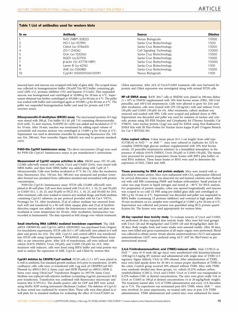
mucosal layer and mucosa was scrapped with help of glass slide. The scraped tissuewas collected in homogenization buffer (50 mM Tris-HCl buffer containing gly-cerol (20% v/v), protease inhibitor (1%) and heparin (3 U/ml)). This suspendedmucosa was homogenized and centrifuged at 10,000×g for 20 min at 4 °C. Super-natant obtained was further centrifuged at 105,000 × g for 60 min at 4 °C. The pelletwas washed with buffer and centrifuged again at 105,000 × g for 60 min at 4 °C. Thepellet was suspended homogenization buffer and used for protein and CYPenzymes assays.
Ethoxyresorufin-O-deethylase (EROD) assay. The microsomal proteins (0.5 mg)were mixed with 200 μL Tris buffer (0.1 M, pH 7.4) containing ethoxyresorufin(0.01 mM). To start reaction, NADPH (0.1 mM) was added and incubated at 37 °Cfor 10 min. After 10 min, reaction was terminated by adding equal volume ofacetonitrile and reaction mixture was centrifuged at 13,000 × g for 10 min at 4 °C.Supernatant was used to determine resorufin by measuring fluorescence (Ex. 530nm, Em. 580 nm). Pure resorufin (Sigma Aldrich) was used to generate standardcurve.
P450-Glo Cyp1A1 luminiscence assay. The above microsomes (20 μg) were usedfor P450-Glo Cyp1A1 luminiscence assays as per manufacturer’s instructions.
Measurement of Cyp1A1 enzyme activities in vitro. EROD assay: HT-29 cells(15,000 cells/well) treated with vehicle, UroA and UAS03 (24 h), were rinsed withHBSS buffer, and then fresh HBSS buffer was added along with 5 μM of 7-ethoxyresorufin. Cells were further incubated at 37 °C for 1 h. After the incubationtime, fluorescence (Exc. 530 nm, Em. 580 nm) was measured and product (resor-ufin) formed was calculated from calibration standard and normalized with proteinconcentration.
P450-Glo Cyp1A1 luminiscence assay: HT29 cells (25,000 cells/well) wereplated in 48 well plate. Cell were then treated with UroA (0.1, 1, 10, 25, and 50 µM)or UAS03 (0.1, 1, 10, 25, and 50 µM) or FICZ (0.1, 1, 10, 25, and 50 nM) for 24 h.After treatment, cells were washed to remove any residual drugs, and fresh mediumcontaining Cyp1A1 substrate (as per protocol provided with kit Cat.# V8751;Promega) for 3 h. After incubation, 25 µl of culture medium was removed fromeach well and transferred to a 96-well white opaque plate and 25 µl of luciferindetection reagent was added to initiate the luminescence reaction and plate wasincubated at room temperature for 20 min. After incubation, luminescence wasrecorded in luminometer. The data reported as fold change over vehicle treatment.
Small interfering RNA (siRNA) mediated knockdown experiment. The AhRsiRNA (SR300136) and Cyp1A1 siRNA (SR301093) was purchased from Origene.For knockdown experiments, HT29 cells (0.5 × 10⁶ cells/well) were plated in 6 wellplate and grown for 24 h. The AhR, Cyp1A1 and control-siRNA was transfectedinto HT29 cells using Lipofectamine ® RNAiMAX reagent (ThermoFisher Scien-tific) as per intruction given. After 24 h of transfections, cell were induced withvehicle (0.01% DMSO), UroA (50 µM), and UAS03 (50 µM) for 24 h. Aftertreatment with inducers, cells were lysed using RIPA buffer and total protein wasused to analyse the expression of AhR, Cyp1A1 and Cldn4 by western blot.
Cyp1A1 deletion by CRISPR/Cas9 method. HT29 cells (1.5 × 10⁵) were plated in6-well in antibiotic free standard growth medium 24 h prior to transfection. At 60%confluency cells, cells were co-transfected with 2 µg each of CRISPR/Cas9 KOPlasmid (sc-400511-KO-2; Santa cruz) and HDR Plasmid (sc-400511-HDR-2;Santa cruz) using UltraCruz® Transfection Reagent (sc-395739; Santa Cruz).Medium was replaced with selective medium (containing 4 µg/mL puromycin) 96 hpost transfection. Transfection was confirmed with fluorescence microscopy andwestern blot (CYP1A1). The double postive cells for GFP and RFP were sortedusing MoFlo XDP sorting instrument (Beckman Coulter). The deletion of Cyp1A1in these sorted was confirmed by western blots. These cells were then plated in 6-well plate for in standard medium for evaluating the effect of UroA/UAS03 on
Cldn4 expression. After 24 h of UroA/UAS03 treatment cells were harvested forprotein and Cldn4 expression was investigated along with normal HT29 cells.
NF-κB EMSA assay. RAW 264.7 cells or BMDM were plated in 100 mm dishes(1 × 10⁶) in DMEM supplemented with 10% fetal bovine serum (FBS), 100 U/mlpenicillin, and 100 U/ml streptomycin. Cells were allowed to grow for 24 h andafter incubation, cells were treated with LPS (50 ng/mL) with and without UroA(50 µM) and UAS03 (50 µM) for 6 h. After treatments, culture medium wasremoved and washed with PBS. Cells were scraped and pelleted down in PBS.Supernatant was discarded and pellet was used for isolation of nuclear and cyto-solic protein using NE-PER Nuclear and Cytoplasmic kit (Thermo Scientific; Cat#78833). Later nuclear protein (2 µg) was used for EMSA using Non-RadioactiveEMSA Kits with IR Fluo-Probes for Nuclear factor kappa B p65 (Viagene BiotechInc Cat # IRTF282 60).
Colon explant culture. Colon tissue pieces (0.5–1 cm length) from wild type(C57BL/6) or Nrf2−/− or AhR−/− mice were cultured in triplicates for 24 h incomplete DMEM-high glucose medium (supplemented with 10% fetal bovineserum, 1X penicillin-streptomycin solution) in a humidified atmosphere in thepresence of vehicle (0.01% DMSO), UroA (50 µM) or UAS03 (50 µM). The tissueswere processed for protein preparation (tissue lysates with RIPA plus buffer) ortotal RNA isolation. These tissue lysates or RNA were used to determine theexpression of Nrf2, Cldn4 and AhR.
Tissue processing for RNA and protein analysis. Mice were treated with asdescribed in results section. Mice were euthanized with CO2 asphyxiation followedby cervical dislocation. Colon was dissected out and luminal contents were flushedout with cold PBS (containing PMSF and Sodium orthvandate). Small portion ofcolon was snap frozen in liquid nitrogen and stored at −80 °C for RNA analysis.For preparation of protein samples, colon was opened longitudinally and mucosawas scraped in ice-cold 1X PBS using pre-chilled glass slide and centrifuged at300 × g for 10 min at 4 °C. Supernatant was discarded and pellet was suspended inRIPA buffer (containing 1X protease inhibitor) and vortexed at high speed. After30 min incubation on ice, samples were centrifuged at 13,000 × g for 20 min at 4 °C.Supernatant was collected and protein was quantified using BCA protein quanti-fication kit. The lysates were used appropriately for western blots.
28-day repeated dose toxicity study. To evaluate toxicity of UroA and UAS03,we performed 28-days repeated dose toxicity study. Mice were fed (oral gavage)with UroA (20 and 40 mg/kg/day) and UAS03 (20 and 40 mg/kg/day) daily for28 days. Body weight, food, and water intake were assessed weekly. After 28 days,mice were killed and gross examination of all major organs were performed. Bloodwas collected to obtain serum. Serum alanine aminotransferase (ALT) and asparateaminotransferase (AST) were analyzed using ALT/ AST kit (BioVision) as perinstructional manual.
2,4,6-Trinitrobenzenesulfonic acid (TNBS)-induced colitis. Male C57BL/6 orNrf2−/− mice (6–8 week old age mice) were anesthetized with ketamine/xylazine(100 mg/12.5 mg/kg IP) mixture and administered with single dose of TNBS (2.5mg/mice; Sigma Aldrich, USA) in 50% ethanol. After administration of TNBS,mice were held upside down for 30–60 s to ensure proper distribution of TNBS inthe colon. Control group received 50% ethanol without TNBS. Mice with TNBSwere randomly divided into three groups, viz. vehicle (0.25% sodium carbox-ymethylcellulose (CMC)), UroA and UAS03. UroA or UAS03 was resuspended in0.25% sodium-CMC at desired concentrations. The mice were given orally Veh orUroA or UAS03 in 100 μl at desired concentrations (4 or 20 mg/kg/body weight).The treatment started after 12 h of TNBS administration and every 12 h thereafterup to 72 h. The experiment was terminated post 60 h TNBS, where AhR−/− micewere involved. In some experiments, we treated only once at post 12 h TNBSadministration. TNBS administered and control mice were euthanized for tissue
Table 1 List of antibodies used for western blots
Sr no Antibody Source Dilution
1 Nrf2 (NBP1-32822) Novus Biologicals 1:10002 HO-1 (sc-10789) Santa Cruz Biotechnology 1:5003 Cldn4 (sc-376643) Santa Cruz Biotechnology 1:10004 ZO-1 (5406) Cell Signaling Technology 1:20005 Ocln (sc-133256) Santa Cruz Biotechnology 1:10006 NQO1 (sc32793) Santa Cruz Biotechnology 1:10007 β-actin (Sc-47778 HRP) Santa Cruz Biotechnology 1:50008 Lamin B (sc-6216) Santa Cruz Biotechnology 1:1009 AhR (sc-133088) Santa Cruz Biotechnology 1:100010 Cyp1A1 (H00001543-D01P) Novus Biologicals 1:100
NATURE COMMUNICATIONS | https://doi.org/10.1038/s41467-018-07859-7 ARTICLE
NATURE COMMUNICATIONS | (2019) 10:89 | https://doi.org/10.1038/s41467-018-07859-7 | www.nature.com/naturecommunications 15

and plasma collection after 80 h of TNBS/ethanol treatment. Mice were examinedfor colitis phenotype.
DSS-induced colitis. Acute experimental colitis in mice was induced by giving 3%(w/v) colitis grade DSS (MP Biomedicals) in drinking water for 7 days. Controlanimal received drinking water without DSS. All colitis group mice were randomlydivided into three groups viz. vehicle treated (0.25% Na-CMC), UroA (20 mg/kg/day) and UAS03 (20 mg/kg/day) on the 4th and 6th day of DSS treatment. After7 days, animals were put back on regular water for a period of 7 days. For chronicDSS colitis model, we used three cycles of 2.0% (w/v) DSS and each DSS cycleconsisted or 7 days followed by 10 days of regular water and mice were treated withUroA (20 mg/kg/day) on every 4th and 6th day of DSS cycle.
Assessment of colitis severity and tissue collection. Mice were evaluated dailyfor change in body weight, stool consistency, and rectal bleeding and score wasgiven and combined to obtained disease activity index67. After euthanasia, thecolon was removed and flushed with PBS containing (1 mM PMSF and 0.2 mMsodium orthovanadate). Colon length and colon weight were measured and smallparts of colon were excised for myeloperoxidase (MPO) activity and RNA isolation.Tissues for MPO and RNA extraction were snap frozen in liquid nitrogen andstored in −80 °C until further analysis. Tissue for histological examination wasstored in 10% phosphate buffered saline formalin. Blood was collected and serumwas separated by centrifugation at 3500 × g for 15 min. Serum cytokines (IL-6,TNF-α; Biolegend) and chemokines (CXCL1; R&D Systems) levels were measuredby ELISA according to manufacturer’s instructions.
In vivo intestinal permeability assay. The gut barrier function was evaluated byin vivo intestinal permeability using FITC-Dextran (MW 4000; FD4, Sigma-Aldrich, USA)68. Briefly, mice were orally administered with FITC-dextran (60 mg/100 gm body weight). Mice were fasted for 4 h prior to euthanization. The FITC-dextran concentration in serum was determined using the standard curve of FITC-dextran in serum (excitation, 485 nm; emission, 525 nm; BMG LABTECH).
Myeloperoxidase (MPO) activity. The MPO activity in the colons was deter-mined using the following procedure69. Briefly, colon tissue was homogenized in0.5% (w/v) hexadecyltrimethylammonium bromide (H6269; Sigma-aldrich, USA)in 50 mM PBS, pH 6.0. This homogenate underwent 3 freeze-thaw cycles and10–15 s sonication to obtain homogenous suspension. The supernatant from thissuspension was collected after centrifugation at 13000 × g for 20 min at 4 °C. Thesupernatant (10 µl) was then added to 50 mM potassium phosphate buffer (pH 6.0)containing 0.167 mg/ml o-dianisidine (Sigma-Aldrich, USA) and 0.0005% H2O2
(Sigma-Aldrich USA) and absorbance was taken at 450 nm (BMG, LABTECH) at2 min interval. Units of MPO in each sample was determined by considering thatone unit (U) of MPO= 1 µmol of H2O2 split with molar extinction coefficient of1.13 × 10−2 nm/min and MPO in each sample calculated by using [ΔA(t2− t1)]/Δmin × (1.13 × 10⁻²) formula and MPO units were normalized with per mg tissue.
Histopathology. Collected colon tissue were fixed in 10% buffered formaldehydesolution overnight and fixed tissue underwent standard histopathological proces-sing. Briefly, after fixation tissue underwent dehydration and cleaning with xylenebefore paraffin embedding. The paraffin section of 5 µm were cut (Leica micro-tome) and stained for H&E staining. The H&E images were captured using AperioScanscope. H&E sections were scored blindly using index scoring described byErben et al.70.
Data availabilityThe RNA-seq data described in this MS is deposited with GEO # GSE113581. Thehyperlink for RNA seq as follows: https://urldefense.proofpoint.com/v2/url?u=https-3A__www.ncbi.nlm.nih.gov_geo_query_acc.cgi-3Facc-3DGSE113581&d=DwIBAg&c=OAG1LQNACBDguGvBeNj18Swhr9TMTjS-x4O_KuapPgY&r=gYAe1Rux-xOAaWAT6YX2N_noYeYJBx7FLRBVFljZPt0&m=aQROwFtABYMDtrIp4k7mNRkL77nfCBUH4EqXd2nQLK0&s=RH4q-x-nZSE8JU035mWKJs4mmznYY-E3Nvx9CkS_QKs&e=. The full raw data that supportthe findings of this study are available from the corresponding author upon reasonablerequest. The Source Data underlying all the bar graphs in the Figures are provided inthe Source Data File.
Received: 9 January 2018 Accepted: 28 November 2018
References1. Lynch, S. V. & Pedersen, O. The human intestinal microbiome in health and
disease. N. Engl. J. Med. 375, 2369–2379 (2016).
2. Levy, M., Kolodziejczyk, A. A., Thaiss, C. A. & Elinav, E. Dysbiosis and theimmune system. Nat. Rev. Immunol. 17, 219–232 (2017).
3. Sonnenburg, J. L. & Backhed, F. Diet-microbiota interactions as moderators ofhuman metabolism. Nature 535, 56–64 (2016).
4. Belkaid, Y. & Hand, T. W. Role of the microbiota in immunity andinflammation. Cell 157, 121–141 (2014).
5. Dzutsev, A. et al. Microbes and cancer. Annu. Rev. Immunol. 35, 199–228(2017).
6. Tamboli, C. P., Neut, C., Desreumaux, P. & Colombel, J. F. Dysbiosis ininflammatory bowel disease. Gut 53, 1–4 (2004).
7. Gopalakrishnan, V., Helmink, B. A., Spencer, C. N., Reuben, A. & Wargo, J. A.The Influence of the gut microbiome on cancer, immunity, and cancerimmunotherapy. Cancer Cell 33, 570–580 (2018).
8. Landy, J. et al. Tight junctions in inflammatory bowel diseases andinflammatory bowel disease associated colorectal cancer. World J.Gastroenterol. 22, 3117–3126 (2016).
9. Schulzke, J. D. et al. Epithelial tight junctions in intestinal inflammation. Ann.N. Y. Acad. Sci. 1165, 294–300 (2009).
10. Vindigni, S. M., Zisman, T. L., Suskind, D. L. & Damman, C. J. The intestinalmicrobiome, barrier function, and immune system in inflammatory boweldisease: a tripartite pathophysiological circuit with implications for newtherapeutic directions. Ther. Adv. Gastroenterol. 9, 606–625 (2016).
11. Heber, D. Multitargeted therapy of cancer by ellagitannins. Cancer Lett. 269,262–268 (2008).
12. Sak, K. Cytotoxicity of dietary flavonoids on different human cancer types.Pharmacogn. Rev. 8, 122–146 (2014).
13. Habauzit, V. & Morand, C. Evidence for a protective effect of polyphenols-containing foods on cardiovascular health: an update for clinicians. Ther. Adv.Chronic Dis. 3, 87–106 (2012).
14. Gbinigie, O. A., Onakpoya, I. J. & Spencer, E. A. Evidence for the effectivenessof pomegranate supplementation for blood pressure management is weak: asystematic review of randomized clinical trials. Nutr. Res. 46, 38–48(2017).
15. Stockton, A., Farhat, G., McDougall, G. J. & Al-Dujaili, E. A. S. Effect ofpomegranate extract on blood pressure and anthropometry in adults: adouble-blind placebo-controlled randomised clinical trial. J. Nutr. Sci. 6, e39(2017).
16. Razani, Z., Dastani, M. & Kazerani, H. R. Cardioprotective effects ofpomegranate (Punica granatum) juice in patients with ischemic heart disease.Phytother. Res. 31, 1731–1738 (2017).
17. Derosa, G. & Maffioli, P., . & Sahebkar, A. Ellagic acid and its role in chronicdiseases. Adv. Exp. Med. Biol. 928, 473–479 (2016).
18. Cerda, B., Periago, P., Espin, J. C. & Tomas-Barberan, F. A. Identification ofurolithin a as a metabolite produced by human colon microflora from ellagicacid and related compounds. J. Agric. Food Chem. 53, 5571–5576 (2005).
19. Selma, M. V., Beltran, D., Garcia-Villalba, R., Espin, J. C. & Tomas-Barberan,F. A. Description of urolithin production capacity from ellagic acid oftwo human intestinal Gordonibacter species. Food Funct. 5, 1779–1784(2014).
20. Espin, J. C., Larrosa, M., Garcia-Conesa, M. T. & Tomas-Barberan, F.Biological significance of urolithins, the gut microbial ellagic acid-derivedmetabolites: the evidence so far. Evid.-Based Complement. Alternat. Med.2013, 270418 (2013).
21. Tomas-Barberan, F. A. et al. Urolithins, the rescue of ‘old’ metabolites tounderstand a ‘new’ concept: metabotypes as a nexus between phenolicmetabolism, microbiota dysbiosis and host health status. Mol. Nutr. Food Res.61, 1–35 (2016).
22. Ryu, D. et al. Urolithin A induces mitophagy and prolongs lifespan in C.elegans and increases muscle function in rodents. Nat. Med. 22, 879–888(2016).
23. Saha, P. et al. Gut Microbiota conversion of dietary ellagic acid into bioactivephytoceutical urolithin a inhibits heme peroxidases. PLoS One 11, e0156811(2016).
24. Tsukita, S. & Furuse, M. Claudin-based barrier in simple and stratified cellularsheets. Curr. Opin. Cell Biol. 14, 531–536 (2002).
25. Loboda, A., Damulewicz, M., Pyza, E., Jozkowicz, A. & Dulak, J. Role of Nrf2/HO-1 system in development, oxidative stress response and diseases:an evolutionarily conserved mechanism. Cell. Mol. Life Sci. 73, 3221–3247(2016).
26. Beischlag, T. V., Luis Morales, J., Hollingshead, B. D. & Perdew, G. H. The arylhydrocarbon receptor complex and the control of gene expression. Crit. Rev.Eukaryot. Gene Expr. 18, 207–250 (2008).
27. Yeager, R. L., Reisman, S. A., Aleksunes, L. M. & Klaassen, C. D. Introducingthe “TCDD-inducible AhR-Nrf2 gene battery”. Toxicol. Sci. 111, 238–246(2009).
28. Miao, W., Hu, L., Scrivens, P. J. & Batist, G. Transcriptional regulation of NF-E2 p45-related factor (NRF2) expression by the aryl hydrocarbon receptor-xenobiotic response element signaling pathway: direct cross-talk between
ARTICLE NATURE COMMUNICATIONS | https://doi.org/10.1038/s41467-018-07859-7
16 NATURE COMMUNICATIONS | (2019) 10:89 | https://doi.org/10.1038/s41467-018-07859-7 | www.nature.com/naturecommunications

phase I and II drug-metabolizing enzymes. J. Biol. Chem. 280, 20340–20348(2005).
29. Antoniou, E. et al. The TNBS-induced colitis animal model: an overview. Ann.Med. Surg. 11, 9–15 (2016).
30. Smith, P. D. et al. Intestinal macrophages and response to microbialencroachment. Mucosal Immunol. 4, 31–42 (2011).
31. Bain, C. C. & Mowat, A. M. Macrophages in intestinal homeostasis andinflammation. Immunol. Rev. 260, 102–117 (2014).
32. Gaya, P., Peiroténa, A., Medinaa, M., Álvarezb, I. & Landete, J. M.Bifidobacterium pseudocatenulatum INIA P815: The first bacterium abl e toproduce urolithins A and B from ellagic acid. J. Funct. Foods 45, 95–99 (2018).
33. Larrosa, M. et al. Anti-inflammatory properties of a pomegranate extract andits metabolite urolithin-A in a colitis rat model and the effect of coloninflammation on phenolic metabolism. J. Nutr. Biochem. 21, 717–725 (2010).
34. Forster, C. Tight junctions and the modulation of barrier function in disease.Histochem. Cell Biol. 130, 55–70 (2008).
35. Arrieta, M. C., Bistritz, L. & Meddings, J. B. Alterations in intestinalpermeability. Gut 55, 1512–1520 (2006).
36. Konig, J. et al. Human intestinal barrier function in health and disease. Clin.Transl. Gastroenterol. 7, e196 (2016).
37. Capaldo, C. T. & Nusrat, A. Cytokine regulation of tight junctions. Biochim.Biophys. Acta 1788, 864–871 (2009).
38. Esser, C. & Rannug, A. The aryl hydrocarbon receptor in barrier organphysiology, immunology, and toxicology. Pharmacol. Rev. 67, 259–279(2015).
39. Stockinger, B., Di Meglio, P., Gialitakis, M. & Duarte, J. H. The arylhydrocarbon receptor: multitasking in the immune system. Annu. Rev.Immunol. 32, 403–432 (2014).
40. Furumatsu, K. et al. A role of the aryl hydrocarbon receptor in attenuation ofcolitis. Dig. Dis. Sci. 56, 2532–2544 (2011).
41. Goettel, J. A. et al. AHR activation is protective against colitis driven by T cellsin humanized mice. Cell Rep. 17, 1318–1329 (2016).
42. Schiering, C. et al. Feedback control of AHR signalling regulates intestinalimmunity. Nature 542, 242–245 (2017).
43. Gimenez-Bastida, J. A. et al. Ellagitannin metabolites, urolithin A glucuronideand its aglycone urolithin A, ameliorate TNF-alpha-induced inflammationand associated molecular markers in human aortic endothelial cells. Mol.Nutr. Food Res. 56, 784–796 (2012).
44. Monteleone, I. et al. Aryl hydrocarbon receptor-induced signals up-regulateIL-22 production and inhibit inflammation in the gastrointestinal tract.Gastroenterology 141, 237–248 (2011). 248 e231.
45. Shin, S. et al. NRF2 modulates aryl hydrocarbon receptor signaling: influenceon adipogenesis. Mol. Cell. Biol. 27, 7188–7197 (2007).
46. Hayes, J. D., Dinkova-Kostova, A. T. & McMahon, M. Cross-talk betweentranscription factors AhR and Nrf2: lessons for cancer chemoprevention fromdioxin. Toxicol. Sci. 111, 199–201 (2009).
47. Mitsuishi, Y., Motohashi, H. & Yamamoto, M. The Keap1-Nrf2 system incancers: stress response and anabolic metabolism. Front. Oncol. 2, 200(2012).
48. Al-Sawaf, O. et al. Nrf2 in health and disease: current and future clinicalimplications. Clin. Sci. 129, 989–999 (2015).
49. Khor, T. O. et al. Nrf2-deficient mice have an increased susceptibility todextran sulfate sodium-induced colitis. Cancer Res. 66, 11580–11584(2006).
50. Kobayashi, E. H. et al. Nrf2 suppresses macrophage inflammatory response byblocking proinflammatory cytokine transcription. Nat. Commun. 7, 11624(2016).
51. Wardyn, J. D., Ponsford, A. H. & Sanderson, C. M. Dissecting molecularcross-talk between Nrf2 and NF-kappaB response pathways. Biochem. Soc.Trans. 43, 621–626 (2015).
52. Sekine, H. et al. Hypersensitivity of aryl hydrocarbon receptor-deficient miceto lipopolysaccharide-induced septic shock. Mol. Cell. Biol. 29, 6391–6400(2009).
53. Benson, J. M. & Shepherd, D. M. Aryl hydrocarbon receptor activation byTCDD reduces inflammation associated with Crohn’s disease. Toxicol. Sci.120, 68–78 (2011).
54. Fukumoto, S. et al. Identification of a probiotic bacteria-derived activator ofthe aryl hydrocarbon receptor that inhibits colitis. Immunol. Cell Biol. 92,460–465 (2014).
55. Metidji, A. et al. The environmental sensor AHR protects from inflammatorydamage by maintaining intestinal stem cell homeostasis and barrier integrity.Immunity 49, 353–362 e355 (2018).
56. Novak, E. A. & Mollen, K. P. Mitochondrial dysfunction in inflammatorybowel disease. Front. Cell Dev. Biol. 3, 62 (2015).
57. Delpre, G., Avidor, I., Steinherz, R., Kadish, U. & Ben-Bassat, M.Ultrastructural abnormalities in endoscopically and histologically normal andinvolved colon in ulcerative colitis. Am. J. Gastroenterol. 84, 1038–1046(1989).
58. Rodenburg, W. et al. Impaired barrier function by dietary fructo-oligosaccharides (FOS) in rats is accompanied by increased colonicmitochondrial gene expression. BMC Genomics 9, 144 (2008).
59. Kurowska-Stolarska, M. et al. IL-33 amplifies the polarization of alternativelyactivated macrophages that contribute to airway inflammation. J. Immunol.183, 6469–6477 (2009).
60. Kowapradit, J. et al. In vitro permeability enhancement in intestinal epithelialcells (Caco-2) monolayer of water soluble quaternary ammonium chitosanderivatives. AAPS PharmSciTech 11, 497–508 (2010).
61. Andrews S. FastQC: A Quality Control Tool for High Throughput SequenceData. (2014).
62. Kim, D. et al. TopHat2: accurate alignment of transcriptomes in the presenceof insertions, deletions and gene fusions. Genome Biol. 14, R36 (2013).
63. Flicek, P. et al. Ensembl 2014. Nucleic Acids Res. 42, D749–D755 (2014).64. Trapnell, C. et al. Differential analysis of gene regulation at transcript
resolution with RNA-seq. Nat. Biotechnol. 31, 46–53 (2013).65. Trapnell, C. et al. Differential gene and transcript expression analysis of RNA-
seq experiments with TopHat and Cufflinks. Nat. Protoc. 7, 562–578 (2012).66. Singh, R. et al. Evaluation of memory enhancing clinically available
standardized extract of Bacopa monniera on P-glycoprotein and cytochromeP450 3A in Sprague-Dawley rats. PLoS ONE 8, e72517 (2013).
67. Murthy, S. N. et al. Treatment of dextran sulfate sodium-induced murinecolitis by intracolonic cyclosporin. Dig. Dis. Sci. 38, 1722–1734 (1993).
68. Furuta, G. T. et al. Hypoxia-inducible factor 1-dependent induction ofintestinal trefoil factor protects barrier function during hypoxia. J. Exp. Med.193, 1027–1034 (2001).
69. Kim J. J., Shajib M. S., Manocha M. M., Khan W. I. Investigating intestinalinflammation in DSS-induced model of IBD. J. Vis. Exp. 60, 3678 (2012).
70. Erben, U. et al. A guide to histomorphological evaluation of intestinalinflammation in mouse models. Int. J. Clin. Exp. Pathol. 7, 4557–4576 (2014).
AcknowledgementsThis work is financially supported by NIH/NCI (R21CA216090), NIH/NIGMS CoBREgrant (P20GM125504-01) and pilot grants from Microbiology and Immunology (U of L),Rounsavall Foundation and The Jewish Heritage Fund for Excellence ResearchEnhancement Grant and JGBCC to V.R.J.; V.R.J. and R.S. are partially supported byfunds from NIH/NCI (CA191683). Part of RNA-Seq experiment was performed withassistance of the U of L Genomics Facility, which is supported by NIH P20GM103436(KY IDeA Networks of Biomedical Research Excellence), NIH P30GM106396 (UofL J. G.Brown Cancer Center Phase III CoBRE), the J. G. Brown Foundation, and user fees. Wethank JGBCC imaging core facilites at U of L. This work is financially supported byDepartment of Biotechnology (DBT) (BT/PR12490/AAQ/3/716/2015) to P.K.V., andcore-funds from the Institute for Stem Cell Biology and Regenerative Medicine (inStem).P.K.V. is supported by Ramalingaswami ReEntry Fellowship (DBT), India. S.C. is sup-ported by Department of Science and Technology (DST) under the Scheme for YoungScientists and Technologies program (SP/YO/078/2017). A.A.H. is supported by SeniorResearch Fellowship by Council of Scientific & Industrial Research (CSIR). We thankanimal house facility/members and Central Imaging & Flow Cytometry, NMR facilities atinStem and NCBS.
Author contributionsV.R.J. and P.K.V. conceived, designed the experiments, and analyzed the data. R.S., S.C.,S.R.B., B.B., B.H., N.G.K., A.A.H., T.S., and P.P. designed, performed the experiments,and analyzed the data. M.V.-K., M.G.I.L., G.M.D., G.W.D., H.-G.Z., and B.H. contributedthrough invaluable discussions. X.C., E.C.R., and S.J.W. assisted in analyzing RNA-Seqdata. A.H. scored the inflammation of colons. R.S., B.H., P.K.V., and V.R.J. wrote thepaper, all authors discussed the results. P.K.V. and V.R.J. supervised the project.
Additional informationSupplementary Information accompanies this paper at https://doi.org/10.1038/s41467-018-07859-7.
Competing interests: V.R.J., P.K.V., H.B., R.S., S.C., and A.A.H. hold a patentapplication related to this technology (US patent application number: 62/671,737).P.K.V.’s interests were reviewed and are managed by the Institute for Stem Cell Biologyand Regenerative Medicine (inStem) in accordance with their Conflict of Interestpolicies. The remaining authors declare no competing interests.
Reprints and permission information is available online at http://npg.nature.com/reprintsandpermissions/
Journal peer review information: Nature Communications thanks the anonymousreviewers for their contributions to the peer review of this work.
Publisher’s note: Springer Nature remains neutral with regard to jurisdictional claims inpublished maps and institutional affiliations.
NATURE COMMUNICATIONS | https://doi.org/10.1038/s41467-018-07859-7 ARTICLE
NATURE COMMUNICATIONS | (2019) 10:89 | https://doi.org/10.1038/s41467-018-07859-7 | www.nature.com/naturecommunications 17

Open Access This article is licensed under a Creative CommonsAttribution 4.0 International License, which permits use, sharing,
adaptation, distribution and reproduction in any medium or format, as long as you giveappropriate credit to the original author(s) and the source, provide a link to the CreativeCommons license, and indicate if changes were made. The images or other third partymaterial in this article are included in the article’s Creative Commons license, unlessindicated otherwise in a credit line to the material. If material is not included in thearticle’s Creative Commons license and your intended use is not permitted by statutoryregulation or exceeds the permitted use, you will need to obtain permission directly fromthe copyright holder. To view a copy of this license, visit http://creativecommons.org/licenses/by/4.0/.
© The Author(s) 2019
ARTICLE NATURE COMMUNICATIONS | https://doi.org/10.1038/s41467-018-07859-7
18 NATURE COMMUNICATIONS | (2019) 10:89 | https://doi.org/10.1038/s41467-018-07859-7 | www.nature.com/naturecommunications