energy landscape of adenylate kinase: phosphoryl transfer and conformational transitions
TRANSCRIPT
Sunday, March 6, 2011 17a
[1] Hyun Ho Park, Emmanuelle Logette, Stefan Raunser, Solange Cuenin,Thomas Walz, Jurg Tschopp and Hao Wu (2007). Death domain assemblymechanism revealed by crystal structure of the oligomeric PIDDosome corecomplex. Cell 128: 533-546.[2] LiweiWang, Jin Kuk Yang, Venkataraman Kabaleeswaran, Amanda J Rice,Anthony C Cruz, Ah Young Park, Qian Yin, Ermelinda Damko, Se Bok Jang,Stefan Raunser, Carol V Robinson, Richard M Siegel, Thomas Walz & HaoWu (2010). The Fas-FADD death domain complex structure reveals the basisof DISC assembly and disease mutations’’. Nature Struct. Mol. Biol. in press.[3] Su-Chang Lin, Yu-Chih Lo and Hao Wu (2010). Helical assembly in theMyD88-IRAK4-IRAK2complex inTLR/IL-1R signaling.Nature, 465: 885–90.
89-MiniSympBcl-2 Family Proteins Regulate the Ability of Ceramide Channelsto Permeabilize the Mitochondrial Outer Membrane to ProteinsMarco Colombini1, Meenu N. Perera1, Vidyaramanan Ganesan1,Leah J. Siskind2.1University of Maryland, College Park, MD, USA, 2Medical University ofSouth Carolina, Charleston, SC, USA.The sphingolipid, ceramide, forms channels capable of translocating proteinsthrough membranes. These channels can form in the mitochondrial outer mem-brane at ceramide levels found to be present in that membrane early in apopto-sis. For these channels to be good candidates for the protein release pathwaythat is a key, decision-making step in apoptosis, they need to be controlledby Bcl-2 family proteins. Full length Bcl-xL favors ceramide channel disassem-bly and activated, full length Bax favors the formation of large channels. Theseact at the low nanomolar level. Thus their mode of action on ceramide channelsis consistent with their role in controlling the release of proteins from mito-chondria. These proteins interact directly with the ceramide channels and areable to influence the size and stability of the channels whether the channelsare formed on mitochondrial membranes or phospholipid membranes. Thebinding of Bcl-xL to the ceramide channel seems to form a 1:1 complex, theinfluence of the interaction propagating throughout the structure in an allostericmanner. The influence of Bax seems to involve multiple interactions, favoringa larger channel through an induced-fit mechanism. Alterations in either theprotein structure or the ceramide structure can alter the interaction, providingclues to the site of interaction. Supported by a grant from NSF (MCB-0641208).
90-MiniSympStructural Insights into Cytomegalovirus Resistance to ApoptosisNico Tjandra1, Junhe Ma1, Frank Edlich2, Richard Youle2.1NHLBI, NIH, Bethesda, MD, USA, 2NINDS, NIH, Bethesda, MD, USA.A host defense mechanism against viral invasion is elimination of the infectedcells. This process depends on the finely tuned regulation of programmed celldeath or apoptosis. Some viruses, however through evolution have been able tocounteract this host defense by encoding in its genes proteins that can block ap-optosis. The cytomegalovirus (CMV) genome encodes a protein, vMIA that in-hibits apoptosis through the mitochondria pathway. The Bcl-2 family ofproteins that can be divided into at least two subfamilies controls this apoptosispathway. The subfamily including Bcl-2 and Bcl-xL inhibits apoptosis whereasthe Bax subfamily consisting of Bax and Bak promote apoptosis. Bax is a cyto-solic protein that translocates onto the mitochondria upon apoptosis induction.This is a crucial initiation event for the apoptosis process. The vMIA protein ofCMV interacts specifically with Bax, recruits it onto the mitochondria mem-brane and inhibits its apoptotic function. We investigated the interaction be-tween a peptide from vMIA, which retains most of its affinity, and Bax usingsolution nuclear magnetic resonance (NMR). Due to the nature of the proteininteractions non-conventional NMR approach was employed to study the pep-tide-protein complex. The structure that we determined revealed a unique bind-ing site on Bax that leads to its inhibition. This was confirmed by pointmutations. Our findings provide the molecular mechanism for apoptosis inhibi-tion by the vMIA protein of cytomegalovirus.
91-MiniSympThe Novel Fe-S Protein Miner1- Regulator of Crosstalk BetweenApoptosis and Autophagy?Patricia A. Jennings.UC San Diego, San Diego, CA, USA.The mitochondrial protein MitoNEET is an important new target in diabetestherapy. The endoplasmic reticulum proteinMiner1, a member of the newly dis-covered NEET family of iron-sulfur proteins, is essential for health and longev-ity.Mis-splicing of theMiner1 gene leading to deletion of the iron-sulfur domaincauses Wolfram Syndrome 2 (WFS2), an inherited juvenile-onset fatal disease.Interestingly, Miner1 was recently identified as a novel regulator of crosstalkbetween apoptosis and autophagy response in cells via specific protein-protein
interactions. We are exploring the how environmental stress and tuning of theredox-potentials of the NEET proteins regulate their interactions with pro- andanti-apoptotic factors. The structural biochemical and biophysical results arediscussed in relation to possible roles of MitoNEET and Miner1 in cellular Fe-S management and redox regulation of apoptotic/autophagic response.
PLATFORM F: Protein Dynamics - Experiment& Simulation
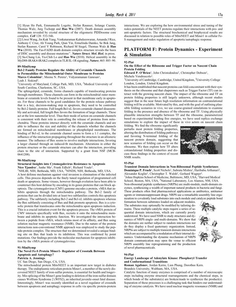
92-PlatOn the Effect of the Ribosome and Trigger Factor on Nascent ChainProtein FoldingEdward P. O’Brien1, John Christodoulou2, Christopher Dobson1,Michele Vendruscolo1.1University of Cambridge, Cambridge, United Kingdom, 2University CollegeLondon, London, United Kingdom.It has been established that nascent proteins can fold concomitant with their syn-thesis on the ribosome and that chaperones such as Trigger Factor (TF) can in-teract with the growing nascent chain. The impact of the ribosome and TF onprotein folding properties is still largely unknown, although recent studiessuggest that in the near future high resolution information on contranslationalfolding will be available. Motivated by this, and with the goal of outlining plau-sible folding scenarios in vivo, we use coarse-grained simulations to examinefolding in the presence and absence of the ribosome and TF. Using physicallyplausible interaction strengths between TF and the ribosome, parameterizedbased on experimental binding free energies, we have used replica exchangesimulations to explore the impact of these in vivo actors on nascent chain
folding. We find the ribosome significantlyperturbs most protein folding properties,altering the distribution of folding pathwaysand favoring N-terminal folding of thenascent protein domain, suggesting thatnew scenarios of folding can occur on theribosome. We then explore how TF alterscotranslational folding properties and dis-cuss these findings in the context of recent NMR results.93-PlatTransient Domain Interactions in Non-Ribosomal Peptide SynthetasesDominique P. Frueh1, Scott Nichols1, Subrata Mishra1, Haribabu Arthanari2,Alexander Koglin3, Christopher T. Walsh2, Gerhard Wagner2.1Johns Hopkins School of Medicine, Baltimore, MD, USA, 2Harvard MedicalSchool, Boston, MA, USA, 3National Laboratory, Los Alamos, NM, USA.Non-ribosomal peptide synthetases (NRPSs) are multi-module, multi-domain en-zymes, synthesizing a wealth of important natural products in bacteria and fungi.These products often find pharmaceutical applications as antibiotics, antitumoragents or immunosuppressant drugs. NRPS use a remarkable assembly line orga-nization to covalently load substrates into each module and catalyze peptide bond
formation between substrates loaded on adjacent modules.The substrates may optionally be modified by tailoring do-mains. These multiple catalytic steps require a series of se-quential domain interactions, which are currently poorlyunderstood.We have usedNMR to study structures and dy-namics of NRPS single- and multi-domains. We show thatthe domains are neither subject to random interactions nordo they display a fixed quaternary organization. Instead,NRPSs are subject tomultiple transient domain interactionswhich are accompanied by amodulation of their internal dy-namics. Understanding the dynamic mechanism of NRPSdomain communication may open the venue to efficientNRPS assembly line reprogramming and the production of novel pharmaceuticals.94-PlatEnergy Landscape of Adenylate Kinase: Phosphoryl Transferand Conformational TransitionsRoman Agafonov, Jordan Kerns, Lien Phung, Dorothee Kern.Brandeis University, Waltham, MA, USA.Catalytic function of many enzymes is comprised of a number of microscopicsteps including enzyme structural rearrangements and the chemical steps, inwhich bonds of the substrate(s) are broken/made to synthesize the product(s).Separation of these processes is a challenging task that hinders our understand-ing of enzyme catalysis. We have used nuclear magnetic resonance (NMR) and
18a Sunday, March 6, 2011
rapid-quenching techniques to independently study these processes in adeny-late kinase (ADK) and to characterize their energetic contribution at atomic res-olution. Adenylate kinase is an important enzyme that catalyzes a reversiblereaction: 2ADP4ATPþAMP, and is involved in maintaining energy homeo-stasis in a cell. Two binding sites of ADK and three different ligands result ina variety of biochemical states (determined by the bound ligands). Each ofthese states has different dynamic properties making the overall structural dy-namics of ADK complex. We have tackled this problem by engineering a loss-of-function ADKmutant (with greatly reduced rate of turnover), which allowedus to ‘‘trap’’ the enzyme in well-defined biochemical states. Our results lead toa detailed energy profile of ADK, providing insights into the molecular mech-anism of its functioning. This work reveals fundamental principles of enzymecatalysis and highlights the role of protein’s intrinsic dynamics.
95-PlatQuadrupolar-Order Deuterium NMR Relaxation ProvidesNew Light on Dynamics of Retinal in RhodopsinAndrey V. Struts1, Gilmar F.J. Salgado2, Michael F. Brown1.1Department of Chemistry, University of Arizona, Tucson, AZ, USA,2Departement de Chimie, Ecole Normale Superieure, Paris, France.Deuterium NMR relaxation of quadrupolar order (R1Q) was applied to studydynamics and ligand-protein interactions underlying rhodopsin activation. Rho-dopsin was regenerated with retinal 2H-labeled at the C5-, C9-, and C13-methylgroups and recombined with POPC bilayers [1-3]. The 2H NMR relaxationrates were measured in the dark state of rhodopsin in the temperature rangefrom �30 to �150 �C. In our previous studies, we measured relaxation ratesof Zeeman order (R1Z) in the dark, Meta I, and Meta II states of rhodopsin.The dynamical parameters involve the spectral densities of motion, whichdepend on correlation times and can be expressed in terms of a pre-exponentialfactor (rotational diffusion constant,D0, or jump rate, k0, for mobility in absenceof a barrier) and the corresponding barrier height (activation energy Ea). Thevalues of D0, k0, and Eadescribe local packing of retinal within the bindingpocket of rhodopsin and the changes occurring in the activation process. TheR1Z relaxation rates enable determination of dynamical parameters, but theydo not establish the anisotropy of methyl rotation. Simultaneous fitting of tem-perature dependences of the R1Z and R1Q relaxation rates indicates the off-axialrotation of the methyl groups is at least an order of magnitude slower than theaxial motion. The new R1Q data confirm previous conclusions on methyl groupdynamics and their interactions with the binding pocket [1]. Taken together, theR1Z and R1Q data allow the motional spectral densities to be individually deter-mined, and afford a new way of investigating the dynamics of ligand-protein in-teractions in membranes. [1] A.V. Struts et al. (2010) Nature Struct. Mol. Biol.(in press). [2] G.F.J. Salgado et al. (2006) JACS 128, 11067. [3] A.V. Struts et al.(2007) J. Mol. Biol. 372, 50.
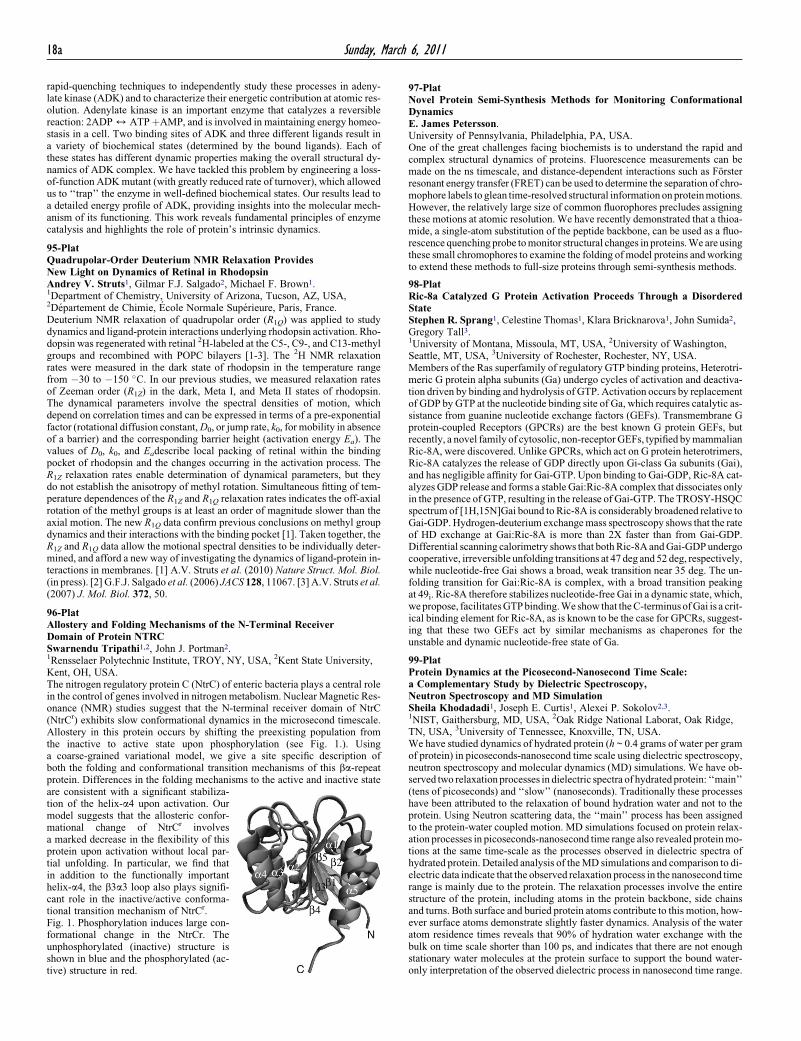
96-PlatAllostery and Folding Mechanisms of the N-Terminal ReceiverDomain of Protein NTRCSwarnendu Tripathi1,2, John J. Portman2.1Rensselaer Polytechnic Institute, TROY, NY, USA, 2Kent State University,Kent, OH, USA.The nitrogen regulatory protein C (NtrC) of enteric bacteria plays a central rolein the control of genes involved in nitrogen metabolism. Nuclear Magnetic Res-onance (NMR) studies suggest that the N-terminal receiver domain of NtrC(NtrCr) exhibits slow conformational dynamics in the microsecond timescale.Allostery in this protein occurs by shifting the preexisting population fromthe inactive to active state upon phosphorylation (see Fig. 1.). Usinga coarse-grained variational model, we give a site specific description ofboth the folding and conformational transition mechanisms of this ba-repeatprotein. Differences in the folding mechanisms to the active and inactive state
are consistent with a significant stabiliza-tion of the helix-a4 upon activation. Ourmodel suggests that the allosteric confor-mational change of NtrCr involvesa marked decrease in the flexibility of thisprotein upon activation without local par-tial unfolding. In particular, we find thatin addition to the functionally importanthelix-a4, the b3a3 loop also plays signifi-cant role in the inactive/active conforma-tional transition mechanism of NtrCr.Fig. 1. Phosphorylation induces large con-formational change in the NtrCr. Theunphosphorylated (inactive) structure isshown in blue and the phosphorylated (ac-tive) structure in red.97-PlatNovel Protein Semi-Synthesis Methods for Monitoring ConformationalDynamicsE. James Petersson.University of Pennsylvania, Philadelphia, PA, USA.One of the great challenges facing biochemists is to understand the rapid andcomplex structural dynamics of proteins. Fluorescence measurements can bemade on the ns timescale, and distance-dependent interactions such as Forsterresonant energy transfer (FRET) can be used to determine the separation of chro-mophore labels to glean time-resolved structural information on proteinmotions.However, the relatively large size of common fluorophores precludes assigningthese motions at atomic resolution. We have recently demonstrated that a thioa-mide, a single-atom substitution of the peptide backbone, can be used as a fluo-rescence quenching probe tomonitor structural changes in proteins.We are usingthese small chromophores to examine the folding of model proteins andworkingto extend these methods to full-size proteins through semi-synthesis methods.
98-PlatRic-8a Catalyzed G Protein Activation Proceeds Through a DisorderedStateStephen R. Sprang1, Celestine Thomas1, Klara Bricknarova1, John Sumida2,Gregory Tall3.1University of Montana, Missoula, MT, USA, 2University of Washington,Seattle, MT, USA, 3University of Rochester, Rochester, NY, USA.Members of the Ras superfamily of regulatory GTP binding proteins, Heterotri-meric G protein alpha subunits (Ga) undergo cycles of activation and deactiva-tion driven by binding and hydrolysis of GTP. Activation occurs by replacementof GDP by GTP at the nucleotide binding site of Ga, which requires catalytic as-sistance from guanine nucleotide exchange factors (GEFs). Transmembrane Gprotein-coupled Receptors (GPCRs) are the best known G protein GEFs, butrecently, a novel family of cytosolic, non-receptorGEFs, typified bymammalianRic-8A, were discovered. Unlike GPCRs, which act on G protein heterotrimers,Ric-8A catalyzes the release of GDP directly upon Gi-class Ga subunits (Gai),and has negligible affinity for Gai-GTP. Upon binding to Gai-GDP, Ric-8A cat-alyzes GDP release and forms a stable Gai:Ric-8A complex that dissociates onlyin the presence of GTP, resulting in the release of Gai-GTP. The TROSY-HSQCspectrum of [1H,15N]Gai bound to Ric-8A is considerably broadened relative toGai-GDP.Hydrogen-deuterium exchangemass spectroscopy shows that the rateof HD exchange at Gai:Ric-8A is more than 2X faster than from Gai-GDP.Differential scanning calorimetry shows that bothRic-8A andGai-GDPundergocooperative, irreversible unfolding transitions at 47 deg and 52 deg, respectively,while nucleotide-free Gai shows a broad, weak transition near 35 deg. The un-folding transition for Gai:Ric-8A is complex, with a broad transition peakingat 49i. Ric-8A therefore stabilizes nucleotide-free Gai in a dynamic state, which,wepropose, facilitatesGTPbinding.We show that theC-terminusofGai is a crit-ical binding element for Ric-8A, as is known to be the case for GPCRs, suggest-ing that these two GEFs act by similar mechanisms as chaperones for theunstable and dynamic nucleotide-free state of Ga.
99-PlatProtein Dynamics at the Picosecond-Nanosecond Time Scale:a Complementary Study by Dielectric Spectroscopy,Neutron Spectroscopy and MD SimulationSheila Khodadadi1, Joseph E. Curtis1, Alexei P. Sokolov2,3.1NIST, Gaithersburg, MD, USA, 2Oak Ridge National Laborat, Oak Ridge,TN, USA, 3University of Tennessee, Knoxville, TN, USA.We have studied dynamics of hydrated protein (h ~ 0.4 grams of water per gramof protein) in picoseconds-nanosecond time scale using dielectric spectroscopy,neutron spectroscopy and molecular dynamics (MD) simulations. We have ob-served two relaxation processes in dielectric spectra of hydrated protein: ‘‘main’’(tens of picoseconds) and ‘‘slow’’ (nanoseconds). Traditionally these processeshave been attributed to the relaxation of bound hydration water and not to theprotein. Using Neutron scattering data, the ‘‘main’’ process has been assignedto the protein-water coupled motion. MD simulations focused on protein relax-ation processes in picoseconds-nanosecond time range also revealed proteinmo-tions at the same time-scale as the processes observed in dielectric spectra ofhydrated protein. Detailed analysis of theMD simulations and comparison to di-electric data indicate that the observed relaxation process in the nanosecond timerange is mainly due to the protein. The relaxation processes involve the entirestructure of the protein, including atoms in the protein backbone, side chainsand turns. Both surface and buried protein atoms contribute to this motion, how-ever surface atoms demonstrate slightly faster dynamics. Analysis of the wateratom residence times reveals that 90% of hydration water exchange with thebulk on time scale shorter than 100 ps, and indicates that there are not enoughstationary water molecules at the protein surface to support the bound water-only interpretation of the observed dielectric process in nanosecond time range.