endosulfan inhibits proliferation through the notch
TRANSCRIPT

lable at ScienceDirect
Environmental Pollution 221 (2017) 26e36
Contents lists avai
Environmental Pollution
journal homepage: www.elsevier .com/locate/envpol
Endosulfan inhibits proliferation through the Notch signaling pathwayin human umbilical vein endothelial cells*
Jialiu Wei a, b, Lianshuang Zhang a, b, Lihua Ren a, b, Jin Zhang a, b, Yang Yu a, b, Ji Wang a, b,Junchao Duan a, b, Cheng Peng c, Zhiwei Sun a, b, Xianqing Zhou a, b, *
a Department of Toxicology and Hygienic Chemistry, School of Public Health, Capital Medical University, 100069, Beijing, Chinab Beijing Key Laboratory of Environmental Toxicology, Capital Medical University, 100069, Beijing Chinac National Research Centre for Environmental Toxicology (Entox), Member of Queensland Alliance for Environmental Health Science (QAEHS), TheUniversity of Queensland, Coopers Plains, 4108, Brisbane, QLD, Australia
a r t i c l e i n f o
Article history:Received 13 April 2016Received in revised form20 August 2016Accepted 30 August 2016Available online 7 December 2016
Keywords:EndosulfanCell cycle arrestCytoskeletonNotch signaling pathwayHUVECs
* This paper has been recommended for acceptance* Corresponding author. Department of Toxicolo
School of Public Health, Capital Medical University, 10E-mail addresses: [email protected], xianqingz
http://dx.doi.org/10.1016/j.envpol.2016.08.0830269-7491/© 2016 Elsevier Ltd. All rights reserved.
a b s t r a c t
Our previous research showed that endosulfan triggers the extrinsic coagulation pathway by damagingendothelial cells and causes hypercoagulation of blood. To identify the mechanism of endosulfan-impaired endothelial cells, we treated human umbilical vein endothelial cells (HUVECs) with differentconcentrations of endosulfan, with and without an inhibitor for Notch, N-[N-(3, 5-difluorophenacetyl)-1-alanyl]eS-Phenylglycinet-butylester (DAPT, 20 mM), or a reactive oxygen species (ROS) scavenger, N-Acetyl-L-cysteine (NAC, 3 mM), for 24 h. The results showed that endosulfan could inhibit cell viability/proliferation by increasing the release of lactate dehydrogenase (LDH), arresting the cell cycle in both Sand G2/M phases, and inducing apoptosis in HUVECs. We also found that endosulfan can damage mi-crofilaments, microtubules, and nuclei; arrest mitosis; remarkably increase the expressions of Dll4,Notch1, Cleaved-Notch1, Jagged1, Notch4, Hes1, and p21; and significantly induce ROS and malondial-dehyde production in HUVECs. The presence of DAPT antagonized the above changes of cycle arrest,proliferation inhibition, and expressions of Dll4, Notch1, Cleaved-Notch1, Hes1, and p21 caused byendosulfan; however, NAC could attenuate LDH release; ROS and malondialdehyde production;apoptosis; and the expression levels of Dll4, Notch1, Cleaved-Notch1, Notch4, and Hes1 induced byendosulfan. These results demonstrated that endosulfan inhibited proliferation through the Notchsignaling pathway as a result of oxidative stress. In addition, endosulfan can damage the cytoskeletonand block mitosis, which may add another layer of toxic effects on endothelial cells.
© 2016 Elsevier Ltd. All rights reserved.
1. Introduction
Endosulfan (6,7,8,9,10,10-hexachloro-1,5,5a, 6,9,9ahexahydro-6,9-methano-2,4,3-benzodioxathiepine-3-oxide) is an organo-chlorine pesticide listed as a kind of persistent organic pollutant(POP) by Stockholm Convention in 2011 (Becker et al., 2011).However, it has been used in agriculture and viticulture worldwidein the past 50 years (Gandhi et al., 2015). Importantly, endosulfanhas been a ubiquitous contaminant, and it was detected in a widevariety of environmental media because of its potential transport
by Prof. von Hippel Frank A.gy and Hygienic Chemistry,0069, Beijing, [email protected] (X. Zhou).
(Kafilzadeh et al., 2015). Environmental endosulfan residues aremainly absorbed into the body through the gastrointestinal tract,respiratory tract, and skin (Abdul Majeed et al., 2014). Studies havedemonstrated that exposure to endosulfan could affect differentorgan systems and physiological functions in mammals, includingreproductive (Du et al., 2015), nervous (Enhui et al., 2016), endo-crine (Senthilkumaran, 2015), immune (Zhao et al., 2014), hepatic(Moses and Peter, 2010), and cardiovascular systems (Ozmen,2013).
Cardiovascular diseases (CVD) have been the primary cause ofmortality worldwide (Balfour et al., 2016; Bundy and He, 2016; Cruzet al., 2016). Studies have proved that POPs are associated withchanges in LDL-cholesterol, which suggests that exposure to POPs isrelated to atherosclerosis and CVD (Kim et al., 2015a; Penell et al.,2014). In an attempted suicide, endosulfan ingestion overdosecaused abnormal heart rate and blood pressure (Moon and Lee,

J. Wei et al. / Environmental Pollution 221 (2017) 26e36 27
2013). Moreover, endosulfan can disrupt cellular homeostasis andlead to toxic changes in the hearts of rabbits, which indicates thatexposure to endosulfan may be associated with CVD (Ozmen,2013).
Endothelial dysfunction has been recognized in CVD as theprimum movens in the pathogenesis of multiple cardiovascularevents that damage the vascular wall, form atherosclerotic plaque,and consequently promote vascular injury (Cimellaro et al., 2016). Astudy showed that endosulfan could infuse toxicity to endothelialcells leading to endothelial dysfunction (Li et al., 2015). Our pre-vious study has also found that endosulfan induces hyper-coagulation of blood by triggering the extrinsic coagulationpathway resulting from the damaging of endothelial cells (Weiet al., 2015; Zhang et al., 2015); however, the mechanism ofendosulfan-induced endothelial dysfunction remains unclear.
Furthermore, endosulfan could damage endothelial cellsthrough proliferation inhibition and apoptosis induced by reactiveoxygen species (ROS) in human HaCaT keratinocytes (Antherieuet al., 2007). The Notch signaling pathway has also been provedto regulate adhesion, proliferation, and migration in endothelialcells (Kofler et al., 2011), which includes five transmembrane li-gands [Jagged1, Jagged2, Delta-like (Dll) 1, Dll3, and Dll4] and fourreceptors (Notch1e4), and all of them have been recognized inmammalian cells. Two receptors (Notch1 and 4) and three ligands(Jagged1, Dll1, and Dll4) were identified in vascular endothelial cells(Kume, 2012). However, the functions of Jagged1 and Dll4 aredifferent. Dll4-dependent Notch activation could prevent endo-thelial tip cell formation and inhibit vessel branching (Sainson andHarris, 2007), and Dll4 inhibition could lead to unrestricted pro-liferation in endothelial cells (Gu et al., 2009). Moreover, cardiacdefects and developmental abnormality of vasculature were foundin Jagged-null mouse, suggesting that Jagged1 plays a vital role inthe development (Pedrosa et al., 2015). However, whether the twoopposing Notch signaling pathways are involved in endosulfan-inhibited proliferation and apoptosis remains unknown. N-[N-(3,5-difluorophenacetyl)-1-alanyl]eS-phenylglycinet-butylester(DAPT) has been shown to be a specific inhibitor of Notch signaling(Ma et al., 2007) and N-acetyl-L-cysteine (NAC) has been used as aROS scavenger (Ma et al., 2016). Therefore, the present study wasdesigned to further clarify the mechanism of endosulfan-inducedcytotoxicity by investigating the effect of the Notch signalingpathway on it using NAC and DAPT as the regulators.
2. Materials and methods
2.1. Cell culture and treatment
Human umbilical vein endothelial cells (HUVECs) were obtainedfrom Shanghai Institutes for Biological Sciences, China. Cells werecultured in DMEM (HyClone, USA) complemented with 10% fetalbovine serum (Gibco, USA) and incubated at 37 �C in a humid at-mospherewith 5% CO2. Endosulfan containing a and b isomers (7:3)was kindly donated by Jiangsu Kuaida Agrochemical Co. Ltd.(Nantong, China). Endosulfan was dissolved in DMSO and used forthe experiment after dilution with DMEM. A control group of cellswas added to DMEM containing an equal volume of 0.1% DMSO.NAC was purchased from KeyGen Biotech (China) and DAPT wasobtained from Selleck Chemicals (USA).
2.2. Cell viability assay
The viability of HUVECs was detected by a cell counting kit(CCK)-8 (KeyGen). Briefly, cells (1 � 104 cells per well) wereadhered to the bottom of 96-well plates for 16e24 h, followed byendosulfan treatment at various concentrations (0.125, 0.25, 0.5, 1,
6, 12, 16, and 32 mg/mL). After 24-h incubation, the CCK-8 reagentwas added to each well at equal volumes, and the viability of thecells was measured by a microplate reader at 492 nm (ThermoMultiskan MK3, USA).
2.3. Assessment of lactate dehydrogenase release
In addition to analyzing the integrity of the cell membrane,lactate dehydrogenase (LDH) release was assessed by a commercialdetection kit (Jiancheng, China) according to manufacturer's in-structions. After HUVECs were treated with various doses ofendosulfan and 12 mg/mL endosulfan þ NAC (3 mM) for 24 h, thesupernatants were harvested for the assessment. LDH activity wasanalyzed using 100 mL of cell medium by the microplate reader at450 nm (Thermo MultiskanMK3, USA).
2.4. Assessment of oxidative damage
Briefiy, after treated with various concentrations of endosulfanand 12 mg/mL endosulfan þ NAC (3 mM) for 24 h, HUVECs werelysed with cold RIPA lysis buffer containing 1 mM phenyl-methylsulphonyl fiuoride (DingGuo, China). After being centrifugedat 12,000 rpm for 10 min at 4 �C, the collected supernatants wereprepared for measurement by a malondialdehyde (MDA) kit (Jian-cheng, China).
For the assessment of intracellular ROS level, flow cytometrywas used with an oxidation-sensitive probe, 20,70-dichloro-fluorescein diacetate (DCFH-DA) (JianCheng, China). After treat-ment with various concentrations of endosulfan for 24 h, the cellswere washed and coincubated with 10 mM DCFH-DA diluted inserum-free DMEM at 37 �C for 30 min. After incubation, the cellswere rinsed with cold PBS and resuspended for measurement.Fluorescence intensity was quantified using a flow cytometer(Becton-Dickinson, USA).
2.5. Observation of mitosis
Briefly, HUVECs treated with 0 and 12 mg/mL endosulfan wereobserved under a real-time inverted phase contrast microscope(UltraVIEW VoX, USA) for 24 h. Cells at different stages wereexamined, and images were captured every 10 min in ninerandomly selected visual fields. The percentage of normalmitosis ofHUVECs was calculated by counting HUVECs from six random vi-sual fields per group.
2.6. Measurement of cell apoptosis
Apoptosis in HUVECs was detected using annexin V and a pro-pidium iodide (PI) assay kit (KeyGen). After being treated withvarious dosages of endosulfan and 12 mg/mL endosulfan þ NAC(3 mM) for 24 h, HUVECs were suspended in a binding buffer andstainedwith 5 mL Annexin V-FITC for 15min, followed by treatmentwith 5 mL PI at room temperature. The cells were then loaded on aflow cytometer (Millipore, USA), and the data from 10,000 cells/sample were collected. The apoptosis rates of early and late stageswere together regarded as the total apoptosis rate.
2.7. Cell cycle assays
Distributions of cell cycle were measured using a detection kitfor cell cycle (KeyGen). HUVECs (1.0 � 106/well) were plated andtreated in six-well plates (three wells per group). After beingtreated with different concentrations of endosulfan and 12 mg/mLendosulfanþ DAPT (20 mM) for 24 h, the cells were fixed in ice-cold70% ethanol at 4 �C overnight. Then the cells were incubated at

J. Wei et al. / Environmental Pollution 221 (2017) 26e3628
37 �C for 30 min with 100 mL RNase A and 400 mL PI. Finally, thesamples were analyzed using a flow cytometer (FC500, BeckmanCoulter, USA).
2.8. Measurement of cell proliferation
Briefly, HUVECs were stained with 5 mM of fluorescent probeCFDASE (KeyGen) at 37 �C for 15 min, and then washed with PBStwice and cultured in six-well plates for 24 h. Then the cells wereincubated with different concentrations of endosulfan and 12 mg/mL endosulfanþDAPT (20 mM) for 24 h, collected, and resuspendedin 500 mL of PBS for analysis. The average fluorescence intensity ofHUVECs was analyzed using a flow cytometer (BD FACS Aria, USA).
2.9. Damage assessment of microfilaments, microtubules, and cellnuclei
Cells were harvested and fixed for 10 min with 3.7% formalde-hyde at room temperature. After washing with PBS containing 0.1%Triton X-100 thrice, the cells were divided into three parts: somewere incubated with Hoechst 33258 (5 mg mL�1) (KeyGen) for20 min for staining nuclei, some were stained with Actin-TrackerGreen (200 nM) (KeyGen) for microfilaments, and the microtu-bules of some cells were treatedwith Tubulin-Tracker Red (250 nM)(KeyGen). Laser confocal microscopy (Leica TCS SP5, Germany) wasused to monitor the distribution of fluorescence. Twenty cells fromfour randomly selected visual fields per group were examined. Theaverage fluorescence intensity of microfilaments, microtubules,and nuclei and diameters of nuclei were analyzed by Volocity Demo6.0.
2.10. Determination of the Notch signaling pathway activation
To analyze whether endosulfan influences the expressions ofcellular factors involved in the Notch signaling pathway, expres-sions of Jagged1 [1:1000, Cell Signaling Technology (CST), USA],Dll4 (1:1000, Abcam, USA), Notch1 (1:1000, CST, USA), Cleaved-Notch1 (1:1000, CST, USA), Notch4 (1:1000, Abcam, USA), Hes1(1:1000, CST, USA), and p21 (1:200, rabbit antibodies, Boster, China)in HUVECs were assessed byWestern blot. As an internal control, b-actin (1:1000, CST, USA) was also detected. The densitometricanalysis of the protein bands was performed by Image Lab™ Soft-ware (Bio-Rad, USA).
2.11. Statistical analysis
The data were expressed as mean ± standard deviation. Statis-tical analyses were performed using SPSS 17.0 software. Differencesamong the groups were analyzed by one-way analysis of variance(ANOVA) followed by comparing the differences between variousgroups. Differences were considered statistically significant atP<0.05.
3. Results
3.1. Cytotoxicity of HUVECs induced by endosulfan
The viability of HUVECs gradually decreased with increasingendosulfan levels compared to that of the cells in the control groupin a dose-dependent manner (Fig. 1A). In the middle- (6 mg/mL) andhigh- (12 mg/mL) dose groups, LDH release increased compared tothat in the control group, whereas it significantly reduced in the12 mg/mL endosulfan þ NAC (3 mM) group compared to that in the12 mg/mL group (P < 0.05) (Fig. 1B). Doses of endosulfan wereselected at 1, 6, and 12 mg/mL for further experiments on the basis
of the CCK-8 assay.
3.2. Apoptosis of HUVECs induced by endosulfan
As shown in Fig. 2, the apoptotic rate significantly increased inthe middle- (6 mg/mL) and high- (12 mg/mL) dose endosulfangroups compared to that in the control group, whereas it obviouslydecreased in the 12 mg/mL endosulfan þ NAC (3 mM) groupcompared to that in the 12 mg/mL endosulfan group (P < 0.05)(Fig. 2A and B).
3.3. Production of oxidative stress by exposure to endosulfan
To obtain a clear insight into endosulfan-induced cytotoxicity,we assessed the intracellular generation of ROS and MDA levels.After HUVECs were treated with endosulfan for 24 h, both intra-cellular ROS and MDA levels significantly increased in 6 and 12 mg/mL dose groups compared to those in the control group. ROS andMDA levels in the 12 mg/mL endosulfan þ NAC (3 mM) group weresignificantly reduced compared to those in the 12 mg/mL endo-sulfan group (P < 0.05) (Fig. 2C and E).
3.4. Cell cycle and proliferation after endosulfan treatment
As presented in Fig. 3A and B, the percentage of cells in the S andG2/M phases increased and that in the G0/G1 phase decreased inthe treatment group compared to that in the control group. Thepercentage of HUVECs in the S phase increased in a dose-dependent manner, that in the G2/M phase increased only in thehigh-dose (12 mg/mL) endosulfan group, and that in the G0/G1phase decreased in the 6 and 12 mg/mL endosulfan groupscompared to the control group (P < 0.05). Furthermore, the per-centage of cells in the G2/M phase decreased, whereas that in theG0/G1 phase progressively increased in the 12 mg/mLendosulfan þ DAPT (20 mM) group compared with the 12 mg/mLendosulfan group (P < 0.05). The results of proliferation test alsoshowed that the level of average fluorescence intensity significantlyincreased in the 6 and 12 mg/mL endosulfan groups compared tothat in the control group, whereas it decreased in the 12 mg/mLendosulfan þ DAPT (20 mM) group compared to that in the 12 mg/mL endosulfan group (P < 0.05) (Fig. 3C and D).
3.5. Changes in the cytoskeleton and mitosis induced by endosulfan
Results showed that microfilaments and microtubules hadmaldistribution when endosulfan concentration increased (Fig. 4Aand B). Endosulfan could decrease the fluorescence intensity ofmicrofilaments andmicrotubules significantly in a dose-dependentmanner, which demonstrated that the microfilaments and micro-tubules were damaged by endosulfan (Table 1) (P<0.05). Cells wereround and nuclei were dyed homogeneously without endosulfantreatment, whereas karyopyknosis was clearly observed withmiddle (6 mg/mL) and high (12 mg/mL) doses of endosulfan. Theaverage diameters of nuclei significantly decreased in the 12 mg/mLendosulfan group (9.53 ± 1.02 mm) compared to that in the controlgroup (13.68 ± 0.70 mm) although there was no significant variationin the fluorescence intensity in the endosulfan-treated groups(P < 0.05) (Fig. 4C and Table 1). Furthermore, there were no obviouschanges in the 12 mg/mL endosulfan þ NAC (3 mM) and 12 mg/mLendosulfan þ DAPT (20 mM) groups compared with the 12 mg/mLendosulfan group. The results also suggested that failure of com-plete mitosis dramatically increased and cells ultimately died afterthey were treated with 12 mg/mL endosulfan (Fig. 4E and G).

Fig. 1. Effects of endosulfan on the cytotoxicity of HUVECs. (A) Cell viability was assessed by the CCK-8 assay. (B) LDH release in cells treated with different dosages of endosulfan for24 h * indicates significant difference compared with the 0 mg/mL endosulfan group, # indicates significant difference compared with the 12 mg/mL endosulfan group. The data areexpressed as mean ± S.D. (P<0.05).
J. Wei et al. / Environmental Pollution 221 (2017) 26e36 29
3.6. Notch signaling pathway activated by endosulfan
The results showed that the expressions of Dll4, Cleaved-Notch1, Hes1, and p21 obviously increased after treatment withendosulfan (P<0.05). The expression level of Notch1 was thehighest in the 6 mg/mL dosage group and significantly decreased inDAPT and NAC-treated groups. The expressions of Jagged1 andNotch4were upregulated at all three dosages (1, 6, and 12 mg/mL) ofendosulfan compared to that in the control group and significantlydecreased in the high-dose (12 mg/mL) endosulfan group comparedwith the 1 mg/mL endosulfan group (P < 0.05). DAPT and NAC couldattenuate them in HUVECs (Fig. 5A and B).
4. Discussion
Our previous study showed that endosulfan could lead to bloodhypercoagulation resulting from damage and apoptosis of endo-thelial cells (Wei et al., 2015). Apoptosis of endothelial cells wasregarded as the key issue of research in revealing the molecularmechanisms of atherosclerotic vascular diseases (Lai and Kan,2015).
The present results showed that endosulfan treatment causedsignificant decrease in cell viability; however, the apoptotic ratesand LDH release were increased in HUVECs. This further explainsthe injury of vascular endothelial tissue and cells observed in rats inour previous study (Wei et al., 2015; Zhang et al., 2015). The presentresults were also similar to the results of de Lavor et al. who showedthat cell viability reduction could result in LDH release andapoptosis (de Lavor et al., 2015). To acquire closer mechanisticinsight into endosulfan-induced endothelial cytotoxicity, the cellcycle and proliferation in HUVECs were studied. The present studyshowed that endosulfan arrested the HUVECs in both S and G2/Mphases and inhibited the proliferation of HUVECs. However, Li et al.found that HUVECswere arrested in the G1 phase but not in the G2/M phase when treated with 60-mM endosulfan for 48 h, which wasnot in accordancewith our present results and could have been dueto different dosages and exposure time of endosulfan (Li et al.,2015). Cell cycle block may lead to the inhibition of cell prolifera-tion. Apoptosis may arise from the occurrence of depletion of ATP,DNA damage and cell cycle arrest (Kanno and Nishizaki, 2011). Cell
cycle arrest can be caused by oxidative stress-induced DNAbreakage. Our previous study showed that endosulfan could induceoxidative DNA damage in vessel endothelial cells (Wei et al., 2015).Cells have a resumable cell cycle arrest in response to instant ormodest DNA damage, whereas extended or serious DNA damagemay result in case of continuous cell cycle block (Lukin et al., 2015).In addition, the activation of the DNA damage response pathwaycould mediate cell cycle arrest (Abraham, 2001; Puente et al., 2014).Cells can activate genome surveillance in response to DNA damageto sense and repair damaged or abnormally structured DNA andmaintain the genome stability (Abraham, 2001; Wang et al., 2011).Cell cycle arrest allows cells time to repair DNA damages andresultant genemutations. However, when the DNA damages are tooserious and exceeded the self-repairing capacity of the cells,apoptosis would take place (White, 1993). The present studyillustrated that endosulfan could cause cell cycle arrest andapoptosis, which may be because of the DNA damage caused byendosulfan in HUVECs. In addition, cytoskeleton plays a pivotal rolein cell proliferation and mitosis (Kim et al., 2015b; Ritchey andChakrabarti, 2014). Cytoskeleton consists of microfilaments andmicrotubules. Our study suggested that endosulfan can signifi-cantly induce damages in the microfilaments, reduce microtubules,and form karyopyknosis. A similar study also showed that endo-sulfan can modulate cytoskeletal architecture in HepG2 cells (Peyreet al., 2012), and cytoskeleton may play a pivotal role in theexecution phase of apoptosis organization (Oropesa Avila et al.,2015). It was reported that abnormal actin-based cytoskeletoncould influence the structure and function of spindle, which led to adelayed mitosis or even cell death (Sandquist et al., 2016). Forexample, vinblastine, a microtubule-targeting agents, was provedto arrest mitosis and induce apoptosis subsequently (Bates et al.,2011). The present results indicate that endosulfan may impairmicrofilaments, microtubules, and cell nucleis and block mitosis.
We further investigated the cell signaling pathway ofendosulfan-induced cell cycle arrest. The Notch signaling pathwayregulates proliferation in endothelial cells (Kofler et al., 2011) andplays critical roles in cell cycle and proliferation. AfterNotcheligand binding and a series of proteolytic cuts, the Notchintracellular domain (NICD) is translocated into the nuclei, leadingto the expressions of downstream target genes (Guo et al., 2011;

Fig. 2. Effects of endosulfan on oxidative stress, damage, and apoptosis of HUVECs. (A) aee: 0 mg/mL endosulfan group (a), 1 mg/mL endosulfan group (b), 6 mg/mL endosulfan group(c), 12 mg/mL endosulfan group (d), 12 mg/mL endosulfan þ NAC (3 mM) group (e). (B) HUVECs treated with endosulfan showed an increase in the apoptosis rate. (C, D) Fluorescenceintensity of ROS was measured using flow cytometry. aee: 0 mg/mL endosulfan group (a), 1 mg/mL endosulfan group (b), 6 mg/mL endosulfan group (c), 12 mg/mL endosulfan group(d), 12 mg/mL endosulfan þ NAC (3 mM) group (e). (E) MDA level in HUVECs treated with various dosages of endosulfan for 24 h * indicates significant difference compared with the0 mg/mL endosulfan group, # indicates significant difference compared with the 12 mg/mL endosulfan group. The data are expressed as mean ± S.D. (P<0.05).
J. Wei et al. / Environmental Pollution 221 (2017) 26e3630
Tan et al., 2014). NICD1 and NICD4 are the active intracellular do-mains of Notch1 and Notch4, respectively. Endothelial Jagged1 canactivate Notch4 and regulate vascular maturation by modulatingdownstream of Dll4/Notch1 signaling (Pedrosa et al., 2015). Thepresent study showed that endosulfan gradually induces theupregulation of Dll4, cleaved-Notch1, Hes1, and p21with increasingdosages. We found that cleaved-Notch1, compared to Notch1, wassignificantly increased in the HUVECs treated with endosulfan,suggesting that Notch1 activation plays a vital role in endosulfan-induced adverse events of cellular biology. Although endosulfanalso increased the expressions of Jagged1 and Notch4, the upre-gulated expressions gradually decreased with increasing
endosulfan levels. The results also suggested that endosulfan couldactivate both the Dll4/cleaved-Notch1/Hes1/p21 pathway and Jag-ged1/Notch4 pathway, with the latter being more sensitive toendosulfan exposure than the former. Benedito et al. also reportedthat Jagged1 could inhibit Dll4-induced Notch activation to pro-mote angiogenesis in endothelial cells (Benedito et al., 2009). Hes1is the downstream effector of the Notch pathway involved in cellcycles (Dahlberg et al., 2011). The significantly increased proteinlevels of cleaved Notch1 and Notch target genes proved the acti-vation of the Notch signaling pathway in endothelial cells (El Hindyet al., 2013). P21, the target gene of Hes1, is an inhibitor for cyclin-dependent kinase that negatively mediated cell cycle and induced
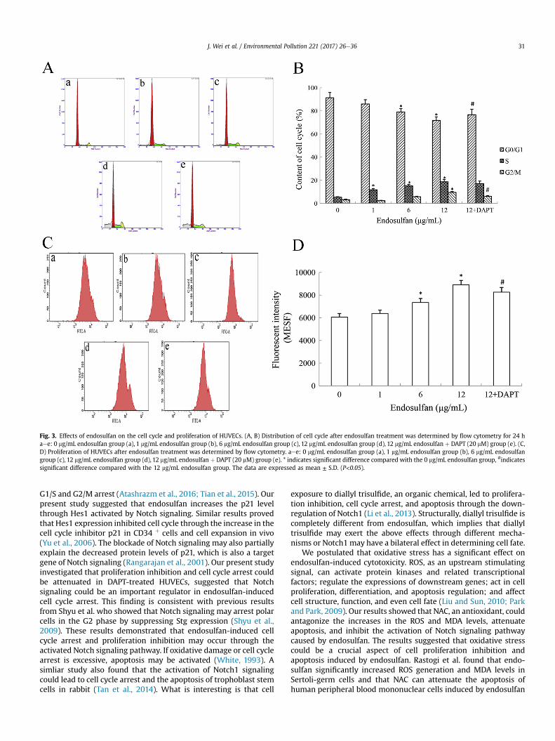
Fig. 3. Effects of endosulfan on the cell cycle and proliferation of HUVECs. (A, B) Distribution of cell cycle after endosulfan treatment was determined by flow cytometry for 24 haee: 0 mg/mL endosulfan group (a), 1 mg/mL endosulfan group (b), 6 mg/mL endosulfan group (c), 12 mg/mL endosulfan group (d), 12 mg/mL endosulfan þ DAPT (20 mM) group (e). (C,D) Proliferation of HUVECs after endosulfan treatment was determined by flow cytometry. aee: 0 mg/mL endosulfan group (a), 1 mg/mL endosulfan group (b), 6 mg/mL endosulfangroup (c), 12 mg/mL endosulfan group (d), 12 mg/mL endosulfan þ DAPT (20 mM) group (e). * indicates significant difference compared with the 0 mg/mL endosulfan group, #indicatessignificant difference compared with the 12 mg/mL endosulfan group. The data are expressed as mean ± S.D. (P<0.05).
J. Wei et al. / Environmental Pollution 221 (2017) 26e36 31
G1/S and G2/M arrest (Atashrazm et al., 2016; Tian et al., 2015). Ourpresent study suggested that endosulfan increases the p21 levelthrough Hes1 activated by Notch signaling. Similar results provedthat Hes1 expression inhibited cell cycle through the increase in thecell cycle inhibitor p21 in CD34 þ cells and cell expansion in vivo(Yu et al., 2006). The blockade of Notch signaling may also partiallyexplain the decreased protein levels of p21, which is also a targetgene of Notch signaling (Rangarajan et al., 2001). Our present studyinvestigated that proliferation inhibition and cell cycle arrest couldbe attenuated in DAPT-treated HUVECs, suggested that Notchsignaling could be an important regulator in endosulfan-inducedcell cycle arrest. This finding is consistent with previous resultsfrom Shyu et al. who showed that Notch signaling may arrest polarcells in the G2 phase by suppressing Stg expression (Shyu et al.,2009). These results demonstrated that endosulfan-induced cellcycle arrest and proliferation inhibition may occur through theactivated Notch signaling pathway. If oxidative damage or cell cyclearrest is excessive, apoptosis may be activated (White, 1993). Asimliar study also found that the activation of Notch1 signalingcould lead to cell cycle arrest and the apoptosis of trophoblast stemcells in rabbit (Tan et al., 2014). What is interesting is that cell
exposure to diallyl trisulfide, an organic chemical, led to prolifera-tion inhibition, cell cycle arrest, and apoptosis through the down-regulation of Notch1 (Li et al., 2013). Structurally, diallyl trisulfide iscompletely different from endosulfan, which implies that diallyltrisulfide may exert the above effects through different mecha-nisms or Notch1 may have a bilateral effect in determining cell fate.
We postulated that oxidative stress has a significant effect onendosulfan-induced cytotoxicity. ROS, as an upstream stimulatingsignal, can activate protein kinases and related transcriptionalfactors; regulate the expressions of downstream genes; act in cellproliferation, differentiation, and apoptosis regulation; and affectcell structure, function, and even cell fate (Liu and Sun, 2010; Parkand Park, 2009). Our results showed that NAC, an antioxidant, couldantagonize the increases in the ROS and MDA levels, attenuateapoptosis, and inhibit the activation of Notch signaling pathwaycaused by endosulfan. The results suggested that oxidative stresscould be a crucial aspect of cell proliferation inhibition andapoptosis induced by endosulfan. Rastogi et al. found that endo-sulfan significantly increased ROS generation and MDA levels inSertoli-germ cells and that NAC can attenuate the apoptosis ofhuman peripheral blood mononuclear cells induced by endosulfan

Fig. 4. Effects of endosulfan on cytoskeleton and mitosis in HUVECs. Images 7e12 are the 4 � magnified versions of 1e6, respectively. Microfilament (A), microtubule (B), and cellnucleus (C) were incubated with Actin-Tracker Green, Tubulin-Tracker Red, and Hoechst 33258 solution, respectively. (D) Merged graphs of A, B, and C. Cells were observed using areal-time inverted phase contrast microscope (200 � ) after treatment with 0 (E) and 12 mg/mL (F) endosulfan for 24 h. (G) The percentage of normal mitosis of HUVECs wascalculated after treatment with 0 and 12 mg/mL endosulfan for 24 h. White arrow indicates regular mitosis, whereas the black indicates the failure of mitosis. *indicates significantdifference between the two groups. The data are expressed as mean ± S.D. (P<0.05).
J. Wei et al. / Environmental Pollution 221 (2017) 26e3632
(Rastogi et al., 2014). Excessive ROS can cause oxidative damage inDNA, resulting in DNA modifications, such as single- and double-strand breaks. Endosulfan has been reported to increase the DNAdamage resulting from excessive ROS in zebrafish (Shao et al.,2012). Kamarehei found that ROS could activate Notch signalingin SK-N-MC cells and induce apoptosis (Kamarehei andYazdanparast, 2014). Because the Notch pathway is a highly con-servative signaling system, the above Notch-medicated cell prolif-eration inhibition and apoptosis by endosulfan may be similar in
HUVECs. We found that endosulfan can cause apoptosis, induce cellcycle arrest and proliferation inhibition, damage cytoskeleton andcell nuclei, and activate the Notch signaling pathway in HUVECs.Proliferation inhibition by endosulfan is possibly induced by theNotch signaling pathway as a result of oxidative stress. Therefore,the present findings determine the mechanism of endosulfan-induced cardiovascular toxicity and demonstrate that exposure toendosulfan may be an underlying risk factor for the cardiovascularsystem.

Fig. 4. (continued).
Table 1Damage to microfilaments, microtubules, and nuclei in HUVECs induced by endosulfan.
Concentration (mg mL�1) Fluorescence intensity ofmicrofilaments Fluorescence intensity ofmicrotubules Fluorescence intensity of nuclei Diameters of nuclei (mm)
0 90.89 ± 6.22 70.18 ± 4.89 25.05 ± 2.07 13.68 ± 0.701 60.24 ± 2.09* 66.18 ± 3.65 20.68 ± 2.27 13.79 ± 1.256 53.95 ± 6.78* 53.02 ± 9.97 29.88 ± 5.07 11.98 ± 0.5512 32.74 ± 4.74* 51.70 ± 4.59* 27.91 ± 4.02 9.53 ± 1.02*
12 þ NAC (3 mM) 27.29 ± 4.37 67.23 ± 8.31 31.07 ± 4.01 11.92 ± 1.6912 þ DAPT (20 mM) 29.55 ± 1.73 51.56 ± 12.44 29.96 ± 4.83 10.68 ± 2.43
*indicates significant difference compared with the 0 mg/mL endosulfan group. The data are expressed as mean ± S.D. (P<0.05).
J. Wei et al. / Environmental Pollution 221 (2017) 26e36 33

Fig. 5. Effects of endosulfan on the Notch signaling pathway in HUVECs. (A) Expressions of Dll4, Notch1, Cleaved-Notch1, Jagged1, Notch4, Hes1, and p21. (B) The relativedensitometric analysis of Dll4, Notch1, Cleaved-Notch1 (a), Jagged1, Notch4 (b), Hes1, and p21 (c) expressions after treatment with different concentrations of endosulfan for 24 h*indicates significant difference compared with the 0 mg/mL endosulfan group, **indicates significant difference between two different groups. The data are expressed as mean ± S.D.(P<0.05).
J. Wei et al. / Environmental Pollution 221 (2017) 26e3634
5. Conclusions
The present study revealed a vital molecular mechanism ofendosulfan-induced toxicity in endothelial cells through the Notchsignaling pathway. Given that the Notch signaling pathway iscentral to determine cell fate and cellular processes, the current
findings have biological implications in cellular organisms. There-fore, blocking the Notch signaling pathwaymay be a promising wayto attenuate the toxicity of endosulfan on endothelial cells. Inaddition, this finding may have key implications in the preventionof CVD where the Notch signaling pathway can be explored as apreclinical evaluation.

J. Wei et al. / Environmental Pollution 221 (2017) 26e36 35
Acknowledgments
This study was supported by National Natural Science Founda-tion of China (No. 31370430).
References
Abdul Majeed, S., Nambi, K.S., Taju, G., Sarath Babu, V., Farook, M.A., SahulHameed, A.S., 2014. Development and characterization of a new gill cell linefrom air breathing fish Channa striatus (Bloch 1793) and its application intoxicology: direct comparison to the acute fish toxicity. Chemosphere 96,89e98.
Abraham, R.T., 2001. Cell cycle checkpoint signaling through the ATM and ATR ki-nases. Genes Dev. 15, 2177e2196.
Antherieu, S., Ledirac, N., Luzy, A.P., Lenormand, P., Caron, J.C., Rahmani, R., 2007.Endosulfan decreases cell growth and apoptosis in human HaCaT keratinocytes:partial ROS-dependent ERK1/2 mechanism. J. Cell Physiol. 213, 177e186.
Atashrazm, F., Lowenthal, R.M., Woods, G.M., Holloway, A.F., Karpiniec, S.S.,Dickinson, J.L., 2016. Fucoidan suppresses the growth of human acute pro-myelocytic leukemia cells in vitro and in vivo. J. Cell Physiol. 231, 688e697.
Balfour Jr., P.C., Ruiz, J.M., Talavera, G.A., Allison, M.A., Rodriguez, C.J., 2016. Car-diovascular disease in hispanics/latinos in the United States. J. Lat. Psychol. 4,98e113.
Bates, D.J., Salerni, B.L., Lowrey, C.H., Eastman, A., 2011. Vinblastine sensitizes leu-kemia cells to cyclin-dependent kinase inhibitors, inducing acute cell cyclephase-independent apoptosis. Cancer Biol. Ther. 12, 314e325.
Becker, L., Scheringer, M., Schenker, U., Hungerbuhler, K., 2011. Assessment of theenvironmental persistence and long-range transport of endosulfan. Environ.Pollut. 159, 1737e1743.
Benedito, R., Roca, C., Sorensen, I., Adams, S., Gossler, A., Fruttiger, M., Adams, R.H.,2009. The notch ligands Dll4 and Jagged1 have opposing effects on angiogen-esis. Cell 137, 1124e1135.
Bundy, J.D., He, J., 2016. Hypertension and related cardiovascular disease burden inChina. Ann. Glob. Health 82, 227e233.
Cimellaro, A., Perticone, M., Fiorentino, T.V., Sciacqua, A., Hribal, M.L., 2016. Role ofendoplasmic reticulum stress in endothelial dysfunction. Nutr. Metab. Car-diovasc Dis. 26, 863e871.
Cruz, A.B., Pitz, H.D., Veber, B., Bini, L.A., Maraschin, M., Zeni, A.L., 2016. Assessmentof bioactive metabolites and hypolipidemic effect of polyphenolic-rich redcabbage extract. Pharm. Biol. 1e7.
Dahlberg, A., Delaney, C., Bernstein, I.D., 2011. Ex vivo expansion of human he-matopoietic stem and progenitor cells. Blood 117, 6083e6090.
de Lavor, M.S., Binda, N.S., Fukushima, F.B., Caldeira, F.M., da Silva, J.F., Silva, C.M., deOliveira, K.M., Martins Bde, C., Torres, B.B., Rosado, I.R., Gomez, R.S.,Gomez, M.V., de Melo, E.G., 2015. Ischemia-reperfusion model in rat spinalcord: cell viability and apoptosis signaling study. Int. J. Clin. Exp. Pathol. 8,9941e9949.
Du, H., Wang, M., Wang, L., Dai, H., Wang, M., Hong, W., Nie, X., Wu, L., Xu, A., 2015.Reproductive toxicity of endosulfan: implication from germ cell apoptosismodulated by mitochondrial dysfunction and genotoxic response genes inCaenorhabditis elegans. Toxicol. Sci. 145, 118e127.
El Hindy, N., Keyvani, K., Pagenstecher, A., Dammann, P., Sandalcioglu, I.E., Sure, U.,Zhu, Y., 2013. Implications of Dll4-Notch signaling activation in primary glio-blastoma multiforme. Neuro Oncol. 15, 1366e1378.
Enhui, Z., Na, C., MengYun, L., Jia, L., Dan, L., Yongsheng, Y., Ying, Z., DeFu, H., 2016.Isomers and their metabolites of endosulfan induced cytotoxicity and oxidativedamage in SH-SY5Y cells. Environ. Toxicol. 31, 496e504.
Gandhi, D., Tarale, P., Naoghare, P.K., Bafana, A., Krishnamurthi, K., Arrigo, P.,Saravanadevi, S., 2015. An integrated genomic and proteomic approach toidentify signatures of endosulfan exposure in hepatocellular carcinoma cells.Pestic. Biochem. Physiol. 125, 8e16.
Gu, J.W., Young, E., Busby, B., Covington, J., Johnson, J.W., 2009. Oral administrationof pyrrolidine dithiocarbamate (PDTC) inhibits VEGF expression, tumor angio-genesis, and growth of breast cancer in female mice. Cancer Biol. Ther. 8,514e521.
Guo, S., Liu, M., Gonzalez-Perez, R.R., 2011. Role of Notch and its oncogenic signalingcrosstalk in breast cancer. Biochim. Biophys. Acta 1815, 197e213.
Kafilzadeh, F., Ebrahimnezhad, M., Tahery, Y., 2015. Isolation and identification ofendosulfan-degrading bacteria and evaluation of their bioremediation in korriver, Iran. Osong Public Health Res. Perspect. 6, 39e46.
Kamarehei, M., Yazdanparast, R., 2014. Modulation of notch signaling pathway toprevent H2O2/menadione-induced SK-N-MC cells death by EUK134. Cell Mol.Neurobiol. 34, 1037e1045.
Kanno, T., Nishizaki, T., 2011. Sphingosine induces apoptosis in hippocampal neu-rons and astrocytes by activating caspase-3/-9 via a mitochondrial pathwaylinked to SDK/14-3-3 protein/Bax/cytochrome c. J. Cell Physiol. 226, 2329e2337.
Kim, S.A., Kim, K.S., Lee, Y.M., Jacobs, D.R., Lee, D.H., 2015a. Associations of organ-ochlorine pesticides and polychlorinated biphenyls with total, cardiovascular,and cancer mortality in elders with differing fat mass. Environ. Res. 138, 1e7.
Kim, Y.W., Eom, B.W., Kook, M.C., Kim, H.S., Kim, M.K., Hwang, H.L., Chandra, V.,Poojan, S., Song, Y., Koh, J.S., Bae, C.D., Ro, J., Hong, K.M., 2015b. Clinical impli-cations of proliferation activity in T1 or T2 male gastric cancer patients. Exp.Mol. Med. 47, e193.
Kofler, N.M., Shawber, C.J., Kangsamaksin, T., Reed, H.O., Galatioto, J., Kitajewski, J.,2011. Notch signaling in developmental and tumor angiogenesis. Genes Cancer2, 1106e1116.
Kume, T., 2012. Ligand-dependent Notch signaling in vascular formation. Adv. Exp.Med. Biol. 727, 210e222.
Lai, W.K., Kan, M.Y., 2015. Homocysteine-induced endothelial dysfunction. Ann.Nutr. Metab. 67, 1e12.
Li, Y., Zhang, J., Zhang, L., Si, M., Yin, H., Li, J., 2013. Diallyl trisulfide inhibits pro-liferation, invasion and angiogenesis of osteosarcoma cells by switching onsuppressor microRNAs and inactivating of Notch-1 signaling. Carcinogenesis 34,1601e1610.
Li, S., Xu, D., Guo, J., Sun, Y., 2015. Inhibition of cell growth and induction ofinflammation by endosulfan in HUVEC-C cells. Environ. Toxicol.
Liu, X., Sun, J., 2010. Endothelial cells dysfunction induced by silica nanoparticlesthrough oxidative stress via JNK/P53 and NF-kappaB pathways. Biomaterials 31,8198e8209.
Lukin, D.J., Carvajal, L.A., Liu, W.J., Resnick-Silverman, L., Manfredi, J.J., 2015. p53Promotes cell survival due to the reversibility of its cell-cycle checkpoints. Mol.Cancer Res. 13, 16e28.
Ma, A., Boulton, M., Zhao, B., Connon, C., Cai, J., Albon, J., 2007. A role for notchsignaling in human corneal epithelial cell differentiation and proliferation.Invest. Ophthalmol. Vis. Sci. 48, 3576e3585.
Ma, Y.M., Peng, Y.M., Zhu, Q.H., Gao, A.H., Chao, B., He, Q.J., Li, J., Hu, Y.H., Zhou, Y.B.,2016. Novel CHOP activator LGH00168 induces necroptosis in A549 human lungcancer cells via ROS-mediated ER stress and NF-kappaB inhibition. Acta Phar-macol. Sin. 37, 1381e1390.
Moon, H.J., Lee, J.W., 2013. Availability of intravenous lipid emulsion therapy onendosulfan-induced cardiovascular collapse. Am. J. Emerg. Med. 31 (886),e881e882.
Moses, V., Peter, J.V., 2010. Acute intentional toxicity: endosulfan and other or-ganochlorines. Clin. Toxicol. (Phila) 48, 539e544.
Oropesa Avila, M., Fernandez Vega, A., Garrido Maraver, J., Villanueva Paz, M., DeLavera, I., De La Mata, M., Cordero, M.D., Alcocer Gomez, E., Delgado Pavon, A.,Alvarez Cordoba, M., Cotan, D., Sanchez-Alcazar, J.A., 2015. Emerging roles ofapoptotic microtubules during the execution phase of apoptosis. Cytoskelet.Hob. 72, 435e446.
Ozmen, O., 2013. Cardiotoxicity and apoptotic activity in subacute endosulfantoxicity and the protective effect of vitamin C in rabbits: a pathological study.J. Environ. Pathol. Toxicol. Oncol. 32, 53e58.
Park, E.J., Park, K., 2009. Oxidative stress and pro-inflammatory responses inducedby silica nanoparticles in vivo and in vitro. Toxicol. Lett. 184, 18e25.
Pedrosa, A.R., Trindade, A., Fernandes, A.C., Carvalho, C., Gigante, J., Tavares, A.T.,Dieguez-Hurtado, R., Yagita, H., Adams, R.H., Duarte, A., 2015. Endothelial Jag-ged1 antagonizes Dll4 regulation of endothelial branching and promotesvascular maturation downstream of Dll4/Notch1. Arterioscler. Thromb. Vasc.Biol. 35, 1134e1146.
Penell, J., Lind, L., Salihovic, S., van Bavel, B., Lind, P.M., 2014. Persistent organicpollutants are related to the change in circulating lipid levels during a 5 yearfollow-up. Environ. Res. 134, 190e197.
Peyre, L., Zucchini-Pascal, N., de Sousa, G., Rahmani, R., 2012. Effects of endosulfanon hepatoma cell adhesion: epithelial-mesenchymal transition and anoikisresistance. Toxicology 300, 19e30.
Puente, B.N., Kimura, W., Muralidhar, S.A., Moon, J., Amatruda, J.F., Phelps, K.L.,Grinsfelder, D., Rothermel, B.A., Chen, R., Garcia, J.A., Santos, C.X., Thet, S.,Mori, E., Kinter, M.T., Rindler, P.M., Zacchigna, S., Mukherjee, S., Chen, D.J.,Mahmoud, A.I., Giacca, M., Rabinovitch, P.S., Aroumougame, A., Shah, A.M.,Szweda, L.I., Sadek, H.A., 2014. The oxygen-rich postnatal environment inducescardiomyocyte cell-cycle arrest through DNA damage response. Cell 157,565e579.
Rangarajan, A., Talora, C., Okuyama, R., Nicolas, M., Mammucari, C., Oh, H., Aster, J.C.,Krishna, S., Metzger, D., Chambon, P., Miele, L., Aguet, M., Radtke, F., Dotto, G.P.,2001. Notch signaling is a direct determinant of keratinocyte growth arrest andentry into differentiation. EMBO J. 20, 3427e3436.
Rastogi, D., Narayan, R., Saxena, D.K., Chowdhuri, D.K., 2014. Endosulfan inducedcell death in Sertoli-germ cells of male Wistar rat follows intrinsic mode of celldeath. Chemosphere 94, 104e115.
Ritchey, L., Chakrabarti, R., 2014. Aurora A kinase modulates actin cytoskeletonthrough phosphorylation of Cofilin: implication in the mitotic process. Biochim.Biophys. Acta 1843, 2719e2729.
Sainson, R.C., Harris, A.L., 2007. Anti-Dll4 therapy: can we block tumour growth byincreasing angiogenesis? Trends Mol. Med. 13, 389e395.
Sandquist, J.C., Larson, M.E., Hine, K.J., 2016. Myosin-10 independently influencesmitotic spindle structure and mitotic progression. Cytoskelet. Hob. 73, 351e364.
Senthilkumaran, B., 2015. Pesticide- and sex steroid analogue-induced endocrinedisruption differentially targets hypothalamo-hypophyseal-gonadal systemduring gametogenesis in teleosts - a review. Gen. Comp. Endocrinol. 219,136e142.
Shao, B., Zhu, L., Dong, M., Wang, J., Wang, J., Xie, H., Zhang, Q., Du, Z., Zhu, S., 2012.DNA damage and oxidative stress induced by endosulfan exposure in zebrafish(Danio rerio). Ecotoxicology 21, 1533e1540.
Shyu, L.F., Sun, J., Chung, H.M., Huang, Y.C., Deng, W.M., 2009. Notch signaling anddevelopmental cell-cycle arrest in Drosophila polar follicle cells. Mol. Biol. Cell20, 5064e5073.
Tan, T., Lu, B., Zhang, J., Niu, Y., Si, W., Wei, Q., Ji, W., 2014. Notch1 signaling an-tagonizes transforming growth factor-beta pathway and induces apoptosis in

J. Wei et al. / Environmental Pollution 221 (2017) 26e3636
rabbit trophoblast stem cells. Stem Cells Dev. 23, 813e822.Tian, C., Yu, Y., Jia, Y., Zhu, L., Zhang, Y., 2015. HES1 activation suppresses prolifer-
ation of leukemia cells in acute myeloid leukemia. Ann. Hematol. 94,1477e1483.
Wang, J., Engle, S., Zhang, Y., 2011. A new in vitro system for activating the cell cyclecheckpoint. Cell Cycle 10, 500e506.
Wei, J., Zhang, L., Wang, J., Guo, F., Li, Y., Zhou, X., Sun, Z., 2015. Endosulfan inducingblood hypercoagulability and endothelial cells apoptosis via the death receptorpathway in Wistar rats. Toxicol. Res. 4, 1282e1288.
White, E., 1993. Death-defying acts: a meeting review on apoptosis. Genes Dev. 7,
2277e2284.Yu, X., Alder, J.K., Chun, J.H., Friedman, A.D., Heimfeld, S., Cheng, L., Civin, C.I., 2006.
HES1 inhibits cycling of hematopoietic progenitor cells via DNA binding. StemCells 24, 876e888.
Zhang, L., Wei, J., Guo, F., Duan, J., Li, Y., Shi, Z., Yang, Y., Zhou, X., Sun, Z., 2015.Endosulfan activates the extrinsic coagulation pathway by inducing endothelialcell injury in rats. Environ. Sci. Pollut. Res. 22, 15722e15730.
Zhao, Y.Z., Jia, J., Li, Y.B., Guo, C.X., Zhou, X.Q., Sun, Z.W., 2014. Effects of endosulfanon the immune function of erythrocytes, and potential protection by testos-terone propionate. J. Toxicol. Sci. 39, 701e710.