endonuclease-independent insertion provides an alternative
TRANSCRIPT

Published online 21 May 2007 Nucleic Acids Research, 2007, Vol. 35, No. 11 3741–3751doi:10.1093/nar/gkm317
Endonuclease-independent insertion provides analternative pathway for L1 retrotranspositionin the human genomeShurjo K. Sen, Charles T. Huang, Kyudong Han and Mark A. Batzer*
Department of Biological Sciences, Biological Computation and Visualization Center, Center for BioModularMulti-Scale Systems, Louisiana State University, Baton Rouge, LA 70803, USA
Received February 15, 2007; Revised April 15, 2007; Accepted April 16, 2007
ABSTRACT
LINE-1 elements (L1s) are a family of highly suc-cessful retrotransposons comprising »17% of thehuman genome, the majority of which have insertedthrough an endonuclease-dependent mechanismtermed target-primed reverse transcription. Recentin vitro analyses suggest that in the absence of non-homologous end joining proteins, L1 elements mayutilize an alternative, endonuclease-independentpathway for insertion. However, it remains unknownwhether this pathway operates in vivo or in cell lineswhere all DNA repair mechanisms are functional.Here, we have analyzed the human genome todemonstrate that this alternative pathway for L1insertion has been active in recent human evolutionand characterized 21 loci where L1 elements haveintegrated without signs of endonuclease-relatedactivity. The structural features of these loci suggesta role for this process in DNA double-strand breakrepair. We show that endonuclease-independentL1 insertions are structurally distinguishable fromclassical L1 insertion loci, and that they areassociated with inter-chromosomal translocationsand deletions of target genomic DNA.
INTRODUCTION
Long interspersed element-1 (LINE-1 or L1) is aubiquitous retrotransposon family in the humangenome, with �520 000 insertions comprising �17% oftotal genomic sequence (1,2). A full-length L1 elementis �6-kb long and contains two open readingframes (ORF1 and ORF2) (3). While ORF1 encodes anRNA-binding protein with nucleic acid chaperone activity(4), ORF2 encodes for endonuclease (EN) and reversetranscriptase (RT) activities (5,6), and both ORFs arerequired for L1 retrotransposition (7,8). In addition to
insertional mutagenesis (9–11), L1 elements have also beenassociated with exon shuffling, creation of deletionsthrough unequal homologous recombination and intra-chromosomal and inter-chromosomal translocation ofgenomic sequence (12–14). As such, the dynamic natureof L1 elements makes them important agents of genomicrearrangement (15,16).The currently accepted model for genomic integration
of L1 elements is termed target-site primed reversetranscription (TPRT) (17,18) (Figure 1). During TPRT,the L1 EN cleaves one strand of the target DNA at a motifapproaching the consensus 50-TTTT/A-30 (where ‘‘/’’denotes the cleavage site), producing a free 30-hydroxyl(5,19). Next, the L1 RNA anneals to the nick site using its30 poly (A) tail, and the L1 RT initiates reversetranscription using the L1 RNA as a template. Cleavageof the second DNA strand by the L1 EN usually occurs7–20 base pairs downstream of the initial nicking site,creating staggered breaks in the target DNA that are laterfilled in to form direct repeats flanking the newly insertedelement (termed target site duplications or TSDs) (20).Integration of the newly synthesized cDNA and comple-tion of second-strand synthesis are the remaining steps inthe TPRT model; however, the order in which they occurand their exact mechanism remain unclear (21). Apartfrom the presence of TSDs, other structural hallmarks ofTPRT-mediated L1 insertion include frequent 50 trunca-tions (or truncation/inversions) and intact 30 ends withvariable-length A-rich tails (20).In recent years, increasing evidence from cell culture
retrotransposition assays suggests that in addition toTPRT-mediated insertion, a second, less-characterizedL1 integration pathway may exist that is independentof L1-encoded endonuclease (18,22). However, witha few isolated exceptions (23–25), the majority ofendonuclease-independent (ENi) L1 insertions have beenrecovered in cell lines lacking one or more components ofthe cellular non-homologous end joining (NHEJ) mecha-nism, a principal form of DNA double-strand break(DSB) repair (26). Consequently, whether ENi L1
*To whom correspondence should be addressed. Tel: þ1 225 578 7102; Fax: þ1 225 578 7113; Email: [email protected]
� 2007 The Author(s)
This is an Open Access article distributed under the terms of the Creative Commons Attribution Non-Commercial License (http://creativecommons.org/licenses/
by-nc/2.0/uk/) which permits unrestricted non-commercial use, distribution, and reproduction in any medium, provided the original work is properly cited.
Downloaded from https://academic.oup.com/nar/article-abstract/35/11/3741/2402690by gueston 23 February 2018

insertion occurs at detectable frequencies whennormal DNA repair pathways are functional has beenthe subject of continued debate (3,22,27–29). Addi-tionally, existing analyses of human genomic L1 elements(20,30), by focusing solely on TPRT-mediated insert-ions, have left this question unanswered in a systematicfashion.In this study, we have utilized computational analyses
of the draft sequence of the human genome to recover L1elements that utilized this alternative pathway of integra-tion (which we term non-classical L1 insertion or NCLI).We report 21 loci where L1 elements appear to haveinserted without any hallmarks of endonuclease activity.In each case, we verified the ancestral (i.e. no L1insertion) state of the loci by re-sequencing the ortholo-gous positions in the common chimpanzee and rhesusmacaque genomes. Overall, our results suggest that NCLIhas been active in recent human evolution, and that itprovides an alternative ‘non-selfish’ pathway for L1integration in the human genome. Interestingly, we findthat NCLI loci are clustered in gene-rich regions of thegenome, in contrast to the distribution of the morecommon TPRT-mediated L1 insertions. Based on theunique structural features of NCLI-mediated L1 ele-ments, we suggest that this process may be capable ofrepairing genomic lesions and that it may confer a slightselective advantage to what may be the otherwisedeleterious nature of the L1 family. We conclude thatnon-LTR retrotransposons may have a previouslyunrecognized role in maintaining human genomicintegrity.
MATERIALS AND METHODS
Computational screening for putative ENi L1 insertions
To identify NCLI loci in the publicly available humangenome, we first downloaded the file chromOut.zip fromthe UCSC Genome Bioinformatics website (http://hgdownload.cse.ucsc.edu/downloads.html#human). Thisarchive contains output files from the RepeatMasker(RM) software package (http://www.repeatmasker.org/)run at the �s (sensitive) setting on individual humanchromosomes. For this project, the archived files corre-sponded to RM output from the May 2004 freeze of thehuman genome (hg17). Next, using our own script, weextracted all L1 insertions from each chromosome. To findelements missing the segment of the 30 UTR normally usedduring TPRT-mediated insertion, we developed a set ofcomputer programs that scanned the comprehensive list ofL1 elements to find all elements truncated beyond 20 basesfrom the 30 end. We chose the 20 bp truncation limit fortwo specific reasons. Firstly, from aligning six previouslypublished consensus sequences of relatively young L1elements, we found the shortest length of the poly(A) tailto be 13 bp. Secondly, we added a 7 bp window to the13-bp poly(A) tail to account for the possibility of smallinternal deletions near the 30 end of the L1 insertions thatwould mimic the appearance of a 30-truncated insertion.As RM assigns a size of 6155 bp to full-length L1 elementsfrom subfamilies L1Hs and L1PA2, our initial output filesthus contained sets of L1 insertions ending at position6135 or lower. To verify the effectiveness of this strategy,for each chromosome, we manually inspected sets of
Figure 1. Comparison of TPRT and NCLI L1 insertions. (A) Classical TPRT-mediated L1 insertion in the human genome. First-strand cleavageby the L1 EN (red arrowhead) at the 50-TTTT/A-30 consensus (red dotted box) allows L1 mRNA (blue line) to anneal to genomic DNA using itspoly(A) tail. RT activity of L1 ORF2 (green oval) synthesizes L1 cDNA (purple line) using L1 mRNA as template and 30 OH from nicked genomicDNA as primer. Second-strand cleavage (blue arrowhead) occurs 7–20 bp downstream from first-strand cleavage site, creating staggered nicks whichare later filled in to form TSDs (blue dotted boxes). Attachment of the L1 cDNA and synthesis of the second strand complete the insertion process.TSD sequences for this diagram are from a 637-bp human L1 element located at chr1:65036188–65036824. (B) Schematic representation of an NCLIevent. Following creation of a genomic double-strand break (red thunderbolt), free-floating L1 mRNA (blue line) attaches to newly separated endsusing small stretches of complementary bases. Once gap is bridged, it may be filled in by DNA synthesis by either the L1 RT, cellular repairpolymerases or both. L1 insertion thus created lacks structural features of TPRT-mediated insertion.
3742 Nucleic Acids Research, 2007, Vol. 35, No. 11
Downloaded from https://academic.oup.com/nar/article-abstract/35/11/3741/2402690by gueston 23 February 2018
50 loci on either side of this truncation limit. The sets ofL1 elements with 30 truncations520 bp did not return anyloci matching all of these three criteria; absence of TSDsof any length, absence of a poly(A) tail and significantdeviation from the consensus L1 EN cleavage site. Thus,these L1 elements most likely integrated into the genomethrough traditional TPRT-mediated insertion. As such,after visual inspection of the computational output, allloci that we selected for further experimental verificationcame from the set of insertions with 30 truncations 20 bpor longer. To further narrow our list to relatively youngL1 insertions, we discarded all elements 42% divergedfrom their respective consensus sequences according to theRM algorithm. We rejected all L1 insertions that hadTSDs of any length, even if they bordered a 30 truncatedelement. Our RM output parsing software accountedfor L1 elements fragmented by small insertions/deletionsand for truncated/inverted L1 insertions, both of whichcommonly occur during the TPRT process and aresometimes annotated by RM as separate insertions. Allthe computer programs are available from the authorsupon request.
Manual inspection of sequence and verification ofancestral (pre-insertion) status
To confirm the ancestral (i.e. no insertion) stage forcomputationally recovered NCLI loci, we extracted10 000 bp of flanking sequence on either side of the L1element. First, we ran each extracted segment (L1insertion plus flanking sequence) through RM to verifythat the potential NCLI candidates were not fragmentsof 30 intact L1 elements separated by large blocks ofintervening non-L1 sequence. We then used the BLATsoftware package (http://www.genome.ucsc.edu/cgi-bin/hgBlat) to construct triple alignments of the human,chimpanzee and rhesus macaque genomes at each locus.Next, we manually inspected each alignment to verify thatthe 50 and 30 ends of each putative human NCLI eventcorresponded to either gaps or extra, non-L1 sequence inthe ancestral sequence (the presence of non-L1 sequenceindicated a deletion in the ancestral genome whoseboundaries exactly matched the human L1 insertion). Inaddition, to further confirm the endonuclease-independentnature of putative NCLI loci, we analyzed them for
divergence from the TTTT/A L1-EN cleavage siteconsensus, based on an earlier analysis of EN sitepreferences (22). This left us with a final data set of21 potential NCLI loci that fit all four of the followingcriteria: 30 truncation, absence of TSDs, absence ofa poly(A) tail and significant divergence from the L1-ENconsensus.
PCR amplification and DNA sequence analysis of NCLI loci
To experimentally confirm that these 21 loci representedtruncated L1 insertions rather than deletions of the30 UTR, we designed oligonucleotide primers in the non-repetitive sequence flanking the L1 elements and amplifiedthem by PCR on a panel of five primate species (Figure 2),including Homo sapiens (HeLa; cell line ATCC CCL-2),Pan troglodytes (common chimpanzee; cell lineAG06939B), Gorilla gorilla (Western lowland gorilla; cellline AG05251), Macaca mulatta (Rhesus macaque; cellline NG07098) and Chlorocebus aethiops (Green monkey;cell line ATCC CCL70). PCR amplification of NCLI lociwas performed in 25 ml reactions using 10–50 ng genomicDNA, 200 nM of each oligonucleotide primer, 200 mMdNTPs in 50mM KCl, 1.5mM MgCl2, 10mM Tris–HCl(pH 8.4) and 2.5 units Taq DNA polymerase. Theconditions for the PCR were an initial denaturation stepof 948C for 4min, followed by 32 cycles of 1min ofdenaturation at 948C, 1min of annealing at optimalannealing temperature and 1min of extension at 728C,followed by a final extension step at 728C for 10min. Forloci with large insertions or deletions (42 kb), we usedEx TaqTM polymerase (TaKaRa) and carried out PCR in50 ml reactions following the manufacturer’s suggestedprotocol. PCR amplicons were separated on 1% agarosegels, stained with ethidium bromide and visualized usingUV fluorescence. Detailed information for each locusincluding primer sequences, annealing temperature andPCR product sizes is available from the ‘Publications’section of the Batzer laboratory website (http://batzerlab.lsu.edu).Repetitive DNA may correspond to sites of genome
assembly errors; therefore we re-sequenced all loci fromthe chimpanzee and rhesus macaque genomes to confirmthat the computationally recovered pre-insertion sequencewas accurate. Individual PCR products were purified
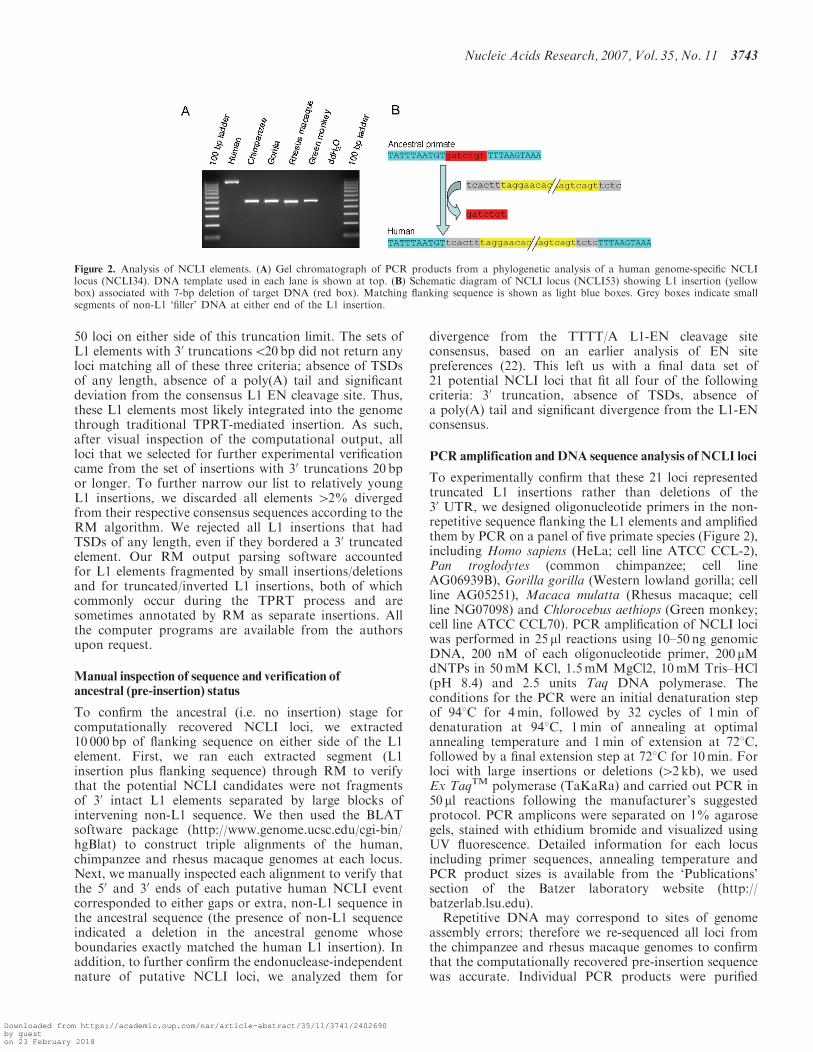
Figure 2. Analysis of NCLI elements. (A) Gel chromatograph of PCR products from a phylogenetic analysis of a human genome-specific NCLIlocus (NCLI34). DNA template used in each lane is shown at top. (B) Schematic diagram of NCLI locus (NCLI53) showing L1 insertion (yellowbox) associated with 7-bp deletion of target DNA (red box). Matching flanking sequence is shown as light blue boxes. Grey boxes indicate smallsegments of non-L1 ‘filler’ DNA at either end of the L1 insertion.
Nucleic Acids Research, 2007, Vol. 35, No. 11 3743
Downloaded from https://academic.oup.com/nar/article-abstract/35/11/3741/2402690by gueston 23 February 2018

from gels using Wizard� gel purification kits (Promega).Amplicons 51.3 kb were cloned into vectors usingTOPO-TA Cloning� kits (Invitrogen) and three colonieswere randomly selected and sequenced in both directionsusing M13 forward and reverse primers to verify that thePCR product matched the computationally recoveredsequence. For PCR products larger than 1.3 kb, gel-purified PCR products were sequenced directly using therespective primers to verify that sequence boundariesmatched the computational predictions. All sequencingwas performed by the chain termination method (31)on an Applied Biosystems ABI3130XL automated DNAsequencer. Analysis of all of the re-sequenced loci showedthat the sequences were exact matches to those in the draftgenome sequence assemblies.
RESULTS
Awhole-genome scan for non-classical L1 insertions
To analyze the human genome sequence for potentialNCLI loci, we combined computational and experimentalapproaches. First, using RM, we computationallyextracted young L1 insertions lacking structural hallmarksof TPRT-mediated ‘classical’ retrotransposition (see‘Materials and Methods’ section). Next, we constructedtriple alignments of the human, chimpanzee and rhesusmacaque genomes at these loci to reconstruct the ancestral(i.e., pre-L1 insertion) state and manually inspected thestructure of each locus to detect signs of non-TPRT medi-ated insertion. Finally, we used PCR and re-sequencingto experimentally verify the sequence architecture forboth the post-insertion and pre-insertion states of theloci (Figure 2). Specifically, the loci included in thisanalysis after experimental confirmation of the computa-tional output possessed all four of the following
characteristics: 30 truncation beyond 25 bp (i.e., 5 bpmore than the minimum truncation level set duringcomputational screening) relative to the L1HS_30endconsensus from the RepeatMaskerLib.embl repetitiveelement library, downloadable from: http://www.girinst.org/repbase/index.html) (32), absence of TSDs of anylength, absence of a poly(A) tail and significant deviationfrom the consensus L1 EN cleavage site. Structuralfeatures of the NCLI loci that were extracted using thisapproach closely mimic ENi L1 insertions reported inearlier cell-culture analyses (18,22,28), further consolidat-ing our hypothesis that they represent products of asimilar insertion mechanism in the human genome. Wefound a total of 21 NCLI loci in the May 2004 freeze ofthe human genome (hg17)(Table 1), of which we were ableto recover the pre-insertion site of seven loci from thechimpanzee genome assembly (panTro2; March 2006freeze) (33) and 14 loci from the rhesus macaquegenome assembly (rheMac2; January 2006 freeze) (34).As we were only interested in NCLI loci for which wecould verify the pre-insertion sequence, we discarded allL1 insertions that were shared between these threegenomes and thus represented older ancestral L1 elements.The L1 elements at NCLI loci ranged between 34 and4410 bp in length, with a total of 12 018 bp L1 DNA(along with 1365 bp of non-L1 sequence) being capturedbetween the matching 50 and 30 ends of the pre-insertionand post-insertion states. In addition, 18 of 21 NCLI lociwere associated with deletions of target site DNA, rangingbetween 5 bp and 14 534 bp and totaling 31 009 bp.
Our estimate of the total number of NCLI events isprobably conservative, given that the RM algorithm weused to detect L1 elements, even at its �s (sensitive)setting, is unable to detect insertions smaller than 30 bp.Given that previous cell culture analyses of DSB repair by
Table 1. Human NCLI loci and insertion site characteristics
Locus Coordinates L1 bp_ins non-L1 bp_ins bp_del L1 seq 50 or 30? AT% �200 bp AT% �20Kb Lineage Intragenic?
NCLI1 chr3:196416805–196421321 4410 107 109 30 59.5 49.04 H C3ORF1NCLI3 chr4:67544153–67545039 589 298 1574 30 63 62.41 H –NCLI9 chr17:36395952–36396018 67 0 0 Both 60.5 60.86 HC KRT40NCLI11 chr19:15679181–15680403 1223 0 2867 Both 67.5 59.16 H –NCLI23 chr2:29588579–29590824 2246 0 17 Both 65 56.7 HC ALKNCLI32 chr4:112069027–112069153 122 5 23 30 60.5 63.86 HC –NCLI33 chr4:60239707–60239936 108 122 2485 30 65.5 67.18 HC –NCLI34 chr4:87186203–87186706 483 21 30 50 70.5 64 H MAPK10NCLI38 chr5:51963332–51963788 441 16 1692 30 53.5 61.77 HC –NCLI40 chr6:4414637–4415321 600 85 0 Bone 58 55.68 HC –NCLI47 chr9:108094757–108094921 160 5 8 50 67.5 60.96 HC –NCLI48 chr10:60661882–60662013 34 98 5928 50 67.5 66.94 HC PHYHIPLNCLI51 chr11:34668952–34669415 464 0 615 Both 58 63.31 H –NCLI52 chr12:59792048–59792392 336 9 46 30 73 65.3 HC –NCLI53 chr12:14711194–14711264 61 10 7 None 67 60.16 H GUCY2CNCLI55 chr13:102553958–102554087 48 62 44 None 52 58.98 HC –NCLI57 chr13:80218694–80218899 202 4 5 50 67.5 64.53 HC –NCLI60 chr16:35125561–35125651 86 0 0 Both 65.5 63.24 H –NCLI61 chr17:3071528–3071879 49 303 14534 50 68 60.46 HC –NCLI64 chr22:45486099–45486153 35 0 1010 Both 67.5 51.07 HC CERKNCLI65 chr22:38619900–38620471 254 318 15 None 63.33 60.75 HC –Total (bp) 12018 1365 31009 Average 63.33 60.13
In the column for ‘Lineage’, H indicates a NCLI event specific to the human genome, while HC indicates an NCLI event shared between the humanand chimpanzee genomes but absent from the rhesus macaque genome.
3744 Nucleic Acids Research, 2007, Vol. 35, No. 11
Downloaded from https://academic.oup.com/nar/article-abstract/35/11/3741/2402690by gueston 23 February 2018

L1-mediated gene conversion have detected insertiontracts as small as 13 bp (35), it is quite possible that thenumber of recent human NCLI events is actually higherthan our estimate. Further support for the existence ofsuch ‘hyphen elements’ (24) in the genome comes fromongoing studies in our lab (Sen, S. K. et al., unpublisheddata), where we find that TPRT can produce severely50 truncated L1 and Alu insertions with a similar minimumsize (�28–30 bp). As such, it is possible that additionalNCLI loci beyond the 21 analyzed here remain undetectedin the human genome.
Alignment of L1 segments involved in NCLI eventswith the full-length consensus sequence of a human-specific L1 subfamily (L1Hs) revealed a tendency tocluster in the downstream half of the L1 consensus, with18 out of 21 NCLI fragments having 50 truncations3000 bp or more in addition to their 30 truncations(Figure 3; supplemental alignment 1, online). Previousanalyses show that most TPRT-mediated genomic L1insertions are severely 50 truncated (20), which may reflectlow processivity of the L1 RT or alternatively, hostsuppression of transcription (36). The analogous tendencyof L1 fragments at NCLI loci to be confined within thedownstream half of the element may either be due tothe same reasons, or may be moderated by the dynamicsof L1 ribonucleoprotein (RNP) positioning at the sitesof DSBs (18).
Random genomic deletions that remove the 30 ends ofclassical TPRT-mediated L1 insertions (including thepoly(A) tail and the downstream TSD) could mimic thesequence architecture of NCLI loci (23). However, byreconstructing the pre-insertion site of all loci (andverifying that the starting point of the 30 flanking sequenceremained unchanged before and after the L1 insertion),
we effectively minimized the chances of including suchevents in our data, as it is unlikely that random deletionswould repeatedly and precisely remove only L1 sequence,leaving the downstream sequence untouched. Also, for the18 NCLI loci that were associated with target sitedeletions, this would require two independent, randomdeletion events to have taken place at exactly the sameposition in two separate primate species, which wouldhave vanishingly small probability. The 30 truncated L1fragment at locus NCLI 40 was not associated with adeletion of target DNA and was followed by an adenosine-rich stretch, making it possible that an internal deletionhad removed the 30 UTR before the poly(A) tail. However,based on the absence of TSDs, high divergence from theL1-EN consensus and presence of non-L1 DNA at both50 and 30 ends, we decided to include it in our analysis.
Analysis of insertion sites reveals divergencefrom L1-EN consensus
To find additional evidence supporting our hypothesisthat NCLI events were created by an endonuclease-independent mechanism, we inspected all loci for devia-tions from the 50-TTTT/A-30 L1-EN consensus cleavagesite. Histograms of divergence scores of NCLI events,compared to two other recent analyses of TPRT-mediatedL1 insertions (Figure 4), revealed a marked shift in themaxima towards an increased number of differences.Statistical comparisons of the amounts of deviation fromthe consensus revealed a highly significant differencebetween the cleavage site preferences of NCLI lociversus a larger set of 282 recent TPRT-mediated L1insertions (22) (unpaired t-test assuming unequal var-iances; P50.0001) (37), further bolstering our conclusion
Figure 3. Schematic diagram of NCLI L1 element length. Length distribution of L1 segments at the 21 NCLI loci in this analysis along the sequenceof a full-length L1 element (L1Hs) as shown by the blue bars. Location of different domains within the L1 element is shown in the lower panel. Ofthe non-coding regions (gray boxes) the 50 UTR contains an internal RNA polII promoter, while a 63-bp spacer (S) separates the two ORFs (purplearrows). 40 kDa ORF1 has RNA-binding and nucleic acid chaperone activities, while 150 kDa ORF2 consists of an NH2-terminal endonuclease (EN)domain, a central reverse transcriptase (RT) domain, and a COOH-terminal zinc-knuckle like domain. The extreme 30 end of the 30UTR consists of avariable poly(A) tail, absent in all 21 NCLI-mediated insertions.
Nucleic Acids Research, 2007, Vol. 35, No. 11 3745
Downloaded from https://academic.oup.com/nar/article-abstract/35/11/3741/2402690by gueston 23 February 2018
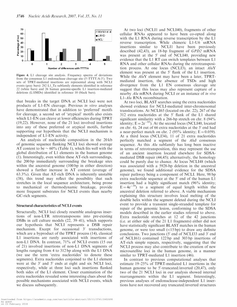
that breaks in the target DNA at NCLI loci were notproducts of L1-EN cleavage. Previous in vitro analyseshave demonstrated that in addition to ‘preferred’ motifsfor cleavage, a second set of ‘atypical’ motifs also existswhich L1-EN can cleave at lower efficiencies during TPRT(19,22). However, none of the 21 loci involved insertionsinto any of these preferred or atypical motifs, furthersupporting our hypothesis that the NCLI mechanism isindependent of L1-EN activity.An analysis of nucleotide composition in the 20 kb
of genomic sequence flanking NCLI loci showed averageAT content to be � 60% (Table 1), which fits well with theglobal distribution of L1 elements in the human genome(1). Interestingly, even within these AT-rich surroundings,the 200 bp immediately surrounding the breakage siteswithin the ancestral genome (100 bp in either direction)showed a further increase in AT content (average of63.3%). Given that AT-rich DNA is inherently unstable(38), this trend may reflect the possibility that suchstretches in the local sequence architecture, being proneto mechanical or thermodynamic breakage, providemore frequent substrates for NCLI events than nearbyGC-rich segments.
Structural characteristics of NCLI events
Structurally, NCLI loci closely resemble analogous inser-tions of non-LTR retrotransposons into pre-existingDSBs in cell culture models (22, 39–41), which supportsour hypothesis that NCLI represents a DSB repairmechanism. Except for occasional 30 transductions,which are a byproduct of the TPRT process (14), classicalL1 insertions are rarely associated with insertions ofnon-L1 DNA. In contrast, 71% of NCLI events (15 outof 21) involved insertions of non-L1 DNA segments oflengths ranging from 4 to 312 bp along with the L1 DNA(we use the term ‘extra nucleotides’ to denote thesesegments). Extra nucleotides conjoined to the L1 elementwere at the 30 and 50 ends at eight and six NCLI loci,respectively, while at three loci such insertions flankedboth sides of the L1 element. Closer examination of theextra nucleotides revealed some interesting clues about thepossible mechanisms associated with NCLI events, whichwe discuss subsequently.
At two loci (NCLI1 and NCLI40), fragments of othercellular RNAs appeared to have been co-opted alongwith the L1 RNA during reverse transcription by the L1reverse transcription. While chimeric L1-U6 snRNAinsertions similar to NCLI1 have been previouslydescribed (42,43), an 18-bp fragment of GPD2 mRNAwas present at the 50 end of NCLI40, providing newevidence that the L1 RT can switch templates between L1RNA and other cellular RNAs during the retrotransposi-tion process. At one locus (NCLI3), an intact AluYelement was present at the 50 flank of the L1 insertion.While the AluY element may have been a later, TPRT-mediated insertion, the absence of TSDs and highdivergence from the L1 EN consensus cleavage sitesuggest that this locus may also represent capture of anearby Alu mRNA during NCLI or an instance of in vivoL1-Alu RNA recombination.
At two loci, BLAST searches using the extra nucleotidesshowed evidence for NCLI-mediated inter-chromosomaltranslocations. At NCLI65 (located on chr. 22), 267 of the312 extra nucleotides at the 50 flank of the L1 sharedsignificant similarity with a 266-bp stretch on chr. 8 (94%identity; E¼ 2e�95). At the second locus (NCLI40, locatedon chr. 6), 24 out of 66 extra nucleotides at the 30 end hada near-perfect match on chr. 2 (95% identity; E¼ 0.059).At a third locus (NCLI34), 11 of 21 extra nucleotidesperfectly matched a segment of the AluJ consensussequence. As this Alu subfamily has long been inactivein terms of retrotransposition, this may represent the useof an ancient insertion located elsewhere for SDSA-mediated DSB repair (44,45); alternatively, the homologycould be purely due to chance. At locus NCLI48 (whichwas associated with a 5928-bp deletion in the ancestralgenome), we found additional evidence for the SDSArepair pathway being a component of NCLI. Here, 98 bpextra nucleotide sequence at the 50 end of the human L1insertion had a highly significant match (96% identity;E¼ 4e�39) to a segment of equal length within theancestral deletion referred to above. A viable mechanismexplaining this structure involves local melting of thedouble helix within the segment deleted during the NCLIevent to provide a transient single-stranded template forrepair of the genomic lesion, conforming to the SDSAmodels described in the earlier studies referred to above.Extra nucleotide stretches at 12 of the 42 junctions(i.e. at either side of the 21 L1 fragments) either did nothave statistically significant BLAST matches in the humangenome, or were too small (515 bp) to draw any definiteconclusions. Two junctions (50 end of NCLI33 and 30 endof NCLI61) contained 122 bp and 303 bp insertions ofAT-rich simple repeats, respectively, suggesting that theNCLI process may also contribute to the creation of newmicrosatellite loci in the human genome, in a mannersimilar to TPRT-mediated L1 insertion (46).
In contrast to previous computational analyses thatestimate 19–25% of TPRT-mediated L1 insertions in thehuman genome to be 50-truncated/inverted (20,47), onlytwo of the 21 NCLI loci in our analysis showed internalrearrangements within the L1 segment. Interestingly,previous analyses of endonuclease-independent L1 inser-tions have not recovered any truncated/inverted structures
Figure 4. L1 cleavage site analysis. Frequency spectra of deviationsfrom the consensus L1 endonuclease cleavage site (50-TTTT/A-30). Twosets of TPRT-mediated insertions are represented along with NCLIevents (gray bars): 282 L1_Ta subfamily elements identified in reference22 (white bars) and 26 human genome-specific L1 insertion-mediateddeletions (L1IMDs) identified in reference 10 (black bars).
3746 Nucleic Acids Research, 2007, Vol. 35, No. 11
Downloaded from https://academic.oup.com/nar/article-abstract/35/11/3741/2402690by gueston 23 February 2018

as well (22). In view of these results, we suggest thatlinearly structured segments in the free-floating L1 mRNAare preferentially captured at the sites of DSBs. Strongsupport for this hypothesis comes from a previous analysisof �K174 DNA fragments transfected into enzymaticallycreated DSBs in a thymidine kinase-deficient mouse cellline, where linear fragments were captured 9X moreefficiently than supercoiled segments (39). Of the twoNCLI loci that showed evidence for rearrangement withinthe L1, NCLI38 was a simple truncation/inversionstructure most likely formed by twin priming (47). LocusNCLI34, where three consecutive L1 fragments formed acomplex structure was more difficult to explain. However,the best BLAST match to the 377 bp highly divergedmiddle segment (98% identity; E¼ 0.0) was locateddownstream on the same chromosome. Thus, our modelfor this locus suggests an initial truncated/inverted NCLIevent followed by a subsequent intra-chromosomal geneconversion which inserted the middle segment. Similarinternal rearrangements in L1Hs elements have beendocumented by a previous analysis (48).
The total amount of deleted sequence between thepre-insertion and post-insertion states of the 21 NCLIevents was 31 009 bp, more than twice the 13 383 bp ofcombined L1 and non-L1 sequence inserted at the sameloci. Of the deleted sequence, almost 50% (14 534 bp)was associated with a single locus (NCLI61). For thislocus, as for all others, we confirmed by both PCRand re-sequencing that the computationally detecteddeletion was authentic and matched the draft genomesequence.
Microhomology between ends of L1 insertsand flanking host DNA
Recent evidence suggests that microhomology between theL1 mRNA and single-stranded overhangs in the genomicDNA flanking the L1-EN cleavage site mediates 50-endattachment during conventional TPRT, while the 30 end ofthe mRNA anneals to the nicked DNA through itspoly(A) tail (21,30). It is possible that a similar mechanismis used for attachment of the L1 RNA to the target DNAduring the NCLI process as well. However, to support thisassumption for NCLI loci, increased levels of microho-mology would have to be present independently at the50 and 30 ends of the L1 insertion rather than at the 50 endalone. To detect such stretches of higher-than-randomcomplementarity at the ends of a NCLI locus, wherever anexact junction was present between the L1 element andflanking pre-insertion host sequences, we located (i) the 50
and 30 extremities of the L1 insertion with respect to theL1Hs consensus sequence and; (ii) the starting points of50 and 30-end flanking sequence (which we identifiedby aligning the pre-insertion and post-insertion states ofthe loci) (Figure 5A). Next, we isolated 6-bp stretchesof sequence extending outwards from these points(i.e upstream of the 50 end and downstream of the30 end) in both the L1Hs consensus and flanking sequenceand aligned them to count the number of complementarybases (at loci where non-L1 DNA was present at one endof the L1 insertion, we only analyzed the other end).
Given that microhomology-mediated single-strandannealing can resolve DSBs when the extent of comple-mentarity is limited to even one match (49), the highnumbers of complementary bases at the L1-genomic DNAjunctions (particularly at the first two positions) noticedseparately at both the 50 and 30 ends of NCLI loci(Figure 5B) strongly suggest that a similar mechanism isindeed likely to facilitate L1 mRNA binding duringNCLI, and further consolidates our hypothesis thatNCLI acts as a DSB repair mechanism.
Genomic environment of NCLI events
To characterize the genomic context in which NCLIevents occur, we scanned 2 Mb of sequence upstream anddownstream of each locus for the presence of known orpredicted human genes using NCBI MapViewer (http://www.ncbi.nlm.nih.gov/mapview/static/MVstart.html).Surprisingly, compared to the vast majority of L1s in thehuman genome which are located in gene-poor regions(1,20), NCLI events were concentrated in areas ofrelatively high gene density (one gene/83 kb), comparedto both the global gene density in the human genome
Figure 5. NCLI microhomology analysis. (A) Complementarity at the50 and 30 ends of NCLI loci. Note that nucleotide positions are countedin opposite directions at the 50 and 30 ends, based on the first twonucleotides that would anneal during mRNA attachment. (B) Numberof matches at each position and the corresponding P-values, thatindicate the likelihood of obtaining the observed numbers of matchesby chance alone. Bases are highlighted gray if they are complementaryto the corresponding nucleotide on the L1 RNA. P-values werecalculated based on a binomial probability distribution, where thechance of success (i.e. complementary pairing) at each position was 1/4and the chance of failure was 3/4.
Nucleic Acids Research, 2007, Vol. 35, No. 11 3747
Downloaded from https://academic.oup.com/nar/article-abstract/35/11/3741/2402690by gueston 23 February 2018

(one gene/150 kb)(50) and the average gene density in thevicinity of TPRT-mediated L1 insertions associated withhuman genomic deletions (one gene/200 kb) (10). Inaddition, 33% of NCLI loci (7 out of 21) were situatedwithin the introns of known genes (Table 1), twicethe figure of 13–17% for TPRT-mediated L1 insertions(20). Interestingly, when we analyzed the genomicsequences corresponding to the 19 NCLI-mediated dele-tions in the ancestral (i.e. chimpanzee or rhesus) genomes,we found that at one locus (NCLI61), a model rhesus gene(LOC721417) from the olfactory receptor family had beendeleted during the L1 insertion process. Although theolfactory receptor gene family is one of the largest inprimate genomes with �1000 members (51) and thedeletion of a single gene is unlikely to create a significantdifference in phenotype, this event further underscores thetendency of NCLI loci to be concentrated in gene-richareas of the genome.
DISCUSSION
An alternative pathway for non-LTR retrotranspositionin the human genome
In this analysis, we address one of the remaining questionsin L1 element biology: does an alternative pathway existfor L1 retrotransposition in the human genome? (3,52)All through the late 1980s until the introduction in 1993 ofthe TPRT model for insertion of the R2Bm non-LTRretrotransposon in Bombyx mori (17), it was thought thatL1 propagation occurred mainly through fortuitousinsertion into DSBs (53,54). Subsequent research estab-lished beyond reasonable doubt that the L1 elements use aTPRT-like process as their predominant insertionalmechanism (5,18,19) and the focus of L1 element biologyhas since shifted to resolving the unanswered questions ofthe TPRT model (21,55). Interestingly though, thehypothesis that an alternative, ENi mechanism actsconcurrently with TPRT in the human genome, thoughoften speculated upon (18,22), has never been fullyinvestigated. Existing whole-genome analyses of L1activity have focused solely on TPRT-mediated insertions,and while ENi L1 retrotransposition has earlier beendetected in cell lines deficient for DNA repair proteins(22), the authors of these studies suggest that, in vivo(i.e. when cellular DNA repair mechanisms functionnormally), such insertions may not be present atdetectable frequencies. Thus, the NCLI loci detected inour study represent the first whole-genome analysis of ENi
L1 insertions in a phenotypically normal genetic back-ground that is also subject to selection (i.e. an extantgenome). Additionally, we find that the structures ofNCLI events recovered in vivo closely mirror thosepreviously found in vitro, reaffirming the validity of cellculture retrotransposition assays as surrogate models foranalyzing retrotransposon biology and determining theimpact that these elements have on the genome.While it remains possible that further NCLI exist in
the human genome that cannot be detected using ourcomputational strategy, TPRT-mediated insertions willregardless be several orders of magnitude more frequent.
This disparity in scale can be explained by the fundamen-tally different natures of the TPRT and NCLI mechan-isms. From the retrotransposon point of view, TPRT is an‘independent’ process, as L1 elements encode boththe endonuclease and RT activities required for self-propagation through this mechanism. As such,TPRT-mediated insertion does not have to depend onpre-existing DSBs to provide integration sites. However,in contrast to the independent and organized nature ofTPRT, structural features of NCLI loci suggest that it isa more random process, depending entirely on thepresence of pre-existing DSBs to provide integrationsites. Additionally, while only �2% of human-specificTPRT-mediated L1 insertions create deletions of targetgenomic DNA (10), the fact that 86% of NCLI loci (18out of 21) are associated with genomic deletions wouldrender it a rather inefficient mechanism, had L1 insertionbeen its sole function. Thus, it is possible that both theseprocesses have co-existed over long periods of time, andwhile TPRT has doubtless been the primary mode ofinsertion, certain beneficial features of NCLI haveprobably contributed to its persistence despite the relativepaucity of these events. The observation that at least sevenNCLI events are restricted to the human lineage andabsent from the chimpanzee and rhesus macaque genomessuggests that this process has been active in recent humangenome evolution subsequent to the divergence of humanand non-human primates.
Mechanistic aspects of NCLI suggest a role in DNA repair
The NCLI loci we analyzed may have been produced bythree separate mechanisms: (i) capture of nearby L1mRNAs at the site of DSBs and subsequent reversetranscription (Figure 1B); (ii) SDSA-mediated DSB repairin which the free-floating ends of a DSB transiently invadelocally melted regions of neighboring double-strandedDNA to provide templates for transcription (44,45) and;(iii) conventional double-strand break-induced recombi-nation (DSBR) (56). Since only three out of 21 NCLI loci(NCLI11, NCLI23 and NCLI51) involve insertions intopre-existing L1 elements, it is unlikely that conventionalDSBR is a mechanism for NCLI, since the presence ofsequence homology between the recombining strands is aprerequisite for this model. Of the other two pathways,while theoretically possible, we believe that SDSA is not apreferred mechanism for NCLI. Firstly, the SDSA path-way is highly efficient at minimizing loss of genomic DNAduring the patching of DSBs (57), which contrasts with the�31 kb of genomic deletion detected at the NCLI loci inour analysis. Secondly, although L1 family insertionscomprise �17% of the genome (1), subfamilies which havebeen active in recent human genome evolution compriseonly a small fraction of this figure, while the vast majorityof insertions belong to older, extinct subfamilies and haveaccumulated large numbers of mutations relative to theoriginal consensus sequence (58–60). In this scenario, it isunlikely that the much smaller fraction of recent L1insertions would be preferentially chosen as templates forSDSA-mediated repair at the 21 NCLI loci in our analysis,which invariably involve relatively young L1 elements
3748 Nucleic Acids Research, 2007, Vol. 35, No. 11
Downloaded from https://academic.oup.com/nar/article-abstract/35/11/3741/2402690by gueston 23 February 2018

with few internal mutations (i.e. 52% divergent by theRM algorithm; see ‘Materials and Methods’ section).However, at two loci (NCLI34 and NCLI48), we did findsome evidence that SDSA may play a minor accessory rolein the NCLI mechanism (see ‘Results’ section).
Consequently, our preferred model for NCLI is that L1mRNAs occasionally act as genomic Band-Aids� bybridging pre-existing DSBs in the genome. Given thatunrepaired DSBs are among the most lethal forms ofDNA damage (26,61), it is not surprising that mammaliancells have evolved highly efficient repair pathways capableof patching DSBs with almost any DNA moleculeavailable in the vicinity (39). Indeed, capture of mobileDNA (including DNA transposons and both LTR andnon-LTR retrotransposons) at the site of genomic DNAlesions seems to be a recurring theme in eukaryotic cells(22,39–41,62). In addition, the exceptionally high levels ofcomplementary bases at the L1-host DNA junctions atNCLI loci support our hypothesis of microhomology-mediated L1 mRNA capture between pre-existing DSBends. A recent analysis shows that the L1-EN createsmany more genomic DSBs than is required for its ownretrotransposition (63), raising an interesting question: aresome of these newly created breaks promptly filled in byNCLI? While we consider this to be a possibility, theNCLI loci analyzed in our study all show significantdeviations from the L1-EN site, making it unlikely thatany of them represent such an occurrence. However,NCLI could be considered a genomic ‘payoff’, throughwhich L1 elements partially compensate for the excess ofDSBs that they create. Additional studies of other non-autonomous L1-dependent retrotransposons such as Aluand SVA elements will provide further insight into the rolethese elements may play in NCLI.
Previous analyses have shown that cellular DNA repairproteins used in the NHEJ pathway mobilize to the sitesof DSBs and compete with the DSBR repair machinerywhere both systems are available (64–66). Given thatboth NCLI and NHEJ are error-prone repair pathwaysassociated with loss of genomic sequence, we consider itquite probable that the NHEJ machinery is co-opted atNCLI loci. Significantly, NHEJ proteins have previouslybeen shown to co-fractionate with non-LTR retrotran-sposon cDNA intermediates, further supporting thehypothesis that they are involved in the genomic integra-tion of mobile DNA (67,68).
CONCLUSION
In this study, we have demonstrated that NCLI hasprovided an alternative, endonuclease-independent path-way for L1 integration during human genome evolution,and highlighted its structural differences as compared tothe more common and well-characterized TPRT-mediatedmode of L1 insertion in the human genome. Based on thesequence architecture of NCLI loci, we propose that thismechanism has been a fortuitous mode for repair ofgenomic lesions. The distinct nature of the TPRT andNCLI processes suggests that they may have differentgenomic implications. TPRT-mediated L1 insertions in
the human genome, apart from creating large numbers ofDSBs, are associated with disruption of functional genesand may be prone to post-insertion ectopic recombina-tion. On the contrary, both the genomic NCLI loci wehave detected and similar insertions in previous cell-culture analyses show definite signs of being variants ofDSB repair, and seven of the loci we have detected arelocated within protein-coding genes, breakage withinwhich would otherwise have had direct consequences onthe phenotype. Thus, it is interesting to speculate thatthis ‘non-selfish’ role of NCLI-mediated insertions inmaintaining genomic integrity may result in a qualitativedifference in the selective regimes acting on the TPRT andNCLI processes (69). Seven of the NCLI events we haverecovered are specific to the human lineage. Assuming thetotal number of human lineage-specific L1 insertions to be�1300–1800 (10,70), NCLI thus occurs at the relativelylow frequency of 0.5% in the human genome. However,extrapolating these numbers to the larger timescale of theprimate radiation, the �520 000 L1 elements in primategenomes may thus include �2000–2800 NCLI events,making this process a significant factor in shaping thearchitecture of the genome. In our opinion, the findingthat both L1 and Alu elements in the human genome arecapable of acting as in vivo molecular Band-Aids� issignificant, as it opens the possibility that active non-LTRretrotransposon families in primate genomes may have arole in maintaining genomic integrity that awaits furthercharacterization.
SUPPLEMENTARY DATA
Supplementary Data are available at NAR Online.
ACKNOWLEDGEMENTS
The authors thank all members of the Batzer laboratoryfor their cooperation and assistance. They would alsolike to express their appreciation to Drs. J.Kim andJ.R. Battista and two anonymous reviewers for theiruseful comments during preparation of the manuscript.They are especially grateful to J.A. Walker for logisticalsupport and to Christopher Faulk for assistance withcomputational analyses. Mike McKenna, PamelaBhattacharya and Alivia Dey provided generous helpwith statistical calculations. S.K.S. expresses his gratitudeto Soma Chowdhury for her support during the course ofthis project. This research was supported by NationalScience Foundation grants BCS-0218338 (M.A.B.) andEPS-0346411 (M.A.B.); National Institutes of HealthRO1GM59290 (M.A.B.), and the State of LouisianaBoard of Regents Support Fund (M.A.B.). Funding topay the Open Access publication charges for this articlewas provided by the National Institutes of Health and theNational Science Foundation.
Conflict of interest statement. None declared.
Nucleic Acids Research, 2007, Vol. 35, No. 11 3749
Downloaded from https://academic.oup.com/nar/article-abstract/35/11/3741/2402690by gueston 23 February 2018
REFERENCES
1. Lander,E.S., Linton,L.M., Birren,B., Nusbaum,C., Zody,M.C.,Baldwin,J., Devon,K., Dewar,K., Doyle,M. et al. (2001) Initialsequencing and analysis of the human genome. Nature, 409,860–921.
2. Smit,A.F. (1996) The origin of interspersed repeats in the humangenome. Curr. Opin. Genet. Dev., 6, 743–748.
3. Moran,J.V. and Gilbert,N. (2002) In Craig,N. L., Craigie,R.,Gellert,M. and Lambowitz,A. M. (eds), Mobile DNA II, ASMPress, Washington, D.C., pp. 836–869.
4. Martin,S.L. (2006) The ORF1 Protein Encoded by LINE-1:Structure and Function During L1 Retrotransposition.J. Biomed. Biotechnol., 2006, 45621.
5. Feng,Q., Moran,J.V., Kazazian,H.H.Jr. and Boeke,J.D. (1996)Human L1 retrotransposon encodes a conserved endonucleaserequired for retrotransposition. Cell, 87, 905–916.
6. Mathias,S.L., Scott,A.F., Kazazian,H.H.Jr, Boeke,J.D. andGabriel,A. (1991) Reverse transcriptase encoded by a humantransposable element. Science, 254, 1808–1810.
7. Wei,W., Gilbert,N., Ooi,S.L., Lawler,J.F., Ostertag,E.M.,Kazazian,H.H., Boeke,J.D. and Moran,J.V. (2001) Human L1retrotransposition: cis preference versus trans complementation.Mol Cell Biol., 21, 1429–1439.
8. Moran,J.V., Holmes,S.E., Naas,T.P., DeBerardinis,R.J., Boeke,J.D.and Kazazian,H.H.Jr. (1996) High frequency retrotransposition incultured mammalian cells. Cell, 87, 917–927.
9. Symer,D.E., Connelly,C., Szak,S.T., Caputo,E.M., Cost,G.J.,Parmigiani,G. and Boeke,J.D. (2002) Human l1 retrotranspositionis associated with genetic instability in vivo. Cell, 110, 327–338.
10. Han,K., Sen,S.K., Wang,J., Callinan,P.A., Lee,J., Cordaux,R.,Liang,P. and Batzer,M.A. (2005) Genomic rearrangements byLINE-1 insertion-mediated deletion in the human and chimpanzeelineages. Nucleic Acids Res., 33, 4040–4052.
11. Gilbert,N., Lutz-Prigge,S. and Moran,J.V. (2002) Genomic dele-tions created upon LINE-1 retrotransposition. Cell, 110, 315–325.
12. Moran,J.V., DeBerardinis,R.J. and Kazazian,H.H.Jr. (1999) Exonshuffling by L1 retrotransposition. Science, 283, 1530–1534.
13. Burwinkel,B. and Kilimann,M.W. (1998) Unequal homologousrecombination between LINE-1 elements as a mutational mechan-ism in human genetic disease. J. Mol. Biol., 277, 513–517.
14. Pickeral,O.K., Makalowski,W., Boguski,M.S. and Boeke,J.D.(2000) Frequent human genomic DNA transduction driven byLINE-1 retrotransposition. Genome Res., 10, 411–415.
15. Kazazian,H.H.Jr. and Moran,J.V. (1998) The impact of L1 retro-transposons on the human genome. Nat. Genet., 19, 19–24.
16. Kazazian,H.H.Jr. (2000) L1 retrotransposons shape the mammaliangenome. Science, 289, 1152–1153.
17. Luan,D.D., Korman,M.H., Jakubczak,J.L. and Eickbush,T.H.(1993) Reverse transcription of R2Bm RNA is primed by a nick atthe chromosomal target site: a mechanism for non-LTR retro-transposition. Cell, 72, 595–605.
18. Cost,G.J., Feng,Q., Jacquier,A. and Boeke,J.D. (2002) Human L1element target-primed reverse transcription in vitro. EMBO J., 21,5899–5910.
19. Cost,G.J. and Boeke,J.D. (1998) Targeting of human retrotran-sposon integration is directed by the specificity of the L1endonuclease for regions of unusual DNA structure. Biochemistry,37, 18081–18093.
20. Szak,S.T., Pickeral,O.K., Makalowski,W., Boguski,M.S.,Landsman,D. and Boeke,J.D. (2002) Molecular archeology of L1insertions in the human genome. Genome Biol., 3, research0052.
21. Zingler,N., Willhoeft,U., Brose,H.P., Schoder,V., Jahns,T.,Hanschmann,K.M., Morrish,T.A., Lower,J. and Schumann,G.G.(2005) Analysis of 5’ junctions of human LINE-1 and Aluretrotransposons suggests an alternative model for 50-end attach-ment requiring microhomology-mediated end-joining. Genome Res.,15, 780–789.
22. Morrish,T.A., Gilbert,N., Myers,J.S., Vincent,B.J., Stamato,T.D.,Taccioli,G.E., Batzer,M.A. and Moran,J.V. (2002) DNA repairmediated by endonuclease-independent LINE-1 retrotransposition.Nat. Genet., 31, 159–165.
23. Mager,D.L., Henthorn,P.S. and Smithies,O. (1985) A ChineseG gamma þ (A gamma delta beta)zero thalassemia deletion:
comparison to other deletions in the human beta-globin gene clusterand sequence analysis of the breakpoints. Nucleic Acids Res., 13,6559–6575.
24. Audrezet,M.P., Chen,J.M., Raguenes,O., Chuzhanova,N.,Giteau,K., Le Marechal,C., Quere,I., Cooper,D.N. and Ferec,C.(2004) Genomic rearrangements in the CFTR gene: extensive allelicheterogeneity and diverse mutational mechanisms. Hum. Mutat., 23,343–357.
25. Van de Water,N., Williams,R., Ockelford,P. and Browett,P. (1998)A 20.7 kb deletion within the factor VIII gene associated withLINE-1 element insertion. Thromb. Haemost., 79, 938–942.
26. Burma,S., Chen,B.P. and Chen,D.J. (2006) Role of non-homologous end joining (NHEJ) in maintaining genomic integrity.DNA Repair (Amst.), 5, 1042–1048.
27. Farkash,E.A. and Prak,E.T. (2006) DNA damage and l1retrotransposition. J. Biomed. Biotechnol., 10.1155/JBB/2006/37285.
28. Farkash,E.A., Kao,G.D., Horman,S.R. and Prak,E.T. (2006)Gamma radiation increases endonuclease-dependent L1 retrotran-sposition in a cultured cell assay. Nucleic Acids Res., 34, 1196–1204.
29. Eickbush,T.H. (2002) Repair by retrotransposition. Nat. Genet., 31,126–127.
30. Martin,S.L., Li,W.L., Furano,A.V. and Boissinot,S. (2005) Thestructures of mouse and human L1 elements reflect their insertionmechanism. Cytogenet. Genome. Res., 110, 223–228.
31. Sanger,F., Nicklen,S. and Coulson,A.R. (1977) DNA sequencingwith chain-terminating inhibitors. Proc. Natl Acad. Sci. USA, 74,5463–5467.
32. Jurka,J. (2000) Repbase update: a database and an electronicjournal of repetitive elements. Trends Genet., 16, 418–420.
33. TCSAC. (2005) Initial sequence of the chimpanzee genome andcomparison with the human genome. Nature, 437, 69–87.
34. Rhesus Macaque Genome Sequencing and Analysis Consortium(2007) The Rhesus macaque genome sequence informs biomedicaland evolutionary analyses. Science, 316, 222–234.
35. Tremblay,A., Jasin,M. and Chartrand,P. (2000) A double-strandbreak in a chromosomal LINE element can be repaired by geneconversion with various endogenous LINE elements in mouse cells.Mol. Cell. Biol., 20, 54–60.
36. Gilbert,N., Lutz,S., Morrish,T.A. and Moran,J.V. (2005) Multiplefates of L1 retrotransposition intermediates in cultured human cells.Mol. Cell. Biol., 25, 7780–7795.
37. Ruxton,G.D. (2006) The unequal variance t-test is an underusedalternative to Student’s t-test and the Mann–Whitney U test.Behav. Ecology, 17, 688–690.
38. Chalikian,T.V., Volker,J., Plum,G.E. and Breslauer,K.J. (1999)A more unified picture for the thermodynamics of nucleic acidduplex melting: a characterization by calorimetric and volumetrictechniques. Proc. Natl Acad. Sci. USA, 96, 7853–7858.
39. Lin,Y. and Waldman,A.S. (2001) Promiscuous patching of brokenchromosomes in mammalian cells with extrachromosomal DNA.Nucleic Acids Res., 29, 3975–3981.
40. Ichiyanagi,K., Nakajima,R., Kajikawa,M. and Okada,N. (2007)Novel retrotransposon analysis reveals multiple mobility pathwaysdictated by hosts. Genome Res., 17, 33–41.
41. Teng,S.C., Kim,B. and Gabriel,A. (1996) Retrotransposon reverse-transcriptase-mediated repair of chromosomal breaks. Nature, 383,641–644.
42. Buzdin,A., Ustyugova,S., Gogvadze,E., Vinogradova,T.,Lebedev,Y. and Sverdlov,E. (2002) A new family of chimericretrotranscripts formed by a full copy of U6 small nuclear RNAfused to the 30 terminus of l1. Genomics, 80, 402–406.
43. Buzdin,A., Ustyugova,S., Gogvadze,E., Lebedev,Y., Hunsmann,G.and Sverdlov,E. (2003) Genome-wide targeted search for humanspecific and polymorphic L1 integrations. Hum. Genet., 112,527–533.
44. Formosa,T. and Alberts,B.M. (1986) DNA synthesisdependent on genetic recombination: characterization ofa reaction catalyzed by purified bacteriophage T4 proteins. Cell,47, 793–806.
45. Nassif,N., Penney,J., Pal,S., Engels,W.R. and Gloor,G.B. (1994)Efficient copying of nonhomologous sequences from ectopicsites via P-element-induced gap repair. Mol. Cell. Biol., 14,1613–1625.
3750 Nucleic Acids Research, 2007, Vol. 35, No. 11
Downloaded from https://academic.oup.com/nar/article-abstract/35/11/3741/2402690by gueston 23 February 2018

46. Ovchinnikov,I., Troxel,A.B. and Swergold,G.D. (2001) GenomicCharacterization of Recent Human LINE-1 Insertions: EvidenceSupporting Random Insertion. Genome Res., 11, 2050–2058.
47. Ostertag,E.M. and Kazazian,H.H.Jr. (2001) Twin priming: aproposed mechanism for the creation of inversions in l1 retro-transposition. Genome Res., 11, 2059–2065.
48. Myers,J.S., Vincent,B.J., Udall,H., Watkins,W.S., Morrish,T.A.,Kilroy,G.E., Swergold,G.D., Henke,J., Henke,L et al. (2002) Acomprehensive analysis of recently integrated human Ta L1elements. Am. J. Hum. Genet., 71, 312–326.
49. Pfeiffer,P., Thode,S., Hancke,J. and Vielmetter,W. (1994)Mechanisms of overlap formation in nonhomologous DNA endjoining. Mol. Cell. Biol., 14, 888–895.
50. IHGSC. (2004) Finishing the euchromatic sequence of the humangenome. Nature, 431, 931–945.
51. Young,J.M. and Trask,B.J. (2002) The sense of smell: genomicsof vertebrate odorant receptors. Hum. Mol. Genet., 11, 1153–1160.
52. Ostertag,E.M. and Kazazian,H.H.Jr. (2001) Biology of mammalianL1 retrotransposons. Annu. Rev. Genet., 35, 501–538.
53. Voliva,C.F., Martin,S.L., Hutchison,A.III and Edgell,M.H. (1984)Dispersal process associated with the L1 family of interspersedrepetitive DNA sequences. J. Mol. Biol., 178, 795–813.
54. Edgell,M.H., Hardies,S.C., Loeb,D.D., Shehee,W.R., Padgett,R.W.,Burton,F.H., Comer,M.B., Casavant,N.C., Funk,F.D. et al. (1987)The L1 family in mice. Prog. Clin. Biol. Res., 251, 107–129.
55. Babushok,D.V., Ostertag,E.M., Courtney,C.E., Choi,J.M. andKazazian,H.H.Jr. (2006) L1 integration in a transgenic mousemodel. Genome Res., 16, 240–250.
56. Liang,F., Han,M., Romanienko,P.J. and Jasin,M. (1998) Homology-directed repair is a major double-strand break repair pathway inmammalian cells. Proc. Natl Acad. Sci. USA, 95, 5172–5177.
57. McVey,M., Larocque,J.R., Adams,M.D. and Sekelsky,J.J. (2004)Formation of deletions during double-strand break repair inDrosophila DmBlm mutants occurs after strand invasion.Proc. Natl Acad. Sci. USA, 101, 15694–15699.
58. Smit,A.F., Toth,G., Riggs,A.D. and Jurka,J. (1995) Ancestral,mammalian-wide subfamilies of LINE-1 repetitive sequences.J. Mol. Biol., 246, 401–417.
59. Khan,H., Smit,A. and Boissinot,S. (2006) Molecular evolution andtempo of amplification of human LINE-1 retrotransposons since theorigin of primates. Genome Res., 16, 78–87.
60. Mathews,L.M., Chi,S.Y., Greenberg,N., Ovchinnikov,I. andSwergold,G.D. (2003) Large differences between LINE-1amplification rates in the human and chimpanzee lineages.Am. J. Hum. Genet., 72, 739–748.
61. Jackson,S.P. (2002) Sensing and repairing DNA double-strandbreaks. Carcinogenesis, 23, 687–696.
62. Yu,X. and Gabriel,A. (1999) Patching broken chromosomes withextranuclear cellular DNA. Mol. Cell, 4, 873–881.
63. Gasior,S.L., Wakeman,T.P., Xu,B. and Deininger,P.L. (2006) TheHuman LINE-1 Retrotransposon Creates DNA Double-strandBreaks. J. Mol. Biol., 357, 1383–1393.
64. Rapp,A. and Greulich,K.O. (2004) After double-strand breakinduction by UV-A, homologous recombination and nonhomolo-gous end joining cooperate at the same DSB if both systems areavailable. J. Cell. Sci., 117, 4935–4945.
65. Drouet,J., Frit,P., Delteil,C., de Villartay,J.P., Salles,B. andCalsou,P. (2006) Interplay between Ku, Artemis, and theDNA-dependent protein kinase catalytic subunit at DNA ends.J. Biol. Chem., 281, 27784–27793.
66. Drouet,J., Delteil,C., Lefrancois,J., Concannon,P., Salles,B. andCalsou,P. (2005) DNA-dependent protein kinase and XRCC4-DNAligase IV mobilization in the cell in response to DNA double strandbreaks. J. Biol. Chem., 280, 7060–7069.
67. Downs,J.A. and Jackson,S.P. (1999) Involvement of DNAend-binding protein Ku in Ty element retrotransposition.Mol. Cell. Biol., 19, 6260–6268.
68. Downs,J.A. and Jackson,S.P. (2004) A means to a DNA end: themany roles of Ku. Nat. Rev. Mol. Cell. Biol., 5, 367–378.
69. Boissinot,S., Entezam,A. and Furano,A.V. (2001) Selectionagainst deleterious LINE-1-containing loci in the human lineage.Mol. Biol. Evol., 18, 926–935.
70. Lee,J., Cordaux,R., Han,K., Wang,J., Hedges,D.J., Liang,P. andBatzer,M.A. (2007) Different evolutionary fates of recently inte-grated human and chimpanzee LINE-1 retrotransposons. Gene, 390,18–27.
Nucleic Acids Research, 2007, Vol. 35, No. 11 3751
Downloaded from https://academic.oup.com/nar/article-abstract/35/11/3741/2402690by gueston 23 February 2018