endodontic surgery: is it an obsolete clinical procedure?
TRANSCRIPT
Manali R Srinivasan, Cruz Nishanthine
40
Endodontic Surgery: Is it an Obsolete Clinical Procedure?1Manali R Srinivasan, 2Cruz Nishanthine
JODE
10.5005/jp-journals-10047-0032
ABSTRACTEndodontic surgery has developed the perception as an unnecessary procedure, with the emergence of implants. This opinion has been based on endodontic surgery being performed using obsolete concepts and techniques that compromised the potential for clinical success, frequently resulting in the persistence of patient symptoms, periapical pathology, and, ultimately, extraction of the treated tooth. With the emerging concepts of magnification, advanced principles of soft and hard tissue management, use of tissue regenerative root-end filling materials, and enhanced principles of wound closure, surgical endodontics has emerged as a highly predictable and compara-tively painless procedure. Periapical surgery should be regarded as the method of choice when orthograde endodontic therapy cannot be performed. The present study describes indications of endodontic surgery and a few cases successfully managed using endodontic surgery.
Keywords: Endodontic surgery, Periapical surgery, Periradicu-lar surgery, Persistent periapical infection.
How to cite this article: Srinivasan MR, Nishanthine C. Endodontic Surgery: Is it an Obsolete Clinical Procedure? J Oper Dent Endod 2017;2(1):40-44.
Source of support: Nil
Conflict of interest: None
INTRODUCTION
Periapical surgery is often considered as the final resort to maintain a tooth with persistent periradicular pathosis that cannot be treated by routine endodontic treatment.1 The presence of microorganisms, such as bacteria, fungi, viruses, and other microbial irritants in root canal space is considered as the causative factors for persistent periradicular disease.2 The main goal of surgical or nonsurgical root canal treatment is to prevent bacterial leakage from the root canal system into the periradicular tissues.3 Most root canal treatment failures could be due to improper debridement of the root canal space, but some cases of failure could be the presence
of persistent microorganisms that can thrive even in apparently well-treated teeth in dentinal tubules, canal irregularities, isthmus areas, and deltas.4 Nonsurgical endodontic treatment is a highly predictable treatment option in most cases of persistent periapical pathosis. It should be the first choice for attempting to correct obvious deficiencies in the previous treatment.4
Periapical surgery should be considered as a routine clinical procedure. It is often a last effort to maintain a tooth with a periapical lesion that cannot be managed with conventional endodontic treatment.3 Surgical intervention should not be considered as a substitute for improperly managed conventional root canal treat-ment. Knowing when to choose surgical intervention is imperative, as is the expertise in performing the surgical procedure.5
The goal of periradicular surgery is to access the affected area, remove the diseased tissue, evaluate the root circumference and root canal system, seal off all the potential routes of microbial escape,4 and place a biocompatible seal in the form of root end filling that can stimulate regeneration of periodontium.3
Endodontic surgery is indicated in cases where there is a need for surgical drainage, failed nonsurgical endodontic treatment, irretrievable root canal filling material with intraradicular post, calcific metamorpho-sis of the pulp space, iatrogenic errors like instrument separation, nonnegotiable ledging, root perforation, symptomatic overfilling, and also in cases of corrective surgery like root resorptive defects, root caries, root resection, hemisection, bicuspidization, biopsy and for exploratory surgery.4,5
This report highlights a series of cases in which endo-dontic surgery was performed not only for failed root canal therapy, but for other few indications.
CASE REPORTS
Case 1
A 25-year-old female patient reported with a chief complaint of pain and swelling in her left upper front tooth. On clinical examination, a swelling was present at the periapical region of maxillary left central incisor. Patient had a metal ceramic fixed partial denture in 12, 11, 21 (Fig. 1A). She was very specific in retaining the bridge. Radiographic examination revealed radiolucency in the periapical region of 21. Treatment plan of surgical
1Professor and Head, 2Senior Lecturer1,2Department of Conservative Dentistry and Endodontics, Sri Venkateswara Dental College and Hospital, Chennai, Tamil Nadu, India
Corresponding Author: Manali R Srinivasan, Professor and Head, Department of Conservative Dentistry and Endodontics, Sri Venkateswara Dental College and Hospital, Chennai, Tamil Nadu India, Phone: +914427435059, e-mail: [email protected]
CASE REPORT

Endodontic Surgery: Is it an Obsolete Clinical Procedure?
Journal of Operative Dentistry and Endodontics, January-June 2017;2(1):40-44 41
JODE
management of periapical lesion was explained to the patient and a written informed consent was obtained. Following local anesthetic administration, a full-thickness mucoperiosteal flap was raised and dehiscence of the labial bone in relation to 21 was noted. An incomplete vertical root fracture was detected in the root of 21 (Fig. 1B). The prognosis of vertical root fracture was explained to patient. Treatment plan was modified. Extraction was performed followed by replacement of existing fixed partial denture.
Case 2
A 25-year-old male patient reported with the chief com-plaint of pain and swelling for the past 2 months. Patient gave a history of trauma 3 years back. Medical history was noncontributory. On intraoral examination, a swelling was present in relation to the apical region of maxillary left central and lateral incisor (21, 22) region. Clinically, the tooth was sensitive to percussion and palpation but failed to respond to pulp sensibility testing. Grade I mobility was noted in 21 and 22. On radiographic evalu-ation, large periapical radiolucency was observed at the apex of 21, 22, 23 (Fig. 2A). Treatment plan of nonsurgi-cal endodontics was explained to the patient. Multiple visit endodontic treatment was performed in 21, 22, 23 with calcium hydroxide as an intracanal medicament for 5 weeks. Symptoms and swelling did not resolve follow-ing nonsurgical management, hence, a modified treat-ment plan of surgical endodontics was explained. After getting informed consent from the patient root canals were reinstrumented, debrided, and obturated. Following this, surgical management was initiated. A full-thickness rectangular mucoperiosteal flap was elevated in relation
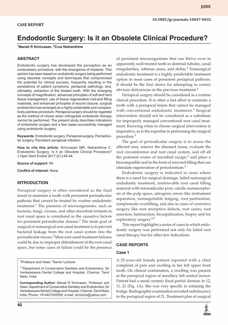
to 21, 22, 23. A large osseous defect measuring 2 × 1 cm was observed (Fig. 2B). The osseous defect was refined using slow-speed bone cutting bur no 703 under water coolant. Root ends of 21, 22, 23 were inspected under magnification. Granulation tissue was curetted and sent for histopathological examination. Root end resection of 3 mm was done (Fig. 2B). The surgical site was irrigated with normal saline and chlorhexidine. The flap was repo-sitioned and simple interrupted sutures were placed. A postoperative radiograph was taken (Fig. 2C).
Case 3
A 28-year-old female patient reported with a chief com-plaint of dislodged crown and pain in her right upper front tooth (#11). She gave a history of root canal treatment for #11, 6 months back but discontinued the treatment. The patient’s medical history was noncontributory. On clinical examination, the upper right central incisor was sensitive to percussion; probing depth was within the normal range. A nonsurgical retreatment was planned. Access cavity was refined and a K file #25 was placed. On radiographic examination, the file was seen perfo-rating the distal aspect of 11 at the middle third of the root surface (Fig. 3A). Treatment plan was modified to a surgical management of the perforation. Root canal was renegotiated and copiously irrigated with normal saline and 2% chlorhexidine. Sodium hypochlorite was avoided to prevent seepage through the perforation site. Tooth was temporarily restored using intermediate restorative material (IRM). After obtaining the patient’s consent, a full-thickness mucoperiosteal flap was reflected under local anesthesia (2% lignocaine hydrochloride with adrenaline 1:80,000). On inspection, root perforation was
Figs 1A and B: (A) Periapical swelling in relation to 21; and (B) vertical root fracture in 21 extending to coronal to middle third of the root
A B
Manali R Srinivasan, Cruz Nishanthine
42
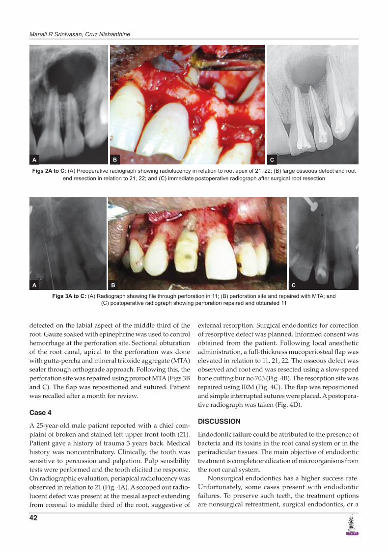
detected on the labial aspect of the middle third of the root. Gauze soaked with epinephrine was used to control hemorrhage at the perforation site. Sectional obturation of the root canal, apical to the perforation was done with gutta-percha and mineral trioxide aggregate (MTA) sealer through orthograde approach. Following this, the perforation site was repaired using proroot MTA (Figs 3B and C). The flap was repositioned and sutured. Patient was recalled after a month for review.
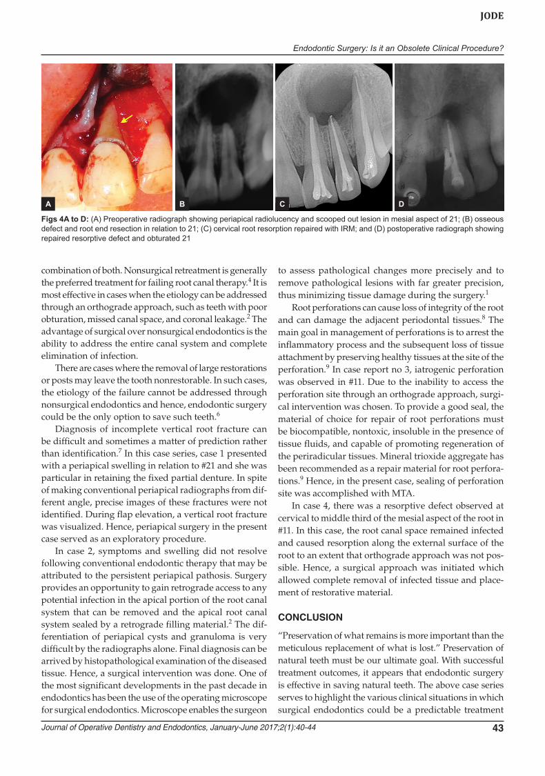
Case 4
A 25-year-old male patient reported with a chief com-plaint of broken and stained left upper front tooth (21). Patient gave a history of trauma 3 years back. Medical history was noncontributory. Clinically, the tooth was sensitive to percussion and palpation. Pulp sensibility tests were performed and the tooth elicited no response. On radiographic evaluation, periapical radiolucency was observed in relation to 21 (Fig. 4A). A scooped out radio-lucent defect was present at the mesial aspect extending from coronal to middle third of the root, suggestive of
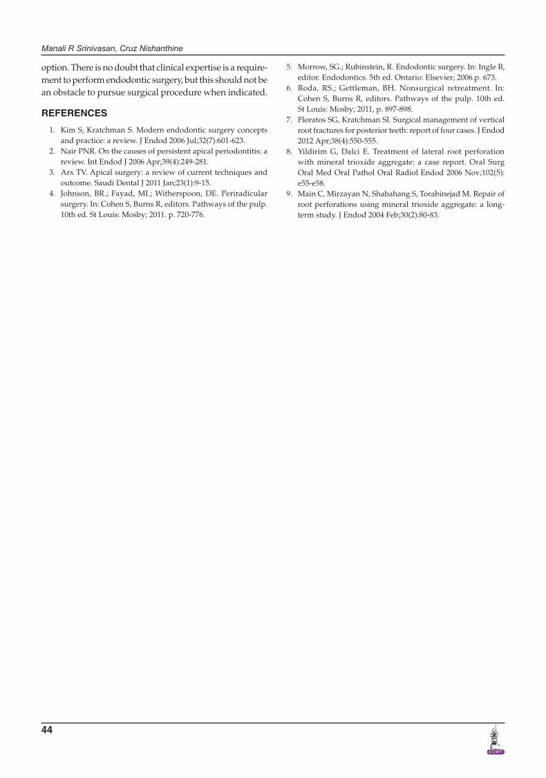
external resorption. Surgical endodontics for correction of resorptive defect was planned. Informed consent was obtained from the patient. Following local anesthetic administration, a full-thickness mucoperiosteal flap was elevated in relation to 11, 21, 22. The osseous defect was observed and root end was resected using a slow-speed bone cutting bur no 703 (Fig. 4B). The resorption site was repaired using IRM (Fig. 4C). The flap was repositioned and simple interrupted sutures were placed. A postopera-tive radiograph was taken (Fig. 4D).
DISCUSSION
Endodontic failure could be attributed to the presence of bacteria and its toxins in the root canal system or in the periradicular tissues. The main objective of endodontic treatment is complete eradication of microorganisms from the root canal system.
Nonsurgical endodontics has a higher success rate. Unfortunately, some cases present with endodontic failures. To preserve such teeth, the treatment options are nonsurgical retreatment, surgical endodontics, or a
Figs 3A to C: (A) Radiograph showing file through perforation in 11; (B) perforation site and repaired with MTA; and (C) postoperative radiograph showing perforation repaired and obturated 11
Figs 2A to C: (A) Preoperative radiograph showing radiolucency in relation to root apex of 21, 22; (B) large osseous defect and root end resection in relation to 21, 22; and (C) immediate postoperative radiograph after surgical root resection
A B C
A B C
Endodontic Surgery: Is it an Obsolete Clinical Procedure?
Journal of Operative Dentistry and Endodontics, January-June 2017;2(1):40-44 43
JODE
combination of both. Nonsurgical retreatment is generally the preferred treatment for failing root canal therapy.4 It is most effective in cases when the etiology can be addressed through an orthograde approach, such as teeth with poor obturation, missed canal space, and coronal leakage.2 The advantage of surgical over nonsurgical endodontics is the ability to address the entire canal system and complete elimination of infection.
There are cases where the removal of large restorations or posts may leave the tooth nonrestorable. In such cases, the etiology of the failure cannot be addressed through nonsurgical endodontics and hence, endodontic surgery could be the only option to save such teeth.6
Diagnosis of incomplete vertical root fracture can be difficult and sometimes a matter of prediction rather than identification.7 In this case series, case 1 presented with a periapical swelling in relation to #21 and she was particular in retaining the fixed partial denture. In spite of making conventional periapical radiographs from dif-ferent angle, precise images of these fractures were not identified. During flap elevation, a vertical root fracture was visualized. Hence, periapical surgery in the present case served as an exploratory procedure.
In case 2, symptoms and swelling did not resolve following conventional endodontic therapy that may be attributed to the persistent periapical pathosis. Surgery provides an opportunity to gain retrograde access to any potential infection in the apical portion of the root canal system that can be removed and the apical root canal system sealed by a retrograde filling material.2 The dif-ferentiation of periapical cysts and granuloma is very difficult by the radiographs alone. Final diagnosis can be arrived by histopathological examination of the diseased tissue. Hence, a surgical intervention was done. One of the most significant developments in the past decade in endodontics has been the use of the operating microscope for surgical endodontics. Microscope enables the surgeon
to assess pathological changes more precisely and to remove pathological lesions with far greater precision, thus minimizing tissue damage during the surgery.1
Root perforations can cause loss of integrity of the root and can damage the adjacent periodontal tissues.8 The main goal in management of perforations is to arrest the inflammatory process and the subsequent loss of tissue attachment by preserving healthy tissues at the site of the perforation.9 In case report no 3, iatrogenic perforation was observed in #11. Due to the inability to access the perforation site through an orthograde approach, surgi-cal intervention was chosen. To provide a good seal, the material of choice for repair of root perforations must be biocompatible, nontoxic, insoluble in the presence of tissue fluids, and capable of promoting regeneration of the periradicular tissues. Mineral trioxide aggregate has been recommended as a repair material for root perfora-tions.9 Hence, in the present case, sealing of perforation site was accomplished with MTA.
In case 4, there was a resorptive defect observed at cervical to middle third of the mesial aspect of the root in #11. In this case, the root canal space remained infected and caused resorption along the external surface of the root to an extent that orthograde approach was not pos-sible. Hence, a surgical approach was initiated which allowed complete removal of infected tissue and place-ment of restorative material.
CONCLUSION
“Preservation of what remains is more important than the meticulous replacement of what is lost.” Preservation of natural teeth must be our ultimate goal. With successful treatment outcomes, it appears that endodontic surgery is effective in saving natural teeth. The above case series serves to highlight the various clinical situations in which surgical endodontics could be a predictable treatment
Figs 4A to D: (A) Preoperative radiograph showing periapical radiolucency and scooped out lesion in mesial aspect of 21; (B) osseous defect and root end resection in relation to 21; (C) cervical root resorption repaired with IRM; and (D) postoperative radiograph showing repaired resorptive defect and obturated 21
A B C D
Manali R Srinivasan, Cruz Nishanthine
44
option. There is no doubt that clinical expertise is a require-ment to perform endodontic surgery, but this should not be an obstacle to pursue surgical procedure when indicated.
REFERENCES
1. Kim S, Kratchman S. Modern endodontic surgery concepts and practice: a review. J Endod 2006 Jul;32(7):601-623.
2. Nair PNR. On the causes of persistent apical periodontitis: a review. Int Endod J 2006 Apr;39(4):249-281.
3. Arx TV. Apical surgery: a review of current techniques and outcome. Saudi Dental J 2011 Jan;23(1):9-15.
4. Johnson, BR.; Fayad, MI.; Witherspoon, DE. Periradicular surgery. In: Cohen S, Burns R, editors. Pathways of the pulp. 10th ed. St Louis: Mosby; 2011. p. 720-776.
5. Morrow, SG.; Rubinstein, R. Endodontic surgery. In: Ingle B, editor. Endodontics. 5th ed. Ontario: Elsevier; 2006.p. 673.
6. Roda, RS.; Gettleman, BH. Nonsurgical retreatment. In: Cohen S, Burns R, editors. Pathways of the pulp. 10th ed. St Louis: Mosby; 2011, p. 897-898.
7. Floratos SG, Kratchman SI. Surgical management of vertical root fractures for posterior teeth: report of four cases. J Endod 2012 Apr;38(4):550-555.
8. Yildirim G, Dalci E. Treatment of lateral root perforation with mineral trioxide aggregate: a case report. Oral Surg Oral Med Oral Pathol Oral Radiol Endod 2006 Nov;102(5): e55-e58.
9. Main C, Mirzayan N, Shabahang S, Torabinejad M. Repair of root perforations using mineral trioxide aggregate: a long-term study. J Endod 2004 Feb;30(2):80-83.