endocrine system - midland independent school district
TRANSCRIPT
CHAPTER 16
ENDOCRINESYSTEM
KEY TERMSadenohypophysisadrenal glandsendocrinegonadshormoneneurohypophysis
parathyroidprostaglandin (PG)target cellthymusthyroidtropic hormone
The endocrine systems and nervous system both func-tion to achieve and maintain stability of the internalenvironment. Each system may work alone or in con-
cert with others as a single neuroendocrine system, per-forming the same general functions within the body: com-munication, integration, and control.
Both the endocrine system and the nervous system per-form their regulatory functions by means of chemical mes-sengers sent to specific cells. In the nervous system, neuronssecrete neurotransmitter molecules to signal nearby cells thathave the appropriate receptor molecules. In the endocrinesystem, secreting cells send hormone (from the Greek hor-maein, “to excite”) molecules by way of the bloodstream tosignal specific target cells throughout the body. Tissues andorgans that contain endocrine target cells are called target tis-sues and target organs, respectively. As with postsynaptic cells,endocrine target cells must have the appropriate receptor tobe influenced by the signaling chemical. Many cells have re-ceptors for neurotransmitters and hormones, so they can beinfluenced by both types of chemicals.
Whereas neurotransmitters are sent over very short dis-tances across a synapse, hormones diffuse into the blood to
CHAPTER OUTLINEHormones, 486
Classification of Hormones, 486Steroid Hormones, 486Nonsteroid Hormones, 486
How Hormones Work, 488General Principles of Hormone Action, 488Mechanism of Steroid Hormone Action, 489Mechanisms of Nonsteroid Hormone Action, 489
Regulation of Hormone Secretion, 491Prostaglandins, 494Pituitary Gland, 495
Structure of the Pituitary Gland, 495Adenohypophysis (Anterior Pituitary), 496
Growth Hormone, 496Prolactin, 496Tropic Hormones, 496Control of Secretion in the Adenohypophysis, 498
Neurohypophysis (Posterior Pituitary), 500Antidiuretic Hormone, 500Oxytocin, 501
Pineal Gland, 502Thyroid Gland, 502
Structure of the Thyroid Gland, 502Thyroid Hormone, 502Calcitonin, 503
Parathyroid Glands, 505Structure of the Parathyroid Glands, 505Parathyroid Hormone, 505
Adrenal Glands, 506Structure of the Adrenal Glands, 506Adrenal Cortex, 506
Mineralocorticoids, 507Glucocorticoids, 508Gonadocorticoids, 509
Adrenal Medulla, 509Pancreatic Islets, 510
Structure of the Pancreatic Islets, 510Pancreatic Hormones, 510
Gonads, 513Testes, 513Ovaries, 513
Placenta, 513Thymus, 514Gastric and Intestinal Mucosa, 514Heart, 514Cycle of Life, 518The Big Picture, 518Mechanisms of Disease, 518Case Study, 521
484

be carried to nearly every point in the body. The nervous sys-tem can directly control only muscles and glands that are in-nervated with efferent fibers, whereas the endocrine systemcan regulate most cells in the body. The effects of neuro-transmitters are rapid and short-lived compared with the ef-fects of hormones, which appear more slowly and lastlonger. Table 16-1 compares endocrine structure and func-tion with nervous structure and function (Figure 16-1).
Endocrine glands secrete their products, hormones, di-rectly into the blood. Because they do not have ducts, theyare often called “ductless glands.” This characteristic distin-guishes endocrine glands from exocrine glands, which secretetheir products into ducts (see Chapter 5, p. 131). Many en-docrine glands are made of glandular epithelium, whosecells manufacture and secrete hormones. However, a few en-docrine glands are made of neurosecretory tissue. Neurose-cretory cells are simply modified neurons that secrete chem-ical messengers that diffuse into the bloodstream rather thanacross a synapse. In such cases, the chemical messenger iscalled a hormone rather than a neurotransmitter. For exam-ple, when norepinephrine is released by neurons, diffusesacross a synapse, and binds to an adrenergic receptor in apostsynaptic neuron, we call norepinephrine a neurotrans-
mitter. On the other hand, we call norepinephrine a hormonewhen it diffuses into the blood (because there is no postsy-naptic cell present), then binds to an adrenergic receptor ina distant target cell.
Glands of the endocrine system are widely scatteredthroughout the body. New discoveries in endocrinology con-tinue to add to the long list of hormone-secreting tissues.However, even the most newly discovered endocrine tissuesand their hormones operate according to some basic physi-ological principles. In this chapter, we will focus our discus-sion primarily on the major endocrine glands. Figure 16-2and Table 16-2 summarize the names and locations of thesemajor endocrine glands. After you are familiar with the ba-sic principles of endocrinology and the major examples ofglands and their hormones, you will be prepared for addi-tional examples that you will encounter as you continueyour study of the human body.
Endocrine System Chapter 16 485
Table 16-1 Comparison of Features of the Endocrine System and Nervous System
Feature Endocrine System Nervous System
Overall Function
Control by regulatory feedback loops
Effector tissues
Effector cells
Chemical MessengerCells that secrete the chemical messenger
Distance traveled (and method of travel)
by chemical messenger
Location of receptor in effector cell
Characteristics of regulatory effects
Regulation of effectors to maintain
homeostasis
Yes (endocrine reflexes)
Endocrine effectors: virtually all tissues
Target cells (throughout the body)
Hormone
Glandular epithelial cells or neurosecretory
cells (modified neurons)
Long (by way of circulating blood)
On the plasma membrane or within the cell
Slow to appear, long-lasting
Regulation of effectors to maintain
homeostasis
Yes (nervous reflexes)
Nervous effectors: muscle and glan-
dular tissue only
Postsynaptic cells (in muscle and
glandular tissue only)
Neurotransmitter
Neurons
Short (across a microscopic synapse)
On the plasma membrane
Appear rapidly, short-lived
Endocrinecell
Bloodstream
Hormone
Slow
Fast
Neuron
Postsynapticcell
Neurotransmitter
Targetcell
A B
Figure 16-1 Mechanisms of endocrine (A) and nervous (B) signals.
1. What is meant by the term target cell?2. Describe how the nervous system and the endocrine
system differ in the way they control effectors.

HORMONES
CLASSIFICATION OF HORMONESHormone molecules can be classified in various useful ways.For example, when classified by general function, hormonescan be identified as tropic hormones (hormones that targetother endocrine glands and stimulate their growth and se-
cretion), sex hormones (hormones that target reproductivetissues), anabolic hormones (hormones that stimulate an-abolism in their target cells), and many other functionalnames. Another useful way to classify hormones is by theirchemical structure. Because this method of classifying hor-mones is so widely used, we will briefly describe it in the fol-lowing paragraphs.
Steroid HormonesAll of the many hormones secreted by endocrine tissues canbe classified simply as steroid or nonsteroid (Figure 16-3).Steroid hormone molecules are manufactured by endocrinecells from cholesterol, an important type of lipid in the hu-man body (see Chapter 2, p. 57). As Figure 16-4 shows, be-cause all steroid hormones are derived from a commonmolecule, cholesterol, they have a characteristic chemicalgroup at the core of each molecule. Because steroids arelipid-soluble, they can easily pass through the phospholipidplasma membrane of target cells. Examples of steroid hor-mones include cortisol, aldosterone, estrogen, progesterone,and testosterone (Figures 16-3 and 16-4).
Nonsteroid HormonesNonsteroid hormones are synthesized primarily from aminoacids rather than from cholesterol (Figure 16-5). Some non-steroid hormones are protein hormones. These hormones arelong, folded chains of amino acids, a structure typical of pro-tein molecules of any sort (see Chapter 2, p. 51). Includedamong the protein hormones are insulin, parathyroid hor-mone, and others listed in Figure 16-3. Protein hormones thathave carbohydrate groups attached to their amino acid chainsare often classified separately as glycoprotein hormones.
Another major category of nonsteroid hormones consistsof the peptide hormones. Peptide hormones such as oxy-tocin and antidiuretic hormone are smaller than the proteinhormones. They are each made of a short chain of amino
486 Unit 3 Communication, Control, and Integration
Table 16-2 Names and Locations of MajorEndocrine Glands
Name Location
Hypothalamus Cranial cavity (brain)
Pituitary gland Cranial cavity
(hypophysis cerebri)
Pineal gland Cranial cavity (brain)
Thyroid gland Neck
Parathyroid glands Neck
Thymus Mediastinum
Adrenal glands Abdominal cavity (retroperitoneal)
Pancreatic islets Abdominal cavity (pancreas)
Ovaries Pelvic cavity
Testes Scrotum
Placenta Pregnant uterus
Figure 16-2 Locations of the major endocrine glands.

Endocrine System Chapter 16 487
Steroid
HORMONES
Nonsteroid
Peptides
Cortisol (hydrocortisone)AldosteroneEstrogenProgesteroneTestosterone
Proteins Glycoproteins
Growth hormone (GH)Prolactin (PRL)Parathyroid hormone (PTH)CalcitoninAdrenocorticotropic hormone (ACTH)InsulinGlucagon
Antidiuretic hormone (ADH)OxytocinMelanocyte-stimulating homone (MSH)SomatostatinThyrotropin-releasing hormone (TRH)Gonadotropin-releasing hormone (GnRH)Atrial natriuretic hormone (ANH)
Amines NorepinephrineEpinephrineMelatonin
Iodinated amino acidsThyroxine (T4)Triiodothyronine (T3)
Follicle-stimulating hormone (FSH)Luteinizing hormone (LH)Thyroid-stimulating hormone (TSH)Chorionic gonadotropin (CG)
Amino acid derivatives
Figure 16-3 Chemical classification of hormones.

acids, as Figure 16-5, B, shows. Examples of peptide hor-mones are listed in Figure 16-3.
Yet another category of nonsteroid hormones consists ofthe amino acid derivative hormones. Each of these hormones isderived from only a single amino acid molecule. There are twomajor subgroups within this category. One subgroup, theamine hormones, is synthesized by modifying a single mole-cule of the amino acid, tyrosine. Amine hormones such as ep-inephrine and norepinephrine are produced by neurosecre-tory cells (where they are secreted as hormones) and byneurons (where they are secreted as neurotransmitters). An-other subgroup of amino acid derivatives produced by thethyroid gland are all synthesized by adding iodine (I) atoms toa tyrosine molecule (Figure 16-5, C). Examples of hormonesderived from single amino acids are listed in Figure 16-3.
HOW HORMONES WORKGeneral Principles of Hormone ActionAs previously stated, hormones signal a cell by binding tospecific receptors on or in the cell. In a “lock-and-key”mechanism, hormones will bind only to receptor moleculesthat “fit” them exactly. Any cell with one or more receptorsfor a particular hormone is said to be a target of that hor-mone (Figure 16-6). Cells usually have many different typesof receptors; therefore, they are target cells of many differenthormones.
Each different hormone-receptor interaction producesdifferent regulatory changes within the target cell. Thesecellular changes are usually accomplished by altering thechemical reactions within the target cell. For example,some hormone-receptor interactions initiate synthesis ofnew proteins. Other hormone-receptor interactions trigger
488 Unit 3 Communication, Control, and Integration
Figure 16-5 Nonsteroid hormone structure. As these examples show, protein hormone molecules (A) aremade of long, folded strands of amino acids. B, Peptide hormone molecules are smaller strands of amino acids.C, Amino acid derivatives are, as their name implies, derived from a single amino acid.
Figure 16-4 Steroid hormone structure. Asthese examples show, steroid hormone mole-cules are very similar in structure to cholesterol(top), from which they are all derived.
1. How are steroid hormones able to pass through a cell’splasma membrane easily?
2. Name some of the different general types of nonsteroidhormones. Can you give an example of each?
A
B C

the activation or inactivation of certain enzymes and thusaffect the metabolic reactions regulated by those enzymes.Still other hormone-receptor interactions regulate cells byopening or closing specific ion channels in the plasmamembrane. Specific mechanisms of hormone-receptor in-teractions are outlined in the next section.
Different hormones may work together to enhance eachother’s influence on a target cell. In a phenomenon calledsynergism, combinations of hormones have a greater effecton a target cell than the sum of the effects that each wouldhave if acting alone. Combined hormone actions may ex-hibit instead the phenomenon of permissiveness. Permis-siveness occurs when a small amount of one hormone allowsa second hormone to have its full effect on a target cell; thefirst hormone “permits” the full action of the second hor-mone. A common type of combined action of hormones isseen in the phenomenon of antagonism. In antagonism, onehormone produces the opposite effect of another hormone.Antagonism between hormones can be used to “fine tune”the activity of target cells with great accuracy, signaling thecell exactly when (and by how much) to increase or decreasea certain cellular process.
As previously stated, hormones travel to their target cellsby way of the circulating bloodstream. This means that allhormones travel throughout the body. Because they only af-fect their target cells, however, the effects of a particular hor-mone may be limited to specific tissues in the body. Somehormone molecules are attached to plasma proteins whilethey are carried along the bloodstream. Such hormonesmust free themselves from the plasma protein to leave theblood and combine with their receptors. Because blood car-ries hormones nearly everywhere in the body, even wherethere are no target cells, endocrine glands produce morehormone molecules than actually hit their target. Unusedhormones usually are quickly excreted by the kidneys or bro-ken down by metabolic processes.
Mechanism of Steroid Hormone ActionSteroid hormones are lipids and thus are not very soluble inblood plasma, which is mostly water. Instead of traveling inthe plasma as free molecules, they attach to soluble plasmaproteins. As you can see in Figure 16-7, a steroid hormonemolecule dissociates from its carrier before approaching thetarget cell. Because steroid hormones are lipid-soluble andthus can pass into cells easily, it is not surprising that theirreceptors are normally found inside the cell rather than onthe surface of the plasma membrane. After a steroid hor-mone molecule has diffused into its target cell, it passes intothe nucleus where it binds to a mobile receptor molecule toform a hormone-receptor complex. Some hormones must beactivated by enzymes before they can bind to their receptors.Because steroid hormone receptors are not attached to theplasma membrane, but seem to move freely in the nucleo-plasm, this model of hormone action has been called themobile-receptor hypothesis.
Once formed, the hormone-receptor complex activates acertain gene sequence to begin transcription of messengerRNA (mRNA) molecules. The newly formed mRNA mole-cules then move out of the nucleus into the cytosol, wherethey associate with ribosomes and begin synthesizing pro-tein molecules.
The new protein molecules synthesized by the target cellwould not have been made if not for the arrival of the steroidhormone molecule. Steroid hormones regulate cells by regu-lating their production of certain critical proteins, such asenzymes that control intracellular reactions or membraneproteins that alter the permeability of a cell.
This mechanism of steroid hormone action implies sev-eral things about the effects of these hormones. For onething, the more hormone-receptor complexes formed, themore mRNA molecules are transcribed, the more new pro-tein molecules are formed, and thus the greater the magni-tude of the regulatory effect. In short, the amount of steroidhormone present determines the magnitude of a target cell’sresponse. Also, because transcription and protein synthesistake some time, responses to steroid hormones are oftenslow—from 45 minutes to several days before the full effectis seen.
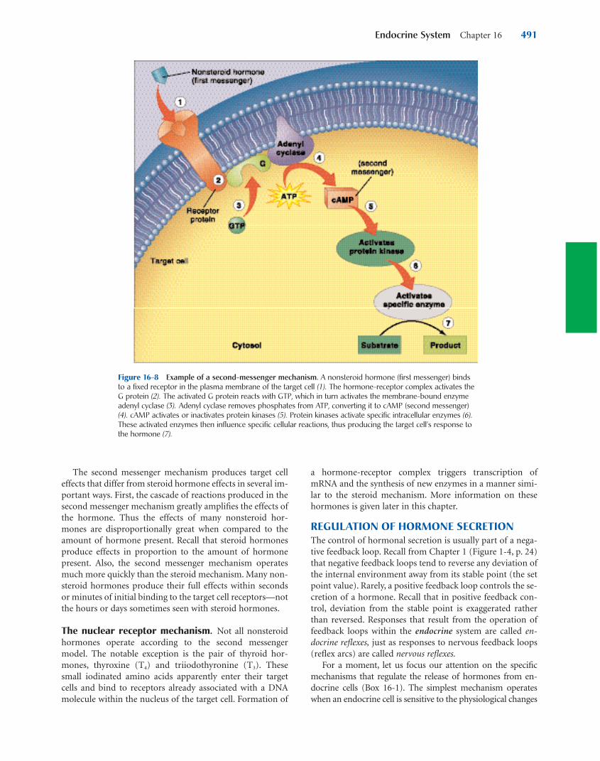
Mechanisms of Nonsteroid Hormone ActionThe second messenger mechanism. Nonsteroid hormonestypically operate according to a mechanism originally calledthe second messenger hypothesis. This concept of hormoneaction—first proposed several decades ago by Dr. Earl W.Sutherland—was a milestone in endocrinology for which hereceived the 1971 Nobel Prize in Medicine and Physiology.According to this concept, a nonsteroid hormone moleculeacts as a “first messenger,” delivering its chemical message tofixed receptors in the target cell’s plasma membrane. The“message” is then passed into the cell where a “second mes-senger” triggers the appropriate cellular changes. This con-cept of nonsteroid hormone action is also called the fixed-membrane-receptor hypothesis.
In the example illustrated in Figure 16-8, formation ofthe hormone-receptor complex causes a membrane protein,
Endocrine System Chapter 16 489
Figure 16-6 The target cell concept. A hormone acts only on cellsthat have receptors specific to that hormone because the shape ofthe receptor determines which hormone can react with it. This is anexample of the lock-and-key model of biochemical reactions.

called the G protein, to bind to a nucleotide called guanosinetriphosphate (GTP). This, in turn, activates another mem-brane protein, adenyl cyclase. Adenyl cyclase is an enzymethat promotes the removal of two phosphate groups fromadenosine triphosphate (ATP) molecules in the cytosol. Theproduct thus formed is cyclic adenosine monophosphate(cAMP). The cAMP molecule acts as a “second messenger”within the cell. cAMP activates protein kinases, a set of en-zymes that activate other types of enzymes. It is this final setof specific enzymes, which are now activated, that catalyzethe cellular reactions that characterize the target cell’s re-sponse. In short, the hormone “first messenger” binds to amembrane receptor, triggering formation of an intracellular“second messenger,” which activates a cascade of chemicalreactions that produces the target cell’s response.
Since the time Sutherland first began his pioneering work,other second messenger systems have been discovered. As amatter of fact, the study of second messenger mechanisms isstill a very active area of research, with new discoveries con-tinuing to be revealed in scientific journals around the world.Although most nonsteroid hormones seem to use cAMP as
the second messenger, we now know that a few hormones usecompounds such as inositol triphosphate (IP3) and cyclicguanosine monophosphate (GMP) as the second messenger.
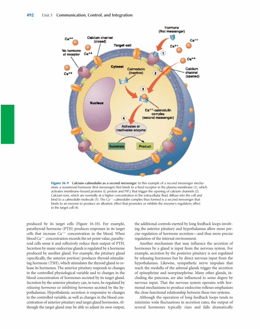
Still other hormones produce their effects by triggeringthe opening of calcium (Ca��) channels in the target cell’smembranes, as you can see in Figure 16-9. Binding of a hor-mone to a fixed membrane receptor activates a chain ofmembrane proteins (G protein and phosphodiesterase,PIP2) that in turn trigger the opening of calcium channels inthe plasma membrane. Ca�� ions that enter the cytosolwhen the channels open bind to an intracellular moleculecalled calmodulin. The Ca��-calmodulin complex thusformed acts as a second messenger, influencing the enzymesthat produce the target cell’s response.
Recent research findings also show that in second mes-senger systems, the hormone-receptor complexes may betaken into the cell by means of endocytosis. Although thepurpose of this may be primarily to break down the com-plexes and recycle the receptors, the hormone-receptor com-plex may continue to have physiological effects after it istaken into the cell.
490 Unit 3 Communication, Control, and Integration
Figure 16-7 Steroid hormone mechanism. According to the mobile-receptor hypothesis, lipid-solublesteroid hormone molecules detach from a carrier protein (1) and pass through the plasma membrane (2). Thehormone molecules then pass into the nucleus where they bind with a mobile receptor to form a hormone-receptor complex (3). This complex then binds to a specific site on a DNA molecule (4), triggering transcriptionof the genetic information encoded there (5). The resulting mRNA molecule moves to the cytosol, where it as-sociates with a ribosome, initiating synthesis of a new protein (6). This new protein—usually an enzyme orchannel protein—produces specific effects in the target cell (7).
The second messenger mechanism produces target celleffects that differ from steroid hormone effects in several im-portant ways. First, the cascade of reactions produced in thesecond messenger mechanism greatly amplifies the effects ofthe hormone. Thus the effects of many nonsteroid hor-mones are disproportionally great when compared to theamount of hormone present. Recall that steroid hormonesproduce effects in proportion to the amount of hormonepresent. Also, the second messenger mechanism operatesmuch more quickly than the steroid mechanism. Many non-steroid hormones produce their full effects within secondsor minutes of initial binding to the target cell receptors—notthe hours or days sometimes seen with steroid hormones.
The nuclear receptor mechanism. Not all nonsteroidhormones operate according to the second messengermodel. The notable exception is the pair of thyroid hor-mones, thyroxine (T4) and triiodothyronine (T3). Thesesmall iodinated amino acids apparently enter their targetcells and bind to receptors already associated with a DNAmolecule within the nucleus of the target cell. Formation of
a hormone-receptor complex triggers transcription ofmRNA and the synthesis of new enzymes in a manner simi-lar to the steroid mechanism. More information on thesehormones is given later in this chapter.
REGULATION OF HORMONE SECRETIONThe control of hormonal secretion is usually part of a nega-tive feedback loop. Recall from Chapter 1 (Figure 1-4, p. 24)that negative feedback loops tend to reverse any deviation ofthe internal environment away from its stable point (the setpoint value). Rarely, a positive feedback loop controls the se-cretion of a hormone. Recall that in positive feedback con-trol, deviation from the stable point is exaggerated ratherthan reversed. Responses that result from the operation offeedback loops within the endocrine system are called en-docrine reflexes, just as responses to nervous feedback loops(reflex arcs) are called nervous reflexes.
For a moment, let us focus our attention on the specificmechanisms that regulate the release of hormones from en-docrine cells (Box 16-1). The simplest mechanism operateswhen an endocrine cell is sensitive to the physiological changes
Endocrine System Chapter 16 491
Figure 16-8 Example of a second-messenger mechanism. A nonsteroid hormone (first messenger) binds to a fixed receptor in the plasma membrane of the target cell (1). The hormone-receptor complex activates theG protein (2). The activated G protein reacts with GTP, which in turn activates the membrane-bound enzymeadenyl cyclase (3). Adenyl cyclase removes phosphates from ATP, converting it to cAMP (second messenger)(4). cAMP activates or inactivates protein kinases (5). Protein kinases activate specific intracellular enzymes (6).These activated enzymes then influence specific cellular reactions, thus producing the target cell’s response tothe hormone (7).
produced by its target cells (Figure 16-10). For example,parathyroid hormone (PTH) produces responses in its targetcells that increase Ca�� concentration in the blood. Whenblood Ca�� concentration exceeds the set point value, parathy-roid cells sense it and reflexively reduce their output of PTH.Secretion by many endocrine glands is regulated by a hormoneproduced by another gland. For example, the pituitary gland(specifically, the anterior portion) produces thyroid-stimulat-ing hormone (TSH), which stimulates the thyroid gland to re-lease its hormones. The anterior pituitary responds to changesin the controlled physiological variable and to changes in theblood concentration of hormones secreted by its target gland.Secretion by the anterior pituitary can, in turn, be regulated byreleasing hormones or inhibiting hormones secreted by the hy-pothalamus. Hypothalamic secretion is responsive to changesin the controlled variable, as well as changes in the blood con-centration of anterior pituitary and target gland hormones. Al-though the target gland may be able to adjust its own output,
the additional controls exerted by long feedback loops involv-ing the anterior pituitary and hypothalamus allow more pre-cise regulation of hormone secretion—and thus more preciseregulation of the internal environment.
Another mechanism that may influence the secretion ofhormones by a gland is input from the nervous system. Forexample, secretion by the posterior pituitary is not regulatedby releasing hormones but by direct nervous input from thehypothalamus. Likewise, sympathetic nerve impulses thatreach the medulla of the adrenal glands trigger the secretionof epinephrine and norepinephrine. Many other glands, in-cluding the pancreas, are also influenced to some degree bynervous input. That the nervous system operates with hor-monal mechanisms to produce endocrine reflexes emphasizesthe close functional relationship between these two systems.
Although the operation of long feedback loops tends tominimize wide fluctuations in secretion rates, the output ofseveral hormones typically rises and falls dramatically
492 Unit 3 Communication, Control, and Integration
Figure 16-9 Calcium-calmodulin as a second messenger. In this example of a second messenger mecha-nism, a nonsteroid hormone (first messenger) first binds to a fixed receptor in the plasma membrane (1), whichactivates membrane-bound proteins (G protein and PIP2) that trigger the opening of calcium channels (2).Calcium ions, which are normally at a higher concentration in the extracellular fluid, diffuse into the cell andbind to a calmodulin molecule (3). The Ca��-calmodulin complex thus formed is a second messenger thatbinds to an enzyme to produce an allosteric effect that promotes or inhibits the enzyme’s regulatory effect in the target cell (4).

within a short period. For example, the concentration ofinsulin—a hormone that can correct a rise in blood glucoseconcentration—increases to a high level just after a mealhigh in carbohydrates. The level of insulin decreases only af-ter the blood glucose concentration returns to its set pointvalue. Likewise, threatening stimuli can cause a sudden, dra-matic increase in the secretion of epinephrine from theadrenal medulla as part of the fight-or-flight response.
Specific examples of feedback control of hormone secre-tion are given later in this chapter.
Endocrine System Chapter 16 493
Lactation
Decreases
Ca��
Blood Ca++
concentration
Detected by
Parathyroidglands
Cell of parathyroid(secretes PTH)
Sensor-integratorPTH
Bloodvessel
Correction signal viaparathyroid hormone (PTH)
release
Bone
Setpoint
Bloodvessels
Effector
Ca++ Osteoclast(reabsorbs Ca++ )
Increases via Ca++
release into blood
Controlledvariable
Figure 16-10 Endocrine feedback loop. In this example of a short feedback loop, each parathyroid gland issensitive to changes in the physiological variable its hormone (PTH) controls—blood calcium (Ca��) concentra-tion. When lactation (milk production) in a pregnant woman consumes Ca�� and thus lowers blood Ca�� con-centration, the parathyroids sense the change and respond by increasing their secretion of parathyroidhormone (PTH). PTH stimulates osteoclasts in bone to release more Ca�� from storage in bone tissue (amongother effects), which increases maternal blood Ca�� concentration to the set point level.
1. Name some ways in which hormones can work togetherto regulate a tissue.
2. Why is the concept of steroid hormone action called themobile-receptor hypothesis?
3. Why is the concept of nonsteroid hormone action often calledthe second messenger hypothesis? Why is it known as the fixed-membrane-receptor hypothesis?
4. Name some of the ways that the secretion of an endocrine cellcan be controlled.
PROSTAGLANDINSBefore continuing with our discussion of endocrineglands and hormones, let us pause a moment to considerthe prostaglandins (PGs) and related compounds. Theprostaglandins are a unique group of lipid molecules that
serve important and widespread integrative functions inthe body but do not meet the usual definition of a hormone.Prostaglandins are derived from 20-carbon unsaturated fattyacids and contain a 5-carbon ring (Figure 16-11). Althoughthey may be secreted directly into the bloodstream, theyare rapidly metabolized, so that circulating levels are ex-tremely low. The term tissue hormone is appropriate be-cause the secretion is produced in a tissue and diffusesonly a short distance to other cells within the same tissue.Whereas typical hormones integrate activities of widelyseparated organs, prostaglandins tend to integrate activi-ties of neighboring cells.
There are at least 16 different prostaglandins, falling intonine structural classes—prostaglandins A through prosta-glandins I. Prostaglandins have been isolated and identified
494 Unit 3 Communication, Control, and Integration
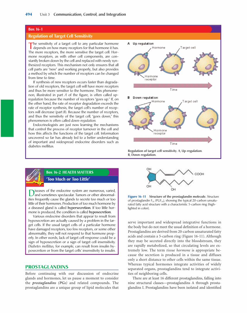
The sensitivity of a target cell to any particular hormonedepends on how many receptors for that hormone it has.
The more receptors, the more sensitive the target cell. Hor-mone receptors, as with other cell components, are con-stantly broken down by the cell and replaced with newly syn-thesized receptors. This mechanism not only ensures that allcell parts are “new” and working properly, but also providesa method by which the number of receptors can be changedfrom time to time.
If synthesis of new receptors occurs faster than degrada-tion of old receptors, the target cell will have more receptorsand thus be more sensitive to the hormone. This phenome-non, illustrated in part A of the figure, is often called up-regulation because the number of receptors “goes up.” If, onthe other hand, the rate of receptor degradation exceeds therate of receptor synthesis, the target cell’s number of recep-tors will decrease (part B). Because the number of receptors,and thus the sensitivity of the target cell, “goes down,” thisphenomenon is often called down-regulation.
Endocrinologists are just now learning the mechanismsthat control the process of receptor turnover in the cell andhow this affects the functions of the target cell. Informationuncovered so far has already led to a better understandingof important and widespread endocrine disorders such asdiabetes mellitus.
Box 16-1
Regulation of Target Cell Sensitivity
Regulation of target cell sensitivity. A, Up-regulation. B, Down-regulation.
Box 16-2 HEALTH MATTERS
“Too Much or Too Little”
Diseases of the endocrine system are numerous, varied,and sometimes spectacular. Tumors or other abnormal-
ities frequently cause the glands to secrete too much or toolittle of their hormones. Production of too much hormone bya diseased gland is called hypersecretion. If too little hor-mone is produced, the condition is called hyposecretion.
Various endocrine disorders that appear to result fromhyposecretion are actually caused by a problem in the tar-get cells. If the usual target cells of a particular hormonehave damaged receptors, too few receptors, or some otherabnormality, they will not respond to that hormone prop-erly. In other words, lack of target cell response could be asign of hyposecretion or a sign of target cell insensitivity.Diabetes mellitus, for example, can result from insulin hy-posecretion or from the target cells’ insensitivity to insulin.
OH
OHOH
COOH
Figure 16-11 Structure of the prostaglandin molecule. Structureof prostaglandin F2a (PGF2a), showing the typical 20-carbon unsatu-rated fatty acid structure with a characteristic 5-carbon ring (high-lighted in color).

from a variety of tissues. The first prostaglandin was discov-ered in semen, so it was attributed to the prostate gland(hence the name, prostaglandin). Later, researchers foundthat the seminal vesicles, not the prostate, secreted theprostaglandin that they had found. Other tissues known tosecrete prostaglandins include the kidneys, lungs, iris, brain,and thymus. As a group, the prostaglandins have diversephysiological effects and are among the most varied and po-tent of any naturally occurring biological compounds. Theyare intimately involved in overall endocrine regulation by in-fluencing adenyl cyclase–cAMP interaction within the cell’splasma membrane (see Figure 16-8). Specific biological ef-fects depend on the class of prostaglandin.
Intraarterial infusion of prostaglandins A (PGAs) resultsin an immediate fall in blood pressure accompanied by anincrease in regional blood flow to several areas, including thecoronary and renal systems. PGAs apparently produce thiseffect by causing relaxation of smooth muscle fibers in thewalls of certain arteries and arterioles.
Prostaglandins E (PGEs) have an important role in vari-ous vascular, metabolic, and gastrointestinal functions. Vas-cular effects include regulation of red blood cell deformabil-ity and platelet aggregation (see Chapter 17). PGEs also havea role in systemic inflammations such as fever. Common an-tiinflammatory agents, such as aspirin, produce some oftheir effects by inhibiting PGE synthesis. PGE also regulateshydrochloric acid secretion in the stomach, helping to pre-vent gastric ulcers.
Prostaglandins F (PGFs) have an especially importantrole in the reproductive system. They cause uterine musclecontractions, so they have been used to induce labor andthus accelerate delivery of a baby. PGFs also affect intestinalmotility and are required for normal peristalsis.
In addition to prostaglandins, various tissues also synthe-size other fatty acid compounds that are structurally andfunctionally similar to prostaglandins. Examples include thethromboxanes and leukotrienes. As with prostaglandins, thesecompounds may also be referred to as tissue hormones be-cause of their local, yet potent, regulatory effects.
The potential therapeutic use of prostaglandins and re-lated compounds, which are found in almost every body tis-sue and are capable of regulating hormone activity on thecellular level, has been described as the most revolutionarydevelopment in medicine since the advent of antibiotics.They are likely to play increasingly important roles in thetreatment of such diverse conditions as hypertension, coro-nary thrombosis, asthma, and ulcers.
PITUITARY GLAND
STRUCTURE OF THE PITUITARY GLANDThe pituitary gland, or hypophysis, is a small but mightystructure. It measures only 1.2 to 1.5 cm (about 1⁄2 inch)across. By weight, it is even less impressive—only about 0.5 g (1⁄60 ounce)! And yet so crucial are the functions of theanterior lobe of the pituitary gland that, years ago, it was re-ferred to as the “master gland.”
The hypophysis has a well-protected location within theskull on the ventral surface of the brain (Figure 16-12). It liesin the pituitary fossa of the sella turcica and is covered by a
Endocrine System Chapter 16 495
Figure 16-12 Location and structure of the pituitary gland (hypophysis). The pituitary gland is locatedwithin the sella turcica of the skull’s sphenoid bone and is connected to the hypothalamus by a stalklike in-fundibulum. The infundibulum passes through a gap in the portion of the dura mater that covers the pituitary(the pituitary diaphragm). The inset shows that the pituitary is divided into an anterior portion, the adenohy-pophysis, and a posterior portion, the neurohypophysis. The adenohypophysis is further subdivided into thepars anterior and pars intermedia. The pars intermedia is almost absent in the adult pituitary.
1. Why are prostaglandins sometimes called tissue hormones?
2. Why are prostaglandins considered to be important inclinical applications?

portion of the dura mater called the pituitary diaphragm.The gland has a stemlike stalk, the infundibulum, whichconnects it to the hypothalamus of the brain.
Although the pituitary looks like one gland, it actuallyconsists of two separate glands—the adenohypophysis, oranterior pituitary gland, and the neurohypophysis, or poste-rior pituitary gland. In the embryo, the adenohypophysis de-velops from an upward projection of the pharynx and iscomposed of regular endocrine tissue. The neurohypoph-ysis, on the other hand, develops from a downward projec-tion of the brain and is composed of neurosecretory tissue.These histological differences are incorporated into theirnames—adeno means “gland” and neuro means “nervous.”As you may suspect, the hormones secreted by the adenohy-pophysis serve very different functions from those releasedby the neurohypophysis.
ADENOHYPOPHYSIS (ANTERIOR PITUITARY)The adenohypophysis, the anterior portion of the pituitarygland, is divided into two parts—the pars anterior and thepars intermedia. The pars anterior forms the major portionof the adenohypophysis and is divided from the tiny pars in-termedia by a narrow cleft and some connective tissue (Fig-ure 16-12).
The tissue of the adenohypophysis is composed of irreg-ular clumps of secretory cells supported by fine connectivetissue fibers and surrounded by a rich vascular network.
Traditionally, histologists have identified three types ofcells according to their affinity for certain types of stains:chromophobes (literally “afraid of color”), acidophils (“acid[stain] lover”), and basophils (“base [stain] lover”). All threetypes are visible in the photomicrograph shown in Fig-ure 16-13. Currently, however, cells of the adenohypophysisare more often classified by their secretions into five types:
1. Somatotrophs—secrete growth hormone (GH)2. Corticotrophs—secrete adrenocorticotropic
hormone (ACTH) 3. Thyrotrophs—secrete thyroid-stimulating hormone
(TSH)4. Lactotrophs—secrete prolactin (PRL)5. Gonadotrophs—secrete luteinizing hormone (LH)
and follicle-stimulating hormone (FSH)
Figure 16-14 summarizes the hormones of the adenohy-pophysis and shows the primary locations of their targetcells.
Growth HormoneGrowth hormone (GH), or somatotropin (STH), is thoughtto promote bodily growth indirectly by stimulating the liverto produce certain growth factors, which, in turn, accelerateamino acid transport into cells. Rapid entrance of aminoacids from the blood into the cells allows protein anabolismwithin the cells to accelerate. Increased protein anabolism al-lows increased rate of growth. GH promotes the growth ofbone, muscle, and other tissues.
In addition to stimulating protein anabolism, GH alsostimulates fat metabolism. GH accelerates mobilization oflipids from storage in adipose cells and also speeds up the ca-tabolism of those lipids after they have entered another cell.In this way, GH tends to shift a cell’s use of nutrients awayfrom carbohydrate (glucose) catabolism and toward lipidcatabolism as an energy source. Because less glucose is thenremoved from the blood by cells, the blood glucose levelstend to rise. Thus GH is said to have a hyperglycemic effect.Insulin (from the pancreas) has the opposite effect—it pro-motes glucose entry into cells, producing a hypoglycemic ef-fect. Therefore GH and insulin function as antagonists. Thebalance between these two hormones is vital to maintaininga homeostasis of blood glucose levels.
GH affects metabolism in these ways:
• It promotes protein anabolism (growth, tissue repair)
• It promotes lipid mobilization and catabolism
• It indirectly inhibits glucose metabolism
• It indirectly increases blood glucose levels
ProlactinProlactin (PRL), produced by acidophils in the pars anterior,is also called lactogenic hormone. Both names of this hormonesuggest its function in “generating” or initiating milk secre-tion (lactation). During pregnancy, a high level of PRL pro-motes the development of the breasts in anticipation of milksecretion. At the birth of an infant, PRL in the mother stim-ulates the mammary glands to begin milk secretion.
Hypersecretion of PRL may cause lactation in nonnurs-ing women, disruption of the menstrual cycle, and impo-tence in men. Hyposecretion of PRL is usually insignificantexcept in women who want to nurse their children. Milkproduction cannot be initiated or maintained without PRL.
Tropic HormonesTropic hormones are hormones that have a stimulating effecton other endocrine glands. These hormones stimulate the
496 Unit 3 Communication, Control, and Integration
aa
bb
Figure 16-13 Histology of the adenohypophysis. In this light mi-crograph, nonstaining chromophobes are indicated by arrowheads.Examples of hormone-secreting cells are labeled a (acidophil) and b (basophil).

Endocrine System Chapter 16 497
Box 16-3 HEALTH MATTERS
Growth Hormone Abnormalities
Hypersecretion of GH during the growth years (beforeossification of the epiphyseal plates) causes an abnor-
mally rapid rate of skeletal growth. This condition is knownas gigantism (see figure, left). Hypersecretion after skeletalfusion has occurred can result in acromegaly, a condition inwhich cartilage still left in the skeleton continues to formnew bone. This abnormal growth may result in a distortedappearance because of the enlargement of the hands, feet,face, jaw (causing separation of the teeth), and other bodyparts. Overlying soft tissue may also be affected—for in-stance, the skin often thickens and the pores become morepronounced.
Hyposecretion of GH during growth years may result instunted body growth, known as pituitary dwarfism (see fig-ure, right). Formerly, patients were treated only with GH ex-tracted from human tissues. Since the 1980s, the availability ofhuman GH produced by genetically engineered bacteria hasmade the treatment obtainable for many more patients. How-ever, concerns have been raised about possible adverse sideeffects associated with human GH from bacterial sources.
Growth hormone abnormalities. The man on the left exhibits gi-gantism and the man on the right exhibits pituitary dwarfism. Thetwo men in the middle are of average height.
Figure 16-14 Pituitary hormones. Some of the major hormones of the adenohypophysis and neurohypoph-ysis and their principal target organs.

development of their target glands and tend to stimulatesynthesis and secretion of the target hormone. Four princi-pal tropic hormones are produced and secreted by the ba-sophils of the pars anterior:
1. Thyroid-stimulating hormone (TSH), or thyrotropin,promotes and maintains the growth and develop-ment of its target gland—the thyroid. TSH alsocauses the thyroid gland to secrete its hormones.
2. Adrenocorticotropic hormone (ACTH), or adreno-corticotropin, promotes and maintains normal growthand development of the cortex of the adrenal gland.ACTH also stimulates the adrenal cortex to synthe-size and secrete some of its hormones.
3. Follicle-stimulating hormone (FSH), stimulatesstructures within the ovaries, primary follicles, togrow toward maturity. Each follicle contains a devel-oping egg cell (ovum), which is released from theovary during ovulation. FSH also stimulates the folli-cle cells to synthesize and secrete estrogens (femalesex hormones). In the male, FSH stimulates the de-
velopment of the seminiferous tubules of the testesand maintains spermatogenesis (sperm production)by them.
4. Luteinizing hormone (LH) stimulates the formationand activity of the corpus luteum of the ovary. Thecorpus luteum (meaning “yellow body”) is the tissueleft behind when a follicle ruptures to release its eggduring ovulation. The corpus luteum secretes proges-terone and estrogens when stimulated by LH. LH alsosupports FSH in stimulating the maturation of folli-cles. In males, LH stimulates interstitial cells in thetestes to develop, then synthesize and secrete testos-terone (the male sex hormone).
FSH and LH are called gonadotropins because they stim-ulate the growth and maintenance of the gonads (ovariesand testes). During childhood the adenohypophysis secretesinsignificant amounts of the gonadotropins. A few years be-fore puberty, gonadotropin secretion is gradually increased.Then, suddenly, their secretion spurts, and the gonads arestimulated to develop and begin their normal functions.
Control of Secretion in the AdenohypophysisThe cell bodies of neurons in certain parts of the hypothala-mus synthesize chemicals that their axons secrete into theblood. These chemicals, generally called releasing hor-mones, travel through a complex of small blood vesselscalled the hypophyseal portal system (Figure 16-15). A por-tal system is an arrangement of blood vessels in which bloodexiting one tissue is immediately carried to a second tissuebefore being returned to the heart and lungs for oxygenationand redistribution. The hypophyseal portal system carriesblood from the hypothalamus directly to the adenohypoph-ysis, where the target cells of the releasing hormones are lo-cated. The advantage of a portal system in the hypophysis isthat a small amount of hormone can be delivered directly toits target tissue without the great dilution that would occurin the general circulation. The releasing hormones that ar-rive in the adenohypophysis by means of this portal systeminfluence the secretion of hormones by acidophils and ba-
498 Unit 3 Communication, Control, and Integration
Box 16-4 HEALTH MATTERS
Tropic Hormone Abnormalities
Hypersecretion of the tropic hormones may result froma pituitary tumor. The tropic hormones, produced at
higher than normal levels, cause hypersecretion in their tar-get glands. This may result in various effects throughout thebody. Early hypersecretion of gonadotropins may lead toan abnormally early onset of puberty.
Hyposecretion of tropic hormones often causes theirtarget glands to secrete less than a normal amount of theirhormones. This may disrupt reproduction, kidney function,overall metabolism, and other processes.
Hypothalamicneurosecretorycell
Releasing hormones
Target cells in adenohypophysisAnterior
hypophyseal vein
Superior hypophysealartery
A
S
I
P
Figure 16-15 Hypophyseal portal system. Neurons in the hypo-thalamus secrete releasing hormones into veins that carry the releas-ing hormones directly to the vessels of the adenohypophysis, thusbypassing the normal circulatory route.

sophils. In this manner, the hypothalamus directly regulatesthe secretion of the adenohypophysis. You can see that thesupposed “master gland” really has a master of its own—thehypothalamus.
The following is a list of some of the important hormonessecreted by the hypothalamus into the hypophyseal portalsystem:
• Growth hormone-releasing hormone (GRH)
• Growth hormone-inhibiting hormone (GIH) (alsocalled somatostatin)
• Corticotropin-releasing hormone (CRH)
• Thyrotropin-releasing hormone (TRH)
• Gonadotropin-releasing hormone (GnRH)
• Prolactin-releasing hormone (PRH)
• Prolactin-inhibiting hormone (PIH)
Table 16-3 lists the functions of each releasing hormone.Before consulting the table, try to deduce their functionsfrom their names.
Through negative feedback mechanisms, the hypothala-mus adjusts the secretions of the adenohypophysis, and theadenohypophysis adjusts the secretions of its target glands,which in turn adjust the activity of their target tissues. Forexample, Figure 16-16 shows the negative feedback controlof the secretion of TSH and thyroid hormone (T3 and T4).
Before leaving the subject of control of pituitary secre-tion, we want to call attention to another concept about thehypothalamus. It functions as an important part of thebody’s complex machinery for responding to stress situa-tions. For example, in severe pain or intense emotions, thecerebral cortex—especially the limbic area—sends impulsesto the hypothalamus. The impulses stimulate the hypothala-mus to secrete its releasing hormones into the hypophysealportal veins. Circulating quickly to the adenohypophysis,they stimulate it to secrete more of its hormones. These, in
Endocrine System Chapter 16 499
Table 16-3 Hormones of the Hypothalamus
Hormone Source Target Principal Action
Growth hormone-releasing
hormone (GRH)
Growth hormone-inhibiting
hormone (GIH), or somatostatin
Corticotropin-releasing
hormone (CRH)
Thyrotropin-releasing
hormone (TRH)
Gonadotropin-releasing
hormone (GNRH)
Prolactin-releasing
hormone (PRH)
Prolactin-inhibiting
hormone (PIH)
Hypothalamus
Hypothalamus
Hypothalamus
Hypothalamus
Hypothalamus
Hypothalamus
Hypothalamus
Adenohypophysis (somatotrophs)
Adenohypophysis (somatotrophs)
Adenohypophysis (corticotrophs)
Adenohypophysis (thyrotrophs)
Adenohypophysis (gonadotrophs)
Adenohypophysis (corticotrophs)
Adenohypophysis (corticotrophs)
Stimulates secretion (release) of
growth hormone
Inhibits secretion of growth
hormone
Stimulates release of adrenocor-
ticotropic hormone (ACTH)
Stimulates release of thyroid-
stimulating hormone (TSH)
Stimulates release of go-
nadotropins (FSH and LH)
Stimulates secretion of prolactin
Inhibits secretion of prolactin
Figure 16-16 Negative feedback control by the hypothalamus. In this example, the secretion of thyroid hormone (T3 and T4) is regulated by a number of negative feedback loops. A long negativefeedback loop (thin line) allows the CNS to influence hypothalamicsecretion of thyrotropin-releasing hormone (TRH) by nervous feed-back from the targets of T3/T4 (and from other nerve inputs). The secretion of TRH by the hypothalamus and thyroid-stimulatinghormone (TSH) by the adenohypophysis is also influenced by shorterfeedback loops (thicker lines), allowing great precision in the controlof this system.

turn, stimulate increased activity by the pituitary’s targetstructures. In essence, what the hypothalamus does throughits releasing hormones is to translate nerve impulses intohormone secretion by endocrine glands. Thus the hypothal-amus links the nervous system to the endocrine system. It integrates the activities of these two great integrating systems—particularly, it seems, in times of stress. When survival is threatened, the hypothalamus can take over theadenohypophysis and thus gain control of literally every cellin the body.
The mind-body link provided by the hypothalamus hastremendous implications. It means that the cerebrum can domore than just receive sensory impulses and send out im-pulses to muscles and glands. It means that our thoughts andemotions—our minds—can, by way of the hypothalamus,influence the functions of all of our billions of cells. In short,the brain has two-way contact with every tissue of the body.
Thus the state of the body can influence mental processes,and the state of the mind can affect the functioning of thebody. Therefore both psychosomatic (mind influencing thebody) and somatopsychic (body influencing the mind) rela-tionships exist between human body systems and the brain.
NEUROHYPOPHYSIS (POSTERIOR PITUITARY)The neurohypophysis serves as a storage and release site fortwo hormones: antidiuretic hormone (ADH) and oxytocin(OT). The cells of the neurohypophysis do not themselvesmake these hormones. Instead, neurons whose bodies are ineither the supraoptic or the paraventricular nuclei of the hy-pothalamus synthesize them (Figure 16-17).
From the cell bodies of these neurons in the hypothala-mus, the hormones pass down along axons (in the hypothal-amohypophysial tract) into the neurohypophysis. Instead ofthe chemical-releasing factors that triggered secretion ofhormones from the adenohypophysis, release of ADH andOT into the blood is controlled by nervous stimulation.
Antidiuretic HormoneThe term antidiuresis literally means “opposing the produc-tion of a large urine volume.” And this is exactly what ADH
500 Unit 3 Communication, Control, and Integration
Box 16-5 HEALTH MATTERS
Clinical Evidence of Feedback Control
Clinical facts furnish interesting evidence about feedbackcontrol of hormone secretion by the anterior pituitary
gland and its target glands. For instance, patients who havethe pituitary gland removed (hypophysectomy) surgicallyor by radiation must be given hormone replacement ther-apy for the rest of their lives. If not, they will develop thyroid, adrenocortical, and gonadotropic deficiencies—deficiencies of the anterior pituitary’s target gland hor-mones. Another well-known clinical fact is that estrogendeficiency develops in women between 40 and 50 years ofage. By then the ovaries seem to have tired of producinghormones and ovulating each month. They no longer re-spond to FSH stimulation, so estrogen deficiency develops,brings about menopause, and persists after menopause.What therefore would you deduce is true of the bloodconcentration of FSH after menopause? Apply the principlethat a low concentration of a target gland hormone stim-ulates tropic hormone secretion by the anterior pituitarygland and you will understand that FSH levels rise, indeed,after menopause.
Supraopticnucleus
Paraventricularnucleus
Neurosecretory cells
Hypothalamus
Neurohypophysis
Optic chiasma
Adenohypophysis
S
A P
I
Figure 16-17 Relationship of the hypothalamus and neuro-hypophysis. Neurosecretory cells have their cell bodies in the hypothalamus and their axon terminals in the neurohypophysis. Thushormones synthesized in the hypothalamus are actually releasedfrom the neurohypophysis.
1. What are the two main divisions of the pituitary called?How are they distinguished by location and histology?
2. Name three hormones produced by the adenohypoph-ysis and give their main function.
3. What is a tropic hormone? A releasing hormone?

does—it prevents the formation of a large volume of urine.In preventing large losses of fluid through the excretion ofdilute urine, ADH helps the body conserve water. In otherwords, ADH maintains water balance in the body. When thebody dehydrates, the increased osmotic pressure of theblood is detected by special osmoreceptors near the supraop-tic nucleus. This triggers the release of ADH from the neu-rohypophysis. ADH causes water to be reabsorbed from thetubules of the kidney and returned to the blood (see Chap-ter 28). This increases the water content of the blood, restor-ing the osmotic pressure to its normal lower level.
OxytocinOxytocin has two actions: it stimulates contraction of uterinemuscles and it causes milk ejection from the breasts of lactat-
ing women. Under the influence of OT, milk-producing alve-olar cells release their secretion into the ducts of the breast.This is very important because milk cannot be removed bysuckling unless it has first been ejected into the ducts.Throughout nursing, the mechanical and psychological stim-ulation of the baby’s suckling action trigger the release ofmore OT. In other words, OT secretion is regulated by a pos-itive feedback mechanism: the baby suckles, which increasesOT levels, which provides more milk, so the baby continuesto suckle, which increases OT levels, and so on. OT, togetherwith prolactin, ensures successful nursing. Prolactin preparesthe breast for milk production and stimulates cells to producemilk. The milk is not released, however, until OT permits it to do so.
It is OT’s other action—its stimulation of uterine contractions—that gives it its name: oxytocin (literally “swiftchildbirth”). OT stimulates the uterus to strengthen thestrong, muscular labor contractions that occur during child-birth. OT secretion is regulated here again by means of apositive feedback mechanism. After they have begun, uterinecontractions push on receptors in the pelvis, which triggersthe release of more OT, which again pushes on the pelvic re-ceptors, and so on. The wavelike contractions continue tosome degree after childbirth, which helps the uterus expelthe placenta and then return to its unstretched shape. Com-mercial preparations of OT have been given to stimulatecontractions after childbirth to lessen the danger of uterinehemorrhage. Important characteristics of the hormones se-creted by the pituitary—both the adenohypophysis and theneurohypophysis—are summarized in Table 16-4.
Endocrine System Chapter 16 501
Box 16-6 HEALTH MATTERS
Antidiuretic Hormone (ADH) Abnormalities
Hyposecretion of ADH can lead to diabetes insipidus, acondition in which the patient produces abnormally
large amounts of urine. ADH, administered under thename vasopressin (Pitressin), can alleviate this symptom.Studies have shown that ADH may be involved in learningand memory, so investigators are looking into the possi-bility of administering ADH to reverse the memory loss as-sociated with senility.
Table 16-4 Hormones of the Pituitary Gland (Hypophysis)
Hormone Source Target Principal Action
Growth hormone (GH)
(somatotropin [STH])
Prolactin (PRL)
(lactogenic hormone)
Thyroid-stimulating
hormone (TSH)*
Adrenocorticotropic
hormone (ACTH)*
Follicle-stimulating
hormone (FSH)*
Luteinizing
hormone (LH)*
Antidiuretic
hormone (ADH)
Oxytocin (OT)
*Tropic hormones.
Adenohypophysis
(somatotrophs)
Adenohypophysis
(lactotrophs)
Adenohypophysis
(thyrotrophs)
Adenohypophysis
(corticotrophs)
Adenohypophysis
(gonadotrophs)
Adenohypophysis
(gonadotrophs)
Neurohypophysis
Neurohypophysis
General
Mammary glands (alve-
olar secretory cells)
Thyroid gland
Adrenal cortex
Gonads (primary sex
organs)
Gonads
Kidney
Uterus and mammary
glands
Promotes growth by stimulating protein anabolism and
fat mobilization
Promotes milk secretion
Stimulates development and secretion in the thyroid
gland
Promotes development and secretion in the adrenal
cortex
Female: promotes development of ovarian follicle; simu-
lates estrogen secretion
Male: promotes development of testis; stimulates sperm
production
Female: triggers ovulation; promotes development of
corpus luteum
Male: stimulates production of testosterone
Promotes water retention by kidney tubules
Stimulates uterine contractions; stimulates ejection of
milk into mammary ducts

PINEAL GLANDThe pineal gland, or pineal body, is a tiny (1 cm [or about 3⁄8 in]) pine cone–shaped structure located on the dorsal as-pect of the brain’s diencephalon region (see Figure 16-12). Itis a member of two systems because it acts as a part of thenervous system (it receives visual nerve stimuli) and as a partof the endocrine system (it secretes a hormone).
Although full understanding of the pineal gland is a longway off, we do know that it functions to support the body’sbiological clock. It is the biological clock that regulates ourpatterns of eating (hunger), sleeping, reproduction (femalereproductive cycle), and behavior. One hypothesis states thatvisual signals received by the pineal allow it to determine daylength and lunar cycles (changing phases of the moon). Day-length information helps to keep daily and seasonal cycles“on time,” whereas lunar-cycle information helps keep themenstrual cycle “on time.” Melatonin, the principal pinealsecretion, is thought to induce sleep.
Melatonin, whose secretion is inhibited by the presenceof sunlight, may also affect a person’s mood. A mental dis-order, called seasonal affective disorder (SAD), in which apatient suffers severe depression only in winter (when daylength is shorter), has been linked to the pineal gland. Pa-tients suffering from this “winter depression” are often ad-vised to expose themselves to special high-intensity lightsfor several hours each evening during the winter months.Apparently, light stimulates the pineal gland for a longerperiod, which reduces the blood levels of mood-alteringmelatonin. The symptoms of depression are thus reducedor eliminated.
THYROID GLAND
STRUCTURE OF THE THYROID GLANDTwo large lateral lobes and a narrow connecting isthmusmake up the thyroid gland (Figure 16-18). There is often athin wormlike piece of thyroid tissue, called the pyramidallobe, extending upward from the isthmus. The weight of thegland in the adult is variable, but it’s around 30 g (1 oz). Thethyroid is located in the neck, on the anterior and lateral sur-faces of the trachea, just below the larynx.
Thyroid tissue is composed of tiny structural units calledfollicles, the site of thyroid hormone synthesis. Each follicle isa small hollow sphere with a wall of simple cuboidal glandu-lar epithelium (Figure 16-19). The interior is filled with athick fluid called thyroid colloid. The colloid is produced bythe cuboidal cells of the follicle wall (follicular cells) and con-tains protein-iodine complexes known as thyroglobulins—theprecursors of thyroid hormones.
THYROID HORMONEThe substance that is often called thyroid hormone (TH) isactually two different hormones. The most abundant TH is
502 Unit 3 Communication, Control, and Integration
Figure 16-18 Thyroid and parathyroid glands. Note the relationships of the thyroid and parathyroid glandsto each other, to the larynx (voice box), and to the trachea.
1. Where are the hormones of the neurohypophysis man-ufactured? From which location in the body are they re-leased into the bloodstream?
2. Name the two hormones of the neurohypophysis.3. How does the pineal gland adjust the body’s biological clock?

tetraiodothyronine (T4), or thyroxine. The other is calledtriiodothyronine (T3). One molecule of T4 contains four io-dine atoms, and one molecule of T3 contains three iodineatoms. After synthesizing a preliminary form of its hor-mones, the thyroid gland stores considerable amounts ofthem before secreting them. This is unusual because none ofthe other endocrine glands stores its hormones in anotherform for later release. T3 and T4 form in the colloid of the fol-licles on globulin molecules, forming thyroglobulin com-plexes. When they are to be released, T3 and T4 detach fromthe globulin and enter the blood. Once in the bloodstream,however, they attach to plasma proteins, principally a globu-lin called thyroxine-binding globulin (TBG) and albumin,and circulate as a hormone-globulin complex. When theynear their target cells, T3 and T4 detach from the plasmaglobulin.
Although the thyroid gland releases about 20 times moreT4 than T3, T3 is much more potent than T4 and is consideredby physiologists to be the principal thyroid hormone. Why isthis? T4 binds more strongly to plasma globulins than T3, so
T4 is not removed from the blood by target cells as quickly asT3. The small amount of T4 that enters target tissues is usu-ally converted to T3. Add this to the fact that experimentshave shown that T3 binds more efficiently than T4 to nuclearreceptors in target cells and the evidence is overwhelmingthat T3 is the principal thyroid hormone. Although T4 mayinfluence target cells to some extent, its major importance isas a precursor to T3. Such hormone precursors are oftencalled prohormones.
Thyroid hormone helps regulate the metabolic rate of allcells, as well as the processes of cell growth and tissue differ-entiation. Because thyroid hormone can potentially interactwith any cell in the body, it is said to have a “general” target.
CALCITONINBesides thyroid hormone (T3 and T4), the thyroid gland alsoproduces a hormone called calcitonin (CT). You might won-der why some hormones of the thyroid qualify for the name“thyroid hormone,” whereas calcitonin does not. The answerlies in the simple fact that for many years, we had no ideathat a hormone other than thyroid hormone was producedby the thyroid gland. By the time calcitonin was discovered,and later shown to be made in the thyroid gland, the termthyroid hormone was too well established to change it easily.
Produced by parafollicular cells (cells between the thyroidfollicles), or simply c cells, calcitonin influences the process-ing of calcium by bone cells. Calcitonin apparently controlscalcium content of the blood by increasing bone formationby osteoblasts and inhibiting bone breakdown by osteo-clasts. This means more calcium is removed from the bloodby the osteoblasts, and less calcium is released into the bloodby osteoclasts. Calcitonin, then, tends to decrease blood cal-cium levels and promote conservation of hard bone matrix.Parathyroid hormone, discussed later, is an antagonist to cal-citonin, because it has the opposite effects. Together, calci-tonin and parathyroid hormone help maintain calciumhomeostasis (see Figure 16-21).
Hormones of the thyroid gland are summarized inTable 16-5.
Endocrine System Chapter 16 503
Thyroidfollicle
Follicularcells
Parafollicularcells
Colloid infollicle
Figure 16-19 Thyroid gland tissue. Note that each of the folliclesis filled with colloid. (x 140.)
Table 16-5 Hormones of the Thyroid and Parathyroid Glands
Hormone Source Target Principal Action
Triiodothyronine (T3)
Tetraiodothyronine (T4),
or thyroxine
Calcitonin (CT)
Parathyroid hormone
(PTH) or parathormone
Thyroid gland (fol-
licular cells)
Thyroid gland (fol-
licular cells)
Thyroid gland (para-
follicular cells)
Parathyroid glands
General
General
Bone tissue
Bone tissue
and kidney
Increases rate of metabolism
Increases rate of metabolism (usually converted to T3 first)
Increases calcium storage in bone, lowering blood Ca�� levels
Increases calcium removal from storage in bone and produces
the active form of vitamin D in the kidneys, increasing ab-
sorption of calcium by intestines and increasing blood
Ca�� levels

504 Unit 3 Communication, Control, and Integration
Box 16-7 HEALTH MATTERS
Thyroid Hormone Abnormalities
Hypersecretion of thyroid hormone occurs in Graves dis-ease, which is thought to be an autoimmune condition.
Graves disease patients may suffer from unexplained weightloss, nervousness, increased heart rate, and exophthalmos(protrusion of the eyeballs resulting, in part, to edema of tis-sue at the back of the eye socket; see part A of the figure).
Hyposecretion of thyroid hormone during growth yearsmay lead to cretinism. Cretinism is a condition characterizedby a low metabolic rate, retarded growth and sexual develop-ment, and, possibly, mental retardation. People with profoundmanifestations of this condition are said to have deformeddwarfism (as opposed to the proportional dwarfism causedby hyposecretion of growth hormone). Hyposecretion later inlife produces a condition characterized by decreased meta-bolic rate, loss of mental and physical vigor, gain in weight,loss of hair, yellow dullness of the skin, and myxedema.Myxedema is a swelling (edema) and firmness of the skincaused by accumulation of mucopolysaccharides in the skin.
In a condition called simple goiter, the thyroid enlargeswhen there is a lack of iodine in the diet (part B of the fig-
ure). This condition is an interesting example of how thefeedback control mechanisms illustrated in Figure 16-16 op-erate. Because iodine is required for the synthesis of T3 andT4, lack of iodine in the diet results in a drop in the produc-tion of these hormones. When the reserve (in thyroid col-loid) is exhausted, feedback informs the hypothalamus andadenohypophysis of the deficiency. In response, the secre-tion of thyrotropin-releasing hormone (TRH) and thyroid-stimulating hormone (TSH) increases in an attempt to stim-ulate the thyroid to produce more thyroid hormone.Because there is no iodine available to do this, the only ef-fect is to increase the size of the thyroid gland. This infor-mation feeds back to the hypothalamus and adenohypoph-ysis, and both increase their secretions in response. Thus thethyroid gets larger and larger and larger—all in a futile at-tempt to increase thyroid hormone secretion to normal lev-els. This condition is still common in areas of the worldwhere the soil and water contain little or no iodine. The useof iodized salt has dramatically reduced the incidence ofsimple goiter in the United States.
Thyroid hormone abnormalities. A, Exophthalmos goiter. B, Simple goiter.
A
B
PARATHYROID GLANDS
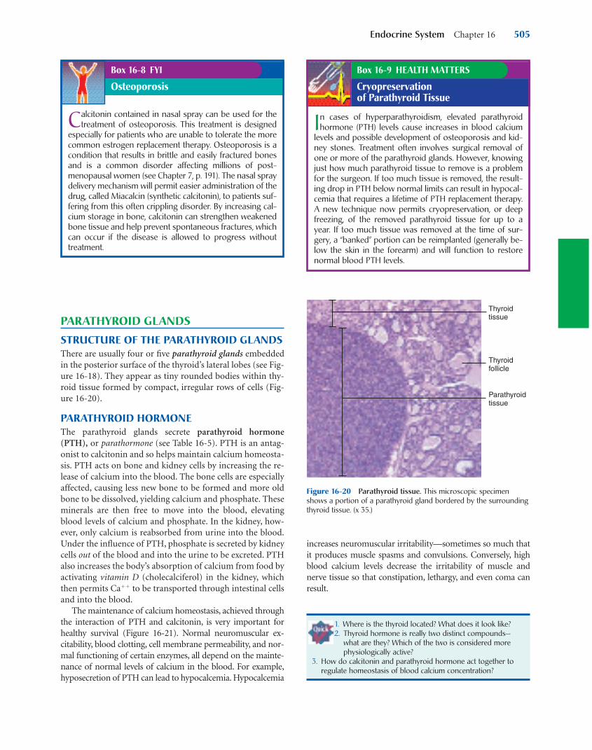
STRUCTURE OF THE PARATHYROID GLANDSThere are usually four or five parathyroid glands embeddedin the posterior surface of the thyroid’s lateral lobes (see Fig-ure 16-18). They appear as tiny rounded bodies within thy-roid tissue formed by compact, irregular rows of cells (Fig-ure 16-20).
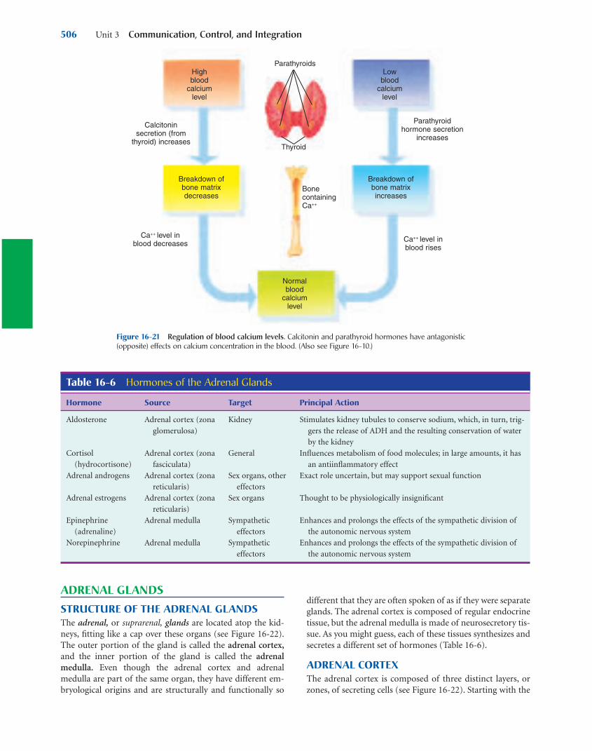
PARATHYROID HORMONEThe parathyroid glands secrete parathyroid hormone(PTH), or parathormone (see Table 16-5). PTH is an antag-onist to calcitonin and so helps maintain calcium homeosta-sis. PTH acts on bone and kidney cells by increasing the re-lease of calcium into the blood. The bone cells are especiallyaffected, causing less new bone to be formed and more oldbone to be dissolved, yielding calcium and phosphate. Theseminerals are then free to move into the blood, elevatingblood levels of calcium and phosphate. In the kidney, how-ever, only calcium is reabsorbed from urine into the blood.Under the influence of PTH, phosphate is secreted by kidneycells out of the blood and into the urine to be excreted. PTHalso increases the body’s absorption of calcium from food byactivating vitamin D (cholecalciferol) in the kidney, whichthen permits Ca�� to be transported through intestinal cellsand into the blood.
The maintenance of calcium homeostasis, achieved throughthe interaction of PTH and calcitonin, is very important forhealthy survival (Figure 16-21). Normal neuromuscular ex-citability, blood clotting, cell membrane permeability, and nor-mal functioning of certain enzymes, all depend on the mainte-nance of normal levels of calcium in the blood. For example,hyposecretion of PTH can lead to hypocalcemia. Hypocalcemia
increases neuromuscular irritability—sometimes so much thatit produces muscle spasms and convulsions. Conversely, highblood calcium levels decrease the irritability of muscle andnerve tissue so that constipation, lethargy, and even coma canresult.
Endocrine System Chapter 16 505
Box 16-8 FYI
Osteoporosis
Calcitonin contained in nasal spray can be used for thetreatment of osteoporosis. This treatment is designed
especially for patients who are unable to tolerate the morecommon estrogen replacement therapy. Osteoporosis is acondition that results in brittle and easily fractured bonesand is a common disorder affecting millions of post-menopausal women (see Chapter 7, p. 191). The nasal spraydelivery mechanism will permit easier administration of thedrug, called Miacalcin (synthetic calcitonin), to patients suf-fering from this often crippling disorder. By increasing cal-cium storage in bone, calcitonin can strengthen weakenedbone tissue and help prevent spontaneous fractures, whichcan occur if the disease is allowed to progress withouttreatment.
Thyroid tissue
Thyroid follicle
Parathyroidtissue
Figure 16-20 Parathyroid tissue. This microscopic specimenshows a portion of a parathyroid gland bordered by the surroundingthyroid tissue. (x 35.)
Box 16-9 HEALTH MATTERS
Cryopreservation of Parathyroid Tissue
In cases of hyperparathyroidism, elevated parathyroidhormone (PTH) levels cause increases in blood calcium
levels and possible development of osteoporosis and kid-ney stones. Treatment often involves surgical removal ofone or more of the parathyroid glands. However, knowingjust how much parathyroid tissue to remove is a problemfor the surgeon. If too much tissue is removed, the result-ing drop in PTH below normal limits can result in hypocal-cemia that requires a lifetime of PTH replacement therapy.A new technique now permits cryopreservation, or deepfreezing, of the removed parathyroid tissue for up to ayear. If too much tissue was removed at the time of sur-gery, a “banked” portion can be reimplanted (generally be-low the skin in the forearm) and will function to restorenormal blood PTH levels.
1. Where is the thyroid located? What does it look like?2. Thyroid hormone is really two distinct compounds—
what are they? Which of the two is considered morephysiologically active?
3. How do calcitonin and parathyroid hormone act together toregulate homeostasis of blood calcium concentration?
Highblood
calciumlevel
ParathyroidsLow
bloodcalcium
level
Thyroid
Breakdown ofbone matrixdecreases
Calcitoninsecretion (from
thyroid) increases
Ca++ level inblood decreases
Normalblood
calciumlevel
BonecontainingCa++
Ca++ level inblood rises
Breakdown ofbone matrixincreases
Parathyroidhormone secretion
increases
ADRENAL GLANDS
STRUCTURE OF THE ADRENAL GLANDSThe adrenal, or suprarenal, glands are located atop the kid-neys, fitting like a cap over these organs (see Figure 16-22).The outer portion of the gland is called the adrenal cortex,and the inner portion of the gland is called the adrenalmedulla. Even though the adrenal cortex and adrenalmedulla are part of the same organ, they have different em-bryological origins and are structurally and functionally so
different that they are often spoken of as if they were separateglands. The adrenal cortex is composed of regular endocrinetissue, but the adrenal medulla is made of neurosecretory tis-sue. As you might guess, each of these tissues synthesizes andsecretes a different set of hormones (Table 16-6).
ADRENAL CORTEXThe adrenal cortex is composed of three distinct layers, orzones, of secreting cells (see Figure 16-22). Starting with the
506 Unit 3 Communication, Control, and Integration
Figure 16-21 Regulation of blood calcium levels. Calcitonin and parathyroid hormones have antagonistic(opposite) effects on calcium concentration in the blood. (Also see Figure 16-10.)
Table 16-6 Hormones of the Adrenal Glands
Hormone Source Target Principal Action
Aldosterone
Cortisol
(hydrocortisone)
Adrenal androgens
Adrenal estrogens
Epinephrine
(adrenaline)
Norepinephrine
Adrenal cortex (zona
glomerulosa)
Adrenal cortex (zona
fasciculata)
Adrenal cortex (zona
reticularis)
Adrenal cortex (zona
reticularis)
Adrenal medulla
Adrenal medulla
Kidney
General
Sex organs, other
effectors
Sex organs
Sympathetic
effectors
Sympathetic
effectors
Stimulates kidney tubules to conserve sodium, which, in turn, trig-
gers the release of ADH and the resulting conservation of water
by the kidney
Influences metabolism of food molecules; in large amounts, it has
an antiinflammatory effect
Exact role uncertain, but may support sexual function
Thought to be physiologically insignificant
Enhances and prolongs the effects of the sympathetic division of
the autonomic nervous system
Enhances and prolongs the effects of the sympathetic division of
the autonomic nervous system

zone directly under the outer connective tissue capsule ofthe gland, they are zona glomerulosa, zona fasciculata, andzona reticularis. Cells of the outer zone secrete a class ofhormones called mineralocorticoids. Cells of the middle zonesecrete glucocorticoids. The inner zone secretes smallamounts of glucocorticoids and gonadocorticoids (sex hor-mones). All of these cortical hormones are steroids, so, to-gether, they are known as corticosteroids.
MineralocorticoidsMineralocorticoids, as their name suggests, have an impor-tant role in regulating how mineral salts (electrolytes) areprocessed in the body. In the human, aldosterone is the onlyphysiologically important mineralocorticoid. Its primaryfunction is in the maintenance of sodium homeostasis in theblood. Aldosterone accomplishes this by increasing sodiumreabsorption in the kidneys. Sodium ions are reabsorbedfrom the urine back into the blood in exchange for potas-sium or hydrogen ions. In this way, aldosterone not only ad-justs blood sodium levels but can also influence potassiumand pH levels in the blood.
Because the reabsorption of sodium ions causes water toalso be reabsorbed (partly by triggering the secretion ofADH), aldosterone promotes water retention by the body. Al-
together, aldosterone can increase sodium and water reten-tion and promote the loss of potassium and hydrogen ions.
Aldosterone secretion is controlled mainly by the renin-angiotensin mechanism and by blood potassium concentra-tion. The renin-angiotensin mechanism (Figure 16-23) op-erates as indicated in this sequence of steps:
1. When the incoming blood pressure in the kidneysdrops below a certain level, a piece of tissue near thevessels (the juxtaglomerular apparatus) secretes renininto the blood.
2. Renin, an enzyme, causes angiotensinogen (a normalconstituent of blood) to be converted to angiotensin I.
3. Angiotensin I circulates to the lungs, where convert-ing enzymes in the capillaries split the molecule,forming angiotensin II.
4. Angiotensin II circulates to the adrenal cortex, whereit stimulates the secretion of aldosterone.
5. Aldosterone causes increased reabsorption ofsodium, which causes increased water retention.As water is retained, the volume of blood increases.The increased volume of blood creates higher bloodpressure—which then causes the renin-angiotensinmechanism to stop.
Endocrine System Chapter 16 507
Figure 16-22 Structure of the adrenal gland. The zona glomerulosa of the cortex secretes aldosterone. Thezona fasciculata secretes abundant amounts of glucocorticoids, chiefly cortisol. The zona reticularis secretesminute amounts of sex hormones and glucocorticoids. A portion of the medulla is visible at lower right in thephotomicrograph (x 35) and at the bottom of the drawing.

The renin-angiotensin mechanism is a negative feed-back mechanism that helps maintain homeostasis of bloodpressure.
GlucocorticoidsThe chief glucocorticoids secreted by the zona fasciculata ofthe adrenal cortex are cortisol (also called hydrocortisone),cortisone, and corticosterone. Of these, only cortisol is se-creted in significant quantities in the human. Glucocorti-coids affect every cell in the body. Although much remains tobe discovered about their precise mechanisms of action, wedo know enough to make some generalizations:
• Glucocorticoids accelerate the breakdown of proteinsinto amino acids (except in liver cells). These “mobi-lized” amino acids move out of the tissue cells andinto the blood. From there, they circulate to the liver
cells, where they are changed to glucose in a processcalled gluconeogenesis. A prolonged high blood con-centration of glucocorticoids in the blood, therefore,results in a net loss of tissue proteins (“tissuewasting”) and hyperglycemia (high blood glucose).Glucocorticoids are protein-mobilizing, gluco-neogenic, and hyperglycemic.
• Glucocorticoids tend to accelerate mobilization ofboth lipids from adipose cells and lipid catabolism bynearly every cell in the body. In other words, gluco-corticoids tend to cause a shift from carbohydrate ca-tabolism to lipid catabolism as an energy source. Themobilized lipids may also be used in the liver for glu-coneogenesis. This effect contributes to the hyper-glycemic effect already observed.
• Glucocorticoids are essential for maintaining anormal blood pressure. Without adequate amounts
508 Unit 3 Communication, Control, and Integration
Box 16-10 HEALTH MATTERS
Adrenal Cortical Hormone Abnormalities
Hypersecretion of cortisol from the adrenal cortex oftenproduces a collection of symptoms called Cushing syn-
drome. Hypersecretion of glucocorticoids results in a redis-tribution of body fat. The fatty “moon face” and thin, red-dened skin characteristic of Cushing syndrome is shown inpart A of the figure. Part B shows the face of the same boy 4 months after treatment. Hypersecretion of aldosterone, al-dosteronism, leads to increased water retention and muscleweakness resulting from potassium loss. Hypersecretion ofandrogens can result from tumors of the adrenal cortex,called virilizing tumors. They are so called because the in-creased blood level of male hormones in women can causethem to acquire male characteristics. People with Cushing
syndrome may suffer from all of the symptoms describedabove.
Hyposecretion of mineralocorticoids and glucocorti-coids, as in Addison disease, may lead to a drop in bloodsodium and blood glucose, an increase in blood potassiumlevels, dehydration, and weight loss.
Pharmacological preparations of glucocorticoids havebeen used for many years to temporarily relieve the symp-toms of severe inflammatory conditions such as rheumatoidarthritis. More recently, over-the-counter creams and oint-ments containing hydrocortisone have become available foruse in treating the pain, itching, swelling, and redness of skinrashes.
Cushing syndrome. A, Fatty “moon face” in a body with Cushing syndrome. B, The face of the same boy 4 monthsafter treatment.
A
B

of glucocorticoids in the blood, the hormones norep-inephrine and epinephrine cannot produce theirvasoconstricting effect on blood vessels, and bloodpressure falls. In other words, glucocorticoids exhibitpermissiveness in that they permit norepinephrineand epinephrine to have their full effects. When glu-cocorticoids are present in high concentrations for aprolonged period of time, they may elevate bloodpressure beyond normal (hypertension).
• A high blood concentration of glucocorticoids ratherquickly causes a marked decrease in the number ofwhite blood cells, called eosinophils, in the blood(eosinopenia) and marked atrophy of lymphatictissues. The thymus gland and lymph nodes are par-ticularly affected. This, in turn, leads to a decrease inthe number of lymphocytes and plasma cells in theblood. Because of the decreased number of lympho-cytes and plasma cells (antibody-processing cells),antibody formation decreases. Antibody formation isan important part of immunity—the body’s defenseagainst infection.
• Normal amounts of glucocorticoids act with epi-nephrine, a hormone secreted by the adrenalmedulla, to bring about normal recovery from injuryproduced by inflammatory agents. How they act to-gether to bring about this antiinflammatory effect isstill uncertain.
• Glucocorticoid secretion increases as part of thestress response. One advantage gained by increasedsecretion may be the increase in glucose available forskeletal muscles needed in fight-or-flight responses.However, prolonged stress can lead to immune dys-
function, probably as a result of prolonged exposureto high levels of glucocorticoids (see Chapter 22).
• Except during the stress response, glucocorticoid se-cretion is controlled mainly by means of a negativefeedback mechanism that involves ACTH from theadenohypophysis.
Gonadocorticoids The term gonadocorticoid refers to sex hormones that are re-leased from the zona fasciculata and zona reticularis of theadrenal cortex rather than the gonads. The normal adrenalcortex secretes small amounts of male hormones (andro-gens). Normally, there is not enough androgen produced togive women masculine characteristics, but it is sufficient toinfluence the appearance of pubic and axillary hair in bothboys and girls.
ADRENAL MEDULLAThe adrenal medulla is composed of neurosecretory tissue;that is, tissue composed of neurons specialized to secretetheir products into the blood rather than across a synapse.Actually, the medullary cells are modified versions of sym-pathetic postganglionic fibers of the autonomic nervous sys-tem. They are innervated by sympathetic preganglionicfibers, so that when the sympathetic nervous system is acti-vated (as in the stress response), the medullary cells secretetheir hormones directly into the blood.
The adrenal medulla secretes two important hormones,both of which are in the class of nonsteroid hormones calledcatecholamines. Epinephrine, or adrenaline, accounts forabout 80% of the medulla’s secretion. The other 20% is nor-epinephrine (NE or NR). You may recall that norepinephrine
Endocrine System Chapter 16 509
25
3 4
1
Angiotensin II
AldosteroneAngiotensin I
Angiotensinogen
Lungs
Adrenalcortex
Blood vesselKidney
Renin
Figure 16-23 Renin-angiotensin mechanism for regulating aldosterone secretion. The numbers corre-spond to the steps outlined in the text.

is also the neurotransmitter produced by postganglionicsympathetic fibers. Sympathetic effectors such as the heart,smooth muscle, and glands have receptors for norepineph-rine. Both epinephrine and norepinephrine produced by theadrenal medulla can bind to the receptors of sympathetic ef-fectors to prolong and enhance the effects of sympatheticstimulation by the autonomic nervous system (Figure 16-24).
PANCREATIC ISLETS
STRUCTURE OF THE PANCREATIC ISLETSThe pancreas is an elongated gland (12 to 15 cm [or about5 to 6 inches] long) weighing up to 100 g (3.5 ounces) (Fig-ure 16-25). The “head” of the gland lies in the C-shaped be-ginning of the small intestine (duodenum), with its bodyextending horizontally behind the stomach and its tailtouching the spleen.
The tissue of the pancreas is composed of both endocrineand exocrine tissues. The endocrine portion is made up ofscattered, tiny islands of cells, called pancreatic islets (isletsof Langerhans) that account for only about 2% or 3% of the
total mass of the pancreas. These hormone-producing isletsare surrounded by cells called acini, which secrete a serousfluid containing digestive enzymes into ducts that drain intothe small intestine (see Figure 16-25). The digestive roles ofthe pancreas are discussed in Chapters 25 and 26. For themoment, we will concentrate on the endocrine part of thisgland, the pancreatic islets.
Each of the one to two million pancreatic islets in the pan-creas contains a combination of four primary types of en-docrine cells, all joined to each other by gap junctions. Eachtype of cell secretes a different hormone, but the gap junc-tions may allow for some coordination of these functions asa single secretory unit. One type of pancreatic islet cell is thealpha cell (also called the A cell), which secretes the hormoneglucagon. Beta cells (B cells) secrete the hormone insulin;delta cells (D cells) secrete the hormone somatostatin; andpancreatic polypeptide cells (F, or PP, cells) secrete pancre-atic polypeptide. Beta cells, which account for about three-fourths of all the pancreatic islet cells, are usually found nearthe center of each islet, whereas cells of the other three typesare more often found in the outer portion. Figure 16-26shows how the different cell types can be distinguished by themicroscope with special staining techniques.
PANCREATIC HORMONESThe pancreatic islets produce several hormones, the mostimportant of which are described in Table 16-7 and in thefollowing paragraphs:
510 Unit 3 Communication, Control, and Integration
Adrenal medullasecretory cell
Adrenalmedulla
Bloodstream
Hypothalamus
Sympatheticpreganglionic
fiber
Sympatheticpostganglionicfiber
Sympatheticeffector cell(target)
Autonomicganglion
Sympatheticpreganglionic
fiber
ANS(sympathetic
division)
➝➝
➝➝➝
▲
Figure 16-24 Combined nervous and endocrine influence on sympathetic effectors. A sympathetic centerin the hypothalamus sends efferent impulses through preganglionic fibers. Some preganglionic fibers synapsewith postganglionic fibers that deliver norepinephrine across a synapse with the effector cell. Other pregan-glionic fibers synapse with postganglionic neurosecretory cells in the adrenal medulla. These neurosecretorycells secrete epinephrine and norepinephrine into the bloodstream, where they travel to the target cells (sympa-thetic effectors). Compare this figure with Figure 16-1.
1. Distinguish between the histology of the adrenal cortexand the adrenal medulla.
2. Name some of the effects of cortisol in the body.3. How does the function of the adrenal medulla overlap with the
function of the autonomic nervous system?

Endocrine System Chapter 16 511
Acini(exocrine cells that produce digestive juice)
Pancreatic islets(groups of endocrine cells)
Pancreatic duct(for digestive juice)
Commonbile duct
S
R L
I
Figure 16-25 Pancreas. Two pancreatic islets, or hormone-producing areas, are evident among the pancre-atic cells that produce the pancreatic digestive juice. The pancreatic islets are more abundant in the tail of thepancreas than in the body or head.
A B
Figure 16-26 Cells of the pancreatic islet. A special technique of microscopy, called immunofluorescentstaining, allows cells to be distinguished by the different molecules that they contain. A, Micrograph shows acentral cluster of beta, or b, cells outlined by green dots. The green dots are insulin molecules stained by aspecial type of antibody coupled to a fluorescent dye. B, In this micrograph, peripheral alpha, or a, cells arestained green and delta, or d, cells are stained red. The central beta cells are not stained in this specimen, sothey appear only as black spaces in the micrograph.

• Glucagon, produced by alpha cells, tends to increaseblood glucose levels by stimulating the conversion ofglycogen to glucose in liver cells. It also stimulatesgluconeogenesis (transformation of fatty acids andamino acids into glucose) in liver cells. The glucoseproduced via the breakdown of glycogen and by glu-coneogenesis is released into the bloodstream, pro-ducing a hyperglycemic effect.
• Insulin, produced by beta cells, tends to promote themovement of glucose, amino acids, and fatty acidsout of the blood and into tissue cells. Hence insulintends to lower the blood concentrations of these foodmolecules and to promote their metabolism by tissuecells. The antagonistic effects that glucagon andinsulin have on blood glucose levels are summarizedin Figure 16-27 and discussed further in Chapter 27.(See Box 16-12 for a discussion of diabetes mellitus.)
• Somatostatin, produced by delta cells, may affectmany different tissues in the body, but its primaryrole seems to be in regulating the other endocrinecells of the pancreatic islets. Somatostatin inhibits the
secretion of glucagon, insulin, and pancreaticpolypeptide. It also inhibits the secretion of growthhormone (somatotropin) from the anterior pituitary.
• Pancreatic polypeptide is produced by PP (or F) cellsin the periphery of pancreatic islets. Although muchis yet to be learned about pancreatic polypeptide, wedo know that it influences the digestion and distribu-tion of food molecules to some degree.
All four of these pancreatic hormones probably work to-gether as a team to maintain a homeostasis of food mole-cules (glucose, fatty acids, and amino acids). More abouttheir respective roles in overall nutrient metabolism is dis-cussed in Chapter 27.
512 Unit 3 Communication, Control, and Integration
Figure 16-27 Regulation of blood glucose levels. Insulin and glucagon, two of the major pancreatic hor-mones, have antagonistic (opposite) effects on glucose concentration in the blood.
1. Name two of the four principal hormones secreted bythe pancreatic islets.
2. In what way do insulin and glucagon exert antagonisticinfluences on the concentration of glucose in the blood?

GONADSGonads are the primary sex organs in the male (testes; singu-lar, testis) and in the female (ovaries). Each is structured dif-ferently, and each produces its own unique set of hormones.
TESTESThe testes are paired organs within a sac of skin called thescrotum, which hangs from the groin area of the trunk (seeFigure 16-2). Composed mainly of coils of sperm-producingseminiferous tubules, there is a scattering of endocrine inter-stitial cells found in areas between the tubules. These inter-stitial cells produce androgens (male sex hormones); theprincipal androgen is testosterone. Testosterone is responsi-ble for the growth and maintenance of male sexual charac-teristics and for sperm production. Testosterone secretion isregulated principally by gonadotropin (especially luteinizinghormone [LH]) levels in the blood.
OVARIESOvaries are a set of paired glands in the pelvis (see Figure 16-2)that produce several types of sex hormones, including thosedescribed briefly in the following paragraphs:
• Estrogens, including estradiol and estrone, are steroidhormones secreted by the cells of the ovarian folliclesthat promote the development and maintenance offemale sexual characteristics. With other hormones,they are responsible for breast development and theproper sequence of events in the female reproductivecycle (menstrual cycle). More details of its functionare discussed in Chapter 32.
• Progesterone is a hormone whose name, which means“pregnancy-promoting steroid,” is an indicator of itschief function. Secreted by the corpus luteum (thetissue left behind after the rupture of a follicle duringovulation), progesterone (along with estrogen) main-tains the lining of the uterus necessary for successfulpregnancy (gestation). This hormone, along withothers, is discussed in detail in Chapter 32.
Regulation of ovarian hormone secretion is complex, tosay the least, but basically depends on the changing levels offollicle-stimulating hormone (FSH) and LH (gonadotropins)from the adenohypophysis.
PLACENTAAnother reproductive tissue that functions as an importantendocrine gland is the placenta. The placenta, the tissue thatforms on the lining of the uterus as an interface between thecirculatory systems of the mother and developing child,serves as a temporary endocrine gland. The placenta pro-duces human chorionic gonadotropin (hCG). This hor-mone is called “chorionic” because it is secreted by thechorion, a fetal tissue component of the placenta. It is called“gonadotropin” because, as with the gonadotropins of theadenohypophysis, it stimulates development and hormonesecretion by maternal ovarian tissues. Chorionic go-nadotropin secretion is high during the early part of preg-
Endocrine System Chapter 16 513
Table 16-7 Hormones of the Pancreatic Islets
Hormone Source Target Principal Action
Glucagon
Insulin
Somatostatin
Pancreatic
polypeptide
Pancreatic islets (alpha [a]
cells or A cells)
Pancreatic islets (beta [b] cells
or B cells)
Pancreatic islets (delta [d] cells
or D cells)
Pancreatic islets (pancreatic
polypeptide [PP] or F cells)
General
General
Pancreatic cells and
other effectors
Intestinal cells and
other effectors
Promotes movement of glucose from storage and into the
blood
Promotes movement of glucose out of the blood and into
cells
Can have general effects in the body, but primary role
seems to be regulation of secretion of other pancreatic
hormones
Exact function uncertain, but seems to influence absorp-
tion in the digestive tract
Box 16-11 SPORTS AND FITNESS
Steroid Abuse
Some steroid hormones are called anabolic steroids be-cause they stimulate the building of large molecules
(anabolism). Specifically, they stimulate the building of pro-teins in muscle and bone. Steroids such as testosteroneand its synthetic derivatives may be abused by athletes andothers who want to increase their performance. The ana-bolic effects of the hormones increase the mass andstrength of skeletal muscles.
Unfortunately, steroid abuse has other consequences.Prolonged use of testosterone will cause a negative feed-back response of the adenohypophysis: gonadotropin lev-els will drop in response to high blood levels of testos-terone. This may lead to atrophy of the testes and,possibly, permanent sterility. Many other adverse effects,including behavioral abnormalities, are known to resultfrom steroid abuse.

nancy and serves as a signal to the mother’s gonads to main-tain the uterine lining rather than allow it to degenerate andfall away (as in menstruation).
The discovery of hCG many years ago led to the develop-ment of early pregnancy tests. The high levels of hCG in theurine of women who are in the early part of their pregnan-cies can be detected through several means. The most famil-iar test involves the use of an over-the-counter kit, whichtests for hCG in urine by means of an antigen-antibody re-action that can be easily interpreted.
As the placenta develops past the first trimester (3 months)of pregnancy, its production of hCG drops as its productionof estrogens and progesterone increase. The placenta thereforemore or less takes over the job of ovaries in producing thesehormones necessary for a successful pregnancy. More abouthow this works is discussed in Chapter 33.
THYMUSThe thymus is a gland in the mediastinum, just beneath thesternum (see Figure 16-2). It is large in children until pu-berty, when it begins to atrophy. It continues to atrophythroughout adulthood, so that by the time an individualreaches old age, the gland is but a vestige of fat and fibroustissue.
The anatomy of the thymus is described in Chapter 20.Although it is considered to be primarily a lymphatic or-
gan (see Chapter 20), the hormones thymosin and thy-mopoietin have been isolated from thymus tissue and areconsidered to be largely responsible for its endocrine activ-ity. Thymosin and thymopoietin actually refer to two entirefamilies of peptides that, together, have a critical role in thedevelopment of the immune system. Specifically, thymosinand thymopoietin are thought to stimulate the productionof specialized lymphocytes involved in the immune responsecalled T cells. The role of T cells in the immune system is dis-cussed in Chapter 21.
GASTRIC AND INTESTINAL MUCOSAThe mucous lining of the gastrointestinal (GI) tract, like thepancreas, contains cells that produce both endocrine and ex-ocrine secretions. GI hormones such as gastrin, secretin,cholecystokinin-pancreozymin (CCK), and others, have im-portant regulatory roles in coordinating the secretory andmotor activities involved in the digestive process. For exam-ple, secretin is released when acids make contact with the in-testinal mucosa. Secretin carried by the blood triggers its tar-
get cells in the stomach to reduce acid secretion. Secretinalso triggers its target cells in the pancreas to release an alka-line fluid and it acts with CCK to trigger the pancreas to re-lease digestive enzymes. CCK triggers the gallbladder to re-lease more bile, which helps break up fat droplets. In effect,secretin and CCK are signals from the intestine to otherparts of the digestive system that promote an effective coor-dination of GI functions.
The hormone ghrelin is secreted by endocrine cells inthe gastric mucosa. It acts by stimulating the hypothala-mus to boost appetite. Since it also acts on other body tis-sues to slow metabolism and reduce fat burning, it mayplay an important role in contributing to obesity.
Chapter 26 describes the hormonal control of digestionin the stomach and small intestine in more detail (seeTable 26-5, p. 782).
HEARTThe heart is another organ that has a secondary endocrinerole. Although the heart’s main function is to pump blood, aspecific area in its wall contains some hormone-producingcells. These cells produce a hormone called atrial natriuretichormone (ANH). The name of this hormone reveals muchabout its role in the body. The term atrial refers to the factthat ANH is secreted by cells in an upper chamber of theheart called an atrium. Atrial cells increase their secretion ofANH in response to an increase in the stretch of the atrialwall caused by abnormally high blood volume or blood pres-sure. The term natriuretic refers to the fact that its principaleffect is to promote the loss of sodium (Latin, natrium) fromthe body by means of the urine. When sodium is thus lostfrom the internal environment, water follows. Water loss re-sults in a decrease in blood volume (and thus a decrease inblood pressure). We can then state that the primary effect ofANH is to oppose increases in blood volume or blood pres-sure. We can also state that ANH is an antagonist to ADHand aldosterone. ANH is also known by several other names,including atrial natriuretic factor (ANF), atrial natriureticpeptide, and, simply, atrial peptide.
514 Unit 3 Communication, Control, and Integration
1. What are the major hormones secreted by reproductivetissues (gonads and the placenta)?
2. Which gland produces a hormone that regulates the de-velopment of cells important to the immune system?
3. Secretin was the first substance in the body to be identified asa hormone. What structure produces secretin?

Diabetes mellitus is one of the most common endocrinedisorders. It affects more than 14 million Americans and
will have a significant impact on the health of about 7% ofthe population at some point in their lives. Diabetes is bestdescribed as a syndrome—a collection of features that char-acterizes the disease. Although the features of diabetes tendto vary between individuals and on the type, severity, andlength of the illness, each sign or symptom is related in someway to abnormal metabolism of nutrients and the conse-quences that follow (see the figure on p. 517).
In diabetics, an inadequate amount or abnormal type ofinsulin may be produced. In other affected individuals, di-minished numbers of normal insulin receptors or secondmessenger systems in target cells impair glucose entry intocells even if normal insulin in adequate amounts is present.A newly discovered hormone secreted by fat cells, called re-sistin, may also interfere with insulin action. Research on re-sistin may help explain the relationship between obesity andType 2 diabetes (see discussion below). The possible causesof all of these insulin-related problems are still under inves-tigation. Probably one or a combination of factors such asgenetic predisposition, hormone problems, viral infection,nutrition and diet, obesity, autoimmune disorders, and expo-sure to damaging agents are involved in most cases of dia-betes mellitus.
The term insulin resistance refers to an inability of fat,muscle, and other cells to respond normally to insulin andpermit the entry of blood sugar that the hormone is trying toaccomplish. Insulin insensitivity is the opposite of insulin re-sistance. It involves the rapid response of cells to insulin ac-tion and subsequent entry of sugar.
The presence of adequate amounts of normal insulin, andsufficient sensitivity to insulin, is key to the entry of glucoseinto cells. If there are no insulin problems and if the majortarget tissues such as skeletal muscle, fat, and liver have ad-equate, normal insulin receptors and signaling mechanisms,glucose will transfer from blood into cells. In diabetes, glu-cose cannot enter cells normally. The result is one of themost universal symptoms of the disease—chronic elevationin blood glucose levels, a condition called hyperglycemia.
Glucose is normally filtered out of the blood and then re-absorbed from the kidney tubules. However, as blood sugarlevels rise in diabetes, the amount of glucose filtered out ofthe blood exceeds the ability of the kidney tubules to resorbit. The result is a “spilling over” of sugar into the urine. Thiscondition, called glycosuria, causes increased urine produc-tion (polyuria), since additional water is required to carry thesugar load. In effect, the excess glucose acts like an osmoticdiuretic. As large quantities of water are lost in the urine, thebody dehydrates. The resulting sense of excessive and on-going thirst (polydipsia) and the tendency to drink largequantities of liquid are also classic symptoms of the disease.In addition, since cells are deprived of glucose to burn as en-ergy, people with diabetes often suffer from intense and con-tinuous hunger (polyphagia). Their blood sugar level is high,but the cells are literally starving to death, and the body
craves food. In screening for diabetes, health care profes-sionals look for glycosuria and the “three polys”: polyuria,polydipsia, and polyphagia.
The untreated person with diabetes is unable to utilizeglucose for energy, and the body is forced to burn proteinand fat. This abnormal metabolic shift results in fatigue andweight loss. If glucose metabolism is severely restricted, thelarge quantities of fat that must be burned produce toxicquantities of acetoacetic acid and other acidic metabolitescalled ketone bodies. Buildup of these organic acids lowersblood pH, causing acidosis, and disturbs the normal acid-base balance of the body. (Mechanisms that attempt to re-store homeostasis are described in Chapter 30.) Accumula-tion of ketone bodies in the blood results in diabeticketoacidosis. Signs and symptoms include abdominal pain,nausea, vomiting, fruity odor of the breath, possible alter-ations in level of consciousness, coma, and even death, if leftuntreated.
Types of Diabetes MellitusThere are two major types of diabetes mellitus: type 1 andtype 2. Hereditary factors play an important role in bothtypes.
Type 1
Type 1 diabetes mellitus was formerly referred to as juvenile-onset diabetes because it usually strikes before age 30. In thisform of the disease the beta cells of the pancreatic islets aredestroyed and there is an absolute deficiency of insulin pro-duction. Individuals with type 1 diabetes are required to takeinsulin injections daily to prevent ketosis and to control hy-perglycemia. As a result, type 1 diabetes was sometimescalled insulin-dependent diabetes mellitus (IDDM). This formof the disease accounts for only about 10% of the total num-ber of diabetic individuals.
The cause of beta cell destruction in type 1 diabetes is stilluncertain. Current research, however, suggests that type 1diabetes is an autoimmune disease that is probably triggeredby some type of viral infection in genetically susceptible indi-viduals. Anyone who has a parent, brother, or sister with type1 diabetes has about a 5% to 7% chance of getting the dis-ease. If an identical twin has type 1 diabetes, the risk in-creases to about 50%.
Type 2
Type 2 diabetes mellitus, previously called non–insulin-dependent diabetes mellitus (NIDDM), is the most commonform of the disease, accounting for about 90% of all cases.Because it most often occurs after age 40 years, it was for-merly also called maturity-onset diabetes. In susceptible indi-viduals who are overweight, the incidence increases with age.In this form of diabetes, insulin is still produced by the betacells but generally in reduced amounts. In addition, loss ofinsulin receptors on the surface membranes of target cellsleading to insulin resistance also reduces effectiveness ofglucose uptake from the blood.
Endocrine System Chapter 16 515
Box 16-12
Diabetes Mellitus
Continued.
The older name NIDDM was inappropriate because in-sulin injections, often combined with oral diabetic medicines,may be required to control the disease. However, in manytype 2 diabetics, hyperglycemia will frequently respond tochanges in lifestyle that result in eating a balanced diet highin fiber, achieving an ideal body weight, getting adequate ex-ercise, and maintaining body weight within normal limits.
Heredity and ethnic background are important determi-nants in type 2 diabetes. Those who have a family history ofthe disease are particularly susceptible if overweight andsedentary. Native Americans are at increased risk for type 2diabetes. In addition, Hispanics and African Americans aremore likely than Caucasians to develop this type of diabetes.A specific gene defect is responsible for at least some formsof type 2 diabetes. The gene in question is involved in regu-lating insulin secretion.
The symptoms of type 1 diabetes are dramatic and, as aresult, most individuals seek medical care soon after the dis-ease occurs. The symptoms of type 2 diabetes, although ofthe same type as seen in type 1 diabetes, can be much moresubtle and hard to recognize. The American Diabetes Asso-ciation estimates that more than 7 million Americans havediabetes and are unaware of it. Unfortunately, if left un-treated, over time the hyperglycemia and other effects of di-abetes results in many complications affecting almost everyarea of the body. Reduced blood flow caused by a buildup offatty materials in blood vessels (atherosclerosis) is one of themost serious complications. It causes such diverse problemsas heart attack, stroke, and reduced circulation to the ex-tremities, resulting in tingling or numbness in the feet and, insevere cases, gangrene. Retinal changes (diabetic retinopa-thy) may cause blindness in some people with diabetes whohave battled the disease for decades. As you read in Chapter12, nerve damage or neuropathy can also result from dia-betes. Kidney disease is another common diabetic complica-tion. Most authorities agree that careful regulation of bloodsugar levels and reducing cardiovascular risk factors such ashypertension and “bad” (LDL) cholesterol levels are the mostimportant measure that people with diabetes can take to re-duce the number of long-term complications of the disease.
Treatment of Type 1 Diabetes
The discovery of insulin in 1921 was one of the most impor-tant advances in medicine of the last century. Problems as-sociated with the injection of insulin harvested from animalpancreatic tissue were solved when synthetic human insulinbecame available.
Insulin can be introduced into the body by regular injec-tion using a needle and syringe or by implanting miniaturizedpumps to deliver the insulin as needed. Oral (non-injectable)
insulin in pill form that can withstand the destructive action ofdigestive enzymes may become a reality in the not-too-distant future. In addition, development of alternate ways toadminister new types of insulin also show promise. Specialinhalers are being tested to deliver a powder form of insulininto the lungs. A liquid aerosol form of insulin is also understudy. It is sprayed into the mouth and absorbed through theepithelial lining of the oral cavity.
Pancreas transplants eliminate the need for insulin injec-tion or administration altogether and the number of suc-cessful pancreatic transplants is increasing with the advent ofnew surgical techniques and more effective anti-rejectiondrugs (see Chapter 21). However, transplantation of islet cellsrather than the entire pancreas shows even greater promisefor treatment of the most serious cases of type 1 diabetes.
One of the newest and most promising of the experimen-tal islet cell transplant techniques is called the “EdmontonProtocol.” It was developed at the University of Alberta in Edmonton, Canada. The procedure begins with the separa-tion and harvesting of islets from a donor pancreas; the isletsare then injected into a diabetic patient’s portal vein that goesto the liver. After injection, the islets pass into the liver, be-come lodged there, and produce insulin. The protocol differsfrom other islet cell transplant procedures by increasing thenumber of functional islet cells that can be recovered from adonor pancreas and by elimination of steroids and othersubstances that decrease rejection rates. The procedure,while still experimental, shows great promise in treating se-vere type 1 diabetics.
Treatment of Type 2 Diabetes
If the lifestyle changes described earlier in this discussion arenot totally effective in lowering elevated blood glucose levelsin individuals with type 2 diabetes, medicines called oral hy-poglycemic agents may be prescribed. One group of oralhypoglycemic agents, called sulfonylureas, act by stimulat-ing beta cells to increase insulin supplies (for example, Mi-cronase and Glucotrol). Other short-acting “insulin stimula-tors” act like sulfonylureas but much more rapidly (forexample, Prandin and Starlix). Another group of oral agents,called biguanides, act by inhibiting the release of glucosefrom the liver (for example, Glucophage). Others, such as thedrugs Avandia and Actos (thiazolidinediones or TZDs) reduceblood levels of resistin, thus decreasing insulin resistance.Newer oral treatments for type 2 diabetes often involve ad-ministration of lower doses of more than one drug. A popu-lar combination called Glucovance combines metformin(Glucophage) and the sulfonurea glyburide (Micronase). Inaddition, oral agents are also given in combination with in-sulin injections in many patients with type 2 diabetes.
516 Unit 3 Communication, Control, and Integration
Box 16-12
Diabetes Mellitus—cont’d
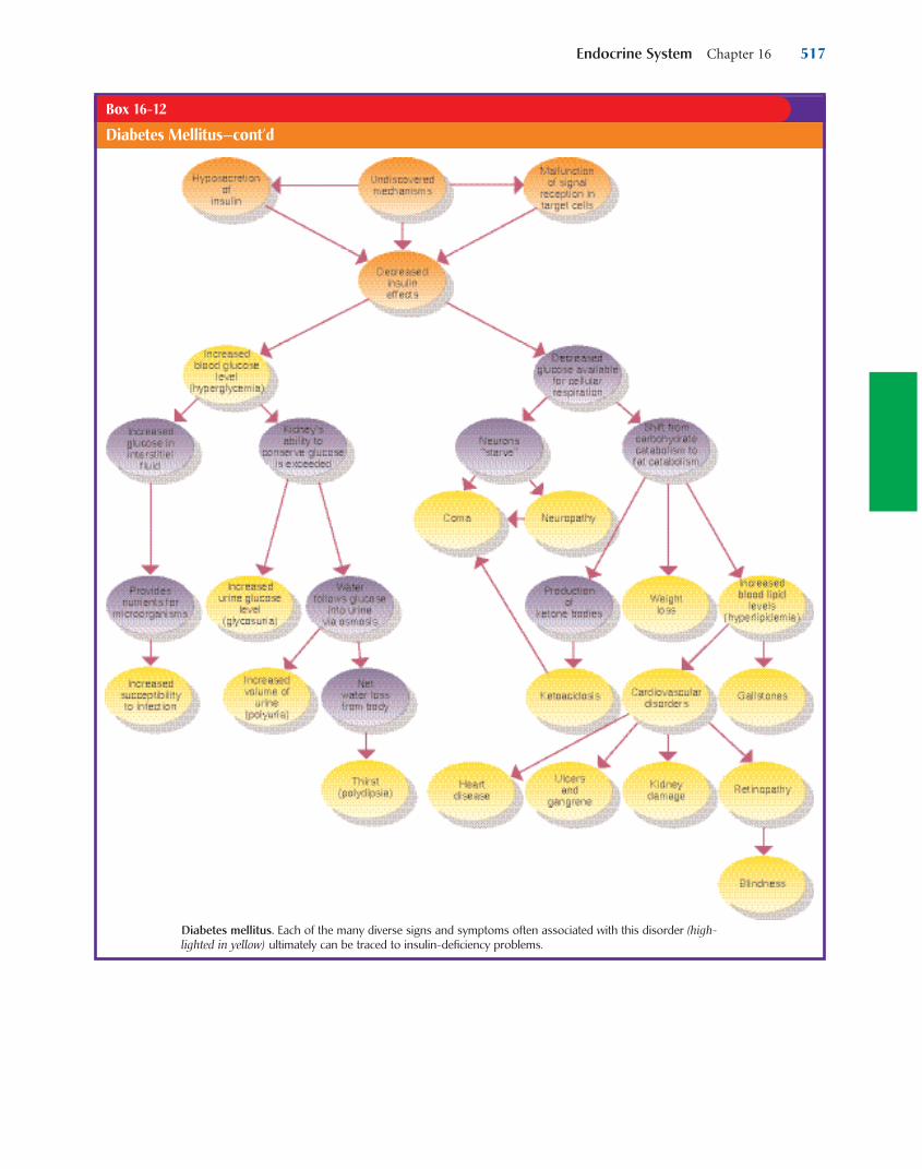
Endocrine System Chapter 16 517
Box 16-12
Diabetes Mellitus—cont’d
Diabetes mellitus. Each of the many diverse signs and symptoms often associated with this disorder (high-lighted in yellow) ultimately can be traced to insulin-deficiency problems.

518 Unit 3 Communication, Control, and Integration
Endocrine regulation of body processes first begins dur-ing early development in the womb. By the time a baby is
born, many of the hormones are already at work influenc-ing the activity of target cells throughout the body. As amatter of fact, new evidence suggests that it is a hormonalsignal from the fetus to the mother that signals the onset oflabor and delivery. Many of the basic hormones are activefrom birth, but most of the hormones related to reproduc-
tive functions are not produced or secreted until puberty.Secretion of male reproductive hormones follows the samepattern as most nonreproductive hormones: continuous se-cretion from puberty until there is a slight tapering off in lateadulthood. The secretion of female reproductive hormonessuch as estrogens also declines late in life, but more sud-denly and completely—often during or just at the end ofmiddle adulthood.
CYCLE OF LIFE
Endocrine System
It is important to appreciate the precision of control affordedby the partnership of the two major regulatory systems: the
endocrine system and the nervous system. The neuroen-docrine system is able to finely adjust the availability and pro-cessing of nutrients through a diverse array of mechanisms:growth hormone, thyroid hormone, cortisol, epinephrine, so-matostatin, autonomic nervous regulation, and so on. Theabsorption, storage, and transport of calcium ions are kept inbalance by the antagonistic actions of calcitonin and parathy-roid hormone (and its effects on vitamin D). Reproductiveability is triggered, developed, maintained, and timed by the complex interaction of the nervous system with follicle-stimulating hormone, luteinizing hormone, estrogen, proges-terone, testosterone, chorionic gonadotropin, prolactin, oxy-tocin, and melatonin. Nearly every process in the human
organism is kept in balance by the incredibly complex, butprecise, interaction of all these different nervous and en-docrine regulatory chemicals.
In this chapter, we have seen the many different structuresand regulatory mechanisms that comprise the endocrine sys-tem. Some of the more important hormones and their char-acteristics are summarized in tables throughout the chapter.We have seen how the hormones interact with each other, aswell as how their functions complement those of the nervoussystem. Although there is still much to be learned, we can seethat a basic understanding of hormonal regulatory mecha-nisms is required to fully appreciate the nature of homeosta-sis in the human organism. As we continue our study of hu-man anatomy and physiology, we will often encounter thecritical integrative role played by the endocrine system.
THE BIG PICTURE
The Endocrine System and the Whole Body
Endocrine DisordersAs we have stated throughout this chapter, endocrine disor-ders typically result from either elevated or depressed hor-mone levels (hypersecretion or hyposecretion). At firstthought, this may seem very simple and straightforward. Inreality, however, nothing could be further from the truth. Avariety of specific mechanisms may produce hypersecretionor hyposecretion of hormones. A few of the more well-known mechanisms are briefly explained here. Examples ofa few major endocrine disorders and their mechanisms arelisted in Table 16-8.
Mechanisms of HypersecretionExcessively high blood concentration of a hormone—or anycondition that mimics high hormone levels—is called hyper-secretion. Specific types of hypersecretion are usually namedby placing the prefix hyper- in front of the name of the sourcegland and the suffix -ism at the end. For example, hyper-secretion of thyroid hormone—no matter what the specificcause—is called hyperthyroidism. Hyperthyroidism is not a
disease itself but a condition that characterizes several differ-ent diseases (e.g., Graves’ disease and toxic nodular goiter).
Any of several different mechanisms may be responsiblefor a particular case of hypersecretion. For example, tumorsare often responsible for an abnormal proliferation of en-docrine cells and the resulting increase in hormone secre-tion. Pituitary adenomas, for example, are benign tumorsthat may cause hyperpituitarism. As many as one in five peo-ple may have pituitary adenomas, but the majority of tu-mors are microscopic and asymptomatic. Larger tumorsmay, however, cause hyperpituitarism with a possible out-come of gigantism, or acromegaly.
Another cause of hypersecretion is a phenomenon calledautoimmunity. In autoimmunity, the immune system func-tions abnormally. In Graves’ disease, for example, autoimmuneantibodies against the TSH receptor actually stimulate the re-ceptor and mimic the activity of TSH. As a result, the thyroidgland hypertrophies and excess thyroid hormone is produced.
Another possible cause of hypersecretion of a hormone isa failure of the feedback mechanisms that regulate secretion
MECHANISMS OF DISEASE

Endocrine System Chapter 16 519
Continued
Table 16-8 Examples of Endocrine Conditions
Condition Mechanism Description
Acromegaly
Addison disease
Aldosteronism
Cretinism
Cushing disease
Cushing syndrome
Diabetes insipidus
Gestational diabetes
mellitus (GDM)
Gigantism
Graves disease
Hashimoto disease
Hyperparathyroidism
Hyperthyroidism
(adult)
Hypothyroidism
(adult)
Insulin shock
Type 1 diabetes
mellitus
Myxedema
Hypersecretion of growth hormone
(GH) during adulthood
Hyposecretion of adrenal cortical
hormones (adrenal cortical
insufficiency)
Hypersecretion of aldosterone
Hyposecretion of thyroid hormone
during early development
Hypersecretion of adrenocorti-
cotropic hormone (ACTH)
Hypersecretion (or injection) of
glucocorticoids
Hyposecretion of (or insensitivity
to) antidiuretic hormone (ADH)
Temporary decrease in blood levels
of insulin during pregnancy
Hypersecretion of GH before
age 25
Hypersecretion of thyroid
hormone
Autoimmune damage to thyroid
causing hyposecretion of thyroid
hormone
Hypersecretion of parathyroid
hormone (PTH)
Hypersecretion of thyroid
hormone
Hyposecretion of thyroid hormone
Hypersecretion (or overdose injec-
tion) of insulin, decreased food
intake, and excessive exercise
Hyposecretion of insulin
Extreme hyposecretion of thyroid
hormone during adulthood
Chronic metabolic disorder characterized by gradual enlargement or
elongation of facial bones and extremities
Caused by tuberculosis, autoimmunity, or other factors, this life-
threatening condition is characterized by weakness, anorexia, weight
loss, nausea, irritability, decreased cold tolerance, dehydration, in-
creased skin pigmentation, and emotional disturbance; it may lead
to an acute phase (adrenal crisis) characterized by circulatory shock
Often caused by adrenal hyperplasia, this condition is characterized by
sodium retention and potassium loss—producing Conn syndrome:
severe muscle weakness, hypertension (high blood pressure), kidney
dysfunction, and cardiac problems
Congenital condition characterized by dwarfism, retarded mental devel-
opment, facial puffiness, dry skin, umbilical hernia, and lack of
muscle coordination
Caused by adenoma of the anterior pituitary, increased ACTH causes hy-
persecretion of adrenal cortical hormones, producing Cushing syndrome
Metabolic disorder characterized by fat deposits on upper back, striated
pad of fat on chest and abdomen, rounded “moon” face, muscular
atrophy, edema, hypokalemia (low blood potassium level), and possi-
ble abnormal skin pigmentation; occurs in Cushing disease
Metabolic disorder characterized by extreme polyuria (excessive urina-
tion) and polydipsia (excessive thirst) due to a decrease in the kidney’s
retention of water
Carbohydrate-metabolism disorder occurring in some pregnant women;
characterized by polydipsia, polyuria, overeating, weight loss, fatigue,
and irritability
Condition characterized by extreme skeletal size caused by excess
protein anabolism during skeletal development
Inherited, possibly autoimmune disease characterized by hyperthy-
roidism, exophthalmos (protruding eyes)
Enlargement of thyroid (goiter) is sometimes accompanied by hypothy-
roidism, typically occurring between ages 30 and 50; it is 20 times
more common in females than males
Condition characterized by increased reabsorption of calcium from
bone tissue and kidneys and increased absorption by the gastrointesti-
nal tract; produces hypercalcemia, resulting in confusion, anorexia,
abdominal pain, muscle pain, and fatigue, possibly progressing to cir-
culatory shock, kidney failure, and death
Condition characterized by nervousness, tremor, weight loss, excessive
hunger, fatigue, heat intolerance, heart arrhythmia, and diarrhea, is
caused by a general acceleration of body function
Condition characterized by sluggishness, weight gain, skin dryness, con-
stipation, arthritis, and general slowing of body function, may lead to
myxedema, coma, or death if untreated
Hypoglycemic (low blood glucose) shock characterized by nervousness,
sweating and chills, irritability, hunger, and pallor—progressing to
convulsion, coma, and death if untreated
Inherited condition with sudden childhood onset characterized by
polydipsia, polyuria, overeating, weight loss, fatigue, and irritability,
resulting from the inability of cells to secure and metabolize
carbohydrates
Severe form of adult hypothyroidism characterized by edema of the face
and extremities; often progressing to coma and death

of a particular hormone. For example, a condition called pri-mary hyperparathyroidism is characterized by a failure of theparathyroid gland to adjust its output to compensate forchanges in blood calcium levels. Instead, the parathyroidgland seems to operate independently of the normal feed-back loop and thus overproduces parathyroid hormone.
Mechanisms of HyposecretionDepressed blood hormone levels—or any condition thatmimics low hormone levels—is termed hyposecretion. Spe-cific types of hyposecretion are named in a manner similarto that in which hypersecretion disorders are named: by theaddition of the hypo- prefix and the -ism suffix. For example,hyposecretion of thyroid hormone is called hypothyroidism.
Various different mechanisms have been shown to causehyposecretion of hormones. For example, although most tu-mors cause oversecretion of a hormone, they may insteadcause a gland to undersecrete its hormone(s). Tissue death,perhaps caused by a blockage or other failure of the bloodsupply, can also cause a gland to reduce its hormonal output.Hypopituitarism (hyposecretion by the anterior pituitary)can occur this way. Still another way in which a gland mayreduce its secretion below normal levels is through abnor-mal operation of regulatory feedback loops. An example ofthis is in the case of hyposecretion of testosterone and go-nadotropic hormones in males who abuse anabolic steroids.Men who take testosterone steroids increase their blood con-centration of this hormone above set point levels. The bodyresponds to this overabundance by reducing its own output
of testosterone by reducing its output of gonadotropins.This may lead to sterility and other complications.
Abnormalities of immune function may also cause hy-posecretion. For example, an autoimmune attack on glandu-lar tissue sometimes has the effect of reducing hormone out-put. Some endocrinologists theorize that autoimmunedestruction of pancreatic islet cells, perhaps in combinationwith viral and genetic mechanisms, is a culprit in many casesof type 1 (insulin-dependent) diabetes mellitus.
Many types of hyposecretion disorders have recently beenshown to be caused by insensitivity of the target cells totropic hormones rather than from actual hyposecretion. Afew major types of abnormal responses in target cells are:
• An abnormal decrease in the number of hormone receptors
• Abnormal function of hormone receptors, resultingin failure to bind to hormones properly
• Antibodies bind to hormone receptors, thus blockingbinding of hormone molecules
• Abnormal metabolic response to the hormone-receptor complex by the target cell
• Failure of the target cell to produce enough secondmessenger molecules
Type 2 (non–insulin-dependent) diabetes mellitus isthought to be caused by target cell abnormalities that renderthe cells insensitive to insulin.
520 Unit 3 Communication, Control, and Integration
Table 16-8 Examples of Endocrine Conditions—cont’d
Condition Mechanism Description
Type 2 diabetes
mellitus
Osteoporosis
Pituitary dwarfism
Simple goiter
Sterility
Winter (seasonal)
depression
Insensitivity of target cells to
insulin
Hyposecretion of estrogen in post-
menopausal women
Hyposecretion of GH before age 25
Lack of iodine in diet
Hyposecretion of sex hormones
Hypersecretion of (or hypersensi-
tivity to) melatonin
Carbohydrate-metabolism disorder with slow adulthood onset thought
to be caused by a combination of genetic and environmental factors
and characterized by polydipsia, polyuria, overeating, weight loss,
fatigue, and irritability
Bone disorder characterized by loss of minerals and collagen from bone
matrix, producing holes or porosities that weaken the skeleton
Condition characterized by reduced skeletal size caused by decreased
protein anabolism during skeletal development
Enlargement of thyroid tissue results from the inability of the thyroid
to make thyroid hormone because of a lack of iodine; a positive-
feedback situation develops in which low thyroid hormone levels
trigger hypersecretion of thyroid-stimulating hormone (TSH) by
pituitary—which stimulates thyroid growth
Loss of reproductive function
Abnormal emotional state characterized by sadness and melancholy re-
sulting from exaggerated melatonin effects; melatonin levels are in-
hibited by sunlight so they increase when day length decreases during
winter

Endocrine System Chapter 16 521
twice a day to decrease her heart rate and control her car-diac symptoms.
1. Which of the following reasons is the most likely causeof Sonia Dawson’s increased thyroid function?A. Hyperplasia of the thyroidB. Anterior pituitary tumorC. Thyroid carcinomaD. Autoimmune response
2. After 1 month of treatment Sonia returns to the clinicfor a checkup. She has not been feeling well and is verytired. However, her previous symptoms have resolved.A TSH level from a few days ago is now 7.5, which isgreatly elevated. Which one of the following explana-tions for this recent elevation would be considered rea-sonable in Ms. Dawson’s case?A. Pituitary failure has occurred.B. The thyroid gland has been suppressed too much.C. The hypothalamus has been suppressed.D. The thyroid gland has been overstimulated.
3. Based on the elevated TSH level and the change in Ms.Dawson’s condition, what symptoms would you expecther to exhibit?A. Dyspnea, increased heart rate, and constipationB. Slower heart rate, lethargy, and nervousnessC. Dyspnea, slight fever, and increased heart rate D. Slower heart rate, lethargy, and hair loss
Sonia Dawson, a 30-year-old female, has been diagnosedwith Graves’ disease. She has recently lost 25 pounds
despite an increased appetite. Her vision is blurry; herheart races and pounds. She has been irritable but attrib-utes the irritability to insomnia. She has had bouts of diar-rhea. She feels hot, even if others are comfortable. The col-lars on her clothing are becoming too tight. Her lastmenstrual period was 2 months ago. Before that time hermenses were regular.
On physical examination, the nurse notes that Ms.Dawson is thin, pale, and anxious. She moves restlesslyaround the room. Her eyes are bulging and have a staringappearance. Her eyelids and hands have fine tremors. Herskin is smooth, warm, and moist. She is sweating profusely,although the room temperature is ambient. Her hair is veryfine and soft. She weighs 112 pounds. Her oral temperatureis 99° F (37.2° C), heart rate is 120 at rest, respirations are20, and blood pressure is 110/50. Her thyroid gland is enlarged with auscultation of a bruit. Laboratory results reveal a serum T3 level of 200 µg/dl (high), a T4 level of20 µg/dl (low), and a TSH level that is 2.5 µU/ml, which isnormal. A thyroid antibody test shows a high titer of thy-roglobulin antibodies. Radioactive iodine uptake testshows a 40% uptake in 6 hours, which is elevated.
The decision to treat nonsurgically is made. Ms.Dawson is started on methimazole 10 mg by mouth everyday to suppress her thyroid gland and propranolol 10 mg
CASE STUDY
INTRODUCTIONA. The endocrine and nervous systems function to achieve
and maintain homeostasisB. When the two systems work together, referred to as
the neuroendocrine system, they perform the samegeneral functions: communication, integration, andcontrol
C. In the endocrine system, secreting cells send hormonemolecules via the blood to specific target cells containedin target tissues or target organs
D. Hormones—carried to almost every point in the body;can regulate most cells; effects work more slowly andlast longer than those of neurotransmitters
E. Endocrine glands are “ductless glands”; many are madeof glandular epithelium whose cells manufacture andsecrete hormones; a few endocrine glands are made ofneurosecretory tissue
F. Glands of the endocrine system are widely scatteredthroughout the body (Figure 16-2)
HORMONESA. Classification of hormones
1. Classification by general functiona. Tropic hormones—hormones that target other
endocrine glands and stimulate their growth andsecretion
b. Sex hormones—hormones that target reproduc-tive tissues
c. Anabolic hormones—hormones that stimulateanabolism in target cells
2. Classification by chemical structure (Figure 16-3)a. Steroid hormonesb. Nonsteroid hormones
3. Steroid hormones (Figure 16-4)a. Synthesized from cholesterolb. Lipid-soluble and can easily pass through the
phospholipid plasma membrane of target cellsc. Examples of steroid hormones: cortisol, aldos-
terone, estrogen, progesterone, and testosterone
CHAPTER SUMMARY

4. Nonsteroid hormones (Figure 16-5)a. Synthesized primarily from amino acidsb. Protein hormones—long, folded chains of amino
acids; e.g., insulin and parathyroid hormonec. Glycoprotein hormones—protein hormones with
carbohydrate groups attached to the amino acidchain
d. Peptide hormones—smaller than protein hor-mones; short chain of amino acids; e.g., oxytocinand antidiuretic hormone (ADH)
e. Amino acid derivative hormones—each is derivedfrom a single amino acid molecule(1) Amine hormones—synthesized by modifying
a single molecule of tyrosine; produced byneurosecretory cells and by neurons; e.g., epi-nephrine and norepinephrine
(2) Amino acid derivatives produced by the thy-roid gland; synthesized by adding iodine totyrosine
B. How hormones work1. General principles of hormone action
a. Hormones signal a cell by binding to the targetcell’s specific receptors in a “lock-and-key” mech-anism (Figure 16-6)
b. Different hormone-receptor interactions producedifferent regulatory changes within the target cellthrough chemical reactions
c. Combined hormone actions:(1) Synergism—combinations of hormones act-
ing together have a greater effect on a targetcell than the sum of the effects that eachwould have if acting alone
(2) Permissiveness—when a small amount of onehormone allows a second one to have its fulleffects on a target cell
(3) Antagonism—one hormone produces the op-posite effects of another hormone; used to“fine tune” the activity of target cells withgreat accuracy
d. Endocrine glands produce more hormone mole-cules than actually are needed; the unused hor-mones are quickly excreted by the kidneys or bro-ken down by metabolic processes
2. Mechanism of steroid hormone action (Figure 16-7)a. Steroid hormones are lipid-soluble, and their
receptors are normally found in the target cell’scytosol
b. After a steroid hormone molecule has diffusedinto the target cell, it binds to a receptor moleculeto form a hormone-receptor complex
c. Mobile-receptor hypothesis—the hormone passesinto the nucleus, where it binds to mobile recep-tor and activates a certain gene sequence to begintranscription of mRNA; newly formed mRNAmolecules move into the cytosol, associate with ri-bosomes, and begin synthesizing protein mole-cules that produce the effects of the hormone
d. Steroid hormones regulate cells by regulating pro-duction of certain critical proteins
e. The amount of steroid hormone present deter-mines the magnitude of a target cell’s response
f. Because transcription and protein synthesis taketime, responses to steroid hormones are oftenslow
3. Mechanisms of nonsteroid hormone actiona. The second messenger mechanism—also known
as the fixed-membrane-receptor hypothesis(Figure 16-8)(1) A nonsteroid hormone molecule acts as a
“first messenger” and delivers its chemicalmessage to fixed receptors in the target cell’splasma membrane
(2) The “message” is then passed by way of a G protein into the cell where a “second messen-ger” triggers the appropriate cellular changes
(3) Second messenger mechanism—produces tar-get cell effects that differ from steroid hor-mone effects in several important ways:(a) The effects of the hormone are amplified
by the cascade of reactions(b) There are a variety of second messenger
mechanisms—examples: IP3, GMP,calcium-calmodulin mechanisms (Figure 16-9)
(c) The second messenger mechanism oper-ates much more quickly than the steroidmechanism
b. The nuclear receptor mechanism—small iodi-nated amino acids (T4 and T3) enter the target celland bind to receptors associated with a DNA mol-ecule in the nucleus; this binding triggers tran-scription of mRNA and synthesis of new enzymes
C. Regulation of hormone secretion1. Control of hormonal secretion is usually part of a
negative feedback loop and is called endocrine reflexes(Figure 16-10)
2. Simplest mechanism—when an endocrine gland issensitive to the physiological changes produced by itstarget cells
3. Endocrine gland secretion may also be regulated by ahormone produced by another gland
4. Endocrine gland secretions may be influenced by ner-vous system input; this fact emphasizes the closefunctional relationship between the two systems
PROSTAGLANDINSA. Unique group of lipid molecules (20-carbon fatty acid
with 5-carbon ring) that serves important and wide-spread integrative functions in the body but do notmeet the usual definition of a hormone (Figure 16-11)
B. Called tissue hormones because the secretion is pro-duced in a tissue and diffuses only a short distance toother cells within the same tissue; PGs tend to integrateactivities of neighboring cells
522 Unit 3 Communication, Control, and Integration

C. Many structural classes of prostaglandins have been iso-lated and identified1. Prostaglandin A (PGA)—intraarterial infusion result-
ing in an immediate fall in blood pressure accompa-nied by an increase in regional blood flow to severalareas
2. Prostaglandin E (PGE)—vascular effects: regulationof red blood cell deformability and platelet aggrega-tion; gastrointestinal effects: regulates hydrochloricacid secretion
3. Prostaglandin F (PGF)—especially important in re-productive system, causing uterine contractions; alsoaffects intestinal motility and is required for normalperistalsis
D. Tissues known to secrete PGs—kidneys, lungs, iris,brain, thymus
E. PGs have diverse physiological effects
PITUITARY GLANDA. Structure of the pituitary gland
1. Also known as hypophysis and called the “mastergland”
2. Size: 1.2 to 1.5 cm (about 1⁄2 inch) across; weight: 0.5g (1⁄60 ounce)
3. Located on the ventral surface of the brain within theskull (Figure 16-12)
4. Infundibulum—stemlike stalk that connects pituitaryto the hypothalamus
5. Made up of two separate glands, the adenohypophysis(anterior pituitary gland) and the neurohypophysis(posterior pituitary gland)
B. Adenohypophysis (anterior pituitary)1. Divided into two parts
a. Pars anterior—forms the major portion of theadenohypophysis
b. Pars intermedia2. Tissue is composed of irregular clumps of secretory
cells supported by fine connective tissue fibers andsurrounded by a rich vascular network
3. Three types of cells can be identified according totheir affinity for certain stains (Figure 16-13)a. Chromophobes—do not stainb. Acidophils—stain with acid stainsc. Basophils—stain with basic stains
4. Five functional types of secretory cells exist:a. Somatotrophs—secrete GHb. Corticotrophs—secrete ACTHc. Thyrotrophs—secrete TSHd. Lactotrophs—secrete prolactin (PRL)e. Gonadotrophs—secrete LH and FSH
5. Growth hormone (GH) (Figure 16-14)a. Also known as somatotropin (STH)b. Promotes growth of bone, muscle, and other tis-
sues by accelerating amino acid transport into thecells
c. Stimulates fat metabolism by mobilizing lipidsfrom storage in adipose cells and speeding up ca-
tabolism of the lipids after they have entered an-other cell
d. GH tends to shift cell chemistry away from glu-cose catabolism and toward lipid catabolism as anenergy source; this leads to increased blood glu-cose levels
e. GH functions as an insulin antagonist and is vitalto maintaining homeostasis of blood glucose levels
6. Prolactin (PRL)a. Produced by acidophils in the pars anteriorb. Also known as lactogenic hormonec. During pregnancy, PRL promotes development of
the breasts, anticipating milk secretion; after thebaby is born, PRL stimulates the mother’s mam-mary glands to produce milk
7. Tropic hormones—hormones that have a stimulatingeffect on other endocrine glands; four principal tropichormones are produced and secreted by the basophilsof the pars anteriora. Thyroid-stimulating hormone (TSH), or
thyrotropin—promotes and maintains thegrowth and development of the thyroid; alsocauses the thyroid to secrete its hormones
b. Adrenocorticotropic hormone (ACTH), oradrenocorticotropin—promotes and maintainsnormal growth and development of the cortex ofthe adrenal gland; also stimulates the adrenal cor-tex to secrete some of its hormones
c. Follicle-stimulating hormone (FSH)—in the fe-male, stimulates primary graafian follicles to growtoward maturity; also stimulates the follicle cellsto secrete estrogens; in the male, FSH stimulatesthe development of the seminiferous tubules ofthe testes and maintains spermatogenesis
d. Luteinizing hormone (LH)—in the female, stimu-lates the formation and activity of the corpus lu-teum of the ovary; corpus luteum secretes proges-terone and estrogens when stimulated by LH; LHalso supports FSH in stimulating maturation offollicles; in the male, LH stimulates interstitialcells in the testes to develop and secrete testos-terone; FSH and LH are called gonadotropins be-cause they stimulate the growth and maintenanceof the gonads
8. Control of secretion in the adenohypophysisa. Hypothalamus secretes releasing hormones into
the blood, which are then carried to the hypophy-seal portal system (Figure 16-15)
b. Hypophyseal portal system carries blood from thehypothalamus directly to the adenohypophysiswhere the target cells of the releasing hormonesare located
c. Releasing hormones influence the secretion ofhormones by acidophils and basophils
d. Through negative feedback, the hypothalamus ad-justs the secretions of the adenohypophysis, whichthen adjusts the secretions of the target glands
Endocrine System Chapter 16 523

that, in turn, adjust the activity of their target tis-sues (Figure 16-16)
e. In stress, the hypothalamus translates nerve im-pulses into hormone secretions by endocrineglands, basically creating a mind-body link
C. Neurohypophysis (posterior pituitary)1. Serves as storage and release site for antidiuretic hor-
mone (ADH) and oxytocin (OT), which are synthe-sized in the hypothalamus (Figure 16-17)
2. Release of ADH and OT into the blood is controlledby nervous stimulation
3. Antidiuretic hormone (ADH)a. Prevents the formation of a large volume of urine,
thereby helping the body conserve waterb. Causes a portion of each tubule in the kidney to
reabsorb water from the urine it is formingc. Dehydration triggers the release of ADH
4. Oxytocin (OT)—has two actionsa. Causes milk ejection from the lactating breast;
regulated by positive feedback mechanism; PRLcooperates with oxytocin
b. Stimulates contraction of uterine muscles that oc-curs during childbirth; regulated by positive feed-back mechanism
PINEAL GLANDA. Tiny, pine cone–shaped structure located on the dorsal
aspect of the brain’s diencephalonB. Member of the nervous system, because it receives
visual stimuli, and also a member of the endocrinesystem, because it secretes hormones
C. Pineal gland supports the body’s biological clockD. Principal pineal secretion is melatonin
THYROID GLANDA. Structure of the thyroid gland
1. Made up of two large lateral lobes and a narrow con-necting isthmus (Figure 16-18)
2. A thin wormlike projection of thyroid tissue often ex-tends upward from the isthmus
3. Weight of the thyroid in an adult is approximately 30 g (1 ounce)
4. Located in the neck, on the anterior and lateral sur-faces of the trachea, just below the larynx
5. Composed of follicles (Figure 16-19)a. Small hollow spheresb. Filled with thyroid colloid that contains
thyroglobulinsB. Thyroid hormone
1. Actually two different hormonesa. Tetraiodothyronine (T4), or thyroxine—contains
four iodine atoms; approximately 20 times moreabundant than T3; major importance is as a pre-cursor to T3
b. Triiodothyronine (T3)—contains three iodineatoms; considered to be the principal thyroid hor-mone; T3 binds efficiently to nuclear receptors intarget cells
2. Thyroid gland stores considerable amounts of a pre-liminary form of its hormones prior to secreting them
3. Before being stored in the colloid of follicles, T3 andT4 are attached to globulin molecules, forming thyro-globin complexes
4. On release, T3 and T4 detach from globulin and enterthe bloodstream
5. Once in the blood, T3 and T4 attach to plasma globu-lins and travel as a hormone-globulin complex
6. T3 and, to a lesser extent, T4 detach from plasma glob-ulin as they near the target cells
7. Thyroid hormone—helps regulate the metabolic rateof all cells and cell growth and tissue differentiation;it is said to have a “general” target
C. Calcitonin1. Produced by thyroid gland in the parafollicular cells2. Influences the processing of calcium by bone cells by
decreasing blood calcium levels and promoting con-servation of hard bone matrix
3. Parathyroid hormone acts as antagonist to calcitoninto maintain calcium homeostasis
PARATHYROID GLANDSA. Structure of the parathyroid glands
1. Four or five parathyroid glands embedded in the pos-terior surface of the thyroid’s lateral lobes
2. Tiny, rounded bodies within thyroid tissue formed bycompact, irregular rows of cells (Figure 16-20)
B. Parathyroid hormone (PTH)1. PTH is an antagonist to calcitonin and acts to main-
tain calcium homeostasis (Figure 16-21)2. PTH acts on bone and kidney
a. Causes more bone to be dissolved, yielding calcium and phosphate, which enters the bloodstream
b. Causes phosphate to be secreted by the kidneycells into the urine to be excreted
c. Causes increased intestinal absorption of calciumby stimulating the kidney to produce active vita-min D, which increases calcium absorption in gut
ADRENAL GLANDSA. Structure of the adrenal glands (Figure 16-22)
1. Located on top of the kidneys, fitting like caps2. Made up of two portions (Table 16-6)
a. Adrenal cortex—composed of endocrine tissueb. Adrenal medulla—composed of neurosecretory
tissueB. Adrenal cortex—all cortical hormones are steroids and
known as corticosteroids1. Composed of three distinct layers of secreting cells
a. Zona glomerulosa—outermost layer, directly un-der the outer connective tissue capsule of theadrenal gland; secretes mineralocorticoids
b. Zona fasciculata—middle layer; secretes glucocorticoids
c. Zona reticularis—inner layer; secretes smallamounts of glucocorticoids and gonadocorticoids
524 Unit 3 Communication, Control, and Integration

2. Mineralocorticoidsa. Have an important role in the regulatory process
of sodium in the bodyb. Aldosterone
(1) Only physiologically important mineralocor-ticoid in the human; primary function ismaintenance of sodium homeostasis in theblood by increasing sodium reabsorption inthe kidneys
(2) Aldosterone also increases water retentionand promotes the loss of potassium and hy-drogen ions
(3) Aldosterone secretion is controlled by therenin-angiotensin mechanism and by bloodpotassium concentration (Figure 16-23)
3. Glucocorticoidsa. Main glucocorticoids secreted by the zona
fasciculata are cortisol, cortisone, and corticosterone,with cortisol the only one secreted in significantquantities
b. Affect every cell in the bodyc. Are protein-mobilizing, gluconeogenic, and hyper-
glycemicd. Tend to cause a shift from carbohydrate catabolism to
lipid catabolism as an energy sourcee. Essential for maintaining normal blood pressure by
aiding norepinephrine and epinephrine to have theirfull effect, causing vasoconstriction
f. High blood concentration causes eosinopenia andmarked atrophy of lymphatic tissues
g. Act with epinephrine to bring about normal recoveryfrom injury produced by inflammatory agents
h. Secretion increases in response to stressi. Except during stress response, secretion is mainly
controlled by a negative feedback mechanism involv-ing ACTH from the adenohypophysis
4. Gonadocorticoids—sex hormones (androgens) thatare released from the adrenal cortex
C. Adrenal medulla1. Neurosecretory tissue—tissue composed of
neurons specialized to secrete their products into the blood
2. Adrenal medulla secretes two important hormones—epinephrine and norepinephrine; they are part of theclass of nonsteroid hormones called catecholamines
3. Both hormones bind to the receptors of sympatheticeffectors to prolong and enhance the effects of sym-pathetic stimulation by the ANS (Figure 16-24)
PANCREATIC ISLETSA. Structure of the pancreatic islets (Figure 16-25)
1. Elongated gland, weighing approximately 100 g (3.5 ounce); its head lies in the duodenum, extendshorizontally behind the stomach, and, then, touchesthe spleen
2. Composed of endocrine and exocrine tissuesa. Pancreatic islets (islets of Langerhans)—
endocrine portion
b. Acini—exocrine portion—secretes a serous fluidcontaining digestive enzymes into ducts draininginto the small intestine
3. Pancreatic islets—each islet contains four primarytypes of endocrine glands joined by gap junctionsa. Alpha cells (A cells)—secrete glucagon
(Figure 16-26)b. Beta cells (B cells)—secrete insulin; account for
up to 75% of all pancreatic islet cellsc. Delta cells (D cells)—secrete somatostatind. Pancreatic polypeptide cells (F, or PP, cells)—
secrete pancreatic polypeptidesB. Pancreatic hormones (review Table 16-7)—work as a
team to maintain homeostasis of food molecules(Figure 16-27)1. Glucagon—produced by alpha cells; tends to increase
blood glucose levels; stimulates gluconeogenesis inliver cells
2. Insulin—produced by beta cells; lowers blood concentration of glucose, amino acids, and fatty acids and promotes their metabolism by tissue cells
3. Somatostatin—produced by delta cells; primary roleis regulating the other endocrine cells of the pancre-atic islets
4. Pancreatic polypeptide—produced by F (PP) cells; in-fluences the digestion and distribution of food mole-cules to some degree
GONADSA. Testes (Figure 16-2)
1. Paired organs within the scrotum in the male2. Composed of seminiferous tubules and a scattering
of interstitial cells3. Testosterone is produced by the interstitial cells and
responsible for the growth and maintenance of malesexual characteristics
4. Testosterone secretion is mainly regulated by gon-adotropin levels in the blood
B. Ovaries (Figure 16-2)1. Primary sex organs in the female2. Set of paired glands in the pelvis that produce several
types of sex hormonesa. Estrogens—steroid hormones secreted by ovarian
follicles; promote development and maintenanceof female sexual characteristics
b. Progesterone—secreted by corpus luteum; main-tains the lining of the uterus necessary for suc-cessful pregnancy
c. Ovarian hormone secretion depends on the chang-ing levels of FSH and LH from the adenohypophysis
PLACENTAA. Tissues that form on the lining of the uterus as a con-
nection between the circulatory systems of the motherand developing child
B. Serves as a temporary endocrine gland that produceshuman chorionic gonadotropin, estrogens, and progesterone
Endocrine System Chapter 16 525

526 Unit 3 Communication, Control, and Integration
THYMUS (Figure 16-2)
A. Gland located in the mediastinum just beneath thesternum
B. Thymus is large in children, begins to atrophy atpuberty, and, by old age, the gland is a vestige of fat and fibrous tissue
C. Considered to be primarily a lymphatic organ, but thehormone thymosin has been isolated from thymus tissue
D. Thymosin—stimulates development of T cells
GASTRIC AND INTESTINAL MUCOSAA. The mucous lining of the GI tract contains cells that
produce both endocrine and exocrine secretionsB. GI hormones such as gastrin, secretin, and
cholecystokinin-pancreozymin (CCK) play regulatory roles in coordinating the secretory andmotor activities involved in the digestive process
C. Ghrelin—hormone secreted by endocrine cells ingastric mucosa; stimulates hypothalamus to boost ap-petite; slows metabolism and fat burning; may be a con-tributor to obesity
HEARTA. The heart has a secondary endocrine roleB. Hormone-producing cells produce atrial natriuretic
hormone (ANH)C. ANH’s primary effect is to oppose increases in blood
volume or blood pressure; also an antagonist to ADHand aldosterone
CYCLE OF LIFE: ENDOCRINE SYSTEMA. Endocrine regulation begins in the wombB. Many active hormones are active from birth
1. Evidence that a hormonal signal from fetus to mothersignals the onset of labor
C. Hormones related to reproduction begin at pubertyD. Secretion of male reproductive hormones—
continuous production from puberty, slight decline in late adulthood
E. Secretion of female reproductive hormones declinessuddenly and completely in middle adulthood
THE BIG PICTURE: THE ENDOCRINE SYSTEMAND THE WHOLE BODYA. Nearly every process in the human organism is kept in
balance by the intricate interaction of different nervousand endocrine regulatory chemicals
B. The endocrine system operates with the nervous system to finely adjust the many processes they regulate
C. Neuroendocrine system adjusts nutrient supplyD. Calcitonin, parathyroid hormone, and vitamin D
balance calcium ion useE. The nervous system and hormones regulate
reproduction
1. Define the terms hormone and target organ.2. Describe the characteristic chemical group found at the
core of each steroid hormone.3. Identify the major categories of nonsteroid hormones.4. Identify the sequence of events involved in a second
messenger mechanism.5. What is the function of calmodulin?6. List the classes of prostaglandins. Identify the functions
of three of these classes.7. Name the two subdivisions of the adenohypophysis.8. Discuss and identify, by staining tendency and relative
percentages, the cell types present in the anterior pitu-itary gland.
9. Discuss the functions of growth hormone.10. What effect does growth hormone have on blood
glucose concentration? Fat mobilization and catabo-lism? Protein anabolism?
11. List the four tropic hormones secreted by the basophilsof the anterior pituitary gland. Which of the tropichormones are also called gonadotropins?
12. How does antidiuretic hormone act to alter urinevolume?
13. Describe the positive feedback associated with oxytocin.
14. Discuss the synthesis and storage of thyroxine and tri-iodothyronine. How are they transported in the blood?
15. Discuss the functions of parathyroid hormone.16. List the hormones produced by each “zone” of the
adrenal cortex and describe the actions of these hormones.
17. Discuss the normal function of hormones produced by the adrenal medulla.
18. Identify the hormones produced by each of the celltypes in the pancreatic islets.
19. Identify the “pregnancy-promoting” hormone.20. Where is human chorionic gonadotropin produced?
What does it do?21. Describe the role of atrial natriuretic hormone.22. Identify the conditions resulting from both hypersecre-
tion and hyposecretion of growth hormone duringgrowth years.
23. Describe the face of a patient with Cushing syndrome.24. How does exercise affect diabetes mellitus?
REVIEW QUESTIONS
1. Driving a car requires rapid response of selectedmuscles. The regulation of blood sugar level requiresregulating almost every cell in the body. Based on thecharacteristics of each system, explain why drivingwould be a nervous system function and blood sugarregulation would be an endocrine function.
CRITICAL THINKING QUESTIONS

2. How would you explain the ways in which onehormone interacts with another and its impact on the cell?
3. Compare and contrast the action mechanisms ofsteroid and nonsteroid hormones.
4. Why are thyroid hormones exceptions to the usualmode of nonsteroid hormone functioning?
5. What examples can you find that apply the concept ofa negative feedback loop to the regulation of hormonesecretion?
6. A hyposecretion of which hormone would make it dif-ficult for a mother to nurse her child? How would yousummarize the effects of this hormone?
7. Why do you think the hypothalamus can be called the“mind-body link?”
8. How would you explain the hormonal interaction thathelps maintain the set point value for the glucose inthe blood?
9. Explain how the cell is able to become more or lesssensitive to a specific hormone.
10. What is the relationship between increased blood levelconcentrations of FSH and menopause?
11. A lack of iodine in the diet will cause a simple goiter.Describe the feedback loop that will cause its formation.
12. A friend of yours is considering the use of anabolicsteroids. Based on what you know of their effects, howwould you explain not using these steroids?
Endocrine System Chapter 16 527
CAREER CHOICES
Chiropractor
Chiropractic is a holistic ap-proach to health and illness
(the absence of health), recognizingthe body’s inherent ability to healitself during times of physical,mental, and environmental stress. Ichose to be a Doctor of Chiroprac-tic because I like the mind-bodyconnection concept. I believe healthcomes from within, not from out-
side the body with potions and pills. Many chiropractic andmedical studies have proven the effectiveness of chiropracticcare. I was an athlete with many biomechanical problems,and traditional approaches did not help. Eventually I triedchiropractic, and it worked! Great thinkers such as Hippocrates wrote, “Look well to the spine for the cause ofdisease.” Thomas Edison said, “The doctor of the future willgive no medicines, but will interest his patients in the care ofthe human frame, in diet, and the cause and prevention ofdisease.” I chose to practice in a small-town setting, in a sub-urb of Philadelphia called Media, Pennsylvania.
D.D. Palmer founded chiropractic back in 1885 in Davenport, Iowa. Chiropractic now enjoys worldwide ac-ceptance, even by the medical profession. Chiropractic isthe second largest health care profession in the world. Cur-rently, there are more than 65,000 chiropractors world-wide. I feel chiropractic will continue to grow and be evenfurther accepted, as long as people understand that healthdoes not come from a bottle or a pill, but from within.
Mark Alderman,BS, DC
The reward of chiropractic is quite clear—it helps a pa-tient return to their optimum potential for health. Chiro-practic is not a treatment for disease. The purpose of thechiropractic adjustment is to allow the inborn intelligencewithin the body to be more fully expressed. When the in-nate (inborn) intelligence within the body is fully ex-pressed, the body functions at its maximum potential forhealth and healing. To see this occur is the biggest rewardfor me!
Chiropractors use anatomy and physiology in everydaypractice. We use the five components of subluxation thataffect the physiology of the body. Kinesiopathology dealswith the biomechanics, neuropathology deals with thenerves, myopathology deals with the muscles, histopathol-ogy deals with the cells, and finally, we use the biochemi-cal system. Chiropractic is concerned with the relationshipof the spinal column and the musculoskeletal structures ofthe body to the nervous system. Proper alignment of thespinal column is essential for optimum health because thespine acts as a “switchboard” for the nervous system.When there is nerve interference caused by misalignmentin the spine, known as subluxation, pain can occur, andthe body’s natural defenses are diminished. The chiroprac-tor adjusts the spinal joints to remove subluxation and re-store normal nerve function.
The education of the chiropractor is long and challeng-ing. First you must go to undergraduate school to obtain aB.S. with a concentration in the sciences. Then you attendchiropractic school for another 4 years (8 years is a bigcommitment!). To receive a Doctor of Chiropractic, youhave to take four national boards and a state board in thestate in which you wish to practice.