enamel demineralization around bonded orthdontic ... · enamel demineralization around bonded...
TRANSCRIPT

ENAMEL DEMINERALIZATION AROUND BONDED ORTHDONTIC APPLIANCES USING RESIN COATINGS
by
MICHAEL B. WOOD
JOHN O. BURGESS, COMMITTEE CHAIR DENIZ CAKIR
AMJAD JAVED ANDRE FERREIRA
A THESIS
Submitted to the graduate faculty of The University of Alabama at Birmingham, in partial fulfillment of the requirements for the degree of
Master of Science
BIRMINGHAM, ALABAMA
2011
ii
ENAMEL DEMINERALIZATION AROUND BONDED ORTHODONTIC APPLIANCES USING RESIN COATINGS
BRETT WOOD
CLINICAL DENTISTRY
ABSTRACT
Introduction: Orthodontists work to achieve a favorable esthetic and
functional result, only to have the outcome tainted by the presence of
demineralization upon appliance removal. The presence of WSL’s is a
disappointment to both orthodontists and patients. Several products have been
developed to prevent this unfortunate damage to tooth structure, including “Opalseal”
and “Ortho-coat”. Opalseal is a 38% filled primer that releases and recharges fluoride.
Ortho-coat is a fluoride releasing, light cure, hydrophilic resin that coats both the
bracket and tooth surface preventing decalcification. The aim of this study was to
investigate the barrier effect of Opalseal and Ortho-coat compared to both a fluoride
varnish group and a control group. Methods: Utilizing fifty-four human central
incisors, the teeth were divided into four groups to receive treatment with the
following materials: Opalseal (Ultradent, South Jordan, UT), Ortho-coat (Pulpdent
Corporation, Watertown, MA), Cavity Shield (OMNII Oral Pharmaceuticals, West
Palm Beach, Fla.), and a control using Transbond XT (3M Unitek, St. Paul, MN). The
groups of teeth were treated with the corresponding material following manufacturer’s
instructions and the bonding of an orthodontic button was incorporated into the
procedure. The teeth were then subjected to 10K thermocycles and treated for 48hrs
with 3.8ph demineralization solution. Each tooth was then sectioned and imaged

iii
using a digital microscope (Keyence VHX 600 Series) at 100x magnification. The
depth of demineralization was measured for all groups using two separate distances
from the edge of the orthodontic button corresponding to the center of both the treated
enamel zone and the untreated enamel zone. The results were statistically analyzed
using a mixed model ANOVA (p<0.05). Results: Control, Varnish, Opalseal, and
Ortho-coat showed no statistically significant differences for protection of the treated
enamel zone. The opalseal group showed more demineralization in the untreated
enamel zone. Conclusions: There was no difference in demineralization around
bonded orthodontic appliances between the groups. There was no difference in the
groups relative to the control for the untreated enamel indicating that no fluoride
releasing benefit was seen.

iv
ACKNOWLEDGMENTS
The author thanks Drs. Burgess, Cakir, Ferreira, Javed, Ramp, Mr. Beck, and
the faculty members and residents of the Department of Orthodontics, University of
Alabama School of Dentistry, Birmingham, Alabama.
v
TABLE OF CONTENTS Page
ABSTRACT ........................................................................................................................ ii ACKNOWLEDGMENTS ................................................................................................. iv
LIST OF TABLES ........................................................................................................... vii
LIST OF FIGURES ............................................................................................................................ viii
LIST OF ABBREVIATIONS .......................................................................... ix
INTRODUCTION .......................................................................................................................................... 1
Purpose of this Study ...........................................................................................................3
White Spot Lesions and Orthodontics ............................................................................5 Demineralization Process ...............................................................................................7 Decalcification Prevalence in Orthodontic Patients.......................................................8 Decreasing Decalcification in Orthodontic Patients ................................................... 10 Manual Products Fluoride ....................................................................................................................... 11 Mechanism of Action Fluoride Products Bonding Systems Incorporating Fluoride ................................................................... 15 Enamel Coatings and Sealants .................................................................................... 16 New Materials .............................................................................................................. 17 Present Study ................................................................................................................18 MATERIALS AND METHODS Study Design .................................................................................................................19 Statistical Analysis ........................................................................................................24 RESULTS ................................................................................................................................................................................. 25 DISCUSSION ....................................................................................................................28

vi
CONCLUSIONS................................................................................................................31 REFERENCES ..................................................................................................................32 APPENDIX A INSTITUTIONAL REVIEW BOARD FOR HUMAN USE APPROVAL FORM .............................................................. 40

vii
LIST OF TABLES Table Page 1 Study design ................................................................................................................19

viii
LIST OF FIGURES
Figure Page 1............................................................................................................................................4 2..........................................................................................................................................20 3..........................................................................................................................................25 4..........................................................................................................................................26

ix
LIST OF ABBREVIATIONS PPM parts per million
RMGIC resin-modified glass ionomer cement
WSL white spot lesion
LED light emitting diode
SBS shear bond strength

1
INTRODUCTION Patients are prompted to seek orthodontic care for a myriad of different
reasons. Evaluation forms are full of chief complaints such as misalignment of teeth,
deep bite, underbite, overbite, gap, teeth don’t fit together, front teeth stick out, want
straight teeth, and the list could go on and on. And just as patients present with many
different concerns, there are many different ways to evaluate the result of the treatment
that orthodontists give these patients. The most weight on this list is given to function
and esthetics.
In Andrews’ study of 120 casts of non-orthodontic patients with normal
occlusion, he listed the characteristics that these cases shared and called them the “six
keys to normal occlusion.” These keys are molar relationship, crown angulation,
crown inclination, rotations, spaces, and occlusal plane. He concluded that we have an
excellent example of “natures best” and that in the absence of uncontrollable
abnormalities these principles should be the goal.66 These keys have guided many
orthodontists to what they feel is a stable and functionally acceptable result. Yet some
orthodontist’s believe that function has been overemphasized and that occlusion’s role
in function and temperomandibular disease has been overstated. This is no more
evident in the field of gnathology, which is considered “the science that treats the
biology of the masticatory mechanism as a whole.” Rinchuse and Kandasamy
concluded in their article “the view that occlusion and condyle position are the primary
causes of TMD, and that diagnoses and treatments should be based on these notions,
has been discredited.”67
Simultaneously there has been a patient shift in concern for esthetics. Sarver
2
and Ackerman explored this paradigm shift stating that “throughout most of the 20th
century, the idea persisted that occlusion was the primary objective of orthodontic
treatment, with esthetics playing only a secondary role.” They concluded that as the
role of esthetics continues to grow, new ways to evaluate the parameters that make up
a good smile and face will need to be developed as measuring cephalograms alone is
outdated.68
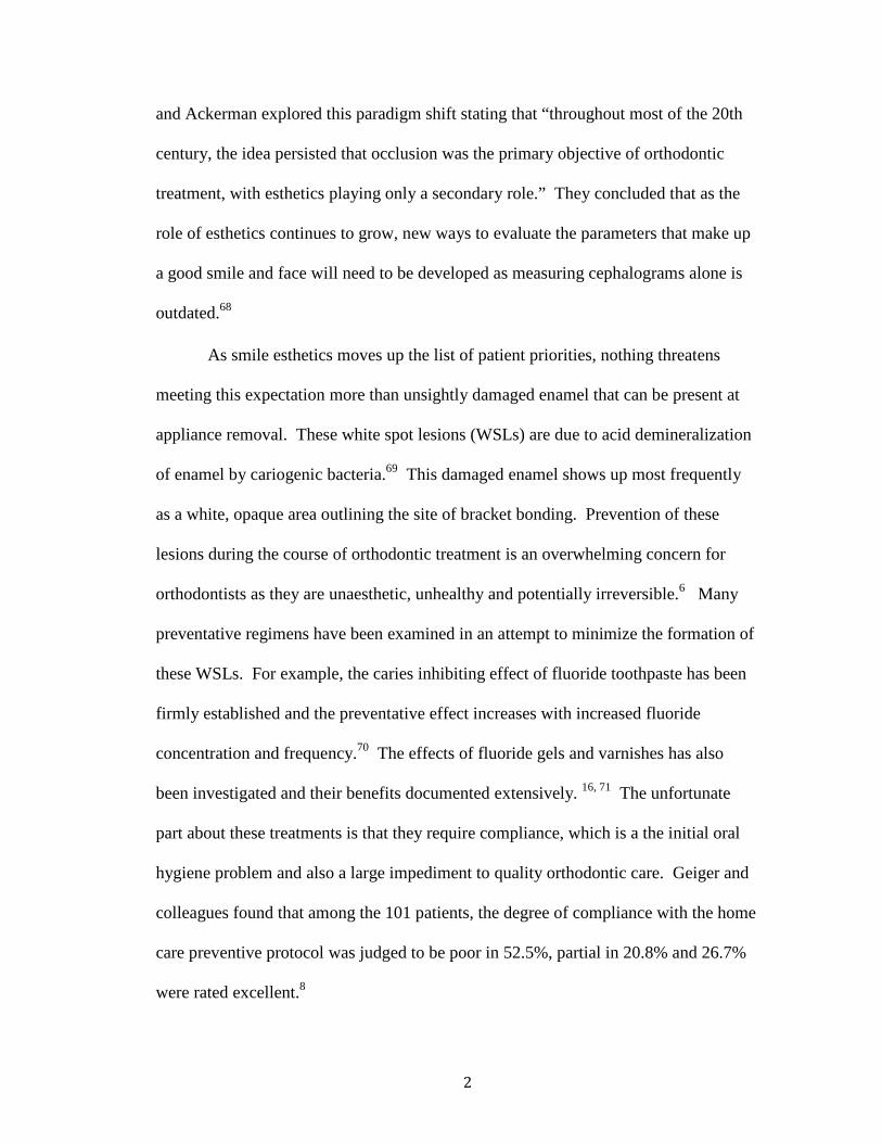
As smile esthetics moves up the list of patient priorities, nothing threatens
meeting this expectation more than unsightly damaged enamel that can be present at
appliance removal. These white spot lesions (WSLs) are due to acid demineralization
of enamel by cariogenic bacteria.69 This damaged enamel shows up most frequently
as a white, opaque area outlining the site of bracket bonding. Prevention of these
lesions during the course of orthodontic treatment is an overwhelming concern for
orthodontists as they are unaesthetic, unhealthy and potentially irreversible.6 Many
preventative regimens have been examined in an attempt to minimize the formation of
these WSLs. For example, the caries inhibiting effect of fluoride toothpaste has been
firmly established and the preventative effect increases with increased fluoride
concentration and frequency.70 The effects of fluoride gels and varnishes has also
been investigated and their benefits documented extensively. 16, 71 The unfortunate
part about these treatments is that they require compliance, which is a the initial oral
hygiene problem and also a large impediment to quality orthodontic care. Geiger and
colleagues found that among the 101 patients, the degree of compliance with the home
care preventive protocol was judged to be poor in 52.5%, partial in 20.8% and 26.7%
were rated excellent.8
3
Considering that cooperation is always part of the equation, new methods of
prevention have been developed in an attempt to prevent WSLs that don’t require
patient effort. Fluoride varnishes have been used but require multiple in-office visits
and also leave an unpleasant yellowish discoloration on teeth. Antimicrobial,
selenium and fluoride-releasing ligatures have also been developed to help prevent
enamel demineralization. However, Wiltshire and colleagues stated that on the basis
of their study, it was doubtful that fluoride-impregnated elastomeric ligature ties
would be able to significantly inhibit enamel demineralization during orthodontics
with plaque present around the brackets.29 The non-compliant products that show the
most promise in prevention are current commercially developed resins.
Ultradent developed Opalseal which is a 38% filled resin applied under brackets that
releases fluoride and recharges fluoride with uptake. Orthocoat was developed by
Pulpdent and is a fluoride releasing, light cure resin that coats both the bracket and the
tooth, prevents food and bacteria from collecting around and under brackets, and
prevents decalcification and staining. There are no studies in the current literature in
which these products are tested together.
Purpose of this Study
The purpose of this study is to compare, in vitro, the barrier effect of Opalseal, Ortho-
coat, fluoride varnish and our transbond xt control on the inhibition of enamel
demineralization.

4
Figure 1. Clinical example of before (top) and after (bottom) treatment in a patient exhibiting severe enamel demineralization.
5
White Spot Lesions and Orthodontics
Decalcification of tooth structure surrounding orthodontic appliances is
unfortunately a prevalent iatrogenic effect of orthodontic treatment. Gorelick has
reported an increase in white spots in 50% of patients after orthodontic treatment.5
Patients begin treatment with a well-aligned, esthetic result in mind and are often
dissatisfied with the lesions that remain after appliance removal. Just as dental caries
is a multifactorial disease, demineralization is a complex process involving the loss of
tooth structure resulting in an altered surface appearance. Dental caries is a state of
imbalance between demineralization and remineralization.2 Brackets, archwires,
ligatures, and other orthodontic appliances confound the use of conventional oral-
hygiene methods which can result in substantial plaque accumulation around
appliances at the level of tooth structure. This creates a cariogenic challenge on what
would normally be smooth, caries-resistant tooth surfaces. Many different treatment
regimens have been developed to limit the formation of white spot lesions. Fluoride is
the basis for a majority of these regimens.
Unfortunately some courses of therapy require patient cooperation so clinicians
have searched for methods that remain in their control. Chadwick et al concluded in a
systematic review that the use of topical fluorides in addition to fluoride toothpaste
appeared to reduce the incidence of decalcification in patients undergoing orthodontic
treatment with fixed appliances. They also found that none of the various preparations
studied appeared to be superior and that it was not possible to recommend which of the
various schedules provided the greatest protection.17 Although, Farhadian concluded

6
that one topical application of high concentration fluoride varnish could decrease
enamel lesion depth around bonded brackets by 40% for three months and found it a
highly advisable course of treatment in susceptible and uncooperative patients.24
Great promise in the war on demineralization has been seen in the area of resin
materials. In a 2006 study using bovine teeth, Paris et al found that lesions coated
with a resin sealant showed almost no progression even in artificially carious
conditions. This gave promise that sealing therapy may be a strategy to arrest lesions
in less-compliant patients.51
A study by Hu and Featherstone compared fluoride varnish with Pro Seal
(Reliance Orthodontics, Itasca, IL), which is a filled, light-cured sealant. They found
that the teeth sealed with Pro Seal had what looked like a normal enamel profile at all
points, indicating almost complete inhibition of demineralization. They also found
that the fluoride varnish group had 30% less demineralization than the control group
but more than the filled resin group. Compliance with fluoride as a solution to this
problem of demineralization was also discussed as a hindrance. Coincidentally, Pro
Seal was also found to effectively resist toothbrush abrasion meaning longer protection
if a non-compliant patient suddenly started brushing.75
There has also been research aimed at how to address demineralization lesions
once they have begun formation. One technique incorporates the use of acid to erode
the surface layer of demineralized enamel in preparation for resin infiltration. The use
of low viscosity resins to fill lesions has been shown to be a viable option for halting
lesion progression. Unfortunately, the highly mineralized enamel surface layer of
these lesions has been thought to decrease penetration of the resin. Meyer-Lueckel et

7
al conducted a study of using commercially available phosphoric acid and two
experimental hydrochloric acid gels to prepare the enamel for infiltration with the
unfilled resin. They found that a 90-120sec etch with hydrochloric acid gel led to an
almost complete removal of the surface layer without lesion damage that could
potentially cause cavitation. Phosphoric acid gel was found to be an insufficient
enamel preparation material.52
Demineralization Process
Extensive research has shown us that dental caries is a multifactorial process
involving food particles acquired in the diet, bacteria in dental plaque and the tooth
surface.76 Mutans streptococci (including Streptococcus mutans and S sobrinus) and
lactobacilli have been identified as most associated with the caries process. These
organisms, which thrive in an acidic environment, colonize the tooth surface and
metabolize fermentable carbohydrates to form glucans and lactic acid. The model
described by Harris and Garcia-Godoy illustrates the complex chemical and physical
events that are initiated once the pH drops in the presence of the production of these
acids.77 As the pH level decreases, the plaque fluid that is normally supersaturated
with calcium and phosphate gradually loses the saturation of these valuable minerals.
A critical pH of approximately 5.5 is reached and acids are able to diffuse through the
acquired pellicle to reach the enamel surface. These acids, that include lactic, acetic,
propionic and formic acid, have the capability to dissolve the calcium phosphate
mineral of tooth enamel or dentin. This is demineralization, or the loss of minerals.10

8
Featherstone stated that the acids diffuse through plaque and into porous enamel and
dissociate to produce hydrogen ions as they penetrate. These hydrogen ions have the
capacity to dissolve the mineral of tooth structure, thereby releasing calcium and
phosphate into solution that diffuse out of the tooth. But as these minerals diffuse out
of the tooth they redeposit the mineral and produce what appears as an intact surface
layer.
The outset of this process has been described by Ogaard and colleagues and
subdivided into two unique initial stages.12 The first is surface softening which is
characterized by the loss of “interprismatic substance” and is most exaggerated at the
outer enamel surface. Remineralization will occur more readily in this phase due to
the ability of the ions to penetrate the outer surface layer.13 The next is categorized as
the subsurface lesion where mineral content is lost at the deeper part of the enamel but
is still covered by a relatively intact mineral rich layer. This has also been described in
its earliest stages as an incipient lesion. It is first observed as a sub-surface white spot
lesion due to the fact that mineral loss changes the refractive index optical property the
eye perceives when compared to the intact and translucent surrounding enamel.
This entire process is a dynamic continuous cycle with the balance shifting to and fro
between demineralization and remineralization. If the demineralization process
continues unabated the lesion will progress into an overt or frank lesion.
Decalcification Prevalence in Orthodontic Patients
Fixed orthodontic appliances pose serious hygiene concerns, especially in non-

9
compliant patients. The increased plaque retention afforded by these appliances can
lead to both white spot lesion formation and gingival inflammation. A 1971 study by
Zachrisson and colleagues showed that there is a definite linear, positive correlation
between white spot lesion development and bacterial plaque accumulation in
orthodontic patients.78
Numerous studies have reported the incidence of demineralization in
orthodontic patients as anywhere from 2%-96%.1,3,5,6,8 A longitudinal study by
Zachrisson and Zachrisson showed that 89% showed signs of demineralization after
treatment and Ogaard showed that only 4% of patients were without WSLs five years
after treatment.6,78 Gorelick and colleagues performed a study examining 2,211
debonded teeth after orthodontic treatment in which there was no preventative fluoride
program other than routine toothpaste. They showed that 49.6% of patients had white
spot formation on at least one tooth but that only 10.8% had WSLs. They reported
that there was a large variation in the number of white spots per patient which
confirms the large range of incidence seen in the literature.5 Boersma et al performed
a study in which 97% of their patients displayed lesions after treatment.79
The demineralization process has been shown to transpire in only four weeks,
which can be the time between appointments for most orthodontists. 2, 7 Alexander,
O’Reily and Geiger all found that lesions can appear in patients in this one month
window of time. And the location of these lesions within the arch is another point of
interest. Mizrahi published two studies evaluating this phenomenon. In 1982 he
stated that the majority of demineralization following orthodontic treatment occurred
in the cervical and middle thirds of the facial surface of the teeth.80 Next, in a study
10
published in 1983, he found that the greatest prevalence of demineralization was seen
in the maxillary and mandibular molars followed by the maxillary incisors ranking as
the second most affected.9 Other sites commonly affected are the cervical margins of
teeth, loose molar bands and the resin enamel junction.7 And as stated earlier, white
spot lesions can be seen up to five years after treatment demonstrating their ability to
resist remineralization.
Decreasing Decalcification in Orthodontic Patients
Manual Products
The difficulty in maintaining oral hygiene with fixed appliances is greatly
increased and failure to do so can increase patients risk for decalicification, caries and
periodontal disease.78 Approximately 20% to 30% of adolescents do not adequately
remove plaque while undergoing orthodontic care.4 And taking into account that
orthodontic treatment can persist for two or more years, a rigid plaque control
program is indispensable.12 It is imperative that oral hygiene education be presented
at the beginning of treatment and reinforce at every visit by verifying that the patient
can remove plaque when problems arise. Mechanical plaque removal is the essential
first step that has to be achieved by daily brushing and flossing. Proper use of a
manual toothbrush may be sufficient, but most clinicians recommend a powered
toothbrush, which removes plaque more effectively. 4,13-15 Wilcoxon found that
counterrotational power toothbrushes are superior to manual toothbrushes in
removing dental plaque from patients with fixed orthodontic appliances.14 Ho and

11
Niederman found that the Sonicare toothbrush was significantly more effective in
supragingival plaque removal and also at reducing gram negative bacteria count than
the manual toothbrush.15 Although these products have been proven to aid
orthodontic patients in maintaining good oral hygiene, proper use and compliance are
necessities in seeing results.
Fluoride
Mechanism of action
The compliance issues associated with the use of manual products have forced
clinicians to seek alternate methods of preventing demineralization. Fluoride has long
been utilized as a supplement to manual products and plays a critical role in
orthodontics. The regular use of fluoride by a patient in orthodontic treatment has
been shown to decrease the prevalence of demineralization.6, 16, 17 Reductions of
dental caries by 40-70% was the result of widespread fluoridation of public water
supplies in many communities across the country.10 The three key characteristics of
fluoride that make it so effective are its abilities to inhibit demineralization, inhibit
bacterial metabolism and enhance remineralization.
Sound tooth enamel contains approximately 20-100ppm fluoride levels
depending on the amount of fluoride available during tooth development. Patients
developing in fluoridated communities are usually on the high end of this scale. And
although these fluoride levels are inherent to tooth structure, investigators have shown
that fluoride in solution is much more effective at preventing demineralization than
that existing in the crystalline structure of enamel.10 Systemic fluoride is incorporated

12
into enamel by combining with hydroxyapatite to form fluoroapatite which is stronger
and more resistant to dissolution.81
Another capability of fluoride is to inhibit the enzyme enolase thereby limiting
bacterial metabolism. Enolase is a bacterial enzyme necessary for the breakdown of
carbohydrates into pyruvic acid. Fluoride combines with hydrogen to penetrate the
cell wall and once inside it dissociates again and the free fluoride acidifies the bacterial
cell and disrupting enolase. Fluoride also competes with bacteria for the binding sites
on enamel to prevent adhesion to the tooth.82
Perhaps the most important characteristic of fluoride is its ability to enhance
remineralization. As saliva bathes tooth structure with its supersaturated chemical
mixture of calcium and phosphate, fluoride acts to speed up this process by absorbing
to the surface and attracting calcium ions.10 These ions are then able to form a new
structure between hydroxyapatite and fluorapatite which is stronger and more acid-
resistant that the previous enamel. Remineralization can occur in saliva alone but
Arends and ten Cate found a twofold rate increase in the presence of 1ppm fluoride
ion.83
Fluoride can help place high-caries risk patients into an equilibrium that will
favor remineralization. And although fluoride in the drinking water has been shown to
reduce dental caries, it doesn’t eradicate the problem. Jeansonne and Feagin
concluded that the action of fluoride in the presence of acid in their study indicated
that popular commercial beverages may serve as excellent vehicles for fluoridation.84
Because even the slightest changes in this equilibrium prove effective, a variety of
fluoride delivery systems have developed over the years to help dentists in the fight
against decay.

13
Fluoride Products
Reports have suggested that topical fluoride in the form of toothpastes, gels,
rinses and fluoride varnishes might reduce or eliminate decalcification during fixed
orthodontic treatment.17 But fluoride can exist in many different forms and delivery
methods. Some common preparations include stannous fluoride, sodium fluoride,
amine fluoride, and acidulated phosphofluoride. The contemporary delivery methods
include rinses, gels and solutions.
A fluoride rinse is one of the most easily accessible forms of fluoride outside
of drinking water and normal dentifrice use. Unfortunately, it is a self-administered
product and is thus related to patient compliance which is a significant problem in the
patients who need it the most.8 Geiger and colleagues found that despite educational
efforts and a free supply of rinse, only 13% of patients complied fully with its use
suggesting the need for motivation and improved compliance with this product. They
also found that there was a statistically significant dose response effect between the
frequency of rinsing and incidence of white spots regardless of oral hygiene status.
However, regardless of compliance issues, they also demonstrated a 25% reduction in
the number of patients exhibiting white spot lesions during the use of this rinsing
program.
Fluoride varnish as a topical delivery method has been used extensively in the
prevention of demineralization around orthodontic brackets.7 This is mostly due to the
compliance issues that have been seen with toothbrushing, flossing and rinsing. The
benefits of a professionally applied fluoride varnish regimen are that it doesn’t require
patient compliance and allows the orthodontist to choose his appliances without

14
worrying about the release of fluoride inherent in certain bonding systems.85 The
orthodontist can bond with resin instead of a fluoride releasing glass ionomer system
thereby avoiding a sacrifice in bond strength. Another benefit of fluoride varnish is
the delivery method that allows increased exposure time of the enamel to the fluoride.
Brudevold and colleagues showed that the efficiency of topical fluoride applications
was directly related to the exposure period of the enamel.86 In a study by Farhadian
and colleagues designed to evaluate the short term effect of topical fluoride in-vivo,
they found that one application can decrease enamel lesion depth by 40% for 3
months.24 The literature has shown that varnish can be beneficial as a preventive
adjunct in reducing demineralization around orthodontic brackets.
Another common fluoride delivery method is the gel. Comparing reports about
fluoride toothpaste alone versus toothpaste with an additional topical fluoride strongly
suggest that additional topical fluorides offer greater protection against
decalcification.17 Alexander and Ripa stated that self-applied topical fluoride
preparations are indicated in orthodontic treatment and may very well represent the
standard of care in the orthodontic community. They found that subjects who used
either gel after brushing with toothpaste at bedtime or the dentifrice twice daily,
showed significantly fewer areas of demineralization than those adding fluoride rinse
at bedtime.11 Stratemann and Shannon found that only 2% of patients on a fluoride gel
regimen developed white spot lesions while 58% of patients without fluoride
developed lesions.87 A couple of popular fluoride gel products are Prevident 5000 and
MI Paste. Prevident has 5000ppm fluoride as has been shown in one study to be
superior to products containing 1100 ppm fluoride.88 MI paste has the active
ingredient CPP-ACP, which is a complex of casein phosphopeptides and amorphous

15
calcium phosphate, and has been shown effective in reducing mean lesion depth
integrated with a paste.89 The only disadvantage of the gels is that it has been shown
that 15-20% of patients develop mild staining after 3-6 months of use.4 Although, this
side effect doesn’t outweigh the inherent benefit of preventing decalcification for
orthodontic patients.
Bonding systems incorporating fluoride
In reaction to the increasing problem of white spot lesions, orthodontic
adhesives have been developed that release fluoride in order to increase tip the balance
toward remineralization in teeth that are facing a cariogenic challenge. This is a
popular fluoride releasing system that dismisses the compliance issue as fluoride is
released at the site of bonding of fixed orthodontic appliances. Fluoride releasing
cement systems include glass ionomers, composite resins and resin-modified glass
ionomers.
The benefit of glass ionomer cements is that not only do they release fluoride,
they also absorb topically applied fluorides in order that they recharge and release over
longer periods of time.90 Fluoride is released gradually, usually more readily in low
pH environments and also releases at the site of bonding which is the closest to the site
of demineralization. The disadvantage of glass ionomer cements is that they have
been shown to have lower bond strengths and higher rates of bond failure than resin
cements.25 Composite resins have been combined with glass ionomers in pursuit of
greater bond strength and fluoride release. Resin-modified glass ionomer cements
(RMGIC) were developed to overcome moisture sensitivity problems of composites

16
and low bond strength of glass ionomers while still possessing qualities of fluoride
release, chemical bond to enamel and adhesion in a wet field.91 This type of bonding
agent has been shown to significantly prevent the development of demineralization.
Sudjalim and colleagues found that while the use of RMGIC’s significantly reduced
enamel demineralization when compared with composite resin controls, there was
concern regarding the reduction in shear and tensile bond strength.27
Enamel coatings and sealants
Enamel sealants have shown recent promise in the fight against
demineralization during orthodontic treatment. Sealant placement has been thought to
increase bond strength, protect against demineralization and protect etched enamel.
Chemical cure sealants have been shown to be ineffective due to their inability to
completely seal smooth enamel surfaces. This is due to the oxygen inhibition of
polymerization when the sealant is in contact with air in a thin layer and only islands
of cured sealant remain where resin pooling occurs.92 One way to quantify these
resins is the degree of conversion, or the percentage of bonds that are reacting. Craig
found that while chemical cure systems have a 35% conversion in the air inhibited
layer, light-initiated polymerization has around 80%.93 However, the unfilled or
lightly filled light-cured sealant hasn’t been shown to provide more protection than the
chemically cured due to mechanical toothbrush abrasion and acid attack intraorally.94
More recently, highly filled light cure cements have been shown to be effective in
reduction of enamel demineralization and also to be resistant to abrasion from
toothbrushing.95 Hu and Featherstone also found that this highly filled cement didn’t
17
affect shear bond strength of adhesive precoated metal brackets.94
New materials
Ortho-coatTM
Ortho-coatTM (Pulpdent Corporation) is a fluoride releasing, light cure resin
that coats the bracket and tooth. It incorporates Pulpdent’s bis-GMA-free Embrace
Technology, which is a patented, hydrophilic resin technology that behaves favorably
in a moist environment. Ortho-coat integrates with the tooth structure and is noted for
its ability to prevent microleakage. The product can be used with both light cure and
chemical cure adhesives. For light cure adhesives, Ortho-coat can be applied
immediately after bracket placement and cured for 20 seconds or it applied at
subsequent orthodontic visits by etching the bracket margin and enamel and placing.
When using with chemical cure adhesives and placing the material after initial
bonding, the air-inhibited layer must be removed with a dry cotton pellet or alcohol.
Tuncer and colleagues performed a study evaluating the effect of Ortho-coat
application on bond strength when used with two different self-etching primers. They
found that the application of enamel-protective resin didn’t affect the bond strength of
orthodontic brackets to enamel with a self-etching primer.96 Abdelnaby and Al-
Wakeel studied the bond strength and microleakage properties of Ortho-coat when
used with transbond xt. They found that curing the resin coat and adhesive
simultaneously showed an increase in shear bond strength (SBS) that was not
significant, but curing the resin and adhesive separately exhibited a significant
increase. It was also shown that Ortho-coat applied in either protocol produced a
18
significant reduction in microleakage probably due to the barrier around the bracket
and adhesive edges.97
Opalseal
Opalseal is a fluoride releasing and recharging light cured primer that is 38%
filled with substantial glass ionomer plus nano-fillers and is compatible with any
bonding system. Opal Seal is used when bonding orthodontic appliances to etched
enamel and the use of a UV “black” light will make the sealant illuminate showing
that the resin containing fluoride is present. Opalseal is applied by etching the tooth
surface, rinsing and applying a thin layer of opalseal. It is then cured for 5 seconds to
avoid bracket skating on placement and then the bracket is cured per manufacturer’s
instructions. There is no current literature available evaluating either the bond strength
or demineralization inhibiting properties of opalseal.
Present Study
Bonded resins have been shown to be effective in reducing enamel
demineralization during orthodontic treatment. (51, 94, 95, 97) No reports have been
conducted to compare Ortho-coat and Opalseal with a fluoride varnish and control.
The specific aim of this study was to compare, in vitro, the barrier effect of Opalseal,
Ortho-coat, fluoride varnish and our Transbond xt control on the inhibition of enamel
demineralization.

19
MATERIALS AND METHODS
Study Design
Forty extracted human central incisor teeth were collected and stored in a 10%
solution of sodium hypochlorite. Approximately one month prior to the study, the
teeth were transferred to distilled water. The study called for four groups of at least
ten teeth with random assignment to each treatment group. Additional teeth were used
in the case of defect or fracture that resulted in exclusion from the study. The study
design and sample sizes are shown in table 1.
Table 1 Study design
Treatment group Specimen Co
Group 1: Control N=13 N=
Group 2: Fluoride Varnish N=14 N=
Group 3: Ortho-coat N=14 N=
Group 4: Opalseal N=13 N=
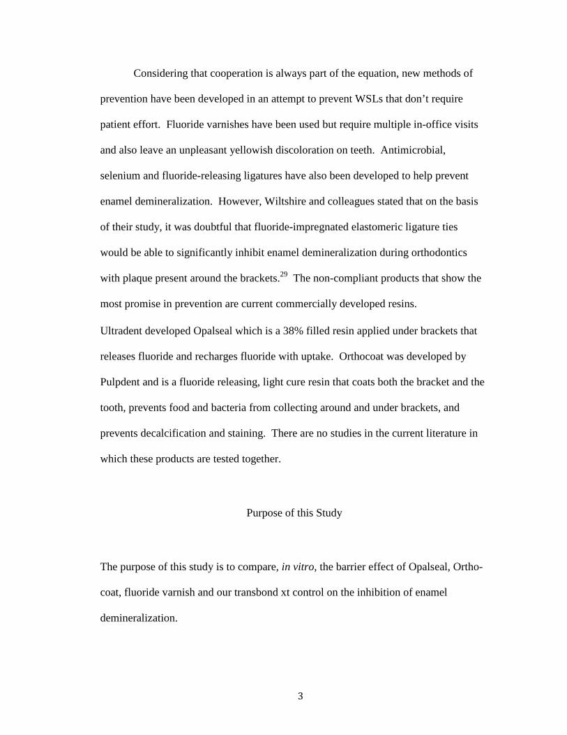
The teeth were sectioned 2mm apical to the cement-enamel junction using a
diamond bur under copious irrigation and the root segment was discarded. The
remaining crown was then prepared using oil-free, water based pumice (Whipmix,
Louisville, KY) to clean the entire enamel surface followed by a rinse and drying
stage. All further isolation methods and bonding techniques were performed under

2.9-x magnification using dental loupes.
placed in the center of the facial surface of the tooth and the remaining exposed tooth
structure was covered adequately with 2 coats of
City of Industry, CA). This was done to isolate the treatment area of the tooth and to
protect the remaining portion of the tooth from demineralization by the acidic
demineralization solution. After the varnish was
removed leaving a 7mm diameter treatment zone on the facial surface of the tooth.
Another segment of tape was then placed over this treatment zone leaving an open
hole 4.9mm in diameter. The purpose of this tape segment was to isolate the etch
portion of the tooth to the 4.9mm diameter circle, thus leaving an untreated enamel
border of 1.02mm width encircling the treatment zone. This created a “bulls
effect of treatment zones that is illustrated in Figure 1
Figure 2. Bonding Diagram
20
using dental loupes. A 7mm diameter masking tape
placed in the center of the facial surface of the tooth and the remaining exposed tooth
structure was covered adequately with 2 coats of Megalast TM nail varnish (
). This was done to isolate the treatment area of the tooth and to
protect the remaining portion of the tooth from demineralization by the acidic
solution. After the varnish was completely dry, the tape circ
removed leaving a 7mm diameter treatment zone on the facial surface of the tooth.
Another segment of tape was then placed over this treatment zone leaving an open
hole 4.9mm in diameter. The purpose of this tape segment was to isolate the etch
tion of the tooth to the 4.9mm diameter circle, thus leaving an untreated enamel
border of 1.02mm width encircling the treatment zone. This created a “bulls
zones that is illustrated in Figure 1.
. Bonding Diagram
tape circle was
placed in the center of the facial surface of the tooth and the remaining exposed tooth
nail varnish (Markwins,
). This was done to isolate the treatment area of the tooth and to
protect the remaining portion of the tooth from demineralization by the acidic
tape circle was
removed leaving a 7mm diameter treatment zone on the facial surface of the tooth.
Another segment of tape was then placed over this treatment zone leaving an open
hole 4.9mm in diameter. The purpose of this tape segment was to isolate the etch
tion of the tooth to the 4.9mm diameter circle, thus leaving an untreated enamel
border of 1.02mm width encircling the treatment zone. This created a “bulls-eye”

21
Bonding Protocol Control
After isolation was completed according to the previous instructions, the
4.9mm diameter hole was coated with 38% phosphoric acid etchant gel (Etch-Rite,
Pulpdent Corporation, Watertown, MA) for 30 seconds. The bonding area was then
rinsed for 5 seconds with sterile water and dried thoroughly with oil/moisture-free air.
A thin layer of transbond xt prime and bond (3M Espe, St Paul, MN) was applied and
air dispersed for 2 seconds. A 3.8mm diameter button was positioned in the center of
the bonding area using transbond xt composite (3M Espe, St Paul, MN), removing all
excess composite. The tape isolation border was removed and the specimen was cured
for 20 seconds using a Valo broadband LED curing light (Opal Orthodontics by
Ultradent, South Jordan, UT) with an intensity of 1700mW/cm2.
Fluoride Varnish
After isolation was completed according to the previous instructions, the
4.9mm diameter hole was coated with 38% phosphoric acid etchant gel (Etch-Rite,
Pulpdent Corporation, Watertown, MA) for 30 seconds. The bonding area was then
rinsed for 5 seconds with sterile water and dried thoroughly with oil/moisture-free air.
A thin layer of Transbond xt prime and bond (3M Espe, St Paul, MN) was applied and
air dispersed for 2 seconds. A 3.8mm diameter button was positioned in the center of
the bonding area using Transbond xt composite (3M Espe, St Paul, MN), removing all
excess composite. The specimen was cured for 20 seconds using a Valo broadband

22
LED curing light (Opal Orthodontics by Ultradent, South Jordan, UT) with an
intensity of 1700mW/cm2. A coat of 5% sodium fluoride Cavity Shield varnish (3M
Espe, St Paul, MN) was applied around the bracket in the treated enamel zone and the
isolation tape was removed.
Ortho-coat
After isolation was completed according to the previous instructions, the
4.9mm diameter hole was coated with 38% phosphoric acid etchant gel (Etch-Rite,
Pulpdent Corporation, Watertown, MA) for 30 seconds. The bonding area was then
rinsed for 5 seconds with sterile water and dried thoroughly with oil/moisture-free air.
A thin layer of Transbond xt prime and bond (3M Espe, St Paul, MN) was applied and
air dispersed for 2 seconds. A 3.8mm diameter button was positioned in the center of
the bonding area using Transbond xt composite (3M Espe, St Paul, MN), removing all
excess composite. The specimen was cured for 20 seconds using a Valo broadband
LED curing light (Opal Orthodontics by Ultradent, South Jordan, UT) with an
intensity of 1700mW/cm2. A thin layer of Ortho-coat was applied around the bracket
margin and the treated enamel zone and then cured using the same method as stated
above. The tape isolation border was then removed.
Opalseal
After isolation was completed according to the previous instructions, the
4.9mm diameter hole was coated with 38% phosphoric acid etchant gel (Etch-Rite,
Pulpdent Corporation, Watertown, MA) for 30 seconds. The bonding area was then

23
rinsed for 5 seconds with sterile water and dried thoroughly with oil/moisture-free air.
A thin layer of Opalseal (Ultradent, South Jordan, UT) was applied and air dispersed
for 2 seconds. A 3.8mm diameter button was positioned in the center of the bonding
area using Transbond xt composite (3M Espe, St Paul, MN), removing all excess
composite. The tape isolation border was removed and the specimen was cured for 20
seconds using a Valo broadband LED curing light (Opal Orthodontics by Ultradent,
South Jordan, UT) with an intensity of 1700mW/cm2.
All four groups of specimen were then subjected to thermocycling for 10,000
cycles over a period of 5 days alternating between 6C and 65C with a 15 second dwell
time. Following the thermocycling procedure, the teeth were placed in sterile water
and stored at 37 degrees for approximately 7 days while the artificial caries
demineralization solution was being prepared.
An artificial caries solution was prepared by first combining 500 mL 1M lactic
acid with 10 mL K2HPO4 stock solution. The pH of the solution was brought up to
4.7 using 1M NaOH. The solution was titrated to pH 3.8, which was verified using a
calibrated sensION 4 pH/ISE meter (Hach Company, Loveland, CO). Each group of
teeth was placed in a separate container of the artificial caries solution and then stored
at 37° C for 48 hrs. The groups were then rinsed copiously with distilled water to
remove all remnants of the demineralization solution and then stored in distilled water
for three days.
At the conclusion of the treatment, the nail varnish was removed and all teeth
were sectioned using the Isomet machine. They were then imaged under 100x
magnification using a digital light microscope (Keyence VHX-600 series, Woodcliff
24
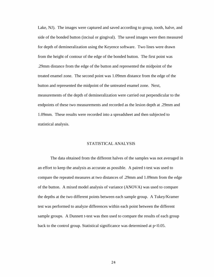
Lake, NJ). The images were captured and saved according to group, tooth, halve, and
side of the bonded button (incisal or gingival). The saved images were then measured
for depth of demineralization using the Keyence software. Two lines were drawn
from the height of contour of the edge of the bonded button. The first point was
.29mm distance from the edge of the button and represented the midpoint of the
treated enamel zone. The second point was 1.09mm distance from the edge of the
button and represented the midpoint of the untreated enamel zone. Next,
measurements of the depth of demineralization were carried out perpendicular to the
endpoints of these two measurements and recorded as the lesion depth at .29mm and
1.09mm. These results were recorded into a spreadsheet and then subjected to
statistical analysis.
STATISTICAL ANALYSIS
The data obtained from the different halves of the samples was not averaged in
an effort to keep the analysis as accurate as possible. A paired t-test was used to
compare the repeated measures at two distances of .29mm and 1.09mm from the edge
of the button. A mixed model analysis of variance (ANOVA) was used to compare
the depths at the two different points between each sample group. A Tukey/Kramer
test was performed to analyze differences within each point between the different
sample groups. A Dunnett t-test was then used to compare the results of each group
back to the control group. Statistical significance was determined at p<0.05.

25
RESULTS
Each tooth was divided in half and each half measured at two points for a total
of four measurements on each tooth. 54 teeth were used so ideally we would have 216
total measurements. But as each tooth was sectioned to obtain the samples, several
specimens fractured and were unsuitable to be used as samples. After specimen with
fractures, flash and cavitations were excluded; the study yielded 167 total
measurements. The 49 exclusions were distributed as follows: Control 8, varnish 18,
Ortho-coat 13 and Opalseal 10.
The mean depths for the midpoints of the treated enamel group and untreated
enamel group and the results of the paired t-test on these data are shown in Figure 2.
Figure 3. Paired t-test means comparision
The results indicate that the differences in lesion depths between the untreated enamel
-.029 166 -8.237 <.0001-.045 10 -2.149 .0572 -.009 10 -1.187 .2625 -.049 11 -2.889 .0147 -.027 8 -2.071 .0721 -.066 10 -3.775 .0036 -.056 8 -4.476 .0021 -.059 11 -3.400 .0059 -.050 9 -3.527 .0064 -.017 8 -1.733 .1213 -.010 9 -1.104 .2982 -.033 8 -3.267 .0114 -.004 10 -.502 .6268
-2.344E-4 8 -.032 .9754 -.013 10 -1.848 .0943 -.022 10 -1.482 .1692 -.007 11 -.871 .4026
Mean Diff. DF t-Value P-Value_.29_, _1.09_: Total_.29_, _1.09_: oc, a, g_.29_, _1.09_: oc, a, i_.29_, _1.09_: oc, b, g_.29_, _1.09_: oc, b, i_.29_, _1.09_: os, a, g_.29_, _1.09_: os, a, i_.29_, _1.09_: os, b, g_.29_, _1.09_: os, b, i_.29_, _1.09_: v, a, g_.29_, _1.09_: v, a, i_.29_, _1.09_: v, b, g_.29_, _1.09_: v, b, i_.29_, _1.09_: xc, a, g_.29_, _1.09_: xc, a, i_.29_, _1.09_: xc, b, g_.29_, _1.09_: xc, b, i
Paired t-testSplit By: Group, A/B, G/IHypothesized Difference =
11 .069 .062 .0111 .091 .034 .0112 .072 .053 .01
9 .078 .052 .0111 .113 .076 .02
9 .088 .056 .0112 .118 .080 .0210 .105 .094 .03
9 .113 .049 .0110 .091 .053 .01
9 .091 .029 .0111 .083 .064 .01
9 .098 .066 .0211 .081 .061 .0111 .084 .070 .0212 .093 .051 .01
Count Mean Std. Dev. Std. Err.oc, a, goc, a, ioc, b, goc, b, ios, a, gos, a, ios, b, gos, b, iv, a, g v, a, iv, b, g v, b, ixc, a, xc, a, i xc, b, xc, b, i
Means Table for _.29_ Effect: Group * A/B * G/I
11 .114 .042 .0111 .100 .043 .0112 .121 .040 .01
9 .105 .035 .0111 .179 .058 .01
9 .144 .050 .0112 .177 .089 .0210 .155 .062 .02
9 .130 .042 .0110 .101 .068 .02
9 .124 .043 .0111 .086 .069 .02
9 .098 .073 .0211 .094 .059 .0111 .106 .078 .0212 .100 .060 .01
Count Mean Std. Dev. Std. Err.oc, a, goc, a, ioc, b, goc, b, ios, a, gos, a, ios, b, gos, b, iv, a, g v, a, iv, b, g v, b, ixc, a, xc, a, i xc, b, xc, b, i
Means Table for _1.09_ Effect: Group * A/B * G/I

26
at .29mm and the treated enamel at 1.09mm were only statistically significant for the
opalseal group. All four measurement points for the opalseal group had statistically
significant differences between the treated and untreated zones of enamel. The
orthocoat and varnish groups had a statistically significant difference only at one
measurement point. The control group showed no statistically significant differences
between the two measurements.
The means were plotted in a graph below to illustrate the relationship of the
mean lesion depths compared with each group at the two different points. The lesion
depths were shown to be the highest for the opalseal group for both the .29mm and
1.09 mm points illustrated in Figure 3.
Figure 4. Interaction Plot for means
0
.02
.04
.06
.08
.1
.12
.14
Cell M
ean
oc, a oc, b os, a os, b v, a v, b xc, a xc, bCell
i
g
Interaction Bar Plot for _.29_Effect: Group * A/B * G/I
0
.02
.04
.06
.08
.1
.12
.14
.16
.18
.2
Cell M
ean
oc, a oc, b os, a os, b v, a v, b xc, a xc, bCell
i
g
Interaction Bar Plot for _1.09_Effect: Group * A/B * G/I
27
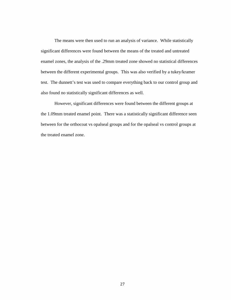
The means were then used to run an analysis of variance. While statistically
significant differences were found between the means of the treated and untreated
enamel zones, the analysis of the .29mm treated zone showed no statistical differences
between the different experimental groups. This was also verified by a tukey/kramer
test. The dunnett’s test was used to compare everything back to our control group and
also found no statistically significant differences as well.
However, significant differences were found between the different groups at
the 1.09mm treated enamel point. There was a statistically significant difference seen
between for the orthocoat vs opalseal groups and for the opalseal vs control groups at
the treated enamel zone.

28
DISCUSSION
The goal of orthodontic treatment is to create a functionally sound occlusion
and an esthetically idyllic smile. White spots that are the product of poor hygiene can
act to spoil this result. Given that studies show that restoration of these lesions very
difficult, it is imperative to take all preventative measures necessary to prevent their
formation. An adhesive resin can offer a unique approach to preventing white spot
lesions that does not depend on patient compliance and can be completed during
bonding to protect the appearance of the teeth. These resins coat the tooth structure
around fixed orthodontic appliances to prevent lesions from beginning and
progressing to cavitation. Previous studies have shown that this can be an effective
way to prevent the demineralization process that causes white spot lesions. 98, 99
However, no published studies have reported results that compare the effectiveness of
some of these newer commercially available products with a traditional bonding
control and a fluoride varnish. The aim of this study was to compare, in vitro, the
barrier effect of Opalseal, Ortho-coat, fluoride varnish and our Transbond xt control
on the inhibition of enamel demineralization.
In the present study, teeth treated with Opalseal showed a significant
difference in mean depths of treated and untreated tooth structure. The four points
measured in the Opalseal group showed mean differences of .066, .056, .059 and
.050mm. This was the only group that had consistently significant differences in the
mean lesion depths between the treated and untreated zones. Both the varnish group
and the ortho-coat group had one of the four points that indicated a statistically

29
significant difference but no consistent pattern. The points measured on each half of
the sample were mirror images of each other at the incisal and gingival portion of the
bonded orthodontic button. Thus the measurements are taken at a sample of tooth
separated only by the thickness of the isomet blade. Therefore, samples that don’t
show consistent statistically significant differences may contain an anomaly that
only presents itself at one measurement point. There have been no studies using
opalseal to study demineralization inhibition with which to compare the current
results.
The interaction bar plot graph in Figure 2 illustrates that lesion depth around
the Opalseal group was greater than the lesion depths in other groups. The means for
the Opalseal untreated enamel point were significantly greater than the means for the
other three groups registering at .177 and .179mm for the gingival measurements of
the samples. This is confirmed by the anova analysis for the untreated zone. There
were statistically significant differences between the opalseal group and both the
control and orthocoat groups at the 1.09 untreated enamel points. This difference can
be attributed to the fact that the opalseal demineralization depths at the untreated zone
were greater than the other groups. There may be two possible explanations for this
finding. The first would be an unintentional bias within the teeth selected for the
opalseal group. The teeth were distributed at random but there may have been a slight
difference in the enamel properties of the teeth placed into the opalseal group making
them more prone to larger demineralization depths. Second, the results indicate that
the fluoride leaching properties of the material, which are expected to provide benefit
to the enamel surrounding the treated zone, had no significant effect. Studies have

30
shown that many adhesives currently in use release fluoride but long-term clinical
studies have shown that this was unsuccessful clinically in preventing
demineralization around brackets.72, 73, 74 However, the benefit of the material is in its
ability to recharge in the presence of fluoridated hygiene products and to show
continued release. Our study design restricted the recharge of fluoride as
demineralization was carried out in an artificial caries solution and were never exposed
to a source of fluoride.
Another significant finding was the fact that there were no statistically
significant differences found between the mean lesion depths of the different groups in
the treated enamel zones. This information is useful in evaluating the barrier effect as
no material added a greater demineralization inhibition effect. The control, fluoride
and Ortho-coat samples all contained a coat of Transbond xt used as the primer to
bond the bracket. The only different group with respect to the barrier around the
button was the opalseal group that used opalseal material for the primer. Wear or
breaks in a lightly or unfilled resin caused by mechanical toothbrush abrasion and
chemical attack could result in significant decalcification. Highly filled sealants have
been found to resist abrasion and remain attached to enamel surfaces.99 Pro Seal,
which is a light-cured, highly-filled sealant, was found to greatly resist toothbrush
abrasion and effectively seal a smooth enamel surface.94 The omission of toothbrush
wear in our study could the reason that the treated enamel zones appeared similar for
all groups and an abrasion study may give different results.

31
CONCLUSIONS
Under the conditions of the present study, the following conclusions are made:
1. There was no difference in demineralization around bonded orthodontic
appliances between the groups.
2. There was no difference in the groups relative to the control for the untreated
enamel indicating that no fluoride releasing benefit was seen.

32
REFERENCES
1. Artun J, Brobakken BO. Prevalence of caries and white spots after orthodontic treatment with multibanded appliances. Eur J Orthod 1986;8:229-231. 2. Øgaard B, Rǿlla G, Arends J. Orthodontic appliances and enamel demineralization. Part 1. Lesion development. Am J Orthod Dentofacial Orthop 1988;94:68-73. 3. Chang, H, Walsh L, Freer T. Enamel demineralization during orthodontic treatment. Aetiology and prevention. Aust Dent J 1997;42(5):322-327. 4. Boyd, R. Enhancing the value of orthodontic treatment: incorporating effective preventive dentistry into treatment. Am J Orthod Dentofacial Orthop 2000;117(5):601-603. 5. Gorelick L, Geiger A, Gwinnett A. Incidence of white spot formation after bonding and banding. Am J Orthod Dentofacial Orthop 1982;81:93-98. 6. Øgaard B. Prevalence of white spot lesions in 19-year-olds: A study on untreated and orthodontically treated persons 5 years after treatment. Am J Orthod Dentofacial Orthop 1989;96:423-427. 7. O’Reilly MM, Featherstone JDB. Demineralization and remineralization around orthodontic appliances. An in vivo study. Am J Orthod Dentofacial Orthop 1987;92:33-40. 8. Geiger AM, Gorelick L, Gwinnett AJ, Griswold PG. The effect of a fluoride program on white spot formation during orthodontic treatment. Am J Orthod Dentofacial Orthop 1988;93:29-37. 9. Mizrahi E. Surface distribution of enamel opacities following orthodontic treatment. Am J Orthod, 1983;81:323-31. 10. Featherstone JDB. Prevention and reversal of dental caries: role of low level fluoride. Comm Dent Oral Epid 1999;27:31-40. 11. Alexander SA, Ripa LW. Effects of self-applied topical fluoride preparations in orthodontic patients. Angle Orthod 2000;70:424-430. 12. Øgaard B, Rǿlla G, Arends J, ten Cate JM. Orthodontic appliances and enamel demineralization. Part 2. Prevention and treatment of lesions. Am J Orthod Dentofacial Orthop, 1988;94:123-28.

33
13. Heintze S, Jost-Brinkmann P, Loundos J. Effectiveness of three different types of electric toothbrushes compared with a manual technique in orthodontic patients. Am J Orthod Dentofacial Orthop, 1996;110:630-638. 14. Wilcoxon D, Ackerman R, Killoy W, Love J, Sakumura J, Tira D. The effectiveness of a counterrotational-action power toothbrush on plaque control in orthodontic patients. Am J Orthod Dentofacial Orthop 1991;99:7-14. 15. Ho H, Niederman R. Effectiveness of the Sonicare toothbrush on reduction of plaque, gingivitis, probing pocket depth and subgingival bacteria in adolescent orthodontic patients. J Clin Dent 1997;8:15-19. 16. Marinho VCC, Higgins JPT, Logan S, Sheiham A. Fluoride mouthrinses for preventing dental caries in children and adolescents. Cochrane Database of Systematic Reviews 2003;3. 17. Chadwick BL, Roy J, Knox J, Treasure ET. The effect of topical fluorides on decalcification in patients with fixed orthodontic appliances: a systematic review. Am J Orthod Dentofacial Orthop 2005;128:601-6. 18. Pfarrer AM, Karlinsey RL. Challenges of implementing new remineralization technologies. Adv Dent Res 2009;21:79-82. 19. Derks A, Katsaros C, Frencken JE, van’t Hof MA, Kuijpers-Jagtman AM. Caries- inhibiting effect of preventive measures during orthodontic treatment with fixed appliances. Caries Res 2004;38:413-420. 20. Bishara SE, Ostby AW. White spot lesions: formation, prevention, and treatment. Semin Orthod 2008;14:174-182. 21. Geiger AM, Gorelick L, Gwinnett AJ, Benson BJ. Reducing white spot lesions in orthodontic populations with fluoride rinsing. Am J Orthod Dentofacial Orthop 1992;101:403-407. 22. Grobler SR, Øgaard B, Rolla G. Uptake and retention of fluoride in sound dental enamel in vivo after a single application of neutral 2 percent sodium fluoride. In: Rolla G, Sonju T, Embery G, eds. Tooth surface interactions and preventive dentistry. London: Information Retrivel Ltd, 1971;17-26.
23. Vivalidi-Rodrigues G, Demito CF, Bowman SJ, et al. The effectiveness of a fluoride varnish in preventing development of white spot lesions. World J Orthod 2006;7:138-144.

34
24. Farhadian N, Amirfarhang M, Eslami B, Mehrabi S. Effect of fluoride varnish on enamel demineralization around brackets: an in-vivo study. Am J Orthod Dentofacial Orthop 2008;133:S95-8.2. 25. Hallgren A, Oliveby A, Twetman S. Fluoride concentration in plaque adjacent to orthodontic brackets retained with glass ionomer cements. Caries Res 1993;23:51-54. 26. Rix D, Foley TF, Mamandras A. A comparison of bond strength of three adhesives: composite resin, hybrid GIC, and glass-filled GIC. Am J Orthod Dentofacial Orthop 2001;119:36-42. 27. Sudjalim, TR, Woods MG, Manton DJ, Reynolds EC. Prevention of demineralization around orthodontic brackets in vitro. Am J Orthod Dentofacial Orthop 2007; 131:705-705. 28. Wiltshire WA. Determination of fluoride from fluoride-releasing elastomeric ligature ties. Am J Orthod Dentofacial Orthop 2007;110:383-387. 29. Wiltshire WA. In Vitro and In Vivo Fluoride Release from Orthodontic Elastomeric Ligature Ties. Am J Orthod Dentofacial Orthop 1999;115:288-92. 30. Reynolds EC, Cain CJ, Webber FL, Black CL, Riley PF, Johnson IH, Perich JW. Anticariogenicity of calcium phosphate complexes of tryptic casein phosphopeptides in the rat. J Dent Res 1995;74(6):1272-1279. 31. Reynolds EC. Remineralization of enamel subsurface lesions by casein phosphopeptide-stabilized calcium phosphate solutions. J Dent Res 1997;76(9):1587- 1595. 32. Reynolds EC. Anticariogenic complexes of amorphous calcium phosphate stabilized by casein phosphopeptides: a review. Spec Care Dent 1998;18:(1),8-16. 33. Llena C, Forner L, Baca P. Anticariogenicity of casein phosphopeptide-amorphous calcium phosphate: a review of the literature. J Contemp Dent Pract 2009;16(3):1-9. 34. Reynolds EC, Riley PF, Storey E. Phosphoprotein inhibition of hydroxyapatite dissolution. Calcif Tissue Int 1982;34:S52-S56. 35. Cross KJ, Huq NL, Reynolds EC. Casein phosphopeptides in oral health- chemistry and clinical applications. Curr Pharm Des 2007;13:793-800.

35
36. GC America. MI Paste: a breakthrough that has everyone smiling. [Product brochure] 2006. 37. Reynolds EC, Cai F, Shen P, Walker GD. Retention in plaque and remineralization of enamel lesions by various forms of calcium in a mouthrinse or sugar-free chewing gum. J Dent Res 2003;82:206-211. 38. Ung M, Huq NL, Cross KJ, Reynolds EC. Characterization of the binding of anticariogenic casein phosphopeptide complexes to the enamel pellicle proteins. Aust Dent J 2004;49:S19-20. 39. Reynolds E. Anticariogenic complexes of amorphous calcium phosphate stabilized by casein phosphate. A review. Spec Care Dent 1998;18(1):8-16. 40. Donly KJ, Sasa IS. Potential remineralization of postorthodontic demineralized enamel and the use of enamel microabrasion and bleaching for esthetics. Semin Orthod 2008;14:220-225. 41. Andersson A, Sköld-Larsson K, Hallgren A, Petersson LG, Twetman S. Effect of a dental cream containing amorphous cream phosphate complexes on white spot lesion regression assessed by laser fluorescence. Oral Health Prev Dent 2007;5(3):229-233. 42. Lijima Y, Cai F, Shen P, Walker G, Reynolds C, Reynolds EC. Acid resistance of enamel subsurface lesions remineralized by a sugar-free chewing gum containing casein phosphopeptide-amorphous calcium phosphate. Caries Res 2004;38:551-556. 43. Croll TP, Bullock GA. Enamel microabrasion for removal of smooth surface decalcification lesions. J Clin Orthod 1994;6:365-370. 44. Donly KJ, O’Neill M, Croll TP. Enamel microabrasion: A microscopic evaluation of the “abrasion effect’. Quintessence Int 1992;23:175-179. 45. Segura A, Donly KJ, Wefel JS, et al. (1997). The effects of microabrasion on demineralization inhibition of enamel surfaces. Quintessence Int 1997;28:463-466. 46. Murdoch-Kinch CA, McLean ME. Minimally invasive dentistry. J Am Dent Assoc 2003;134:87-95. 47. Davila JM, Buonocore MG, Greeley CB, Provenza DV. Adhesive penetration in human artificial and natural white spots. J Dent Res 1975;54:999-1008. 48. Robinson C, Hallsworth AD, Weatherell JA, Kunzel W. Arrest and control of carious lesions: a study based on preliminary experiments with resorcinol-formaldehyde resin. J Dent Res 1976;55:812-818.

36
49. Robinson C, Brookes SJ, Kirkham J, Wood SR, Shore RC. In vitro studies of the penetration of adhesive resins into artificial caries-like lesions. Caries Res 2001;35:136-141. 50. Schmidlin PR, Zehnder M, Pasqualetti T, Imfeld T, Besek MJ. Penetration of a bonding agent into de- and remineralized enamel in vitro. J Adhes Dent 2004;6:111-115. 51. Paris S, Meyer-Lueckel H, Mueller J, Hummel M, Kielbassa AM. Progression of sealed initial bovine enamel lesions under demineralizing conditions in vitro. Caries Res 2006;40:124-129. 52. Meyer-Lueckel H, Paris S, Kielbassa AM. Surface layer erosion of natural caries lesions with phosphoric and hydrochloric acid gels in preparation for resin infiltration. Caries Res 2007;41:223-230. 53. Flaitz CM, Hicks MJ. Role of the acid-etch technique in remineralization of caries- like lesions of enamel: a polarized light and scanning electron microscope study. ASCD J Dent Child 1994;61:21-28. 54. Al-Khateeb S, Exterkate R, Angmar-Mansson B, ten Cate JM. Effect of acid-etching on remineralization of enamel white spot lesions. Acta Odontol Scand 2000;58: 31-36. 55. Paris S, Meyer-Lueckel H, Colfen H, Kielbassa AM. Penetration coefficients of commercially available and experimental composites intended to infiltrate enamel carious lesions. Dent Mater J 2007a;23:742-748. 56. Paris S, Meyer-Lueckel H, Colfen H, Kielbassa AM. Resin infiltration of artificial enamel caries lesions with experimental light curing resins. Dent Mater J 2007b;26:582-588. 57. Paris S, Meyer-Lueckel H, Kielbassa AM. Resin infiltration of natural caries lesions. J Dent Res 2007c;86:662-666. 58. Meyer-Lueckel H, Paris S. Progression of artificial enamel caries lesions after infiltration with experimental light curing resins. Caries Res 2008;42:117-124. 59. Paris S. Meyer-Lueckel H. Influence of application frequency on an infiltrant on enamel lesions. J Dent Res 2008;87(Spec Iss B):1585. 60. Glazer HS. Treating white spots: new caries infiltration technique. Dentistry Today 2009: 82-85.

37
61. http://www.drilling-no-thanks.com 62. Martinez de Pison J. Infiltration: a new treatment for caries. Dental Tribune, May 2009: 19. 63. Paris S, Meyer-Lueckel H. Progression of infiltrated artificial enamel caries lesions in situ. Data on file. DMG. Hamburg, Germany. 64. Øgaard B. White spot lesions during orthodontic treatment: mechanisms and fluoride preventive aspects. Semin Orthod 2008;14:183-103. 65. Walsh L. Evidence that demands a verdict: latest developments in remineralization therapies. Australas Dent Pract March/April 2009: 48-59. 66. Andrews LF. Six keys to normal occlusion. AM J ORTHOD 1972;62:296-309. 67. Rinchuse D, Kandasamy S. Myths of Orthodontic Gnathology. Am J Orthod Dentofacial Orthop 2009; 136:322-30. 68. Sarver DM, Ackerman JL. Orthodontics about face: the reemergence of the esthetic paradigm. Am J Orthod Dentofacial Orthop 2000;117:575-6. 69. Featherstone JDB. The science and practice of caries prevention. J Am Dent Assoc 2000;131:887-99. 70. Marinho VCC, Higgins JPT, Sheiham A, Logan S. Fluoride toothpastes for preventing dental caries in children and adolescents. Cochrane Database of Systematic Reviews 2003;3c. 71. Marinho VCC, Higgins JPT, Logan S, Sheiham A. Fluoride gels for preventing dental caries in children and adolescents. Cochrane Database of Systematic Reviews 2003;3a
72. Mitchell L. An investigation into the effect of a fluoride releasing adhesive on the
prevalence of enamel surface changes associated with directly bonded orthodontic
attachments, Br J Orthod 19 (1992), pp. 207–214.
73. D.T. Millett, J.H. Nunn, R.R. Welbury and P.H. Gordon, Decalcification in
relation to brackets bonded with glass ionomer cement or a resin adhesive, Angle
Orthod 69 (1999), pp. 65–70.
38
74. M. Gaworski, M. Weinstein, A.J. Borislow and L.E. Braitman, Decalcification and bond failure: a comparison of a glass ionomer and a composite resin bonding, Am J Orthod Dentofacial Orthop 116 (1999), pp. 518–521. 75 Hu W, Featherstone JDB. Prevention of enamel demineralization: An in-vitro study Using light-cured filled sealant. Am J Orthod Dentofacial Orthop 2005;128:592-600. 76 Zero, DT. Dental caries process. Dent Clin North Am. 43(4):635-64, 1999 77 Harris NO, Garcia-Godoy F. Primary Preventive Dentistry 5th ed. Appleton and Lange. 1999. 78 Zachrisson BI, Zachrisson S. Caries incidence and oral hygiene during orthodontic treatment. Scand J Dent Res. 79(6):394-401, 1971. 79 Boersma JG, van der Veen HM, Lagerweij MD, Bokhout B, Prahl-Andersen B. Caries prevalence measured with QLF after treatment with fixed orthodontic appliances: influencing factors. Caries Res 2005;39(1):41-7. 80 Mizrahi E. Enamel demineralization following orthodontic treatment. Am J Orthod 1982;82(1):62-67. 81 Zipkin I. Physiological effects of small doses of fluoride. Introduction and Summary, Fluorides and Human Health, World Health Organization. Geneva, pp. 162, 214; 1970. 82 Levine RS. Fluoride and caries prevention: 1. Scientific rationale. Dent Update. 1991;18:105-10 83 ten Cate JM, Arends J. Remineralization of artificial enamel lesion in vitro. Caries Res 1977;11:277-86 84 Jeansonne, B.G. and Feagin, F.F. (1979): Fluoride Action on Acid Resistance of Unaltered Human Surface Enamel, J Oral Pathol 8:207-212. 85 Todd MA, Staley RN, Kanellis MJ, Donly KJ, Wefel JS. Effect of a fluoride varnish on demineralization adjacent to orthodontic brackets. Am J Orthod Dentofac Orthop 1999;116(2):159-67.
86 Brudevold F et al. The chemistry of caries inhibition. J Dent Res1967; 46:37-49.
87 Stratemann NW, Shannon IL. Control of decalcification in orthodontic patients by daily self-administered application of water free 0.4 percent stannous fluoride gel. Am J Orthod 1974;66:273-9.

39
88 Baysan A, Lynch E, Ellwood R, Davies R, Petersson L, Borsboom P. Reversal of primary root caries using dentifrices containing 5,000 and 1,100 ppm fluoride. Caries Res. 35(1):41-6, 2001. 89 Eng AWT. The efficacy of MI Paste™, MI Paste Plus™, and Prevident 5000 Plus™ on preventing enamel demineralization and white spot lesion formation. (Master’s Thesis), Iowa City, Iowa: Univ of Iowa, 2009. 90 Voss A, Hickel F, Holkner S. In vivo bonding of orthodontic brackets with glass ionomer cements. Angle Orthod 1993;63(2):149-53.
91 Cook PA, Youngson CC. An in vitro study of the bond strength of a glass ionomer cement in the direct bonding of orthodontic brackets. British Journal of Orthodontics 1988;15:247-253
92 Zachrisson et al., 1979B.U. Zachrisson, E. Heimgard and I.E. Ruyter et al., Problems with sealants for bracket bonding, Am J. Orthodont. 75 (1979), p. 641
93 Craig RG, ed. Restorative Dental Materials, 10th ed. Mosby-Year Book, Inc. St Louis, 1997 94 Hu W, Featherstone JDB. Prevention of enamel demineralization: An in-vitro study Using light-cured filled sealant. Am J Orthod Dentofacial Orthop 2005;128:592-600.
95 Varkik S, Demirbas E. Effect of light-cured filled sealant on the bond failure rate of orthodontic brackets in vivo. Am J Orthod Dentofacial Orthop 2009; 135(2): 144.e1-144.e4.
96 Tuncer C, Tuncer B, Ulusov C. Effect of fluoride-releasing light-cured resin on shear bond strength of orthodontic brackets. Am J Orthod Dentofacial Orthop 2009; 135(1): 14.e1-14.e6
97 Abdelnaby Y, Al-Wakeel E. Influence of modifying the resin coat application protocol on bond strength and microleakage of metal orthodontic brackets. Angle Orthod 2010; 80(2):378-84
98 Paschos E et al. Effect of different bonding agents on prevention of enamel demineralization around orthodontic brackets. Am J Orthod Dentofacial Orthop 2009; 135:603-612.
99 Benham A, Campbell P, Buschang P. Effectiveness of Pit and Fissure Sealants in Reducing White Spot Lesions during Orthodontic Treatment. Angle Orthod 2009; 79:337-344.

40
APPENDIX A
INSTITUTIONAL REVIEW BOARD FOR HUMAN USE APPROVAL FORM