emergency burr holes: "how to do it" | springerlink
TRANSCRIPT

Emergency burr holes: "How to do it"Wilson et al.
Wilson et al. Scandinavian Journal of Trauma, Resuscitation and Emergency Medicine 2012, 20:24http://www.sjtrem.com/content/20/1/24 (2 April 2012)

COMMENTARY Open Access
Emergency burr holes: “How to do it”Mark H Wilson1,3*, David Wise1,2, Gareth Davies1,2 and David Lockey1,4
Abstract
This paper describes a simple approach to emergency burr hole evacuation of extra-axial intracranial haematomathat can be used in the uncommon situation when life saving specialist neurosurgical intervention is not available.
Keywords: Burr Hole, Extradural haematoma, Remote Medicine
No patients involvedThis paper describes a simple approach to emergency
burr hole evacuation of extra-axial intracranial haema-toma that can be used in the uncommon situation whenlife saving specialist neurosurgical intervention is notavailable.Rapidly expanding intracranial haematomas associated
with fixed dilated pupils are rapidly fatal. A recentlyfixed dilated pupil with corresponding imaging evidenceof an extra-axial haematoma is considered an indicationfor emergency targeted burr hole placement.Extra-axial haematomas (extradural/subdural) by defi-
nition are outside the brain and hence are not a primarybrain injury. It is the delay in removing the compressionof the brain by the clot that causes brain injury anddeath.Ideal treatment is provided by immediate specialist
neurosurgical care. However in many parts of the world,this is not always available and the risks of delay asso-ciated with secondary transfer have to be balanced withthe risks of the procedure being done by a non-specia-list. At one UK neurosurgical centre, the median trans-fer time was 5.25 hours for patients with extraduralhaematoma and 6 hours for subdural haematoma [1].The prolonged transfer of a patient with fixed/dilatedpupils is unlikely to have a good outcome. Transfer ofthis type of patient is analogous to transferring a patientwith other time critical but reversible pathology such asa tension pneumothorax. There are many reports ofnon-specialists successfully performing emergency burrholes [2]. These are often done with household drillsand other makeshift tools which, when successful, has
created media interest [3]. Although there have beensignificant technical advances in the safety of the proce-dure since the time of “exploratory” burr holes, therehas simultaneously been a reduction in the number ofsurgeons either having experience in or being willing toperform the procedure. A number of general surgeonsworking in remote areas of Australia are more confidentin performing simple neurosurgical procedures eventhough they may have no greater training than generalsurgeons closer to neurosurgical centres [4]. This mayparadoxically result in more optimal management inmore remote regions. With adequate training and skillretention, burr hole drainage of acute extradural haema-tomas can be performed by non-neurosurgeons [5].Despite this, We must stress that this procedure shouldonly be performed if it is not possible to transfer apatient to a more appropriate centre in a timely mannerand that this procedure must not delay transfer. A pre-vious study has demonstrated that attempts by local,non-trained personnel can result in delay and worseoutcome [6]. This must not occur.The authors describe a simple approach to burr hole
placement. The important considerations here are thatthe burr hole should be targeted (not exploratory),should be done using the correct tools (and in particulara perforator drill bit with a clutch mechanism, Figure 1)and should not unduly delay the transfer of the patientwho will usually still require an urgent craniotomy.
Emergency “Burr Hole” CraniostomyIndicationPatient with reduced GCS (< 8) with imaging evidenceof an extra-dural haematoma causing midline shift andunequal pupils when timely neurosurgical interventionis not possible. Attempts should always be made to
* Correspondence: [email protected]’s Air Ambulance, The Helipad, The Royal London Hospital, LondonE1 1BB, UKFull list of author information is available at the end of the article
Wilson et al. Scandinavian Journal of Trauma, Resuscitation and Emergency Medicine 2012, 20:24http://www.sjtrem.com/content/20/1/24
© 2012 Wilson et al; licensee BioMed Central Ltd. This is an Open Access article distributed under the terms of the Creative CommonsAttribution License (http://creativecommons.org/licenses/by/2.0), which permits unrestricted use, distribution, and reproduction inany medium, provided the original work is properly cited.
discuss the images and necessity for a procedure with aneurosurgeon.
Contraindications• GCS > 8• No Imaging*• Neurosurgical intervention available in a reason-able time frame.
* Very high clinical suspicion (e.g. a palpable fracturewith an ipsilateral fixed pupil), in an area remote fromCT imaging may be an exception to this. In the futuredevices such as the Infrascanner™ (a handheld portabledevice designed to detect extra-axial haematoma usingnear infra-red light) may lessen the need for formal CTimaging in the emergency setting. Currently however, aCT scan should always be performed, especially if anon-neurosurgeon is contemplating performing theprocedure.
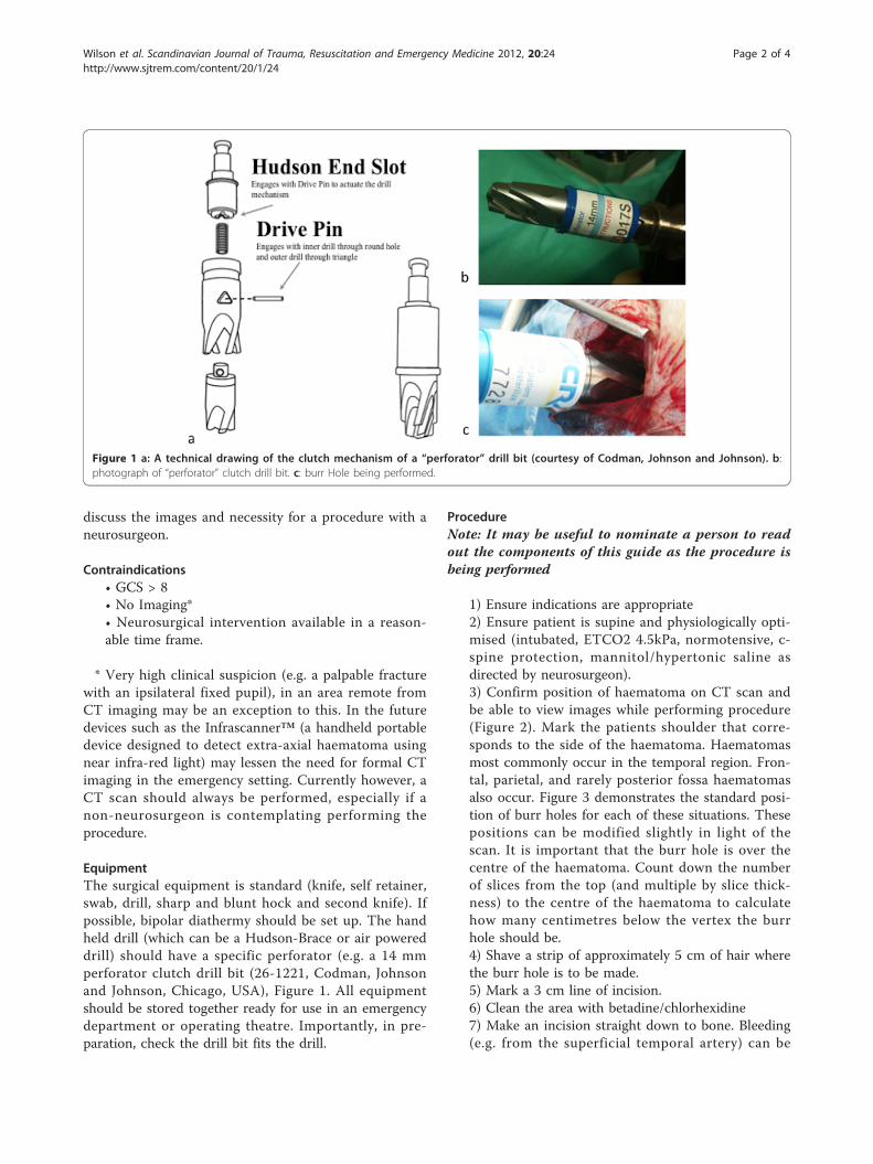
EquipmentThe surgical equipment is standard (knife, self retainer,swab, drill, sharp and blunt hock and second knife). Ifpossible, bipolar diathermy should be set up. The handheld drill (which can be a Hudson-Brace or air powereddrill) should have a specific perforator (e.g. a 14 mmperforator clutch drill bit (26-1221, Codman, Johnsonand Johnson, Chicago, USA), Figure 1. All equipmentshould be stored together ready for use in an emergencydepartment or operating theatre. Importantly, in pre-paration, check the drill bit fits the drill.
ProcedureNote: It may be useful to nominate a person to readout the components of this guide as the procedure isbeing performed
1) Ensure indications are appropriate2) Ensure patient is supine and physiologically opti-mised (intubated, ETCO2 4.5kPa, normotensive, c-spine protection, mannitol/hypertonic saline asdirected by neurosurgeon).3) Confirm position of haematoma on CT scan andbe able to view images while performing procedure(Figure 2). Mark the patients shoulder that corre-sponds to the side of the haematoma. Haematomasmost commonly occur in the temporal region. Fron-tal, parietal, and rarely posterior fossa haematomasalso occur. Figure 3 demonstrates the standard posi-tion of burr holes for each of these situations. Thesepositions can be modified slightly in light of thescan. It is important that the burr hole is over thecentre of the haematoma. Count down the numberof slices from the top (and multiple by slice thick-ness) to the centre of the haematoma to calculatehow many centimetres below the vertex the burrhole should be.4) Shave a strip of approximately 5 cm of hair wherethe burr hole is to be made.5) Mark a 3 cm line of incision.6) Clean the area with betadine/chlorhexidine7) Make an incision straight down to bone. Bleeding(e.g. from the superficial temporal artery) can be
Figure 1 a: A technical drawing of the clutch mechanism of a “perforator” drill bit (courtesy of Codman, Johnson and Johnson). b:photograph of “perforator” clutch drill bit. c: burr Hole being performed.
Wilson et al. Scandinavian Journal of Trauma, Resuscitation and Emergency Medicine 2012, 20:24http://www.sjtrem.com/content/20/1/24
Page 2 of 4

controlled with direct pressure while continuing theprocedure.8) Push the periosteum off the bone with knife/swab9) Insert self-retaining retractor10) Push down firmly with drill and start drillingkeeping drill perpendicular to the skull. Ensure anassistant is holding the head still and ideally applysaline wash as you drill.11) Keep going - do NOT stop (as this will disen-gage the clutch mechanism which can be difficult tore-engage manually)12) Drill until the drill bit stops spinning. Removedrill.13) Use blunt hook to remove remaining bonefragments.14) Extradural blood should now escape.15) If the blood is subdural, very carefully open thedura using a sharp hook to tent the dura up, and anew sharp knife to incise the dura in a cruciate man-ner. Subdural blood is likely to be more clotted anddifficult to extrude than extradural. Manual removalof clot (e.g. with forceps or very careful suction)could be considered, but may damage brain and isunlikely to remove sufficient haematoma. If noblood is found either extra or sub-durally, stop,check side, and check location of hole. DO NOTDELAY TRANSFER.16) If fresh blood is continuing to ooze from thewound, do NOT try to tamponade. Leaving the self-retainer in place may stop the bleeding. Try to
diathermy skin edges; if not available, apply directpressure to wound edges during transfer.
DiscussionThe cranial procedure of burr hole placement hasbecome the sole domain of neurosurgeons particularlyas they can deal with surgical complications. As such,non-neurosurgeons are no longer familiar with the tech-nique. This creates a therapeutic vacuum for patientsremote from specialist care who meet the criteria forurgent burr hole drainage.Central to the ability of non-neurosurgeons to suc-
cessfully performing burr holes is the development ofclutch drill-bits (Figure 1). These cause the drill to dis-engage on penetrating the inner table of the skull sothat the risk of “plunging” is minimised making the pro-cedure considerably safer. If a haematoma is notrelieved, the patient should not have come to any addi-tional harm providing transfer to a neurosurgical centreis not significantly delayed. In remote areas of Australia,
Figure 2 CT scan demonstrating an extradural haematoma.
Figure 3 Diagram demonstrating position of standard burrholes (1, temporal (above zygoma), 2 frontal (over the coronalsuture, approx 10 cm behind and in the mid-pupillary line)and 3 parietal (over the parietal eminence). CT Imagescorrespond. A posterior fossa burr hole can be used in theextremely rare cases of posterior fossa extradural haematoma.). Seetext for indications, requirement for imaging and requirement forneurosurgical discussion. (Image adapted from Head Injuries p134,Mark Wilson Oxford Desk Reference of Trauma Ed Smith, Greavesand Porter 2011).
Wilson et al. Scandinavian Journal of Trauma, Resuscitation and Emergency Medicine 2012, 20:24http://www.sjtrem.com/content/20/1/24
Page 3 of 4

when such neurosurgical procedures are performed bynon-neurosurgeons, outcomes are acceptable [7]. Evenin less remote situations non-neurosurgeons in districtgeneral hospitals in the UK have historically carried outemergency craniotomy [8].For many years it has been known that earlier surgical
intervention is of benefit in the management of headtrauma when an extra-axial collection can be removed[9]. In the future, near infra-red/ultrasound devices ormobile CT, may mean that extra-axial collections can bedetected in remote locations. This will not be of benefitunless the time to surgical relief of increased Intracra-nial Pressure is also shortened.While attempting to remove the mystique and anxiety
surrounding emergency burr hole placement, we empha-sise the importance of avoiding inappropriate interven-tion. However, when faced with a situation wheremortality approaches 100%, a simple technique, usingthe correct equipment can be robust, safe and life-savingeven in the hands of non-specialists.
Author details1London’s Air Ambulance, The Helipad, The Royal London Hospital, LondonE1 1BB, UK. 2Emergency Department, Barts and the London NHS Trust,London E1 1BB, UK. 3Department of Neurosurgery, Imperial Hospitals NHSTrust, London W2 1NY, UK. 4School of Clinical Sciences, Bristol University,Bristol BS10 5NB, UK.
Competing interestsThe authors declare that they have no competing interests.
Received: 24 October 2011 Accepted: 2 April 2012Published: 2 April 2012
References1. Leach P, Childs C, Evans J, Johnston N, Protheroe R, King A: Transfer times
for patients with extradural and subdural haematomas to neurosurgeryin Greater Manchester. Br J Neurosurg 2007, 21(1):11-15.
2. Motohashi O, Kameyama M, Shimosegawa Y, Fujimori K, Sugai K, Onuma T:Single burr hole evacuation for traumatic acute subdural hematoma ofthe posterior fossa in the emergency room. J Neurotrauma 2002,19(8):993-998.
3. Shears R, Get the Black and Decker: Doctor uses household drill to boreinto boy’s skull to save his life after accident swelled brain. Daily Mail2009.
4. Bishop CV, Drummond KJ: Rural neurotrauma in Australia: implications forsurgical training. ANZ J Surg 2006, 76(1-2):53-59.
5. Rinker CF, McMurry FG, Groeneweg VR, Bahnson FF, Banks KL, Gannon DM:Emergency craniotomy in a rural Level III trauma center. J Trauma 1998,44(6):984-989, discussion 89-90.
6. Wester K: Decompressive Surgery for “Pure” Epidural Hematomas: DoesNeurosurgical Expertise Improve the Outcome? Neurosurgery 1999,44(3):495-500.
7. Treacy PJ, Reilly P, Brophy B: Emergency neurosurgery by generalsurgeons at a remote major hospital. ANZ J Surg 2005, 75(10):852-857.
8. Spencer-Jones R, Varley GW, Thomas P, Stevens DB: Helicopter transfer oftrauma patients: the Isle of Man experience. Injury 1993, 24(7):447-450.
9. Mendelow AD, Karmi MZ, Paul KS, Fuller GA, Gillingham FJ: Extraduralhaematoma: effect of delayed treatment. Br Med J 1979,1(6173):1240-1242.
doi:10.1186/1757-7241-20-24Cite this article as: Wilson et al.: Emergency burr holes: “How to do it”.Scandinavian Journal of Trauma, Resuscitation and Emergency Medicine 201220:24.
Submit your next manuscript to BioMed Centraland take full advantage of:
• Convenient online submission
• Thorough peer review
• No space constraints or color figure charges
• Immediate publication on acceptance
• Inclusion in PubMed, CAS, Scopus and Google Scholar
• Research which is freely available for redistribution
Submit your manuscript at www.biomedcentral.com/submit
Wilson et al. Scandinavian Journal of Trauma, Resuscitation and Emergency Medicine 2012, 20:24http://www.sjtrem.com/content/20/1/24
Page 4 of 4