emergence and evolution of the renin angiotensin ... · review emergence and evolution of the...
TRANSCRIPT
REVIEW
Emergence and evolution of the renin–angiotensin–aldosteronesystem
David Fournier & Friedrich C. Luft & Michael Bader &
Detlev Ganten & Miguel A. Andrade-Navarro
Received: 29 December 2011 /Revised: 29 February 2012 /Accepted: 13 March 2012 /Published online: 14 April 2012# The Author(s) 2012. This article is published with open access at Springerlink.com
Abstract The renin–angiotensin–aldosterone system (RAAS)is not the sole, but perhaps the most important volume regula-tor in vertebrates. To gain insights into the function and evo-lution of its components, we conducted a phylogenetic analysisof its main related genes. We found that important parts of thesystem began to appear with primitive chordates and tunicatesand that all major components were present at the divergenceof bony fish, with the exception of the Mas receptor. The Masreceptor first appears after the bony-fish/tetrapod divergence.This phase of evolutionary innovation happened about 400million years ago. We found solid evidence that angiotensino-gen made its appearance in cartilage fish. The presence ofseveral RAAS genes in organisms that lack all the componentsshows that these genes have had other ancestral functionsoutside of their current role. Our analysis underscores theutility of sequence comparisons in the study of evolu-tion. Such analyses may provide new hypotheses as tohow and why in today's population an increased activityof the RAAS frequently leads to faulty salt and volumeregulation, hypertension, and cardiovascular diseases,
opening up new and clinically important research areasfor evolutionary medicine.
Keywords Evolution . Evolutionary medicine . Renin .
Angiotensin . Aldosterone . Volume regulation . Salt .
Hypertension . Cardiovascular diseases . RAAS
Introduction
Multicellular organisms regulate their internal environmentby either modifying the extracellular volume or the soluteconcentration (osmolality). In mammals, the kidney is thefinal common pathway for both of these functions. Low-and high-pressure baroreceptors in various circulatory beds,as well as ion channels and sensors in the distal tubule, inferthe body's volume. Ion channels in the hypothalamus sensethe sodium concentration and extracellular fluid osmolality.Glomerular filtration rate, physical forces along the nephron,the sympathetic nervous system, and the renin–angiotensin–aldosterone system (RAAS) are the volume effectors. Osmo-lality effectors include thirst stimulation, release of the anti-diuretic hormone vasopressin, and the placement of aquaporinwater channels in the renal collecting duct so that concentratedurine can be made. This study focuses on volume regulation.The RAAS is the principal volume-regulatory effector inmammals. It is a major regulator of blood pressure withinthe human body. Therefore, the RAAS has important impli-cations for study of hypertension and other cardiovasculardiseases [1].
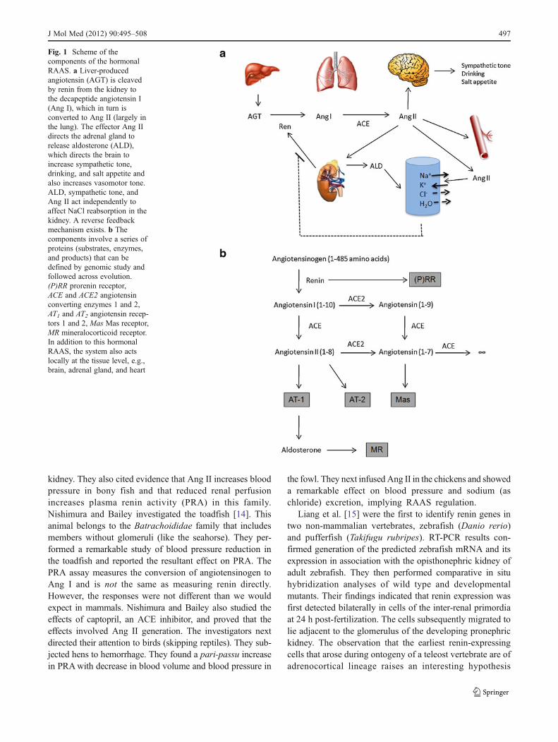
The key effector precursor molecule of the RAAS isangiotensinogen, a protein produced principally in liver(Fig. 1a). Angiotensinogen is cleaved to a 10 amino-acidpeptide, angiotensin (Ang I), by a unique aspartyl proteasecalled renin, which is produced by the juxtaglomerular
Electronic supplementary material The online version of this article(doi:10.1007/s00109-012-0894-z) contains supplementary material,which is available to authorized users.
D. Fournier : F. C. Luft :M. Bader :M. A. Andrade-Navarro (*)Max-Delbrück Center for Molecular Medicine Berlin-Buch,Robert-Rössle-Str. 10,13125 Berlin, Germanye-mail: [email protected]
F. C. Luft :M. Bader :D. GantenCharité–Universitätsmedizin,Berlin, Germany
F. C. LuftExperimental and Clinical Research Center,Berlin, Germany
J Mol Med (2012) 90:495–508DOI 10.1007/s00109-012-0894-z

apparatus (JGA) in the kidney [2, 3]. The angiotensin-converting enzyme (ACE) in turn cleaves Ang I to a smaller,highly active 8 amino-acid peptide, angiotensin II (Ang II).ACE is a matrix metalloproteinase that is particularly highlyconcentrated on pulmonary endothelial cells, despite the factthat numerous functions of the enzyme in other bodily tissuesbesides the vasculature have been uncovered by elegant studiesusing genetically modified mice [4]. Moreover, other enzymesexhibit ACE-like activity, like the chymase, a protein mainlyfound in mast cells, which mostly displays such an activity inthe heart [5]. Ang II acts on the adrenal cortex to releasealdosterone (ALD). ALD acts in the kidney, primarily oncollecting duct cells to effect reabsorption of sodium (andchloride). Ang II also has its own independent sodium reab-sorptive effects in the kidney, acts in brain to stimulate thirstand salt appetite, as well as to increase sympathetic tone, andacts directly on the vessel wall (primarily arterioles) to affectvasoconstriction and to increase blood pressure [6]. Renin is therate-limiting step in Ang II production. Renin release is stimu-lated by baroreflex mechanisms in the JGA, by beta-adrenergicsympathetic innervation of these cells, and by solute delivery,notably chloride content in the tubular fluid at the macula densasegment of the distal tubule. A negative feedback loop inhibitsrenin release that includes Ang II levels, inactivated baroreflexsensors, and sympathetic inhibition.
The RAAS involves a series of proteins working in a net-work (Fig. 1b). Renin, and its precursor molecule prorenin, canoccupy a recently cloned receptor called the prorenin receptor(P)RR that binds both renin and prorenin [7]. The (P)RR cansignal via MAP-kinase pathways and serves to activate prore-nin. Its more global function is currently being investigated;however, the (P)RR has not been found to be implicated involume regulation. Ang I is inert, while Ang II signals throughtwo receptors: the AT1 and the AT2 receptors that have differentfunctions, AT1 displaying two different isoforms (AT1A andAT1B), which have been characterized in mice [8]. Ang I andAng II can be cleaved to further products melding into aseptapeptide called Ang (1-7). The recently discoveredangiotensin-converting enzyme-2 (ACE2) is responsible forthis cleavage activity. Ang (1-7) can signal through a uniquereceptor encoded by the Mas protooncogene, the Mas recep-tor. The steroid hormone ALD, stimulated by Ang II, signalsthrough the mineralocorticoid receptor (MR).
In very general terms, the salt reabsorptive and vaso-constrictor mechanisms are stimulated by Ang II and aremediated by the AT1 receptor. The AT2 receptor has moreameliorative and modulating effects. The primary saltreabsorptive mechanisms are stimulated by ALD via theMR. Ang (1-7) appears to have actions that are generallyopposite to those of Ang II so that its actions may also beameliorative. The complex actions of the RAAS on itstarget organs and cellular messenger systems have beenreviewed [6, 9].
Evolution of the renin–angiotensin system
The evolution of the RAAS can be used to gain insightsabout its molecules and components, for which some ques-tions remain open: Do we know all components of thesystem? Are they all similarly important? When did thissystem emerge and why? Is the system conserved in diverseanimal species? Answering these questions is relevant tocomplete our understanding of how the system functions inhumans, how it is involved in cardiovascular diseases, andwhy pharmacological interference of the RAAS is effectivein their treatment [9, 10].
Today, genomic sequence data can be used to studyRAAS evolution; however, the RAAS has been studied withcomparative physiological techniques that also providedevolutionary insights. Homer Smith, who dominated renalphysiology in the first half of the twentieth century, wasextremely interested in comparative physiology and renalevolution [11]. He described the ureatelic phenotype of thelungfish, which can survive in mud-caked enclaves for overa decade without water. His activities culminated in a sem-inal book, From Fish To Philosopher, published in 1953[12]. In a prescient fashion that would have pleased ClaudeBernard, Smith describes the evolution of the milieu intér-ieur regulation in vertebrates. Smith followed renal evolu-tion with model organisms from the present and what hecould discern from fossil records. His notion was that organ-isms evolved in “relatively” brackish water with electrolytecontents similar to the extracellular environment of today.Evolution offered challenges, such as return to the sea andmovement to fresh (electrolyte poor) water, to land, andeven to the air. Organisms were faced with the problem ofmaintaining their internal environment and also excretingthe end products of protein metabolism. Smith was inter-ested in the different solutions that species have evolvedfor volume regulation and nitrogen excretion. Someorganisms have developed ancillary organs (gills for fish,skin for amphibians, and rectal or orbital glands forsharks, crocodiles, and birds). However, we humans arelargely stuck with “body-is-a-box.” Entry is via the mouthand excretion occurs through the kidneys and to a minorextent via the feces.
In 1977, Taylor pointed out that a renin-like material ispresent in many vertebrate species [13]. He observed noevidence of renin's presence in cartilage fish and concludedthat renin first appeared in bony fish, amphibians, andsubsequent vertebrates. Nishimura and Bailey published aremarkable early paper on the intrarenal RAAS in variousvertebrates [14]. They pointed out that renin activity andgranulated epithelial JGA cells are present in bony fish,dipnoans (lung fish), amphibians, birds, and mammals.Nishimura, Bailey, and others observed that in fish, JGAcells are distributed along small arteries and arterioles of the
496 J Mol Med (2012) 90:495–508
kidney. They also cited evidence that Ang II increases bloodpressure in bony fish and that reduced renal perfusionincreases plasma renin activity (PRA) in this family.Nishimura and Bailey investigated the toadfish [14]. Thisanimal belongs to the Batrachoididae family that includesmembers without glomeruli (like the seahorse). They per-formed a remarkable study of blood pressure reduction inthe toadfish and reported the resultant effect on PRA. ThePRA assay measures the conversion of angiotensinogen toAng I and is not the same as measuring renin directly.However, the responses were not different than we wouldexpect in mammals. Nishimura and Bailey also studied theeffects of captopril, an ACE inhibitor, and proved that theeffects involved Ang II generation. The investigators nextdirected their attention to birds (skipping reptiles). They sub-jected hens to hemorrhage. They found a pari-passu increasein PRAwith decrease in blood volume and blood pressure in
the fowl. They next infused Ang II in the chickens and showeda remarkable effect on blood pressure and sodium (aschloride) excretion, implying RAAS regulation.
Liang et al. [15] were the first to identify renin genes intwo non-mammalian vertebrates, zebrafish (Danio rerio)and pufferfish (Takifugu rubripes). RT-PCR results con-firmed generation of the predicted zebrafish mRNA and itsexpression in association with the opisthonephric kidney ofadult zebrafish. They then performed comparative in situhybridization analyses of wild type and developmentalmutants. Their findings indicated that renin expression wasfirst detected bilaterally in cells of the inter-renal primordiaat 24 h post-fertilization. The cells subsequently migrated tolie adjacent to the glomerulus of the developing pronephrickidney. The observation that the earliest renin-expressingcells that arose during ontogeny of a teleost vertebrate are ofadrenocortical lineage raises an interesting hypothesis
Fig. 1 Scheme of thecomponents of the hormonalRAAS. a Liver-producedangiotensin (AGT) is cleavedby renin from the kidney tothe decapeptide angiotensin I(Ang I), which in turn isconverted to Ang II (largely inthe lung). The effector Ang IIdirects the adrenal gland torelease aldosterone (ALD),which directs the brain toincrease sympathetic tone,drinking, and salt appetite andalso increases vasomotor tone.ALD, sympathetic tone, andAng II act independently toaffect NaCl reabsorption in thekidney. A reverse feedbackmechanism exists. b Thecomponents involve a series ofproteins (substrates, enzymes,and products) that can bedefined by genomic study andfollowed across evolution.(P)RR prorenin receptor,ACE and ACE2 angiotensinconverting enzymes 1 and 2,AT1 and AT2 angiotensin recep-tors 1 and 2, Mas Mas receptor,MR mineralocorticoid receptor.In addition to this hormonalRAAS, the system also actslocally at the tissue level, e.g.,brain, adrenal gland, and heart
J Mol Med (2012) 90:495–508 497

regarding the origin of renin-expressing cells in the meta-nephric kidney of higher vertebrates. Expression of renin inthe adrenal gland of murine embryos indicates that this isalso first in ontogeny in mammals and further supports thatthis renin expression is ancestral to renin's expression in thekidney [16].
Salzet et al. [17] have reviewed RAAS elements in inver-tebrates and vertebrates. Since genome sequences were notavailable at the time of their report, their study could not relyon genomic data, but instead included inferences fromimmunoassays and immunohistochemistry. They recog-nized the presence of ACE-like proteins in the fly and
renin-like enzymes in leeches. They extended their discus-sion to certain vertebrate models. The authors pointed outthat snakes developed anatomical and functional adaptationsand interesting structural peculiarities that are found in theirautonomic, kallikrein-, renin–angiotensin-, and endothelin-related systems.
The physiological evidence indicates that the RAAS wasestablished in bony fishes. Examining the lamprey couldhelp us establish the origin of the system more preciselysince these cyclostomes are the sister taxon of all livingjawed vertebrates, the gnathostomes. Brown et al. estab-lished a radioimmunoassay to measure Ang II in lampreys
Fig. 2 Phylogeny of thespecies whose sequences arestudied in this review. Themain groups displayed are thechordates, which comprise allspecies considered exceptDrosophila and Caenorhabditis,and display a notochord, atleast at some point during theirembryonic development.Vertebrates are speciesdisplaying vertebra and compriseall considered chordate species,with the exception of Ciona andAmphioxus. Finally, thetetrapods comprise all ourvertebrate species, with theexception of fishes, eithercartilage fishes (elephant shark,CallorhinchusMilii), bony fishes(zebrafish, Danio rerio),coelacants, or lungfishes
498 J Mol Med (2012) 90:495–508
[18]. They performed acute volume depletion by removing40 % of the animal's blood volume. This maneuver doubledAng II concentrations. They then exposed the animals to adecrease in salinity (758 to 605 mosm/kg H2O), which rapidlydecreased Ang II with a subsequent increase in Ang II. Inject-ing saline solution intraperitoneally into fresh-water-acclimated lampreys also decreased Ang II concentrations.The results suggest that Ang II may play a role in volumeregulation of these primitive vertebrates. The data are consis-tent with the idea that the Ang II peptide has been around for500 million years. Missing from the authors' data are mass-spectrometry-determined amino acid sequences of the pepti-des. These results are fascinating; however, more precisemethodologies would be important to prove beyond any doubtthat the investigators indeed were dealing with Ang II [19].
DNA sequence analysis of an ancient system
To complement the available physiological evidence on theevolution of the RAAS, we used the information availablefrom the gene and protein sequences in public databases[20]. Complete genomes are especially useful since they allow
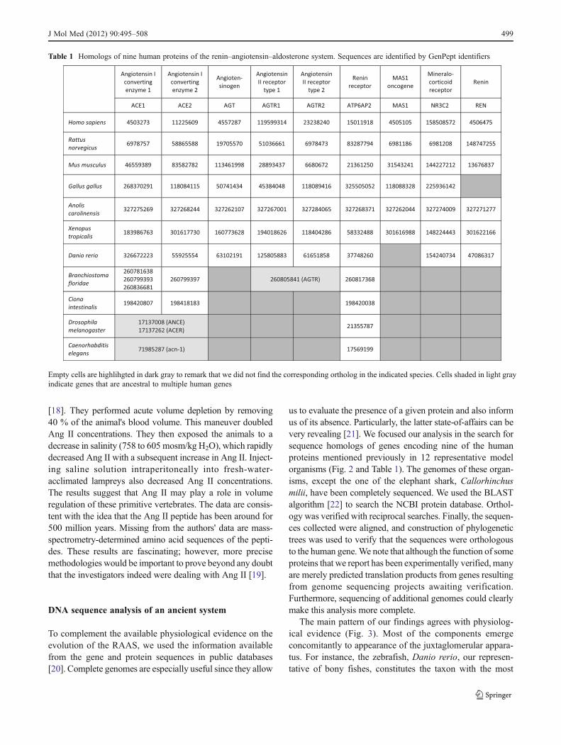
us to evaluate the presence of a given protein and also informus of its absence. Particularly, the latter state-of-affairs can bevery revealing [21]. We focused our analysis in the search forsequence homologs of genes encoding nine of the humanproteins mentioned previously in 12 representative modelorganisms (Fig. 2 and Table 1). The genomes of these organ-isms, except the one of the elephant shark, Callorhinchusmilii, have been completely sequenced. We used the BLASTalgorithm [22] to search the NCBI protein database. Orthol-ogy was verified with reciprocal searches. Finally, the sequen-ces collected were aligned, and construction of phylogenetictrees was used to verify that the sequences were orthologousto the human gene.We note that although the function of someproteins that we report has been experimentally verified, manyare merely predicted translation products from genes resultingfrom genome sequencing projects awaiting verification.Furthermore, sequencing of additional genomes could clearlymake this analysis more complete.
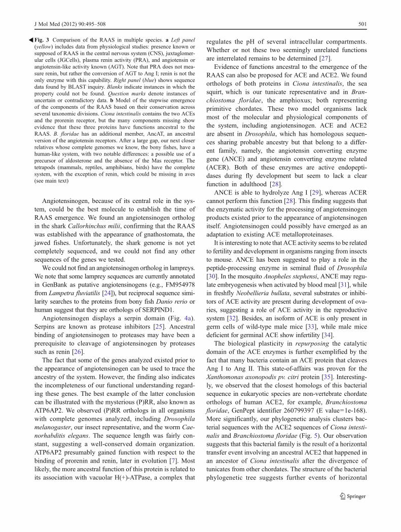
The main pattern of our findings agrees with physiolog-ical evidence (Fig. 3). Most of the components emergeconcomitantly to appearance of the juxtaglomerular appara-tus. For instance, the zebrafish, Danio rerio, our represen-tative of bony fishes, constitutes the taxon with the most
Table 1 Homologs of nine human proteins of the renin–angiotensin–aldosterone system. Sequences are identified by GenPept identifiers
Empty cells are highlihgted in dark gray to remark that we did not find the corresponding ortholog in the indicated species. Cells shaded in light grayindicate genes that are ancestral to multiple human genes
J Mol Med (2012) 90:495–508 499

primitive JGA [23] and already contains orthologs to eightof the nine proteins considered. The exception is the oncogeneMas, which seems to have evolved later in the tetrapod lineage.We found Mas in the frog Xenopus tropicalis, our amphibianrepresentative. Orthologs of the nine genes could be found in alltetrapod species analyzed, indicating the molecular stability of
the pathway. We could not find the renin protein in Gallusgallus, the chicken, representing birds in our study. However,clear homology to full human renin was found in genomicshotgun sequences corresponding to chromosome 26 of Gallusgallus. These sequences have not been yet assembled to thecurrent version of the Gallus gallus genome.
500 J Mol Med (2012) 90:495–508
Angiotensinogen, because of its central role in the sys-tem, could be the best molecule to establish the time ofRAAS emergence. We found an angiotensinogen orthologin the shark Callorhinchus milii, confirming that the RAASwas established with the appearance of gnathostomata, thejawed fishes. Unfortunately, the shark genome is not yetcompletely sequenced, and we could not find any othersequences of the genes we tested.
We could not find an angiotensinogen ortholog in lampreys.We note that some lamprey sequences are currently annotatedin GenBank as putative angiotensinogens (e.g., FM954978from Lampetra fluviatilis [24]), but reciprocal sequence simi-larity searches to the proteins from bony fish Danio rerio orhuman suggest that they are orthologs of SERPIND1.
Angiotensinogen displays a serpin domain (Fig. 4a).Serpins are known as protease inhibitors [25]. Ancestralbinding of angiotensinogen to proteases may have been aprerequisite to cleavage of angiotensinogen by proteasessuch as renin [26].
The fact that some of the genes analyzed existed prior tothe appearance of angiotensinogen can be used to trace theancestry of the system. However, the finding also indicatesthe incompleteness of our functional understanding regard-ing these genes. The best example of the latter conclusioncan be illustrated with the mysterious (P)RR, also known asATP6AP2. We observed (P)RR orthologs in all organismswith complete genomes analyzed, including Drosophilamelanogaster, our insect representative, and the worm Cae-norhabditis elegans. The sequence length was fairly con-stant, suggesting a well-conserved domain organization.ATP6AP2 presumably gained function with respect to thebinding of prorenin and renin, later in evolution [7]. Mostlikely, the more ancestral function of this protein is related toits association with vacuolar H(+)-ATPase, a complex that
regulates the pH of several intracellular compartments.Whether or not these two seemingly unrelated functionsare interrelated remains to be determined [27].
Evidence of functions ancestral to the emergence of theRAAS can also be proposed for ACE and ACE2. We foundorthologs of both proteins in Ciona intestinalis, the seasquirt, which is our tunicate representative and in Bran-chiostoma floridae, the amphioxus; both representingprimitive chordates. These two model organisms lackmost of the molecular and physiological components ofthe system, including angiotensinogen. ACE and ACE2are absent in Drosophila, which has homologous sequen-ces sharing probable ancestry but that belong to a differ-ent family, namely, the angiotensin converting enzymegene (ANCE) and angiotensin converting enzyme related(ACER). Both of these enzymes are active endopepti-dases during fly development but seem to lack a clearfunction in adulthood [28].
ANCE is able to hydrolyze Ang I [29], whereas ACERcannot perform this function [28]. This finding suggests thatthe enzymatic activity for the processing of angiotensinogenproducts existed prior to the appearance of angiotensinogenitself. Angiotensinogen could possibly have emerged as anadaptation to existing ACE metalloproteinases.
It is interesting to note that ACE activity seems to be relatedto fertility and development in organisms ranging from insectsto mouse. ANCE has been suggested to play a role in thepeptide-processing enzyme in seminal fluid of Drosophila[30]. In the mosquito Anopheles stephensi, ANCE may regu-late embryogenesis when activated by blood meal [31], whilein freshfly Neobellieria bullata, several substrates or inhibi-tors of ACE activity are present during development of ova-ries, suggesting a role of ACE activity in the reproductivesystem [32]. Besides, an isoform of ACE is only present ingerm cells of wild-type male mice [33], while male micedeficient for germinal ACE show infertility [34].
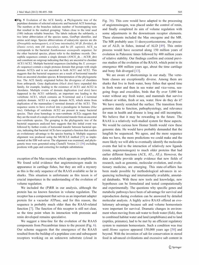
The biological plasticity in repurposing the catalyticdomain of the ACE enzymes is further exemplified by thefact that many bacteria contain an ACE protein that cleavesAng I to Ang II. This state-of-affairs was proven for theXanthomonas axonopodis pv. citri protein [35]. Interesting-ly, we observed that the closest homologs of this bacterialsequence in eukaryotic species are non-vertebrate chordateorthologs of human ACE2, for example, Branchiostomafloridae, GenPept identifier 260799397 (E value01e-168).More significantly, our phylogenetic analysis clusters bac-terial sequences with the ACE2 sequences of Ciona intesti-nalis and Branchiostoma floridae (Fig. 5). Our observationsuggests that this bacterial family is the result of a horizontaltransfer event involving an ancestral ACE2 that happened inan ancestor of Ciona intestinalis after the divergence oftunicates from other chordates. The structure of the bacterialphylogenetic tree suggests further events of horizontal
Fig. 3 Comparison of the RAAS in multiple species. a Left panel(yellow) includes data from physiological studies: presence known orsupposed of RAAS in the central nervous system (CNS), juxtaglomer-ular cells (JGCells), plasma renin activity (PRA), and angiotensin orangiotensin-like activity known (AGT). Note that PRA does not mea-sure renin, but rather the conversion of AGT to Ang I; renin is not theonly enzyme with this capability. Right panel (blue) shows sequencedata found by BLAST inquiry. Blanks indicate instances in which theproperty could not be found. Question marks denote instances ofuncertain or contradictory data. b Model of the stepwise emergenceof the components of the RAAS based on their conservation acrossseveral taxonomic divisions. Ciona intestinalis contains the two ACEsand the prorenin receptor, but the many components missing showevidence that these three proteins have functions ancestral to theRAAS. B. floridae has an additional member, AncAT, an ancestralversion of the angiotensin receptors. After a large gap, our next closerrelatives whose complete genomes we know, the bony fishes, have ahuman-like system, with two notable differences: a possible use of aprecursor of aldosterone and the absence of the Mas receptor. Thetetrapods (mammals, reptiles, amphibians, birds) have the completesystem, with the exception of renin, which could be missing in aves(see main text)
�
J Mol Med (2012) 90:495–508 501

502 J Mol Med (2012) 90:495–508

transfer involving this gene between bacteria. Besides theirenzymatic properties, the ACEs have other non-catalyticfunctions [36]. Bacteria might exploit these other functionsas well.
Domain analysis of this family is interesting because theACE2 orthologs (including the bacterial ones) and Drosoph-ila melanogaster ANCE and ACER possess only a single600 amino-acid peptidase domain (peptidase M2 domain).In contrast, ACE orthologs have two such domains ([37],Figs. 4a and 5). The strong similarity between the N- and C-terminal domains in one Ciona intestinalis and in oneBranchiostoma floridae sequence suggest that the proteinsof the ACE subfamily have undergone multiple independentevents of domain duplication like the one leading to thevertebrate ACEs. In mice, the two domains have differentimportance in the development of hypertension, as has beenshown by selective deletion of one of the two domains [38].
Finally, in mammals, we observed a further member ofthis family containing a single catalytic domain, termedACE3. The function of ACE3 is unknown. The best char-acterized member is the mouse ACE3, which is expressedand produces a protein in sperm; however, gene deletiondoes not render mice infertile [39]. Orthologs of ACE3 existin cow, dog, and rat, while the human counterpart is apseudogene with multiple base deletions, insertions, andstop codons. Moreover, murine ACE3 lacks the residuesnecessary for ACE catalytic activity [40]. ACE3 expressioncould possibly regulate the expression of the parentalACE gene, a mechanism of post-transcriptional generegulation that is known to exist for other pseudogenes[41]. The position of the ACE3 gene in human andmouse genomes downstream from ACE is consistent withthis view. Our sequence analysis indicates that ACE3 isthe result of an N-terminal domain duplication of ACE(Fig. 5). We did not find orthologs of this gene outsideof mammals.
It is important to note that beside ACEs, other enzymesthat can cleave Ang I to Ang II [5] have been found, such aschymase, a protein expressed in the mast cells of heart and
blood vessels [5], and cathepsin G, a secreted serine pepti-dase of neutrophils and mast cells [42]. These proteinsappear to have evolved well after the establishment of theRAAS.
The AT1 and AT2 receptors, also termed AGTR1 andAGTR2, are also evolutionary products of gene duplication.We identified an ancestral sequence of the two proteins inBranchiostoma (AncAT, Fig. 3a; supplementary Figure 1).We did not find any homologue in the most distant Ciona.This finding suggests that this protein family emerged afterdivergence of tunicates from primitive chordates. Again, thefunctional relationship of this gene with Ang II must haveoccurred later in evolution since angiotensinogen evolvedlater (Fig. 3b). The Ang II receptor family may exhibit otherancestral functions.
The MR probably predated angiotensinogen and renin.The gene for the ancient MR protein might have beenduplicated in an ancestral organism before the emergenceof bony fishes [43, 44]. The primordial MR then evolved(duplicated) to become a glucocorticoid receptor (GR). Thisinterpretation suggests that volume regulation is older thanstress-related responses. We traced MR easily to bonyfishes. Our cartilage fish sequence left us with a questionmark (Fig. 3a), and we will have to await additionalsequence information to be able to establish more preciselythe time of emergence of this receptor.
Finally, some inbred strains of laboratory mice (not thewidely used C57Bl/6) harbor a second renin gene (Ren-2)[45]. Ren-2 is mainly expressed in the salivary gland ofmales and is dramatically stimulated by aggression(100,000-fold higher levels in saliva than plasma) [46].Possibly, the aspartyl protease serves to injure a bittenopponent or conceivably the protease serves a protectivefunction for wound healing after a fight by wound licking.Interestingly, increasing angiotensinogen in the mouse byinfusion increases blood pressure substantially. The normal-ly fairly low levels of angiotensinogen in the blood of micecould be a response to the effectiveness of their salivaryrenin. The fact that Ren-2 is strain-specific suggests that theduplication of this gene is a relatively recent event. Arelative paucity of renin substrate in the mouse could havefavored this duplication [47]. Ren-2 actually provided thefirst successful transgenic-rat model of hypertension, under-scoring its amazing effectiveness in cleaving rat (crossspecies) angiotensinogen [48].
Discussion
The important finding of our study is that most of the RAAScomponent members began to appear with primitive chor-dates and tunicates. All of the important components werepresent with the development of bony fishes, with the
Fig. 4 Structural features of nine human proteins relevant to theRAAS. a Domain organization of ACE, ACE2, renin, AGT, (P)RR,and MR. T transmembrane alpha-helix (TM), S signal peptide (SP), Ppro-peptide (PP). Red box on angiotensinogen diagram: Ang Isequence (AG). Red symbols indicate protein cleavage sites. b Solved3D structures of these proteins or homologs (when indicated). ACE2:peptidase domain (fragment 1-615, PDB:1R42); REN and AGT:complex of renin (blue) and AGT (orange). Note the N-terminal ofAGT protruding into the renin molecule for processing (PDB:2X0B);NRC32: steroid binding domain (blue; PDB:2AA2) and DNA bindingdomain (green) with DNA (stick model) from 85 % identical ratglucocorticoid receptor NRC31 (PDB:3G9P); AGTR1/AGTR2 are30 % identical to the CXCR4 chemokine receptor whose structure isshown (TM helices in green; PDB:3OE0). All protein structures arerepresented using the PyMOL Molecular Graphics System software(DeLano Scientific, Palo Alto, California)
�
J Mol Med (2012) 90:495–508 503

504 J Mol Med (2012) 90:495–508
exception of the Mas receptor, which appears in amphibians.We found solid evidence that angiotensinogen made itsappearance in cartilage fishes, but they are still a mysteryas this is the only sequence of the RAAS available so far insharks. This situation is unfortunate as this taxon is ofcrucial importance in the understanding of the evolution ofvolume regulation.
We included the (P)RR in our analysis, although theprotein has no known function in volume regulation. Thereceptor has a component that serves as an important adapterprotein for a vacuolar ATPase, and for this reason, thesequence is probably much older than the RAAS-relatedfunction [7]. The function of this receptor is still not clear,so the time point when its interaction with prorenin andrenin developed remains speculative.
We suggest a time-line for the evolution of the RAAScomponents from Precambrian times to the present (Fig. 6).Our scheme suggests that the emergence of the RAASresulted from the buildup of a peptidase core and subsequentreceptors working on an unknown substrate (cloud in
Fig. 3b). This core would have adapted to the processingof angiotensinogen, was placed under the control of renin,and finally completed its aim of volume regulation withsome adjustments in the downstream receptor elements.These elements included the Mas oncogene and the MR.The MR probably uses 11-deoxycorticosterone, the precur-sor of ALD, in fishes, instead of ALD [49]. This entireprocess would have occurred along 150 million years ofevolution in Paleozoic times followed by 400 million yearsof relative stability. Our findings confirm and extend previ-ous studies of the evolution of the RAAS, which point to itsemergence 400 million years ago, about the time cartilageand bony fish diverged [13].
We are aware of shortcomings in our study. The verte-brate classes are exceptionally diverse. Among them aresharks that live in fresh water, bony fishes that spend timein fresh water and then in sea water and vice-versa, sea-going frogs and crocodiles, birds that fly over 5,000 kmwater without any fresh water, and mammals that survivewithout or within, fresh or sea, water. How do they do it?We have merely scratched the surface. The transition fromgenomic data to function, pathophysiology, and relevancefor man in health and disease is long and still incomplete.We believe that it may be rewarding in the future. TheRAAS is a relatively well-studied system for these aspects.We would be curious how Homer Smith would view thesegenomic data. He would have probably demanded that thelungfish be sequenced. We agree, and the more sequencedata we have, the more predictions we could make and themore likely we will able to eventually identify the molecularevents that led to the interaction of relatively new ligands(renin, angiotensinogen) to much older proteins that hadearlier different functions (ACE, AT1, AT2). However, thedata available provide ample evidence that new fields ofresearch, such as genomic, molecular evolution, and evolu-tionary medicine, are emerging. This state-of-affairs hasbeen made possible by methodological advances in se-quencing technology and internationally available, annotat-ed databanks. With these new tools and knowledge, newhypotheses can be formulated and tested computationallyand experimentally. The questions why specific genes andmetabolic pathways have been of advantage for survival andreproduction during evolution can be answered by detailedmolecular analysis. A highly active RAAS offered an evo-lutionary advantage because salt and volume homeostasiswere important for survival. Dramatic changes in environ-ment when moving from salt water to fresh water (fish), thento combined habitat water and land (amphibians) and to land(reptiles, primates), had to be met by an efficient regulatorysystem to maintain homeostasis. Such a condition was trueuntil Homo sapiens appeared 150,000 years ago [50] andbeyond. With the invention of salt for conservation in storedfood in advanced civilizations and excessive salt content in
Fig. 5 Evolution of the ACE family. a Phylogenetic tree of thepeptidase domains of selected eukaryotic and bacterial ACE homologs.The numbers at the branches indicate number of bootstrapping teststhat resulted in the marked grouping: Values close to the total used(100) indicate reliable branches. The labels indicate the subfamily, atwo letter abbreviation of the species name, GenPept identifier, andamino acid range. Species abbreviations of eukaryotic species are dm(Drosophila melanogaster), ci (Ciona intestinalis), bf (B. floridae), dr(Danio rerio), mm (M. musculus), and hs (H. sapiens). ACE_xacorresponds to the bacterial Xanthomonas axonopodis sequence; forthe other bacterial species, please refer to the database records. Dro-sophila sequences contain a single domain (ANCE_dr, ; ACER_dm)and constitute an outgroup indicating that they are ancestral to chordateACE1/ACE2. Multiple bacterial sequences (including the X. axonopo-dis sequence) contain a single protease domain that groups with singledomain ACE2s and is not ancestral to both ACE1 and ACE2. Thissuggests that the bacterial sequences are a result of horizontal transferfrom an ancestral chordate species. b Interpretation of the phylogenetictree. The ACE family originated before the divergence of chordatesfrom arthropods. Gene duplications (black dots) have expanded thisfamily, for example, leading to the existence of ACE1 and ACE2 inchordates. Multiple events of domain duplication (red dots) havehappened in the ACE1 subfamily, an important one leading to thevertebrate ACE1, which contains an N-terminal and a C-terminalcatalytic domain. ACE3 is a single domain ACE, which stems fromduplication of the mammalian C-terminal domain of the ACE1. Thissequence seems to have evolved into a pseudogene in humans (blueline). Orthologs of vertebrate ACE2 are present in many bacterialspecies. Their close homology to non-vertebrate ACE2s suggests thatthey are the result of a single event of horizontal transfer from an ancestralnon-vertebrate species. The grouping in the phylogenetic tree of thebacterial sequences analyzed here suggests that this initial event wasfollowed by further events of horizontal transfer between bacterial spe-cies, indicating that bacterial ACEs have acquired a function that confersan evolutionary advantage to the species bearing it. Multiple sequencealignment was produced using the MUSCLE method [55] as imple-mented at the EBI web server. The alignment was examined, and phylo-genetic trees were generated using ClustalX Version 2.1 [56] excludingpositions with gaps and correcting for multiple substitutions
�
J Mol Med (2012) 90:495–508 505

industrialized nutrition, the RAAS is in constant overdrive,causing salt and volume overload in the body with ensuinghypertension, stroke, and cardiovascular diseases. There isoverwhelming evidence that salt is an important cause of
hypertension [51, 52]. The gap between the early function ofthe RAAS and the completely different environmental chal-lenges and nutrition today may be one of the evolutionaryreasons why hypertension, the number-one risk factor for
Fig. 6 Time-line of the emergence of the RAAS. Left geological erasand a time-line (scale in millions of years). While most genes appearedin the early Paleozoic, others might have emerged earlier in the Pre-cambrian era and were adapted for their use as part of the RAAS. ACE
is one such example and might have evolved from an initial develop-mental function to physiological actions on volume regulation invertebrates
506 J Mol Med (2012) 90:495–508

mortality worldwide, occurs in about half of the adult pop-ulation nowadays [53].
In conclusion, by studying evolution, we can gain insightinto how our body works and eventually obtain clues onhow to deal with dysfunctions. We suggest that the sustain-ing efforts in better understanding the RAAS will have soonimportance in the study of hypertension and other cardio-vascular diseases. These would be important studies toadvance the concept of an evolutionary medicine [54].
Acknowledgements We would like to thank Norbert Hübner(MDC-Berlin) for his useful comments during the developmentof this review.
Open Access This article is distributed under the terms of the Crea-tive Commons Attribution License which permits any use, distribution,and reproduction in any medium, provided the original author(s) andthe source are credited.
References
1. Cushman DW, Ondetti MA (1999) Design of angiotensin convert-ing enzyme inhibitors. Nat Med 5:1110–1113
2. Ganten D, Hayduk K, Brecht HM, Boucher R, Genest J (1970)Evidence of renin release or production in splanchnic territory.Nature 226:551–552
3. Ganten D, Minnich JL, Granger P, Hayduk K, Brecht HM,Barbeau A, Boucher R, Genest J (1971) Angiotensin-formingenzyme in brain tissue. Science 173:64–65
4. Shen XZ, Xiao HD, Li P, Lin CX, Billet S, Okwan-Duodu D,Adams JW, Bernstein EA, Xu Y, Fuchs S et al (2008) Newinsights into the role of angiotensin-converting enzyme obtainedfrom the analysis of genetically modified mice. J Mol Med (Berl)86:679–684
5. Urata H, Boehm KD, Philip A, Kinoshita A, Gabrovsek J, BumpusFM, Husain A (1993) Cellular localization and regional distribu-tion of an angiotensin II-forming chymase in the heart. J ClinInvest 91:1269–1281
6. Bader M, Ganten D (2008) Update on tissue renin–angiotensin sys-tems. J Mol Med (Berl) 86:615–621
7. Nguyen G, Muller DN (2010) The biology of the (pro)renin receptor.J Am Soc Nephrol 21:18–23
8. Tsuchida S, Matsusaka T, Chen X, Okubo S, Niimura F, NishimuraH, Fogo A, Utsunomiya H, Inagami T, Ichikawa I (1998) Murinedouble nullizygotes of the angiotensin type 1A and 1B receptorgenes duplicate severe abnormal phenotypes of angiotensinogennullizygotes. J Clin Invest 101:755–760
9. Bader M (2010) Tissue renin–angiotensin–aldosterone systems:targets for pharmacological therapy. Annu Rev Pharmacol Toxicol50:439–465
10. Steckelings UM, Paulis L, Unger T, Bader M (2011) Emergingdrugs which target the renin–angiotensin–aldosterone system.Expert Opin Emerg Drugs 16:619–630
11. Schnermann J (2003) Homer W. Smith Award lecture. The juxta-glomerular apparatus: from anatomical peculiarity to physiologicalrelevance. J Am Soc Nephrol 14:1681–1694
12. Smith HW (1953) From fish to philosopher: the story of ourinternal environmentLittle, Brown and Company.
13. Taylor AA (1977) Comparative physiology of the renin–angiotensinsystem. Fed Proc 36:1776–1780
14. Nishimura H, Bailey JR (1982) Intrarenal renin–angiotensinsystem in primitive vertebrates. Kidney Int Suppl 12:S185–S192
15. Liang P, Jones CA, Bisgrove BW, Song L, Glenn ST, Yost HJ,Gross KW (2004) Genomic characterization and expressionanalysis of the first nonmammalian renin genes from zebrafishand pufferfish. Physiol Genomics 16:314–322
16. Jones CA, Sigmund CD, McGowan RA, Kane-Haas CM, GrossKW (1990) Expression of murine renin genes during fetal devel-opment. Mol Endocrinol 4:375–383
17. Salzet M, Deloffre L, Breton C, Vieau D, Schoofs L (2001) Theangiotensin system elements in invertebrates. Brain Res Brain ResRev 36:35–45
18. Brown JA, Cobb CS, Frankling SC, Rankin JC (2005) Activationof the newly discovered cyclostome renin–angiotensin system inthe river lamprey Lampetra fluviatilis. J Exp Biol 208:223–232
19. Rankin JC, Watanabe TX, Nakajima K, Broadhead C, Takei Y(2004) Identification of angiotensin I in a cyclostome, Lampetrafluviatilis. Zoolog Sci 21:173–179
20. Pruitt KD, Tatusova T, Klimke W, Maglott DR (2009) NCBIReference Sequences: current status, policy and new initiatives.Nucleic Acids Res 37:D32–D36
21. Blomme T, Vandepoele K, De Bodt S, Simillion C, Maere S, Vande Peer Y (2006) The gain and loss of genes during 600 millionyears of vertebrate evolution. Genome Biol 7:R43
22. Altschul SF, Madden TL, Schaffer AA, Zhang J, Zhang Z, MillerW, Lipman DJ (1997) Gapped BLAST and PSI-BLAST: a newgeneration of protein database search programs. Nucleic Acids Res25:3389–3402
23. Sokabe H, Ogawa M (1974) Comparative studies of the juxtaglo-merular apparatus. Int Rev Cytol 37:271–327
24. Ragg H, Kumar A, Koster K, Bentele C, Wang Y, Frese MA, Prib N,Kruger O (2009) Multiple gains of spliceosomal introns in a super-family of vertebrate protease inhibitor genes. BMC Evol Biol 9:208
25. Potempa J, Korzus E, Travis J (1994) The serpin superfamily ofproteinase inhibitors: structure, function, and regulation. J BiolChem 269:15957–15960
26. Wang Y, Ragg H (2011) An unexpected link between angiotensi-nogen and thrombin. FEBS Lett 585:2395–2399
27. Sihn G, Rousselle A, Vilianovitch L, Burckle C, Bader M (2010)Physiology of the (pro)renin receptor: Wnt of change? Kidney Int78:246–256
28. Houard X, Williams TA, Michaud A, Dani P, Isaac RE, ShirrasAD, Coates D, Corvol P (1998) The Drosophila melanogaster-related angiotensin-I-converting enzymes Acer and Ance—distinctenzymic characteristics and alternative expression during pupaldevelopment. Eur J Biochem 257:599–606
29. Williams TA, Michaud A, Houard X, Chauvet MT, Soubrier F,Corvol P (1996) Drosophila melanogaster angiotensin I-convertingenzyme expressed in Pichia pastoris resembles the C domain of themammalian homologue and does not require glycosylation for secre-tion and enzymic activity. Biochem J 318(Pt 1):125–131
30. Rylett CM, Walker MJ, Howell GJ, Shirras AD, Isaac RE (2007)Male accessory glands of Drosophila melanogaster make asecreted angiotensin I-converting enzyme (ANCE), suggesting arole for the peptide-processing enzyme in seminal fluid. J Exp Biol210:3601–3606
31. Ekbote U, Coates D, Isaac RE (1999) A mosquito (Anophelesstephensi) angiotensin I-converting enzyme (ACE) is induced bya blood meal and accumulates in the developing ovary. FEBS Lett455:219–222
32. Vandingenen A, Hens K, Macours N, Schoofs L, De Loof A,Huybrechts R (2002) Presence of angiotensin converting enzyme(ACE) interactive factors in ovaries of the grey fleshfly Neobellieriabullata. Comp Biochem Physiol B Biochem Mol Biol 132:27–35
33. Corvol P, Eyries M, Soubrier F (2004) Peptidyl-dipeptidase A/angiotensin I-converting enzyme. In: Barrett AJ, Rawlings ND,
J Mol Med (2012) 90:495–508 507
Woessner JF (eds) Handbook of proteolytic enzymes. ElsevierAcademic Press edn, London
34. Hagaman JR, Moyer JS, Bachman ES, Sibony M, Magyar PL,Welch JE, Smithies O, Krege JH, O'Brien DA (1998) Angiotensin-converting enzyme and male fertility. Proc Natl Acad Sci U S A95:2552–2557
35. Riviere G, Michaud A, Corradi HR, Sturrock ED, Ravi Acharya K,Cogez V, Bohin JP, Vieau D, Corvol P (2007) Characterization of thefirst angiotensin-converting like enzyme in bacteria: ancestor ACE isalready active. Gene 399:81–90
36. Lambert DW, Clarke NE, Turner AJ (2010) Not just angiotensi-nases: new roles for the angiotensin-converting enzymes. Cell MolLife Sci 67:89–98
37. Cornell MJ, Williams TA, Lamango NS, Coates D, Corvol P,Soubrier F, Hoheisel J, Lehrach H, Isaac RE (1995) Cloning andexpression of an evolutionary conserved single-domain angioten-sin converting enzyme from Drosophila melanogaster. J BiolChem 270:13613–13619
38. Ong FS, Lin CX, Campbell DJ, Okwan-Duodu D, Chen X,Blackwell WL, Shah KH, Gonzalez-Villalobos RA, Shen XZ,Fuchs S et al (2012) Increased angiotensin II-induced hypertensionand inflammatory cytokines in mice lacking angiotensin-converting enzyme N domain activity. Hypertension 59:283–290
39. Inoue N, Kasahara T, Ikawa M, Okabe M (2010) Identification anddisruption of sperm-specific angiotensin converting enzyme-3 (ACE3)in mouse. PLoS One 5:e10301
40. Rella M, Elliot JL, Revett TJ, Lanfear J, Phelan A, Jackson RM,Turner AJ, Hooper NM (2007) Identification and characterisationof the angiotensin converting enzyme-3 (ACE3) gene: a novelmammalian homologue of ACE. BMC Genomics 8:194
41. Muro EM, Mah N, Andrade-Navarro MA (2011) Functionalevidence of post-transcriptional regulation by pseudogenes.Biochimie 93:1916–1921
42. Tonnesen MG, Klempner MS, Austen KF, Wintroub BU (1982)Identification of a human neutrophil angiotension II-generatingprotease as cathepsin G. J Clin Invest 69:25–30
43. Bridgham JT, Carroll SM, Thornton JW (2006) Evolution ofhormone-receptor complexity by molecular exploitation. Science312:97–101
44. Carroll SM, Ortlund EA, Thornton JW (2011) Mechanisms for theevolution of a derived function in the ancestral glucocorticoid recep-tor. PLoS Genet 7:e1002117
45. Holm I, Ollo R, Panthier JJ, Rougeon F (1984) Evolution ofaspartyl proteases by gene duplication: the mouse renin gene isorganized in two homologous clusters of four exons. EMBO J3:557–562
46. Pedersen EB, Poulsen K (1983) Aggression-provoked huge releaseof submaxillary mouse renin to saliva. Acta Endocrinol (Copenh)104:510–512
47. Weaver D, Skinner S, Walker L, Sangster M (1991) Phenotypicinhibition of the renin–angiotensin system, emergence of theRen-2 gene, and adaptive radiation of mice. Gen Comp Endocrinol83:306–315
48. Mullins JJ, Peters J, Ganten D (1990) Fulminant hypertension intransgenic rats harbouring the mouse Ren-2 gene. Nature 344:541–544
49. Bury NR, Sturm A (2007) Evolution of the corticosteroid receptorsignalling pathway in fish. Gen Comp Endocrinol 153:47–56
50. McDougall I, Brown FH, Fleagle JG (2005) Stratigraphic place-ment and age of modern humans from Kibish, Ethiopia. Nature433:733–736
51. Luft FC (1989) Salt and hypertension: recent advances andperspectives. J Lab Clin Med 114:215–221
52. He FJ, Burnier M, Macgregor GA (2011) Nutrition in cardiovas-cular disease: salt in hypertension and heart failure. Eur Heart J32:3073–3080
53. Kearney PM, Whelton M, Reynolds K, Muntner P, WheltonPK, He J (2005) Global burden of hypertension: analysis ofworldwide data. Lancet 365:217–223
54. Williams GC, Nesse RM (1991) The dawn of Darwinian medicine.Q Rev Biol 66:1–22
55. Edgar RC (2004) MUSCLE: multiple sequence alignment withhigh accuracy and high throughput. Nucleic Acids Res 32:1792–1797
56. Larkin MA, Blackshields G, Brown NP, Chenna R, McGettiganPA, McWilliam H, Valentin F, Wallace IM, Wilm A, Lopez R et al(2007) Clustal W and Clustal X version 2.0. Bioinformatics23:2947–2948
508 J Mol Med (2012) 90:495–508