embyrogenesis of the cns and its disorders
TRANSCRIPT

Embryogenesis of CNS
and its disorders
Amr Hasan MD Associate Professor of Neurology
Cairo University
Embryogenesis of CNS and its disorders
Embryological
developmental progress
Stage I Dorsal Induction
Formation and Closure of the Neural Tube (2-4 wks)
At 2 weeks
Development of the Neural Tube Begins in the third week and is completed in the fourth
week
Induction the notochord directs the overlying ectoderm to form the neural plate
Neural fold formed by thickening of the neural plate with elevation of its edges
Development of the Neural Tube
Neural tube
The neural folds first contact each other to begin the formation of the neural tube
This fusion initially takes place on the dorsal midline at what will become the cervical levels of the spinal cord The fusion proceeds zipper-like in rostral and caudal directions
Tube closure
23 thickened to form future brain
13 caudal form future spinal cord
Closes like a zipper starting in hind brain
Neural tube closure is followed by disjunction of
cutenous and neural ectoderm
Development of the Neural Tube
During the process the lumen of the neural tube called the central canal is open to the amniotic cavity both rostrally and caudally
The two openings in the neural tube connect the central canal with the amniotic cavity
Anterior neuropore closes at about 24 days and becomes the lamina terminalis
Posterior neuropore closes at about 26 days
Neurulation process by which CNS develops from a hollow structure called the neural tube
Primary neurulation most of the neural tube forms from the neural plate by a process of infolding called primary neurulation This part of the neural tube will give rise to the brain and to the spinal cord through lumbar levels
Secondary neurulation the sacral and coccygeal segments of the spinal cord and their corresponding dorsal and ventral roots are formed secondary neurulation
Neural Crest
Gives rise to
1048698 Pseudounipolar ganglion cells of the spinal and cranial nerve
ganglia
1048698 Schwann cells (neurolemmal sheath cells that form myelin in
the PNS)
1048698 Multipolar ganglion cells of the autonomic ganglia
1048698 Leptomeninges (pia-arachnoid cells)
1048698 Chromaffin cells of the suprarenal medulla
1048698 Pigment cells (melanocytes)
1048698 Odontoblasts (dentine-forming cells)
1048698 Aorticopulmonary septum of the heart
1048698 Parafollicular cells (calcitonin-producing C-cells)
1048698 Skeletal and connective components of the pharyngeal
arches
Stage 2 Ventral Induction
Formation of the Brain Segments and Face (5-10 wks)
Wall will form future brain
Cavity will form future ventricles
ldquo لقد خلقنا االنسان في أحسن تقويم ldquo
سورة التين 4اية
Stage 3
Proliferationdifferentiation
Histogenesis and Migration
(2-5 months)
1-Neural proliferation
Germinal matrix formed lining lateral ventricles and third ventricle at about 7 weeks
2-Neural differentiation
3-Neural migration
Migrate peripherally along specialized radial glial fibres
To cortex along inside-out fashion
4- Cerebral Comissures
Forms from front to back except rostrum forms ldquolastrdquo
Neuronal migration
Neuronal migration
Stage 4 Myelination
Stage 4 Myelination
ndash Inferior to superior posterior to anterior
ndash 5 - 15 months matures by 3 years
ndash Failure developmental delay dysmyelinating disease
Myelination Milestones
Brain stem cerebellum posterior limb of internal capsule term birth
Anterior limb internal capsule two months
Splenium of the corpus callosum three months
Myelination Milestones
Genu corpus callosum six months
Occipital white matter Central five months (T1)fourteen months (T2) Peripheral seven months (T1)fifteen months (T2)
Frontal white matter Central six months (T1)sixteen months (T2) Peripheral eleven months (T1)eighteen months (T2)
Embryological
developmental failure
Congenital brain disorders
Embryological failure
Acquired lesions
Congenital brain disorders
Embryological failure
Acquired lesions
Dorsal Induction 1Stage Formation and Closure of the
Neural Tube
Weeks 3 - 4
Three phases Formation and Closure of the Neural Tube Neurulation
Neural tube defects
(Failure of fusion or dysraphias)
bull Complete or regional disturbance in continuity of
neural tube structures and their coverings
1Open defects (Neurulation defects)
Neural tissue is exposed to the environment or
covered only with a thin membrane
Anencephaly
Open spina bifida (myelocelemeningomyelocele)
Neural tube defects
2 Closed defects (postneurulation defects)
Neural tissue is covered by normal skin
Encaphalocele
Closed spina bifida (meningocele occult
spinabifida)
Split cord malformations
(diastematomyeliadiplomyelia)
3 Arnold-Chiari malformation
Neural tube defects
1Cranioschisis
Anencephaly
Encephalocele
Meningocele
2Rachischisis
Spina bifida
MyeloMeningocele
Meningocele
3 Arnold-Chiari malformation
Encephalocele
Anencephaly
Failure of the brain and skull development
Most severe anomaly
Ultrasound diagnosis as early as twenty
weeks
Polyhydramnios high alpha fetoprotein
Death
Chiari malformations
Chiari Malformation
It was first described by Hans Chiari Austrian
pathologist (1851-1914) in 1891
In this and subsequent papers Chiari also credited Julius
Arnold (1835-1915) Professor of Anatomy at Heidelberg
on the grounds of a previous publication by Arnold
believed by him to be of a Chiari II malformation
It is a condition where there is herniation of the hindbrain
into the upper cervical spine This is classified into
TYPE I
There is herniation of the Cerebellar tonsils into the upper cervical canal
TYPE II
Migration of the medulla oblongata and 4th ventricle into the upper cervical canal
TYPE III
Displacement of the entire cerebellum and the 4th ventricle into the upper cervical canal
Chiari I
Chiari I
Chiari I
Chiari I
Chiari I
Chiari I with syrinx
Associated anomalies are
Spinal cord syrinx 20-40
Ventricles mild to moderate hydrocephalus 20-25
Skeletal anomalies basilar invagination 25-50
Arnold Chiari II
Complex anomaly
Skull and dura
Brain
Spine
cord
Chiari II
Chiari II
ndash Myelomeningoceleis present in nearly all patients
with a Chiari II malformation
ndash Hydrocephalus 90 of cases
ndash Aqueductal stenosis is seen in 50
ndash Medullary kink
ndash Corpus callosum agenesis
ndash Polymicrogyria
ndash Syringomyelia
Arnold Chiari II with syrinx
Chiari II with syrinx
Chiari III
Cervical occipital encephalocele that contains cerebellum
Chiari III
Cervical occipital encephalocele that contains cerebellum
Chiari IV Severe cerebellar hypoplasia
Chiari type 0
a newly identified form of Chiari describes the
absence (or a ldquozerordquo herniation) of the
tonsils below the foramen magnum
Yet Chiari 0 includes the presence of both
symptoms and a syrinx in the spinal cord
This new type is under study and
controversial
Chiari type 0
Ventral Induction 2Stage Formation of the Brain Segments and
Face
Weeks 5-10
Three vesicles (prosencephalon
mesencephalon and rhombencephalon) form
the cerebrum mid-brain cerebellum and
lower brain stem
Division into two hemispheres
Formation Ventral Induction 2Stage of the Brain Segments and Face
Failure Holoprosencephalies Dandy Walker Facial anomalies ndash Basilar invagination ndash KF syndrome ndash Sprengel deformity
Holoprosencephalies
Failure to separate into two hemispheres
ndash Lobar
ndash Semi lobar
ndash Alobar
ndash Septal optic dysplasia
Lobar fusion of only anterior inferior frontal lobes Otherwise the brain appears to be quite normal except for lack of septum pellucidum
Semi lobar Partial separation of the posterior occipital and temporal lobes Frontal brain is fused thalami partially fused
Alobar complete failure no falx single mono-ventricle fused thalami
Septal optic dysplasia most mild form in which there is no septum pellucidum and the optic nerves are very atrophic
Schizencephaly may be present in 50 of these cases
Corpus callosum agenesis may also be seen
Dandy Walker Defective development of the roof of the fourth
ventricle
Posterior fossa cyst (cystic dilation of 4th ventricles) hydrocephalus 80
Large posterior fossaabsent falx in the posterior fossa
cerebellar hypoplasia
The most common accompanying cerebral anomaly is callosal hypogenesis present in as much as 32 of affected patients
Dandy Walker
Dandy Walker
DD arachnoid cyst mega cisterna magna inflammatory cyst dermoid cyst
A mega cisterna magna adult patients with significantly enlarged CSF retrocerebellar cisterns in the posterior fossa with normal cerebellar morphology
Controversy remains around whether mega cisterna magna is a normal anatomical variant or due to volume loss of the cerebellum 1 Mega cisterna magna has also been referred to as Blakes pouch or retrocerebellar
Facial Anomalies
Often found with alobar holoprosencephaly corpus callosal agenesis
Usually midline
Hypertelorism hypotelorism cleft palat
THANK YOU

Embryological
developmental progress
Stage I Dorsal Induction
Formation and Closure of the Neural Tube (2-4 wks)
At 2 weeks
Development of the Neural Tube Begins in the third week and is completed in the fourth
week
Induction the notochord directs the overlying ectoderm to form the neural plate
Neural fold formed by thickening of the neural plate with elevation of its edges
Development of the Neural Tube
Neural tube
The neural folds first contact each other to begin the formation of the neural tube
This fusion initially takes place on the dorsal midline at what will become the cervical levels of the spinal cord The fusion proceeds zipper-like in rostral and caudal directions
Tube closure
23 thickened to form future brain
13 caudal form future spinal cord
Closes like a zipper starting in hind brain
Neural tube closure is followed by disjunction of
cutenous and neural ectoderm
Development of the Neural Tube
During the process the lumen of the neural tube called the central canal is open to the amniotic cavity both rostrally and caudally
The two openings in the neural tube connect the central canal with the amniotic cavity
Anterior neuropore closes at about 24 days and becomes the lamina terminalis
Posterior neuropore closes at about 26 days
Neurulation process by which CNS develops from a hollow structure called the neural tube
Primary neurulation most of the neural tube forms from the neural plate by a process of infolding called primary neurulation This part of the neural tube will give rise to the brain and to the spinal cord through lumbar levels
Secondary neurulation the sacral and coccygeal segments of the spinal cord and their corresponding dorsal and ventral roots are formed secondary neurulation
Neural Crest
Gives rise to
1048698 Pseudounipolar ganglion cells of the spinal and cranial nerve
ganglia
1048698 Schwann cells (neurolemmal sheath cells that form myelin in
the PNS)
1048698 Multipolar ganglion cells of the autonomic ganglia
1048698 Leptomeninges (pia-arachnoid cells)
1048698 Chromaffin cells of the suprarenal medulla
1048698 Pigment cells (melanocytes)
1048698 Odontoblasts (dentine-forming cells)
1048698 Aorticopulmonary septum of the heart
1048698 Parafollicular cells (calcitonin-producing C-cells)
1048698 Skeletal and connective components of the pharyngeal
arches
Stage 2 Ventral Induction
Formation of the Brain Segments and Face (5-10 wks)
Wall will form future brain
Cavity will form future ventricles
ldquo لقد خلقنا االنسان في أحسن تقويم ldquo
سورة التين 4اية
Stage 3
Proliferationdifferentiation
Histogenesis and Migration
(2-5 months)
1-Neural proliferation
Germinal matrix formed lining lateral ventricles and third ventricle at about 7 weeks
2-Neural differentiation
3-Neural migration
Migrate peripherally along specialized radial glial fibres
To cortex along inside-out fashion
4- Cerebral Comissures
Forms from front to back except rostrum forms ldquolastrdquo
Neuronal migration
Neuronal migration
Stage 4 Myelination
Stage 4 Myelination
ndash Inferior to superior posterior to anterior
ndash 5 - 15 months matures by 3 years
ndash Failure developmental delay dysmyelinating disease
Myelination Milestones
Brain stem cerebellum posterior limb of internal capsule term birth
Anterior limb internal capsule two months
Splenium of the corpus callosum three months
Myelination Milestones
Genu corpus callosum six months
Occipital white matter Central five months (T1)fourteen months (T2) Peripheral seven months (T1)fifteen months (T2)
Frontal white matter Central six months (T1)sixteen months (T2) Peripheral eleven months (T1)eighteen months (T2)
Embryological
developmental failure
Congenital brain disorders
Embryological failure
Acquired lesions
Congenital brain disorders
Embryological failure
Acquired lesions
Dorsal Induction 1Stage Formation and Closure of the
Neural Tube
Weeks 3 - 4
Three phases Formation and Closure of the Neural Tube Neurulation
Neural tube defects
(Failure of fusion or dysraphias)
bull Complete or regional disturbance in continuity of
neural tube structures and their coverings
1Open defects (Neurulation defects)
Neural tissue is exposed to the environment or
covered only with a thin membrane
Anencephaly
Open spina bifida (myelocelemeningomyelocele)
Neural tube defects
2 Closed defects (postneurulation defects)
Neural tissue is covered by normal skin
Encaphalocele
Closed spina bifida (meningocele occult
spinabifida)
Split cord malformations
(diastematomyeliadiplomyelia)
3 Arnold-Chiari malformation
Neural tube defects
1Cranioschisis
Anencephaly
Encephalocele
Meningocele
2Rachischisis
Spina bifida
MyeloMeningocele
Meningocele
3 Arnold-Chiari malformation
Encephalocele
Anencephaly
Failure of the brain and skull development
Most severe anomaly
Ultrasound diagnosis as early as twenty
weeks
Polyhydramnios high alpha fetoprotein
Death
Chiari malformations
Chiari Malformation
It was first described by Hans Chiari Austrian
pathologist (1851-1914) in 1891
In this and subsequent papers Chiari also credited Julius
Arnold (1835-1915) Professor of Anatomy at Heidelberg
on the grounds of a previous publication by Arnold
believed by him to be of a Chiari II malformation
It is a condition where there is herniation of the hindbrain
into the upper cervical spine This is classified into
TYPE I
There is herniation of the Cerebellar tonsils into the upper cervical canal
TYPE II
Migration of the medulla oblongata and 4th ventricle into the upper cervical canal
TYPE III
Displacement of the entire cerebellum and the 4th ventricle into the upper cervical canal
Chiari I
Chiari I
Chiari I
Chiari I
Chiari I
Chiari I with syrinx
Associated anomalies are
Spinal cord syrinx 20-40
Ventricles mild to moderate hydrocephalus 20-25
Skeletal anomalies basilar invagination 25-50
Arnold Chiari II
Complex anomaly
Skull and dura
Brain
Spine
cord
Chiari II
Chiari II
ndash Myelomeningoceleis present in nearly all patients
with a Chiari II malformation
ndash Hydrocephalus 90 of cases
ndash Aqueductal stenosis is seen in 50
ndash Medullary kink
ndash Corpus callosum agenesis
ndash Polymicrogyria
ndash Syringomyelia
Arnold Chiari II with syrinx
Chiari II with syrinx
Chiari III
Cervical occipital encephalocele that contains cerebellum
Chiari III
Cervical occipital encephalocele that contains cerebellum
Chiari IV Severe cerebellar hypoplasia
Chiari type 0
a newly identified form of Chiari describes the
absence (or a ldquozerordquo herniation) of the
tonsils below the foramen magnum
Yet Chiari 0 includes the presence of both
symptoms and a syrinx in the spinal cord
This new type is under study and
controversial
Chiari type 0
Ventral Induction 2Stage Formation of the Brain Segments and
Face
Weeks 5-10
Three vesicles (prosencephalon
mesencephalon and rhombencephalon) form
the cerebrum mid-brain cerebellum and
lower brain stem
Division into two hemispheres
Formation Ventral Induction 2Stage of the Brain Segments and Face
Failure Holoprosencephalies Dandy Walker Facial anomalies ndash Basilar invagination ndash KF syndrome ndash Sprengel deformity
Holoprosencephalies
Failure to separate into two hemispheres
ndash Lobar
ndash Semi lobar
ndash Alobar
ndash Septal optic dysplasia
Lobar fusion of only anterior inferior frontal lobes Otherwise the brain appears to be quite normal except for lack of septum pellucidum
Semi lobar Partial separation of the posterior occipital and temporal lobes Frontal brain is fused thalami partially fused
Alobar complete failure no falx single mono-ventricle fused thalami
Septal optic dysplasia most mild form in which there is no septum pellucidum and the optic nerves are very atrophic
Schizencephaly may be present in 50 of these cases
Corpus callosum agenesis may also be seen
Dandy Walker Defective development of the roof of the fourth
ventricle
Posterior fossa cyst (cystic dilation of 4th ventricles) hydrocephalus 80
Large posterior fossaabsent falx in the posterior fossa
cerebellar hypoplasia
The most common accompanying cerebral anomaly is callosal hypogenesis present in as much as 32 of affected patients
Dandy Walker
Dandy Walker
DD arachnoid cyst mega cisterna magna inflammatory cyst dermoid cyst
A mega cisterna magna adult patients with significantly enlarged CSF retrocerebellar cisterns in the posterior fossa with normal cerebellar morphology
Controversy remains around whether mega cisterna magna is a normal anatomical variant or due to volume loss of the cerebellum 1 Mega cisterna magna has also been referred to as Blakes pouch or retrocerebellar
Facial Anomalies
Often found with alobar holoprosencephaly corpus callosal agenesis
Usually midline
Hypertelorism hypotelorism cleft palat
THANK YOU

Development of the Neural Tube
Neural tube
The neural folds first contact each other to begin the formation of the neural tube
This fusion initially takes place on the dorsal midline at what will become the cervical levels of the spinal cord The fusion proceeds zipper-like in rostral and caudal directions
Tube closure
23 thickened to form future brain
13 caudal form future spinal cord
Closes like a zipper starting in hind brain
Neural tube closure is followed by disjunction of
cutenous and neural ectoderm
Development of the Neural Tube
During the process the lumen of the neural tube called the central canal is open to the amniotic cavity both rostrally and caudally
The two openings in the neural tube connect the central canal with the amniotic cavity
Anterior neuropore closes at about 24 days and becomes the lamina terminalis
Posterior neuropore closes at about 26 days
Neurulation process by which CNS develops from a hollow structure called the neural tube
Primary neurulation most of the neural tube forms from the neural plate by a process of infolding called primary neurulation This part of the neural tube will give rise to the brain and to the spinal cord through lumbar levels
Secondary neurulation the sacral and coccygeal segments of the spinal cord and their corresponding dorsal and ventral roots are formed secondary neurulation
Neural Crest
Gives rise to
1048698 Pseudounipolar ganglion cells of the spinal and cranial nerve
ganglia
1048698 Schwann cells (neurolemmal sheath cells that form myelin in
the PNS)
1048698 Multipolar ganglion cells of the autonomic ganglia
1048698 Leptomeninges (pia-arachnoid cells)
1048698 Chromaffin cells of the suprarenal medulla
1048698 Pigment cells (melanocytes)
1048698 Odontoblasts (dentine-forming cells)
1048698 Aorticopulmonary septum of the heart
1048698 Parafollicular cells (calcitonin-producing C-cells)
1048698 Skeletal and connective components of the pharyngeal
arches
Stage 2 Ventral Induction
Formation of the Brain Segments and Face (5-10 wks)
Wall will form future brain
Cavity will form future ventricles
ldquo لقد خلقنا االنسان في أحسن تقويم ldquo
سورة التين 4اية
Stage 3
Proliferationdifferentiation
Histogenesis and Migration
(2-5 months)
1-Neural proliferation
Germinal matrix formed lining lateral ventricles and third ventricle at about 7 weeks
2-Neural differentiation
3-Neural migration
Migrate peripherally along specialized radial glial fibres
To cortex along inside-out fashion
4- Cerebral Comissures
Forms from front to back except rostrum forms ldquolastrdquo
Neuronal migration
Neuronal migration
Stage 4 Myelination
Stage 4 Myelination
ndash Inferior to superior posterior to anterior
ndash 5 - 15 months matures by 3 years
ndash Failure developmental delay dysmyelinating disease
Myelination Milestones
Brain stem cerebellum posterior limb of internal capsule term birth
Anterior limb internal capsule two months
Splenium of the corpus callosum three months
Myelination Milestones
Genu corpus callosum six months
Occipital white matter Central five months (T1)fourteen months (T2) Peripheral seven months (T1)fifteen months (T2)
Frontal white matter Central six months (T1)sixteen months (T2) Peripheral eleven months (T1)eighteen months (T2)
Embryological
developmental failure
Congenital brain disorders
Embryological failure
Acquired lesions
Congenital brain disorders
Embryological failure
Acquired lesions
Dorsal Induction 1Stage Formation and Closure of the
Neural Tube
Weeks 3 - 4
Three phases Formation and Closure of the Neural Tube Neurulation
Neural tube defects
(Failure of fusion or dysraphias)
bull Complete or regional disturbance in continuity of
neural tube structures and their coverings
1Open defects (Neurulation defects)
Neural tissue is exposed to the environment or
covered only with a thin membrane
Anencephaly
Open spina bifida (myelocelemeningomyelocele)
Neural tube defects
2 Closed defects (postneurulation defects)
Neural tissue is covered by normal skin
Encaphalocele
Closed spina bifida (meningocele occult
spinabifida)
Split cord malformations
(diastematomyeliadiplomyelia)
3 Arnold-Chiari malformation
Neural tube defects
1Cranioschisis
Anencephaly
Encephalocele
Meningocele
2Rachischisis
Spina bifida
MyeloMeningocele
Meningocele
3 Arnold-Chiari malformation
Encephalocele
Anencephaly
Failure of the brain and skull development
Most severe anomaly
Ultrasound diagnosis as early as twenty
weeks
Polyhydramnios high alpha fetoprotein
Death
Chiari malformations
Chiari Malformation
It was first described by Hans Chiari Austrian
pathologist (1851-1914) in 1891
In this and subsequent papers Chiari also credited Julius
Arnold (1835-1915) Professor of Anatomy at Heidelberg
on the grounds of a previous publication by Arnold
believed by him to be of a Chiari II malformation
It is a condition where there is herniation of the hindbrain
into the upper cervical spine This is classified into
TYPE I
There is herniation of the Cerebellar tonsils into the upper cervical canal
TYPE II
Migration of the medulla oblongata and 4th ventricle into the upper cervical canal
TYPE III
Displacement of the entire cerebellum and the 4th ventricle into the upper cervical canal
Chiari I
Chiari I
Chiari I
Chiari I
Chiari I
Chiari I with syrinx
Associated anomalies are
Spinal cord syrinx 20-40
Ventricles mild to moderate hydrocephalus 20-25
Skeletal anomalies basilar invagination 25-50
Arnold Chiari II
Complex anomaly
Skull and dura
Brain
Spine
cord
Chiari II
Chiari II
ndash Myelomeningoceleis present in nearly all patients
with a Chiari II malformation
ndash Hydrocephalus 90 of cases
ndash Aqueductal stenosis is seen in 50
ndash Medullary kink
ndash Corpus callosum agenesis
ndash Polymicrogyria
ndash Syringomyelia
Arnold Chiari II with syrinx
Chiari II with syrinx
Chiari III
Cervical occipital encephalocele that contains cerebellum
Chiari III
Cervical occipital encephalocele that contains cerebellum
Chiari IV Severe cerebellar hypoplasia
Chiari type 0
a newly identified form of Chiari describes the
absence (or a ldquozerordquo herniation) of the
tonsils below the foramen magnum
Yet Chiari 0 includes the presence of both
symptoms and a syrinx in the spinal cord
This new type is under study and
controversial
Chiari type 0
Ventral Induction 2Stage Formation of the Brain Segments and
Face
Weeks 5-10
Three vesicles (prosencephalon
mesencephalon and rhombencephalon) form
the cerebrum mid-brain cerebellum and
lower brain stem
Division into two hemispheres
Formation Ventral Induction 2Stage of the Brain Segments and Face
Failure Holoprosencephalies Dandy Walker Facial anomalies ndash Basilar invagination ndash KF syndrome ndash Sprengel deformity
Holoprosencephalies
Failure to separate into two hemispheres
ndash Lobar
ndash Semi lobar
ndash Alobar
ndash Septal optic dysplasia
Lobar fusion of only anterior inferior frontal lobes Otherwise the brain appears to be quite normal except for lack of septum pellucidum
Semi lobar Partial separation of the posterior occipital and temporal lobes Frontal brain is fused thalami partially fused
Alobar complete failure no falx single mono-ventricle fused thalami
Septal optic dysplasia most mild form in which there is no septum pellucidum and the optic nerves are very atrophic
Schizencephaly may be present in 50 of these cases
Corpus callosum agenesis may also be seen
Dandy Walker Defective development of the roof of the fourth
ventricle
Posterior fossa cyst (cystic dilation of 4th ventricles) hydrocephalus 80
Large posterior fossaabsent falx in the posterior fossa
cerebellar hypoplasia
The most common accompanying cerebral anomaly is callosal hypogenesis present in as much as 32 of affected patients
Dandy Walker
Dandy Walker
DD arachnoid cyst mega cisterna magna inflammatory cyst dermoid cyst
A mega cisterna magna adult patients with significantly enlarged CSF retrocerebellar cisterns in the posterior fossa with normal cerebellar morphology
Controversy remains around whether mega cisterna magna is a normal anatomical variant or due to volume loss of the cerebellum 1 Mega cisterna magna has also been referred to as Blakes pouch or retrocerebellar
Facial Anomalies
Often found with alobar holoprosencephaly corpus callosal agenesis
Usually midline
Hypertelorism hypotelorism cleft palat
THANK YOU

Neuronal migration
Neuronal migration
Stage 4 Myelination
Stage 4 Myelination
ndash Inferior to superior posterior to anterior
ndash 5 - 15 months matures by 3 years
ndash Failure developmental delay dysmyelinating disease
Myelination Milestones
Brain stem cerebellum posterior limb of internal capsule term birth
Anterior limb internal capsule two months
Splenium of the corpus callosum three months
Myelination Milestones
Genu corpus callosum six months
Occipital white matter Central five months (T1)fourteen months (T2) Peripheral seven months (T1)fifteen months (T2)
Frontal white matter Central six months (T1)sixteen months (T2) Peripheral eleven months (T1)eighteen months (T2)
Embryological
developmental failure
Congenital brain disorders
Embryological failure
Acquired lesions
Congenital brain disorders
Embryological failure
Acquired lesions
Dorsal Induction 1Stage Formation and Closure of the
Neural Tube
Weeks 3 - 4
Three phases Formation and Closure of the Neural Tube Neurulation
Neural tube defects
(Failure of fusion or dysraphias)
bull Complete or regional disturbance in continuity of
neural tube structures and their coverings
1Open defects (Neurulation defects)
Neural tissue is exposed to the environment or
covered only with a thin membrane
Anencephaly
Open spina bifida (myelocelemeningomyelocele)
Neural tube defects
2 Closed defects (postneurulation defects)
Neural tissue is covered by normal skin
Encaphalocele
Closed spina bifida (meningocele occult
spinabifida)
Split cord malformations
(diastematomyeliadiplomyelia)
3 Arnold-Chiari malformation
Neural tube defects
1Cranioschisis
Anencephaly
Encephalocele
Meningocele
2Rachischisis
Spina bifida
MyeloMeningocele
Meningocele
3 Arnold-Chiari malformation
Encephalocele
Anencephaly
Failure of the brain and skull development
Most severe anomaly
Ultrasound diagnosis as early as twenty
weeks
Polyhydramnios high alpha fetoprotein
Death
Chiari malformations
Chiari Malformation
It was first described by Hans Chiari Austrian
pathologist (1851-1914) in 1891
In this and subsequent papers Chiari also credited Julius
Arnold (1835-1915) Professor of Anatomy at Heidelberg
on the grounds of a previous publication by Arnold
believed by him to be of a Chiari II malformation
It is a condition where there is herniation of the hindbrain
into the upper cervical spine This is classified into
TYPE I
There is herniation of the Cerebellar tonsils into the upper cervical canal
TYPE II
Migration of the medulla oblongata and 4th ventricle into the upper cervical canal
TYPE III
Displacement of the entire cerebellum and the 4th ventricle into the upper cervical canal
Chiari I
Chiari I
Chiari I
Chiari I
Chiari I
Chiari I with syrinx
Associated anomalies are
Spinal cord syrinx 20-40
Ventricles mild to moderate hydrocephalus 20-25
Skeletal anomalies basilar invagination 25-50
Arnold Chiari II
Complex anomaly
Skull and dura
Brain
Spine
cord
Chiari II
Chiari II
ndash Myelomeningoceleis present in nearly all patients
with a Chiari II malformation
ndash Hydrocephalus 90 of cases
ndash Aqueductal stenosis is seen in 50
ndash Medullary kink
ndash Corpus callosum agenesis
ndash Polymicrogyria
ndash Syringomyelia
Arnold Chiari II with syrinx
Chiari II with syrinx
Chiari III
Cervical occipital encephalocele that contains cerebellum
Chiari III
Cervical occipital encephalocele that contains cerebellum
Chiari IV Severe cerebellar hypoplasia
Chiari type 0
a newly identified form of Chiari describes the
absence (or a ldquozerordquo herniation) of the
tonsils below the foramen magnum
Yet Chiari 0 includes the presence of both
symptoms and a syrinx in the spinal cord
This new type is under study and
controversial
Chiari type 0
Ventral Induction 2Stage Formation of the Brain Segments and
Face
Weeks 5-10
Three vesicles (prosencephalon
mesencephalon and rhombencephalon) form
the cerebrum mid-brain cerebellum and
lower brain stem
Division into two hemispheres
Formation Ventral Induction 2Stage of the Brain Segments and Face
Failure Holoprosencephalies Dandy Walker Facial anomalies ndash Basilar invagination ndash KF syndrome ndash Sprengel deformity
Holoprosencephalies
Failure to separate into two hemispheres
ndash Lobar
ndash Semi lobar
ndash Alobar
ndash Septal optic dysplasia
Lobar fusion of only anterior inferior frontal lobes Otherwise the brain appears to be quite normal except for lack of septum pellucidum
Semi lobar Partial separation of the posterior occipital and temporal lobes Frontal brain is fused thalami partially fused
Alobar complete failure no falx single mono-ventricle fused thalami
Septal optic dysplasia most mild form in which there is no septum pellucidum and the optic nerves are very atrophic
Schizencephaly may be present in 50 of these cases
Corpus callosum agenesis may also be seen
Dandy Walker Defective development of the roof of the fourth
ventricle
Posterior fossa cyst (cystic dilation of 4th ventricles) hydrocephalus 80
Large posterior fossaabsent falx in the posterior fossa
cerebellar hypoplasia
The most common accompanying cerebral anomaly is callosal hypogenesis present in as much as 32 of affected patients
Dandy Walker
Dandy Walker
DD arachnoid cyst mega cisterna magna inflammatory cyst dermoid cyst
A mega cisterna magna adult patients with significantly enlarged CSF retrocerebellar cisterns in the posterior fossa with normal cerebellar morphology
Controversy remains around whether mega cisterna magna is a normal anatomical variant or due to volume loss of the cerebellum 1 Mega cisterna magna has also been referred to as Blakes pouch or retrocerebellar
Facial Anomalies
Often found with alobar holoprosencephaly corpus callosal agenesis
Usually midline
Hypertelorism hypotelorism cleft palat
THANK YOU

Neuronal migration
Stage 4 Myelination
Stage 4 Myelination
ndash Inferior to superior posterior to anterior
ndash 5 - 15 months matures by 3 years
ndash Failure developmental delay dysmyelinating disease
Myelination Milestones
Brain stem cerebellum posterior limb of internal capsule term birth
Anterior limb internal capsule two months
Splenium of the corpus callosum three months
Myelination Milestones
Genu corpus callosum six months
Occipital white matter Central five months (T1)fourteen months (T2) Peripheral seven months (T1)fifteen months (T2)
Frontal white matter Central six months (T1)sixteen months (T2) Peripheral eleven months (T1)eighteen months (T2)
Embryological
developmental failure
Congenital brain disorders
Embryological failure
Acquired lesions
Congenital brain disorders
Embryological failure
Acquired lesions
Dorsal Induction 1Stage Formation and Closure of the
Neural Tube
Weeks 3 - 4
Three phases Formation and Closure of the Neural Tube Neurulation
Neural tube defects
(Failure of fusion or dysraphias)
bull Complete or regional disturbance in continuity of
neural tube structures and their coverings
1Open defects (Neurulation defects)
Neural tissue is exposed to the environment or
covered only with a thin membrane
Anencephaly
Open spina bifida (myelocelemeningomyelocele)
Neural tube defects
2 Closed defects (postneurulation defects)
Neural tissue is covered by normal skin
Encaphalocele
Closed spina bifida (meningocele occult
spinabifida)
Split cord malformations
(diastematomyeliadiplomyelia)
3 Arnold-Chiari malformation
Neural tube defects
1Cranioschisis
Anencephaly
Encephalocele
Meningocele
2Rachischisis
Spina bifida
MyeloMeningocele
Meningocele
3 Arnold-Chiari malformation
Encephalocele
Anencephaly
Failure of the brain and skull development
Most severe anomaly
Ultrasound diagnosis as early as twenty
weeks
Polyhydramnios high alpha fetoprotein
Death
Chiari malformations
Chiari Malformation
It was first described by Hans Chiari Austrian
pathologist (1851-1914) in 1891
In this and subsequent papers Chiari also credited Julius
Arnold (1835-1915) Professor of Anatomy at Heidelberg
on the grounds of a previous publication by Arnold
believed by him to be of a Chiari II malformation
It is a condition where there is herniation of the hindbrain
into the upper cervical spine This is classified into
TYPE I
There is herniation of the Cerebellar tonsils into the upper cervical canal
TYPE II
Migration of the medulla oblongata and 4th ventricle into the upper cervical canal
TYPE III
Displacement of the entire cerebellum and the 4th ventricle into the upper cervical canal
Chiari I
Chiari I
Chiari I
Chiari I
Chiari I
Chiari I with syrinx
Associated anomalies are
Spinal cord syrinx 20-40
Ventricles mild to moderate hydrocephalus 20-25
Skeletal anomalies basilar invagination 25-50
Arnold Chiari II
Complex anomaly
Skull and dura
Brain
Spine
cord
Chiari II
Chiari II
ndash Myelomeningoceleis present in nearly all patients
with a Chiari II malformation
ndash Hydrocephalus 90 of cases
ndash Aqueductal stenosis is seen in 50
ndash Medullary kink
ndash Corpus callosum agenesis
ndash Polymicrogyria
ndash Syringomyelia
Arnold Chiari II with syrinx
Chiari II with syrinx
Chiari III
Cervical occipital encephalocele that contains cerebellum
Chiari III
Cervical occipital encephalocele that contains cerebellum
Chiari IV Severe cerebellar hypoplasia
Chiari type 0
a newly identified form of Chiari describes the
absence (or a ldquozerordquo herniation) of the
tonsils below the foramen magnum
Yet Chiari 0 includes the presence of both
symptoms and a syrinx in the spinal cord
This new type is under study and
controversial
Chiari type 0
Ventral Induction 2Stage Formation of the Brain Segments and
Face
Weeks 5-10
Three vesicles (prosencephalon
mesencephalon and rhombencephalon) form
the cerebrum mid-brain cerebellum and
lower brain stem
Division into two hemispheres
Formation Ventral Induction 2Stage of the Brain Segments and Face
Failure Holoprosencephalies Dandy Walker Facial anomalies ndash Basilar invagination ndash KF syndrome ndash Sprengel deformity
Holoprosencephalies
Failure to separate into two hemispheres
ndash Lobar
ndash Semi lobar
ndash Alobar
ndash Septal optic dysplasia
Lobar fusion of only anterior inferior frontal lobes Otherwise the brain appears to be quite normal except for lack of septum pellucidum
Semi lobar Partial separation of the posterior occipital and temporal lobes Frontal brain is fused thalami partially fused
Alobar complete failure no falx single mono-ventricle fused thalami
Septal optic dysplasia most mild form in which there is no septum pellucidum and the optic nerves are very atrophic
Schizencephaly may be present in 50 of these cases
Corpus callosum agenesis may also be seen
Dandy Walker Defective development of the roof of the fourth
ventricle
Posterior fossa cyst (cystic dilation of 4th ventricles) hydrocephalus 80
Large posterior fossaabsent falx in the posterior fossa
cerebellar hypoplasia
The most common accompanying cerebral anomaly is callosal hypogenesis present in as much as 32 of affected patients
Dandy Walker
Dandy Walker
DD arachnoid cyst mega cisterna magna inflammatory cyst dermoid cyst
A mega cisterna magna adult patients with significantly enlarged CSF retrocerebellar cisterns in the posterior fossa with normal cerebellar morphology
Controversy remains around whether mega cisterna magna is a normal anatomical variant or due to volume loss of the cerebellum 1 Mega cisterna magna has also been referred to as Blakes pouch or retrocerebellar
Facial Anomalies
Often found with alobar holoprosencephaly corpus callosal agenesis
Usually midline
Hypertelorism hypotelorism cleft palat
THANK YOU

Stage 4 Myelination
Stage 4 Myelination
ndash Inferior to superior posterior to anterior
ndash 5 - 15 months matures by 3 years
ndash Failure developmental delay dysmyelinating disease
Myelination Milestones
Brain stem cerebellum posterior limb of internal capsule term birth
Anterior limb internal capsule two months
Splenium of the corpus callosum three months
Myelination Milestones
Genu corpus callosum six months
Occipital white matter Central five months (T1)fourteen months (T2) Peripheral seven months (T1)fifteen months (T2)
Frontal white matter Central six months (T1)sixteen months (T2) Peripheral eleven months (T1)eighteen months (T2)
Embryological
developmental failure
Congenital brain disorders
Embryological failure
Acquired lesions
Congenital brain disorders
Embryological failure
Acquired lesions
Dorsal Induction 1Stage Formation and Closure of the
Neural Tube
Weeks 3 - 4
Three phases Formation and Closure of the Neural Tube Neurulation
Neural tube defects
(Failure of fusion or dysraphias)
bull Complete or regional disturbance in continuity of
neural tube structures and their coverings
1Open defects (Neurulation defects)
Neural tissue is exposed to the environment or
covered only with a thin membrane
Anencephaly
Open spina bifida (myelocelemeningomyelocele)
Neural tube defects
2 Closed defects (postneurulation defects)
Neural tissue is covered by normal skin
Encaphalocele
Closed spina bifida (meningocele occult
spinabifida)
Split cord malformations
(diastematomyeliadiplomyelia)
3 Arnold-Chiari malformation
Neural tube defects
1Cranioschisis
Anencephaly
Encephalocele
Meningocele
2Rachischisis
Spina bifida
MyeloMeningocele
Meningocele
3 Arnold-Chiari malformation
Encephalocele
Anencephaly
Failure of the brain and skull development
Most severe anomaly
Ultrasound diagnosis as early as twenty
weeks
Polyhydramnios high alpha fetoprotein
Death
Chiari malformations
Chiari Malformation
It was first described by Hans Chiari Austrian
pathologist (1851-1914) in 1891
In this and subsequent papers Chiari also credited Julius
Arnold (1835-1915) Professor of Anatomy at Heidelberg
on the grounds of a previous publication by Arnold
believed by him to be of a Chiari II malformation
It is a condition where there is herniation of the hindbrain
into the upper cervical spine This is classified into
TYPE I
There is herniation of the Cerebellar tonsils into the upper cervical canal
TYPE II
Migration of the medulla oblongata and 4th ventricle into the upper cervical canal
TYPE III
Displacement of the entire cerebellum and the 4th ventricle into the upper cervical canal
Chiari I
Chiari I
Chiari I
Chiari I
Chiari I
Chiari I with syrinx
Associated anomalies are
Spinal cord syrinx 20-40
Ventricles mild to moderate hydrocephalus 20-25
Skeletal anomalies basilar invagination 25-50
Arnold Chiari II
Complex anomaly
Skull and dura
Brain
Spine
cord
Chiari II
Chiari II
ndash Myelomeningoceleis present in nearly all patients
with a Chiari II malformation
ndash Hydrocephalus 90 of cases
ndash Aqueductal stenosis is seen in 50
ndash Medullary kink
ndash Corpus callosum agenesis
ndash Polymicrogyria
ndash Syringomyelia
Arnold Chiari II with syrinx
Chiari II with syrinx
Chiari III
Cervical occipital encephalocele that contains cerebellum
Chiari III
Cervical occipital encephalocele that contains cerebellum
Chiari IV Severe cerebellar hypoplasia
Chiari type 0
a newly identified form of Chiari describes the
absence (or a ldquozerordquo herniation) of the
tonsils below the foramen magnum
Yet Chiari 0 includes the presence of both
symptoms and a syrinx in the spinal cord
This new type is under study and
controversial
Chiari type 0
Ventral Induction 2Stage Formation of the Brain Segments and
Face
Weeks 5-10
Three vesicles (prosencephalon
mesencephalon and rhombencephalon) form
the cerebrum mid-brain cerebellum and
lower brain stem
Division into two hemispheres
Formation Ventral Induction 2Stage of the Brain Segments and Face
Failure Holoprosencephalies Dandy Walker Facial anomalies ndash Basilar invagination ndash KF syndrome ndash Sprengel deformity
Holoprosencephalies
Failure to separate into two hemispheres
ndash Lobar
ndash Semi lobar
ndash Alobar
ndash Septal optic dysplasia
Lobar fusion of only anterior inferior frontal lobes Otherwise the brain appears to be quite normal except for lack of septum pellucidum
Semi lobar Partial separation of the posterior occipital and temporal lobes Frontal brain is fused thalami partially fused
Alobar complete failure no falx single mono-ventricle fused thalami
Septal optic dysplasia most mild form in which there is no septum pellucidum and the optic nerves are very atrophic
Schizencephaly may be present in 50 of these cases
Corpus callosum agenesis may also be seen
Dandy Walker Defective development of the roof of the fourth
ventricle
Posterior fossa cyst (cystic dilation of 4th ventricles) hydrocephalus 80
Large posterior fossaabsent falx in the posterior fossa
cerebellar hypoplasia
The most common accompanying cerebral anomaly is callosal hypogenesis present in as much as 32 of affected patients
Dandy Walker
Dandy Walker
DD arachnoid cyst mega cisterna magna inflammatory cyst dermoid cyst
A mega cisterna magna adult patients with significantly enlarged CSF retrocerebellar cisterns in the posterior fossa with normal cerebellar morphology
Controversy remains around whether mega cisterna magna is a normal anatomical variant or due to volume loss of the cerebellum 1 Mega cisterna magna has also been referred to as Blakes pouch or retrocerebellar
Facial Anomalies
Often found with alobar holoprosencephaly corpus callosal agenesis
Usually midline
Hypertelorism hypotelorism cleft palat
THANK YOU
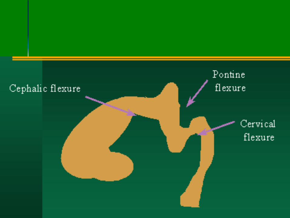
Stage 4 Myelination
ndash Inferior to superior posterior to anterior
ndash 5 - 15 months matures by 3 years
ndash Failure developmental delay dysmyelinating disease
Myelination Milestones
Brain stem cerebellum posterior limb of internal capsule term birth
Anterior limb internal capsule two months
Splenium of the corpus callosum three months
Myelination Milestones
Genu corpus callosum six months
Occipital white matter Central five months (T1)fourteen months (T2) Peripheral seven months (T1)fifteen months (T2)
Frontal white matter Central six months (T1)sixteen months (T2) Peripheral eleven months (T1)eighteen months (T2)
Embryological
developmental failure
Congenital brain disorders
Embryological failure
Acquired lesions
Congenital brain disorders
Embryological failure
Acquired lesions
Dorsal Induction 1Stage Formation and Closure of the
Neural Tube
Weeks 3 - 4
Three phases Formation and Closure of the Neural Tube Neurulation
Neural tube defects
(Failure of fusion or dysraphias)
bull Complete or regional disturbance in continuity of
neural tube structures and their coverings
1Open defects (Neurulation defects)
Neural tissue is exposed to the environment or
covered only with a thin membrane
Anencephaly
Open spina bifida (myelocelemeningomyelocele)
Neural tube defects
2 Closed defects (postneurulation defects)
Neural tissue is covered by normal skin
Encaphalocele
Closed spina bifida (meningocele occult
spinabifida)
Split cord malformations
(diastematomyeliadiplomyelia)
3 Arnold-Chiari malformation
Neural tube defects
1Cranioschisis
Anencephaly
Encephalocele
Meningocele
2Rachischisis
Spina bifida
MyeloMeningocele
Meningocele
3 Arnold-Chiari malformation
Encephalocele
Anencephaly
Failure of the brain and skull development
Most severe anomaly
Ultrasound diagnosis as early as twenty
weeks
Polyhydramnios high alpha fetoprotein
Death
Chiari malformations
Chiari Malformation
It was first described by Hans Chiari Austrian
pathologist (1851-1914) in 1891
In this and subsequent papers Chiari also credited Julius
Arnold (1835-1915) Professor of Anatomy at Heidelberg
on the grounds of a previous publication by Arnold
believed by him to be of a Chiari II malformation
It is a condition where there is herniation of the hindbrain
into the upper cervical spine This is classified into
TYPE I
There is herniation of the Cerebellar tonsils into the upper cervical canal
TYPE II
Migration of the medulla oblongata and 4th ventricle into the upper cervical canal
TYPE III
Displacement of the entire cerebellum and the 4th ventricle into the upper cervical canal
Chiari I
Chiari I
Chiari I
Chiari I
Chiari I
Chiari I with syrinx
Associated anomalies are
Spinal cord syrinx 20-40
Ventricles mild to moderate hydrocephalus 20-25
Skeletal anomalies basilar invagination 25-50
Arnold Chiari II
Complex anomaly
Skull and dura
Brain
Spine
cord
Chiari II
Chiari II
ndash Myelomeningoceleis present in nearly all patients
with a Chiari II malformation
ndash Hydrocephalus 90 of cases
ndash Aqueductal stenosis is seen in 50
ndash Medullary kink
ndash Corpus callosum agenesis
ndash Polymicrogyria
ndash Syringomyelia
Arnold Chiari II with syrinx
Chiari II with syrinx
Chiari III
Cervical occipital encephalocele that contains cerebellum
Chiari III
Cervical occipital encephalocele that contains cerebellum
Chiari IV Severe cerebellar hypoplasia
Chiari type 0
a newly identified form of Chiari describes the
absence (or a ldquozerordquo herniation) of the
tonsils below the foramen magnum
Yet Chiari 0 includes the presence of both
symptoms and a syrinx in the spinal cord
This new type is under study and
controversial
Chiari type 0
Ventral Induction 2Stage Formation of the Brain Segments and
Face
Weeks 5-10
Three vesicles (prosencephalon
mesencephalon and rhombencephalon) form
the cerebrum mid-brain cerebellum and
lower brain stem
Division into two hemispheres
Formation Ventral Induction 2Stage of the Brain Segments and Face
Failure Holoprosencephalies Dandy Walker Facial anomalies ndash Basilar invagination ndash KF syndrome ndash Sprengel deformity
Holoprosencephalies
Failure to separate into two hemispheres
ndash Lobar
ndash Semi lobar
ndash Alobar
ndash Septal optic dysplasia
Lobar fusion of only anterior inferior frontal lobes Otherwise the brain appears to be quite normal except for lack of septum pellucidum
Semi lobar Partial separation of the posterior occipital and temporal lobes Frontal brain is fused thalami partially fused
Alobar complete failure no falx single mono-ventricle fused thalami
Septal optic dysplasia most mild form in which there is no septum pellucidum and the optic nerves are very atrophic
Schizencephaly may be present in 50 of these cases
Corpus callosum agenesis may also be seen
Dandy Walker Defective development of the roof of the fourth
ventricle
Posterior fossa cyst (cystic dilation of 4th ventricles) hydrocephalus 80
Large posterior fossaabsent falx in the posterior fossa
cerebellar hypoplasia
The most common accompanying cerebral anomaly is callosal hypogenesis present in as much as 32 of affected patients
Dandy Walker
Dandy Walker
DD arachnoid cyst mega cisterna magna inflammatory cyst dermoid cyst
A mega cisterna magna adult patients with significantly enlarged CSF retrocerebellar cisterns in the posterior fossa with normal cerebellar morphology
Controversy remains around whether mega cisterna magna is a normal anatomical variant or due to volume loss of the cerebellum 1 Mega cisterna magna has also been referred to as Blakes pouch or retrocerebellar
Facial Anomalies
Often found with alobar holoprosencephaly corpus callosal agenesis
Usually midline
Hypertelorism hypotelorism cleft palat
THANK YOU

Myelination Milestones
Genu corpus callosum six months
Occipital white matter Central five months (T1)fourteen months (T2) Peripheral seven months (T1)fifteen months (T2)
Frontal white matter Central six months (T1)sixteen months (T2) Peripheral eleven months (T1)eighteen months (T2)
Embryological
developmental failure
Congenital brain disorders
Embryological failure
Acquired lesions
Congenital brain disorders
Embryological failure
Acquired lesions
Dorsal Induction 1Stage Formation and Closure of the
Neural Tube
Weeks 3 - 4
Three phases Formation and Closure of the Neural Tube Neurulation
Neural tube defects
(Failure of fusion or dysraphias)
bull Complete or regional disturbance in continuity of
neural tube structures and their coverings
1Open defects (Neurulation defects)
Neural tissue is exposed to the environment or
covered only with a thin membrane
Anencephaly
Open spina bifida (myelocelemeningomyelocele)
Neural tube defects
2 Closed defects (postneurulation defects)
Neural tissue is covered by normal skin
Encaphalocele
Closed spina bifida (meningocele occult
spinabifida)
Split cord malformations
(diastematomyeliadiplomyelia)
3 Arnold-Chiari malformation
Neural tube defects
1Cranioschisis
Anencephaly
Encephalocele
Meningocele
2Rachischisis
Spina bifida
MyeloMeningocele
Meningocele
3 Arnold-Chiari malformation
Encephalocele
Anencephaly
Failure of the brain and skull development
Most severe anomaly
Ultrasound diagnosis as early as twenty
weeks
Polyhydramnios high alpha fetoprotein
Death
Chiari malformations
Chiari Malformation
It was first described by Hans Chiari Austrian
pathologist (1851-1914) in 1891
In this and subsequent papers Chiari also credited Julius
Arnold (1835-1915) Professor of Anatomy at Heidelberg
on the grounds of a previous publication by Arnold
believed by him to be of a Chiari II malformation
It is a condition where there is herniation of the hindbrain
into the upper cervical spine This is classified into
TYPE I
There is herniation of the Cerebellar tonsils into the upper cervical canal
TYPE II
Migration of the medulla oblongata and 4th ventricle into the upper cervical canal
TYPE III
Displacement of the entire cerebellum and the 4th ventricle into the upper cervical canal
Chiari I
Chiari I
Chiari I
Chiari I
Chiari I
Chiari I with syrinx
Associated anomalies are
Spinal cord syrinx 20-40
Ventricles mild to moderate hydrocephalus 20-25
Skeletal anomalies basilar invagination 25-50
Arnold Chiari II
Complex anomaly
Skull and dura
Brain
Spine
cord
Chiari II
Chiari II
ndash Myelomeningoceleis present in nearly all patients
with a Chiari II malformation
ndash Hydrocephalus 90 of cases
ndash Aqueductal stenosis is seen in 50
ndash Medullary kink
ndash Corpus callosum agenesis
ndash Polymicrogyria
ndash Syringomyelia
Arnold Chiari II with syrinx
Chiari II with syrinx
Chiari III
Cervical occipital encephalocele that contains cerebellum
Chiari III
Cervical occipital encephalocele that contains cerebellum
Chiari IV Severe cerebellar hypoplasia
Chiari type 0
a newly identified form of Chiari describes the
absence (or a ldquozerordquo herniation) of the
tonsils below the foramen magnum
Yet Chiari 0 includes the presence of both
symptoms and a syrinx in the spinal cord
This new type is under study and
controversial
Chiari type 0
Ventral Induction 2Stage Formation of the Brain Segments and
Face
Weeks 5-10
Three vesicles (prosencephalon
mesencephalon and rhombencephalon) form
the cerebrum mid-brain cerebellum and
lower brain stem
Division into two hemispheres
Formation Ventral Induction 2Stage of the Brain Segments and Face
Failure Holoprosencephalies Dandy Walker Facial anomalies ndash Basilar invagination ndash KF syndrome ndash Sprengel deformity
Holoprosencephalies
Failure to separate into two hemispheres
ndash Lobar
ndash Semi lobar
ndash Alobar
ndash Septal optic dysplasia
Lobar fusion of only anterior inferior frontal lobes Otherwise the brain appears to be quite normal except for lack of septum pellucidum
Semi lobar Partial separation of the posterior occipital and temporal lobes Frontal brain is fused thalami partially fused
Alobar complete failure no falx single mono-ventricle fused thalami
Septal optic dysplasia most mild form in which there is no septum pellucidum and the optic nerves are very atrophic
Schizencephaly may be present in 50 of these cases
Corpus callosum agenesis may also be seen
Dandy Walker Defective development of the roof of the fourth
ventricle
Posterior fossa cyst (cystic dilation of 4th ventricles) hydrocephalus 80
Large posterior fossaabsent falx in the posterior fossa
cerebellar hypoplasia
The most common accompanying cerebral anomaly is callosal hypogenesis present in as much as 32 of affected patients
Dandy Walker
Dandy Walker
DD arachnoid cyst mega cisterna magna inflammatory cyst dermoid cyst
A mega cisterna magna adult patients with significantly enlarged CSF retrocerebellar cisterns in the posterior fossa with normal cerebellar morphology
Controversy remains around whether mega cisterna magna is a normal anatomical variant or due to volume loss of the cerebellum 1 Mega cisterna magna has also been referred to as Blakes pouch or retrocerebellar
Facial Anomalies
Often found with alobar holoprosencephaly corpus callosal agenesis
Usually midline
Hypertelorism hypotelorism cleft palat
THANK YOU

Congenital brain disorders
Embryological failure
Acquired lesions
Congenital brain disorders
Embryological failure
Acquired lesions
Dorsal Induction 1Stage Formation and Closure of the
Neural Tube
Weeks 3 - 4
Three phases Formation and Closure of the Neural Tube Neurulation
Neural tube defects
(Failure of fusion or dysraphias)
bull Complete or regional disturbance in continuity of
neural tube structures and their coverings
1Open defects (Neurulation defects)
Neural tissue is exposed to the environment or
covered only with a thin membrane
Anencephaly
Open spina bifida (myelocelemeningomyelocele)
Neural tube defects
2 Closed defects (postneurulation defects)
Neural tissue is covered by normal skin
Encaphalocele
Closed spina bifida (meningocele occult
spinabifida)
Split cord malformations
(diastematomyeliadiplomyelia)
3 Arnold-Chiari malformation
Neural tube defects
1Cranioschisis
Anencephaly
Encephalocele
Meningocele
2Rachischisis
Spina bifida
MyeloMeningocele
Meningocele
3 Arnold-Chiari malformation
Encephalocele
Anencephaly
Failure of the brain and skull development
Most severe anomaly
Ultrasound diagnosis as early as twenty
weeks
Polyhydramnios high alpha fetoprotein
Death
Chiari malformations
Chiari Malformation
It was first described by Hans Chiari Austrian
pathologist (1851-1914) in 1891
In this and subsequent papers Chiari also credited Julius
Arnold (1835-1915) Professor of Anatomy at Heidelberg
on the grounds of a previous publication by Arnold
believed by him to be of a Chiari II malformation
It is a condition where there is herniation of the hindbrain
into the upper cervical spine This is classified into
TYPE I
There is herniation of the Cerebellar tonsils into the upper cervical canal
TYPE II
Migration of the medulla oblongata and 4th ventricle into the upper cervical canal
TYPE III
Displacement of the entire cerebellum and the 4th ventricle into the upper cervical canal
Chiari I
Chiari I
Chiari I
Chiari I
Chiari I
Chiari I with syrinx
Associated anomalies are
Spinal cord syrinx 20-40
Ventricles mild to moderate hydrocephalus 20-25
Skeletal anomalies basilar invagination 25-50
Arnold Chiari II
Complex anomaly
Skull and dura
Brain
Spine
cord
Chiari II
Chiari II
ndash Myelomeningoceleis present in nearly all patients
with a Chiari II malformation
ndash Hydrocephalus 90 of cases
ndash Aqueductal stenosis is seen in 50
ndash Medullary kink
ndash Corpus callosum agenesis
ndash Polymicrogyria
ndash Syringomyelia
Arnold Chiari II with syrinx
Chiari II with syrinx
Chiari III
Cervical occipital encephalocele that contains cerebellum
Chiari III
Cervical occipital encephalocele that contains cerebellum
Chiari IV Severe cerebellar hypoplasia
Chiari type 0
a newly identified form of Chiari describes the
absence (or a ldquozerordquo herniation) of the
tonsils below the foramen magnum
Yet Chiari 0 includes the presence of both
symptoms and a syrinx in the spinal cord
This new type is under study and
controversial
Chiari type 0
Ventral Induction 2Stage Formation of the Brain Segments and
Face
Weeks 5-10
Three vesicles (prosencephalon
mesencephalon and rhombencephalon) form
the cerebrum mid-brain cerebellum and
lower brain stem
Division into two hemispheres
Formation Ventral Induction 2Stage of the Brain Segments and Face
Failure Holoprosencephalies Dandy Walker Facial anomalies ndash Basilar invagination ndash KF syndrome ndash Sprengel deformity
Holoprosencephalies
Failure to separate into two hemispheres
ndash Lobar
ndash Semi lobar
ndash Alobar
ndash Septal optic dysplasia
Lobar fusion of only anterior inferior frontal lobes Otherwise the brain appears to be quite normal except for lack of septum pellucidum
Semi lobar Partial separation of the posterior occipital and temporal lobes Frontal brain is fused thalami partially fused
Alobar complete failure no falx single mono-ventricle fused thalami
Septal optic dysplasia most mild form in which there is no septum pellucidum and the optic nerves are very atrophic
Schizencephaly may be present in 50 of these cases
Corpus callosum agenesis may also be seen
Dandy Walker Defective development of the roof of the fourth
ventricle
Posterior fossa cyst (cystic dilation of 4th ventricles) hydrocephalus 80
Large posterior fossaabsent falx in the posterior fossa
cerebellar hypoplasia
The most common accompanying cerebral anomaly is callosal hypogenesis present in as much as 32 of affected patients
Dandy Walker
Dandy Walker
DD arachnoid cyst mega cisterna magna inflammatory cyst dermoid cyst
A mega cisterna magna adult patients with significantly enlarged CSF retrocerebellar cisterns in the posterior fossa with normal cerebellar morphology
Controversy remains around whether mega cisterna magna is a normal anatomical variant or due to volume loss of the cerebellum 1 Mega cisterna magna has also been referred to as Blakes pouch or retrocerebellar
Facial Anomalies
Often found with alobar holoprosencephaly corpus callosal agenesis
Usually midline
Hypertelorism hypotelorism cleft palat
THANK YOU

Congenital brain disorders
Embryological failure
Acquired lesions
Dorsal Induction 1Stage Formation and Closure of the
Neural Tube
Weeks 3 - 4
Three phases Formation and Closure of the Neural Tube Neurulation
Neural tube defects
(Failure of fusion or dysraphias)
bull Complete or regional disturbance in continuity of
neural tube structures and their coverings
1Open defects (Neurulation defects)
Neural tissue is exposed to the environment or
covered only with a thin membrane
Anencephaly
Open spina bifida (myelocelemeningomyelocele)
Neural tube defects
2 Closed defects (postneurulation defects)
Neural tissue is covered by normal skin
Encaphalocele
Closed spina bifida (meningocele occult
spinabifida)
Split cord malformations
(diastematomyeliadiplomyelia)
3 Arnold-Chiari malformation
Neural tube defects
1Cranioschisis
Anencephaly
Encephalocele
Meningocele
2Rachischisis
Spina bifida
MyeloMeningocele
Meningocele
3 Arnold-Chiari malformation
Encephalocele
Anencephaly
Failure of the brain and skull development
Most severe anomaly
Ultrasound diagnosis as early as twenty
weeks
Polyhydramnios high alpha fetoprotein
Death
Chiari malformations
Chiari Malformation
It was first described by Hans Chiari Austrian
pathologist (1851-1914) in 1891
In this and subsequent papers Chiari also credited Julius
Arnold (1835-1915) Professor of Anatomy at Heidelberg
on the grounds of a previous publication by Arnold
believed by him to be of a Chiari II malformation
It is a condition where there is herniation of the hindbrain
into the upper cervical spine This is classified into
TYPE I
There is herniation of the Cerebellar tonsils into the upper cervical canal
TYPE II
Migration of the medulla oblongata and 4th ventricle into the upper cervical canal
TYPE III
Displacement of the entire cerebellum and the 4th ventricle into the upper cervical canal
Chiari I
Chiari I
Chiari I
Chiari I
Chiari I
Chiari I with syrinx
Associated anomalies are
Spinal cord syrinx 20-40
Ventricles mild to moderate hydrocephalus 20-25
Skeletal anomalies basilar invagination 25-50
Arnold Chiari II
Complex anomaly
Skull and dura
Brain
Spine
cord
Chiari II
Chiari II
ndash Myelomeningoceleis present in nearly all patients
with a Chiari II malformation
ndash Hydrocephalus 90 of cases
ndash Aqueductal stenosis is seen in 50
ndash Medullary kink
ndash Corpus callosum agenesis
ndash Polymicrogyria
ndash Syringomyelia
Arnold Chiari II with syrinx
Chiari II with syrinx
Chiari III
Cervical occipital encephalocele that contains cerebellum
Chiari III
Cervical occipital encephalocele that contains cerebellum
Chiari IV Severe cerebellar hypoplasia
Chiari type 0
a newly identified form of Chiari describes the
absence (or a ldquozerordquo herniation) of the
tonsils below the foramen magnum
Yet Chiari 0 includes the presence of both
symptoms and a syrinx in the spinal cord
This new type is under study and
controversial
Chiari type 0
Ventral Induction 2Stage Formation of the Brain Segments and
Face
Weeks 5-10
Three vesicles (prosencephalon
mesencephalon and rhombencephalon) form
the cerebrum mid-brain cerebellum and
lower brain stem
Division into two hemispheres
Formation Ventral Induction 2Stage of the Brain Segments and Face
Failure Holoprosencephalies Dandy Walker Facial anomalies ndash Basilar invagination ndash KF syndrome ndash Sprengel deformity
Holoprosencephalies
Failure to separate into two hemispheres
ndash Lobar
ndash Semi lobar
ndash Alobar
ndash Septal optic dysplasia
Lobar fusion of only anterior inferior frontal lobes Otherwise the brain appears to be quite normal except for lack of septum pellucidum
Semi lobar Partial separation of the posterior occipital and temporal lobes Frontal brain is fused thalami partially fused
Alobar complete failure no falx single mono-ventricle fused thalami
Septal optic dysplasia most mild form in which there is no septum pellucidum and the optic nerves are very atrophic
Schizencephaly may be present in 50 of these cases
Corpus callosum agenesis may also be seen
Dandy Walker Defective development of the roof of the fourth
ventricle
Posterior fossa cyst (cystic dilation of 4th ventricles) hydrocephalus 80
Large posterior fossaabsent falx in the posterior fossa
cerebellar hypoplasia
The most common accompanying cerebral anomaly is callosal hypogenesis present in as much as 32 of affected patients
Dandy Walker
Dandy Walker
DD arachnoid cyst mega cisterna magna inflammatory cyst dermoid cyst
A mega cisterna magna adult patients with significantly enlarged CSF retrocerebellar cisterns in the posterior fossa with normal cerebellar morphology
Controversy remains around whether mega cisterna magna is a normal anatomical variant or due to volume loss of the cerebellum 1 Mega cisterna magna has also been referred to as Blakes pouch or retrocerebellar
Facial Anomalies
Often found with alobar holoprosencephaly corpus callosal agenesis
Usually midline
Hypertelorism hypotelorism cleft palat
THANK YOU

Neural tube defects
1Cranioschisis
Anencephaly
Encephalocele
Meningocele
2Rachischisis
Spina bifida
MyeloMeningocele
Meningocele
3 Arnold-Chiari malformation
Encephalocele
Anencephaly
Failure of the brain and skull development
Most severe anomaly
Ultrasound diagnosis as early as twenty
weeks
Polyhydramnios high alpha fetoprotein
Death
Chiari malformations
Chiari Malformation
It was first described by Hans Chiari Austrian
pathologist (1851-1914) in 1891
In this and subsequent papers Chiari also credited Julius
Arnold (1835-1915) Professor of Anatomy at Heidelberg
on the grounds of a previous publication by Arnold
believed by him to be of a Chiari II malformation
It is a condition where there is herniation of the hindbrain
into the upper cervical spine This is classified into
TYPE I
There is herniation of the Cerebellar tonsils into the upper cervical canal
TYPE II
Migration of the medulla oblongata and 4th ventricle into the upper cervical canal
TYPE III
Displacement of the entire cerebellum and the 4th ventricle into the upper cervical canal
Chiari I
Chiari I
Chiari I
Chiari I
Chiari I
Chiari I with syrinx
Associated anomalies are
Spinal cord syrinx 20-40
Ventricles mild to moderate hydrocephalus 20-25
Skeletal anomalies basilar invagination 25-50
Arnold Chiari II
Complex anomaly
Skull and dura
Brain
Spine
cord
Chiari II
Chiari II
ndash Myelomeningoceleis present in nearly all patients
with a Chiari II malformation
ndash Hydrocephalus 90 of cases
ndash Aqueductal stenosis is seen in 50
ndash Medullary kink
ndash Corpus callosum agenesis
ndash Polymicrogyria
ndash Syringomyelia
Arnold Chiari II with syrinx
Chiari II with syrinx
Chiari III
Cervical occipital encephalocele that contains cerebellum
Chiari III
Cervical occipital encephalocele that contains cerebellum
Chiari IV Severe cerebellar hypoplasia
Chiari type 0
a newly identified form of Chiari describes the
absence (or a ldquozerordquo herniation) of the
tonsils below the foramen magnum
Yet Chiari 0 includes the presence of both
symptoms and a syrinx in the spinal cord
This new type is under study and
controversial
Chiari type 0
Ventral Induction 2Stage Formation of the Brain Segments and
Face
Weeks 5-10
Three vesicles (prosencephalon
mesencephalon and rhombencephalon) form
the cerebrum mid-brain cerebellum and
lower brain stem
Division into two hemispheres
Formation Ventral Induction 2Stage of the Brain Segments and Face
Failure Holoprosencephalies Dandy Walker Facial anomalies ndash Basilar invagination ndash KF syndrome ndash Sprengel deformity
Holoprosencephalies
Failure to separate into two hemispheres
ndash Lobar
ndash Semi lobar
ndash Alobar
ndash Septal optic dysplasia
Lobar fusion of only anterior inferior frontal lobes Otherwise the brain appears to be quite normal except for lack of septum pellucidum
Semi lobar Partial separation of the posterior occipital and temporal lobes Frontal brain is fused thalami partially fused
Alobar complete failure no falx single mono-ventricle fused thalami
Septal optic dysplasia most mild form in which there is no septum pellucidum and the optic nerves are very atrophic
Schizencephaly may be present in 50 of these cases
Corpus callosum agenesis may also be seen
Dandy Walker Defective development of the roof of the fourth
ventricle
Posterior fossa cyst (cystic dilation of 4th ventricles) hydrocephalus 80
Large posterior fossaabsent falx in the posterior fossa
cerebellar hypoplasia
The most common accompanying cerebral anomaly is callosal hypogenesis present in as much as 32 of affected patients
Dandy Walker
Dandy Walker
DD arachnoid cyst mega cisterna magna inflammatory cyst dermoid cyst
A mega cisterna magna adult patients with significantly enlarged CSF retrocerebellar cisterns in the posterior fossa with normal cerebellar morphology
Controversy remains around whether mega cisterna magna is a normal anatomical variant or due to volume loss of the cerebellum 1 Mega cisterna magna has also been referred to as Blakes pouch or retrocerebellar
Facial Anomalies
Often found with alobar holoprosencephaly corpus callosal agenesis
Usually midline
Hypertelorism hypotelorism cleft palat
THANK YOU
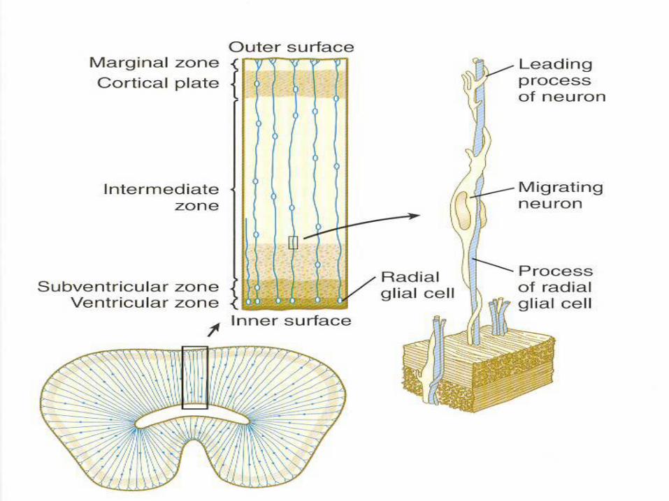
Chiari I
Chiari I
Chiari I
Chiari I
Chiari I
Chiari I with syrinx
Associated anomalies are
Spinal cord syrinx 20-40
Ventricles mild to moderate hydrocephalus 20-25
Skeletal anomalies basilar invagination 25-50
Arnold Chiari II
Complex anomaly
Skull and dura
Brain
Spine
cord
Chiari II
Chiari II
ndash Myelomeningoceleis present in nearly all patients
with a Chiari II malformation
ndash Hydrocephalus 90 of cases
ndash Aqueductal stenosis is seen in 50
ndash Medullary kink
ndash Corpus callosum agenesis
ndash Polymicrogyria
ndash Syringomyelia
Arnold Chiari II with syrinx
Chiari II with syrinx
Chiari III
Cervical occipital encephalocele that contains cerebellum
Chiari III
Cervical occipital encephalocele that contains cerebellum
Chiari IV Severe cerebellar hypoplasia
Chiari type 0
a newly identified form of Chiari describes the
absence (or a ldquozerordquo herniation) of the
tonsils below the foramen magnum
Yet Chiari 0 includes the presence of both
symptoms and a syrinx in the spinal cord
This new type is under study and
controversial
Chiari type 0
Ventral Induction 2Stage Formation of the Brain Segments and
Face
Weeks 5-10
Three vesicles (prosencephalon
mesencephalon and rhombencephalon) form
the cerebrum mid-brain cerebellum and
lower brain stem
Division into two hemispheres
Formation Ventral Induction 2Stage of the Brain Segments and Face
Failure Holoprosencephalies Dandy Walker Facial anomalies ndash Basilar invagination ndash KF syndrome ndash Sprengel deformity
Holoprosencephalies
Failure to separate into two hemispheres
ndash Lobar
ndash Semi lobar
ndash Alobar
ndash Septal optic dysplasia
Lobar fusion of only anterior inferior frontal lobes Otherwise the brain appears to be quite normal except for lack of septum pellucidum
Semi lobar Partial separation of the posterior occipital and temporal lobes Frontal brain is fused thalami partially fused
Alobar complete failure no falx single mono-ventricle fused thalami
Septal optic dysplasia most mild form in which there is no septum pellucidum and the optic nerves are very atrophic
Schizencephaly may be present in 50 of these cases
Corpus callosum agenesis may also be seen
Dandy Walker Defective development of the roof of the fourth
ventricle
Posterior fossa cyst (cystic dilation of 4th ventricles) hydrocephalus 80
Large posterior fossaabsent falx in the posterior fossa
cerebellar hypoplasia
The most common accompanying cerebral anomaly is callosal hypogenesis present in as much as 32 of affected patients
Dandy Walker
Dandy Walker
DD arachnoid cyst mega cisterna magna inflammatory cyst dermoid cyst
A mega cisterna magna adult patients with significantly enlarged CSF retrocerebellar cisterns in the posterior fossa with normal cerebellar morphology
Controversy remains around whether mega cisterna magna is a normal anatomical variant or due to volume loss of the cerebellum 1 Mega cisterna magna has also been referred to as Blakes pouch or retrocerebellar
Facial Anomalies
Often found with alobar holoprosencephaly corpus callosal agenesis
Usually midline
Hypertelorism hypotelorism cleft palat
THANK YOU

Chiari I
Chiari I
Chiari I
Chiari I
Chiari I with syrinx
Associated anomalies are
Spinal cord syrinx 20-40
Ventricles mild to moderate hydrocephalus 20-25
Skeletal anomalies basilar invagination 25-50
Arnold Chiari II
Complex anomaly
Skull and dura
Brain
Spine
cord
Chiari II
Chiari II
ndash Myelomeningoceleis present in nearly all patients
with a Chiari II malformation
ndash Hydrocephalus 90 of cases
ndash Aqueductal stenosis is seen in 50
ndash Medullary kink
ndash Corpus callosum agenesis
ndash Polymicrogyria
ndash Syringomyelia
Arnold Chiari II with syrinx
Chiari II with syrinx
Chiari III
Cervical occipital encephalocele that contains cerebellum
Chiari III
Cervical occipital encephalocele that contains cerebellum
Chiari IV Severe cerebellar hypoplasia
Chiari type 0
a newly identified form of Chiari describes the
absence (or a ldquozerordquo herniation) of the
tonsils below the foramen magnum
Yet Chiari 0 includes the presence of both
symptoms and a syrinx in the spinal cord
This new type is under study and
controversial
Chiari type 0
Ventral Induction 2Stage Formation of the Brain Segments and
Face
Weeks 5-10
Three vesicles (prosencephalon
mesencephalon and rhombencephalon) form
the cerebrum mid-brain cerebellum and
lower brain stem
Division into two hemispheres
Formation Ventral Induction 2Stage of the Brain Segments and Face
Failure Holoprosencephalies Dandy Walker Facial anomalies ndash Basilar invagination ndash KF syndrome ndash Sprengel deformity
Holoprosencephalies
Failure to separate into two hemispheres
ndash Lobar
ndash Semi lobar
ndash Alobar
ndash Septal optic dysplasia
Lobar fusion of only anterior inferior frontal lobes Otherwise the brain appears to be quite normal except for lack of septum pellucidum
Semi lobar Partial separation of the posterior occipital and temporal lobes Frontal brain is fused thalami partially fused
Alobar complete failure no falx single mono-ventricle fused thalami
Septal optic dysplasia most mild form in which there is no septum pellucidum and the optic nerves are very atrophic
Schizencephaly may be present in 50 of these cases
Corpus callosum agenesis may also be seen
Dandy Walker Defective development of the roof of the fourth
ventricle
Posterior fossa cyst (cystic dilation of 4th ventricles) hydrocephalus 80
Large posterior fossaabsent falx in the posterior fossa
cerebellar hypoplasia
The most common accompanying cerebral anomaly is callosal hypogenesis present in as much as 32 of affected patients
Dandy Walker
Dandy Walker
DD arachnoid cyst mega cisterna magna inflammatory cyst dermoid cyst
A mega cisterna magna adult patients with significantly enlarged CSF retrocerebellar cisterns in the posterior fossa with normal cerebellar morphology
Controversy remains around whether mega cisterna magna is a normal anatomical variant or due to volume loss of the cerebellum 1 Mega cisterna magna has also been referred to as Blakes pouch or retrocerebellar
Facial Anomalies
Often found with alobar holoprosencephaly corpus callosal agenesis
Usually midline
Hypertelorism hypotelorism cleft palat
THANK YOU

Chiari I
Chiari I
Chiari I
Chiari I with syrinx
Associated anomalies are
Spinal cord syrinx 20-40
Ventricles mild to moderate hydrocephalus 20-25
Skeletal anomalies basilar invagination 25-50
Arnold Chiari II
Complex anomaly
Skull and dura
Brain
Spine
cord
Chiari II
Chiari II
ndash Myelomeningoceleis present in nearly all patients
with a Chiari II malformation
ndash Hydrocephalus 90 of cases
ndash Aqueductal stenosis is seen in 50
ndash Medullary kink
ndash Corpus callosum agenesis
ndash Polymicrogyria
ndash Syringomyelia
Arnold Chiari II with syrinx
Chiari II with syrinx
Chiari III
Cervical occipital encephalocele that contains cerebellum
Chiari III
Cervical occipital encephalocele that contains cerebellum
Chiari IV Severe cerebellar hypoplasia
Chiari type 0
a newly identified form of Chiari describes the
absence (or a ldquozerordquo herniation) of the
tonsils below the foramen magnum
Yet Chiari 0 includes the presence of both
symptoms and a syrinx in the spinal cord
This new type is under study and
controversial
Chiari type 0
Ventral Induction 2Stage Formation of the Brain Segments and
Face
Weeks 5-10
Three vesicles (prosencephalon
mesencephalon and rhombencephalon) form
the cerebrum mid-brain cerebellum and
lower brain stem
Division into two hemispheres
Formation Ventral Induction 2Stage of the Brain Segments and Face
Failure Holoprosencephalies Dandy Walker Facial anomalies ndash Basilar invagination ndash KF syndrome ndash Sprengel deformity
Holoprosencephalies
Failure to separate into two hemispheres
ndash Lobar
ndash Semi lobar
ndash Alobar
ndash Septal optic dysplasia
Lobar fusion of only anterior inferior frontal lobes Otherwise the brain appears to be quite normal except for lack of septum pellucidum
Semi lobar Partial separation of the posterior occipital and temporal lobes Frontal brain is fused thalami partially fused
Alobar complete failure no falx single mono-ventricle fused thalami
Septal optic dysplasia most mild form in which there is no septum pellucidum and the optic nerves are very atrophic
Schizencephaly may be present in 50 of these cases
Corpus callosum agenesis may also be seen
Dandy Walker Defective development of the roof of the fourth
ventricle
Posterior fossa cyst (cystic dilation of 4th ventricles) hydrocephalus 80
Large posterior fossaabsent falx in the posterior fossa
cerebellar hypoplasia
The most common accompanying cerebral anomaly is callosal hypogenesis present in as much as 32 of affected patients
Dandy Walker
Dandy Walker
DD arachnoid cyst mega cisterna magna inflammatory cyst dermoid cyst
A mega cisterna magna adult patients with significantly enlarged CSF retrocerebellar cisterns in the posterior fossa with normal cerebellar morphology
Controversy remains around whether mega cisterna magna is a normal anatomical variant or due to volume loss of the cerebellum 1 Mega cisterna magna has also been referred to as Blakes pouch or retrocerebellar
Facial Anomalies
Often found with alobar holoprosencephaly corpus callosal agenesis
Usually midline
Hypertelorism hypotelorism cleft palat
THANK YOU

Chiari I
Chiari I
Chiari I with syrinx
Associated anomalies are
Spinal cord syrinx 20-40
Ventricles mild to moderate hydrocephalus 20-25
Skeletal anomalies basilar invagination 25-50
Arnold Chiari II
Complex anomaly
Skull and dura
Brain
Spine
cord
Chiari II
Chiari II
ndash Myelomeningoceleis present in nearly all patients
with a Chiari II malformation
ndash Hydrocephalus 90 of cases
ndash Aqueductal stenosis is seen in 50
ndash Medullary kink
ndash Corpus callosum agenesis
ndash Polymicrogyria
ndash Syringomyelia
Arnold Chiari II with syrinx
Chiari II with syrinx
Chiari III
Cervical occipital encephalocele that contains cerebellum
Chiari III
Cervical occipital encephalocele that contains cerebellum
Chiari IV Severe cerebellar hypoplasia
Chiari type 0
a newly identified form of Chiari describes the
absence (or a ldquozerordquo herniation) of the
tonsils below the foramen magnum
Yet Chiari 0 includes the presence of both
symptoms and a syrinx in the spinal cord
This new type is under study and
controversial
Chiari type 0
Ventral Induction 2Stage Formation of the Brain Segments and
Face
Weeks 5-10
Three vesicles (prosencephalon
mesencephalon and rhombencephalon) form
the cerebrum mid-brain cerebellum and
lower brain stem
Division into two hemispheres
Formation Ventral Induction 2Stage of the Brain Segments and Face
Failure Holoprosencephalies Dandy Walker Facial anomalies ndash Basilar invagination ndash KF syndrome ndash Sprengel deformity
Holoprosencephalies
Failure to separate into two hemispheres
ndash Lobar
ndash Semi lobar
ndash Alobar
ndash Septal optic dysplasia
Lobar fusion of only anterior inferior frontal lobes Otherwise the brain appears to be quite normal except for lack of septum pellucidum
Semi lobar Partial separation of the posterior occipital and temporal lobes Frontal brain is fused thalami partially fused
Alobar complete failure no falx single mono-ventricle fused thalami
Septal optic dysplasia most mild form in which there is no septum pellucidum and the optic nerves are very atrophic
Schizencephaly may be present in 50 of these cases
Corpus callosum agenesis may also be seen
Dandy Walker Defective development of the roof of the fourth
ventricle
Posterior fossa cyst (cystic dilation of 4th ventricles) hydrocephalus 80
Large posterior fossaabsent falx in the posterior fossa
cerebellar hypoplasia
The most common accompanying cerebral anomaly is callosal hypogenesis present in as much as 32 of affected patients
Dandy Walker
Dandy Walker
DD arachnoid cyst mega cisterna magna inflammatory cyst dermoid cyst
A mega cisterna magna adult patients with significantly enlarged CSF retrocerebellar cisterns in the posterior fossa with normal cerebellar morphology
Controversy remains around whether mega cisterna magna is a normal anatomical variant or due to volume loss of the cerebellum 1 Mega cisterna magna has also been referred to as Blakes pouch or retrocerebellar
Facial Anomalies
Often found with alobar holoprosencephaly corpus callosal agenesis
Usually midline
Hypertelorism hypotelorism cleft palat
THANK YOU

Chiari I
Chiari I with syrinx
Associated anomalies are
Spinal cord syrinx 20-40
Ventricles mild to moderate hydrocephalus 20-25
Skeletal anomalies basilar invagination 25-50
Arnold Chiari II
Complex anomaly
Skull and dura
Brain
Spine
cord
Chiari II
Chiari II
ndash Myelomeningoceleis present in nearly all patients
with a Chiari II malformation
ndash Hydrocephalus 90 of cases
ndash Aqueductal stenosis is seen in 50
ndash Medullary kink
ndash Corpus callosum agenesis
ndash Polymicrogyria
ndash Syringomyelia
Arnold Chiari II with syrinx
Chiari II with syrinx
Chiari III
Cervical occipital encephalocele that contains cerebellum
Chiari III
Cervical occipital encephalocele that contains cerebellum
Chiari IV Severe cerebellar hypoplasia
Chiari type 0
a newly identified form of Chiari describes the
absence (or a ldquozerordquo herniation) of the
tonsils below the foramen magnum
Yet Chiari 0 includes the presence of both
symptoms and a syrinx in the spinal cord
This new type is under study and
controversial
Chiari type 0
Ventral Induction 2Stage Formation of the Brain Segments and
Face
Weeks 5-10
Three vesicles (prosencephalon
mesencephalon and rhombencephalon) form
the cerebrum mid-brain cerebellum and
lower brain stem
Division into two hemispheres
Formation Ventral Induction 2Stage of the Brain Segments and Face
Failure Holoprosencephalies Dandy Walker Facial anomalies ndash Basilar invagination ndash KF syndrome ndash Sprengel deformity
Holoprosencephalies
Failure to separate into two hemispheres
ndash Lobar
ndash Semi lobar
ndash Alobar
ndash Septal optic dysplasia
Lobar fusion of only anterior inferior frontal lobes Otherwise the brain appears to be quite normal except for lack of septum pellucidum
Semi lobar Partial separation of the posterior occipital and temporal lobes Frontal brain is fused thalami partially fused
Alobar complete failure no falx single mono-ventricle fused thalami
Septal optic dysplasia most mild form in which there is no septum pellucidum and the optic nerves are very atrophic
Schizencephaly may be present in 50 of these cases
Corpus callosum agenesis may also be seen
Dandy Walker Defective development of the roof of the fourth
ventricle
Posterior fossa cyst (cystic dilation of 4th ventricles) hydrocephalus 80
Large posterior fossaabsent falx in the posterior fossa
cerebellar hypoplasia
The most common accompanying cerebral anomaly is callosal hypogenesis present in as much as 32 of affected patients
Dandy Walker
Dandy Walker
DD arachnoid cyst mega cisterna magna inflammatory cyst dermoid cyst
A mega cisterna magna adult patients with significantly enlarged CSF retrocerebellar cisterns in the posterior fossa with normal cerebellar morphology
Controversy remains around whether mega cisterna magna is a normal anatomical variant or due to volume loss of the cerebellum 1 Mega cisterna magna has also been referred to as Blakes pouch or retrocerebellar
Facial Anomalies
Often found with alobar holoprosencephaly corpus callosal agenesis
Usually midline
Hypertelorism hypotelorism cleft palat
THANK YOU
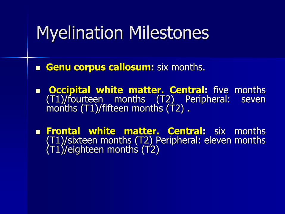
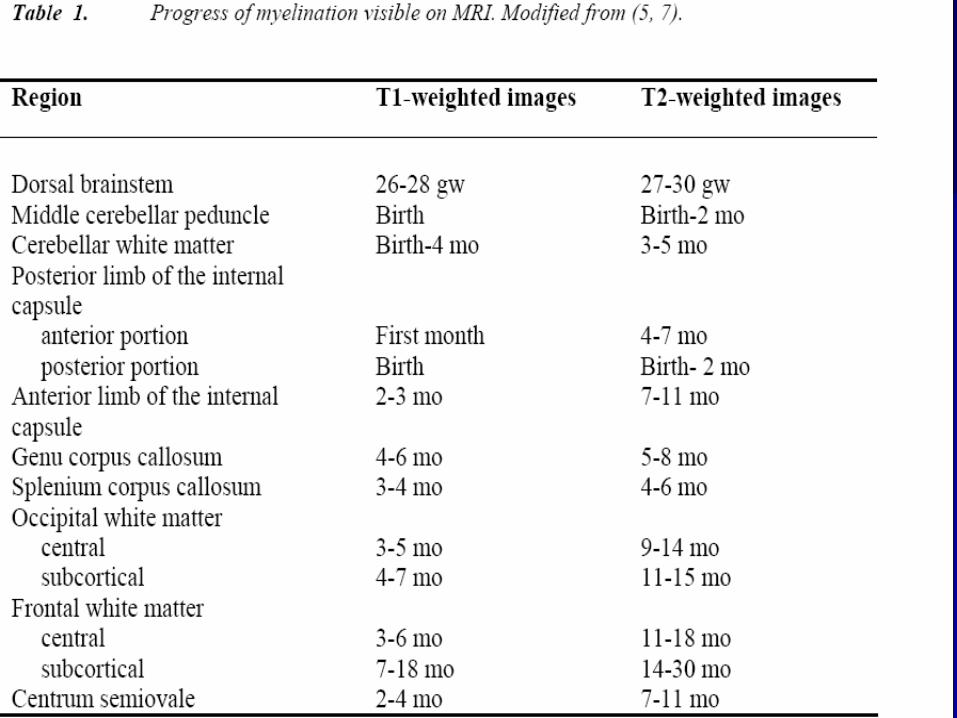

Chiari II
Chiari II
ndash Myelomeningoceleis present in nearly all patients
with a Chiari II malformation
ndash Hydrocephalus 90 of cases
ndash Aqueductal stenosis is seen in 50
ndash Medullary kink
ndash Corpus callosum agenesis
ndash Polymicrogyria
ndash Syringomyelia
Arnold Chiari II with syrinx
Chiari II with syrinx
Chiari III
Cervical occipital encephalocele that contains cerebellum
Chiari III
Cervical occipital encephalocele that contains cerebellum
Chiari IV Severe cerebellar hypoplasia
Chiari type 0
a newly identified form of Chiari describes the
absence (or a ldquozerordquo herniation) of the
tonsils below the foramen magnum
Yet Chiari 0 includes the presence of both
symptoms and a syrinx in the spinal cord
This new type is under study and
controversial
Chiari type 0
Ventral Induction 2Stage Formation of the Brain Segments and
Face
Weeks 5-10
Three vesicles (prosencephalon
mesencephalon and rhombencephalon) form
the cerebrum mid-brain cerebellum and
lower brain stem
Division into two hemispheres
Formation Ventral Induction 2Stage of the Brain Segments and Face
Failure Holoprosencephalies Dandy Walker Facial anomalies ndash Basilar invagination ndash KF syndrome ndash Sprengel deformity
Holoprosencephalies
Failure to separate into two hemispheres
ndash Lobar
ndash Semi lobar
ndash Alobar
ndash Septal optic dysplasia
Lobar fusion of only anterior inferior frontal lobes Otherwise the brain appears to be quite normal except for lack of septum pellucidum
Semi lobar Partial separation of the posterior occipital and temporal lobes Frontal brain is fused thalami partially fused
Alobar complete failure no falx single mono-ventricle fused thalami
Septal optic dysplasia most mild form in which there is no septum pellucidum and the optic nerves are very atrophic
Schizencephaly may be present in 50 of these cases
Corpus callosum agenesis may also be seen
Dandy Walker Defective development of the roof of the fourth
ventricle
Posterior fossa cyst (cystic dilation of 4th ventricles) hydrocephalus 80
Large posterior fossaabsent falx in the posterior fossa
cerebellar hypoplasia
The most common accompanying cerebral anomaly is callosal hypogenesis present in as much as 32 of affected patients
Dandy Walker
Dandy Walker
DD arachnoid cyst mega cisterna magna inflammatory cyst dermoid cyst
A mega cisterna magna adult patients with significantly enlarged CSF retrocerebellar cisterns in the posterior fossa with normal cerebellar morphology
Controversy remains around whether mega cisterna magna is a normal anatomical variant or due to volume loss of the cerebellum 1 Mega cisterna magna has also been referred to as Blakes pouch or retrocerebellar
Facial Anomalies
Often found with alobar holoprosencephaly corpus callosal agenesis
Usually midline
Hypertelorism hypotelorism cleft palat
THANK YOU

Chiari III
Cervical occipital encephalocele that contains cerebellum
Chiari III
Cervical occipital encephalocele that contains cerebellum
Chiari IV Severe cerebellar hypoplasia
Chiari type 0
a newly identified form of Chiari describes the
absence (or a ldquozerordquo herniation) of the
tonsils below the foramen magnum
Yet Chiari 0 includes the presence of both
symptoms and a syrinx in the spinal cord
This new type is under study and
controversial
Chiari type 0
Ventral Induction 2Stage Formation of the Brain Segments and
Face
Weeks 5-10
Three vesicles (prosencephalon
mesencephalon and rhombencephalon) form
the cerebrum mid-brain cerebellum and
lower brain stem
Division into two hemispheres
Formation Ventral Induction 2Stage of the Brain Segments and Face
Failure Holoprosencephalies Dandy Walker Facial anomalies ndash Basilar invagination ndash KF syndrome ndash Sprengel deformity
Holoprosencephalies
Failure to separate into two hemispheres
ndash Lobar
ndash Semi lobar
ndash Alobar
ndash Septal optic dysplasia
Lobar fusion of only anterior inferior frontal lobes Otherwise the brain appears to be quite normal except for lack of septum pellucidum
Semi lobar Partial separation of the posterior occipital and temporal lobes Frontal brain is fused thalami partially fused
Alobar complete failure no falx single mono-ventricle fused thalami
Septal optic dysplasia most mild form in which there is no septum pellucidum and the optic nerves are very atrophic
Schizencephaly may be present in 50 of these cases
Corpus callosum agenesis may also be seen
Dandy Walker Defective development of the roof of the fourth
ventricle
Posterior fossa cyst (cystic dilation of 4th ventricles) hydrocephalus 80
Large posterior fossaabsent falx in the posterior fossa
cerebellar hypoplasia
The most common accompanying cerebral anomaly is callosal hypogenesis present in as much as 32 of affected patients
Dandy Walker
Dandy Walker
DD arachnoid cyst mega cisterna magna inflammatory cyst dermoid cyst
A mega cisterna magna adult patients with significantly enlarged CSF retrocerebellar cisterns in the posterior fossa with normal cerebellar morphology
Controversy remains around whether mega cisterna magna is a normal anatomical variant or due to volume loss of the cerebellum 1 Mega cisterna magna has also been referred to as Blakes pouch or retrocerebellar
Facial Anomalies
Often found with alobar holoprosencephaly corpus callosal agenesis
Usually midline
Hypertelorism hypotelorism cleft palat
THANK YOU

Chiari III
Cervical occipital encephalocele that contains cerebellum
Chiari IV Severe cerebellar hypoplasia
Chiari type 0
a newly identified form of Chiari describes the
absence (or a ldquozerordquo herniation) of the
tonsils below the foramen magnum
Yet Chiari 0 includes the presence of both
symptoms and a syrinx in the spinal cord
This new type is under study and
controversial
Chiari type 0
Ventral Induction 2Stage Formation of the Brain Segments and
Face
Weeks 5-10
Three vesicles (prosencephalon
mesencephalon and rhombencephalon) form
the cerebrum mid-brain cerebellum and
lower brain stem
Division into two hemispheres
Formation Ventral Induction 2Stage of the Brain Segments and Face
Failure Holoprosencephalies Dandy Walker Facial anomalies ndash Basilar invagination ndash KF syndrome ndash Sprengel deformity
Holoprosencephalies
Failure to separate into two hemispheres
ndash Lobar
ndash Semi lobar
ndash Alobar
ndash Septal optic dysplasia
Lobar fusion of only anterior inferior frontal lobes Otherwise the brain appears to be quite normal except for lack of septum pellucidum
Semi lobar Partial separation of the posterior occipital and temporal lobes Frontal brain is fused thalami partially fused
Alobar complete failure no falx single mono-ventricle fused thalami
Septal optic dysplasia most mild form in which there is no septum pellucidum and the optic nerves are very atrophic
Schizencephaly may be present in 50 of these cases
Corpus callosum agenesis may also be seen
Dandy Walker Defective development of the roof of the fourth
ventricle
Posterior fossa cyst (cystic dilation of 4th ventricles) hydrocephalus 80
Large posterior fossaabsent falx in the posterior fossa
cerebellar hypoplasia
The most common accompanying cerebral anomaly is callosal hypogenesis present in as much as 32 of affected patients
Dandy Walker
Dandy Walker
DD arachnoid cyst mega cisterna magna inflammatory cyst dermoid cyst
A mega cisterna magna adult patients with significantly enlarged CSF retrocerebellar cisterns in the posterior fossa with normal cerebellar morphology
Controversy remains around whether mega cisterna magna is a normal anatomical variant or due to volume loss of the cerebellum 1 Mega cisterna magna has also been referred to as Blakes pouch or retrocerebellar
Facial Anomalies
Often found with alobar holoprosencephaly corpus callosal agenesis
Usually midline
Hypertelorism hypotelorism cleft palat
THANK YOU

Chiari type 0
Ventral Induction 2Stage Formation of the Brain Segments and
Face
Weeks 5-10
Three vesicles (prosencephalon
mesencephalon and rhombencephalon) form
the cerebrum mid-brain cerebellum and
lower brain stem
Division into two hemispheres
Formation Ventral Induction 2Stage of the Brain Segments and Face
Failure Holoprosencephalies Dandy Walker Facial anomalies ndash Basilar invagination ndash KF syndrome ndash Sprengel deformity
Holoprosencephalies
Failure to separate into two hemispheres
ndash Lobar
ndash Semi lobar
ndash Alobar
ndash Septal optic dysplasia
Lobar fusion of only anterior inferior frontal lobes Otherwise the brain appears to be quite normal except for lack of septum pellucidum
Semi lobar Partial separation of the posterior occipital and temporal lobes Frontal brain is fused thalami partially fused
Alobar complete failure no falx single mono-ventricle fused thalami
Septal optic dysplasia most mild form in which there is no septum pellucidum and the optic nerves are very atrophic
Schizencephaly may be present in 50 of these cases
Corpus callosum agenesis may also be seen
Dandy Walker Defective development of the roof of the fourth
ventricle
Posterior fossa cyst (cystic dilation of 4th ventricles) hydrocephalus 80
Large posterior fossaabsent falx in the posterior fossa
cerebellar hypoplasia
The most common accompanying cerebral anomaly is callosal hypogenesis present in as much as 32 of affected patients
Dandy Walker
Dandy Walker
DD arachnoid cyst mega cisterna magna inflammatory cyst dermoid cyst
A mega cisterna magna adult patients with significantly enlarged CSF retrocerebellar cisterns in the posterior fossa with normal cerebellar morphology
Controversy remains around whether mega cisterna magna is a normal anatomical variant or due to volume loss of the cerebellum 1 Mega cisterna magna has also been referred to as Blakes pouch or retrocerebellar
Facial Anomalies
Often found with alobar holoprosencephaly corpus callosal agenesis
Usually midline
Hypertelorism hypotelorism cleft palat
THANK YOU
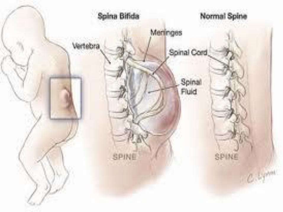
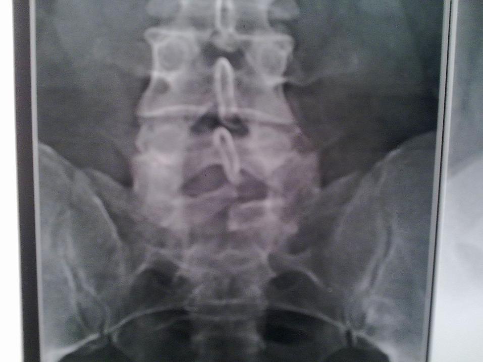

Dandy Walker
Dandy Walker
DD arachnoid cyst mega cisterna magna inflammatory cyst dermoid cyst
A mega cisterna magna adult patients with significantly enlarged CSF retrocerebellar cisterns in the posterior fossa with normal cerebellar morphology
Controversy remains around whether mega cisterna magna is a normal anatomical variant or due to volume loss of the cerebellum 1 Mega cisterna magna has also been referred to as Blakes pouch or retrocerebellar
Facial Anomalies
Often found with alobar holoprosencephaly corpus callosal agenesis
Usually midline
Hypertelorism hypotelorism cleft palat
THANK YOU

Dandy Walker
DD arachnoid cyst mega cisterna magna inflammatory cyst dermoid cyst
A mega cisterna magna adult patients with significantly enlarged CSF retrocerebellar cisterns in the posterior fossa with normal cerebellar morphology
Controversy remains around whether mega cisterna magna is a normal anatomical variant or due to volume loss of the cerebellum 1 Mega cisterna magna has also been referred to as Blakes pouch or retrocerebellar
Facial Anomalies
Often found with alobar holoprosencephaly corpus callosal agenesis
Usually midline
Hypertelorism hypotelorism cleft palat
THANK YOU

Facial Anomalies
Often found with alobar holoprosencephaly corpus callosal agenesis
Usually midline
Hypertelorism hypotelorism cleft palat
THANK YOU
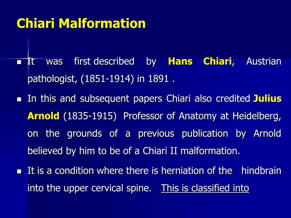
THANK YOU