elevated soluble fms-like tyrosine kinase-1 levels in ...€¦(ck) kinase (ck-mb), myoglobin, ......
TRANSCRIPT

Elevated Soluble fms-Like Tyrosine Kinase-1 Levels inAcute Coronary Occlusion
Navin K. Kapur, Kevin S. Heffernan, Adil A. Yunis, Tuan A. Nguyen, Mark J. Aronovitz,Peter Parpos, Szuhuei Wilson, Corey K. Baker, Michele L. Esposito, Ameer Shah,
Carey D. Kimmelstiel, Andrew Weintraub, Richard H. Karas, Michael E. Mendelsohn
Objective—Early recognition of an acute coronary occlusion (ACO) improves clinical outcomes. Soluble fms-like tyrosinekinase-1 (sFLT1) is an endothelium-derived protein induced by hypoxia. We tested whether sFLT1 are elevated in ACO.
Methods and Results—Serum sFLT1 levels were measured by enzyme-linked immunosorbent assay in patients withST-segment elevations and angiographically confirmed ACO referred for emergent catheterization, and they werecompared with unstable angina/non-ST-elevation myocardial infarction and 2 control groups. To further explore sFLT1release, a mouse model of ACO and in vitro human coronary artery endothelial cell injury were used. sFLT1 levels wereincreased in ACO compared with unstable angina/non-ST-elevation myocardial infarction, catheterized controls, orhealthy volunteers (200.7�15.5 versus 70.7�44.0 versus 10.2�4.0 versus 11.7�1.7 pg/mL respectively, P�0.001versus ACO). At presentation, all ACO patients had elevated sFLT1 levels (�15 pg/mL, 99th percentile in controls),whereas 57% had levels of the MB isoform of creatine kinase levels �10 ng/mL (P�0.01) and 85% had ultrasensitivetroponin I levels �0.05 ng/mL (P�0.05). Within 60 minutes after symptom onset, sFLT1 was more sensitive than theMB isoform of creatine kinase or ultrasensitive troponin I for ACO (100% versus 20% versus 20% respectively; P�0.01for each). Within 60 minutes of ACO in mice, sFLT1 levels were elevated. Hypoxia and thrombin increased sFLT1levels within 15 minutes in human coronary artery endothelial cells.
Conclusion—sFLT1 levels may be an early indicator of endothelial hypoxia in ACO. (Arterioscler Thromb Vasc Biol.2011;31:00-00.)
Key Words: acute coronary syndromes � coronary artery disease � coronary heart disease � endothelium� thrombosis
Acute myocardial infarction (AMI) is a major cause ofmorbidity and mortality worldwide. During acute coro-
nary occlusion (ACO), a delay in diagnosis beyond 2 hoursafter symptom onset reduces the benefits of reperfusiontherapy by �50%.1–3 In most cases, ACO is clinicallydiagnosed based on electrocardiographic (ECG) evidence ofST-segment elevation (STE) and symptoms consistent withmyocardial ischemia. However, STE alone is not specific tocoronary occlusion. Conversely, patients with ACO may haveno evidence of STE.4 The difficulty of timely identification ofACO is increased further because currently used biomarkersfor the diagnosis of AMI,5 such as the MB isoform of creatine(CK) kinase (CK-MB), myoglobin, and standard or ultrasen-sitive troponin require cardiomyocyte damage and may not bespecific to coronary occlusion.6,7 Therefore, relying on theECG or biomarkers of myocyte necrosis to distinguish ACOfrom noncardiac causes of chest pain may be accompanied byboth false-positive and negative results.
Human vascular endothelial growth factor receptor-1, alsoknown as fms-like tyrosine kinase-1 (FLT1), was originally
identified in human placental tissue and later characterized asa receptor for the angiogenic factors vascular endothelialgrowth factor-A, vascular endothelial growth factor-B, andplacental growth factor.8 FLT1 is highly expressed by endo-thelial cells, vascular smooth muscle cells, and monocytes.9,10
A truncated form of FLT1 that circulates in the blood isknown as soluble FLT1 (sFLT1).11,12 Several investigatorshave reported increased circulating sFLT1 levels in acutecoronary syndromes (ACSs), preeclampsia, and sepsis.13–16
Hypoxia increases sFLT1 expression in placental explants,peripheral blood monocytes, and aortic vascular rings.17–19
Hypoxia induces sFLT1 expression by either generation of asplice variant of FLT1 or by proteolytic cleavage of the FLT1ectodomain.20 Given the well-noted hypoxia that occursduring ACO, we postulated that circulating levels of sFLT1may be elevated in ACO. To explore this hypothesis, weprospectively measured sFLT1 serum levels in patients re-ferred for emergent percutaneous coronary revascularizationwith active chest pain, STE, and angiographically confirmedACO. We identified significantly elevated sFLT1 serum
Received on: May 26, 2010; final version accepted on: October 26, 2010.From the Molecular Cardiology Research Institute, Division of Cardiology, Tufts Medical Center, Boston, Mass.Correspondence to Navin K. Kapur, MD, Molecular Cardiology Research Institute, Division of Cardiology, Tufts Medical Center, 800 Washington St,
Box 80, Boston, MA 02111. E-mail [email protected]© 2010 American Heart Association, Inc.
Arterioscler Thromb Vasc Biol is available at http://atvb.ahajournals.org DOI: 10.1161/ATVBAHA.110.215897
1
by guest on April 13, 2017
http://atvb.ahajournals.org/D
ownloaded from

levels during the acute phase of coronary artery occlusion andfurther studied the timing of sFLT1 release in a mouse modelof left coronary occlusion and in human coronary arteryendothelial cells (HCAECs) in vitro.
MethodsPatient PopulationIn this prospective, observational study, we enrolled 60 patientspresenting to Tufts Medical Center with either STE (n�30) orunstable angina/non-ST-elevation myocardial infarction (UA/NSTEMI; n�30) within 12 hours of symptom onset who werereferred for emergent percutaneous coronary angiography and inter-vention. All STE subjects were required to have active chest pain onarrival to the catheterization laboratory and angiographically con-firmed ACO, which was defined as thrombolysis in myocardialinfarction flow (TIMI) �1. Subjects with UA/NSTEMI were definedby American College of Cardiology/American Heart Associationguidelines2 and were required to have had chest pain within 12 hoursof catheterization. Other inclusion criteria were as follows: age�18 years and �90 years, sinus rhythm, and ability to provideinformed consent. Patients with hemodynamic or clinical insta-bility, perceived interference with standard clinical care ofpatients, unsuccessful reperfusion, pregnancy, active or remotecancer, renal failure (estimated glomerular filtration rate �30), orliver transaminases �2 times the upper limit of normal wereexcluded. All eligible STE patients who agreed to enroll hadblood sampled at the time of arterial sheath insertion and at 24and 48 hours after revascularization.
Two control groups were enrolled. First, 25 healthy volunteerswith no prior medical history and currently taking no medications asdetermined by a screening questionnaire were studied. Second, 20subjects referred for elective cardiac catheterization without evi-dence of an ACS (non-ACS), documented heart failure, or angio-graphic evidence of clinically significant coronary obstruction werestudied. Control subjects were required to be between 21 and 80years of age. For healthy volunteers, serum samples were obtained asa 1-time laboratory draw in our clinical research center. For electivecatheterization controls, serum samples were obtained after femoralartery sheath insertion.
All physicians were blinded to the results of the serum analysis.Patients received standard clinical care for AMI during their indexhospitalization, including serial ECGs and cardiac biomarkers, andpharmacological therapy including aspirin, clopidogrel, anticoagula-tion with unfractionated heparin or bivalirudin, glycoprotein IIb/IIIareceptor inhibitors, HMG-CoA reductase inhibitors, �-blockade, andangiotensin-converting enzyme inhibition as clinically indicated.The institutional review board of Tufts Medical Center approved thisstudy, and all patients provided written informed consent.
sFLT1 Release In VivoSixteen-week-old male, wild-type C57/Bl6 mice underwent leftcoronary ligation as we have previously described.21 After 15, 30,60, and 90 minutes of coronary ligation (n�4 mice/time point),serum samples were obtained from the inferior vena cava. Sham-operated mice (n�4) served as controls.
sFLT1 Release In VitroHCAECs (Cell Applications Inc) or human vascular smooth musclecells (HVSMCs; Clonetics) were cultured to near confluence usingnormal humidified tissue culture incubators with 5% CO2. Forhypoxia experiments, gas-tight modular incubator chambers(Billups-Rothenberg, Del Mar, Calif) were flushed with a gasmixture containing 5% CO2 and 95% N2 for 15 minutes. Serum-freemedium (Dulbecco’s) was incubated in the chamber for 18 hoursunder hypoxic conditions and then transferred to culture dishescontaining nearly confluent cells. CO2/N2 washout yields less than1% O2 in the chamber.22 For hypoxia and thrombin costimulationstudies, HCAECs were treated with recombinant human thrombin (5U/mL; Sigma-Aldrich) in the presence and absence of hypoxia as
described. HCAECs, HVSMCs, and conditioned media were har-vested at various time points for protein and mRNA expressionanalysis.
Real-Time Quantitative PolymeraseChain ReactionRNA from harvested cultured cells was extracted using Trizolreagent (Invitrogen, Carlsbad, Calif). Real-time quantitative poly-merase chain reaction was performed with a 7900HT sequencedetection system (Applied Biosystems, Foster City, Calif). Triplicatesamples were subjected to reverse transcription and real-time poly-merase chain reaction with TaqMan One-Step RT-PCR Master Mixreagents with gene-specific primers and probes designed by PrimerExpress software (Applied Biosystems) (human sFLT: forwardprimer, AGGTGAGCACTGCGGCA; reverse primer, ATGAGTC-CTTTAATGTTTGAC; HIF-1�: forward primer, CGTTCCTTC-GATCAGTTGTC; reverse primer, TCAGTGGTGGCAGTGG-TAGT). Target gene expression was normalized to total RNAcontent by quantification of GAPDH gene expression.
Biomarker AssaysFor sFLT1 and myoglobin ELISA, blood samples were collectedusing serum separator tubes and allowed to clot for 30 minutesbefore centrifugation at 2000g for 15 minutes. Serum samples wereimmediately stored at �80°C. sFLT1 and myoglobin levels weremeasured in duplicate for each serum sample using commerciallyavailable quantitative sandwich ELISA kits (sFLT1: R&D Systems;myoglobin: Calbiotech, Inc) according to the manufacturers’ instruc-tions. Time to completion of the human sFLT1 ELISA was 4.5hours. The total CK, CK-MB, and ultrasensitive troponin I (us-Tn-I)levels were measured by the central clinical laboratory using theAdvia Centaur TnI Ultra Immunoassay System (Siemens). Formouse sFLT1 analysis, serum sFLT1 levels were measured using amouse Soluble FLT1 ELISA kit (R&D Systems). For in vitro sFLT1analysis, conditioned medium from incubated HCAECs was iso-lated, spun at 10 000 rpm for 5 minutes to remove cellular debris,and then analyzed using a human Soluble FLT1 ELISA kit (R&DSystems). All biomarker assays were performed by blinded observ-ers. The intraassay and interassay coefficients of variation for thesFLT1 assays were 3% and 6%, respectively.
Statistical AnalysisData are expressed as means�SE. Normality of distribution wasassessed using Kolmogorov-Smirnof and Shapiro-Wilk tests andQ-Q plots. Pairwise comparisons were made using analysis ofvariance (ANOVA) for continuous variables and the �2/Fisher’sexact tests for categorical values. Post hoc comparisons were madewith the Scheffe method where appropriate. Analysis of covariancewas used to adjust for potential confounders where appropriate.ANOVA with repeated measures examined change in outcomevariables over time. When a significant main effect was detected,appropriate post hoc comparisons were made. Pearson’s correlationcoefficients assessed relationships between variables of interest.Stepwise multiple regression analysis examined correlates of sFLT1in our cohort with ACO. Variables entered into the model includedcardiovascular risk factors (age, gender, presence/absence of hyper-tension, diabetes, hyperlipidemia, family history, smoking status)and history of coronary artery disease. The quality-of-fit of regres-sion models was checked by the Hosmer-Lemeshow test. Area underthe receiver operating characteristic curve was examined to deter-mine the predictive power of sFLT1 for detecting presence of ACO.For in vitro data, 2-tailed t tests and 1-way ANOVA with aBonferroni correction for intergroup comparisons were used. Allstatistical analyses were performed using SigmaStat Version 3.1(Systat Software, Inc) and Statistical Package for the Social Sciences(SPSS, version 16.0.1, SPSS, Inc, Chicago, Ill). P�0.05 denotessignificant differences.
2 Arterioscler Thromb Vasc Biol February 2011
by guest on April 13, 2017
http://atvb.ahajournals.org/D
ownloaded from

ResultssFLT1 Expression in ACOThe baseline characteristics of the overall study groups areprovided in Table 1. Groups did not differ in age, gender, orrace. Clinical characteristics of ACO, UA/NSTEMI, andnon-ACS patients are presented in Table 2. Among the 30patients presenting with ACO, intracoronary thrombus wasevident by angiography in 55%, whereas TIMI 0 or 1 flowwas observed in all subjects. Mean Killip classification score(reference) for the ACO group was 1.7�0.09. Unfractionatedheparin and eptifibatide infusions were administered in 27 of30 patients (90%), with bivalirudin used as the primaryanticoagulant in the remaining 3 patients (10%). Among the30 UA/NSTEMI patients, mean TIMI risk score was3.6�1.6.
sFLT1 levels were most significantly elevated in subjectswith ACO compared with UA/NSTEMI, non-ACS controls,or healthy volunteers (200.7�15pg/mL versus 70.7�43 ver-sus 10.2�4 versus 11.7�1.7, respectively, P�0.001 for eachgroup versus ACO; Figure 1A). According to our univariateanalysis, there was a significant main effect for elevatedsFLT1 levels in ACO compared with the other 3 groups(P�0.001, F-statistic�110.4). Posthoc comparisons usingthe Scheffe method revealed that ACO patients had highervalues compared with all other groups, including UA/NSTEMI (P�0.001). UA/NSTEMI patients also had highervalues compared with both healthy controls and non-ACSpatients (P�0.001). There were no differences betweenhealthy controls and non-ACS patients (P�1.000). Observedpower for this 4-group comparison was high (100%), with apartial ETA2 of 0.77. Group differences in sFLT1 remainedafter covarying for demographic and medication differencesbetween controls and study subjects (P�0.05).
At presentation, 100% of ACO patients had elevatedsFLT1 levels greater than the 99th percentile of values
measured in normal healthy controls (�15 pg/mL), whereasonly 57% (P�0.01 versus sFLT1) and 85% (P�0.03 versussFLT1) had significantly elevated levels of CK-MB orus-Tn-I (�10 and �0.05 ng/mL, respectively; both arepreviously established clinical cut points; Figure 1B). Using athreshold sFLT1 value of 15 pg/mL, the area under thereceiver operating characteristic curve for sFLT1 discerningACO from healthy controls was 1.00 (P�0.001). AmongACO patients, admission sFLT1 levels inversely correlatedwith peak CK (r��0.510, P�0.01), peak CK-MB(r��0.375, P�0.05), and echocardiographic left ventricularejection fraction (r��0.388, P�0.05). Among UA/NSTEMIpatients, admission sFLT1 levels did not correlate with TIMIrisk score or TIMI flow.
As a first step toward understanding the potential utility ofsFLT1 for identifying ACO, we used the time from symptomonset as a clinical marker for the onset of coronary occlusion.Across all time points after symptom onset, sFLT1 levelswere significantly elevated above the 99th percentile ofnormal values (P�0.001) (Figure 2A). sFLT1 levels weresignificantly higher in subjects who presented within 60minutes of symptom onset compared with patients whopresented more than 360 minutes after symptom onset(P�0.05). In contrast, levels of CK-MB and us-Tn-I were notsignificantly increased above normal values (CK-MB �10and us-Tn-I �0.05 ng/mL) in subjects presenting within 120
Table 1. Baseline Characteristics of the OverallStudy Population
ACO(n�30)
UA/NSTEMI(n�30)
Non-ACS(n�20)
HealthyControls(n�25)
Age, years 61�15 63�12 59�9 54�8
Male, n (%) 21 (70%) 18 (60%) 13 (65%) 11 (55%)
Caucasian, n (%) 28 (93%) 27 (90%) 17 (85%) 17 (85%)
Body mass index, kg/m2 30�7 30�8 32�6 32�8
Hypertension, n (%) 18 (60%)*†‡ 25 (83%)* 20 (100%)* 0 (0%)
Diabetes mellitus, n (%) 6 (20%) 13 (43%)* 4 (20%) 0 (0%)
Dyslipidemia, n (%) 17 (57%)*† 26 (87%)* 14 (70%)* 0 (0%)
Active smoking, n (%) 14 (47%)* 5 (17%) 6 (30%)* 0 (0%)
PVD, n (%) 3 (10%) 4 (13%) 1 (5%) 0 (0%)
Coronary arterydisease, n (%)
7 (23%)† 15 (50%)* 6 (30%)* 0 (0%)
Prior myocardialinfarction, n (%)
3 (10%) 8 (27%)* 3 (15%) 0 (0%)
*Significantly different from healthy controls (P�0.05).†Significantly different from non-ACS (P�0.05).‡Significantly different from UA/NSTEMI (P�0.05).
Table 2. Clinical Characteristics of Catheterized Patients
ACO UA/NSTEMINon-ACS
Catheterized
Time from symptom onset tocatheterization (minutes)
262.8�198 NA NA
Reperfusion time (minutes) 61�31 NA NA
Admission left ventricularejection fraction (mean�SD)
43�14%* 48�15% 56�9%
Admission chemistries
Sodium 137�3.5 139�2.3 139�2.1
Creatinine 0.9�0.3 1.1�0.4 0.89�0.22
Glucose 131�33 120�32 109�32
White blood cell count 11.4�2.9*† 9.0�4.3 7.7�2.6
Hemoglobin 13.3�1.7 13.0�1.6 14.0�1.4
Platelets 244�78 216�94 246�82
Medications on admission, %
Aspirin 30%*† 80% 90%
Clopidogrel 7%† 67%* 20%
�-blockers 37%*† 83% 75%
Angiotensin-convertingenzyme inhibitor
27% 40% 45%
Calcium channel blocker 10% 13% 5%
Angiotensin receptor block 7% 10% 5%
Aldosterone antagonist 3% 10% 5%
Diuretic 17% 20% 15%
Antidyslipidemic agent 37%*† 77% 65%
NA indicates not applicable.*Significantly different from non-ACS (P�0.05).†Significantly different from UA/NSTEMI (P�0.05).
Kapur et al sFLT1 Levels in Acute Coronary Occlusion 3
by guest on April 13, 2017
http://atvb.ahajournals.org/D
ownloaded from

minutes after symptom onset. CK-MB and us-Tn-I levelswere higher at �360 minutes from chest pain onset comparedwith all other time points (P�0.05). Across all time pointsfollowing chest pain onset, sFLT1 levels exhibited 100%sensitivity for diagnosing an ACO (Figure 2B). In contrast,mean values of CK-MB and us-Tn-I were slightly increasedabove normal values (CK-MB �10 and us-Tn-I �0.05ng/mL), but these values did not reach statistical significancein subjects presenting within 120 minutes after symptomonset. The proportion of subjects presenting within 60 min-utes who had positive sFLT1 levels (100%) was higher thanthe same individuals with positive CK-MB (20%; P�0.001)or us-Tn-I (20%; P�0.01) levels. In subjects presenting 60 to120 minutes after symptom onset, the sensitivity of sFLT1levels remained higher than CK-MB (100% versus 17%,respectively, P�0.01). No statistically significant differencewas observed between the sensitivity of sFLT1 and us-Tn-I(100% versus 83%) at 60 to 120 minutes after symptomonset. These data support the idea that sFLT1 may haveclinical utility as a biomarker of ACO, especially within thefirst 60 to 120 minutes after symptom onset.
CK-MB and us-Tn-I levels were higher at �360 minutesfrom chest pain onset compared with all other time points(P�0.05). Across all time points following chest pain onset,sFLT1 levels exhibited 100% sensitivity for diagnosing anACO (Figure 2B). In contrast, total CK, CK-MB, myoglobinand us-Tn-I levels exhibited relatively lower diagnostic sen-sitivity within 120 minutes of symptom onset. The proportionof subjects presenting within 60 minutes who had positivesFLT1 levels (100%) was higher than the same individualswith positive CK-MB (20%; P�0.001) or us-Tn-I (20%;P�0.01) levels. In subjects presenting 60 to 120 minutes aftersymptom onset, the sensitivity of sFLT1 levels remainedhigher than CK-MB (100% versus 17%, respectively,P�0.01). No statistically significant difference was observedbetween the sensitivity of sFLT1 and us-Tn-I (100% versus83%) at 60 to 120 minutes after symptom onset. These data
support that sFLT1 may have clinical utility as a biomarker ofACO, especially within the first 60 to 120 minutes aftersymptom onset.
Stepwise multiple regression analysis showed that cardio-vascular risk factors were not significant correlates of sFLT1levels in patients with ACO, because none of the selectedvariables (age, gender, age, gender, presence/absence ofhypertension, diabetes, hyperlipidemia, family history, smok-ing status, coronary artery disease) entered into the linearmodel (P�0.05). Moreover, sFLT1 values at the time ofpresentation were not significantly influenced by the absenceor presence of cardiovascular risk factors. There was nocorrelation between sFLT1 levels at the time of presentation,indices of renal function, left ventricular ejection fraction onadmission, or peak CK-MB in patients with ACO (P�0.05).
According to stepwise multiple regression, hypertensionwas a significant predictor of sFLT1 levels in ACO andUA/NSTEMI patients combined (standardized ���0.35;R2�0.12, P�0.008). sFLT1 levels were significantly lower inpatients with hypertension compared patients without hyper-tension (118.3�88.7 versus 194.8�90.5 pg/mL, P�0.05). Ina separate model, medication history was examined. Accord-ing to stepwise multiple regression, the only significantmedication that predicted sFLT1 levels were � blockers(standardized ���0.54; R2�0.29, P�0.001). After adjust-ing for � blocker use with blockwise multiple regression,hypertension was no longer a predictor of sFLT1. Moreover,covarying for � blocker use abolished group differences insFLT1 between hypertensives versus normotensives(P�0.05). Thus the lower sFLT1 levels seen in patients withhypertension appear to be driven by their significantly higheruse of � blockers.
sFLT1 levels were highest at time of presentation anddecreased significantly at both 24 and 48 hours followingrestoration of coronary artery patency by PCI (Figure 3). Incontrast to the reduction in sFLT1 levels following revascu-larization, myocyte-based markers increased at 24 hours
Figure 1. sFLT1 levels at presentation for STE myocardial infarction. A, Increased sFLT1 in ACO or UA/NSTEMI versus healthy or non-ACS control. Levels were highest in patients with ACO. B, At presentation, sFLT1 levels exhibited greater sensitivity for ACO comparedwith us-Tn-I or CK-MB.
4 Arterioscler Thromb Vasc Biol February 2011
by guest on April 13, 2017
http://atvb.ahajournals.org/D
ownloaded from

postrevascularization after restoration of coronary artery pa-tency. For example, CK-MB values at 24 hours were higherthan values at the time of presentation (P�0.05). Similarly,us-Tn-I values were significantly higher at 24 hours com-pared with the values at presentation (P�0.05).
sFLT1 Release in Coronary OcclusionTo study the timing of sFLT1 release after coronary occlu-sion, healthy adult male mice underwent left coronary liga-tion followed by serum sampling at various time points.Serum sFLT1 levels were significantly increased within 30minutes of coronary occlusion as compared with sham-operated controls (Figure 4; ANOVA P�0.01). These find-ings support our observation that circulating sFLT1 levels areelevated during the acute phase coronary occlusion.
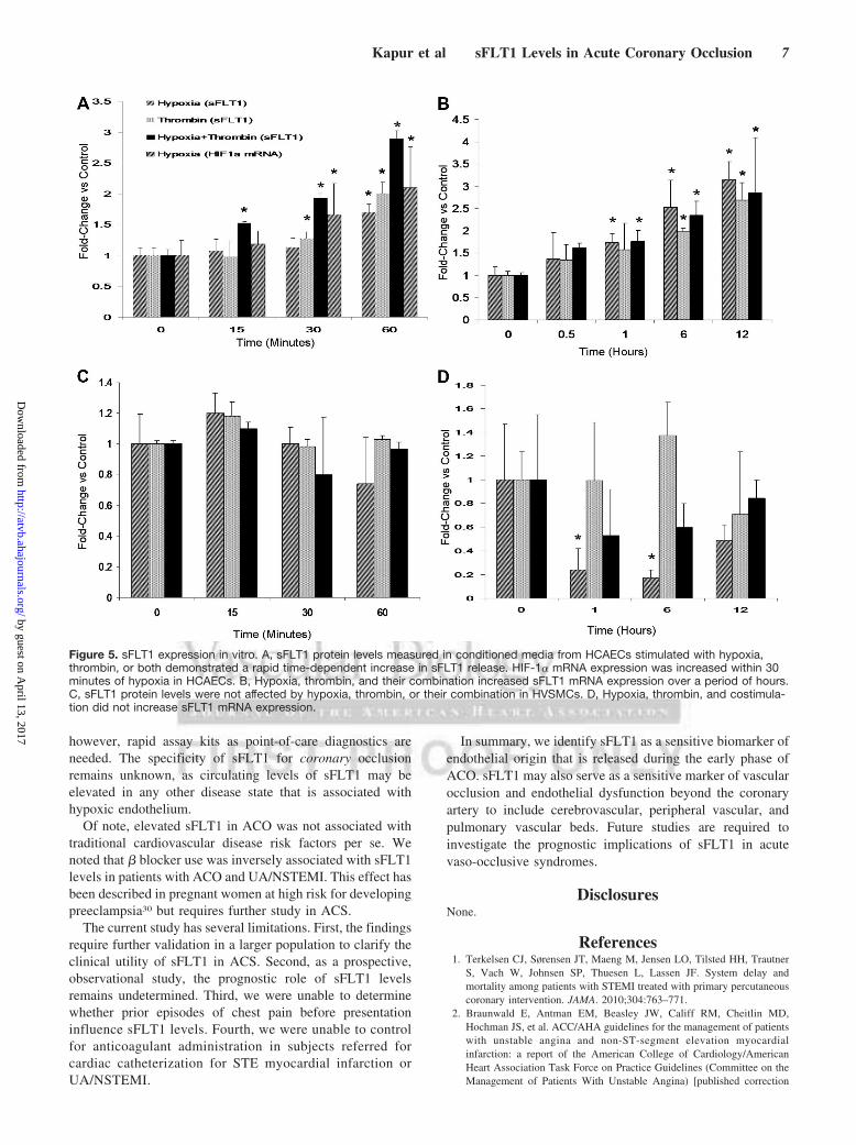
sFLT1 Expression in Coronary Endothelial CellsTo explore the timing of sFLT1 expression in vitro,hypoxic HCAECs and HVSMCs were independently stud-ied in the presence and absence of exogenous thrombin.
sFLT1 protein detected in the conditioned media of hy-poxic HCAECs by ELISA increased by 60 minutes ofexposure (ANOVA P�0.01). Thrombin alone increasedsFLT1 levels by 60 minutes in HCAECs; however, levelswere significantly increased within 15 minutes of costimu-lation with both hypoxia and thrombin (Figure 5A). As acontrol, HIF-1� mRNA levels increased in a time-dependent manner in hypoxic HCAECs (ANOVA �0.05)(Figure 5A).
In HCAECs, hypoxia induced a time-dependent increase insFLT1 mRNA expression within 30 minutes (ANOVAP�0.01) Thrombin and costimulation with both hypoxia andthrombin induced a time-dependent increase in sFLT1mRNA expression over hours (Figure 5B). No increase insFLT1 mRNA was observed in HVSMCs treated with hyp-oxia, thrombin, or their combination. No change in sFLT1protein release was observed with hypoxia in HVSMCs(Figure 5C). These data suggest that sFLT1 protein releasefrom HCAECs occurs within minutes in the presence of bothhypoxia and thrombin.
Figure 2. Clinical utility of sFLT1 in STE myocardial infarction. A, Biomarker levels grouped according to time of symptom onset. Foldchange above cutoff values in sFLT1 (�15 pg/mL) (I), CK-MB (�10 ng/mL) (II), and us-Tn-I levels (�0.05 ng/mL) (III) are shown. sFLT1levels were increased in patients presenting within 60 minutes of symptom onset compared with patients presenting �360 minutesafter symptom onset. B, Across all time points of minutes after chest pain onset, sFLT1 levels exhibited 100% sensitivity for diagnosingan ACO, with greater sensitivity than CK-MB at 60 and 60 to 120 minutes (P�0.01) and greater sensitivity than us-Tn-I at 60 minutes(P�0.01).
Kapur et al sFLT1 Levels in Acute Coronary Occlusion 5
by guest on April 13, 2017
http://atvb.ahajournals.org/D
ownloaded from

DiscussionOur findings indicate that ACO leads to rapid release ofsFLT1 into the systemic circulation. We have shown thatserum sFLT1 levels are increased in patients with ACOcompared with control subjects. During the early period aftersymptom onset, sFLT1 levels were elevated and more sensi-tive for the diagnosis of ACO than CK-MB or ultrasensitivetroponin I. Furthermore, we observed rapid release of sFLT1protein in a mouse model of ACO and by coronary endothe-lial cells exposed to hypoxia and thrombin. These datasuggest that sFLT1 is an early indicator of endothelial originfor ACO.
These data may have clinical implications that requirefurther study in a larger population of patients. First, theability to rapidly identify an ACO could improve clinicaloutcomes by allowing therapeutic interventions with lessdelay. Second, because sFLT1 originates from the endothe-lium, myocyte necrosis is not required for detection in theserum. As a result, sFLT1 may potentially be able todiscriminate between ACO and other causes of direct myo-
cyte injury such as myocarditis. The role of sFLT1 in thecontext of direct myocyte injury requires further study. Third,circulating sFLT1 retains its ability to bind vascular endothe-lial growth factor and placental growth factor, suggesting thatduring ACO, sFLT1 may attenuate the benefit of proangio-genic cytokines. Our data support the need for furtherinvestigation of sFLT1 in patients with ACS.
Early detection of ACO with a highly sensitive biomarkerin conjunction with the ECG has the potential to improveclinical outcomes by reducing treatment delay.2,23 Given therisk and cost associated with emergent coronary angiography,reducing the number of procedures performed for false-positive ECGs may be important. Finally, field diagnosticsfor patients with suspected ACO may be limited in underde-veloped regions where an ECG is unavailable. In these cases,the development of a rapid, diagnostic test with high sensi-tivity within minutes after symptom onset may affect clinicaloutcomes.
At present, CK-MB, troponins, and myoglobin exhibitexcellent sensitivity (above 90%) for the diagnosis of AMIwithin 24 hours of presentation.2 However, within the first 1hour of symptom onset, we observed lower sensitivity forbiomarkers of myocardial necrosis (myoglobin, 75%; CK-MB,20%; us-Tn-I, 20%). These levels are consistent with reportsshowing poor sensitivity for myocyte-based markers in theacute phase of coronary occlusion.24–29 Recently, ultrasensi-tive troponin assays have demonstrated improved sensitivityfor detecting an AMI.6 However, patients presenting withmyocardial injury from any cause may yield a positiveultrasensitive troponin test result, compromising the value ofthese tests for identifying patients presenting specifically withan ACO. Furthermore, despite advancements in troponinassay development, myocyte necrosis is still required forthese assays to be useful. As a result, an endothelium-derivedprotein such as sFLT1 may promote rapid diagnosis andearlier intervention and thus improve clinical outcomes. Torealize the potential benefits of sFLT1 in clinical practice,
Figure 3. STE myocardial infarctionbiomarker levels at 24 and 48 hours fol-lowing presentation. Serum sFLT1 levelsdeclined within 24 hours after revascular-ization and remained low at 48-hourfollow-up, whereas CK-MB and us-Tn-Ilevels were lower at presentationcompared with 24 hours afterrevascularization.
Figure 4. sFLT1 levels after left coronary ligation in vivo. SerumsFLT1 levels were increased within 30 minutes of left coronaryocclusion in mice.
6 Arterioscler Thromb Vasc Biol February 2011
by guest on April 13, 2017
http://atvb.ahajournals.org/D
ownloaded from
however, rapid assay kits as point-of-care diagnostics areneeded. The specificity of sFLT1 for coronary occlusionremains unknown, as circulating levels of sFLT1 may beelevated in any other disease state that is associated withhypoxic endothelium.
Of note, elevated sFLT1 in ACO was not associated withtraditional cardiovascular disease risk factors per se. Wenoted that � blocker use was inversely associated with sFLT1levels in patients with ACO and UA/NSTEMI. This effect hasbeen described in pregnant women at high risk for developingpreeclampsia30 but requires further study in ACS.
The current study has several limitations. First, the findingsrequire further validation in a larger population to clarify theclinical utility of sFLT1 in ACS. Second, as a prospective,observational study, the prognostic role of sFLT1 levelsremains undetermined. Third, we were unable to determinewhether prior episodes of chest pain before presentationinfluence sFLT1 levels. Fourth, we were unable to controlfor anticoagulant administration in subjects referred forcardiac catheterization for STE myocardial infarction orUA/NSTEMI.
In summary, we identify sFLT1 as a sensitive biomarker ofendothelial origin that is released during the early phase ofACO. sFLT1 may also serve as a sensitive marker of vascularocclusion and endothelial dysfunction beyond the coronaryartery to include cerebrovascular, peripheral vascular, andpulmonary vascular beds. Future studies are required toinvestigate the prognostic implications of sFLT1 in acutevaso-occlusive syndromes.
DisclosuresNone.
References1. Terkelsen CJ, Sørensen JT, Maeng M, Jensen LO, Tilsted HH, Trautner
S, Vach W, Johnsen SP, Thuesen L, Lassen JF. System delay andmortality among patients with STEMI treated with primary percutaneouscoronary intervention. JAMA. 2010;304:763–771.
2. Braunwald E, Antman EM, Beasley JW, Califf RM, Cheitlin MD,Hochman JS, et al. ACC/AHA guidelines for the management of patientswith unstable angina and non-ST-segment elevation myocardialinfarction: a report of the American College of Cardiology/AmericanHeart Association Task Force on Practice Guidelines (Committee on theManagement of Patients With Unstable Angina) [published correction
Figure 5. sFLT1 expression in vitro. A, sFLT1 protein levels measured in conditioned media from HCAECs stimulated with hypoxia,thrombin, or both demonstrated a rapid time-dependent increase in sFLT1 release. HIF-1� mRNA expression was increased within 30minutes of hypoxia in HCAECs. B, Hypoxia, thrombin, and their combination increased sFLT1 mRNA expression over a period of hours.C, sFLT1 protein levels were not affected by hypoxia, thrombin, or their combination in HVSMCs. D, Hypoxia, thrombin, and costimula-tion did not increase sFLT1 mRNA expression.
Kapur et al sFLT1 Levels in Acute Coronary Occlusion 7
by guest on April 13, 2017
http://atvb.ahajournals.org/D
ownloaded from

appears in J Am Coll Cardiol. 2001;38:294–295]. J Am Coll Cardiol.2000;36:970–1062.
3. Hand M, Brown C, Horan M, Simons-Morton D. The National HeartAttack Alert Program: progress at 5 years. J Thromb Thrombolysis.1998;6:19.
4. Aufderheide TP, Hendley GE, Woo J, Lawrence S, Valley V, TeichmanSL. A prospective evaluation of prehospital 12-lead ECG application inchest pain patients. J Electrocardiol. 1992;24:8–13.
5. American Heart Association. Heart and Stroke Disease Statistics Update2009.
6. Friess U, Stark M. Cardiac markers: a clear cause for point-of-caretesting. Anal Bioanal Chem. 2009;393:1453–1462.
7. Keller T, Zeller T, Peetz D, Tzikas S, Roth A, Czyz E, Bickel C, BaldusS, Warnholtz A, Frohlich M, Sinning CR, Eleftheriadis MS, Wild PS,Schnabel RB, Lubos E, Jachmann N, Genth-Zotz S, Post F, Nicaud V,Tiret L, Lackner KJ, Munzel TF, Blankenberg S. Sensitive troponin Iassay in early diagnosis of acute myocardial infarction. N Engl J Med.2009;361:868–877.
8. Maglione D, Guerriero V, Viglietto G, Delli-Bovi P, Persico MG. Iso-lation of human placenta cDNA coding for a protein related to thevascular permeability factor. Proc Natl Acad Sci. 1991;88:9267–9271.
9. Kendall RL, Wang G, DiSalvo J, Thomas KA. Specificity of vascularendothelial growth factor receptor ligand binding domains. BiochemBiophys Res Commun. 1994;201:326–330.
10. Matthews W, Jordan CT, Wiegand GW, Pardoll D, Lemischka IR. Areceptor tyrosine kinase specific to hematopoietic stem and progenitorcell-enriched populations. Cell. 1991;65:1143–1152.
11. Terman BL, Carrion ME, Kovacs E, Rasmussen BA, Eddy RL, ShowsTB. Identification of a new endothelial cell growth factor receptortyrosine kinase. Oncogene. 1991;6:1677–1683.
12. Ma L, Wang X, Zhang Z, Zhou X, Chen A, Yao L. Identification of theligand-binding domain of human vascular endothelial growth factorreceptor Flt-1. Biotechnol Appl Biochem. 2001;34:199–204.
13. Chung NA, Makin AJ, Lip GY. Measurement of the soluble angiopoietinreceptor tie-2 in patients with coronary artery disease: development andapplication of an immunoassay. Eur J Clin Invest. 2003;33:529–535.
14. Onoue K, Uemura S, Takeda Y, Somekawa S, Iwama H, Nishida T,Morikawa Y, Nakagawa H, Tsutsumi T, Sung JH, Takemoto Y, Soeda T,Okayama S, Ishigami K, Kawata H, Horii M, Nakajima T, Saito Y.Usefulness of soluble Fms-like tyrosine kinase-1 as a biomarker of acutesevere heart failure in patients with acute myocardial infarction. Am JCardiol. 2009;104:1478–1483.
15. Levine RJ, Maynard SE, Qian C, Lim KH, England LJ, Yu KF,Schisterman EF, Thadhani R, Sachs BP, Epstein FH, Sibai BM, SukhatmeVP, Karumanchi SA. Circulating angiogenic factors and the risk ofpreeclampsia. N Engl J Med. 2004;12:672–683.
16. Yano K, Liaw PC, Mullington JM, Shih SC, Okada H, Bodyak N, KangPM, Toltl L, Belikoff B, Buras J, Simms BT, Mizgerd JP, Carmeliet P,Karumanchi SA, Aird WC. Vascular endothelial growth factor is animportant determinant of sepsis morbidity and mortality. J Exp Med.2006;203:1447–1458.
17. Nagamatsu T, Fujii T, Kusumi M, Li Z, Yamashita T, Osuga Y,Momoeda M, Kozuma S, Taketani Y. Cytotrophoblasts up-regulatesoluble Fms-like tyrosine kinase-1 expression under reduced oxygen: animplication for the placental vascular development and the pathophys-iology of preeclampsia. Endocrinology. 2004;145:4838–4845.
18. Karumanchi SA, Bdolah Y. Hypoxia and sFlt-1 in preeclampsia: the“chicken-and-egg” question. Endocrinology. 2004;145:4835–4837.
19. Parenti A, Brogelli L, Filippi S, Donnini S, Ledda F. Effect of hypoxiaand endothelial loss on vascular smooth muscle cell responsiveness toVEGF-A: role of flt1/VEGF receptor-1. Cardiovasc Res. 2002;55:201–212.
20. Rahimi N, Golde TE, Meyer RD. Identification of ligand-induced pro-teolytic cleavage and ectodomain shedding of VEGFR-1/FLT1 inleukemic cancer cells. Cancer Res. 2009;69:2607–2614.
21. Patten RD, Aronovitz MJ, Deras-Mejia L, Pandian NG, Hanak GG, SmithJJ, Mendelsohn ME, Konstam MA. Ventricular remodeling in a mousemodel of myocardial infarction. Am J Physiol. 1998;274:H1812–H1820.
22. Imamura T, Kikuchi H, Herraiz MT, Park DY, Mizukami Y, Mino-Kenduson M, Lynch MP, Rueda BR, Benita Y, Xavier RJ, Chung DC.HIF-1� and HIF-2� have divergent roles in colon cancer. Int J Cancer.2009;124:763–771.
23. Brady WJ, Perron AD, Martin ML, Beagle C, Aufderheide TP. Cause ofST segment abnormality in ED chest pain patients. Am J Emerg Med.2001;19:25–28.
24. Pedersen SH, Galatius S, Hansen PR, Mogelvang R, Abildstrom SZ,Sørensen R, Davidsen U, Galloe A, Abildgaard U, Iversen A, Bech J,Madsen JK, Jensen JS. Field triage reduces treatment delay and improveslong-term clinical outcome in patients with acute ST-segment elevationmyocardial infarction treated with primary percutaneous coronary inter-vention. J Am Coll Cardiol. 2009;54:2296–2302.
25. Karras DJ, Kane DL. Serum markers in the emergency departmentdiagnosis of acute myocardial infarction. Emerg Med Clin North Am.2001;19:321–337.
26. Hamm CW, Goldmann BU, Heeschen C, Kreymann G, Berger J,Meinertz T. Emergency room triage of patients with acute chest pain bymeans of rapid testing for cardiac troponin T or troponin I. N Engl J Med.1997;337:1648–1653.
27. Pope JH, Selker HP. Diagnosis of acute cardiac ischemia. Emerg MedClin North Am. 2003;21:27–59.
28. Balk EM, Ioannidis JP, Salem D, Chew PW, Lau J. Accuracy ofbiomarkers to diagnose acute cardiac ischemia in the emergencydepartment: a meta-analysis. Ann Emerg Med. 2001;37:478–494.
29. Morrow DA, Cannon CP, Jesse RL, Newby LK, Ravkilde J, Storrow AB,Wu AH, Christenson RH. National Academy of Clinical BiochemistryLaboratory Medicine Practice Guidelines: clinical characteristics andutilization of biochemical markers in acute coronary syndromes.Circulation. 2007;115:e356–e375.
30. Carr DB, Tran LT, Brateng DA, Kawamura C, Shofer JB, KarumanchiSA, Easterling TR. Hemodynamically-directed atenolol therapy is asso-ciated with a blunted rise in maternal sFLT-1 levels during pregnancy.Hypertens Pregnancy. 2009;28:42–55.
8 Arterioscler Thromb Vasc Biol February 2011
by guest on April 13, 2017
http://atvb.ahajournals.org/D
ownloaded from
Kimmelstiel, Andrew Weintraub, Richard H. Karas and Michael E. MendelsohnParpos, Szuhuei Wilson, Corey K. Baker, Michele L. Esposito, Ameer Shah, Carey D.
Navin K. Kapur, Kevin S. Heffernan, Adil A. Yunis, Tuan A. Nguyen, Mark J. Aronovitz, PeterElevated Soluble fms-Like Tyrosine Kinase-1 Levels in Acute Coronary Occlusion
Print ISSN: 1079-5642. Online ISSN: 1524-4636 Copyright © 2010 American Heart Association, Inc. All rights reserved.
Greenville Avenue, Dallas, TX 75231is published by the American Heart Association, 7272Arteriosclerosis, Thrombosis, and Vascular Biology
published online November 11, 2010;Arterioscler Thromb Vasc Biol.
http://atvb.ahajournals.org/content/early/2010/11/11/ATVBAHA.110.215897.citationWorld Wide Web at:
The online version of this article, along with updated information and services, is located on the
http://atvb.ahajournals.org//subscriptions/
at: is onlineArteriosclerosis, Thrombosis, and Vascular Biology Information about subscribing to Subscriptions:
http://www.lww.com/reprints
Information about reprints can be found online at: Reprints:
document. Question and AnswerPermissions and Rightspage under Services. Further information about this process is available in the
which permission is being requested is located, click Request Permissions in the middle column of the WebCopyright Clearance Center, not the Editorial Office. Once the online version of the published article for
can be obtained via RightsLink, a service of theArteriosclerosis, Thrombosis, and Vascular Biologyin Requests for permissions to reproduce figures, tables, or portions of articles originally publishedPermissions:
by guest on April 13, 2017
http://atvb.ahajournals.org/D
ownloaded from