elevated level of carbon dioxide affects metabolism and … · elevated level of carbon dioxide...
TRANSCRIPT

MARINE ECOLOGY PROGRESS SERIESMar Ecol Prog Ser
Vol. 419: 95–108, 2010doi: 10.3354/meps08841
Published November 30
INTRODUCTION
Estuarine ecosystems are areas of high biologicaldiversity and productivity that are normally exposed toa high degree of natural and anthropogenic stressincluding pollution, fluctuations in temperature, salin-ity, dissolved oxygen (O2) and carbon dioxide (CO2)levels, and water pH. Among these stressors, environ-mental hypercapnia (elevated CO2 levels) and associ-ated acidification of the seawater play an important yetcurrently not well understood role. Although seawater
has substantial buffering capacity, variation in seawa-ter chemistry due to factors such as hypercapniacaused by biological activity, freshwater inputs, andrun-off from acidic soils leads to substantial shifts ofseawater pH in estuaries. In estuaries, pH can vary ona daily and seasonal basis from the values typical forthe open ocean (7.8 to 8.2) down to pH 6.0 to 7.0(Cochran & Burnett 1996, Hubertz & Cahoon 1999,Keppler & Ringwood 2002), and periods of low pH, typ-ically associated with elevated CO2 levels, may persistin some estuaries for a prolonged period of time, up to
© Inter-Research 2010 · www.int-res.com*Corresponding authors. Email: [email protected];[email protected]
Elevated level of carbon dioxide affects metabolismand shell formation in oysters Crassostrea virginica
Elia Beniash1,*, Anna Ivanina2, Nicholas S. Lieb1, Ilya Kurochkin2, Inna M. Sokolova2,*
1Department of Oral Biology, University of Pittsburgh, 589 Salk Hall, 3501 Terrace Street, Pittsburgh, Pennsylvania 15261, USA2Department of Biology, University of North Carolina at Charlotte, 9201 University City Blvd., Charlotte,
North Carolina 28223, USA
ABSTRACT: Estuarine organisms are exposed to periodic strong fluctuations in seawater pH drivenby biological carbon dioxide (CO2) production, which may in the future be further exacerbated by theocean acidification associated with the global rise in CO2. Calcium carbonate-producing marine spe-cies such as mollusks are expected to be vulnerable to acidification of estuarine waters, since ele-vated CO2 concentration and lower pH lead to a decrease in the degree of saturation of water withrespect to calcium carbonate, potentially affecting biomineralization. Our study demonstrates thatthe increase in CO2 partial pressure (pCO2) in seawater and associated decrease in pH within theenvironmentally relevant range for estuaries have negative effects on physiology, rates of shell depo-sition and mechanical properties of the shells of eastern oysters Crassostrea virginica (Gmelin). HighCO2 levels (pH ~7.5, pCO2 ~3500 µatm) caused significant increases in juvenile mortality ratesand inhibited both shell and soft-body growth compared to the control conditions (pH ~8.2, pCO2
~380 µatm). Furthermore, elevated CO2 concentrations resulted in higher standard metabolic rates inoyster juveniles, likely due to the higher energy cost of homeostasis. The high CO2 conditions also ledto changes in the ultrastructure and mechanical properties of shells, including increased thickness ofthe calcite laths within the hypostracum and reduced hardness and fracture toughness of the shells,indicating that elevated CO2 levels have negative effects on the biomineralization process. Thesedata strongly suggest that the rise in CO2 can impact physiology and biomineralization in marinecalcifiers such as eastern oysters, threatening their survival and potentially leading to profound eco-logical and economic impacts in estuarine ecosystems.
KEY WORDS: Hypercapnia · Ocean acidification · Calcification · Shell structure · Energy metabolism ·Oxygen consumption · Mollusks
Resale or republication not permitted without written consent of the publisher

Mar Ecol Prog Ser 419: 95–108, 2010
between 4 and 5 mo in summer and early fall (Cochran& Burnett 1996, Keppler & Ringwood 2002; see alsolong-term water pH data for eastern US estuaries athttp://cdmo.baruch.sc.edu/). Given that pH is mea-sured on a log scale, even relatively small changes inpH result in a considerable change in concentrations ofhydrogen and hydroxide ions, which may have impor-tant physiological consequences for the resident biota.
The effects of seasonal and diurnal hypercapniaexperienced by estuarine organisms are likely tobecome exacerbated in the coming years due to globalclimate change and ocean acidification driven byanthropogenically released CO2 (Caldeira & Wickett2003, 2005). The uptake of atmospheric CO2 is espe-cially pronounced in surface ocean waters (<250 mdepth), where nearly 50% of anthropogenic CO2 isabsorbed, making near-shore habitats including estu-aries vulnerable to ocean acidification (Feely et al.2008, Doney et al. 2009). While photosynthetic auto-trophs may benefit from mildly elevated CO2 levels,the combination of higher CO2 concentrations andlower pH in estuaries may have adverse effects onother marine organisms, ranging from shifts in acid-base homeostasis and changes in metabolic functionsand energy balance, to negative impacts on biominer-alization rates (Pörtner et al. 2004, 2005, Doney et al.2009). Marine calcifiers (organisms with calcium car-bonate, CaCO3, skeletons) are expected to be sensitiveto CO2-driven changes in ocean chemistry, such asdecrease in pH and the associated decline in CaCO3
saturation of seawater, although the direction andmagnitude of the CO2-driven effects on calcificationrates can significantly vary between species (Feely etal. 2004, Iglesias-Rodriguez et al. 2008, Clark et al.2009, Ries et al. 2009). However, the complex effects ofelevated CO2 on the physiology of these organisms arestill poorly understood, hampering our ability to accu-rately predict the consequences of elevated CO2 onestuarine ecosystems.
Mollusks (Mollusca), the major group of marine cal-cifiers in estuarine and coastal waters, accumulatelarge amounts of carbonate in their shells. The shellsare produced by specialized epithelial cells of the man-tle with the assistance of CaCO3-transporting hemo-cytes (blood cells) (Wheller 1992, Mount et al. 2004).Bivalve shells consist of 3 major layers: the outermostproteinaceous layer called periostracum, and 2 miner-alized layers called ostracum (middle layer) and hypos-tracum (inner layer), composed primarily of CaCO3
crystals (Wheller 1992). Shells protect mollusks frompredators and parasites, and their mechanical proper-ties such as fracture toughness are far superior to thoseof geological CaCO3 due to their unique structuralorganization and composition (Smith et al. 1999,Kamat et al. 2000). Shells also play a systemic role in
the metabolism of mollusks, participating in the cap-ture and deposition of respiratory CO2 in the shell min-eral (Wilbur 1964, Wheller 1992) and in buffering ofextracellular pH during environmental anaerobiosis(Crenshaw 1972, Sokolova et al. 2000b). An increase inthe CO2 concentration in seawater and the associatedreduction in the degree of saturation with respect toaragonite and calcite can impair shell deposition andincrease shell dissolution rates, weakening the shellsand affecting their functional properties in bivalves(Andersson et al. 2005, Orr et al. 2005, Gazeau et al.2007, Kurihara et al. 2007, Kurihara 2008, Doney et al.2009, Ries et al. 2009). Moreover, the energy costs ofbiomineralization may contribute to the basal meta-bolic costs of marine calcifiers, especially when CaCO3
is lost due to erosion in acidic seawater (Palmer 1992,Day et al. 2000, Wood et al. 2008). Thus, CO2-drivenacidification of seawater may also have indirect nega-tive impacts on mollusks by increasing basal metaboliccosts and diverting energy from other processes suchas growth or reproduction. Yet, the effects of hyper-capnia and associated acidification of seawater onshell formation and properties as well as on basalmetabolism in mollusks have not been extensivelystudied, and more research is critically needed tounderstand molluscan physiology and calcificationunder high CO2 conditions.
Eastern oysters Crassostrea virginica are commonmollusks and major reef builders/ecosystem engineersin estuaries of the western Atlantic. They deposit largeamounts of CaCO3 in their massive shells (nearly 90 to1000 times more per year and unit area than most othermollusks) (Gutierrez et al. 2003). The shells of juvenileand adult C. virginica are made predominantly of low-magnesium (Mg) calcite (Carter et al. 1998, Mount etal. 2004, Checa et al. 2007b, Esteban-Delgado et al.2008). The shells are composed of an outer prismaticand an inner foliated layer. The foliated layer hasa plywood-like structure which consists of roughly200 nm thick mineral sheets called folia or laths, sepa-rated by an organic matrix (Sikes et al. 1998, Lee &Choi 2007). Laths are composed of single crystals ofcalcite, with their c-axes tilted 20 to 30° with respect tothe shell surface normal, in the direction opposite tothe direction of the shell growth, and with one of thefaces forming the growing edges of laths (Checa et al.2007a).
The relative simplicity of shell mineralogy of oysterssimplifies interpretations of elevated CO2 levels onbiomineralization and shell properties. Moreover, gen-eral metabolic physiology and biochemistry of oystersis well understood compared to other marine bivalves(Shumway 1982, Kennedy et al. 1996, Sokolova 2004,Kurochkin et al. 2009 and references therein), makingthem an attractive model for studies of physiological
96
Beniash et al.: Effects of hypercapnia on oysters
and metabolic effects of elevated CO2. The goals of thepresent study were to determine the effects of elevatedCO2 levels on biomineralization and metabolic physi-ology of juvenile and adult Crassostrea virginica. Weassessed growth, mineralogy, and structural organiza-tion in the shells in juvenile mollusks exposed tohypercapnic conditions; studied the effects of hyper-capnia on basal metabolism and cellular energy statusof juvenile and adult oysters; and compared the ex-pression of 3 genes involved in calcification, i.e. car-bonic anhydrase (CAn), voltage-dependent Ca2+ chan-nel, and H+ ATPase (Carre et al. 2006), under normo-and hypercapnic conditions in adult C. virginica. Thedata presented here contribute to a better understand-ing of the potential impacts of environmental hyper-capnia (such as is currently observed in many estuariesof the southeastern USA) as well as ocean acidificationin estuaries on this key group of marine calcifiers.
MATERIALS AND METHODS
Animal maintenance and experimental exposures.Juvenile (3 wk post-metamorphosis, <1 mm shelllength) and adult Crassostrea virginica (age >2 yr, 8 to12 cm shell length) were obtained from commercialoyster suppliers (J & B Aquafood and Taylor ShellfishFarms for juveniles and adults, respectively). The oys-ters were acclimated for 5 to 7 d at 20°C and a salinityof 30 in re-circulating water tanks with artificial sea-water (ASW; Instant Ocean®, Kent Marine) prior toexperimentation.
For hypercapnic treatments, CO2-enriched air wasvigorously bubbled through the seawater using certi-fied gas mixtures containing 0.5% CO2, 21% O2, andbalance nitrogen (Roberts Oxygen). Gas content of themixtures was analyzed by the manufacturer and certi-fied to be accurate within 10% of the target value(Roberts Oxygen). The control (normocapnic) treat-ments were bubbled with ambient air. In both cases,gas flow through the seawater was adjusted in such away that further increases in flow rate did not result ina pH change, indicating that experimental systemswere at steady-state with respect to gas saturation.The steady-state pCO2 levels achieved in these treat-ments were ~380 and ~3500 µatm for normocapnic andhypercapnic exposures, respectively (see Table 1 and‘Seawater parameters’ below for more details).
Oysters were divided into 8 different batches(~50 juveniles or 5 adults batch–1) and randomlyassigned to either hypercapnic or normocapnic treat-ment. For each treatment, 4 replicate tanks (5 l) wereset up, 2 of them containing 50 juveniles each, and 2with 5 adults each. Water temperature was maintainedat 21 ± 1°C and salinity at 30 ± 0.5 in all tanks. Water
was changed every other day. The experimental incu-bations were 20 wk for juveniles and 2 wk for adults.During the preliminary acclimation and experimentalincubations, oysters were fed ad libitum every otherday with a commercial algal blend (5 ml tank–1) con-taining Nannochloropsis oculata, Phaeodactylum tri-cornutum, and Chlorella sp. with a cell size of 2 to 20µm (DT’s Live Marine Phytoplankton). The feedingregime used higher algal concentrations than recom-mended by the manufacturer (1 ml l–1 vs. 0.4 ml l–1
every other day recommended by DT’s Live MarinePhytoplankton); however, in our experience this higherfeeding regime provides better results for long-termmaintenance of good physiological condition of oysters(I. M. Sokolova unpubl. data). Algae were added to thetanks immediately following the water change.
In adults, no mortality was detected throughout theexperiment. Due to their small size, juveniles’ mortalitycould be determined only under a dissecting micro-scope. To minimize handling disturbance, mortalitywas assessed at the beginning of the experiment (toensure that all animals were alive) and when theexperiment was concluded.
Seawater parameters. Water pH was measured dailyusing a pH electrode (pH meter Model 1671, JencoInstruments) calibrated with National Institute of Stan-dards and Technology (NIST) standard pH solutions(NBS standards). Due to the fact that longer equilibra-tion times may be needed to stabilize the liquid junc-tion potential of the electrode in high ionic-strengthmedia such as seawater, the electrode was incubatedfor 1 h in seawater before the measurements. SeawaterpH was stable throughout the experimental exposures,did not differ between the replicate tanks within nor-mocapnic or hypercapnic exposures, and was 8.2 ± 0.1and 7.5 ± 0.1 (mean ± SD) in normocapnia and hyper-capnia conditions, respectively (n = 231, repeated-measures ANOVA: p < 0.05). These pH values arewithin the normal range currently found in estuaries,e.g. 7.4 to 8.2 for typical high-salinity sites (salinity 29to 35) (Burnett 1997, Hubertz & Cahoon 1999, Keppler& Ringwood 2002). O2 levels in experimental tankswere periodically tested during the exposures usingClark-type oxygen probes (YSI 5331 Oxygen probe)connected to a YSI 5300A Biological Oxygen monitor,and ranged between 97 and 100% of air saturationthroughout all exposures.
For water chemistry analysis, seawater samples werecollected at the beginning, in the middle, and at theend of the experimental exposures in air-tight 50 mlcontainers without air space to eliminate potential gasexchange, stabilized by mercuric chloride poisoning(Dickson et al. 2007) and immediately shipped to theChesapeake Biological Laboratory (Solomons, MD) foranalysis. Samples were kept in the dark at +4°C during
97

Mar Ecol Prog Ser 419: 95–108, 2010
shipping and storage, and analyzed within a week ofcollection. Total dissolved inorganic carbon (DIC) con-centrations were measured by Nutrient AnalyticalServices (Chesapeake Biological Laboratory) using aShimadzu TOC5000 gas analyzer equipped with anondispersive infrared sensor (NDIR) detector for CO2
determination (Shimadzu Scientific Instruments). Sam-ples were measured immediately after opening to min-imize gas exchange. Three to 5 replicates were run foreach sample, and precision of the analysis was 1% orbetter for technical replicates from the same sample.Temperature, salinity, and pH were measured for eachsample at the time of collection, and along with thetotal DIC levels were used to calculate pCO2, alkalin-ity, and the average degree of saturation (Ω) for calciteand aragonite in seawater using co2sys software (Pier-rot et al. 2006). For co2sys settings, we used the NBSscale of seawater pH, constants from Millero et al.(2006), and the KSO4 constant from Dickson (1990) (asimplemented in Pierrot et al. 2006), and concentrationsof silicate and phosphate for Instant Ocean® seawater(0.17 and 0.04 µmol kg–1, respectively, at a salinity of30). Water chemistry data for this subset of samples(n = 7 to 14) are given in Table 1; temperature, salinity,and pH of this subset did not significantly differ fromthe remainder of the experimental seawater samplesfor which only temperature, salinity, and pH weremeasured (ANOVA: p > 0.05).
Whole-organism O2 consumption rates. O2 con-sumption rates (MO2) were measured after 20 wk and2 wk of normo- and hypercapnic exposure in juvenileand adult Crassostrea virginica, respectively. In ju-veniles, MO2 was determined by closed-system re-spirometry in 3 ml water-jacketed chambers using
Clarke-type oxygen electrodes (Qubit Systems) at20°C in ASW at a salinity of 30 and the same CO2 con-centration as during experimental exposures. For eachmeasurement, several similarly sized juveniles (2 to 8,based on the individual size) were placed into thechamber and allowed to recover from the handlingstress for at least 30 min. The total wet mass of thebatch was 17 to 90 mg, and the juveniles were selectedin such a way that individual masses within each batchdid not vary by more than 15%. The chambers werethen closed and O2 consumption of the juveniles wasmonitored for 20 to 30 min. Decline in O2 concentrationin the respirometry chambers was linear over thisperiod, and O2 levels were never <85% of air satura-tion. Respiration of each batch of juveniles was mea-sured twice, with a brief (10 to 15 min) recovery periodin the open chamber between measurements, and datafrom the 2 replicates were averaged. Total wet bodymass of juveniles was determined, and wet tissue masswas calculated from the total wet mass by subtractingthe relative weight of the shell determined in the pre-sent study (0.38 and 0.22% of the total wet mass injuveniles exposed to normocapnia or hypercapnia,respectively, based on ‘Shell and soft-tissue mass mea-surements’ described below). Throughout this paper,juveniles exposed to normocapnia or hypercapnia arereferred to as normocapnic or hypercapnic juveniles,respectively. After the MO2 determinations, all juve-nile oysters were collected and stored in 70% ethanoluntil further analysis of body size and shell character-istics.
O2 consumption in adults was measured usingClark-type oxygen probes (YSI 5331 Oxygen probe)connected to a YSI 5300A Biological Oxygen monitor.
Oysters were placed into a flow-through respiration chamber andallowed to acclimate overnight. Waterflow (20 ml min–1) was adjusted so thatanimals consumed <25% of O2 at alltimes. O2 consumption was continu-ously monitored for 2 to 10 h, usingAcqKnowledge software (Biopac Sys-tems) at 20°C in ASW at a salinity of 30and the same CO2 concentration asduring experimental exposures. Ourpilot studies have shown that short-term exposure to reduced O2 and ele-vated CO2 in closed or flow-throughrespirometry chambers has no effectson metabolic rates of oysters providedthat O2 levels do not decline to <75%of air saturation (data not shown). Thisis typical for most bivalves, includingoysters (Shumway 1982, Le Moullac etal. 2007). After measurements, adult
98
Control High CO2 exposure(environmental (environmental
normocapnia; n = 14) hypercapnia; n = 7)
Salinity 30.1 ± 0.2 30.0 ± 0.1 (ns)Temperature (°C) 20.0 ± 0.1 20.0 ± 0.1 (ns)pH (NBS scale) 8.3 ± 0.1 7.5 ± 0.0**pCO2 (µatm) 385.4 ± 22.4 3523.3 ± 222.0***Total alkalinity (mmol kg–1 SW) 3320.1 ± 454.0 3341.8 ± 242.9 (ns)DIC (mmol kg–1 SW) 2899.4 ± 364.9 3384.8 ± 245.7**[CO3
2–] (mmol kg–1 SW) 337.9 ± 79.6 57.4 ± 4.63***Ω calcite 8.4 ± 2.0 1.42 ± 0.1***Ω aragonite 5.4 ± 1.3 0.9 ± 0.1***
Table 1. Water chemistry parameters during the experimental exposures.Salinity, temperature, pH, and dissolved inorganic carbon (DIC) were de-termined in samples from experimental tanks at the beginning, middle, and endof experimental exposures. Other parameters were calculated using co2sys soft-ware ver. 1.05 (available at http://cdiac.ornl.gov/oceans/ co2rprt.html; Pierrot etal. 2006). Data are means ± SD. Differences between normocapnic and hyper-capnic conditions were tested using generalized linear model (GLM) ANOVA.ns: not significant (p > 0.05), **p < 0.01, ***p < 0.001. NBS: National Bureau of
Standards. SW: seawater. Ω: degree of saturation

Beniash et al.: Effects of hypercapnia on oysters
oysters were dissected and soft-tissue mass deter-mined. Gill and mantle tissues were immediatelyshock-frozen in liquid nitrogen for further determina-tion of mRNA expression and measurements of tissuemetabolite concentration.
Respiration rates were corrected for the electrodedrift, normalized to the average wet tissue mass ofexperimental oysters (1 mg and 10 g for juveniles andadults, respectively) as described elsewhere (Lannig etal. 2006) and expressed as µmol O2 h–1 g–1 wet tissuemass. Calibration of oxygen electrodes, data acquisi-tion, and MO2 calculations were performed as de-scribed in Sokolova (2004). O2 solubility of the seawa-ter was 1.54 µmol O2 l–1 Torr–1 at 20°C and a salinity of30 (Dejours 1975).
Shell and soft-tissue mass measurements. Eachjuvenile mollusk was given a unique code and placedin an individual container, flash-frozen, lyophilized,and stored in a desiccator at –80°C. The lyophilizedmollusks were weighed individually using a microbal-ance XP 56 (Metler-Toledo) with a precision of 0.01 mgor better to determine their total dry mass (Mt). Todetermine the mass of the soft tissues, out of these lio-phylized and weighted mollusks 10 individuals withknown Mt (5 tank–1) were randomly selected from eachtreatment group. They were individually incubated inaqueous 2% Na hypochlorite (NaOCl) for 2 min toremove the soft tissues. Individual shells were brieflyrinsed in large volumes of distilled deionized water(DDW), followed by a rinse in alcohol, freeze-dried,and weighed again to determine the mass of the shells(Msh). The shell mass was subtracted from the Mt of thesame individual to determine the dry mass of soft tis-sues (Mst). Wet-tissue mass was calculated from thedry-tissue mass assuming 80% body water contentdetermined in our earlier studies on Crassostreavirginica (data not shown).
To determine the projected shell area, juvenile oys-ters were placed in a Petri dish and their right (upper)shell valves were photographed under a dissectingmicroscope. The projected areas were calculated usingthe ImageJ 1.64 image processing package andexpressed in mm2.
Shell mineral characterization using Fourier trans-form infrared (FTIR) spectroscopy. Ten individualjuveniles (5 from each replicate tank) were collectedfrom normocapnic and hypercapnic conditions after20 wk of incubation. The soft tissue was removed asdescribed above (‘Shell and soft-tissue mass measure-ments’) using NaOCl. For FTIR spectroscopy we usedtotal pulverized shells; however, because the size ofthe initial shells at the beginning of the experimentswas very small (<1 mm), the fraction of the old shell inthe total shell volume is minuscule. The shells werelyophilized, ground, and studied in the transmittance
mode in KBr pellets using a Bruker Vertex 70 FTIRspectrometer. The 600–1000 cm–1 regions of the spec-tra were isolated, baseline-corrected, and normalized,and the ν2 and ν4 peak positions and heights weremeasured using the Spectrum 5.1 software package(Perkin-Elmer). The relative crystallinity of the shellswas determined using the ν2:ν4 band intensity ratio(Beniash et al. 1997, Gueta et al. 2007, Chu et al. 2008).
Scanning electron microscopy (SEM) of shell struc-ture. For SEM and micromechanical experiments (see‘Micromechanical tests of the shells’ below), we usedonly the new portion of the juvenile shells grown dur-ing the laboratory exposures. The area of the new shellgrowth was clearly identifiable in juveniles due to thefact that the old shell grown in the field was darker incolor and had a rougher surface than the new shellgrown in the laboratory. The shells were prepared forSEM analysis and microhardness testing as describedelsewhere (Bartlett et al. 2004, 2005, Baldassarri et al.2008). Briefly, the lyophilized right valves of the shellsof oyster juveniles were mounted and polymerized inepoxy resin (Epofix, ESM) at room temperature. Theembedded shells were cut in the plane normal to thesurface, which transects the shell from the acute apicaltip to its most distal edge, using a slow-speed water-cooled diamond saw (IsoMet, Buehler). The sampleswere then polished with Metadi diamond suspensions(Buehler), with the final diamond particle size of0.25 µm in DDW equilibrated with aragonite. Foursamples per group were carbon-coated and analyzedusing a Jeol 6330 SEM in the back-scattered electronmode (BSE SEM) as described elsewhere (Bartlett etal. 2004, 2005, Baldassarri et al. 2008). At least 4 micro-graphs were taken for each sample at a magnificationof 15 000×, accelerating voltage set at 10 000 kV and5 to 7 mm working distance. At this magnification, thefield of view covered an area of 8 × 6 µm. For eachmicrograph, the longest distance in the direction per-pendicular to the direction of the laths was determinedand divided by the number of laths in the field of view.The average lath thickness values in each sample wascalculated based on data from the individual micro-graphs.
Micromechanical tests of the shells. The samples formicrohardness tests were prepared the same way asfor BSE SEM. Microhardness was determined using astandard Vickers hardness test. The samples weretested dry using a Leco microindenter equipped with aVickers diamond indenter at 0.49 N load and 5 sdwelling time, and 10 to 15 indentations were acquiredper sample. Values were averaged for each shell andexpressed as Vickers hardness numbers. Three sam-ples from each treatment group were tested. The frac-ture toughness (Kc) was determined using Anstis’equation as previously described (Anstis et al. 1981,
99

Mar Ecol Prog Ser 419: 95–108, 2010
Baldassarri et al. 2008), with a Young’s modulus valueof 73 GPa based on Lee et al. (2008). Indentations from8 control shells and 10 hypercapnic shells were ana-lyzed.
Expression of transcripts of biomineralization-related genes. Gene expression was determined in thedistal edge of the mantle (along the edge of the normalshell growth) and gill tissue of adult Crassostrea vir-ginica incubated for 2 wk in hypercapnic or normocap-nic seawater. Total RNA was extracted from the gilland mantle tissues using TRI Reagent (Sigma) accord-ing to the manufacturer’s protocol. cDNA was obtainedfrom 1 to 5 µg total RNA using SuperScriptTM IIIreverse transcriptase (Invitrogen) according to themanufacturer’s instruction. Expression of mRNA ofCA, voltage-dependent Ca2+ channel, and H+ ATPaseV-type subunit was measured by quantitative real-time PCR using a LightCycler® 2.0 Real Time PCR Sys-tem (Roche Applied Science) and the QuantiTect SYBRGreen PCR kit (Qiagen) according to the manufactur-ers’ instructions, and normalized against the expres-sion for β-actin and an internal cDNA standard asdescribed elsewhere (Pfaffl 2001, Sanni et al. 2008)(Table 2). A cDNA sample obtained from the gill tissueof a control adult oyster was used as internal standardin all analyses. Mantle is involved in shell formation inoysters; gills are not involved in biomineralization andwere used as a reference tissue (Wheeler 1992).
Determination of metabolites. For the determinationof tissue metabolite concentration, gills were quicklyexcised from adult oysters exposed for 2 wk to hyper-capnic or control conditions and immediately frozen inliquid nitrogen. Deproteinized tissue extracts wereobtained using 0.6 mol l–1 perchloric acid (PCA) with150 mmol l–1 EDTA as described elsewhere (Sokolovaet al. 2000a) and stored at –80°C. Concentrations ofATP, ADP, AMP, L-alanine, and acetate were mea-
sured spectrophotometrically using standard enzy-matic tests (Bergmeyer 1985).
Statistical analysis. Differences in the means forshell parameters, MO2, tissue metabolite levels, andlevels of mRNA expression were tested usingrepeated-measures ANOVA with CO2 concentrationsand tissue type (for mRNA expression) or CO2 concen-trations only (for all other traits) as fixed factors, repli-cate exposure tanks as a random factor (block), andindividual oyster samples as a repeated-measures vari-able within the block. Fisher’s least significant differ-ence (LSD) test was used for post hoc comparisons.Mortality of juveniles under different CO2 conditionswas compared using a chi-square test. There were nosignificant differences in any of the studied parametersbetween the replicate tanks within each experimentalcondition. Statistical analyses were performed usingSAS 8.2 software (SAS Institute). Differences wereconsidered significant at p < 0.05 and marginally sig-nificant at 0.10 < p ≤ 0.05. Data are presented as mean± standard error of the mean unless otherwise indi-cated. For factor effects and their interactions inANOVA, F-values with the corresponding degrees offreedom for the factor and error effects, as well as theprobabilities of Type I error (p) are given. For post hocpairwise comparisons, only p-values are presented.
RESULTS
Effects of elevated CO2 on mortality and shellcharacteristics of juvenile oysters
Hypercapnic exposure resulted in a significant in-crease of mortality in juvenile oysters (77% vs. 46% inhypercapnia and normocapnia, respectively; n = 89 to98, χ2 = 18.4, p < 0.0001). Average dry shell mass was
100
Gene Primer names Primer sequence (5’ to 3’) Amplification efficiency (E)
Carbonic anhydrase FW: CarbAnh-F23 AGA GGA ACA CCG TAT CGG AGC CA 1.65 ± 0.01RV: CarbAnh-R155 ATG TCA ATG GGC GAC TGC CG (n = 4)
Voltage-dependent FW: Cvir_CaChannel FW274 TGC TGC TGA CAA ACT GAA CCA GTG 1.99 ± 0.13Ca2+ channel RV: Cvir_CaChannel RV440 TGG GGG AAG GCT GGA GTT TG (n = 4)
H+ ATPase V-type FW: Cvir_VacH+ATPase FW302 GGA CTG GTC GTA GCT GCT GTC ATC 2.13 ± 0.14subunit RV: Cvir_VacH+ATPase RV471 GGT TGT TGT GCC GTT CCA CG (n = 4)
β-actin FW: Act-Cv-F437 CAC AGC CGC TTC CTC ATC CTC C 1.83 ± 0.06RV: Act-Cv-R571 CCG GCG GAT TCC ATA CCA AGG (n = 4)
Table 2. Primer sequences used for quantitative PCR. Gene sequences for the target genes for Crassostrea virginica were ob-tained from the Marine Genomics database (www.marinegenomics.org, sequence accession nos. MGID94539, MGID95078, andMGID93334 for carbonic anhydrase, voltage-dependent Ca2+ channel, and H+ ATPase V-type subunit, respectively), and thesequence for β-actin was obtained from GenBank (accession no. X75894.1). Annealing temperature used in PCR) (Tann) was 55°C
for all genes. PCR amplification efficiencies (E) were determined as described by Pfaffl (2001). FW: forward, RV: reverse
Beniash et al.: Effects of hypercapnia on oysters
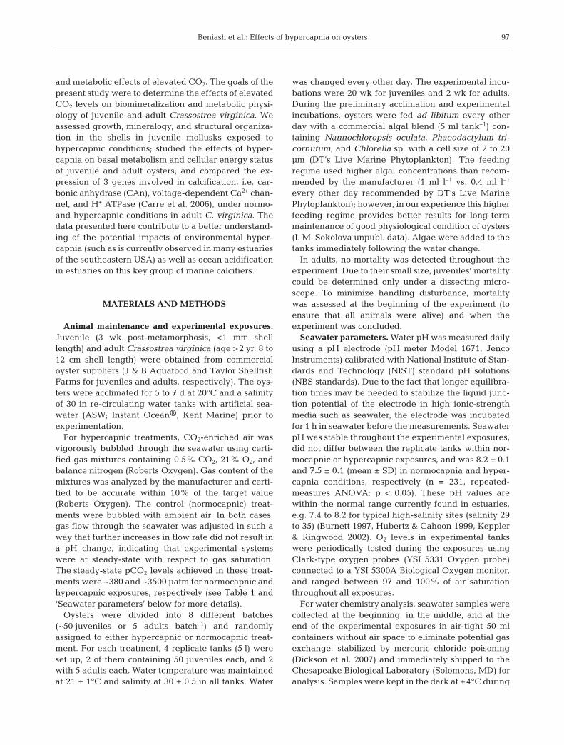
39% lower in hypercapnic juveniles compared to theirnormocapnic counterparts (5.03 ± 0.43 mg vs. 8.28 ±0.69 mg; ANOVA: p = 0.003, n = 10; Fig. 1A). The aver-age soft-tissue mass was also lower in juveniles exposedto hypercapnia (0.04 mg) compared to controls (0.16 mg)(ANOVA: p = 0.04, n = 10; Fig. 1B). In contrast, shell areadid not differ between juveniles exposed to normocapniaand hypercapnia (10.6 ± 0.6 and 9.2 ± 0.5 mm2, n = 46and 22, respectively; ANOVA: p = 0.087)
SEM ultrastructural analysis revealed that calciticlaths of the shells of juveniles from hypercapnic treat-ments were on average 227 nm thick, compared to191 nm in the control shells (ANOVA: p = 0.01, n = 4;Fig. 1C). No other differences in shell structure wereobserved.
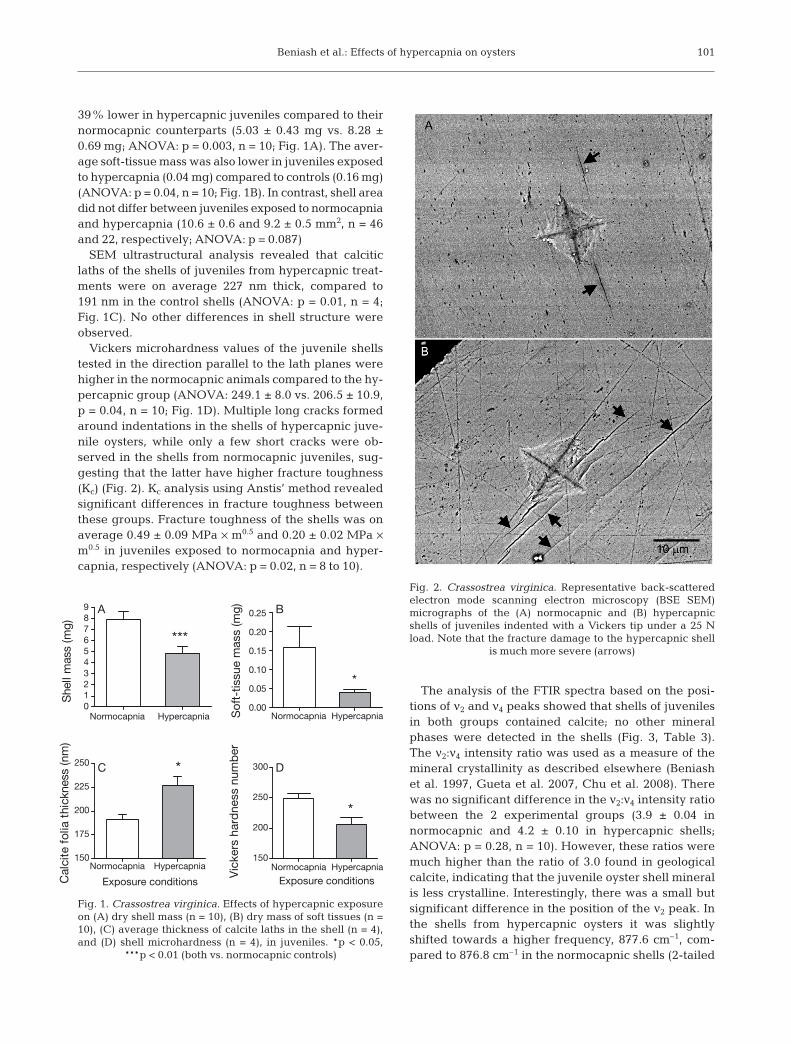
Vickers microhardness values of the juvenile shellstested in the direction parallel to the lath planes werehigher in the normocapnic animals compared to the hy-percapnic group (ANOVA: 249.1 ± 8.0 vs. 206.5 ± 10.9,p = 0.04, n = 10; Fig. 1D). Multiple long cracks formedaround indentations in the shells of hypercapnic juve-nile oysters, while only a few short cracks were ob-served in the shells from normocapnic juveniles, sug-gesting that the latter have higher fracture toughness(Kc) (Fig. 2). Kc analysis using Anstis’ method revealedsignificant differences in fracture toughness betweenthese groups. Fracture toughness of the shells was onaverage 0.49 ± 0.09 MPa × m0.5 and 0.20 ± 0.02 MPa ×m0.5 in juveniles exposed to normocapnia and hyper-capnia, respectively (ANOVA: p = 0.02, n = 8 to 10).
The analysis of the FTIR spectra based on the posi-tions of ν2 and ν4 peaks showed that shells of juvenilesin both groups contained calcite; no other mineralphases were detected in the shells (Fig. 3, Table 3).The ν2:ν4 intensity ratio was used as a measure of themineral crystallinity as described elsewhere (Beniashet al. 1997, Gueta et al. 2007, Chu et al. 2008). Therewas no significant difference in the ν2:ν4 intensity ratiobetween the 2 experimental groups (3.9 ± 0.04 innormocapnic and 4.2 ± 0.10 in hypercapnic shells;ANOVA: p = 0.28, n = 10). However, these ratios weremuch higher than the ratio of 3.0 found in geologicalcalcite, indicating that the juvenile oyster shell mineralis less crystalline. Interestingly, there was a small butsignificant difference in the position of the ν2 peak. Inthe shells from hypercapnic oysters it was slightlyshifted towards a higher frequency, 877.6 cm–1, com-pared to 876.8 cm–1 in the normocapnic shells (2-tailed
101
Normocapnia Hypercapnia
Normocapnia Hypercapnia
0123456789
***
Shell
mass
(m
g)
Normocapnia Hypercapnia
Normocapnia Hypercapnia
0.00
0.05
0.10
0.15
0.20
0.25
*
So
ft-t
issue m
ass (m
g)
150
175
200
225
250 *
Exposure conditionsCalc
ite f
olia
thic
kness
(nm
)
150
200
250
300
*
Exposure conditions
Vic
kers
hard
ness n
um
ber
A B
C D
Fig. 1. Crassostrea virginica. Effects of hypercapnic exposureon (A) dry shell mass (n = 10), (B) dry mass of soft tissues (n =10), (C) average thickness of calcite laths in the shell (n = 4),and (D) shell microhardness (n = 4), in juveniles. *p < 0.05,
***p < 0.01 (both vs. normocapnic controls)
Fig. 2. Crassostrea virginica. Representative back-scatteredelectron mode scanning electron microscopy (BSE SEM)micrographs of the (A) normocapnic and (B) hypercapnicshells of juveniles indented with a Vickers tip under a 25 Nload. Note that the fracture damage to the hypercapnic shell
is much more severe (arrows)

Mar Ecol Prog Ser 419: 95–108, 2010
t-test: p < 0.0001, n = 10). This shift may indicate smalldifferences in the structural organization of calcitecrystals between normocapnic and hypercapnic shells.
Effects of elevated CO2 on respiration rates andtissue energy status of juvenile and adult oysters
In juveniles, hypercapnia induced a doubling of thestandard metabolic rate (SMR) (ANOVA: p = 0.003, n = 5to 12), whereas in adults there was a smaller non-significant increase by approximately 15% (Fig. 4). Nodifference in the levels of anaerobic end-products
102
Fig. 3. Crassostrea virginica. Representative absorbance Fouriertransform infrared (FTIR) spectra of geological calcite (toppanel), and oyster shell calcite from a normocapnic juvenile oys-ter shell (middle panel) and a hypercapnic juvenile oyster shell(lower panel). Reference positions of ν2 and ν4 peaks in different
calcium carbonate polymorphs are given in Table 3
Mineral ν4 position ν2 position phase (cm–1) (cm–1)
Calcite 875–876 713Aragonite 857 Split peaks at 713 and 700Amorphous 866 No peakcalcium carbonate
Table 3. Positions of ν2 and ν4 peaks of major calcium carbon-ate polymorphs (after Chester & Elderfield 1967, Beniash etal. 1997, Vagenas et al. 2003, Gueta et al. 2007). The refer-ence positions of the ν2 and ν4 peaks on the Fourier transforminfrared (FTIR) spectra were used for identification of thecalcium carbonate polymorphs of juvenile oyster Crassostreavirginica shells, and the ν2:ν4 intensity ratio was used as a
measure of the mineral crystallinity
05
1015202530354045 *
Normocapnia Hypercapnia
Normocapnia Hypercapnia
Exposure conditions
SM
R (µm
ol O
2 h
–1 g
–1 w
et
mass)
0.0
0.5
1.0
1.5
2.0
2.5
A
B
Juveniles
Adults
Fig. 4. Crassostrea virginica. Standard metabolic rates (SMR)of (A) juveniles and (B) adults exposed to different atmos-pheric CO2 levels. SMR was measured as oxygen consump-tion rate (MO2) at 20°C and normalized for 1 mg (juveniles) or10 g (adults) soft-tissue mass. Differences in mass-specificMO2 between adults and juveniles reflect allometric effectson metabolism. *p < 0.05 (vs. normocapnic value). n = 5 to 12

Beniash et al.: Effects of hypercapnia on oysters
(L-alanine or acetate) was found between normocapnicand hypercapnic adult oysters (acetate: 0.16 ± 0.04 and0.50 ± 0.21 µmol g–1; L-alanine: 8.44 ± 0.63 and 8.79 ±0.75 µmol g–1 wet tissue mass, respectively; n = 9 to 10,ANOVA: p = 0.16 to 0.69). ADP levels were significantlyhigher in hypercapnic oysters (0.26 ± 0.03 vs. 0.46 ± 0.06in adults maintained in normocapnia and hypercapnia,respectively; ANOVA: p = 0.02, n = 9 to 10), whereasATP and AMP levels did not significantly differ betweenthe 2 groups (p = 0.25 and 0.26, n = 9 to 10, respectively)(Fig. 5A). The ADP:ATP ratio was also higher in adultoysters exposed to high CO2 levels (0.38 ± 0.12 vs. 0.25 ±0.03 in hypercapnia and normocapnia, respectively, n =8 to 10); however, this difference was not statistically sig-nificant (ANOVA: p = 0.26, n = 8 to 10) due to the largevariation in the hypercapnia-exposed group.
Effects of CO2 on mRNA expression ofbiomineralization-related genes in adult oysters
CA expression levels were significantly higher in themantle compared to the gills of adult oysters exposedto normocapnia and hypercapnia (ANOVA: F1, 33 =
20.08, p = 0.004 for tissue effect) (Fig. 5B). The effectsof hypercapnia on CA expression significantly differedbetween the 2 tissue types (ANOVA: p = 0.01 for theinteraction between tissue type and exposure condi-tions, n = 8 to 10) such that hypercapnic exposureresulted in an increase of CA mRNA expression in themantle and a decline in the gills. Within each tissuetype, this change was only marginally significant in thegills and in the mantle tissue (Fig. 5B). Expression lev-els of mRNA for H+ ATPase were higher in the mantle(H+ ATPase to β-actin ratio: 0.37 ± 0.03 and 0.22 ± 0.06in mantle and gills, respectively; ANOVA: p = 0.0004,n = 8 to 10) whereas the voltage-dependent Ca2+ chan-nel mRNA was more highly expressed in the gills (Ca2+
channel to β-actin ratio: 1.15 ± 0.09 and 1.50 ± 0.23 inmantle and gills, respectively; ANOVA: p = 0.01, n = 8to 10). Neither of these 2 genes showed a difference inexpression between normocapnic and hypercapnicconditions (ANOVA: p = 0.75 to 0.93, n = 8 to 10).
DISCUSSION
Effects of elevated CO2 on survival, growth, andmetabolism
Our results show that elevated CO2 levels have neg-ative impacts on survival and somatic growth, as wellas shell deposition rates and mechanical properties injuveniles of a model marine bivalve, Crassostrea vir-ginica. Acidification of seawater to pH values between7.4 and 7.6, i.e. within the pH range found in estuarinehabitats and which is typical for many estuaries in thesoutheastern USA in summer (Cochran & Burnett 1996,Keppler & Ringwood 2002; see also long-term waterpH data for eastern US estuaries at http://cdmo.baruch.sc.edu/), was earlier shown to reduce viability of mol-luscan embryos and larvae (Kurihara et al. 2007, Kuri-hara 2008, Ellis et al. 2009, Talmage & Gobler 2009),and the present study indicates that such acidification(pH 7.5) also severely affects survivability of post-metamorphic stages (juveniles). High mortality was ac-companied by a significant decrease in somatic growthrates in juvenile oysters under hypercapnic conditions,with a nearly 4 times lower soft body mass in juvenilesmaintained in hypercapnia. These results are in gen-eral agreement with recent literature reports of nega-tive effects of elevated CO2 levels on survival andgrowth rates in mollusks (Gazeau et al. 2007, Beesleyet al. 2008, Ellis et al. 2009) and other marine calcifiers(Kurihara 2008, Clark et al. 2009, Doney et al. 2009).
Remarkably, hypercapnic exposure resulted in ele-vated SMR in juveniles, and to a lesser extent in adultoysters, indicating higher costs of basal metabolism ina high CO2 environment. To date, few studies have
103
Normocapnia Hypercapnia0.0
0.5
1.0
1.5
2.0
2.5
3.0
3.5ATP
ADP
AMP
*
Σ Adenylates
Exposure conditions
Ad
en
yla
te level
(µm
ol g
–1 w
et
mass
)
Mantle Gill0.0
0.5
1.0
1.5
2.0
2.5
3.0
3.5
4.0
4.5Normocapnia
Hypercapnia
p = 0.07
p = 0.06
CA
/β-a
ctin
mR
NA
ratio
A
B
Fig. 5. Crassostrea virginica. Effects of different atmosphericCO2 levels on (A) levels of adenylates and (B) expression ofcarbonic anhydrase (CA) in adults. Adenylates were measuredin gill tissues, and carbonic anhydrase mRNA expression ingills and mantle. *p < 0.05 (vs. normocapnic value). n = 8 to 10

Mar Ecol Prog Ser 419: 95–108, 2010
determined the effects of hypercapnia on metabolicrates in marine calcifiers, including mollusks. In bluemussels Mytilus edulis, the oxygen consumption ratedecreased after exposure to hypercapnic seawater atpH 7.3 for 20 h (adults) or 90 d (juveniles) (Michaelidiset al. 2005). Studies in cephalopods showed a slightdecrease (in jumbo squid Dosidicus gigas at pH 7.6;Rosa & Seibel 2008) or no change (in cuttlefish Sepiaofficinalis at pH 7.1; Gutowska et al. 2008) in SMR dur-ing short-term (24 h) exposure to hypercapnia. Inter-estingly, in the jumbo squid a hypercapnia-induceddecrease in SMR was significant at elevated tempera-tures (20 and 25°C) but not at 10°C, suggesting that inthis species, hypercapnia induces metabolic depres-sion only when combined with other stressors (Rosa &Seibel 2008). Overall, the present and earlier studiessuggest that metabolic response to hypercapnia inmollusks may be species-specific and depend on CO2
concentration and duration of exposure. In the presentstudy, the hypercapnia-induced increase in SMR wasassociated with higher tissue ADP levels and elevatedADP:ATP ratios, likely supporting higher aerobicfluxes in oysters under high CO2 conditions. Notably,no accumulation of anaerobic end products (L-alanineor acetate) was observed during hypercapnic expo-sures, indicating that aerobic respiration was sufficientto cover the basal energy demand of the organism. Themechanisms by which hypercapnia affects the SMRlevels are currently not known. Most likely, high CO2
affects different aspects of mollusk physiology, such asadditional energy costs for acid–base regulation andcompensatory increases in expression of biomineral-ization-related enzymes (e.g. CA). Irrespective of theexact mechanisms, elevated basal metabolic costs candivert energy from other essential ATP-requiring pro-cesses such as growth and reproduction (Calow 1989,Calow & Forbes 1998) and thus may explain lowerrates of somatic growth in hypercapnia-exposed juve-niles. Interestingly, elevated seawater pCO2 (632 to1480 µatm) has also been shown to delay metamorpho-sis in Crassostrea virginica larvae (Talmage & Gobler2009), consistent with the notion of the energy trade-off between basal metabolism, and growth and devel-opment of oysters at high CO2 levels.
Effects of elevated CO2 on juvenile shell deposition
Exposure to high CO2 levels resulted in a substantialdecrease in the rate of shell deposition in juvenile oys-ters, resulting in an approximately 40% decrease inshell mass in hypercapnic oysters. It is worth notingthat this reduction in shell deposition rate was founddespite the fact that the seawater remained saturatedwith respect to calcite (Ωcalcite = 1.42 in hypercapnic
exposures). A recent study also reported a decrease inthe calcification rate of adult Crassostrea virginica withdecreasing CaCO3 saturation state of seawater; thisdecrease was noticeable even before the seawaterbecame undersaturated with respect to aragonite andcalcite (Ries et al. 2009). Another study in C. virginicalarvae reported a significant decrease in larval growth(measured as total length) at pCO2 of 632 and 1480µatm (pH of 7.9 and 7.5, respectively, at a salinity of 28)despite the fact that Ωcalcite remained above saturation(2.97 to 1.43) (Talmage & Gobler 2009). Other changesin water chemistry (such as a decrease in salinity typi-cal for many estuaries) may further depress the degreeof CaCO3 saturation, thereby decreasing the drivingforce for shell deposition and increasing the shell dis-solution rates. Interestingly, the average shell area wasnot affected by CO2 levels in the present study, sug-gesting that oysters were depositing thinner shells dur-ing the hypercapnic exposure. This contrasts with anearlier study of C. virginica larvae conducted in brack-ish estuarine waters (salinity of 18) under conditionsundersaturated for aragonite where both the shell areaand calcification rate were negatively affected byhypercapnia (Miller et al. 2009).
Overall, as discussed in ‘Effects of elevated CO2 onsurvival, growth, and metabolism’, a decrease in shelldeposition in response to elevated CO2 levels appearsto be a widespread phenomenon among marine bi-valves. The mechanisms of this reduction are not fullyunderstood. Two possible (not mutually exclusive)explanations can account for this decrease. First, adecrease in the degree of saturation of water withrespect to CaCO3 at elevated CO2 concentrations cancause slower rates of mineral deposition and fastershell dissolution (Michaelidis et al. 2005). Second, slowshell growth may reflect an overall decrease in somaticgrowth due to the energy deficiency and a greater pro-portion of the organism’s energy diverted from growthto basal maintenance, reflected in a higher SMR. Ourdata suggest that both of these mechanisms may con-tribute to reduced shell growth in oysters. Indeed, a 2-fold increase in SMR and a 4-fold reduction of the soft-tissue mass in hypercapnic juveniles suggest thatbioenergetic mechanisms are implicated in the reduc-tion of shell growth during hypercapnia. Moreover,because the shell is directly secreted by the mantlecells, a smaller soft-body size in hypercapnia-exposedindividuals would lead to smaller shells. On the otherhand, a trend towards an increase in CA expression inthe mantle tissues of adult oysters in hypercapnia maysuggest that oysters produce more CA (an enzymewhich converts CO2 into bicarbonate and hence in-creases the driving force toward mineralization) tocompensate for the changes in carbonate chemistry ofextrapallial fluid. Future studies (outside the scope of
104

Beniash et al.: Effects of hypercapnia on oysters
the present work) will need to determine whetherthere is also an increase in enzymatic activity of CA inthe mantle tissue in hypercapnic oysters.
Effects of elevated CO2 on ultrastructure andmechanical properties of juvenile oyster shells
The ultrastructure and mechanical properties ofshells were significantly altered by high CO2, and nodifferences were found in the shell mineralogybetween hypercapnic and normocapnic animals. Ourobservation of thicker mineral laths in the hypercapnicanimals is intriguing. One might expect that a de-crease in the degree of calcite saturation should slowthe rate of mineral deposition, leading to the formationof thinner laths. In contrast, our results show anincrease in lath thickness in hypercapnia. A possibleexplanation for this phenomenon could be a decreasein cell-division rates in mantle tissue (due to the over-all decrease in the somatic growth rates) that leads to aslow-down in mantle growth and increase of the timeinterval for deposition of a single folium. However,at present this mechanism remains speculative andrequires further investigation.
The foliated layer of Crassostrea virginica and otheroyster species are multilayered nanomaterials, com-prised of thin calcite laths bound together by the extra-cellular matrix molecules (Checa et al. 2007a, Lee et al.2008). The mechanical properties of the shells aredetermined by the morphology and organization ofcrystalline laths and by the shell matrix component(Lee et al. 2008). Our results clearly show that theexposure of juvenile oysters to hypercapnia had a neg-ative impact on the mechanical properties of theirshells. Oyster shells formed in hypercapnic conditionsdemonstrated significantly lower hardness and frac-ture-toughness values. Given that the crystallinity ofcalcite did not change significantly in the hypercapnicshells, it is unlikely that the observed decrease in themechanical strength of shells is associated with achange in the crystallographic characteristics of calcitemineral. More likely, it reflects changes in shell ultra-structure, such as the reported increase in the shelllath thickness. It has been shown that in multilayerednanomaterials, hardness and strength are inverselyproportional to the thickness of the layers (Anderson &Li 1995, He et al. 1997). This phenomenon of simulta-neously increased toughness and hardness in the lay-ered materials at the nanoscale is not fully understood.One possible explanation is that in a material withthinner layers, the cracks propagating during its defor-mation on average will travel shorter distances unin-terrupted through the stiff and brittle crystalline layersand interface more often with softer organic rich lay-
ers, which will deflect the cracks leading to moretreacherous crack paths and hence toughening of thematerial (Fratzl et al. 2007, Zhang et al. 2010). Alterna-tively, lower hardness values of hypercapnic shells canbe due to the alterations in the organic matrix (Lee etal. 2008); however, currently we do not have the datato support this hypothesis.
Perspectives and significance
Current models of ocean acidification predict adecline in seawater pH by 0.3 to 0.5 by the year 2100and by 0.8 to 1.4 by the year 2300, depending on theCO2 emission scenario (Caldeira & Wickett 2005).While the experimental conditions in the present studyreflect extreme acidification, which is not expected tooccur in the open ocean before the year 2300, and maynever be reached under the more optimistic emissionscenarios (Caldeira & Wickett 2005), they are environ-mentally relevant to the present and the immediatefuture of estuaries. Currently, prolonged bouts ofextreme hypercapnia associated with the reduction inseawater pH down to between 6 and 7.5 are typical formany estuaries, including oyster habitats in the south-eastern USA (Pritchard 1967, Cochran & Burnett 1996,Burnett 1997, Ringwood & Keppler 2002). In some ofthese estuaries, low pH values (7 to 7.5) can persist forseveral months from late spring until early autumn(Cochran & Burnett 1996, Ringwood & Keppler 2002;see also seawater pH data for estuaries in the south-eastern USA at http://cdmo.baruch.sc.edu/). Further-more, Feely et al. (2008) have recently shown that thenaturally occurring upwelling of CO2-enriched deepwaters is amplified by the rise in atmospheric CO2, cre-ating conditions in which vast coastal areas areexposed to acidified waters for prolonged periods oftime. Given the complex dynamics of the carbonatesystem in estuarine and coastal waters, it is not yetfully understood to what degree global climate changeand ocean acidification will affect carbonate chemistryin these areas, but it is likely that the global increase inCO2 may worsen the situation in many estuarine andcoastal waters that already experience a wide range ofpH fluctuations and acidification stress.
Overall, the results of the present study demonstratethat an increase in CO2 concentration negatively im-pacts eastern oysters in a number of ways. The hyper-capnic exposure leads to higher mortality, slower bodyand shell growth rates, higher energy demands forbasal maintenance, slightly elevated CA expression inthe mantle, and lower hardness of the shells. Weaken-ing of the shells reduces their protective properties andcan make the mollusks more vulnerable to predatorsand parasites, reducing their chances for survival.
105

Mar Ecol Prog Ser 419: 95–108, 2010
These data demonstrate that the continuing rise in at-mospheric CO2 can lead to negative impacts on molluskpopulations similar to those earlier shown for othermarine calcifiers such as corals (Anthony et al. 2008),especially in estuarine and coastal populations, whichalready experience a high degree of acidification andhypercapnic stress. Given the high degree of pH andCO2 stress already experienced by estuarine bivalves,their existing physiological adaptations may be insuffi-cient to cope with the additional CO2 load on theseecosystems. Population decline of estuarine bivalveswould lead to dramatic changes in ocean ecosystems ona global scale and could have a negative economicimpact on coastal communities around the world.
Acknowledgments. This work was supported by funds pro-vided by NSF award IOS-0951079 and North Carolina SeaGrant Minigrant (R/MG-0906) to I.M.S and E.B., and a UNCCharlotte Faculty Research Grant to I.M.S. We also thank 4anonymous reviewers for their useful comments on an earlierversion of this manuscript.
LITERATURE CITED
Anderson PM, Li C (1995) Hall-petch relations for multilay-ered materials. Nanostruct Mater 5:349–362
Andersson AJ, Mackenzie FT, Lerman A (2005) Coastal oceanand carbonate systems in the high CO2 world of theanthropocene. Am J Sci 305:875–918
Anstis GR, Chantikul P, Lawn BR, Marshall DB (1981) A criti-cal evaluation of indentation techniques for measuringfracture toughness: I. Direct crack measurements. J AmCeram Soc 64:533–538
Anthony KRN, Kline DI, Diaz-Pulido G, Dove S, Hoegh-Guld-berg O (2008) Ocean acidification causes bleaching andproductivity loss in coral reef builders. Proc Natl Acad SciUSA 105:17442–17446
Baldassarri M, Margolis HC, Beniash E (2008) Compositionaldeterminants of mechanical properties of enamel. J DentRes 87:645–649
Bartlett JD, Beniash E, Lee DH, Smith CE (2004) Decreasedmineral content in MMP-20 null mouse enamel is promi-nent during the maturation stage. J Dent Res 83:909–913
Bartlett JD, Dwyer SE, Beniash E, Skobe Z, Payne-Ferreira TL(2005) Fluorosis: a new model and new insights. J DentRes 84:832–836
Beesley A, Lowe DM, Pascoe CK, Widdicombe S (2008)Effects of CO2-induced seawater acidification on thehealth of Mytilus edulis. Clim Res 37:215–225
Beniash E, Aizenberg J, Addadi L, Weiner S (1997) Amor-phous calcium carbonate transforms into calcite duringsea urchin larval spicule growth. Proc R Soc Lond B BiolSci 264:461–465
Bergmeyer HU (1985) Methods of enzymatic analysis. Vol VI.Metabolites 1: carbohydrates. Vol VIII. Metabolites 3:lipids, amino acids and related compounds. VCH Verlags-gesellschaft, Weinheim
Burnett LE (1997) The challenges of living in hypoxic andhypercapnic aquatic environments. Am Zool 37:633–640
Caldeira K, Wickett ME (2003) Oceanography: anthropogeniccarbon and ocean pH. Nature 425:365
Caldeira K, Wickett ME (2005) Ocean model predictions of
chemistry changes from carbon dioxide emissions to theatmosphere and ocean. J Geophys Res Oceans 110:C09S04 doi:10.1029/2004JC002671
Calow P (1989) Proximate and ultimate responses to stress inbiological systems. Biol J Linn Soc 37:173–181
Calow P, Forbes VE (1998) How do physiological responses tostress translate into ecological and evolutionary pro-cesses? Comp Biochem Physiol A 120:11–16
Carre M, Bentaleb I, Bruguier O, Ordinola E, Barrett NT,Fontugne M (2006) Calcification rate influence on trace ele-ment concentrations in aragonitic bivalve shells: evidencesand mechanisms. Geochim Cosmochim Acta 70: 4906–4920
Carter JG, Barrera E, Tevesz MJS (1998) Thermal potentia-tion and mineralogical evolution in the Bivalvia (mol-lusca). J Paleontol 72:991–1010
Checa AG, Esteban-Delgado FJ, Rodriguez-Navarro AB(2007a) Crystallographic structure of the foliated calcite ofbivalves. J Struct Biol 157:393–402
Checa AG, Jimenez-Lopez C, Rodriguez-Navarro A, MachadoJ (2007b) Precipitation of aragonite by calcitic bivalves inMg-enriched marine waters. Mar Biol 150:819–827
Chester R, Elderfield H (1967) The application of infra-redabsorption spectroscopy to carbonate mineralogy. Sedi-mentology 9:5–21
Chu V, Regev L, Weiner S, Boaretto E (2008) Differentiatingbetween anthropogenic calcite in plaster, ash and naturalcalcite using infrared spectroscopy: implications inarchaeology. J Archaeol Sci 35:905–911
Clark D, Lamare M, Barker M (2009) Response of sea urchinpluteus larvae (Echinodermata: Echinoidea) to reducedseawater pH: a comparison among a tropical, temperate,and a polar species. Mar Biol 156:1125–1137
Cochran RE, Burnett LE (1996) Respiratory responses of thesalt marsh animals, Fundulus heteroclitus, Leiostomusxanthurus, and Palaemonetes pugio to environmentalhypoxia and hypercapnia and to the organophosphatepesticide, azinphosmethyl. J Exp Mar Biol Ecol 195:125–144
Crenshaw MA (1972) The inorganic composition of molluscanextrapallial fluid. Biol Bull 143:506–512
Day EG, Branch GM, Viljoen C (2000) How costly is mollus-can shell erosion? A comparison of two patellid limpetswith contrasting shell structures. J Exp Mar Biol Ecol243:185–208
Dejours P (1975) Principles of comparative respiratory physi-ology. Elsevier/North Holland Press, New York, NY
Dickson AG (1990) Standard potential of the reaction: AgCl(s)+ 1/2H2 (g) = Ag(s) + HCl(aq), and the constant of the ionHSO4
– in synthetic sea water from 273.15 to 318.15 K.J Chem Thermodyn 22:113–127
Dickson AG, Sabine CL, Christian JR (eds) (2007) Guide tobest practices for ocean CO2 measurements. PICES SpecPubl 3
Doney SC, Fabry VJ, Feely RA, Kleypas JA (2009) Oceanacidification: the other CO2 problem. Annu Rev Mar Sci1:169–192
Ellis RP, Bersey J, Rundle SD, Hall-Spencer JM, Spicer JI(2009) Subtle but significant effects of CO2 acidified sea-water on embryos of the intertidal snail, Littorina obtusata.Aquat Biol 5:41–48
Esteban-Delgado FJ, Harper EM, Checa AG, Rodriguez-Navarro AB (2008) Origin and expansion of foliatedmicrostructure in pteriomorph bivalves. Biol Bull 214:153–165
Feely RA, Sabine CL, Lee K, Berelson W, Kleypas J, Fabry VJ,Millero FJ (2004) Impact of anthropogenic CO2 on theCaCO3 system in the oceans. Science 305:362–366
106

Beniash et al.: Effects of hypercapnia on oysters
Feely RA, Sabine CL, Hernandez-Ayon JM, Ianson D, Hales B(2008) Evidence for upwelling of corrosive ‘acidified’water onto the continental shelf. Science 320:1490–1492
Fratzl P, Gupta HS, Fischer FD, Kolednik O (2007) Hinderedcrack propagation in materials with periodically varyingYoung’s modulus — lessons from biological materials. AdvMater 19:2657–2661
Gazeau F, Quiblier C, Jansen JM, Gattuso JP, Middelburg JJ,Heip CHR (2007) Impact of elevated CO2 on shellfishcalcification. Geophys Res Lett 34:L07603 doi:10.1029/2006GL028554
Gueta R, Natan A, Addadi L, Weiner S, Refson K, Kronik L(2007) Local atomic order and infrared spectra of biogeniccalcite. Angew Chem Int Ed 46:291–294
Gutierrez JL, Jones CG, Strayer DL, Iribarne OO (2003) Mol-lusks as ecosystem engineers: the role of shell productionin aquatic habitats. Oikos 101:79–90
Gutowska MA, Pörtner HO, Melzner F (2008) Growth andcalcification in the cephalopod Sepia officinalis under ele-vated seawater pCO2. Mar Ecol Prog Ser 373:303–309
He JL, Li WZ, Li HD (1997) Simulation of nacre with TiN/Ptmultilayers and a study of their hardness. J Mater Res12:3140–3145
Hubertz E, Cahoon L (1999) Short-term variability of waterquality parameters in two shallow estuaries of NorthCarolina. Estuaries Coasts 22:814–823
Iglesias-Rodriguez MD, Halloran PR, Gittins JR, Green DRHand others (2008) Phytoplankton calcification in a high-CO2 world. Science 320:336–340
Kamat S, Su X, Ballarini R, Heuer AH (2000) Structural basisfor the fracture toughness of the shell of the conch Strom-bus gigas. Nature 405:1036–1040
Kennedy VS, Newell RIE, Eble AF (eds) (1996) The easternoyster Crassostrea virginica. Maryland Sea Grant, CollegePark, MD
Keppler CJ, Ringwood AH (2002) Effects of metal exposureson juvenile clams, Mercenaria mercenaria. Bull EnvironContam Toxicol 68:43–48
Kurihara H (2008) Effects of CO2-driven ocean acidificationon the early developmental stages of invertebrates. MarEcol Prog Ser 373:275–284
Kurihara H, Kato S, Ishimatsu A (2007) Effects of increasedseawater pCO2 on early development of the oyster Cras-sostrea gigas. Aquat Biol 1:91–98
Kurochkin IO, Ivanina AV, Eilers S, Downs CA, May LA,Sokolova IM (2009) Cadmium affects metabolic responsesto prolonged anoxia and reoxygenation in eastern oystersCrassostrea virginica. Am J Physiol Regul Integr CompPhysiol 297:R1262–R1272
Lannig G, Flores JF, Sokolova IM (2006) Temperature-depen-dent stress response in oysters, Crassostrea virginica: pol-lution reduces temperature tolerance in oysters. AquatToxicol 79:278–287
Le Moullac G, Queau I, Le Souchu P, Pouvreau S, Moal J, LeCoz JR, Damain JF (2007) Metabolic adjustments in theoyster Crassostrea gigas according to oxygen level andtemperature. Mar Biol Res 3:357–366
Lee SW, Choi CS (2007) The correlation between organicmatrices and biominerals (myostracal prism and folia) ofthe adult oyster shell, Crassostrea gigas. Micron 38:58–64
Lee SW, Kim GH, Choi CS (2008) Characteristic crystal orien-tation of folia in oyster shell, Crassostrea gigas. Mater SciEng C 28:258–263
Michaelidis B, Ouzounis C, Paleras A, Pörtner HO (2005)Effects of long-term moderate hypercapnia on acid-basebalance and growth rate in marine mussels Mytilus gallo-provincialis. Mar Ecol Prog Ser 293:109–118
Miller AW, Reynolds AC, Sobrino C, Riedel GF (2009) Shell-fish face uncertain future in high CO2 world: influence ofacidification on oyster larvae calcification and growth inestuaries. PLoS ONE 4:e5661
Millero FJ, Graham TB, Huang F, Bustos-Serrano H, Pierrot D(2006) Dissociation constants of carbonic acid in seawateras a function of salinity and temperature. Mar Chem 100:80–94
Mount AS, Wheeler AP, Paradkar RP, Snider D (2004) Hemo-cyte-mediated shell mineralization in the eastern oyster.Science 304:297–300
Orr JC, Fabry VJ, Aumont O, Bopp L and others (2005)Anthropogenic ocean acidification over the twenty-firstcentury and its impact on calcifying organisms. Nature437:681–686
Palmer AR (1992) Calcification in marine molluscs: Howcostly is it? Proc Natl Acad Sci USA 89:1379–1382
Pfaffl MW (2001) A new mathematical model for relativequantification in real-time RT-PCR. Nucleic Acids Res 29:2002–2007
Pörtner HO, Langenbuch M, Reipschlager A (2004) Biologicalimpact of elevated ocean CO2 concentrations: lessons fromanimal physiology and earth history. J Oceanogr 60:705–718
Pierrot D, Lewis E, Wallace DWR (2006) MS Excel programdeveloped for CO2 System Calculations. ORNL/CDIAC-105a. Carbon Dioxide Information Analysis Center, OakRidge National Laboratory, US Department of Energy,Oak Ridge, TN
Pörtner HO, Langenbuch M, Michaelidis B (2005) Synergisticeffects of temperature extremes, hypoxia, and increases inCO2 on marine animals: from earth history to globalchange. J Geophys Res Oceans 110:C09S10 doi:10.1029/2004JC002561
Pritchard DW (1967) What is an estuary: physical viewpoint.In: Lauff GH (ed) Estuaries. American Association for theAdvancement of Science, Washington, DC, p 3–5
Ries JB, Cohen AL, McCorkle DC (2009) Marine calcifiersexhibit mixed responses to CO2-induced ocean acidifica-tion. Geology 37:1131–1134
Ringwood AH, Keppler CJ (2002) Water quality variationand clam growth: Is pH really a non-issue in estuaries?Estuaries 25:901–907
Rosa R, Seibel BA (2008) Synergistic effects of climate-relatedvariables suggest future physiological impairment in a topoceanic predator. Proc Natl Acad Sci USA 105:20776–20780
Sanni B, Williams K, Sokolov EP, Sokolova IM (2008) Effectsof acclimation temperature and cadmium exposure onmitochondrial aconitase and LON protease from a modelmarine ectotherm, Crassostrea virginica. Comp BiochemPhysiol C 147:101–112
Shumway SE (1982) Oxygen consumption in oysters: anoverview. Mar Biol Lett 3:1–23
Sikes CS, Wheeler AP, Wierzbicki A, Dillaman RM, De Luca L(1998) Oyster shell protein and atomic force microscopy ofoyster shell folia. Biol Bull 194:304–316
Smith BL, Schaffer TE, Viani M, Thompson JB and others(1999) Molecular mechanistic origin of the toughness ofnatural adhesives, fibres and composites. Nature 399:761–763
Sokolova IM (2004) Cadmium effects on mitochondrial func-tion are enhanced by elevated temperatures in a marinepoikilotherm, Crassostrea virginica Gmelin (Bivalvia:Ostreidae). J Exp Biol 207:2639–2648
Sokolova IM, Bock C, Pörtner HO (2000a) Resistance to fresh-water exposure in White Sea Littorina spp. I. Anaerobic
107

Mar Ecol Prog Ser 419: 95–108, 2010
metabolism and energetics. J Comp Physiol B 170:91–103Sokolova IM, Bock C, Pörtner HO (2000b) Resistance to fresh-
water exposure in White Sea Littorina spp. II. Acid-baseregulation. J Comp Physiol B 170:105–115
Talmage SC, Gobler CJ (2009) The effects of elevated carbondioxide concentrations on the metamorphosis, size, andsurvival of larval hard clams (Mercenaria mercenaria),bay scallops (Argopecten irradians), and Eastern oysters(Crassostrea virginica). Limnol Oceanogr 54:2072–2080
Vagenas NV, Gatsouli A, Kontoyannis CG (2003) Quantitativeanalysis of synthetic calcium carbonate polymorphs usingFT-IR spectroscopy. Talanta 59:831–836
Wheeler AP (1992) Mechanisms of molluscan shell formation.In: Bonucci E (ed) Calcification in biological systems. CRCPress, Boca Raton, FL, p 179–215
Wilbur KM (1964) Shell formation and regeneration. In:Wilbur KM, Yonge CM (eds) Physiology of Mollusca. Aca-demic Press, New York, NY, p 243
Wood HL, Spicer JI, Widdicombe S (2008) Ocean acidificationmay increase calcification rates, but at a cost. Proc R SocLond B Biol Sci 275:1767–1773
Zhang JY, Liu G, Zhang X, Zhang GJ, Sun J, Ma E (2010) Amaximum in ductility and fracture toughness in nanostruc-tured Cu/Cr multilayer films. Scripta Mater 62: 333–336
108
Editorial responsibility: Gretchen Hofmann,Santa Barbara, California, USA
Submitted: January 19, 2010; Accepted: September 23, 2010Proofs received from author(s): November 23, 2010