electron tomography - download.e-bookshelf.de filebegun, computerized axial tomography (cat) was...
TRANSCRIPT

Electron TomographySecond Edition

Electron TomographyMethods for Three-DimensionalVisualization of Structures in the Cell
Second Edition
Joachim FrankEditor

Joachim FrankWadsworth Center andState University of New YorkAlbany, NY 10201USA
Cover Illustration
Upper two panels: various topologies exhibited by the inner membrane of rat livermitochondria. Chyong Ere Hsieh, Christian Renken, and Michael Marko, Resource for theVisualization of Biological Complexity, Wadsworth Center, Albany, New York. (Design:Michael Watters.)Lower panel: principle of three-dimensional reconstruction from a tilt series.After Baumeisteret al., Trends Cell Biol. 9 (1999) 81–85. (Design: Paxton Provitera and Michael Watters.)
Library of Congress Control Number: 200620791
ISBN-10: 0-387-31234-X Printed on acid-free paperISBN-13: 978-0387-31234-7
©Springer Science+Business Media, LLC
All rights reserved. This work may not be translated or copied in whole or in part without thewritten permission of the publisher (Springer Science+Business Media, LLC, 233 Spring Street,New York, NY 10013, USA), except for brief excerpts in connection with reviews or scholarlyanalysis. Use in connection with any form of information storage and retrieval, electronicadaptation, computer software, or by similar or dissimilar methodology now known or hereafterdeveloped is forbidden.
The use in this publication of trade names, trademarks, service marks, and similar terms, evenif they are not identified as such, is not to be taken as an expression of opinion as to whetheror not they are subject to proprietary rights.
9 8 7 6 5 4 3 2 1
springer.com

Preface to the First Edition (1992)
Some physicists may be drawn to biology because of the challenge that liesin the vast complexity of biological matter; what attracted me initially wasthe curious paradox that makes electron microscopy of macromoleculespossible—phase contrast, the contrast that arises not despite but because of,the imperfections of the objective lens. It is the capricious nature of suchdetails that carries the promise of future problems finding totally unex-pected (and sometimes surprisingly simple) solutions. Once engaged inelectron microscopy, as a student I was in awe of the wide range of formsin which living matter is organized, but I was also frustrated by the centrallimitation of the instrument—that it renders these structures only in theconfusing, highly ambiguous form of projections.
Three-dimensional information about an object is usually obtained ina cumbersome way, by a process that does not leave the object intact,namely by cutting and slicing, and by stacking or geometrically relating theresulting images. Consider the origins of anatomy, which set itself the taskof making a three-dimensional image of the body with all its organs. Itstarted as a heretical undertaking because it required dissection, intrusioninto the body, violating its sancity which was being upheld by the RomanChurch. Because of the need for dissection, the teaching of anatomy in theMiddle Ages was a clandestine operation performed by candlelight in a win-dowless hall, with the corpse lying on a table that was specially designed tohide it rapidly, in case the authorities stormed the premises. Perspectiveanatomical drawings and three-dimensional models emerged as the resultof an intense visual, tactile and visceral effort on the part of the scholar.Centuries after this type of three-dimensional imaging with the scalpel wasbegun, computerized axial tomography (CAT) was invented, a miraculoustool to look inside a living body without a single cut.
This book deals with a similar revolution (albeit on a different timescale) in the study of the cell’s ultrastructure, brought about by the appli-cation of tomographic techniques to electron microscopy. For a long time,structural information about cell components had to be inferred fromimages of thin sections, the thickness being limited by the path length of100-kV electrons in biological matter. The limitations of sectioning are well
v

known: it produces distortions and material loss, and additional errors arisein the attempt to stack the section images to form a three-dimensional rep-resentation. Organelles of complex shape have proved difficult or impossi-ble to study in this way. The problem is solved by increasing the voltage tothe range of 400 to 1000 kV, thereby increasing the penetration thickness,and using a series of views rather than a single one to generate a ‘true’ three-dimensional image. Again, an inside look is obtained into the structure,which remains intact during the investigation.
Similar techniques have been developed for macromolecular assem-blies that are in a much smaller size range and require no increase involtage. Thus, electron tomography has filled a large gap: for the first time,all hierarchies of structural organization, ranging from the level of atomicstructure (explored by X-ray crystallography) to the architecture of the cell(explored by confocal scanning light microscopy) can now be studied byquantitative three-dimensional imaging techniques that require no symme-try or order. Although this book deals only with the mid-level of structuralorganization in this vast logarithmic range, the challenges posed by theexplosive increase in the amount of data, and the need to make them acces-sible in some ‘nested’ way are becoming evident. Clearly, the revolution inthe biology of the cell will not be complete until a system of data storage,retrieval and visualization is found that is capable of mapping out the intrin-sic complexity of the cell’s components—the cell as a walk-in world, one ofthe momentous challenges of computational biology.
This book emerged as the result of a long and sometimes tedious inter-action with the contributors. I was lucky to find authors that were not onlyexperts in their fields but also enthusiastic to cooperate and share my vision.I am very grateful for their patience and endurance. Special thanks go toMichael Radermacher and Bruce McEwen, who discussed with me theconcept of the book. I also wish to acknowledge valuable suggestions byPawel Penczek and Terry Wagenknecht, who helped me read and reconcilethe contributions. Finally, I thank Amelia McNamara of Plenum for initiat-ing an endeavor that allowed me to illuminate this stimulating topic frommany directions.
vi PREFACE TO THE FIRST EDITION (1992)

Preface to the Second Edition
Electron tomography has come of age. The technique, which had long ledan existence as a more or less esoteric exercise of a few determined groups,has largely become main-stream. Packaged software that can be boughtwith the electron microscope has alleviated the need for special trainingand has made electron tomography accessible to scientists with diversebackgrounds, including those with little or no background in mathematics,physics or computer science. High-visibility papers and reviews haveappeared with stunning three-dimensional images depicting the organiza-tion of the cell or a particular organelle. In some cases, such as the mito-chondrion, long-standing ideas about the architecture have turned out tobe utterly false. As a result of this development, today’s cell biologists confronted with vexing problems of spatial organization are more likely toconsider an investment in 3D imaging. Depending on temperament, extentof funding and determination, this investment can take the form of collab-oration with one of the existing NCRR/NIH-supported BiotechnologyCenters, collaboration with a colleague in the same institution or an effortto install an electron microscope equipped with an automated tomographykit in their own laboratories.
The first edition of this book brought together a group of experts inthe fundamental and practical aspects of the technique. While the materialin the mathematically oriented chapters is still relevant, new ideas haveemerged on how to optimize the results, and a literature has sprung uparound the applications of the different approaches. Updated chapters bythe original contributors will therefore be useful at this point. Additionalmathematical/computational tools have gained importance, namely thosethat aid in the interpretation of the reconstructed volumes. Among theseare techniques for denoising, segmentation, docking and fitting. I am grate-ful to all contributors for the great investment of time and effort they haveput in this endeavor, not only in drafting their chapters, but also in helpingme review all the material for consistency and accuracy.
Joachim Frank,December 14, 2005
vii

Contributors
Montserrat Bárcena • Universiteit Utrecht, Department of MolecularCell Biology, Krupt Building, Room West 511, Padualaan 8, 3584 CHUtrecht, The Netherlands. [email protected]
Sami S. Brandt • Helsinki University of Technology, Laboratory ofComputational Engineering, PO Box 9203, FI-02015, [email protected]
Jose-Maria Carazo • Centro Nacional de Biotecnologia (CSIC), Uni-versidad Autónoma, 28049 Cantoblanco, Madrid, Spain. [email protected]
Achilleas S. Frangakis • EMBL, European Molecular Biology Labora-tory, Meyerhofstr. 1, D-69117 Heidelberg, Germany. [email protected]
Joachim Frank • Howard Hughes Medical Institute, Health Research,Inc. at the Wadsworth Center, Department of Biomedical Sciences, StateUniversity of New York at Albany, Empire State Plaza, Albany, NY 12201-0509, USA. [email protected]
Peter W. Hawkes • CEMES-CNR, BP 94347, F-31055 Toulouse Cedex4, France. [email protected]
Reiner Hegerl • Max Planck Institute for Biochemistry, Am Klopfer-spitz 18, D-82152 Martinsried, Germany. hegerl@ biochem.mpg.de
Gabor T. Herman • Department of Computer Sciences, The GraduateCenter, City University of New York, New York, NY 10016, [email protected]
Chyong-Ere Hsieh • Resource for Visualization of Biological Com-plexity, Wadsworth Center, Empire State Plaza, PO Box 509, Albany, NY12201 USA. [email protected]
ix
Qiang Ji • Electrical, Computer, and Systems Engineering Department,Rensselaer Polytechnic Institute, Troy, NY 12180, USA [email protected]
Ming Jiang • Electrical, Computer, and Systems Engineering Depart-ment, Rensselaer Polytechnic Institute, Troy, NY 12180, [email protected]
Abraham J. Koster • Leiden University Medical Center, Molecular CellBiology, Eindhovenweg 20, Leiden, The Netherlands. [email protected]
Jun Liu • Institute of Molecular Biophysics, Florida State University,Tallahassee, FL 32306-4380, USA. [email protected]
Pradeep K. Luther • Imperial College, Biomedical Sciences Division,Sir Alexander Fleming Building, Exhibition Road, London SW7 2AZ, [email protected]
Carmen Mannella • Resource for Visualization of Biological Com-plexity, Wadsworth Center, Empire State Plaza, PO Box 509, Albany, NY12201, USA. [email protected]
Roberto Marabini • Escuela Politécnica Superior, UniversidadAutónoma, 28049 Cantoblanco, Madrid, Spain. [email protected]
Michael Marko • Resource for Visualization of Biological Complexity,Wadsworth Center, Empire State Plaza, PO Box 509, Albany, NY 12201,USA. [email protected]
David N. Mastronarde • The Boulder Laboratory for 3D ElectronMicroscopy of Cells, Department of Molecular, Cellular, and Developmen-tal Biology, Campus Box 347, University of Colorado, Boulder CO 80309,USA. [email protected]
Bruce F. McEwen • Resource for Visualization of Biological Complexity,Wadsworth Center, Empire State Plaza, PO Box 509, Albany, NY 12201-0509, USA. [email protected]
Pawel A. Penczek • The University of Texas–Houston Medical School,Department of Biochemistry and Molecular Biology, 6431 Fannin, MSB6.218, Houston, TX 77030, USA. [email protected]
Michael Radermacher • University of Vermont College of Medicine,Department of Molecular Physiology & Biophysics, HSRF Building, Rm120, Burlington, VT 05405, USA. [email protected]
x CONTRIBUTORS

Bimal K. Rath • Wadsworth Center, Empire State Plaza, PO Box 509,Albany, NY 12201-0509, USA. [email protected]
Carlos O. S. Sorzano • Escuela Politécnica Superior, Universidad SanPablo–CEU, 28668 Boadilla del Monte, Madrid, Spain. [email protected]
Kenneth A. Taylor • Institute of Molecular Biophysics, Florida StateUniversity, Tallahassee, FL 32306-4380, USA. [email protected]
Xun Wang • Electrical, Computer, and Systems Engineering Depart-ment, Rensselaer Polytechnic Institute, Troy, NY 12180, [email protected]
Hanspeter Winkler • Institute of Molecular Biophysics, Florida StateUniversity, Tallahassee, FL 32306-4380, USA. [email protected]
Elmar Zeitler • Fritz-Haber-Institut der Max-Planck-Gesellschaft,Faradayweg 4–6, D-14195 Berlin, Germany. [email protected]
CONTRIBUTORS xi

Contents
Introduction: Principles of Electron Tomography . . . . . . . . . . . . . . . . 1Joachim Frank
Chapter 1Sample Shrinkage and Radiation Damage of Plastic Sections . . . . . . 17Pradeep K. Luther
Chapter 2Electron Tomography of Frozen-hydrated Sections of Cells and Tissues . . . . . . . . . . . . . . . . . . . . . . . . . . . . . . . . . . . . . . . . . . . . . . . . . 49Michael Marko, Chyong-Ere Hsieh and Carmen A. Mannella
Chapter 3The Electron Microscope as a Structure Projector . . . . . . . . . . . . . . . 83Peter W. Hawkes
Chapter 4Cryotomography: Low-dose Automated Tomography of Frozen-hydrated Specimens . . . . . . . . . . . . . . . . . . . . . . . . . . . . . . . . . 113Abraham J. Koster and Montserrat Bárcena
Chapter 5Fiducial Marker and Hybrid Alignment Methods for Single- and Double-axis Tomography . . . . . . . . . . . . . . . . . . . . . . . . . . . . . . . . . . . 163David N. Mastronarde
Chapter 6Markerless Alignment in Electron Tomography . . . . . . . . . . . . . . . . . 187Sami S. Brandt
xiii
Chapter 7Algorithms for Three-dimensional Reconstruction From the Imperfect Projection Data Provided by Electron Microscopy . . . . . . 217Jose-Maria Carazo, Gabor T. Herman, Carlos O. S. Sorzano and Roberto Marabini
Chapter 8Weighted Back-projection Methods . . . . . . . . . . . . . . . . . . . . . . . . . . 245Michael Radermacher
Chapter 9Reconstruction with Orthogonal Functions . . . . . . . . . . . . . . . . . . . . . 275Elmar Zeitler
Chapter 10Resolution in Electron Tomography . . . . . . . . . . . . . . . . . . . . . . . . . . 307Pawel A. Penczek and Joachim Frank
Chapter 11Denoising of Electron Tomograms . . . . . . . . . . . . . . . . . . . . . . . . . . . 331Reiner Hegerl and Achilleas S. Frangakis
Chapter 12Segmentation of Three-dimensional Electron Tomographic Images . . . . . . . . . . . . . . . . . . . . . . . . . . . . . . . . . . . . . . . . . . . . . . . . . 353Achilleas S. Frangakis and Reiner Hegerl
Chapter 13Segmentation of Cell Components Using Prior Knowledge . . . . . . . . 371Ming Jiang, Qiang Ji, Xun Wang and Bruce F. McEwen
Chapter 14Motif Search in Electron Tomography . . . . . . . . . . . . . . . . . . . . . . . . 401Achilleas S. Frangakis and Bimal K. Rath
Chapter 15Localization and Classification of Repetitive Structures in Electron Tomograms of Paracrystalline Assemblies . . . . . . . . . . . . . . 417Kenneth A. Taylor, Jun Liu and Hanspeter Winkler
Index . . . . . . . . . . . . . . . . . . . . . . . . . . . . . . . . . . . . . . . . . . . . . . . . . . 441
xiv CONTENTS
1
Joachim Frank • Howard Hughes Medical Institute, Health Research, Inc. at theWadsworth Center, Department of Biomedical Sciences, State University of New York atAlbany, Empire State Plaza, Albany, NY 12201-0509, USA
Introduction: Principles of Electron Tomography
Joachim Frank
1. What is Electron Tomography? . . . . . . . . . . . . . . . . . . . . . . . . . . . . . 12. A Historical Perspective . . . . . . . . . . . . . . . . . . . . . . . . . . . . . . . . . . 23. The Principle of 3D Reconstruction . . . . . . . . . . . . . . . . . . . . . . . . . 84. How this Book is Organized . . . . . . . . . . . . . . . . . . . . . . . . . . . . . . . 11
References . . . . . . . . . . . . . . . . . . . . . . . . . . . . . . . . . . . . . . . . . . . . . 13
1. WHAT IS ELECTRON TOMOGRAPHY?
Tomography is a method for reconstructing the interior of an object fromits projections. The word tomography literally means the visualization ofslices, and is applicable, in the strict sense of the word, only in the narrowcontext of the single-axis tilt geometry: for instance, in medical computer-ized axial tomography (CAT-scan imaging), the detector–source arrange-ment is tilted relative to the patient around a single axis (Fig. 1a). In electronmicroscopy, where the beam direction is fixed, the specimen holder is tiltedaround a single axis (Fig. 1b). However, the usage of this term has recentlybecome more liberal, encompassing arbitrary geometries, provided that thespecimen is actively tilted into multiple angles. In line with this relaxed con-vention, we will use the term electron tomography for any technique thatemploys the transmission electron microscope to collect projections of anobject that is tilted in multiple directions and uses these projections toreconstruct the object in its entirety. Excluded from this definition are‘single-particle’ techniques that make use of multiple occurrences of the

object in different orientations, with or without the additional aid of symmetry (Fig. 1c). These techniques are covered elsewhere (non-symmet-ric: Frank, 1996, 2006; symmetric: Glaeser et al., 2007).
The terms ‘3D imaging’ and ‘3D electron microscopy’ have come intouse as general terms to denote the capabilities of the instrument combinedwith the necessary computational tools to obtain a 3D image of an object’sinterior. For instance, a new series of Gordon Conferences was started in1985 under the title ‘Three-dimensional Electron Microscopy of Macro-molecules’, with the intention of providing a forum for scientists approach-ing the study of biological structure with both crystallographic andnon-crystallographic techniques. (The term 3D electron microscopy mayactually sound misleading since it conjectures an instrument with true 3Dimaging performance. Such an instrument was actually conceived (Hoppe,1972; Typke et al., 1976) but never advanced beyond the blueprint stage.)
2. A HISTORICAL PERSPECTIVE
3D imaging techniques are now commonplace in many areas ofscience, and it is difficult to recall that they have emerged only within thepast 30 years; before that time, computers were simply too slow to be usefulin processing 3D data on a routine basis, although much of the mathemati-cal theory was well developed.
We may consider Plato’s simile of the cave as a precursor to the recon-struction problem: here our ignorance of the essence of reality is depicted
2 JOACHIM FRANK
FIGURE 1. Three popular data collection geometries in 3D construction. (a) CAT-scangeometry, with the patient being stationary and a rigid source–detector arrangementtilted by equal increments; (b) equivalent single-axis tilt geometry in the transmissionelectron microscope, with the source–detector arrangement being stationary and thespecimen tilted by equal increments; (c) as (b), but tilting replaced by the multiple inci-dence of molecules found in different random orientations.

by the situation of a man in a cave who watches shadows on the walls ofhis domicile; the shadows are all he sees of the world outside, and, becauseof the scantness of the information he receives, his comprehension of realityis severely limited. Similarly, a single projection, sometimes actually calleda ‘shadowgraph’, of an object, is totally insufficient to establish its 3D shape.If we were prevented from changing the angle of view, we would be in asimilar situation to the man in the cave, although without the dire existen-tial ramifications.
The history of tomography (see also the brief account by Herman andLewitt, 1979) is a history of intellectual challenges in a number of unrelatedfields of science. As Elmar Zeitler recounts in Chapter 9, the same mathe-matical solution to the reconstruction problem that was found by Radon(1917) has had to be rediscovered numerous times. Two Nobel Prizes aredirectly related to 3D reconstruction: one that was shared by A. Cormackand G. N. Hounsfield in 1979 for the development of computerized axial tomography, and one in 1982 to Aaron Klug, in part for his pioneer-ing work in the 3D reconstruction of molecular structures from their elec-tron micrographs.
Klug traces the origins of 3D reconstruction in electron microscopyin his Nobel lecture (Klug, 1983). His laboratory, the Molecular BiologyLaboratory of the Medical Research Council (MRC), is the appropriatestarting point for a brief history of 3D imaging in electron microscopy. The predisposition of this institute for initiating quantitative structure res-earch with the electron microscope is obvious, considering its historic role in the development of protein crystallography under Max Perutz’sleadership.
DeRosier and Klug (1968) considered the problem of reconstructingthe helical structure of the T4 phage tail from its projection (Fig. 2). To puttheir contribution into perspective, we must skip ahead and give a basicoutline of the principle underlying 3D reconstruction. According to a fun-damental mathematical theorem, the measurement of a projection yields asingle central plane of the object’s 3D Fourier transform.The Fourier trans-form, an alternative representation of the object, is a breakdown of theobject’s density distribution into sine waves. The Fourier transform consti-tutes a complete description of the object in the sense that, if we know thestrengths (amplitudes) and phase shifts of all sine waves traveling in all pos-sible directions and having wavelengths down to d/2, then the object is com-pletely known to a resolution of d. The projection theorem thus suggests arecipe for reconstructing the object from its projections: by tilting the objectthrough a range of ±90°, we effectively sample its Fourier transform on abundle of planes all intersecting one another on a single axis. It is clear thatthe angular spacing must be close enough to prevent information loss; inparticular far away from the axis where the planes are maximally spacedand where the information on sine waves with the smallest wavelengths issituated.
INTRODUCTION: PRINCIPLES OF ELECTRON TOMOGRAPHY 3

The application of this method to electron microscopy poses a problembecause the tilt range is normally restricted for several reasons, the mostimportant of which is the need to support the specimen on some type ofgrid that obstructs the electron path at high angles. Therefore, the angularrange in commercial instruments does not usually exceed ±60°. Special tiltstages have been designed that push the range to ±85° (Chalcroft andDavey, 1984). However, when the object is contained in a thick plasticsection, the increased path length of electrons traversing the sections at highangles also become a serious problem. One way to overcome this restric-tion is the development of tilt stages for cylindrical mounting of objects with360° rotation capability. For instance, Barnard et al. (1992) placed a testobject (spores) at the edge of an ultrathin glass capillary. Apart from thesespecial cases, the experimental restriction to a range of about ±60° applies,which means that in the general case of an object without symmetry, a sig-nificant portion of the Fourier transform simply cannot be measured.
4 JOACHIM FRANK
FIGURE 2. Principle of 3D reconstruction: the projections of the object furnish dif-ferent central sections of the object’s Fourier transform. If the number of projectionsis sufficient (making use of symmetries where possible), then the complete Fouriertransform can be regenerated by interpolation, and from this the original object canbe retrieved by inverse Fourier transformation. (Reproduced from DeRosier and Klug(1968), by permission of Macmillan Journals, Ltd.)

In contrast, when an object does possess symmetries, then the meas-urement of any projection yields other symmetry-related projections simul-taneously. Another way of saying this is that, in this case, only part of theFourier transform needs to be known for the entire Fourier transform tobe generated. Among symmetric objects, those with helical symmetry, suchas the T4 phage tail studied by DeRosier and Klug (1968), have a specialposition in that a single projection may be sufficient to generate the entireFourier transform.
As early as 1970, Crowther and co-workers at the MRC formulated theapproach to be used for reconstructing objects with or without symmetrywith great clarity, and they also derived a general formula linking resolu-tion, object size and number of projections. The first particle with icosahe-dral symmetry was reconstructed in 1970 (Crowther et al., 1970b).Subsequently, Henderson and Unwin (1975) developed the reconstructionof single-layer, ‘two-dimensional’ crystals in the general crystallographicframework (see Amos et al., 1982).
It is now necessary to illuminate the substantial contributions to thefield by another group closely linked to crystallography: the group of WalterHoppe at the Max Planck Institute in Munich (later relocated to Martins-ried). Hoppe envisaged the prospect of 3D reconstruction in electronmicroscopy in imaging objects not amenable to crystallographic techniques.Consequently, he pursued almost exclusively the development of methodsaimed at reconstructing objects lacking symmetry or crystalline order.Progress in this direction was initially slow because many tools of data pro-cessing had yet to be developed or adopted from other fields. The recon-struction of the fatty acid synthetase molecule in 1974 (Hoppe et al., 1974)represented a significant achievement, which marked the beginning of elec-tron tomography in the proper sense of the term. At that time, essentiallyall important tools were in place: the use of correlation functions for thealignment of projections, the Smith–Cormack scheme of 3D reconstruction(Cormack, 1964; Smith et al., 1973) and the first sophisticated image-processing software system of modular design dedicated to electronmicroscopy applications (see Hegerl and Altbauer, 1982).
However, work in several other laboratories during that same periodpointed to the deleterious effects of radiation damage, which made thequantitative interpretation of images taken with the standard imaging con-ditions questionable, and cast serious doubts on the significance of 3D infor-mation obtained by multiple exposure of the same object. According toUnwin and Henderson (1975), high-resolution information (at least to 7 Å)is preserved when the total dose is kept below 1 e/Å2.Thus, it became appar-ent that 3D reconstruction would produce biologically significant resultsonly under two rather narrowly defined circumstances: (i) when applied tomacromolecular structures, only those data collection schemes are accept-able that make use of multiple occurrences of the same molecules, byextracting different projections from different ‘repeats’ of the molecule; and
INTRODUCTION: PRINCIPLES OF ELECTRON TOMOGRAPHY 5
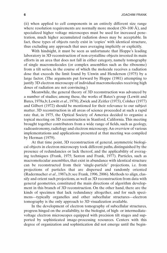
(ii) when applied to cell components in an entirely different size rangewhere resolution requirements are normally more modest (50–100 Å), andspecialized higher voltage microscopes must be used for increased pene-tration, much higher accumulated radiation doses may be acceptable. Infact, these types of objects rarely exist in ‘copies’ with identical structure,thus excluding any approach that uses averaging implicitly or explicitly.
With hindsight, it must be seen as unfortunate that Hoppe’s leadinglaboratory in 3D reconstruction of non-crystalline objects invested its mainefforts in an area that does not fall in either category, namely tomographyof single macromolecules (or complex assemblies such as the ribosome)from a tilt series, in the course of which the molecule receives a radiationdose that exceeds the limit found by Unwin and Henderson (1975) by alarge factor. (The arguments put forward by Hoppe (1981) attempting tojustify 3D electron microscopy of individual macromolecules receiving highdoses of radiation are not convincing.)
Meanwhile, the general theory of 3D reconstruction was advanced bya number of studies; among these, the works of Bates’s group (Lewitt andBates, 1978a,b; Lewitt et al., 1978), Zwick and Zeitler (1973), Colsher (1977)and Gilbert (1972) should be mentioned for their relevance to our subjectmatter. 3D reconstruction in all areas of science proceeded at such a rapidrate that, in 1975, the Optical Society of America decided to organize atopical meeting on 3D reconstruction in Stanford, California. This meetingbrought together contributors from a wide range of fields, such as geology,radioastronomy, radiology and electron microscopy.An overview of variousimplementations and applications presented at that meeting was compiledby Herman (1979).
At that time point, 3D reconstruction of general, asymmetric biologi-cal objects in electron microscopy took different paths, distinguished by thepresence of redundancies or lack thereof, and the applicability of averag-ing techniques (Frank, 1975; Saxton and Frank, 1977). Particles, such asmacromolecular assemblies, that exist in abundance with identical structurecan be reconstructed from their ‘single-particle’ projections, i.e. from projections of particles that are dispersed and randomly oriented (Radermacher et al., 1987a,b; see Frank, 1996, 2006). Methods to align, clas-sify and orient such projections, as well as 3D reconstruction from data withgeneral geometries, constituted the main directions of algorithm develop-ment in this branch of 3D reconstruction. On the other hand, there are thekinds of specimen that lack redundancy altogether, and for such speci-mens—typically organelles and other subcellular structures—electrontomography is the only approach to 3D visualization available.
In the development of electron tomography of subcellular structures,progress hinged on the availability, to the biologist, of high- or intermediate-voltage electron microscopes equipped with precision tilt stages and sup-ported by sophisticated image-processing resources. Centers with thisdegree of organization and sophistication did not emerge until the begin-
6 JOACHIM FRANK

ning of the 1980s when the National Institute of Health’s Biotechnologyprogram started to support three high-voltage microscopes dedicated to thebiological sciences in the USA1. Thus, the pace of development of this tech-nology was rather slow, especially considering the state of the art thatalready existed when Hoppe et al.’s fatty acid synthetase study (Hoppe etal., 1974) was published. However, perhaps the most important factor deter-mining the pace with which 3D imaging with the electron microscope devel-oped has been the speed and memory of computers. It must be realized thatelectron tomography posed computational problems of such magnitudethat, until the beginning of the 1990s, only groups with access to mainframeswere able to make significant progress. Other important factors were theslow progress toward automation of data collection and the need for image-processing software capable of handling the numerous combinations ofoperations that are encountered in the analysis of electron microscopicdata.
Finally, the 1990s brought a breakthrough toward full automation, asaffordable CCD cameras grew large enough to cope with the field sizesencountered in electron tomography, and electron microscopes were inte-grated with fast computer control. Here the work by Abraham Koster, oneof the contributors to this volume (Chapter 4), deserves special mention(Koster et al., 1992). Nowadays, thanks to his and others’ pioneering work,commercial instruments come equipped with the necessary gadgetry andsoftware to perform low-dose data collection, as well as preliminary recon-struction, on the spot. Thus, with the new generation of powerful and smartelectron microscopes, the drawing Walter Hoppe once used to illustrate boththe potential (the capability of 3D imaging) and limitations (radiationdamage) of electron tomography (Fig. 3a) has to be substantially revised(Fig. 3b).
INTRODUCTION: PRINCIPLES OF ELECTRON TOMOGRAPHY 7
FIGURE 3. Electron tomography then (a) and now (b). (a) Adapted from Hoppe (1983);(b) adapted from B. Carragher, unpublished drawing.
1 University of Colorado in Boulder, Colorado; University of Wisconsin in Madison; and NewYork State Department of Health in Albany, New York. Of these, only the one in Albanyhas remained in operation.

3. THE PRINCIPLE OF 3D RECONSTRUCTION
The principle of 3D reconstruction becomes clear from a formulationof the fundamental relationship between an object and its projections. Anunderstanding of the basic concept of the Fourier transform is needed forthis formulation. A brief introduction is provided in the following. For amore detailed introduction, the reader is referred to the specialized litera-ture such as Bracewell (1999). A compilation of definitions and formulaefor the case of discrete data is provided in the appendix of a book on 3Delectron microscopy of macromolecular assemblies by the author (Frank,2006).
The Fourier transform provides an alternative representation of anobject by breaking it down into a series of trigonometric basis functions. Formathematical expediency,complex exponential waves of the form exp[2piRr]are used instead of the more familiar sine and cosine functions.The argumentvector describing a location in 3D space is r = (x, y, z), while R = (X, Y, Z) is aso-called spatial frequency vector,which gives both the direction of travel of aspatial wave and the number of full spatial oscillations per unit length. Fromsuch spatial waves, the object can be built up by linear superposition:
(1)
with the complex coefficients cn. The 3D Fourier transform may be visual-ized as a 3D scheme (‘Fourier space’) in which the coefficients cn arearranged, on a regular grid, according to the position of the spatial fre-quency vector. Each coefficient cn contains the information on the associ-ated wave’s amplitude (or strength),
(2)
and phase (or shift of the spatial wave in its travelling direction, with respectto the origin),
(3)
The projection theorem offers a way to sample the Fourier transformof an object by measuring its projections. According to this theorem, the 2DFourier transform of a projection of the object is identical to a central sectionof the object’s 3D Fourier transform. Thus, by tilting the object into manyorientations, one is, in principle, able to measure its entire Fourier trans-form. Obviously, the projections must be collected with a small angularincrement and, ideally, over the full angular range. Then, after the Fouriersummation in equation (1) is performed, the object can be retrieved. Asalways, the devil is in the details, as evidenced by the lengths and depths ofthe treatises by specialists found in this book.
fnn
n
arctanI m cRe c
= { }{ }
a cn n=
o c inn
nr R r( ) = [ ]∑ exp 2p
8 JOACHIM FRANK

The angular increment Δq is evidently determined by two parameters(Fig. 4): (i) the mesh size of the Fourier space grid; and (ii) the size of theregion, in Fourier space, that needs to be filled. These quantities are in turndetermined by object diameter and resolution:
1. The mesh size must be smaller than 1/D, where D is the objectdiameter.
2. The region in Fourier space for which data must be acquired is asphere with radius 1/d, where d is the resolution distance, i.e. thesize of the smallest feature to be visualized in the reconstruction.
According to these essentially geometrical requirements, the minimumnumber of (equispaced) projections works out to be (Bracewell and Riddle,1967; Crowther et al., 1970a):
(4)
Reconstruction methods may be classified according to the way inwhich projections are collected or, alternatively, according to the way inwhich the object is retrieved from its measured projections. The formerrelates to the experiment, while the latter relates to the mathematical andcomputational aspects of reconstruction as discussed in Chapters 6, 7 and8. As to the data collection geometries, there are three that have gainedpractical importance in electron microscopy: single-axis, double-axis andconical tilting.
Single-axis tilting is simply achieved by rotation of a side-entry rod inthe electron microscope, whereas double-axis tilting involves a second tiltcapability around an axis perpendicular to the first (Fig. 5a), and conicaltilting provides a rotation capability in the inclined plane defined by the
NDd
= p
INTRODUCTION: PRINCIPLES OF ELECTRON TOMOGRAPHY 9
FIGURE 4. Sampling in Fourier spacefor single-axis tilting with equal incre-ments Δq. For explanation, see text.Adapted from Frank (1992).

first tilt (Fig. 5b). It is easy to see, by invoking the projection theorem, thatdouble-axis and conical tilting provide a much wider coverage of Fourierspace if the maximum tilt angle is the same in all cases (Fig. 6a–c). However,the price to be paid for this information gain is a >2-fold increase in totaldose (Frank and Radermacher, 1986; Radermacher and Hoppe, 1980).Because of this disadvantage, and the more elaborate experimental proce-dure, conical data collection has not been widely used in experimental pro-tocols where a single biological structure is multiply exposed. (Conical data
10 JOACHIM FRANK
FIGURE 5. Schematic diagrams showing the principle of side-entry tilt stages withtwo degrees of freedom. (a) Double-tile stage. aa is the principal tilt axis, correspon-ding to the long axis of the rod. bb is the second tilt axis. q and y are the correspon-ding tilt angles. n denotes the normal to the specimen plane. Tilting around the secondaxis is actuated by translation (indicated by arrows) of sliding rods which engagewheels attached to the turret T. (b) Tilt-rotation stage for conical geometry. Again ndenotes the normal to the specimen plane. q is the tilt angle, and j the rotation anglein the plane of the circular turret T. Rotation is actuated by a cable pulled in the direc-tion of the arrow, with return movement provided by a spring S. The turret is held ina stable position by retaining pins P. Adapted from Turner (1981).
FIGURE 6. Coverage of 3D Fourier space in the case of three data collection geome-tries: (a) single-axis; (b) double-axis; and (c) conical. In each case, equal angular incre-ments are depicted. From Lanzevecchia et al. (2005); reproduced with permission ofElsevier.

collection, of course, has found widespread application in reconstructionsof macromolecules in single-particle form from their projections (seeRadermacher et al., 1987b).) Lately, the idea of using the conical tilt geom-etry in tomography has been revived by Lanzavecchia’s group, with remark-able success (Lanzavechia et al., 2005; Zampighi et al., 2005).
4. HOW THIS BOOK IS ORGANIZED
The sequence of chapters in this book essentially follows the flow ofinformation in electron tomography, proceeding from specimen prepara-tion, to data collection in the instrument, and then to the techniques usedfor alignment, reconstruction and interpretation of the resulting tomo-graphic volumes.
We start with the question of to what extent the object reconstructedfrom electron microscopic projections resembles the biological object. Thisquestion has three different aspects to it: one that has to do with the rela-tionship between the native biological object and the specimen investigatedin the electron microscope; the second with the relationship between thatspecimen and the images formed by the electron microscope; and a thirdwith the relationship between the set of multiple projections and the final3D image.
Two chapters deal specifically with the specimen preparation aspectand the question of fidelity to the original biological structure. The first, byPradeep Luther (Chapter 1), examines the quality of specimen preparationin plastic sections, and the damage inflicted by the beam, with special atten-tion to the problem of section shrinkage. Knowledge of the behavior of thespecimen is of crucial importance in planning an experiment that requiresmultiple exposure of the same specimen. The other chapter, by MikeMarko, Chyongere Hsieh and Carmen Mannella (Chapter 2), describesexperience gained with the new, promising technique of sectioning frozen-hydrated biological material prepared by high-pressure freezing for thepurpose of electron tomography.
Peter Hawkes (Chapter 3) explores the conditions that must be satis-fied in the imaging by electron microscopy for the observed projections tobe regarded as simple line integrals of an object function. It is of coursealways possible to apply the reconstruction procedure ‘blindly’ to a set ofexperimental images and obtain a 3D density map, or ‘object function’. Thequestion is whether this reconstructed object has any meaning, or even atractable relationship to the physical object the images originated from. Byimplication, the simple projection relationship and the image formation inbright field under the usual weak object assumptions yield a very elegantlinear system description of the imaging and reconstruction process. As thespecimen thickness increases, which is the case for cell sections investigatedby electron tomography, multiple scattering increasingly interferes with the
INTRODUCTION: PRINCIPLES OF ELECTRON TOMOGRAPHY 11

linear system concept, but energy filtering (also covered in this chapter) caneffectively increase the thickness range of validity.
Low-dose data collection, in the now common automated mode, iscovered in a chapter by the pioneer of automated tomography, AbrahamKoster, together with Montserrat Bárcena (Chapter 4). This chapter goesinto all the necessary details regarding optimum settings, data collectionprotocols and the important considerations of dose fractionation.
Electron tomographic reconstruction requires that projections bealigned to a common frame of reference.The use of gold bead markers is nowroutine, and therefore it is justified that a chapter be devoted to the mathe-matical basis of marker-based alignment. David Mastronarde (Chapter 5),author of the well-known IMOD software, gives an expert introduction intothis subject. However, the search for a reliable markerless alignment methodcontinues, since the electron-opaque markers produce artifacts in the recon-struction volume that cannot be removed computationally. Sami Brandt(Chapter 6),one of the pioneers of markerless alignment techniques,has con-tributed an authoritative chapter on recent approaches to this problem.
Three chapters are devoted to the theory of reconstruction, address-ing different issues that arise due to the peculiarities of data collection andnumerical computation. We first present the chapter by Jose-Maria Carazo,Gabor Herman and co-workers (Chapter 7), which gives an overview onthe approaches to the inverse problem presented by the reconstructionfrom a finite number of projections. This same chapter also introduces iter-ative algebraic methods, such as algebraic reconstruction techniques (ART)and related techniques. Next, Michael Radermacher (Chapter 8) goes intothe details of weighted back-projection for general geometries, and formu-lates the algorithms underlying the computer programs now widely used inthe field. Weighted back-projection methods have a special position in thepractical implementation of 3D reconstruction, mainly because of theirmathematical tractability and high computational efficiency. Radermachersummarizes the rationales and important formulae of weighted back-pro-jection methods for regular and general geometries. Finally, in this sectionon the mathematics of reconstruction, Elmar Zeitler (Chapter 9) has con-tributed a chapter that presents an elegant general framework of recon-struction using special functions of mathematical physics, a chapter thatbrilliantly illuminates the inter-relationships of all approaches to recon-struction in use today.
Although the theoretical resolution obtainable in tomographic recon-structions is well known, the problem of how to measure the actual reso-lution achieved has been elusive. The chapter by Pawel Penczek and thisauthor (Chapter 10) addresses this important issue with a new approach.
The remaining chapters in this book deal with different aspects ofinterpretation of the reconstruction. The first step is the removal of noise,either by simple Fourier filtration or more advanced “denoising” proce-dures. These procedures are described by Reiner Hegerl and Achilleas
12 JOACHIM FRANK

Frangakis (Chapter 11). Segmentation is obviously the most importantaspect as it is instrumental for the assignment of meaning to the differentparts of a density map. Three chapters deal with segmentation, namely oneauthored by Achilleas Frangakis and Reiner Hegerl (Chapter 12), with seg-mentation based on local characteristics of the density distribution; thesecond, by Ming Jiang et al. (Chapter 13), on model-based segmentationmaking use of level set methods.The third chapter in this category addressessegmentation by rigid body motif search using cross-correlation. Thischapter is co-authored by investigators who each have made separate con-tributions to this area of research: Achilleas Frangakis and Bimal Rath(Chapter 14). Another aspect of interpretation comes up when we try tocharacterize quasi-periodic structures, as presented by the complex organ-ization of muscle trapped in the rigor state. We are then dealing with mul-tiple versions of a 3D motif, which may allow the tracking of a physiologicalprocess evolving in time and space—a promising method of analysisdescribed in the final chapter by Ken Taylor, Jun Liu and Hanspeter Winkler(Chapter 15).
ACKNOWLEDGEMENTS
This work was supported by HHMI and by Grant P41 RR01219 fromthe National Center for Research Resources (NCRR/NIH) and R37 GM29169. I would like to thank Michael Watters for assistance with the illustrations.
REFERENCES
Amos, L. A., Henderson, R. and Unwin, P. N. T. (1982). Three-dimensional structure determi-nation by electron microscopy of 2-dimensional crystals. Prog. Biophys. Mol. Biol. 39:183–231.
Andrews, H. C. (1970). Computer Techniques in Image Processing. Academic Press, New York.Barnard, D. P., Turner, J.N., Frank, J. and McEwen, B.F. (1992). A 360° single-axis tilt stage for
the high-voltage electron microscope. J. Microsc. 167:39–48.Bracewell, R. N. (1999). The Fourier Transform and Its Applications, 3rd edn. McGraw-Hill,
New York.Bracewell, R. N. and Riddle, A. C. (1967). Inversion of fan-beam scans in radio astronomy.
Astrophys. Soc. 150:427–434.Chalcroft, J. P. and Davey, C. L. (1984).A simply constructed extreme-tilt holder for the Philips
eucentric goniometer stage. J. Microsc. 134:41–48.Colsher, J. G. (1977). Iterative three-dimensional image reconstruction from tomographic
projections. Comput. Graphics Image Process. 6:513–537.Cormack, A. M. (1964). Representation of a function by its line integrals, with some radio-
logical applications. I. J. Appl. Phys. 35:2908–2912.Crowther, R. A., Amos, L. A., Finch, J. T. and Klug, A. (1970a). Three-dimensional reconstruc-
tion of spherical viruses by Fourier synthesis from electron micrographs. Nature 226:421–425.
INTRODUCTION: PRINCIPLES OF ELECTRON TOMOGRAPHY 13

Crowther, R. A., DeRosier, D. J. and Klug, A. (1970b). The reconstruction of a three-dimensional structure from projections and its application to electron microscopy. Proc.R. Soc. B 317:319–340.
DeRosier, D. and Klug, A. (1968). Reconstruction of 3-dimensional structures from electronmicrographs. Nature 217:130–134.
Frank, J. (1975).Averaging of low exposure electron micrographs of nonperiodic objects. Ultra-microscopy 1:159–162.
Frank, J. (1992). Introduction. In Electron Tomography (J. Frank, ed.) pp. 1–13. Plenum, NewYork.
Frank, J. (1996). Three-dimensional Electron Microscopy of Macromolecules. Academic Press,San Diego.
Frank, J. (2006). Three-dimensional Electron Microscopy of Macromolecules, 2nd edn. OxfordUniversity Press, New York.
Frank, J. and Radermacher, M. (1986). Three-dimensional reconstruction of nonperiodicmacromolecular assemblies from electron micrographs. In Advanced Techniques in Elec-tron Microscopy III (J. K. Koehler, ed.). Springer-Verlag, New York, pp. 1–72.
Gilbert, P. F. C. (1972). The reconstruction of a three-dimensional structure from projectionsand its application to electron microscopy. II. Direct methods. Proc. R. Soc. B 182:89–117.
Glaeser, R. M., Downing, K. H., Chiu, W., Frank, J. and DeRosier, D. (2007). Electron Crystal-lography of Biological Macromolecules. Oxford University Press, New York.
Hegerl, R. and Altbauer, A. (1982). The ‘EM’ program system. Ultramicroscopy 9:109–116.
Herman, G.T. (ed.) (1979). Image Reconstruction from Projections. Springer-Verlag, Berlin.Henderson, R. and Unwin, P. N. T. (1975). Three-dimensional model of purple membrane
obtained by electron microscopy. Nature 257:28–32.Herman, G. T. and Lewitt, R. M. (1979). Overview of image reconstruction from projections,
in Image Reconstruction from Projections (G. T. Herman, ed.). Springer-Verlag, Berlin, pp.1–7.
Hoppe, W. (1972 ). Drei-dimensional abbildende Elektronenmikroskope. Z. Naturforsch. 27a:919–929.
Hoppe, W. (1981). Three-dimensional electron microscopy. Annu. Rev. Biophys. Bioeng. 10:563–592.
Hoppe, W. (1983). Elektronenbeugung mit dem Transmissions-Elektronenmikroskop alsphasenbestimmendem Diffraktometer-von der Ortsfrequenzfilterung zur dreidimension-alen Strukturanalyse an Ribosomen. Angew. Chem. 95:465–494.
Hoppe, W., Gassmann, J., Hunsmann, N., Schramm, H. J. and Sturm, M. (1974). Three-dimensional reconstruction of individual negatively stained fatty-acid synthetase molecules from tilt series in the electron microscope. Hoppe-Seyler’s Z. Physiol. Chem.355:1483–1487.
Klug,A. (1983). From macromolecules to biological assemblies (Nobel lecture). Angew. Chem.22:565–636.
Koster, A. J., Chen, J. W., Sedat, J. W. and Agard, D. A. (1992). Automated microscopy for elec-tron tomography. Ultramicroscopy 46:207–227.
Lanzavecchia, S., Cantele, F., Bellon, P. L., Zampighi, L., Kreman, M.,Wright, E., and Zampighi,G. A. (2005). Conical tomography of freeze-fracture replicas: a method for the study of integral membrane proteins inserted in phospholipids bilayers. J. Struct. Biol. 149:87–98.
Lewitt, R. M. and Bates, R. H. T. (1978a). Image reconstruction from projections. I: Generaltheoretical considerations. Optik (Stuttgart) 50:19–33.
Lewitt, R. M. and Bates, R. H .T. (1978b). Image reconstruction from projections. III: Projec-tion completion methods (theory). Optik (Stuttgart) 50:189–204.
Lewitt, R. M., Bates, R. H. T. and Peters, T. M. (1978). Image reconstruction from projections.II: Modified back-projection methods. Optik (Stuttgart) 50:85–109.
14 JOACHIM FRANK

Radermacher, M. and Hoppe, W. (1980). Properties of 3D reconstruction from projections byconical tilting compared to single axis tilting. In Proceedings of the 7th European Con-gress on Electron Microscopy, Den Haag, Vol. I, pp. 132–133.
Radermacher, M., Wagenknechet, T., Verschoor, A. and Frank, J. (1987a). Three-dimensionalstructure of the large ribosomal subunit from Escherichia coli. EMBO J. 6:1107–1114.
Radermacher, M., Wagenknecht, T., Verschoor, A. and Frank, J. (1987b). Three-dimensionalreconstruction from single-exposure random conical tilt series applied to the 50S riboso-mal subunit of Escherichia coli. J. Microsc. 146:113–136.
Radon, J. (1917). Über die Bestimmung von Funktionen durch ihre Integralwerte längsgewisser Mannigfaltigkeiten. Berichte über die Verhandlungen der Königlich SächsischenGesellschaft der Wissenschaften zu Leipzig. Math. Phys. Klasse 69:262–277.
Saxton, W. O. and Frank, J. (1977). Motif detection in quantum noise-limited electron micro-graphs by cross-correlation. Ultramicroscopy 2:219–227.
Smith, P. R., Peters, T. M. and Bates, R. H. T. (1973). Image reconstruction from a finite numberof projections. J. Phys. A 6:361–382.
Turner, J. N. (1981). Stages and stereo pair recording. Methods Cell Biol. 23:33–51.Typke, D., Hoppe, W., Sessier, W. and Burger, M. (1976). Conception of a 3-D imaging electron
microscopy. In Proceedings of the 6th European Congress on Electron Microscopy (D. G.Brandon, ed.), Vol. 1, Tal International, Israel, pp. 334–335.
Unwin, P. N. T. and Henderson, R. (1975). Molecular structure determination by electronmicroscopy of unstained crystalline specimens. J. Mol. Biol. 94:425–440.
Zampighi, G., Zampighi, L., Fain., N., Wright, E. M., Cantele, F. and Lanzavecchia, S. (2005).Conical tomography II: a method for the study of cellular organelles in thin sections.J. Struct. Biol. 151:263–274.
Zwick, M. and Zeitler, E. (1973). Image reconstruction from projections. Optik 38:550–565.
INTRODUCTION: PRINCIPLES OF ELECTRON TOMOGRAPHY 15

1
Sample Shrinkage and RadiationDamage of Plastic Sections
Pradeep K. Luther
1. Introduction . . . . . . . . . . . . . . . . . . . . . . . . . . . . . . . . . . . . . . . . . . . 182. On Radiation Damage . . . . . . . . . . . . . . . . . . . . . . . . . . . . . . . . . . . 183. On Sample Preparation . . . . . . . . . . . . . . . . . . . . . . . . . . . . . . . . . . . 20
3.1. Fast Freezing/Freeze Substitution . . . . . . . . . . . . . . . . . . . . . . . 203.2. Conventional Processing . . . . . . . . . . . . . . . . . . . . . . . . . . . . . . 21
4. Methods of Measuring Sample Thickness . . . . . . . . . . . . . . . . . . . . . 214.1. Methods Not Involving the Electron Microscope . . . . . . . . . . . 214.2. Methods of Thickness Measurement By Electron
Microscopy . . . . . . . . . . . . . . . . . . . . . . . . . . . . . . . . . . . . . . . . . 245. Studies on Shrinkage of Plastic Sections . . . . . . . . . . . . . . . . . . . . . . 31
5.1. Early Studies . . . . . . . . . . . . . . . . . . . . . . . . . . . . . . . . . . . . . . . 315.2. Characterization of Shrinkage of Plastic Sections . . . . . . . . . . . 315.3. Investigation of Parameters That May Reduce
Section Shrinkage . . . . . . . . . . . . . . . . . . . . . . . . . . . . . . . . . . . 345.4. Shrinkage Studies of Native Samples (Not Embedded
in Plastic) . . . . . . . . . . . . . . . . . . . . . . . . . . . . . . . . . . . . . . . . . . 355.5. Shrinkage Occurs Along Directions Lacking
Restraint . . . . . . . . . . . . . . . . . . . . . . . . . . . . . . . . . . . . . . . . . . 355.6. Is Shrinkage Uniform in Z? . . . . . . . . . . . . . . . . . . . . . . . . . . . . 37
6. Effect of Low Temperatures on Radiation Damage and Sample Shrinkage . . . . . . . . . . . . . . . . . . . . . . . . . . . . . . . . . . . . . . . 386.1. Characterization of Shrinkage in Native (Unembedded)
Crystals at Low Temperature . . . . . . . . . . . . . . . . . . . . . . . . . . . 386.2. Characterization of Shrinkage of Plastic Sections at
Low Temperature . . . . . . . . . . . . . . . . . . . . . . . . . . . . . . . . . . . . 39
17
Pradeep K. Luther • Imperial College London, Biomedical Sciences Division, Sir AlexanderFleming Building, Exhibition Road, London SW7 2AZ, UK

7. Review of Sample Preparation and Imaging Protocols in PastTomography Studies . . . . . . . . . . . . . . . . . . . . . . . . . . . . . . . . . . . . . 417.1. Comments on Sample Preparation and Tomography . . . . . . . . 417.2. Tomography of the Desmosome . . . . . . . . . . . . . . . . . . . . . . . . 417.3. Studies on the Golgi Apparatus . . . . . . . . . . . . . . . . . . . . . . . . . 43
8. Conclusions and Recommendations . . . . . . . . . . . . . . . . . . . . . . . . . 44References . . . . . . . . . . . . . . . . . . . . . . . . . . . . . . . . . . . . . . . . . . . . . 45
1. INTRODUCTION
Just as fossil insects embalmed in amber are extraordinarily preserved, soare biological samples that have been embedded in plastic for electronmicroscopy.The success of embedding samples in plastic lies in the astound-ing resilience of the sections in the electron microscope, albeit after initialchanges. The electron microscope image results from projection of thesample density in the direction of the beam, i.e. through the depth of thesection, and therefore is independent of physical changes in this direction.In contrast, the basis of electron tomography is the constancy of the physical state of the whole section during the time that different views atincremental tilt angle steps are recorded.
The shrinkage of a plastic section in each dimension, especially thedepth, when viewed in the electron microscope, is now a well known phe-nomenon. Knowledge of the shrinkage behavior of a section of a sampleembedded in a particular plastic is of crucial importance when embarkingon the electron tomography of the sample. In the last 15 years, the mostimportant advances in electron tomography have been the development ofautomated methods of recording tilt series and direct imaging onto CCDcameras (Koster et al., 1997; Koster and Barcena, Chapter 4 in this volume).These advances have enabled tremendous savings in labor but also in thetotal dose experienced by a sample. In this chapter, we review the studiescarried out on shrinkage behavior of samples embedded in various resinsand we review the protocols that have been followed by the leading pro-ponents of electron tomography.
2. ON RADIATION DAMAGE
Several researchers have written reviews on the effects of the electronbeam on biological samples (Egerton et al., 2004; Glaeser and Taylor, 1978;Grubb, 1974; Lamvik, 1991; Reimer, 1989; Stenn and Bahr, 1970). Electronmicroscope radiation has the primary effect of producing intense ionizationin organic materials, which results in the formation of free radicals and ions.This causes bond scission and molecular fragments to be formed. Theseprimary effects occur at all temperatures. At room temperature, the freeradicals and molecular fragments can undergo diffusion, and produce cross-
18 PRADEEP K. LUTHER

linking or further chain scission. Damage to secondary structure occurs atan electron dose of <100e/nm2. Further exposure causes the tertiary struc-ture to undergo dramatic reorganization following loss of specific groupsand altered structural composition. The dominant effect finally is that ofmass loss from the sample, which preferentially involves H and O in com-parison with C and N. The mass loss is accompanied or followed closely byshrinkage of the sample normal to the beam.
We can reduce the radiation damage on an organic sample by coolingthe sample to cryotemperatures and by using low-dose techniques (to bedescribed later). One of the main effects of electron irradiation at conven-tional illumination levels is to cause specimen shrinkage normal to theplane of the sample. The effect of the shrinkage in reciprocal space is illus-trated in Fig. 1. The example considered is a structure based on cubic
SAMPLE SHRINKAGE AND RADIATION DAMAGE OF PLASTIC SECTIONS 19
FIGURE 1. Illustration of the ‘missing wedge’ problem due to specimen shrinkage inthe electron microscope. (a) Projection of a cubic crystal (for example) viewed edge-onwith the c axis parallel to the electron beam. (b) Reciprocal lattice for the projection in(a). A conventional tilt holder in the electron microscope can be tilted in the range inthe range of –60° to +60°. When a tilt series is recorded about a single tilt axis, and the3D transform calculated by combining the individual transforms, data will be missingfrom the 3D transform inside the ‘wedge’ AOB of angle 60°. If the sample thicknessreduces by 50% as in (c), then the corresponding reciprocal lattice shown in (d) isstretched 100% along c*. The projection for the 101 diffraction spot, comfortablyincluded in the tilt series for the unshrunk sample (a and b), now lies within the missingwedge. In relation to the original reciprocal lattice, the missing wedge is now describedin (a) by A’OB’ and has an angle of 98°. For 50% shrinkage, the tilt holder effectivelycovers only the range –41° to +41° in relation to the original sample.

symmetry (Fig. 1a), which collapses in thickness by 50% (Fig. 1c). With aconventional tilt holder, a series of views about a single tilt axis are recordedin the range –60° to +60°. The 3D transform in (Fig. 1b), obtained by com-bining the transforms of the individual views, has missing from it data inthe 60° wedge AOB. In the case of the 50% collapsed sample (Fig. 1c), thereciprocal space is stretched by 100% in the corresponding direction (Fig.1d). The volume of the 3D transform that can be sampled is now muchreduced, e.g. the spot 101 present in (Fig. 1b) is missing from the transform(Fig. 1d). In relation to the original sample, the missing wedge (A′OB′) inthe 3D transform has an angle of 98°. The effective tilt range is now only±41°. Sample shrinkage therefore directly reduces the resolution normal tothe sample plane in a tomogram of a plastic-embedded sample. Hence wemust make every effort to curtail the shrinkage.
It is appropriate to describe the terminology of Amos et al. (1982) forthe various imaging modes in the electron microscope and the electron doseinvolved for a single image in each case. A dose on the sample of 50–400e/nm2 is considered as a very low dose, which is appropriate for veryhigh resolution studies of unstained crystalline specimens. A dose of∼1000–2000e/nm2 is termed a minimal dose, which is used for stained ornon-crystalline specimens. Conventional microscopy for single imagesinvolves doses on the order of ∼5000–50,000e/nm2 due to the time involvedin searching and focusing. Minimal and low-dose methods require a searchof suitable areas to be done at very low magnification, about ×2000, duringwhich the dose should be extremely low (∼2e/nm2). Modern electron micro-scopes provide low-dose imaging modes in which the focusing is done athigh magnification on areas adjacent to the area of interest, followed byimage recording at the desired magnification.
3. ON SAMPLE PREPARATION
3.1. Fast Freezing/Freeze Substitution
To produce plastic-embedded samples with the best possible preser-vation, rapid freezing followed by freeze substitution has been the most suc-cessful method. Various methods have been used for rapid freezing. Sosa et al. (1994) successfully froze muscle fibers by ‘plunge-freezing’, whichinvolves rapid propulsion of the sample into a trough of cold cryogen, e.g.liquid ethane, at –180°C. ‘Slam-freezing’ involves rapidly propelling asample onto a highly polished metal block (usually copper) cooled by liquidhelium or liquid nitrogen. Spectacular fast-freeze deep-etch replica imagesof various samples were obtained in the pioneering studies by Heuser(Heuser, 1989; Heuser et al., 1987). Details of the slam-freezing methodapplied to striated muscle fibres to capture different activity states havebeen described (Craig et al., 1992; Hirose et al., 1993; Liu et al., 2004a; Padronet al., 1988).
20 PRADEEP K. LUTHER