electron beam tomography ebt. i’ve never heard of it, (and it doesn’t sound good) electrons...
TRANSCRIPT

Electron Beam Tomography
EBT

I’ve never heard of it,(and it doesn’t sound good)
• Electrons – Atomic particles– Have mass
• Wouldn’t a beam of particulate radiation passing through the body be extremely hazardous?

Yes!

EBT does NOT pass an electron beam through the patient
• The e- beam is focused beneath the patient at 4 stationary tungsten targets where X-rays are produced

What is the radiation dose with EBT?
• An advantage of the EBT scanner over conventional scanners is instead of exposing the entire circumference of the body to the X-ray beam, the EBT X-ray beam enters from the back. Thus, anterior structures such as the breast and thyroid are subjected to a lesser dose of radiation (17% of the entrance skin dose). EBT scanning is usually 1/5th to 1/10th the radiation exposure as Spiral CT scanning.

Comparing Radiation Doses
EBT Coronary Calcium Scan 50 to 70 mrem 0.5 to 0.70 mSv
Standard Abdominal X-ray 48 mrem 0.48 mSv
Standard Coronary Angiogram 500 to 1000 mrem 5 to 10 mSv
Background Sunshine Radiation in 1 year
300 mrem 3.0 mSv

How are EBT Images Obtained?
Due to the near constant motion of the heart, the scans are triggered by the ECG signal at 80% of the R-R interval, near end diastole before atrial contraction, thus minimizing the effect of cardiac motion.
Due to the lack of mechanical motion these scans are fast enough to be shot during the moment the heart is still.

Electron Beam Tomographyof the Heart
• Why was it done?– EBTs were first used to find calcium buildup in
heart arteries, which can increase your risk for coronary artery disease (CAD). It may be used as a tool to find hardening of the arteries in people who have a high chance of developing atherosclerosis

What do cardiac EBTs Show?
• The most significant finding of an EBT exam is the amount of calcified plaque in your coronary arteries. Calcified plaque in the coronary arteries is easy to see on an EBT scan because it has become hard and dense.

CAD in the US
The well-known risk factors for heart disease:
•high cholesterol•high blood pressure•diabetes
can be useful for indicating how healthy a heart is. But they don't tell the whole story; even if you don't have any known risk factors, you could still be at risk.

For 450,000 Americans each year the very first symptom of a heart problem is a sudden heart attack. For one-third of them, that first attack is fatal. That's what makes an EBT imaging study so valuable. It is the first and only screening test approved by the Food and Drug Administration that can actually look inside the heart.
CAD in the US

Calcified Plaque and MI
• A heart attack happens when plaque ruptures in your coronary arteries, leading to the sudden blockage of blood flow to the heart. There's a well-established relationship between coronary plaque and calcified plaque. If you have more of one, you'll have more of the other. Due to this association, EBT is the most definitive method available for detecting early stage coronary-artery disease.

The Progression of an Acute MI
Coronary artery calcification is part of the development of atherosclerosis; it occurs exclusively in atherosclerotic arteries and is absent in the normal vessel wall.


Who Should Have a EBT?
• An EBT exam is recommended if you are a man between the ages of 40 and 70 or a woman between 45 and 75. It is particularly recommended if you have other risk factors for heart disease such as high cholesterol, high blood pressure, a history of smoking, diabetes or a family history of heart disease.

EBCT serial transaxial images are obtained in 100 ms with a scan slice thickness of 3 to 6 mm. 30 to 40 adjacent axial scans usually are obtained. The rapid image-acquisition time virtually eliminates motion artifact related to cardiac contraction.
A study for coronary calcium is completed within 10 to 15 minutes, requiring only a few seconds of scanning time.
How Long Will the Test Take?

The Future of EBT
• Virtual Angiograms
– Due to the rapid progression of medical software the reconstructed images are becoming more and more detailed

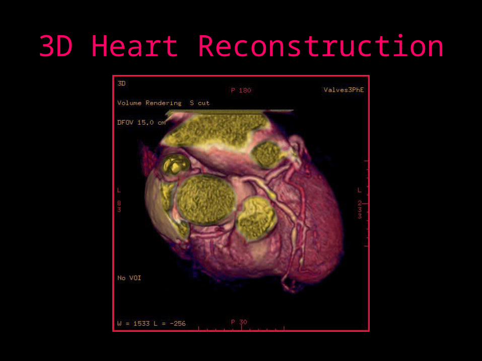
3D Heart Reconstruction

A Virtual Angiogram - Flythrough

The Future of EBTBeyond the Heart
• Bowel Scans
– Virtual Colonoscopy
– 3D Surface Scans

Bowel Scan - Surface

Bowel Scan with Polyp

A Virtual Colonoscopy

• Lung Scans
• Bone Scans
• Sinus Scans
The Future of EBTBeyond the Heart

Lung Scan
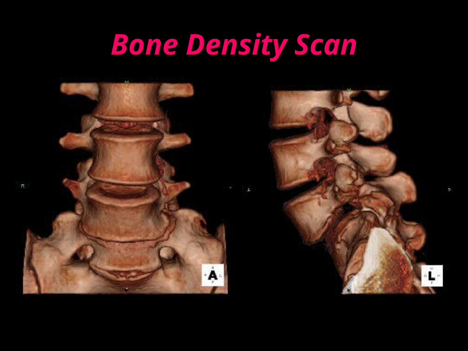
Bone Density Scan

EBCT of the Sinuses