electrodeposition of nano size hydroxyapatite...
TRANSCRIPT
Iranian Journal of Materials Science and Engineering, Vol. 3, Numbers 3 and 4, Summer and Autumn 2006
1
ELECTRODEPOSITION OF NANO SIZE HYDROXYAPATITE
COATING ON Ti ALLOY
M. Saremi and B. Mottaghi Golshan [email protected]
Department of Materials Science and Engineering, Faculty of Engineering, Tehran University, Tehran,
Iran
Abstract: A film of osteoconductive and biocompatible material on biomedical metallic implants
can create bioactivity of the implant and shorten healing time. Hydroxyapatite, that is the most
important mineral part of human bone, was coated on Ti6Al4V using cathodic electrodeposition
process. Pulse electrodeposition technique was used and the effects of different parameters such as
potential, duty cycle (on time/ (on time+ off time)), temperature and current density on the
morphology of the deposits were examined. Nano size deposits were formed under controlled
temperature and optimization of voltage and current density.
Keywords: Hydroxyapatite, Electrodeposition, pulse electrodeposition, morphology.
1. INTRODUCTION
Calcium phosphates are the most important
inorganic constituents of biological hard tissues
[1] that in the form of hydroxyapatite (HA), with
the formula of Ca10 (PO4) 6(OH) 2, are present in
bone and teeth [2]. Biologically formed calcium
phosphates are often nanocrystals that are
precipitated under mild conditions (ambient
pressure, near room temperature) [1], but their
poor mechanical properties are not favorable for
bone-repair purposes [3]. A solution to this
problem has been its use as coating for metallic
implants, in which the good mechanical
properties of metal is combined with the
biocompatibility of HA [4].
The morphology and crystal structure of HA is
important for its biocompatibility and
osteoconductivity; moreover in recent years,
there are growing approaches toward nano
particles and nano coatings which can provide
improved properties on their applications. Nano
hydroxyapatite coating can have structure that is
more matched to the bone structure in which the
implants should function and have lower
sintering temperature [5].
There are several techniques to produce HA
coating on metallic implants including sol-gel
[6], pulse laser deposition [7], plasma spraying
[7], electrophoretic and electrolytic deposition
[8], among them electrodeposition is more
attractive due to its low cost, ease of operation
and the possibility to cover complex shape
surfaces.
Electrodeposition of HA is based on cathodic
reactions composed of two steps of oxygen
reduction to produce hydroxyl ions followed by
precipitation of calcium phosphates under
alkaline condition on the surface. Wile
crystallization by itself composed of nucleation
and growth, controlling electrodeposition
parameters can control the microstructure and
composition of HA deposits. Pulse
electrodeposition in which the amount and
duration of applied current and potential are
controlled is a good means to obtain nano
deposits [9].
The aim of this work is to apply HA on Ti alloy
by pulse electrodeposition and to study the effect
of electrodeposition parameters on the
morphology of the crystals.
2. EXPERIMENTALS
Commercial Ti6Al4V alloy were cut into
samples of of 20×30 mm having 3mm thickness
grand with abrasive papers No.180, (some were
mirror polished) degreased with acetone and
washed in distilled water.
Conventional and pulse electrodeposition were
carried out in 0.042M Ca (NO3)2 and 0.025M
NH4H2PO4 solution at pH4.1. Electrodeposition
was also conduced at different potentials of 3,
4.5 and 6 volts, duty cycles; 0.4, 0.5, 0.05, at
M. Saremi, B. Mottaghi Golshan
2
room temperature and at 12°C. The duty cycle is
described as ton/(ton+ toff) in which ton is time of
applied pulse potential and toff is time of no
current/potential. AMEL model 568
potentiostat/golvanostat was used for constant
current as electrodeposition
Morphology of the HP deposits were examined
using Scanning Electron Microscope (SEM)
Model CamScan Mv 2300. The crystal structures
were characterized using X-ray diffractometere
Model Philips Xtert.
3. RESULTS AND DISCUSSION
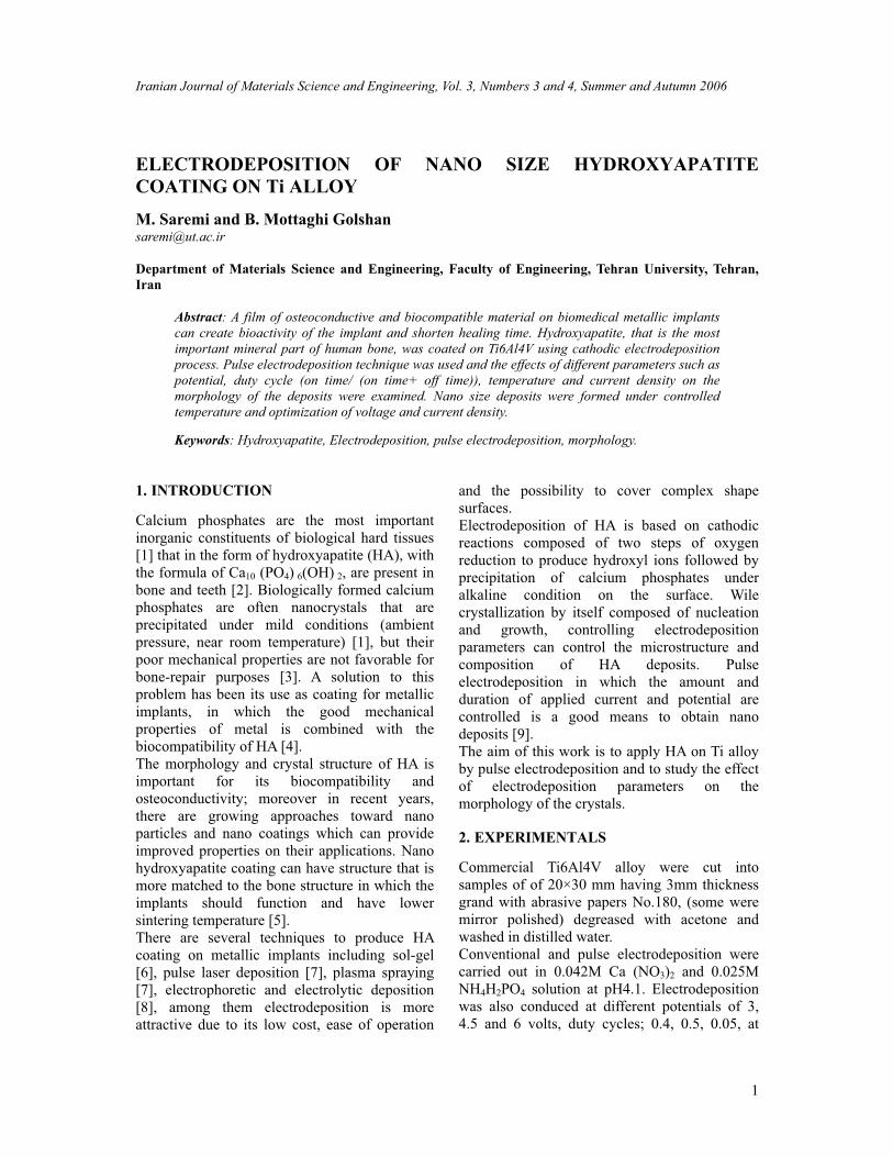
Fig.1 shows the SEM micrograph of the HA
deposit obtained using conventional
electrodeposition at 3V applied potential. A
micro size flake type structure having hexagonal
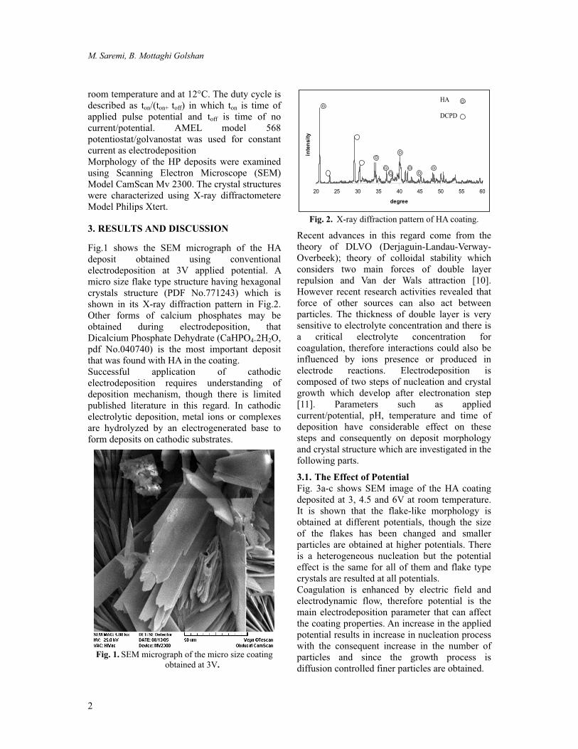
crystals structure (PDF No.771243) which is
shown in its X-ray diffraction pattern in Fig.2.
Other forms of calcium phosphates may be
obtained during electrodeposition, that
Dicalcium Phosphate Dehydrate (CaHPO4.2H2O,
pdf No.040740) is the most important deposit
that was found with HA in the coating.
Successful application of cathodic
electrodeposition requires understanding of
deposition mechanism, though there is limited
published literature in this regard. In cathodic
electrolytic deposition, metal ions or complexes
are hydrolyzed by an electrogenerated base to
form deposits on cathodic substrates.
Fig. 1. SEM micrograph of the micro size coating
obtained at 3V.
20 25 30 35 40 45 50 55 60
degree
intensity
HA
DCPD
Fig. 2. X-ray diffraction pattern of HA coating.
Recent advances in this regard come from the
theory of DLVO (Derjaguin-Landau-Verway-
Overbeek); theory of colloidal stability which
considers two main forces of double layer
repulsion and Van der Wals attraction [10].
However recent research activities revealed that
force of other sources can also act between
particles. The thickness of double layer is very
sensitive to electrolyte concentration and there is
a critical electrolyte concentration for
coagulation, therefore interactions could also be
influenced by ions presence or produced in
electrode reactions. Electrodeposition is
composed of two steps of nucleation and crystal
growth which develop after electronation step
[11]. Parameters such as applied
current/potential, pH, temperature and time of
deposition have considerable effect on these
steps and consequently on deposit morphology
and crystal structure which are investigated in the
following parts.
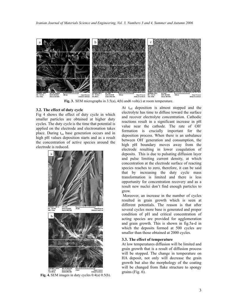
3.1. The Effect of Potential
Fig. 3a-c shows SEM image of the HA coating
deposited at 3, 4.5 and 6V at room temperature.
It is shown that the flake-like morphology is
obtained at different potentials, though the size
of the flakes has been changed and smaller
particles are obtained at higher potentials. There
is a heterogeneous nucleation but the potential
effect is the same for all of them and flake type
crystals are resulted at all potentials.
Coagulation is enhanced by electric field and
electrodynamic flow, therefore potential is the
main electrodeposition parameter that can affect
the coating properties. An increase in the applied
potential results in increase in nucleation process
with the consequent increase in the number of
particles and since the growth process is
diffusion controlled finer particles are obtained.
Iranian Journal of Materials Science and Engineering, Vol. 3, Numbers 3 and 4, Summer and Autumn 2006
3
a b c
Fig. 3. SEM micrographs in 3.5(a), 4(b) and6 volt(c) at room temperature.
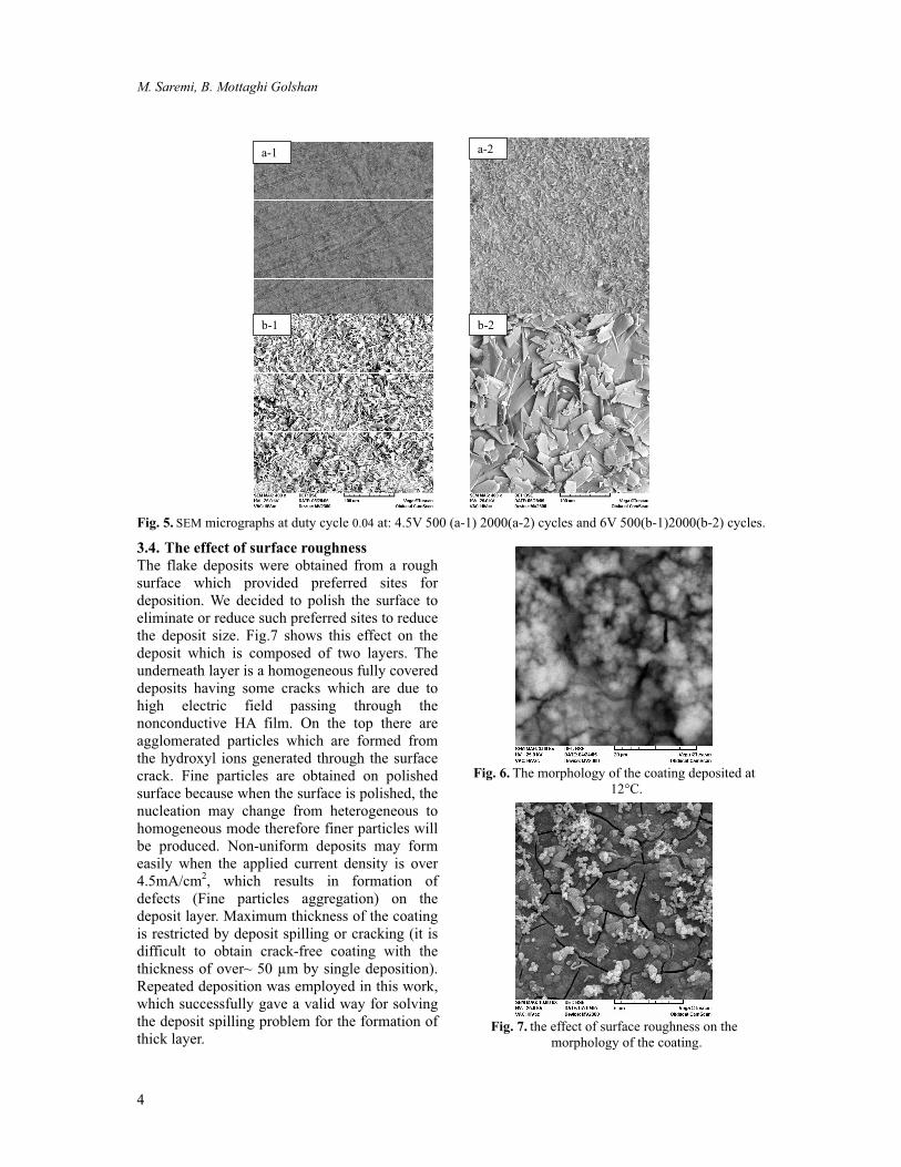
3.2. The effect of duty cycle
Fig 4 shows the effect of duty cycle in which
smaller particles are obtained at higher duty
cycles. The duty cycle is the time that potential is
applied on the electrode and electronation takes
place. During ton base generation occurs and in
high pH values deposition starts and as a result
the concentration of active species around the
electrode is reduced.
a
b
Fig. 4. SEM images in duty cycles 0.4(a) 0.5(b).
At toff deposition is almost stopped and the
electrolyte has time to diffuse toward the surface
and recover electrolyte concentration. Cathodic
reactions result in a significant increase in pH
value near the cathode. The rate of OH-
formation is crucially important for the
deposition process. When there is an unbalance
between OH- generation and consumption, the
high pH boundary moves away from the
electrode resulting in lower coagulation of
deposits. This is due to pulsating diffusion layer
and pulse limiting current density, at which
concentration at the electrode surface of reacting
species reaches to zero, therefore, it can be said
that by increasing the duty cycle mass
transformation is limited and there is less
opportunity for concentration recovery and as a
result new nuclei don’t find enough particles to
grow.
Moreover, an increase in the number of cycles
resulted in grain growth which is seen at
different potentials. The reason is that after
several cycles more base is generated and proper
condition of pH and critical concentration of
acting species are provided for agglomeration
and grain growth. This is shown in fig.5a-d in
which the deposits formed at 500 cycles are
smaller than those obtained at 2000 cycles.
3.3. The effect of temperature
At low temperatures diffusion will be limited and
grain growth that is a result of diffusion process
will be stopped. The change in temperature on
HA deposit, not only will decrease the grain
growth but also the morphology of the coating
will be changed from flake structure to spongy
grains (Fig. 6).
M. Saremi, B. Mottaghi Golshan
4
a-1 a-2
b-1 b-2
Fig. 5. SEM micrographs at duty cycle 0.04 at: 4.5V 500 (a-1) 2000(a-2) cycles and 6V 500(b-1)2000(b-2) cycles.
3.4. The effect of surface roughness
The flake deposits were obtained from a rough
surface which provided preferred sites for
deposition. We decided to polish the surface to
eliminate or reduce such preferred sites to reduce
the deposit size. Fig.7 shows this effect on the
deposit which is composed of two layers. The
underneath layer is a homogeneous fully covered
deposits having some cracks which are due to
high electric field passing through the
nonconductive HA film. On the top there are
agglomerated particles which are formed from
the hydroxyl ions generated through the surface
crack. Fine particles are obtained on polished
surface because when the surface is polished, the
nucleation may change from heterogeneous to
homogeneous mode therefore finer particles will
be produced. Non-uniform deposits may form
easily when the applied current density is over
4.5mA/cm2, which results in formation of
defects (Fine particles aggregation) on the
deposit layer. Maximum thickness of the coating
is restricted by deposit spilling or cracking (it is
difficult to obtain crack-free coating with the
thickness of over~ 50 µm by single deposition).
Repeated deposition was employed in this work,
which successfully gave a valid way for solving
the deposit spilling problem for the formation of
thick layer.
Fig. 6. The morphology of the coating deposited at
12°C.
Fig. 7. the effect of surface roughness on the
morphology of the coating.

Iranian Journal of Materials Science and Engineering, Vol. 3, Numbers 3 and 4, Summer and Autumn 2006
5
3.5. The effect of current density
By increasing current density from 1mA/cm2 to
4 mA/cm2, on a polished surface the
electronation is increased and more base was
formed on the surface. At high pH values (and
because the preferred sites on the polished
surface were also reduced and the temperature
was adjusted to 12 C) the conditions was fully
provided for a homogeneous nucleation of small
size deposits to form on the surface. Fig 8 shows
the deposition of nano size particles on the
surface as a result of the provision of all of the
above mentioned effective electrochemical
parameters. The thickness of the coating is
bellow 50 microns depending on the time of
deposition, but repeated deposition can be used
where thicker deposits is needed.
Fig. 8. The effect of current density.
4. CONCLUSIONS
Based on the above mentioned points it can be
concluded that:
Electrodeposition is a favorable process to
produce HA coating.
Electrodeposition parameters have a marked
effect on the morphology and crystal size of
the coating particles.
The most uniform coatings were obtained at
applied potential of 4.5V and at 12°C.
Nano structured hydroxyapatite coating can
be obtained under controlled current and
potential during pulse electrodeposition.
REFERENCES
1. Sergey Dorozhkinand, Matthis Epple Angew
in “ Biological and Medical Significance of
Calcium Phosphates”.Chem.Int Ed.41,2002,
3130-3146.
2. Gultekil Goller,Faik Nuzhet Oktar in “
Sintering Effects on Mechanical Properties
of Biologically Derived Dentine
Hydroxyapatite”. Material Letters 56, (2002),
142-147.
3. Wen Shi,Akira Kamya,Jun Zhu, Akira Watazu
in “ Properties of Titanium Biomaterial
Fabricated By Sinter Bonding of
Titanium/Hydroxyapatite Composite Surface
Coated Layer to Pure Bulk Titanium”.
Materials science and engineering A337,
(2002), 104-109.
4. P. Monderagon-Cortez, G. Vargas-Gutierrez in
“ Electrophoretic Deposition of
Hydroxyapatite Sub micron Particles at High
Voltages”. Materials Letters 58, (2004), 1336-
1339
5. Wengian Weng,Sam Zhang,Kui Cheng, Haibo
Qu in “Sol-Gel Preparation of Bioactive
Apatite Films”.Surf.Coat.Technol,167(2003)
292-296.
6. C. K. Wang, J. H. Chern Lin in “Pulse Laser
Deposition of Hydroxyapatite”.Biomaterials
18,(1997),1331-1338
7. Cong Wang, J. Mo, Wen Cheng, Ruifang
Zhang in “ Thick Hydroxyapatite Coatings By
Electrophoretic Deposition”.Materials Letters
57(2002) 99-105.
8. M. C. Kue, S. K. Yen in “ The Process Of
Electrochemical Deposition Hydroxyapatite on
Biomedical Titanium at Room Temperature”.
Materials Science and engineering C,
20(2002)153-160.
9. I. Zhitomirsky in “Cathodic Electrodeposition
of Ceramic and Organoceramic Materials.
Fundamental Aspects”. Advanced in Colloid
and Interface Science. 97(2002) 279-317.
10. Subir Bhattacharjee, Menachem Elimelech,
Michal Borkovec.”DLVO Interaction between
Colloidal Particles: Beyond Derjaguin’s
Approximation”. Croatia Chemica Acta 71
(1998) 883-903.
11. Derek Pletcher and Frank C.
Walsh.”Industerial elrctrochemistery” 2nd
edition.London, New York Academic
Press,1993, p.167.